CN101802002A - Antibodies that bind human protein tyrosine phosphatase beta (HPTP beta) and uses thereof - Google Patents
Antibodies that bind human protein tyrosine phosphatase beta (HPTP beta) and uses thereofDownload PDFInfo
- Publication number
- CN101802002A CN101802002ACN200780011778ACN200780011778ACN101802002ACN 101802002 ACN101802002 ACN 101802002ACN 200780011778 ACN200780011778 ACN 200780011778ACN 200780011778 ACN200780011778 ACN 200780011778ACN 101802002 ACN101802002 ACN 101802002A
- Authority
- CN
- China
- Prior art keywords
- antibody
- angiogenesis
- disease
- antibodies
- fragment
- Prior art date
- Legal status (The legal status is an assumption and is not a legal conclusion. Google has not performed a legal analysis and makes no representation as to the accuracy of the status listed.)
- Granted
Links
Images


Classifications
- A—HUMAN NECESSITIES
- A61—MEDICAL OR VETERINARY SCIENCE; HYGIENE
- A61P—SPECIFIC THERAPEUTIC ACTIVITY OF CHEMICAL COMPOUNDS OR MEDICINAL PREPARATIONS
- A61P1/00—Drugs for disorders of the alimentary tract or the digestive system
- C—CHEMISTRY; METALLURGY
- C07—ORGANIC CHEMISTRY
- C07K—PEPTIDES
- C07K16/00—Immunoglobulins [IGs], e.g. monoclonal or polyclonal antibodies
- C07K16/18—Immunoglobulins [IGs], e.g. monoclonal or polyclonal antibodies against material from animals or humans
- A—HUMAN NECESSITIES
- A61—MEDICAL OR VETERINARY SCIENCE; HYGIENE
- A61K—PREPARATIONS FOR MEDICAL, DENTAL OR TOILETRY PURPOSES
- A61K39/00—Medicinal preparations containing antigens or antibodies
- A61K39/395—Antibodies; Immunoglobulins; Immune serum, e.g. antilymphocytic serum
- A—HUMAN NECESSITIES
- A61—MEDICAL OR VETERINARY SCIENCE; HYGIENE
- A61P—SPECIFIC THERAPEUTIC ACTIVITY OF CHEMICAL COMPOUNDS OR MEDICINAL PREPARATIONS
- A61P1/00—Drugs for disorders of the alimentary tract or the digestive system
- A61P1/02—Stomatological preparations, e.g. drugs for caries, aphtae, periodontitis
- A—HUMAN NECESSITIES
- A61—MEDICAL OR VETERINARY SCIENCE; HYGIENE
- A61P—SPECIFIC THERAPEUTIC ACTIVITY OF CHEMICAL COMPOUNDS OR MEDICINAL PREPARATIONS
- A61P1/00—Drugs for disorders of the alimentary tract or the digestive system
- A61P1/04—Drugs for disorders of the alimentary tract or the digestive system for ulcers, gastritis or reflux esophagitis, e.g. antacids, inhibitors of acid secretion, mucosal protectants
- A—HUMAN NECESSITIES
- A61—MEDICAL OR VETERINARY SCIENCE; HYGIENE
- A61P—SPECIFIC THERAPEUTIC ACTIVITY OF CHEMICAL COMPOUNDS OR MEDICINAL PREPARATIONS
- A61P11/00—Drugs for disorders of the respiratory system
- A—HUMAN NECESSITIES
- A61—MEDICAL OR VETERINARY SCIENCE; HYGIENE
- A61P—SPECIFIC THERAPEUTIC ACTIVITY OF CHEMICAL COMPOUNDS OR MEDICINAL PREPARATIONS
- A61P13/00—Drugs for disorders of the urinary system
- A61P13/12—Drugs for disorders of the urinary system of the kidneys
- A—HUMAN NECESSITIES
- A61—MEDICAL OR VETERINARY SCIENCE; HYGIENE
- A61P—SPECIFIC THERAPEUTIC ACTIVITY OF CHEMICAL COMPOUNDS OR MEDICINAL PREPARATIONS
- A61P17/00—Drugs for dermatological disorders
- A—HUMAN NECESSITIES
- A61—MEDICAL OR VETERINARY SCIENCE; HYGIENE
- A61P—SPECIFIC THERAPEUTIC ACTIVITY OF CHEMICAL COMPOUNDS OR MEDICINAL PREPARATIONS
- A61P17/00—Drugs for dermatological disorders
- A61P17/02—Drugs for dermatological disorders for treating wounds, ulcers, burns, scars, keloids, or the like
- A—HUMAN NECESSITIES
- A61—MEDICAL OR VETERINARY SCIENCE; HYGIENE
- A61P—SPECIFIC THERAPEUTIC ACTIVITY OF CHEMICAL COMPOUNDS OR MEDICINAL PREPARATIONS
- A61P17/00—Drugs for dermatological disorders
- A61P17/06—Antipsoriatics
- A—HUMAN NECESSITIES
- A61—MEDICAL OR VETERINARY SCIENCE; HYGIENE
- A61P—SPECIFIC THERAPEUTIC ACTIVITY OF CHEMICAL COMPOUNDS OR MEDICINAL PREPARATIONS
- A61P19/00—Drugs for skeletal disorders
- A61P19/02—Drugs for skeletal disorders for joint disorders, e.g. arthritis, arthrosis
- A—HUMAN NECESSITIES
- A61—MEDICAL OR VETERINARY SCIENCE; HYGIENE
- A61P—SPECIFIC THERAPEUTIC ACTIVITY OF CHEMICAL COMPOUNDS OR MEDICINAL PREPARATIONS
- A61P19/00—Drugs for skeletal disorders
- A61P19/08—Drugs for skeletal disorders for bone diseases, e.g. rachitism, Paget's disease
- A—HUMAN NECESSITIES
- A61—MEDICAL OR VETERINARY SCIENCE; HYGIENE
- A61P—SPECIFIC THERAPEUTIC ACTIVITY OF CHEMICAL COMPOUNDS OR MEDICINAL PREPARATIONS
- A61P25/00—Drugs for disorders of the nervous system
- A61P25/02—Drugs for disorders of the nervous system for peripheral neuropathies
- A—HUMAN NECESSITIES
- A61—MEDICAL OR VETERINARY SCIENCE; HYGIENE
- A61P—SPECIFIC THERAPEUTIC ACTIVITY OF CHEMICAL COMPOUNDS OR MEDICINAL PREPARATIONS
- A61P27/00—Drugs for disorders of the senses
- A61P27/02—Ophthalmic agents
- A—HUMAN NECESSITIES
- A61—MEDICAL OR VETERINARY SCIENCE; HYGIENE
- A61P—SPECIFIC THERAPEUTIC ACTIVITY OF CHEMICAL COMPOUNDS OR MEDICINAL PREPARATIONS
- A61P29/00—Non-central analgesic, antipyretic or antiinflammatory agents, e.g. antirheumatic agents; Non-steroidal antiinflammatory drugs [NSAID]
- A—HUMAN NECESSITIES
- A61—MEDICAL OR VETERINARY SCIENCE; HYGIENE
- A61P—SPECIFIC THERAPEUTIC ACTIVITY OF CHEMICAL COMPOUNDS OR MEDICINAL PREPARATIONS
- A61P3/00—Drugs for disorders of the metabolism
- A61P3/08—Drugs for disorders of the metabolism for glucose homeostasis
- A61P3/10—Drugs for disorders of the metabolism for glucose homeostasis for hyperglycaemia, e.g. antidiabetics
- A—HUMAN NECESSITIES
- A61—MEDICAL OR VETERINARY SCIENCE; HYGIENE
- A61P—SPECIFIC THERAPEUTIC ACTIVITY OF CHEMICAL COMPOUNDS OR MEDICINAL PREPARATIONS
- A61P31/00—Antiinfectives, i.e. antibiotics, antiseptics, chemotherapeutics
- A61P31/04—Antibacterial agents
- A—HUMAN NECESSITIES
- A61—MEDICAL OR VETERINARY SCIENCE; HYGIENE
- A61P—SPECIFIC THERAPEUTIC ACTIVITY OF CHEMICAL COMPOUNDS OR MEDICINAL PREPARATIONS
- A61P31/00—Antiinfectives, i.e. antibiotics, antiseptics, chemotherapeutics
- A61P31/12—Antivirals
- A61P31/14—Antivirals for RNA viruses
- A61P31/18—Antivirals for RNA viruses for HIV
- A—HUMAN NECESSITIES
- A61—MEDICAL OR VETERINARY SCIENCE; HYGIENE
- A61P—SPECIFIC THERAPEUTIC ACTIVITY OF CHEMICAL COMPOUNDS OR MEDICINAL PREPARATIONS
- A61P33/00—Antiparasitic agents
- A61P33/02—Antiprotozoals, e.g. for leishmaniasis, trichomoniasis, toxoplasmosis
- A—HUMAN NECESSITIES
- A61—MEDICAL OR VETERINARY SCIENCE; HYGIENE
- A61P—SPECIFIC THERAPEUTIC ACTIVITY OF CHEMICAL COMPOUNDS OR MEDICINAL PREPARATIONS
- A61P35/00—Antineoplastic agents
- A—HUMAN NECESSITIES
- A61—MEDICAL OR VETERINARY SCIENCE; HYGIENE
- A61P—SPECIFIC THERAPEUTIC ACTIVITY OF CHEMICAL COMPOUNDS OR MEDICINAL PREPARATIONS
- A61P35/00—Antineoplastic agents
- A61P35/02—Antineoplastic agents specific for leukemia
- A—HUMAN NECESSITIES
- A61—MEDICAL OR VETERINARY SCIENCE; HYGIENE
- A61P—SPECIFIC THERAPEUTIC ACTIVITY OF CHEMICAL COMPOUNDS OR MEDICINAL PREPARATIONS
- A61P37/00—Drugs for immunological or allergic disorders
- A—HUMAN NECESSITIES
- A61—MEDICAL OR VETERINARY SCIENCE; HYGIENE
- A61P—SPECIFIC THERAPEUTIC ACTIVITY OF CHEMICAL COMPOUNDS OR MEDICINAL PREPARATIONS
- A61P37/00—Drugs for immunological or allergic disorders
- A61P37/02—Immunomodulators
- A61P37/04—Immunostimulants
- A—HUMAN NECESSITIES
- A61—MEDICAL OR VETERINARY SCIENCE; HYGIENE
- A61P—SPECIFIC THERAPEUTIC ACTIVITY OF CHEMICAL COMPOUNDS OR MEDICINAL PREPARATIONS
- A61P43/00—Drugs for specific purposes, not provided for in groups A61P1/00-A61P41/00
- A—HUMAN NECESSITIES
- A61—MEDICAL OR VETERINARY SCIENCE; HYGIENE
- A61P—SPECIFIC THERAPEUTIC ACTIVITY OF CHEMICAL COMPOUNDS OR MEDICINAL PREPARATIONS
- A61P7/00—Drugs for disorders of the blood or the extracellular fluid
- A61P7/06—Antianaemics
- A—HUMAN NECESSITIES
- A61—MEDICAL OR VETERINARY SCIENCE; HYGIENE
- A61P—SPECIFIC THERAPEUTIC ACTIVITY OF CHEMICAL COMPOUNDS OR MEDICINAL PREPARATIONS
- A61P9/00—Drugs for disorders of the cardiovascular system
- A—HUMAN NECESSITIES
- A61—MEDICAL OR VETERINARY SCIENCE; HYGIENE
- A61P—SPECIFIC THERAPEUTIC ACTIVITY OF CHEMICAL COMPOUNDS OR MEDICINAL PREPARATIONS
- A61P9/00—Drugs for disorders of the cardiovascular system
- A61P9/08—Vasodilators for multiple indications
- A—HUMAN NECESSITIES
- A61—MEDICAL OR VETERINARY SCIENCE; HYGIENE
- A61P—SPECIFIC THERAPEUTIC ACTIVITY OF CHEMICAL COMPOUNDS OR MEDICINAL PREPARATIONS
- A61P9/00—Drugs for disorders of the cardiovascular system
- A61P9/10—Drugs for disorders of the cardiovascular system for treating ischaemic or atherosclerotic diseases, e.g. antianginal drugs, coronary vasodilators, drugs for myocardial infarction, retinopathy, cerebrovascula insufficiency, renal arteriosclerosis
- A—HUMAN NECESSITIES
- A61—MEDICAL OR VETERINARY SCIENCE; HYGIENE
- A61P—SPECIFIC THERAPEUTIC ACTIVITY OF CHEMICAL COMPOUNDS OR MEDICINAL PREPARATIONS
- A61P9/00—Drugs for disorders of the cardiovascular system
- A61P9/12—Antihypertensives
- C—CHEMISTRY; METALLURGY
- C07—ORGANIC CHEMISTRY
- C07K—PEPTIDES
- C07K16/00—Immunoglobulins [IGs], e.g. monoclonal or polyclonal antibodies
- C07K16/18—Immunoglobulins [IGs], e.g. monoclonal or polyclonal antibodies against material from animals or humans
- C07K16/28—Immunoglobulins [IGs], e.g. monoclonal or polyclonal antibodies against material from animals or humans against receptors, cell surface antigens or cell surface determinants
- C—CHEMISTRY; METALLURGY
- C07—ORGANIC CHEMISTRY
- C07K—PEPTIDES
- C07K16/00—Immunoglobulins [IGs], e.g. monoclonal or polyclonal antibodies
- C07K16/40—Immunoglobulins [IGs], e.g. monoclonal or polyclonal antibodies against enzymes
- A—HUMAN NECESSITIES
- A61—MEDICAL OR VETERINARY SCIENCE; HYGIENE
- A61K—PREPARATIONS FOR MEDICAL, DENTAL OR TOILETRY PURPOSES
- A61K39/00—Medicinal preparations containing antigens or antibodies
- A61K2039/505—Medicinal preparations containing antigens or antibodies comprising antibodies
- C—CHEMISTRY; METALLURGY
- C07—ORGANIC CHEMISTRY
- C07K—PEPTIDES
- C07K2317/00—Immunoglobulins specific features
- C07K2317/20—Immunoglobulins specific features characterized by taxonomic origin
- C07K2317/24—Immunoglobulins specific features characterized by taxonomic origin containing regions, domains or residues from different species, e.g. chimeric, humanized or veneered
- C—CHEMISTRY; METALLURGY
- C07—ORGANIC CHEMISTRY
- C07K—PEPTIDES
- C07K2317/00—Immunoglobulins specific features
- C07K2317/50—Immunoglobulins specific features characterized by immunoglobulin fragments
- C07K2317/54—F(ab')2
- C—CHEMISTRY; METALLURGY
- C07—ORGANIC CHEMISTRY
- C07K—PEPTIDES
- C07K2317/00—Immunoglobulins specific features
- C07K2317/50—Immunoglobulins specific features characterized by immunoglobulin fragments
- C07K2317/55—Fab or Fab'
- C—CHEMISTRY; METALLURGY
- C07—ORGANIC CHEMISTRY
- C07K—PEPTIDES
- C07K2317/00—Immunoglobulins specific features
- C07K2317/50—Immunoglobulins specific features characterized by immunoglobulin fragments
- C07K2317/56—Immunoglobulins specific features characterized by immunoglobulin fragments variable (Fv) region, i.e. VH and/or VL
- C—CHEMISTRY; METALLURGY
- C07—ORGANIC CHEMISTRY
- C07K—PEPTIDES
- C07K2317/00—Immunoglobulins specific features
- C07K2317/70—Immunoglobulins specific features characterized by effect upon binding to a cell or to an antigen
- C07K2317/73—Inducing cell death, e.g. apoptosis, necrosis or inhibition of cell proliferation
- C—CHEMISTRY; METALLURGY
- C07—ORGANIC CHEMISTRY
- C07K—PEPTIDES
- C07K2317/00—Immunoglobulins specific features
- C07K2317/70—Immunoglobulins specific features characterized by effect upon binding to a cell or to an antigen
- C07K2317/75—Agonist effect on antigen
- C—CHEMISTRY; METALLURGY
- C07—ORGANIC CHEMISTRY
- C07K—PEPTIDES
- C07K2317/00—Immunoglobulins specific features
- C07K2317/70—Immunoglobulins specific features characterized by effect upon binding to a cell or to an antigen
- C07K2317/76—Antagonist effect on antigen, e.g. neutralization or inhibition of binding
Landscapes
- Health & Medical Sciences (AREA)
- Chemical & Material Sciences (AREA)
- Organic Chemistry (AREA)
- Life Sciences & Earth Sciences (AREA)
- Medicinal Chemistry (AREA)
- General Health & Medical Sciences (AREA)
- Veterinary Medicine (AREA)
- Public Health (AREA)
- Animal Behavior & Ethology (AREA)
- Pharmacology & Pharmacy (AREA)
- Chemical Kinetics & Catalysis (AREA)
- Nuclear Medicine, Radiotherapy & Molecular Imaging (AREA)
- General Chemical & Material Sciences (AREA)
- Bioinformatics & Cheminformatics (AREA)
- Engineering & Computer Science (AREA)
- Immunology (AREA)
- Molecular Biology (AREA)
- Genetics & Genomics (AREA)
- Biochemistry (AREA)
- Biophysics (AREA)
- Proteomics, Peptides & Aminoacids (AREA)
- Heart & Thoracic Surgery (AREA)
- Cardiology (AREA)
- Diabetes (AREA)
- Dermatology (AREA)
- Rheumatology (AREA)
- Physical Education & Sports Medicine (AREA)
- Oncology (AREA)
- Hematology (AREA)
- Urology & Nephrology (AREA)
- Orthopedic Medicine & Surgery (AREA)
- Communicable Diseases (AREA)
- Tropical Medicine & Parasitology (AREA)
- Virology (AREA)
- Emergency Medicine (AREA)
- AIDS & HIV (AREA)
- Ophthalmology & Optometry (AREA)
- Obesity (AREA)
- Microbiology (AREA)
- Mycology (AREA)
Abstract
Description
Translated fromChinese发明领域field of invention
本发明涉及结合人蛋白酪氨酸磷酸酶β(HPTPβ)的抗体及其抗原结合片段以及它们的用途。The present invention relates to antibodies and antigen-binding fragments thereof that bind to human protein tyrosine phosphatase beta (HPTPβ) and their uses.
发明背景Background of the invention
血管新生是从已有的脉管系统中生成新的血管,它在许多生理和病理过程中起着重要的作用(Nguyen,L.L.等人,Int.Rev.Cytol.,204,1-48(2001))。血管新生是一个复杂的过程,受构成血管里层的内皮细胞与其周围环境间的通讯的调节。在血管新生的早期阶段,组织或肿瘤细胞响应环境刺激诸如缺氧,产生并分泌前血管新生生长因子。这些因子扩散到内皮细胞附近并刺激受体,导致蛋白酶的产生和分泌,所述蛋白酶降解周围的细胞外基质。活化内皮细胞开始向这些生长因子源迁移并扩散到周围组织中(Bussolino,F.,Trends Biochem.Sci.,22,251-256,(1997))。然后内皮细胞停止增殖并分化成管状结构,这是形成稳定成熟的血管的第一步。随后在血管形成的下一步骤中,内皮周围细胞如周细胞和平滑肌细胞汇集在新形成的血管处。Angiogenesis is the generation of new blood vessels from existing vasculature, which plays an important role in many physiological and pathological processes (Nguyen, L.L. et al., Int. Rev. Cytol., 204, 1-48 (2001 )). Angiogenesis is a complex process regulated by communication between the endothelial cells that line blood vessels and their surrounding environment. In the early stages of angiogenesis, tissue or tumor cells produce and secrete pro-angiogenic growth factors in response to environmental stimuli such as hypoxia. These factors diffuse into the vicinity of endothelial cells and stimulate receptors, leading to the production and secretion of proteases that degrade the surrounding extracellular matrix. Activated endothelial cells begin to migrate towards these sources of growth factors and spread into surrounding tissues (Bussolino, F., Trends Biochem. Sci., 22, 251-256, (1997)). Endothelial cells then stop proliferating and differentiate into tubular structures, the first step in the formation of stable, mature blood vessels. Subsequently, in the next step of vascularization, periendothelial cells such as pericytes and smooth muscle cells pool at the newly formed vessel.
血管新生由天然存在的前血管新生生长因子和抗血管新生生成因子的平衡调节。血管内皮生长因子、成纤维细胞生长因子和血管生成素代表了一些潜在的前血管新生生长因子。这些配体结合到它们各自的在内皮细胞表面上的受体酪氨酸激酶上,并转导促进细胞迁移和细胞增殖的信号。尽管已经鉴别出多个调节因子,驱动此过程的分子机制仍未得到充分地了解。Angiogenesis is regulated by a balance of naturally occurring pro-angiogenic growth factors and anti-angiogenic factors. Vascular endothelial growth factor, fibroblast growth factor and angiopoietin represent some potential pro-angiogenic growth factors. These ligands bind to their respective receptor tyrosine kinases on the surface of endothelial cells and transduce signals that promote cell migration and cell proliferation. Although multiple regulators have been identified, the molecular mechanisms driving this process are not well understood.
许多疾病状态是由持久的未调节的或不正确调节的血管新生引起的。在此类疾病状态中,未调节的或不正确调节的血管新生可引起具体的疾病或加剧现有的病理状态。例如,眼内血管新生已经被认为是失明的最常见病因,并构成大约20种眼病的病理学基础。在某些以前就有的病症如关节炎中,新形成的毛细血管侵入关节并破坏软骨。在糖尿病中,视网膜中新形成的毛细血管侵入玻璃体,引起出血和失明。Many disease states result from persistent unregulated or incorrectly regulated angiogenesis. In such disease states, unregulated or improperly regulated angiogenesis can cause a particular disease or exacerbate an existing pathological state. For example, intraocular angiogenesis has been recognized as the most common cause of blindness and underlies the pathology of about 20 eye diseases. In certain pre-existing conditions such as arthritis, newly formed capillaries invade the joint and destroy the cartilage. In diabetes, newly formed capillaries in the retina invade the vitreous, causing bleeding and blindness.
实体瘤的生长和转移也可以是血管新生依赖性的,Folkman等人,“Tumor Angiogenesis,”第10章,第206-32页,The Molecular Basisof Cancer,Mendelsohn等人,eds.,W.B.Saunders(1995)。已有研究显示直径大于2mm的肿瘤必须获得自身的血液供应,它们通过诱导新的毛细血管生长来达到这一目的。在这些新的血管进入肿瘤后,它们为肿瘤生长提供必需的营养和生长因子,以及为肿瘤细胞提供进入循环并转移至更远位点如肝脏、肺或骨的途径(Weidner,New Eng.J.Med.,324,1,1-8(1991))。当在荷瘤动物中用作药物时,血管新生的天然抑制剂可预防小肿瘤的生长(O′Reilly等人,Cell,79,315-28(1994))。在一些方案中,甚至在停止治疗后,应用此类抑制剂引起肿瘤的变小和消失(O′Reilly等人,Cell,88,277-85(1997))。此外,向某些肿瘤供应血管新生的抑制剂可加强它们对其它治疗方案的响应(参见例如Teischer等人,Int.J.Cancer,57,920-25(1994))。Growth and metastasis of solid tumors can also be angiogenesis dependent, Folkman et al., "Tumor Angiogenesis,"
尽管许多疾病状态是由持久的未调节的或不正确调节的新生引起的,但是一些疾病状态可通过促进血管新生来治疗。组织生长和修复是生物体的活动,其中发生了细胞增殖和血管新生。因此伤口修复的一个重要方面是受损组织通过血管新生再次形成血管。Although many disease states result from persistent unregulated or improperly regulated neogenesis, some disease states are treatable by promoting angiogenesis. Tissue growth and repair are biological activities in which cell proliferation and angiogenesis occur. An important aspect of wound repair is therefore the revascularization of damaged tissue through angiogenesis.
愈合缓慢的、不愈合的伤口是老年人群病态延长的一个主要原因。尤其是在卧床不起的患者或糖尿病患者中,常发生严重的不愈合皮肤溃疡。在许多情况下,愈合延误或是由于供血不足,或是由于持续压力,或是由于血管闭合。由于小动脉粥样硬化或静脉停滞引起的毛细血循环不良导致无法修复受损的组织。此类组织常被人体天生的防御系统不影响其增殖的微生物所感染,而人体天生的防御系统需要血管生成良好的组织以有效地消除病原体。因此,多数干预治疗致力于使局部缺血组织恢复血流量,从而使得营养物质和免疫因子能到达伤口位置。Slow-healing, non-healing wounds are a major cause of prolonged morbidity in the elderly population. Severe non-healing skin ulcers often occur, especially in bedridden or diabetic patients. In many cases, delayed healing is due to either insufficient blood supply, continued pressure, or closed blood vessels. Inability to repair damaged tissue due to poor capillary circulation due to arteriolar atherosclerosis or venous stagnation. Such tissues are often infected by microorganisms whose proliferation is not affected by the body's innate defense system, which requires well-vascularized tissues to effectively eliminate pathogens. Therefore, most therapeutic interventions aim to restore blood flow to the ischemic tissue so that nutrients and immune factors can reach the wound site.
大血管中的动脉粥样硬化病变可引起组织局部缺血,此症状可通过调节受影响组织的血管生长得到改善。例如,冠状动脉中的动脉粥样硬化病变会引起绞痛和心肌梗塞,如果病人能通过刺激副动脉的生长恢复血流量,则可预防上述症状。同样,向腿部供血的大动脉中的动脉粥样硬化会引起骨骼肌缺血,限制腿的活动能力,在一些情况下会导致截肢,此类症状也可以通过血管生长疗法改善血流量来进行预防。Atherosclerotic lesions in large vessels can cause tissue ischemia, which can be ameliorated by modulating vascular growth in the affected tissues. For example, atherosclerotic lesions in the coronary arteries that cause angina and myocardial infarction can be prevented if the patient restores blood flow by stimulating the growth of accessory arteries. Likewise, atherosclerosis in the large arteries supplying blood to the legs can cause ischemia of the skeletal muscles, limiting the mobility of the legs and, in some cases, leading to amputation, which can also be prevented with angiogenesis therapy to improve blood flow .
其它疾病诸如糖尿病和高血压的特征在于小血管如细动脉和毛细血管的数量和密度减少。这些小血管对氧气和营养物质的递送是非常重要的。这些血管数量和密度的减少导致高血压和糖尿病的不良后果,包括跛行、局部缺血溃疡、急进型高血压和肾衰竭。这些常见的失调和许多其它不常见的疾病,例如伯格病,都可以通过血管新生疗法增加小血管的数量和密度而得到改善。Other diseases such as diabetes and hypertension are characterized by decreased number and density of small blood vessels such as arterioles and capillaries. These small blood vessels are very important for the delivery of oxygen and nutrients. A reduction in the number and density of these vessels leads to adverse consequences of hypertension and diabetes, including claudication, ischemic ulceration, rapidly progressive hypertension, and renal failure. These common disorders and many others, such as Berger's disease, can be improved by increasing the number and density of small blood vessels with angiogenesis therapy.
因此,存在着对鉴别血管新生调节因子的持续需要。Thus, there is a continuing need to identify regulators of angiogenesis.
根据上述内容,治疗血管新生调节的失调需要鉴别生物化学靶区。然而,血管新生涉及多个生长因子的作用和它们的同源受体酪氨酸激酶(RTKs),Yancopoulos等人,Nature,407,242-248,2000)。例如血管内皮生长因子(VEGF)对胚胎血管系统中的内皮细胞分化成新生儿血管是非常重要的。此外,VEGF在成人血管系统中促进血管发育。外源VEGF的给药促进了侧支血管的发育并改善缺血组织的血流量。In light of the foregoing, treatment of dysregulation of angiogenesis regulation requires the identification of biochemical targets. However, angiogenesis involves the action of multiple growth factors and their cognate receptor tyrosine kinases (RTKs), Yancopoulos et al., Nature, 407, 242-248, 2000). For example, vascular endothelial growth factor (VEGF) is important for the differentiation of endothelial cells in the embryonic vasculature into neonatal blood vessels. Furthermore, VEGF promotes vascular development in the adult vasculature. Administration of exogenous VEGF promotes the development of collateral vessels and improves blood flow in ischemic tissues.
迄今已经确认了三个VEGF RTK,分别是VEGFR1(FLT-1)、VEGFR2(KDR)和VEGFR3(FLT-4)。尽管这些受体是高度保守的,但基于其生物化学特征和生物活性,每个受体具有特异性的和不重叠的功能。三个受体中,VEGFR2据信在血管系统发育过程中和成人的血管新生期间对调节VEGF的功能起着主导作用。然而,VEGFR1和VEGFR3是胚胎血管系统的正常发育所必需的,对成人组织中的血管新生也是非常重要的。由于VEGF的结合和二聚化,VEGFR2激酶结构域的构象变化提高了它的激酶活性,导致特异性酪氨酸残基上的其它二聚体构件的“自体磷酸化”。这些自体磷酸化事件促进了激酶活性的进一步提高,并为细胞内信号分子的结合提供了锚定位点。Three VEGF RTKs have been identified so far, namely VEGFR1 (FLT-1), VEGFR2 (KDR) and VEGFR3 (FLT-4). Although these receptors are highly conserved, each receptor has specific and non-overlapping functions based on its biochemical characteristics and biological activities. Of the three receptors, VEGFR2 is believed to play a predominant role in regulating VEGF function during vascular system development and during angiogenesis in adults. However, VEGFR1 and VEGFR3 are required for the normal development of the embryonic vasculature and are also important for angiogenesis in adult tissues. As a result of VEGF binding and dimerization, a conformational change in the VEGFR2 kinase domain increases its kinase activity, leading to "autophosphorylation" of other dimer building blocks on specific tyrosine residues. These autophosphorylation events promote further increases in kinase activity and provide anchor sites for the binding of intracellular signaling molecules.
然而,只活化单独的血管新生途径可能不足以产生能向缺血组织提供足够血液的、持续的和功能性的血管。这些发现,连同胚胎血管系统发育涉及多个RTK的事实一起,说明了调节多个血管新生途径的生物化学靶区将比单独生长因子的给药更有优势。However, activation of angiogenic pathways alone may not be sufficient to generate persistent and functional vessels capable of supplying sufficient blood to ischemic tissue. These findings, together with the fact that multiple RTKs are involved in the development of embryonic vasculature, suggest that biochemical targets for modulation of multiple angiogenic pathways would have advantages over administration of growth factors alone.
蛋白酪氨酸磷酸酶(PTP)包括一个由密切相关的酶组成的家族,所述酶将包含磷酸酪氨酸残基的蛋白质去磷酸化。近来有证据显示PTP的一个功能是限制RTK的磷酸化和活化。例如,HCPTPA,一种低分子量蛋白酪氨酸磷酸酶,显示与VEGFR2结合并负向调节它在培养的内皮细胞中的活化和在血管新生检测分析法中的生物活性(Huang等人,Journal ofBiological Chemistry,274,38183-38185,1999)。Protein tyrosine phosphatases (PTPs) comprise a family of closely related enzymes that dephosphorylate proteins containing phosphotyrosine residues. Recent evidence suggests that one function of PTPs is to limit the phosphorylation and activation of RTKs. For example, HCPTPA, a low molecular weight protein tyrosine phosphatase, was shown to bind to VEGFR2 and negatively regulate its activation in cultured endothelial cells and its biological activity in an angiogenesis assay (Huang et al., Journal of Biological Chemistry, 274, 38183-38185, 1999).
除了VEGFR2之外,来自另一种RTK(Tie-2,血管生成素(Ang1和Ang2)的受体)的信号输入也是非常重要的。删除小鼠的Ang1或Tie-2基因可导致继发于血管系统发育异常的胚胎死亡(Yancopoulos等人,Nature,407,242-248,2000)。此外,皮肤中Ang1的过表达会增加皮肤血管,而施用外源Ang1会增加缺血骨骼肌中的血流量(Suri等人,Science,282,468-471,1998)。此外,在癌症动物模型中抑制Tie-2的活化会抑制血管新生并限制肿瘤的发展(Lin等人,J Clin.Invest.,100,2072-2078,1997)。除了它的血管新生活性之外,通过外源Ang1的给药来活化Tie-2还可以阻止VEGF介导的血管渗漏和前炎性效果,但是会增加它的血管新生效果(Thurston等人,Nature Medicine,6,460-463,2000)。因此,调节VEGFR2和Tie-2信号转导的生物靶区可产生更好的前血管新生或抗血管新生疗法。In addition to VEGFR2, signal input from another RTK (Tie-2, receptor for angiopoietin (Ang1 and Ang2)) is also very important. Deletion of the Ang1 or Tie-2 genes in mice results in embryonic death secondary to abnormal development of the vascular system (Yancopoulos et al., Nature, 407, 242-248, 2000). Furthermore, overexpression of Ang1 in the skin increases cutaneous blood vessels, while administration of exogenous Ang1 increases blood flow in ischemic skeletal muscle (Suri et al., Science, 282, 468-471, 1998). Furthermore, inhibition of Tie-2 activation in animal models of cancer inhibits angiogenesis and limits tumor development (Lin et al., J Clin. Invest., 100, 2072-2078, 1997). In addition to its angiogenic activity, activation of Tie-2 by administration of exogenous Ang1 also prevented VEGF-mediated vascular leakage and pro-inflammatory effects, but increased its angiogenic effects (Thurston et al. , Nature Medicine, 6, 460-463, 2000). Thus, biological targets to modulate VEGFR2 and Tie-2 signaling could lead to better pro-angiogenic or anti-angiogenic therapies.
已经提出将HPTPβ(首次描述于Kruegar等人,EMBO J.,9(1990))用于调节血管生成素受体型酪氨酸激酶Tie-2的活性,例如WO00/65088)。还提出将HPTPβ用于调节VEGFR2的活性,例如美国专利公布2004/0077065。HPTPβ (first described in Kruegar et al., EMBO J., 9 (1990)) has been proposed for modulating the activity of the angiopoietin receptor-type tyrosine kinase Tie-2, eg WO00/65088). The use of HPTP[beta] has also been proposed to modulate the activity of VEGFR2, eg US Patent Publication 2004/0077065.
期望得到抗体,例如人源化的单克隆抗体,它选择性地调节HPTPβ的活性并从而提高血管新生信号转导,刺激血管生长(血管新生),和/或增加局部缺血组织的血流量,或减少血管新生信号转导,减少血管生长,和/或减少受影响组织的血流量。本文描述了结合HPTPβ并调节血管新生细胞信号转导的抗体及其片段,其中细胞信号转导继而调节血管新生。Antibodies, such as humanized monoclonal antibodies, that selectively modulate the activity of HPTPβ and thereby enhance angiogenic signaling, stimulate blood vessel growth (angiogenesis), and/or increase blood flow to ischemic tissue are desired, Or reduce angiogenic signaling, reduce blood vessel growth, and/or reduce blood flow to affected tissues. Described herein are antibodies and fragments thereof that bind HPTP[beta] and modulate angiogenic cell signaling, which in turn regulates angiogenesis.
发明概述Summary of the invention
本发明涉及结合人蛋白酪氨酸磷酸酶β(HPTPβ)的抗体以及因此调节的血管新生细胞信号转导,它继而调节血管新生。The present invention relates to antibodies that bind human protein tyrosine phosphatase beta (HPTP[beta]) and thus modulate angiogenic cell signaling, which in turn regulates angiogenesis.
在一个实施方案中,本发明涉及一种分离抗体或其结合到人蛋白酪氨酸磷酸酶β的抗原结合片段,其中所述抗体或其抗原结合片段调节血管新生细胞信号转导,它继而调节血管新生。In one embodiment, the invention relates to an isolated antibody or antigen-binding fragment thereof that binds to human protein tyrosine phosphatase beta, wherein said antibody or antigen-binding fragment thereof modulates angiogenic cell signaling, which in turn modulates Angiogenesis.
在另一个实施方案中,本发明涉及结合人蛋白酪氨酸磷酸酶β的N-末端部分的抗体。In another embodiment, the invention relates to antibodies that bind the N-terminal portion of human protein tyrosine phosphatase beta.
在另一个实施方案中,本发明涉及结合人蛋白酪氨酸磷酸酶β的首次FN3重复序列的抗体。In another embodiment, the invention relates to antibodies that bind the first FN3 repeat of human protein tyrosine phosphatase beta.
在另一个实施方案中,本发明涉及结合人蛋白酪氨酸磷酸酶β的首次FN3重复序列的抗体,其中所述人蛋白酪氨酸磷酸酶β的首次FN3重复序列具有序列标识号:11所示的序列或其部分。In another embodiment, the present invention relates to an antibody that binds to the first FN3 repeat sequence of human protein tyrosine phosphatase β, wherein the first FN3 repeat sequence of human protein tyrosine phosphatase β has the sequence identification number: 11 The sequence shown or part thereof.
在另一个实施方案中,本发明涉及一种抗体,其中所述抗体为单克隆抗体。In another embodiment, the invention relates to an antibody, wherein said antibody is a monoclonal antibody.
在另一个实施方案中,本发明涉及一种抗体,其中所述抗体为单克隆抗体R15E6(小鼠杂交瘤,Balbc脾细胞(B细胞),在2006年5月4日保藏于American Type Culture Collection(ATCC),P.O.Box1549,Manassas,VA 20108 USA,其ATCC号为PTA-7580)。In another embodiment, the present invention relates to an antibody, wherein said antibody is monoclonal antibody R15E6 (mouse hybridoma, Balbc splenocytes (B cells), deposited at American Type Culture Collection on May 4, 2006 (ATCC), P.O. Box 1549, Manassas, VA 20108 USA, whose ATCC number is PTA-7580).
在另一个实施方案中,本发明涉及一种具有与R15E6相同的或基本上相同的生物学特性的抗体。In another embodiment, the invention relates to an antibody having the same or substantially the same biological properties as R15E6.
在另一个实施方案中,本发明涉及一种抗体,其中所述抗体或抗原结合片段为人源化的。In another embodiment, the invention relates to an antibody, wherein said antibody or antigen-binding fragment is humanized.
在另一个实施方案中,本发明涉及一种抗体,其中所述抗体包括来自单克隆抗体R15E6的抗原结合区域残基,并且为人源化的。In another embodiment, the invention relates to an antibody, wherein said antibody comprises antigen binding domain residues from monoclonal antibody R15E6 and is humanized.
在另一个实施方案中,本发明涉及一种抗体的抗原结合片段,其中所述片段包括重链可变区和轻链可变区。In another embodiment, the invention relates to an antigen-binding fragment of an antibody, wherein said fragment comprises a heavy chain variable region and a light chain variable region.
在另一个实施方案中,本发明涉及一种抗体的抗原结合片段,其中所述抗原结合片段选自由下列组成的组:Fv片段、Fab片段、Fab′片段和F(ab′)2片段。In another embodiment, the invention relates to an antigen-binding fragment of an antibody, wherein said antigen-binding fragment is selected from the group consisting of Fv fragments, Fab fragments, Fab' fragments and F(ab')2 fragments.
在另一个实施方案中,本发明涉及一种治疗受试者血管新生失调的方法,所述方法包括:确认受试者需要血管新生调节;并向受试者施用有效量的结合HPTPβ并调节血管新生的抗体或其抗原结合片段。In another embodiment, the present invention is directed to a method of treating an angiogenic disorder in a subject, the method comprising: identifying the subject in need of angiogenesis modulation; and administering to the subject an effective amount of Nascent antibodies or antigen-binding fragments thereof.
在另一个实施方案中,本发明涉及一种治疗受试者血管新生失调的方法,其中所述血管新生失调是一种血管新生升高的失调,所述失调选自由下列组成的组:糖尿病视网膜病变、黄斑变性、癌症、镰状细胞性贫血、肉状瘤、梅毒、弹力纤维性假黄瘤、佩吉特氏病、静脉闭塞、动脉闭塞、颈动脉阻塞病、慢性葡萄膜炎/玻璃体炎、分枝杆菌感染、莱姆氏病、系统性红斑狼疮、早产儿视网膜病、伊耳斯氏病、白塞病、感染引起的视网膜炎或脉络膜炎、眼假组织胞浆菌病、白斯特氏病、近视、眼凹、斯特格病变、睫状体平坦部炎、慢性视网膜脱落、高粘滞综合症、弓形体病、外伤和激光术后并发症、红变相关的疾病、以及增殖性玻璃体视网膜病变。In another embodiment, the present invention is directed to a method of treating an angiogenic disorder in a subject, wherein said angiogenic disorder is a disorder of increased angiogenesis, said disorder being selected from the group consisting of: diabetic retina Lesions, macular degeneration, cancer, sickle cell anemia, sarcoid, syphilis, pseudoxanthoma elasticum, Paget's disease, venous occlusion, arterial occlusion, carotid occlusive disease, chronic uveitis/vitreitis , mycobacterial infection, Lyme disease, systemic lupus erythematosus, retinopathy of prematurity, Earl's disease, Behcet's disease, retinitis or choroiditis due to infection, ocular pseudohistoplasmosis, Behcet's disease Tet's disease, myopia, sunken eye, Steiger's disease, pars plana, chronic retinal detachment, hyperviscosity syndrome, toxoplasmosis, trauma and post-laser complications, erythema-related disorders, and Proliferative vitreoretinopathy.
在另一个实施方案中,本发明涉及一种治疗受试者血管新生失调的方法,其中所述血管新生失调是一种血管新生升高的失调,所述失调选自下组:包括但不限于糖尿病视网膜病变、黄斑变性、癌症、类风湿性关节炎、血管瘤、奥斯勒-韦伯-朗迪病、或遗传性出血性毛细血管扩张症、以及实体瘤或血道转移瘤。In another embodiment, the present invention relates to a method of treating an angiogenic disorder in a subject, wherein said angiogenic disorder is a disorder of increased angiogenesis, said disorder being selected from the group consisting of, but not limited to Diabetic retinopathy, macular degeneration, cancer, rheumatoid arthritis, hemangioma, Osler-Weber-Lundy disease, or hereditary hemorrhagic telangiectasia, and solid or hematologic metastases.
在另一个实施方案中,本发明涉及一种治疗受试者血管新生失调的方法,其中所述血管新生失调是一种血管新生升高的失调,所述失调选自由下列组成的组:炎性肠疾病包括克隆氏病和溃疡性结肠炎、牛皮癣、肉状瘤、类风湿性关节炎、血管瘤、奥斯勒-韦伯-朗迪病、或遗传性出血性毛细血管扩张症、实体瘤或血道转移瘤、以及获得性免疫缺损综合症。In another embodiment, the present invention is directed to a method of treating an angiogenic disorder in a subject, wherein said angiogenic disorder is a disorder of increased angiogenesis, said disorder being selected from the group consisting of: inflammatory Bowel disorders including Crohn's disease and ulcerative colitis, psoriasis, sarcoid, rheumatoid arthritis, hemangioma, Osler-Weber-Lundy disease, or hereditary hemorrhagic telangiectasia, solid tumors or hematologic metastases, and acquired immunodeficiency syndrome.
在另一个实施方案中,本发明涉及一种治疗受试者血管新生失调的方法,其中所述血管新生失调是一种血管新生降低的失调,所述失调选自下组:包括但不限于骨骼肌或心肌局部缺血、中风、冠状动脉疾病、周围性血管疾病、冠状动脉疾病、脑血管疾病、糖尿病性神经病以及伤口愈合。In another embodiment, the present invention is directed to a method of treating an angiogenic disorder in a subject, wherein said angiogenic disorder is a disorder of reduced angiogenesis, said disorder being selected from the group consisting of, but not limited to, skeletal Myocardial or myocardial ischemia, stroke, coronary artery disease, peripheral vascular disease, coronary artery disease, cerebrovascular disease, diabetic neuropathy, and wound healing.
在另一个实施方案中,本发明涉及一种治疗受试者血管新生失调的方法,其中所述血管新生失调是一种血管新生降低的失调,所述失调选自由下列组成的组:骨骼肌或心肌局部缺血、中风、冠状动脉疾病、周围性血管疾病、冠状动脉疾病。In another embodiment, the present invention is directed to a method of treating an angiogenic disorder in a subject, wherein said angiogenic disorder is a disorder of reduced angiogenesis selected from the group consisting of skeletal muscle or Myocardial ischemia, stroke, coronary artery disease, peripheral vascular disease, coronary artery disease.
在另一个实施方案中,本发明涉及一种治疗受试者血管新生降低的失调的方法,其中所述血管新生降低的失调为周围性血管疾病。In another embodiment, the present invention is directed to a method of treating a disorder of reduced angiogenesis in a subject, wherein the disorder of reduced angiogenesis is peripheral vascular disease.
在另一个实施方案中,本发明涉及一种治疗受试者血管新生降低的失调的方法,其中所述血管新生降低的失调为冠状动脉疾病。In another embodiment, the present invention is directed to a method of treating a disorder of reduced angiogenesis in a subject, wherein the disorder of reduced angiogenesis is coronary artery disease.
在另一个实施方案中,本发明涉及一种药物组合物,所述组合物包括:结合人蛋白酪氨酸磷酸酶β的抗体或其片段;以及药用载体。In another embodiment, the present invention relates to a pharmaceutical composition, which comprises: an antibody or fragment thereof that binds to human protein tyrosine phosphatase β; and a pharmaceutically acceptable carrier.
在另一个实施方案中,本发明涉及一种药物组合物,所述组合物包括:结合人蛋白酪氨酸磷酸酶β的抗体或其片段,其中所述抗体为单克隆抗体R15E6;以及药用载体。In another embodiment, the present invention relates to a pharmaceutical composition comprising: an antibody or fragment thereof that binds to human protein tyrosine phosphatase β, wherein the antibody is monoclonal antibody R15E6; and carrier.
在另一个实施方案中,本发明涉及一种药物组合物,所述组合物包括:结合人蛋白酪氨酸磷酸酶β的抗体或其片段,其中所述抗体为具有与R15E6相同或基本上相同的生物学特性的单克隆抗体;以及药用载体。In another embodiment, the present invention relates to a pharmaceutical composition comprising: an antibody or fragment thereof that binds to human protein tyrosine phosphatase β, wherein the antibody has the same or substantially the same monoclonal antibodies with biological characteristics; and pharmaceutical carriers.
另一个实施方案中,本发明涉及一种药物组合物,所述组合物包括:结合人蛋白酪氨酸磷酸酶β的抗体或其片段,其中所述抗体或抗原结合片段为人源化的;以及药用载体。In another embodiment, the present invention relates to a pharmaceutical composition comprising: an antibody or fragment thereof that binds human protein tyrosine phosphatase beta, wherein the antibody or antigen-binding fragment is humanized; and Pharmaceutical carrier.
在另一个实施方案中,本发明涉及一种药物组合物,所述组合物包括:结合人蛋白酪氨酸磷酸酶β的抗体或其片段,其中所述抗体包括来自单克隆抗体R15E6的抗原结合区域残基,并且为人源化的;以及药用载体。In another embodiment, the present invention relates to a pharmaceutical composition comprising: an antibody or fragment thereof that binds human protein tyrosine phosphatase beta, wherein said antibody comprises an antigen-binding antibody from monoclonal antibody R15E6 domain residues, and is humanized; and a pharmaceutically acceptable carrier.
附图概述Figure overview
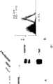
图1:HPTPβECD蛋白的设计和生产。(图A)全长HPTPβ和HPTPβ胞外域-6His融合蛋白的示意图。(图B)来自Ni-NTA柱的咪唑洗脱液银染结果,所述柱中加入了用引导βECD-6His表达的载体转染的HEK293细胞上清液。检测到了一条单独的高分子量条带,与HPTPβ细胞外域-6His蛋白相符合。Figure 1: Design and production of the HPTPβECD protein. (Panel A) Schematic representation of full-length HPTP[beta] and HPTP[beta] ectodomain-6His fusion protein. (Panel B) Silver staining of imidazole eluate from a Ni-NTA column to which supernatants from HEK293 cells transfected with a vector directing the expression of βECD-6His were added. A single high molecular weight band was detected, consistent with the extracellular domain of HPTPβ-6His protein.
图2:R15E6识别内皮细胞上的内源HPTPβ。(图A)内皮细胞溶解产物用对照抗体(泳道1)、R15E6(泳道2)或抗-Tie2和抗-VEGFR2抗体的混合物(泳道3)分别进行免疫沉淀。免疫沉淀物溶解于SDS-PAGE,转移到PVD膜上,用R15E6、抗-Tie2和抗-VEGFR2抗体的混合物通过western印迹法检测。可看到R15E6(泳道2)有一条单独的高分子量条带,与HPTPβ相符合,而对照抗体(泳道1)或抗-Tie2和抗-VEGFR2的混合物(泳道3)无此条带。(图B)内皮细胞经过FACS分析,R15E6(白峰)或无第一抗体对照物(黑峰)。荧光长移说明在完整的内皮细胞表面上R15E6结合HPTPβ。Figure 2: R15E6 recognizes endogenous HPTPβ on endothelial cells. (Panel A) Endothelial cell lysates were immunoprecipitated separately with a control antibody (lane 1), R15E6 (lane 2), or a mixture of anti-Tie2 and anti-VEGFR2 antibodies (lane 3). Immunoprecipitates were resolved by SDS-PAGE, transferred to PVD membranes, and detected by western blotting with a mixture of R15E6, anti-Tie2 and anti-VEGFR2 antibodies. It can be seen that R15E6 (lane 2) has a single high molecular weight band consistent with HPTPβ, which is absent from the control antibody (lane 1) or the mixture of anti-Tie2 and anti-VEGFR2 (lane 3). (Panel B) FACS analysis of endothelial cells, R15E6 (white peaks) or no primary antibody control (black peaks). The long shift in fluorescence indicates that R15E6 binds HPTP[beta] on the surface of intact endothelial cells.
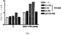
图3:R15E6促进HUVEC中的Tie2受体活化。Tie2活化如实施例4所述在人内皮细胞中进行测量。R15E6剂量依赖性地促进基底的和Ang1诱导的Tie2活化。Figure 3: R15E6 promotes Tie2 receptor activation in HUVECs. Tie2 activation was measured in human endothelial cells as described in Example 4. R15E6 dose-dependently promotes basal and Ang1-induced Tie2 activation.
图4:R15E6促进HUVEC存活。血清饥饿人内皮细胞的存活如实施例4所述进行测量。符合它对Tie2活化的效果,R15E6剂量依赖性地促进基底的和Ang1诱导的内皮细胞存活(图A)。此外,R15E6还剂量依赖性地促进VEGF和FGF介导的内皮细胞存活(图B和图C)。对照抗体不能促进内皮细胞存活(图D)。Figure 4: R15E6 promotes HUVEC survival. The survival of serum starved human endothelial cells was measured as described in Example 4. Consistent with its effect on Tie2 activation, R15E6 dose-dependently promoted basal and Ang1-induced endothelial cell survival (Panel A). In addition, R15E6 dose-dependently promoted VEGF- and FGF-mediated endothelial cell survival (Panels B and C). Control antibody failed to promote endothelial cell survival (Panel D).
图5:R15E6促进HUVEC迁移。人内皮细胞的迁移如实施例4中所述进行测量。R15E6剂量依赖性地促进基底的和VEGF诱导的内皮细胞迁移。Figure 5: R15E6 promotes HUVEC migration. Migration of human endothelial cells was measured as described in Example 4. R15E6 dose-dependently promotes basal and VEGF-induced endothelial cell migration.
图6:R15E6在HUVEC/小珠血管生成检测分析法中促进毛细血管形态发生。人内皮细胞的毛细血管形态发生如实施例4所述在小珠血管生成检测分析法中进行测量。R15E6促进基底的和VEGF诱导的内皮细胞毛细血管形态发生。Figure 6: R15E6 promotes capillary morphogenesis in a HUVEC/bead angiogenesis assay. Capillary morphogenesis of human endothelial cells was measured in a bead angiogenesis assay as described in Example 4. R15E6 promotes basal and VEGF-induced capillary morphogenesis in endothelial cells.
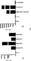
图7:Western印迹分析将R15E6结合表位定位于HPTPβ细胞外域的N-末端FN3重复序列。(图A)通过western分析,R15E6结合所有C-末端平截突变型证明结合表位位于N-末端的2个FN3重复序列。(图B)对小鼠/人嵌合蛋白的分析进一步将R15E6结合表位定位于HPTPβ的N-末端FN3重复序列。Figure 7: Western blot analysis localizes the R15E6 binding epitope to the N-terminal FN3 repeat of the extracellular domain of HPTP[beta]. (Panel A) By western analysis, R15E6 binds to all C-terminal truncated mutants demonstrating that the binding epitope is located at the N-
图8:MSD分析证实R15E6结合表位定位于HPTPβ细胞外域的N-末端FN3重复序列。(图A)通过MSD分析,R15E6结合所有C-末端平截突变型证实结合表位位于N-末端的2个FN3重复序列。(图B)对小鼠/人嵌合蛋白的分析进一步证实R15E6结合表位定位于HPTPβ的N-末端FN3重复序列。Figure 8: MSD analysis confirms that the R15E6 binding epitope localizes to the N-terminal FN3 repeat of the extracellular domain of HPTPβ. (Panel A) R15E6 binds all C-terminal truncated mutants confirming that the binding epitope is located at the N-
图9:MSD分析证明单价R15E6Fab片段也结合HPTPβ的N-末端FN3重复序列。(图A)与完整R15E6抗体相似,R15E6Fab片段结合所有C-末端平截突变型证实结合表位位于N-末端的2个FN3重复序列。(图B)对小鼠/人嵌合蛋白的分析进一步将R15E6Fab片段的结合表位定位于HPTPβ的N-末端FN3重复序列。Figure 9: MSD analysis demonstrates that the monovalent R15E6 Fab fragment also binds the N-terminal FN3 repeat of HPTP[beta]. (Panel A) Similar to the intact R15E6 antibody, the R15E6 Fab fragment binds all C-terminal truncated mutants demonstrating that the binding epitope is located at the N-
图10:单价R15E6Fab片段不能促进Tie2的活化,并阻止Tie2被完整R15E6活化。Figure 10: Monovalent R15E6 Fab fragment fails to promote Tie2 activation and prevents Tie2 activation by intact R15E6.
图11:R15E6Fab片段有效地抑制了内皮细胞的存活。(图A)与Fab片段对照物相比,R15E6Fab片段有效地抑制了内皮细胞的存活。(图B)完整的R15E6的竞争作用减弱了R15E6Fab片段的抑制效果。Figure 11: R15E6 Fab fragment potently inhibits endothelial cell survival. (Panel A) R15E6 Fab fragment effectively inhibited endothelial cell survival compared to Fab fragment control. (Panel B) Competition by intact R15E6 attenuates the inhibitory effect of the R15E6 Fab fragment.
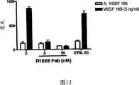
图12:R15E6Fab片段抑制VEGF介导的内皮细胞迁移。Figure 12: R15E6 Fab fragments inhibit VEGF-mediated migration of endothelial cells.
序列列表描述Sequence listing description
序列列表中的每一个核苷酸和蛋白质序列与相应的Genbank或Derwent保藏编号(适用时)以及它们的起源物种一起显示于表I中。Each nucleotide and protein sequence in the sequence listing is shown in Table I together with the corresponding Genbank or Derwent deposit number (where applicable) and their species of origin.
表ITable I
发明详述Detailed description of the invention
本发明涉及结合HPTPβ的抗体及其应用。The present invention relates to antibodies that bind HPTPβ and uses thereof.
标准技术可用于重组DNA、低聚核苷酸合成、以及组织培养和转化(例如电穿孔、微脂粒感染)。可按照厂商的说明书、或本领域中的常规方法、或如本文所述使用酶反应和纯化技术。所述技术和程序一般按照本领域已知的常规方法,并如本说明书中引用和讨论的各种常规的以及较具体的参考文献所述进行实施。除非提供具体的定义,与本文所述的分析化学、有机合成化学、和医药化学有关的所用命名以及试验程序和技术是那些本领域已知的和普遍使用的。标准技术可用于化学合成、化学分析、药物制备、制剂配制和递送、以及治疗病人。Standard techniques can be used for recombinant DNA, oligonucleotide synthesis, and tissue culture and transformation (eg, electroporation, liposome infection). Enzymatic reactions and purification techniques may be employed according to manufacturer's specifications, or as routine in the art, or as described herein. The techniques and procedures are generally performed according to conventional methods known in the art and as described in various general and more specific references that are cited and discussed throughout the present specification. Unless specific definitions are provided, nomenclature used in connection with, and laboratory procedures and techniques of, analytical chemistry, synthetic organic chemistry, and medicinal chemistry described herein are those known and commonly used in the art. Standard techniques can be used for chemical syntheses, chemical analyses, preparation of pharmaceuticals, formulation and delivery of formulations, and treatment of patients.
除非另外指明,应当理解以下术语具有以下含义:Unless otherwise indicated, the following terms shall be understood to have the following meanings:
本文所用的“蛋白质”可与肽和多肽互换。HPTPβ是序列列表中限定的人蛋白酪氨酸磷酸酶。在一些实施方案中,使用HPTPβ的不同片段。下文所述的同源物、直向同源物、片段、变体、和HPTPβ蛋白和基因的突变型均被认为在术语“HPTPβ”的范围内。As used herein, "protein" is interchangeable with peptide and polypeptide. HPTP[beta] is a human protein tyrosine phosphatase defined in the sequence listing. In some embodiments, different fragments of HPTP[beta] are used. Homologues, orthologs, fragments, variants, and mutants of the HPTPβ protein and gene described below are all considered within the scope of the term "HPTPβ".
“片段”是指核苷酸或蛋白质序列的一部分。片段可保留其源蛋白的生物活性。核苷酸序列的片段也可用作杂交探针和引物,或用于调节基因表达,例如反义、siRNA、或小RNA。生物活性部分可以通过下列方法制备:分离本发明的其中一个核苷酸序列的部分、表达被编码的部分(例如,通过体外重组表达),评估被编码蛋白质的活性。"Fragment" refers to a portion of a nucleotide or protein sequence. Fragments retain the biological activity of their source protein. Fragments of nucleotide sequences can also be used as hybridization probes and primers, or to modulate gene expression, such as antisense, siRNA, or small RNA. Biologically active portions can be prepared by isolating a portion of one of the nucleotide sequences of the invention, expressing the encoded portion (eg, by recombinant expression in vitro), and assessing the activity of the encoded protein.
本领域的技术人员也能认识到,得自除了那些序列列表中列出的物种之外的基因和蛋白质,尤其是脊椎动物,也是可用的。此类物种包括但不限于小鼠、大鼠、豚鼠、兔子、狗、猪、山羊、母牛、猴子、黑猩猩、绵羊、仓鼠和斑马鱼。本领域的技术人员还将认识到通过使用来自已知物种序列的探针,cDNA或同源于已知序列的基因序列可以从相同的或替代物种中通过已知的克隆方法获得。此类同源物和直向同源物被设想用于本发明的实践。Those skilled in the art will also recognize that genes and proteins from species other than those listed in the sequence listing, especially vertebrates, are also available. Such species include, but are not limited to, mice, rats, guinea pigs, rabbits, dogs, pigs, goats, cows, monkeys, chimpanzees, sheep, hamsters, and zebrafish. Those skilled in the art will also recognize that by using probes from known species sequences, cDNA or gene sequences homologous to known sequences can be obtained from the same or an alternate species by known cloning methods. Such homologs and orthologs are contemplated for use in the practice of the present invention.
“变体”是指类似的序列。例如,保守变体可包括那些因为遗传密码退化,编码本发明其中一个多肽的氨基酸序列的那些序列。天然存在的等位变体和剪接变体可以用已知的技术鉴定,例如用聚合酶链反应(PCR)、单核苷酸多态性(SNP)分析和杂交技术。为了分离直向同源物和同源物,一般的严紧杂交条件主要通过规定具体序列、序列长度、鸟嘌呤+胞核嘧啶(GC)含量和其它参数被利用。变体核苷酸序列还包括合成来源的核苷酸序列,例如通过使用定点诱变获得的核苷酸序列。变体可包含单独来自基因组位点或与其它序列组合的额外的序列。"Variants" refer to similar sequences. For example, conservative variants may include those sequences that, due to degradation of the genetic code, encode the amino acid sequence of one of the polypeptides of the invention. Naturally occurring allelic and splice variants can be identified using known techniques, such as polymerase chain reaction (PCR), single nucleotide polymorphism (SNP) analysis and hybridization techniques. For the isolation of orthologs and homologues, generally stringent hybridization conditions are utilized mainly by specifying the specific sequence, sequence length, guanine+cytosine (GC) content, and other parameters. Variant nucleotide sequences also include nucleotide sequences of synthetic origin, such as nucleotide sequences obtained by using site-directed mutagenesis. Variants may contain additional sequences derived from genomic loci alone or in combination with other sequences.
本发明的分子也包括平截的和/或突变的蛋白质,其中配体结合或信号转导不需要的蛋白质区域已被删除或修饰。同样,它们也发生突变以改变其配体结合或信号转导活性。此类突变可涉及非保守突变、删除、或氨基酸或蛋白质域的增加。变体蛋白可保留或不保留生物活性。这种变体可以由例如基因多态性或人类操纵产生。The molecules of the invention also include truncated and/or mutated proteins in which regions of the protein not required for ligand binding or signal transduction have been deleted or modified. Likewise, they are mutated to alter their ligand binding or signaling activity. Such mutations may involve non-conservative mutations, deletions, or additions of amino acids or protein domains. Variant proteins may or may not retain biological activity. Such variants may arise, for example, from genetic polymorphisms or human manipulation.
本文还可以想到融合蛋白。使用已知的方法,本领域的技术人员将能够制造本发明的蛋白质的融合蛋白;尽管它不同于与源蛋白的形式,但也是可用的。例如,融合部分可以是信号(或前导)多肽序列,该序列共同翻译或在后翻译指导蛋白质从它的合成部位转移到另一个部位(例如,酵母α因子前导)。作为另外一种选择,可将它加入以有利于本发明蛋白质的纯化或识别(例如,聚His、标记肽、或荧光蛋白)。Fusion proteins are also contemplated herein. Using known methods, one skilled in the art will be able to make a fusion protein of the protein of the invention; although it is in a different form from the source protein, it is useful. For example, the fusion moiety can be a signal (or leader) polypeptide sequence that co-translates or post-translates to direct the transfer of the protein from its site of synthesis to another site (eg, yeast alpha factor leader). Alternatively, it can be added to facilitate purification or identification of proteins of the invention (eg, poly-His, tagged peptides, or fluorescent proteins).
术语“抗原”是指能被选择性结合剂如抗体结合,另外还能在动物中使用以生产能够结合抗原表位的抗体的分子或分子部分。抗原可以有一个或多个表位。The term "antigen" refers to a molecule or portion of a molecule capable of being bound by a selective binding agent, such as an antibody, and which can additionally be used in an animal to produce antibodies capable of binding an antigenic epitope. An antigen can have one or more epitopes.
术语“表位”包括任何抗原决定簇,优选多肽决定簇,能够特异结合到免疫球蛋白或T-细胞受体上。在某些实施方案中,表位决定簇包括化学活性表面基团诸如氨基酸、糖、类脂、磷酰基、或磺酰基,并且在某些实施方案中,表位决定簇可具有特异的三维结构特性,和/或特异的电荷特性。表位是抗原的抗体结合区域。在某些实施方案中,抗体被认为,当它优选地识别其在蛋白质和/或大分子复合混合物中的目标抗原时会特异性地结合抗原。抗体也被认为,当它对抗原表现出比其它相关的和/或不相关的分子更高的亲和力时会特异性地结合抗原。The term "epitope" includes any antigenic determinant, preferably a polypeptide determinant, capable of specifically binding to an immunoglobulin or T-cell receptor. In certain embodiments, epitopic determinants include chemically active surface groups such as amino acids, sugars, lipids, phosphoryl groups, or sulfonyl groups, and in certain embodiments, epitopic determinants may have specific three-dimensional structures characteristics, and/or specific charge characteristics. An epitope is the antibody-binding region of an antigen. In certain embodiments, an antibody is believed to specifically bind an antigen when it preferentially recognizes its target antigen in a complex mixture of proteins and/or macromolecules. An antibody is also believed to specifically bind an antigen when it exhibits a higher affinity for the antigen than other related and/or unrelated molecules.
如本文所用,术语“抗体”(Ab)包括单克隆抗体、多克隆抗体、多特异性抗体(例如双特异性抗体)、单链抗体例如来自骆驼的抗体、抗体片段例如可变区和/或恒定区片段,只要它们表现出期望的生物活性,例如抗原结合活性。术语“免疫球蛋白”(Ig)与本文的“抗体”是可互换的。As used herein, the term "antibody" (Ab) includes monoclonal antibodies, polyclonal antibodies, multispecific antibodies (such as bispecific antibodies), single chain antibodies such as antibodies from camelids, antibody fragments such as variable regions and/or Constant region fragments, as long as they exhibit desired biological activities, such as antigen-binding activity. The term "immunoglobulin" (Ig) is interchangeable with "antibody" herein.
“分离抗体”是已经被鉴定的、和/或分离的、和/或从其天然环境中重新获得的抗体。An "isolated antibody" is an antibody that has been identified, and/or isolated, and/or recovered from its natural environment.
基本的四链抗体单位是一种糖蛋白异四聚体,由两个相同的轻(L)链和两个相同的重链(H)组成(IgM抗体由5个基本的异四聚体单位和一个附加的称为J链的多肽组成,因此包含10个抗原结合位点,然而分泌的IgA抗体可聚合以形成多价的聚合体,所述聚合体包括2至5个基本的4链单位以及J链)。就IgG而言,四链单位一般为约150千道尔顿(kDa)。每个L链通过一个共价二硫键连接到一个H链上,而两个H链取决于H链同种型通过一个或多个二硫键彼此相连。每个H链和L链也有规则排列的链内二硫桥。每个H链在N-末端具有一个可变区(VH),每个α和γ链有三个恒定区(CH)而每个μ和ε同种型有四个CH区。每个L链在N-末端具有可变区(VL),在另一端有恒定区(CL)。VL与VH相连,并且CL与重链(CHl)的第一恒定区相连。据信特殊的氨基酸残基形成轻链和重链可变区之间的界面。VH和VL对一起形成专一的抗原结合位点。不同类别抗体的结构和特性参见例如Basic and Clinical Immunology,第8版,Daniel P.Stites,Abba I.Terr and Tristram G.Parslow(eds.),Appleton & Lange,1994,第6章第71页。The basic four-chain antibody unit is a glycoprotein heterotetramer consisting of two identical light (L) chains and two identical heavy (H) chains (IgM antibodies consist of five basic heterotetrameric units and an additional polypeptide called the J chain, thus containing 10 antigen-binding sites, however secreted IgA antibodies can polymerize to form multivalent aggregates comprising 2 to 5 basic 4-chain units and J chain). For IgG, the four-chain unit is typically about 150 kilodaltons (kDa). Each L chain is linked to one H chain by one covalent disulfide bond, while the two H chains are linked to each other by one or more disulfide bonds depending on the H chain isotype. Each H and L chain also has regularly arranged intrachain disulfide bridges. Each H chain has one variable region (VH ) at the N-terminus, each alpha and gamma chain has three constant regions (CH ) and each mu and epsilon isoform has fourCH regions. Each L chain has a variable region (VL ) at the N-terminus and a constant region (CL ) at the other end.VL is linked toVH , andCL is linked to the first constant region of the heavy chain (CHl ). Particular amino acid residues are believed to form the interface between the light and heavy chain variable regions. Together, theVH andVL pairs form a specific antigen-binding site. For the structure and properties of the different classes of antibodies see eg Basic and Clinical Immunology, 8th Edition, Daniel P. Stites, Abba I. Terr and Tristram G. Parslow (eds.), Appleton & Lange, 1994,
来自任何脊椎动物的L链基于它们的恒定区氨基酸序列可被分成两种不同类型,分别称为k和λ。取决于它们的重链(CH)恒定区的氨基酸序列,免疫球蛋白可以被分为不同的类别或同种型。有五类免疫球蛋白:IgA、IgD、IgE、IgG和IgM,它们各自具有指定的重链α、δ、ε、γ和μ。根据CH序列和功能的相对较小的差异,可将γ类和α类进一步分成亚型,例如,人表达以下亚型:IgG1、IgG2、IgG3、IgG4、IgA1和IgA2。L chains from any vertebrate can be divided into two different types, called kappa and lambda, respectively, based on their constant region amino acid sequences. Depending on the amino acid sequence of the constant region of their heavy chains (CH ), immunoglobulins can be assigned to different classes, or isotypes. There are five classes of immunoglobulins: IgA, IgD, IgE, IgG, and IgM, each of which has designated heavy chains α, δ, ε, γ, and μ. The gamma and alpha classes can be further divided into subtypes based on relatively minor differences in CH sequence and function, for example, humans express the following subtypes: IgG1, IgG2, IgG3, IgG4, IgAl, and IgA2.
驼科家族的成员例如无峰驼、骆驼和单峰驼包含独特的抗体型,所述抗体缺失轻链,并进一步缺失CH1区(Muyldermans,S.,Rev.Mol.Biotechnol.,74,277-302(2001))。这些重链抗体的可变区称为VHH或VHH,并且构建衍生自功能性免疫球蛋白的最小的可利用完整抗原结合片段(15kDa)。Members of the camelid family such as llamas, camels and dromedaries contain unique antibody types that lack light chains and are further deleted from theCH1 region (Muyldermans, S., Rev. Mol. Biotechnol., 74, 277 -302(2001)). The variable regions of these heavy chain antibodies are termedVHH or VHH, and construct the smallest available intact antigen-binding fragment (15 kDa) derived from a functional immunoglobulin.
术语“可变的”是指可变区的某些片段在抗体中的序列差别很大。V区调节抗原结合并且限定特异抗体对其抗原的特异性。然而,可变性未被平均分配到可变区的110个氨基酸序列片段中。相反,V区由相对不变的区域组成,该区域称为框架区(FR),长15至30个氨基酸,它们被较短的高可变区域分隔,该区域称为“高变区”,每个长9至12个氨基酸。每个天然重链和轻链的可变区包括四个主要采用β-折叠构型的FR,它们由三个高变区相连,形成环形连接,并且在一些情况下形成部分β-折叠结构。每条链中的高变区被FR结合到一起,彼此非常接近,并与其它链的高变区一起有助于形成抗体的抗原结合位点。抗体与抗原的结合不直接涉及恒定区,但是所述恒定区表现出多种效应子功能,诸如参与抗体依赖性细胞介导的细胞毒作用(ADCC)。The term "variable" means that certain segments of the variable region vary widely in sequence among antibodies. The V regions regulate antigen binding and define the specificity of a specific antibody for its antigen. However, the variability is not evenly distributed across the 110 amino acid sequence segments of the variable region. In contrast, the V regions consist of relatively invariant regions called framework regions (FRs), 15 to 30 amino acids in length, which are separated by shorter hypervariable regions called "hypervariable regions", Each is 9 to 12 amino acids long. The variable region of each native heavy and light chain consists of four FRs that primarily adopt a β-sheet configuration, connected by three hypervariable regions, forming circular junctions and, in some cases, partial β-sheet structures. The hypervariable regions in each chain are held together by FRs in close proximity to each other and together with the hypervariable regions of the other chains contribute to the antigen-binding site of the antibody. Binding of an antibody to an antigen does not directly involve the constant regions, but the constant regions exhibit various effector functions, such as participation in antibody-dependent cell-mediated cytotoxicity (ADCC).
术语“高变区”当在本文中使用时是指负责抗原结合的抗体氨基酸残基。高变区一般包括来自“互补性决定区”或“CDR”的氨基酸残基(例如VL中的约24至34残基(L1)、50至56残基(L2)和89至97(L3)残基,以及VH中的约1至35残基(H1)、50至65残基(H2)和95至102残基(H3);Kabat等人,Sequences of Proteins ofImmunological Interest,第5版Public Health Service,NationalInstitutes of Health,Bethesda,Md.(1991))和/或那些来自“高变环”的残基。The term "hypervariable region" when used herein refers to the amino acid residues of an antibody that are responsible for antigen binding. Hypervariable regions typically include amino acid residues from "complementarity determining regions" or "CDRs" (e.g., about 24 to 34 residues (L1), 50 to 56 residues (L2), and 89 to 97 (L3) inVL . ) residues, and about 1 to 35 residues (H1), 50 to 65 residues (H2) and 95 to 102 residues (H3) inVH ; Kabat et al., Sequences of Proteins of Immunological Interest, 5th Edition Public Health Service, National Institutes of Health, Bethesda, Md. (1991)) and/or those residues from "hypervariable loops".
如本文所用,术语“单克隆抗体”是指从一组基本上同源的抗体中获得的抗体,即,该组抗体中包括的各个抗体是相同的,除了可能有的微量天然存在的突变。与包括抗不同表位的不同抗体的多克隆抗体相对照,每个单克隆抗体抗一个单独的表位,即,一个单独的抗原决定簇。除了它们的特异性之外,单克隆抗体的优点还在于它们可通过其它抗体被无污染的合成。修饰语“单克隆”将被理解为不需要任何特殊的方法生产抗体。例如,本发明可用的单克隆抗体可通过杂交瘤方法进行制备,或使用在细菌、真核动物或植物细胞中的重组DNA方法进行制造(参见例如美国专利4,816,567)。所述“单克隆抗体”还可使用可用的技术从噬菌体抗体文库中分离,例如Clackson等人,Nature,352:624-628(1991)。As used herein, the term "monoclonal antibody" refers to an antibody obtained from a group of substantially homogeneous antibodies, ie, the individual antibodies included in the group are identical except for possible minor naturally occurring mutations. In contrast to polyclonal antibodies, which comprise different antibodies directed against different epitopes, each monoclonal antibody is directed against a single epitope, ie, a single antigenic determinant. In addition to their specificity, monoclonal antibodies have the advantage that they can be synthesized without contamination by other antibodies. The modifier "monoclonal" is to be understood as not requiring any special method for producing the antibody. For example, monoclonal antibodies useful in the invention can be produced by the hybridoma method, or using recombinant DNA methods in bacterial, eukaryotic, or plant cells (see, eg, US Patent No. 4,816,567). Such "monoclonal antibodies" can also be isolated from phage antibody libraries using available techniques, eg, Clackson et al., Nature, 352:624-628 (1991).
本文的单克隆抗体包括“嵌合”抗体,其中一部分重链和/或轻链与衍生自特定物种的或属于特定抗体类型或亚型的抗体中的对应序列相同或同源,而残余链与衍生自另一种物种或属于另一种抗体类型或亚型的抗体中的对应序列相同或同源,以及此类抗体的片段,只要它们表现出所需的生物活性(参见美国专利4,816,567;和Morrison等人,Proc.Natl.Acad.Sci.”USA,81,6851-6855(1984))。Monoclonal antibodies herein include "chimeric" antibodies in which a portion of the heavy and/or light chains are identical or homologous to corresponding sequences in antibodies derived from a particular species or belonging to a particular antibody class or subtype, and the remaining chains are identical to Identical or homologous to corresponding sequences in antibodies derived from another species or belonging to another antibody class or subtype, and fragments of such antibodies, so long as they exhibit the desired biological activity (see U.S. Patent No. 4,816,567; and Morrison et al., Proc. Natl. Acad. Sci." USA, 81, 6851-6855 (1984)).
“抗体片段”包括多亚基抗体的一部分,优选完整抗体的抗原结合区或可变区。抗体片段的实例包括Fab、Fab′、F(ab′)2、Fab结合物的二聚体和三聚体、Fv、scFv、微型抗体;二聚体、三聚体和四聚体;线性抗体(参见Hudson等人,Nature Med.9,129-134(2003))。"Antibody fragment" includes a portion of a multi-subunit antibody, preferably the antigen-binding or variable region of an intact antibody. Examples of antibody fragments include Fab, Fab', F(ab')2 , dimers and trimers of Fab conjugates, Fv, scFv, minibodies; dimers, trimers and tetramers; linear antibodies (See Hudson et al., Nature Med. 9, 129-134 (2003)).
“Fv”是包含全部抗原结合位点的最小抗体片段。此片段由一个重链可变区和一个轻链可变区通过牢固的非共价结合形成的二聚体组成。从这两个区域发出六个高变环(从H链和L链各发出3个环),产生用于抗原结合的氨基酸残基并赋予抗体抗原结合特异性。然而,甚至单独一个可变区(或仅包括三个抗原特异性的CDR的半个Fv)都具有识别和结合抗原的能力,因此它们被包括在Fv的定义中。"Fv" is the smallest antibody fragment that contains the entire antigen binding site. This fragment consists of a dimer of a heavy chain variable domain and a light chain variable domain through strong non-covalent association. Six hypervariable loops (3 loops each from the H and L chains) emanate from these two regions, generating amino acid residues for antigen binding and conferring antigen binding specificity to the antibody. However, even a single variable domain (or half of an Fv comprising only three antigen-specific CDRs) has the ability to recognize and bind antigen and they are therefore included in the definition of Fv.
“单链Fv”也简写为“sFv”或“scFv”,它是包括连接到单多肽链中的VH和VL抗体域的抗体片段。优选地,sFv多肽还包括在VH和VL域之间的多肽连接肽,它使得sFv形成抗原结合所需的构型。"Single-chain Fv", also abbreviated "sFv" or "scFv", is an antibody fragment comprising theVH andVL antibody domains linked into a single polypeptide chain. Preferably, the sFv polypeptide also includes a polypeptide linker peptide between theVH andVL domains, which allows the sFv to assume the desired configuration for antigen binding.
术语“二聚体、三聚体、和四聚体”是指通过构建sFv片段与VH和VL区之间的短连接片段(约5至10各残基)来制备的小抗体片段,所述短连接片段使得V区形成链间而不是链内的配对,导致多价片段。The terms "dimers, trimers, and tetramers" refer to small antibody fragments prepared by constructing short linker fragments (approximately 5 to 10 residues) between the sFv fragment and the VH and VL regions, which Short connecting segments allow the V regions to form interchain rather than intrachain pairings, resulting in multivalent fragments.
术语“人源化抗体”或“人抗体”是指这样的抗体:包括来自非人类物种(例如小鼠)的重链和轻链可变区序列,但是其中至少一部分VH和/或VL序列已经被改变成较“类似人的”,即与人类种系可变区更相似。一种人源化抗体类型是CDR-移植抗体,其中人CDR序列被引入非人的VH和VL序列以置换对应的非人CDR序列。制造嵌合的CDR移植和人源化抗体的方法是本领域的普通技术人员已知的(参见例如美国专利4,816,567和5,225,539)。一种制造人抗体的方法使用转基因动物如转基因小鼠。这些转基因动物包含被插入它们自己的基因组的、基本部分的人抗体生产基因组,致使在抗体生产中动物自己的内源抗体生产缺乏。制造转基因动物的方法是本领域已知的。此类转基因动物可使用XenoMouseRTM技术或使用“小位点”方法进行制造。制造XenoMiceRTM的方法在美国专利6,162,963、6,150,584、6,114,598和6,075,181中有所描述。使用“小位点”方法制造转基因动物的方法在美国专利5,545,807、5,545,806和5,625,825、以及WO93/12227中有所描述。The term "humanized antibody" or "human antibody" refers to an antibody that includes heavy and light chain variable region sequences from a non-human species (eg, mouse), but in which at least a portion of the VH and/or VL sequences have been Altered to be more "human-like", ie more similar to human germline variable regions. One type of humanized antibody is a CDR-grafted antibody, in which human CDR sequences are introduced into non-human VH and VL sequences to replace the corresponding non-human CDR sequences. Methods of making chimeric CDR-grafted and humanized antibodies are known to those of ordinary skill in the art (see eg, US Patents 4,816,567 and 5,225,539). One method of making human antibodies uses transgenic animals such as transgenic mice. These transgenic animals contain a substantial portion of the human antibody-producing genome inserted into their own genome such that the animal's own endogenous antibody production is absent in antibody production. Methods of making transgenic animals are known in the art. Such transgenic animals can be produced using XenoMouse(TM) technology or using the "mini-site" approach. Methods of making XenoMiceRTM are described in US Patents 6,162,963, 6,150,584, 6,114,598, and 6,075,181. Methods for making transgenic animals using the "small locus" approach are described in US Patent Nos. 5,545,807, 5,545,806, and 5,625,825, and WO 93/12227.
非人抗体的人源化近年来已经变得常规化,现在是本领域技术人员掌握的知识。有几家公司提供制造人源化抗体的服务,例如Xoma、Aries、Medarex、PDL、和Cambridge Antibody Technologies。人源化方案被广泛描述于技术文献中,例如Kipriyanov and Le Gall,MolecularBiotechnol,Vol.26,pp39-60(2004),Humana Press,Totowa,NJ;Lo,Methods Mol.Biol.,Vol.248,pp135-159(2004),HumanaPress,Totowa,NJ;Wu等人,J.Mol.Biol.294,151-162(1999)。Humanization of non-human antibodies has become routine in recent years and is now within the knowledge of those skilled in the art. There are several companies that provide services for manufacturing humanized antibodies, such as Xoma, Aries, Medarex, PDL, and Cambridge Antibody Technologies. Humanization protocols are widely described in the technical literature, for example Kipriyanov and Le Gall, Molecular Biotechnol, Vol.26, pp39-60 (2004), Humana Press, Totowa, NJ; Lo, Methods Mol. Biol., Vol.248, pp135-159 (2004), HumanaPress, Totowa, NJ; Wu et al., J. Mol. Biol. 294, 151-162 (1999).
在某些实施方案中,本发明的抗体可在除杂交瘤细胞系之外的细胞系中表达。可以通过已知方法,使用编码特定抗体的序列转化合适的哺乳动物宿主细胞,以将多核苷酸引入宿主细胞,所述方法包括例如将多核苷酸包封在病毒中(或在病毒载体中)并用病毒(或载体)转化宿主细胞,或通过本领域已知的转染方法,这些方法为美国专利4,399,216、4,912,040、4,740,461和4,959,455所证实。使用的转化程序可取决于需被转化的宿主。将异源多核苷酸引入哺乳动物细胞的方法是本领域已知的,包括但不限于葡聚糖介导的转染、磷酸钙沉淀、凝聚胺介导的转染、原生质体融合、电穿孔、脂质体中的多核苷酸包封、将核酸与带正电的类脂混合、以及将DNA直接显微注射进细胞核。In certain embodiments, antibodies of the invention may be expressed in cell lines other than hybridoma cell lines. Introduction of the polynucleotide into the host cell can be accomplished by transforming a suitable mammalian host cell with the sequence encoding the specific antibody by known methods, including, for example, encapsulation of the polynucleotide in a virus (or in a viral vector) The host cells are transformed with the virus (or vector), or by transfection methods known in the art as demonstrated in US Patent Nos. 4,399,216, 4,912,040, 4,740,461 and 4,959,455. The transformation procedure used may depend on the host to be transformed. Methods for introducing heterologous polynucleotides into mammalian cells are known in the art and include, but are not limited to, dextran-mediated transfection, calcium phosphate precipitation, polybrene-mediated transfection, protoplast fusion, electroporation , Encapsulation of polynucleotides in liposomes, mixing nucleic acids with positively charged lipids, and microinjection of DNA directly into the nucleus.
使用标准连接技术将编码抗体的重链恒定区、重链可变区、轻链恒定区、或轻链可变区的氨基酸序列的核酸分子,或如果需要的话它们的合适组合的片段,插入合适的表达载体。抗体重链恒定区或轻链恒定区可连接到适宜可变区的C末端,并连接到表达载体中。所述载体通常选择在使用的特定宿主细胞中是功能性的载体(即,所述载体与宿主细胞相容以使得基因扩增和/或基因表达可发生)。表达载体参见Methods Enzymol.vol.185(Goeddel,ed.),1990,Academic Press。A nucleic acid molecule encoding the amino acid sequence of the heavy chain constant region, heavy chain variable region, light chain constant region, or light chain variable region of an antibody, or fragments of a suitable combination thereof if desired, is inserted into a suitable ligation technique using standard ligation techniques. expression vector. An antibody heavy or light chain constant region can be linked to the C-terminus of an appropriate variable region and ligated into an expression vector. The vector is typically selected to be functional in the particular host cell used (ie, the vector is compatible with the host cell such that gene amplification and/or gene expression can occur). For expression vectors, see Methods Enzymol. vol. 185 (Goeddel, ed.), 1990, Academic Press.
本发明的抗体及其片段结合HPTPβ并调节血管新生。如上述定义,术语抗体用于表示抗原结合片段。下文进一步描述此类抗体和抗原结合片段的用途。Antibodies and fragments thereof of the invention bind HPTP[beta] and regulate angiogenesis. As defined above, the term antibody is used to denote an antigen-binding fragment. Uses of such antibodies and antigen-binding fragments are described further below.
使用血管新生体外和体内模型的筛选检测分析法Screening assays using in vitro and in vivo models of angiogenesis
本发明的抗体可以使用本领域中已知的血管新生检测分析法进行筛选。此类检测分析法包括测量培养细胞中血管代用品生长或组织外植体的血管结构形成的体外检测分析法,和直接地或不直接地测量血管生长的体内检测分析法(Auerbach,R.,等人(2003),Clin Chem49,32-40,Vailhe,B.,等人(2001),Lab Invest81,439-452)。Antibodies of the invention can be screened using angiogenesis detection assays known in the art. Such assays include in vitro assays that measure the growth of vascular substitutes in cultured cells or the formation of vascular structures in tissue explants, and in vivo assays that directly or indirectly measure vessel growth (Auerbach, R., et al. (2003), Clin Chem 49, 32-40, Vailhe, B., et al. (2001), Lab Invest 81, 439-452).
血管新生的体外模型In Vitro Model of Angiogenesis
大多数这些检测分析法使用培养的内皮细胞或组织外植体,并测量试剂对“血管新生的”细胞应答或对毛细血管样结构形成的功效。体外血管新生检测分析法的实例包括但不限于内皮细胞迁移和增殖、毛细血管形成、内皮新生、主动脉环外植检测分析法和鸡主动脉弓检测分析法。Most of these assays use cultured endothelial cells or tissue explants and measure the efficacy of agents on "angiogenic" cellular responses or on the formation of capillary-like structures. Examples of in vitro angiogenesis assays include, but are not limited to, endothelial cell migration and proliferation, capillary formation, endothelial neogenesis, aortic ring explantation assays, and chicken aortic arch assays.
血管新生的体内模型In vivo model of angiogenesis
在这些检测分析法中,试剂或抗体在生长因子(即VEGF或血管生成素1)存在或不存在的情况下被局部施用或系统施用,并且新血管生长通过直接观察或测量替代标志物如血色素含量或荧光指示剂进行测量。血管新生实例包括但不限于鸡胚绒毛尿囊膜检测分析法、角膜血管新生检测分析法、以及MATRIGELTM塞检测分析法。In these assays, reagents or antibodies are administered topically or systemically in the presence or absence of growth factors (i.e., VEGF or angiopoietin 1), and new blood vessel growth is detected by direct observation or measurement of surrogate markers such as hemoglobin content or fluorescent indicators for measurement. Examples of angiogenesis include, but are not limited to, chicken chorioallantoic membrane assay, corneal angiogenesis assay, and MATRIGEL™ plug assay.
血管新生失调的治疗Treatment of Angiogenesis Disorders
术语“调节”定义为其众所公认的词典含义。因此,术语“调节”的含义包括但不限于通过多种方法上调或下调,以固定、使有序或一致、管理、或引导。在一个方面,抗体可用于治疗“血管新生升高的失调”或“血管新生降低的失调”的方法。如本文所用,“血管新生升高的失调”是一种在疾病、失调、和/或病症的生物学表现中;在导致失调的生物学级联反应中;或作为失调症状,涉及多余的或升高的血管新生的失调。同样,“血管新生降低的失调”是一种在生物学表现中涉及缺少的或降低的血管新生的失调。此血管新生的“涉及”在血管新生升高的/降低的失调中包括但不限于以下内容:The term "conditioning" is defined in its accepted dictionary meaning. Thus, the meaning of the term "regulate" includes, but is not limited to, up- or down-regulation by various means to fix, bring order or conformity, manage, or direct. In one aspect, the antibody is useful in a method of treating a "disorder of increased angiogenesis" or a "disorder of decreased angiogenesis". As used herein, "a disorder of elevated angiogenesis" is one that, in the biological manifestations of a disease, disorder, and/or condition; in the biological cascade leading to the disorder; or as a symptom of a disorder, involves excess or Dysregulation of elevated angiogenesis. Likewise, a "disorder of reduced angiogenesis" is a disorder involving absent or reduced angiogenesis in its biological manifestations. This "involvement" of angiogenesis in a disorder of increased/decreased angiogenesis includes, but is not limited to, the following:
(1)血管新生作为失调或生物学表现的“原因”,血管新生的水平由于感染、自身免疫、外伤、生物力学原因、生活方式、或一些其它原因,在遗传上或者升高或者降低。(1) Angiogenesis As the "cause" of a disorder or biological manifestation, the level of angiogenesis is either genetically increased or decreased due to infection, autoimmunity, trauma, biomechanical reasons, lifestyle, or some other reason.
(2)血管新生作为疾病或失调的可见表现的一部分。也就是说,根据血管新生的增高或降低,疾病或失调是可测量的。从临床观点来说,血管新生指示疾病。然而,血管新生不需要成为疾病或失调的“特征”。(2) Angiogenesis as part of the visible manifestation of a disease or disorder. That is, the disease or disorder is measurable in terms of increased or decreased angiogenesis. From a clinical point of view, angiogenesis is indicative of disease. However, angiogenesis need not be a "signature" of a disease or disorder.
(3)血管新生是导致疾病或失调的生物化学或细胞级联反应的一部分。在这方面,血管新生的调节可中断级联反应,并且可控制疾病。本发明可治疗的血管新生失调的非限制性实例在下文中进行描述。(3) Angiogenesis is part of a biochemical or cellular cascade that leads to a disease or disorder. In this regard, modulation of angiogenesis can interrupt the cascade and control disease. Non-limiting examples of angiogenic disorders treatable by the present invention are described below.
本发明的抗体可用于治疗与视网膜/脉络膜新血管形成相关的疾病,包括但不限于:糖尿病视网膜病变、黄斑变性、癌症、镰状细胞性贫血、肉状瘤、梅毒、弹力纤维性假黄瘤、佩吉特氏病、静脉闭塞、动脉闭塞、颈动脉阻塞病、慢性葡萄膜炎/玻璃体炎、分枝杆菌感染、莱姆氏病、系统性红斑狼疮、早产儿视网膜病、伊耳斯氏病、白塞病、感染引起的视网膜炎或脉络膜炎、眼假组织胞浆菌病、白斯特氏病、近视、眼凹、斯特格病变、睫状体平坦部炎、慢性视网膜脱落、高粘滞综合症、弓形体病、外伤和激光术后并发症。其它疾病包括但不限于红变相关的疾病(虹膜新血管形成),和由维管组织或纤维组织的异常增殖引起的疾病,包括所有形式的增殖性玻璃体视网膜病变,无论其是否与糖尿病相关。The antibodies of the invention can be used to treat diseases associated with retinal/choroidal neovascularization, including but not limited to: diabetic retinopathy, macular degeneration, cancer, sickle cell anemia, sarcoid, syphilis, pseudoxanthoma elasticum , Paget's disease, venous occlusion, arterial occlusion, carotid occlusive disease, chronic uveitis/vitreitis, mycobacterial infection, Lyme disease, systemic lupus erythematosus, retinopathy of prematurity, Earl's disease Behcet's disease, retinitis or choroiditis caused by infection, ocular pseudohistoplasmosis, Bauster's disease, myopia, sunken eye, Steiger's disease, pars plana, chronic retinal detachment, Hyperviscosity syndrome, toxoplasmosis, trauma and post-laser complications. Other diseases include, but are not limited to, diseases associated with erythema (iris neovascularization), and diseases caused by abnormal proliferation of vascular or fibrous tissue, including all forms of proliferative vitreoretinopathy, whether or not associated with diabetes.
本发明的抗体可以用于治疗与慢性炎症相关的疾病。具有慢性炎症症状的疾病包括炎性肠疾病诸如克隆氏病和溃疡性结肠炎、牛皮癣、肉状瘤和类风湿性关节炎。血管新生是这些慢性炎性疾病共同具有的一个关键要素。慢性炎症依赖新生毛细血管的连续形成以维持发炎细胞的流入。发炎细胞的流入和存在产生肉芽瘤,因此维持慢性发炎状态。通过本发明的组合物和方法抑制血管新生将预防肉芽瘤的形成并减轻疾病症状。The antibodies of the invention can be used to treat diseases associated with chronic inflammation. Diseases with symptoms of chronic inflammation include inflammatory bowel diseases such as Crohn's disease and ulcerative colitis, psoriasis, sarcoid and rheumatoid arthritis. Angiogenesis is a key element that these chronic inflammatory diseases share. Chronic inflammation relies on the continuous formation of new capillaries to maintain the influx of inflammatory cells. The influx and presence of inflammatory cells produces granulomas, thus maintaining a chronic inflammatory state. Inhibition of angiogenesis by the compositions and methods of the invention will prevent granuloma formation and reduce disease symptoms.
克隆氏病和溃疡性结肠炎特征在于胃肠道中多个位点的慢性炎症和血管新生。克隆氏病特征在于遍及胃肠道的慢性肉芽瘤炎症,胃肠道由被发炎细胞滚筒状围绕的新生毛细血管组成。防止血管新生抑制了新血管的形成并防止肉芽瘤的形成。克隆氏病是一种慢性透壁性炎性疾病,最常见地影响回肠末梢和结肠,但也可能发生在胃肠道从口腔至肛门以及肛周区域的任何部分。克隆氏病患者一般患有慢性痢疾并伴有腹部疼痛、发烧、厌食、体重降低和腹部水肿。溃疡性结肠炎也是一种慢性的、非特异性的炎症,溃疡疾病发生于结肠黏膜,其特征在于发生血性腹泻。Crohn's disease and ulcerative colitis are characterized by chronic inflammation and angiogenesis at multiple sites in the gastrointestinal tract. Crohn's disease is characterized by chronic granulomatous inflammation throughout the gastrointestinal tract consisting of new capillaries surrounded by a drum of inflammatory cells. Preventing angiogenesis inhibits the formation of new blood vessels and prevents the formation of granulomas. Crohn's disease is a chronic transmural inflammatory disease that most commonly affects the distal ileum and colon, but can occur anywhere in the gastrointestinal tract from the mouth to the anus and the perianal region. Patients with Crohn's disease typically have chronic dysentery with abdominal pain, fever, anorexia, weight loss, and abdominal edema. Ulcerative colitis is also a chronic, nonspecific inflammatory, ulcerative disease of the colonic mucosa and is characterized by the development of bloody diarrhea.
炎性肠疾病还显示肠外表现如皮肤病变。此类病变特征在于发炎和血管新生,可在除胃肠道之外的许多位点发生。本发明的抗体能够通过预防血管新生来治疗这些病变,因此减少发炎细胞的流入和病变形成。Inflammatory bowel disease also shows extraintestinal manifestations such as skin lesions. These lesions are characterized by inflammation and angiogenesis and can occur at many sites other than the gastrointestinal tract. The antibodies of the invention are capable of treating these lesions by preventing angiogenesis, thus reducing the influx of inflammatory cells and lesion formation.
肉状瘤是另一种慢性炎性疾病,其特征在于多系统肉芽瘤失调。此疾病的肉芽瘤可在体内的任何位置形成,因此症状取决于肉芽瘤的位置和疾病是否是活动性的。肉芽瘤通过向发炎细胞提供持续供应的新生毛细血管而产生。Sarcoidosis is another chronic inflammatory disease characterized by multisystem granulomatous dysregulation. The granulomas of this disease can form anywhere in the body, so symptoms depend on where the granulomas are and whether the disease is active. Granulomas arise by providing a constant supply of new capillaries to inflammatory cells.
本发明的抗体还可治疗与牛皮癣相关的慢性炎性病症。牛皮癣是一种皮肤病,是另一种慢性的和周期性发作的疾病,其特征在于大小不一的丘疹和瘢块。防止对维持特征性病变来说必需的新血管形成可导致症状减轻。The antibodies of the invention may also treat chronic inflammatory conditions associated with psoriasis. Psoriasis, a skin disorder, is another chronic and recurrent disease characterized by pimples and blemishes of varying sizes. Prevention of neovascularization necessary to maintain characteristic lesions can lead to symptomatic relief.
类风湿性关节炎是一种慢性炎性疾病,其特征在于周边关节的非特异性炎症。据信关节滑液衬里中的血管经受了血管新生。除了形成新的血管网络之外,内皮细胞还释放因子和反应性氧物质,它们导致血管翳生长和软骨损坏。血管新生中涉及的因子可积极地导致并有助于维持类风湿性关节炎的慢性发炎状态。可根据本发明进行治疗的其它疾病为血管瘤、奥斯勒-韦伯-朗迪病、或遗传性出血性毛细血管扩张症、实体瘤或血道转移瘤、以及获得性免疫缺损综合症。Rheumatoid arthritis is a chronic inflammatory disease characterized by nonspecific inflammation of peripheral joints. The blood vessels in the synovial fluid lining of the joints are believed to undergo angiogenesis. In addition to forming new vascular networks, endothelial cells release factors and reactive oxygen species that lead to pannus growth and cartilage damage. Factors involved in angiogenesis actively contribute to and contribute to the maintenance of the chronic inflammatory state of rheumatoid arthritis. Other diseases which may be treated according to the invention are hemangiomas, Osler-Weber-Rundy disease, or hereditary hemorrhagic telangiectasia, solid tumors or hematologic metastases, and acquired immune deficiency syndromes.
本发明的抗体还可用于治疗“血管新生减少的失调”。如本文所用,“血管新生减少的失调”将认为血管新生对治疗疾病、失调、和/或病症是有益的。所述失调特征在于组织遭受或有遭受局部缺血、感染、和/或愈合困难的风险,当组织由于循环不充分而失去足够的氧合血供应时,发生此类失调。如本文所用,“组织”具有广泛的含义,包括但不限于以下内容:心脏组织,诸如心肌和心室;勃起组织;骨骼肌;神经组织,诸如来自小脑的组织;内部器官,诸如脑、心、胰腺、肝脏、脾、和肺;或广义的身体区域诸如四肢、足部、或肢体末梢如手指或脚趾。The antibodies of the invention are also useful in the treatment of "disorders of reduced angiogenesis". As used herein, a "disorder in which angiogenesis is reduced" shall consider that angiogenesis is beneficial for the treatment of a disease, disorder, and/or condition. The disorders are characterized by tissues suffering from or at risk of ischemia, infection, and/or difficulty in healing, which occur when tissues lose an adequate supply of oxygenated blood due to inadequate circulation. As used herein, "tissue" has a broad meaning, including but not limited to the following: cardiac tissue, such as heart muscle and ventricles; erectile tissue; skeletal muscle; nervous tissue, such as tissue from the cerebellum; internal organs, such as brain, heart, Pancreas, liver, spleen, and lungs; or generalized body regions such as extremities, feet, or extremities such as fingers or toes.
缺血组织生成血管的方法Method for angiogenesis of ischemic tissue
在一个方面,抗体可用于缺血组织生成血管的方法。如本文所用,“缺血组织”是指缺乏足够的血流量的组织。缺血组织的实例包括但不限于由心肌和脑梗塞、肠系膜缺血或肢体缺血、或血管闭合或狭窄引起的缺乏足够供血的组织。在一个实例中,血管闭合可引起氧合血供应的中断。此类血管闭合可由动脉硬化、外伤、外科手术、疾病、和/或其它病因引起。标准常规技术可用于测定组织是否有由不期望的血管闭合引起的缺血损害的风险。例如,在心肌疾病中这些方法包括多种成像技术(例如放射性示踪法、X射线和MRI)和生理学测试。因此诱导血管新生是预防或减轻受血管闭合影响或有受血管闭合影响风险的组织缺血的有效方法。此外,还充分设想了将所述方法用于治疗骨骼肌和心肌局部缺血、中风、冠状动脉疾病、周围性血管疾病、冠状动脉疾病。In one aspect, the antibodies are useful in methods for angiogenesis of ischemic tissue. As used herein, "ischemic tissue" refers to tissue that lacks sufficient blood flow. Examples of ischemic tissue include, but are not limited to, tissue lacking an adequate blood supply resulting from myocardial and cerebral infarction, mesenteric or limb ischemia, or occlusion or stenosis of blood vessels. In one example, occlusion of a blood vessel can cause interruption of the supply of oxygenated blood. Such vessel closure may be caused by arteriosclerosis, trauma, surgery, disease, and/or other etiologies. Standard routine techniques can be used to determine whether tissue is at risk of ischemic damage from undesired vessel closure. For example, in myocardial disease these methods include various imaging techniques (such as radiotracers, X-rays and MRI) and physiological tests. Induction of angiogenesis is therefore an effective approach to prevent or alleviate tissue ischemia affected by or at risk of being affected by vascular occlusion. Furthermore, it is well envisioned that the method will be useful in the treatment of skeletal and myocardial ischemia, stroke, coronary artery disease, peripheral vascular disease, coronary artery disease.
本领域技术人员使用标准技术可测量组织的血管生成。通过测量治疗前和治疗后组织的血流量,测量受试者血管生成的非限制性实例包括SPECT(单光子发射计算体层摄影);PET(正电子发射断层摄影);MRI(核磁共振摄影);以及它们的组合。可使用血管造影术进行宏观血管供应评估。可使用组织学评估在小血管水平定量血管生成。这些技术以及其它技术详述于Simons等人,“Clinical trials incoronaryangiogenesis,”Circulation,102,73-86(2000)。Angiogenesis of tissues can be measured by those skilled in the art using standard techniques. Non-limiting examples of measuring angiogenesis in a subject include SPECT (single photon emission computed tomography); PET (positron emission tomography); MRI (magnetic resonance imaging) by measuring blood flow to tissue before and after treatment. ; and their combinations. Macroscopic vascular supply assessment can be performed using angiography. Angiogenesis can be quantified at the small vessel level using histological assessment. These techniques, as well as others, are described in detail in Simons et al., "Clinical trials incoronary yangiogenesis," Circulation, 102, 73-86 (2000).
修复组织的方法Methods of Repairing Tissue
在一个方面,抗体可用于修复组织的方法中。如本文所用,“修复组织”是指促进组织修复、再生、生长和/或维持,包括但不限于伤口修复或组织工程学。本领域的技术人员知道组织修复需要形成新的血管。继而,组织可能遭受包括但不限于外伤性损伤或病症的损害,所述病症包括关节炎、骨质疏松和其它骨骼失调、以及烧伤。组织也可能遭受由于外科手术、辐射、破口、有毒化学药品、病毒感染或细菌感染、或烧伤引起的损伤的损害。需要修复的组织也包括不愈合的伤口。不愈合伤口的实例包括由糖尿病引起的不愈合皮肤溃疡;或不易愈合的骨折。In one aspect, antibodies are used in methods of repairing tissue. As used herein, "repair tissue" refers to promoting tissue repair, regeneration, growth and/or maintenance, including but not limited to wound repair or tissue engineering. Those skilled in the art know that tissue repair requires the formation of new blood vessels. In turn, the tissue may suffer damage including, but not limited to, traumatic injuries or conditions including arthritis, osteoporosis and other skeletal disorders, and burns. Tissue can also suffer damage from injuries caused by surgery, radiation, cuts, toxic chemicals, viral or bacterial infections, or burns. Tissues in need of repair also include non-healing wounds. Examples of non-healing wounds include non-healing skin ulcers caused by diabetes; or fractures that do not heal easily.
抗体在引导组织再生(GTR)程序中也可用于组织修复。本领域的技术人员在进行创伤性外科手术后,使用此类程序以加速伤口愈合。Antibodies can also be used for tissue repair in guided tissue regeneration (GTR) procedures. Those skilled in the art use such procedures to accelerate wound healing following traumatic surgery.
抗体可用于促进组织修复的方法中,所述方法的特征在于在组织工程学的过程期间促进组织的生长。如本文所用,“组织工程学”是指创造、设计和制造生物假体装置,与合成的或天然的材料组合使用,修复或替换身体组织或器官。因此,本方法可用于增加设计和人体组织在体外的生长,使其可用于以后的移植以修复或置换患病的组织。例如,抗体可用于促进皮瓣移植置换的生长,所述皮瓣移植置换是一种治疗烧伤的疗法。Antibodies are useful in methods of promoting tissue repair characterized by promoting growth of tissue during the procedure of tissue engineering. As used herein, "tissue engineering" refers to the creation, design and manufacture of bioprosthetic devices, used in combination with synthetic or natural materials, to repair or replace body tissues or organs. Thus, the method can be used to augment the design and growth of human tissue in vitro, making it available for later transplantation to repair or replace diseased tissue. For example, antibodies can be used to promote the growth of a flap graft replacement, a therapy for burns.
在组织工程学的另一方面,可将本发明的抗体包括于包含细胞或不包含细胞的装置中,所述装置当被移植到需要再生的位点时会诱导功能性人类组织再生。如上文所述,可使用生物材料引导的组织再生促进骨再生,例如牙周病。因此,可使用抗体促进装配成三维构型置于伤口或其它需要此类修复的组织位置的再生组织的生长。In another aspect of tissue engineering, antibodies of the invention can be included in cell-containing or cell-free devices that induce regeneration of functional human tissue when transplanted into a site in need of regeneration. As mentioned above, biomaterial-guided tissue regeneration can be used to promote bone regeneration, for example in periodontal disease. Thus, antibodies can be used to promote the growth of regenerated tissue that assembles into a three-dimensional configuration for placement in a wound or other tissue location in need of such repair.
在组织工程学的另一方面,可将抗体包括在包含设计用于置换患病的内部组织功能的人类组织的外部或内部装置中。此方法涉及从人体分离细胞,将其与结构母体一起放置,并将新体系移植到体内或在体外使用此体系。例如,可将抗体包括于细胞源性血管移植物中以促进移植物中包含的细胞的生长。设想将本发明的方法用于促进产品诸如软骨和骨、中枢神经系统、肌、肝脏和胰岛(产生胰岛素)细胞中的组织修复、再生和工程学。In another aspect of tissue engineering, antibodies can be included in external or internal devices comprising human tissue designed to replace diseased internal tissue functions. This method involves isolating cells from the human body, placing them with a structural matrix, and transplanting the new system into the body or using the system in vitro. For example, antibodies can be included in cell-derived vascular grafts to promote the growth of cells contained in the graft. The methods of the present invention are envisioned for use in promoting tissue repair, regeneration and engineering in products such as cartilage and bone, central nervous system, muscle, liver and pancreatic islet (insulin-producing) cells.
药物制剂和使用方法Pharmaceutical preparations and methods of use
本发明的抗体可施用于个人以治疗或预防可由本发明的基因和蛋白调节的疾病或失调。如本文所用,术语“治疗”是指施用本发明化合物以减轻宿主体内疾病或不适。因此,术语“治疗”包括预防宿主失调的发生,尤其是当宿主倾向于患有该疾病但尚未诊断出该疾病时;抑制失调;和/或缓和或消除失调。在将本发明方法用于预防失调的情况下,应当理解的是,术语“预防”不需完全抑制病状。(参见Webster的NinthCollegiate Dictionary。)相反,本文所用术语“预防”是指本领域技术人员识别易感染疾病群体的能力,这样就可在疾病开始发作之前,使用本发明的化合物。该术语并不意味着可完全避免病状。用本发明的筛选方法确定的化合物可与其它化合物协同施用。Antibodies of the invention can be administered to individuals to treat or prevent diseases or disorders that can be modulated by the genes and proteins of the invention. As used herein, the term "treating" means administering a compound of the invention to alleviate a disease or disorder in a host. Thus, the term "treating" includes preventing the occurrence of a disorder in a host, especially when the host is predisposed to have the disease but has not been diagnosed; inhibiting the disorder; and/or alleviating or eliminating the disorder. Where the methods of the invention are used to prevent a disorder, it should be understood that the term "prevent" need not completely suppress the condition. (See Webster's Ninth Collegiate Dictionary.) In contrast, the term "prevention" as used herein refers to the ability of one skilled in the art to identify a population susceptible to a disease so that the compounds of the invention can be administered before the onset of the disease begins. The term does not imply that the condition can be avoided entirely. Compounds identified using the screening methods of the invention may be co-administered with other compounds.
已识别的化合物的安全性和治疗功效可通过使用体外或体内技术的标准程序来确定。具有很大治疗指数的化合物为优选的,但是具有较低治疗指数的化合物也是有用的,只要其副作用的程度是可接受的。由体外和体内毒物学和药理学技术获得的数据可用于确定剂量范围。The safety and therapeutic efficacy of identified compounds can be determined by standard procedures using in vitro or in vivo techniques. Compounds with large therapeutic indices are preferred, but compounds with lower therapeutic indices are also useful provided the degree of side effects is acceptable. Data obtained from in vitro and in vivo toxicological and pharmacological techniques can be used in determining dosage ranges.
化合物的效力可进一步通过对动物模型或对血管新生无调节或不正确调节的病人的临床试验来评估。The efficacy of compounds can be further assessed by clinical trials in animal models or in patients with unregulated or improperly regulated angiogenesis.
如本文所用,“药用载体”旨在包括与药物施用相容的所有溶剂、分散介质、包衣、抗菌和抗真菌试剂、等渗和吸收延迟试剂等。药物活性物质的此类介质与试剂的使用为本领域所熟知。除了任何常规的介质与试剂是与活性化合物不相容的,此类介质可以在本发明的组合物中使用。辅助的活性化合物也可被掺入到组合物中。本发明的药学组合物被配制为与其预期的给药途径相容。给药途径的实施例包括肠胃外给药,例如静脉注射、皮层内注射、皮下注射、口服(例如,吸入)、透皮注射(局部用药)、经粘膜给药和直肠给药。用于肠胃外给药、皮内注射或皮下注射应用的溶液或悬浮液包括以下组分:消毒的稀释剂例如注射用水、盐水溶液、非挥发性油、聚乙二醇、甘油、丙二醇或其它合成的溶剂;抗菌剂例如苄醇或对羟基苯甲酸甲酯;抗氧化剂例如抗坏血酸或亚硫酸氢钠;螯合剂例如乙二胺酸;缓冲液例如乙酸盐、柠檬酸盐或磷酸盐、以及用来调节张力的试剂如氯化钠或右旋糖。pH可以用酸或碱来调节例如盐酸或氢氧化钠。肠胃外给药制剂可以装在一次用量的针剂内、一次性注射器或玻璃或由塑料制成的多重剂量小瓶中。As used herein, "pharmaceutically acceptable carrier" is intended to include all solvents, dispersion media, coatings, antibacterial and antifungal agents, isotonic and absorption delaying agents, and the like, compatible with pharmaceutical administration. The use of such media and agents for pharmaceutically active substances is well known in the art. Unless any conventional media and reagents are incompatible with the active compounds, such media can be employed in the compositions of the invention. Supplementary active compounds can also be incorporated into the compositions. A pharmaceutical composition of the invention is formulated to be compatible with its intended route of administration. Examples of routes of administration include parenteral, eg, intravenous, intradermal, subcutaneous, oral (eg, inhalation), transdermal (topical), transmucosal, and rectal administration. Solutions or suspensions for parenteral, intradermal or subcutaneous injection applications include the following components: sterile diluents such as water for injection, saline solution, fixed oils, polyethylene glycol, glycerol, propylene glycol or other synthetic solvents; antimicrobials such as benzyl alcohol or methylparaben; antioxidants such as ascorbic acid or sodium bisulfite; chelating agents such as ethylenediamine; buffers such as acetate, citrate or phosphate, and Reagents such as sodium chloride or dextrose are used to adjust tonicity. The pH can be adjusted with acids or bases such as hydrochloric acid or sodium hydroxide. The preparation for parenteral administration may be presented in ampoules, disposable syringes or multiple dose vials made of glass or plastic.
适于注射的药用组合物包括无菌的水溶液(在水溶性的情况中)或分散体,和用以无菌可注射溶液或分散体临时制备的无菌粉末。对于静脉注射添加,合适载体包括生理盐水、抑菌水、Cremophor ELTM(BASF,Parsippany,N.J.)或磷酸盐缓冲液(PBS)。在所有这些例子中,组合物必须无菌并且其必须是易于以针剂形式存在的流体。在制造和储存条件下必须稳定并且储存必须避免被微生物污染,例如细菌和真菌。载体可以是溶剂或分散介质,包括例如水、乙醇、多元醇(例如甘油、丙二醇、液态聚乙二醇等)、以及它们的合适的混合物。合适的流动性可以通过使用包衣例如卵磷脂、在分散体中维持所需的粒度和使用表面活性剂来维持。可以通过多种抗菌的和抗真菌的试剂来预防微生物,例如对羟基苯甲酸酯、氯代丁醇、酚、抗坏血酸、乙基汞硫代水杨酸钠等。在许多情况下,优选组合物中包含等张剂,例如糖、多元醇例如甘露糖醇、山梨醇和氯化钠。注射组合物的延长吸收可以通过将能延迟吸收的试剂,例如铝一硬脂酸盐和明胶,包含进组合物来实现。Pharmaceutical compositions suitable for injection include sterile aqueous solutions (where water soluble) or dispersions and sterile powders for the extemporaneous preparation of sterile injectable solutions or dispersion. For intravenous addition, suitable carriers include physiological saline, bacteriostatic water, Cremophor EL™ (BASF, Parsippany, NJ) or phosphate buffered saline (PBS). In all of these instances, the composition must be sterile and it must be fluid readily in ampoules. It must be stable under the conditions of manufacture and storage and must be stored against the contamination by microorganisms, such as bacteria and fungi. The carrier can be a solvent or dispersion medium including, for example, water, ethanol, polyol (eg, glycerol, propylene glycol, liquid polyethylene glycol, etc.), and a suitable mixture thereof. Proper fluidity can be maintained through the use of coatings such as lecithin, maintaining the desired particle size in the dispersion and the use of surfactants. Prevention of microorganisms can be achieved by various antibacterial and antifungal agents, such as parabens, chlorobutanol, phenol, ascorbic acid, thimerosal, and the like. In many cases it will be preferred to include isotonic agents, for example sugars, polyalcohols such as mannitol, sorbitol and sodium chloride, in the compositions. Prolonged absorption of injectable compositions can be brought about by including into the compositions agents which delay absorption, for example aluminum monostearate and gelatin.
无菌注射溶液可以如下制备:根据需要,与上文列举成分中的一种或它们的组合一起,将所需量的活性化合物掺入到合适的溶剂中,接着过滤除菌。通常分散体通过将活性化合物掺入到包含基础分散介质和所需的其它成分的无菌载体中来制备。在用以制备无菌注射溶液的无菌粉末的例子中,优选的制备方法是真空干燥和冷冻干燥,从而产生活性成分加上任何额外的所需成分的粉末,这些所需成分来自以前它们无菌过滤后的溶液。Sterile injectable solutions can be prepared by incorporating the active compound in the required amount in an appropriate solvent with one or a combination of ingredients enumerated above, as required, followed by filtered sterilization. Generally dispersions are prepared by incorporating the active compound into a sterile vehicle which contains a basic dispersion medium and the required other ingredients. In the case of sterile powders for the preparation of sterile injectable solutions, the preferred methods of preparation are vacuum drying and freeze-drying, which yield a powder of the active ingredient plus any additional desired ingredient from which they were previously unavailable. Bacterially filtered solution.
系统给药还可以为经粘膜给药或透皮给药方式。为了经粘膜的或透皮注射的给药,适于渗透过障碍的渗透剂被用于制剂中。这些渗透剂是本领域通常已知的,包括例如为经粘膜的给药,洗涤剂、胆汁盐和梭链孢酸衍生物。经粘膜给药可以使用鼻喷剂或栓剂来完成。为透皮注射给药,活性化合物被配制成本领域通常已知的油膏剂、药膏、凝胶或霜膏。Systemic administration can also be transmucosal administration or transdermal administration. For transmucosal or transdermal administration, penetrants suitable to penetrate barriers are used in the formulation. Such penetrants are generally known in the art and include, for example, for transmucosal administration, detergents, bile salts and fusidic acid derivatives. Transmucosal administration can be accomplished using nasal sprays or suppositories. For transdermal administration, the active compounds are formulated into ointments, salves, gels or creams as commonly known in the art.
化合物也可以制备成栓剂(例如,使用常规栓剂基质,如椰子油和其它甘油酯)或用于直肠递送的保留灌肠剂形式。The compounds can also be prepared in the form of suppositories (eg, using conventional suppository bases such as coconut oil and other glycerides) or retention enemas for rectal delivery.
在一个实施方案中,一起制备活性化合物和载体,载体将保护化合物不被身体迅速的消除,例如一种受控的释放制剂,其包括灌输和微胶囊化的递送体系。可使用可生物降解的、生物相容的聚合物,例如乙烯-乙酸乙烯酯、聚酐、聚羟基乙酸、胶原、聚原酸酯和聚乳酸。制备这些制剂的方法为本领域的技术人员所熟知。材料也能从Alza Corporation andNova Pharmaceuticals,Inc.购得。脂质体悬浮液(包括和病毒抗原单克隆抗体一起用于感染细胞的脂质体)也能被用作可药用的载体。这些可按照本领域的技术人员已知的方法进行制备,例如美国专利4,522,811所述的方法。In one embodiment, the active compound is prepared with a carrier that will protect the compound against rapid elimination from the body, such as a controlled release formulation, which includes infusion and microencapsulated delivery systems. Biodegradable, biocompatible polymers can be used, such as ethylene vinyl acetate, polyanhydrides, polyglycolic acid, collagen, polyorthoesters, and polylactic acid. Methods for preparation of such formulations are well known to those skilled in the art. Materials are also commercially available from Alza Corporation and Nova Pharmaceuticals, Inc. Liposomal suspensions (including liposomes used with monoclonal antibodies to viral antigens to infect cells) can also be used as pharmaceutically acceptable carriers. These can be prepared according to methods known to those skilled in the art, for example as described in US Patent 4,522,811.
尤其有利的是,以易于给药和同一化剂量的剂量单位形式来配制口服的或肠胃外给药的组合物。本文所用的“剂量单位形式”是指适用作被治疗个体一体剂量的物理离散单位,每个单位包含预定量的活性化合物以及所需的药物载体,所述预定量由所期望的治疗效果来计算。剂量单位形式的规格被指示自并直接取决于活性化合物的独特性质和想要获得的具体治疗效果,以及本领域组合这样一种用于个体治疗的活性化合物所受的内在限制。It is especially advantageous to formulate oral or parenteral compositions in dosage unit form for ease of administration and uniformity of dosage. "Dosage unit form" as used herein refers to physically discrete units suitable as unitary dosages for the individual to be treated, each unit containing a predetermined quantity of active compound calculated from the desired therapeutic effect, in association with the required pharmaceutical carrier . The specification for dosage unit forms are dictated by and directly depend on the unique properties of the active compound and the particular therapeutic effect desired to be obtained, as well as the limitations inherent in the art of combining such active compounds for individual treatment.
实施例Example
实施例1。HPTPβ细胞外域蛋白的生产Example 1. Production of HPTPβ extracellular domain protein
方法:根据厂商(Origene)说明书从人胎盘文库中克隆全长HPTPβ。所述克隆除了缺失FNIII重复序列#5之外,与以前报导的cDNA克隆(Genbank accession # X54131)相同。通过PCR从全长cDNA(参见下文序列)中克隆编码HPTPβ的完整可溶解细胞外域(ECD)的cDNA,全长cDNA编码AA1-1534,并具有增加的c-末端His-His-His-His-His-His-Gly(6His-Gly)(序列标识号:1)。将所得cDNA克隆进入哺乳动物表达载体,用于在HEK293细胞中的瞬时表达(pShuttle-CMV)或稳定表达(pcDNA3.1(-))。为了获得纯化HPTPβECD(βECD),用βECD表达载体转染HEK293细胞,然后在正常生长条件下,在OptiMEM-无血清(Gibco)培养基中培养24小时。然后重新获得条件培养基,离心以移除残留物(1000rpmx5分钟),在每10mL澄清培养基中加入1mL洗过的Ni-NTA琼脂糖(Qiagen)(500μL包装),4℃振荡培养过夜。第二天将混合物加入柱中,并用20倍柱床体积的50mM NaH2PO4,300mM NaCl,20mM咪唑,pH8的溶液洗脱。纯化的HPTPβ细胞外域蛋白(序列标识号:2)然后用200μL/洗脱液洗脱出六个片段,洗脱液为50mM NaH2PO4,300mM NaCl,250mM咪唑,pH8。使用还原-变性SDS-聚丙烯酰氨凝胶电泳分析片段的蛋白含量,通过银染法(Invitrogen)进行检测并通过质谱进行确认。Methods: Full-length HPTPβ was cloned from a human placenta library according to the manufacturer's (Origene) instructions. The clone was identical to a previously reported cDNA clone (Genbank accession # X54131 ) except for the deletion of
结果:为生产HPTPβ细胞外域的抗体,开发了引导带6-His标签的HPTPβ细胞外域蛋白(图1,图A)表达的表达载体。随后,从表达载体转染的HEK293细胞的条件培养基中将带6-His标签的HPTPβ细胞外域蛋白纯化至接近同质(图1,图B)。Results: For the production of antibodies against the extracellular domain of HPTPβ, an expression vector directing the expression of the extracellular domain protein of HPTPβ with a 6-His tag (Fig. 1, panel A) was developed. Subsequently, the 6-His-tagged HPTPβ ectodomain protein was purified to near homogeneity from the conditioned medium of HEK293 cells transfected with the expression vector (Fig. 1, panel B).
实施例2。HPTPβ细胞外域的单克隆抗体的产生Example 2. Generation of monoclonal antibodies to the extracellular domain of HPTPβ
方法:为了生产HPTPβ细胞外域免疫原,使用EDC化学耦合将纯化的HPTPβ细胞外域-6His蛋白连接到猪的甲状腺球蛋白(Sigma)上(Hockfield,S.等人,(1993)Cold Spring Habor Laboratory Press.Volume1pp.111-201,Immunocytochemistry)。所得HPTPβ细胞外域-甲状腺球蛋白共轭物用pH7.4的PBS进行透析。然后用共轭物(100至200μg)和完全弗氏佐剂的1:1混合物免疫皮下注射成年Balb/c小鼠。2至3周后,对小鼠腹膜内注射或皮下注射不完全弗氏佐剂和共轭物的1:1混合物。重复所述注射4至6周。在第三次注射后7天采集小鼠血清并通过ELISA和western印迹法检测分析HPTPβ细胞外域抗原的免疫反应活性。对抗原显示良好响应的小鼠继续用31计量超长针进行单次脾内注射50μL纯化HPTPβ细胞外域蛋白和与其1:1混合的氢氧化铝(Goding,J.W.(1996)Monoclonal Antibodies:Principles and Practices.第三版,Academic Press Limited.p.145)。简而言之,小鼠用2.5%的阿佛丁进行麻醉,在皮肤和左斜体壁上切开1厘米的切口。把针从脾的后部插到脾的前部进行纵向注射,施用抗原混合物。缝合体壁,并用两个小金属夹封闭皮肤。监控小鼠使其安全恢复。手术后四天移除小鼠的脾,制造单细胞悬浮液用于融合小鼠骨髓瘤细胞以创造杂交瘤细胞系(Spitz,M.,(1986)Methods In Enzymology,Volume121,Eds.John J,Lagone和Helen Van Vunakis。PP.33-41(Academic Press,New York,NY))。所得的杂交瘤在Dulbeccos改进培养基(Gibco)中进行培养,所述培养基补充了15%的胎牛血清(Hyclone)和次黄嘌呤、氨喋呤以及胸腺嘧啶核苷。METHODS: To produce HPTPβ ectodomain immunogens, purified HPTPβ ectodomain-6His protein was linked to porcine thyroglobulin (Sigma) using EDC chemical coupling (Hockfield, S. et al., (1993) Cold Spring Habor Laboratory Press . Volume 1 pp. 111-201, Immunocytochemistry). The resulting HPTP[beta] extracellular domain-thyroglobulin conjugate was dialyzed against PBS pH 7.4. Adult Balb/c mice were then immunized subcutaneously with a 1:1 mixture of the conjugate (100 to 200 μg) and complete Freund's adjuvant. Two to three weeks later, mice were injected intraperitoneally or subcutaneously with a 1:1 mixture of incomplete Freund's adjuvant and conjugate. The injections were repeated for 4 to 6 weeks. Mouse serum was collected 7 days after the third injection, and the immunoreactive activity of HPTPβ extracellular domain antigen was detected and analyzed by ELISA and western blotting. Mice that showed a good response to the antigen continued to perform a single intrasplenic injection of 50 μL of purified HPTPβ extracellular domain protein and aluminum hydroxide mixed with it 1:1 with a 31 gauge ultra-long needle (Goding, J.W. (1996) Monoclonal Antibodies: Principles and Practices . Third Edition, Academic Press Limited. p. 145). Briefly, mice were anesthetized with 2.5% avertin, and a 1 cm incision was made in the skin and wall of the left italic body. The antigen mixture was administered longitudinally by inserting a needle from the posterior to the anterior of the spleen. The body wall is sutured and the skin is closed with two small metal clips. Monitor mice for safe recovery. The spleens of mice were removed four days after surgery, and single-cell suspensions were made for fusing mouse myeloma cells to create hybridoma cell lines (Spitz, M., (1986) Methods In Enzymology, Volume 121, Eds. John J, Lagone and Helen Van Vunakis. PP. 33-41 (Academic Press, New York, NY)). The resulting hybridomas were cultured in Dulbeccos modified medium (Gibco) supplemented with 15% fetal bovine serum (Hyclone) and hypoxanthine, aminopterin and thymidine.
在融合持续15天后,开始筛选阳性杂交瘤8天。生产抗HPTPβ细胞外域抗体的杂交瘤通过在两套96孔板上的ELISA进行鉴别:一套涂敷了带组氨酸标签的HPTPβ细胞外域,另一套涂敷了带组氨酸标签的细菌MurA蛋白作为阴性对照。二次抗体是用辣根过氧化物酶(HRP)(JacksonImmunoresearch)标记的驴抗鼠IgG。将ABTS片剂溶解于pH7.5的TBS缓冲液中引发显色,通过显色反应检测孔内的免疫反应活性。加入100微升的1%SDS中止各个HRP反应混合物,然后用分光光度计读取405nm处的吸光度。杂交瘤产生的抗体与HPTPβ细胞外域-6His相互作用,但不与murA-6His蛋白相互作用,该杂交瘤被用于进一步分析。在96孔板中使用限制性稀释液(每孔0.8个细胞)两次,当超过99%的孔有阳性反应时,定义它具有克隆形成能力。使用同型带分离技术(Roche)测定抗体的同种型。为了获得纯化抗体以供进一步评估,组织培养上清液使用蛋白A或蛋白G柱进行亲和纯化。After fusion lasted for 15 days, selection of positive hybridomas was started for 8 days. Hybridomas producing anti-HPTPβ ectodomain antibodies were identified by ELISA on two sets of 96-well plates: one coated with histidine-tagged HPTPβ ectodomain and the other with histidine-tagged bacteria MurA protein served as a negative control. The secondary antibody was donkey anti-mouse IgG labeled with horseradish peroxidase (HRP) (Jackson Immunoresearch). The ABTS tablet was dissolved in TBS buffer at pH 7.5 to initiate color development, and the immunoreactivity in the wells was detected through the color reaction. Each HRP reaction mixture was quenched by adding 100 microliters of 1% SDS, and the absorbance at 405 nm was read with a spectrophotometer. Antibodies produced by hybridomas that interacted with HPTP[beta] extracellular domain-6His but not murA-6His protein were used for further analysis. A limiting dilution (0.8 cells per well) was used twice in a 96-well plate, and it was defined as having clonogenicity when more than 99% of the wells had a positive reaction. Antibody isotypes were determined using the isotype band separation technique (Roche). To obtain purified antibodies for further evaluation, tissue culture supernatants were affinity purified using protein A or protein G columns.
结果:分离出五个对HPTPβ细胞外域蛋白有免疫反应性的单克隆抗体,命名为R15E6、R12A7、R3A2、R11C3、R15G2和R5A8。Results: Five monoclonal antibodies immunoreactive to HPTPβ extracellular domain protein were isolated and named R15E6, R12A7, R3A2, R11C3, R15G2 and R5A8.
单克隆抗体R15E6在2006年5月4日保藏于American Type CultureCollection(ATCC),P.O.Box1549,Manassas,VA20108USA。Monoclonal antibody R15E6 was deposited in American Type Culture Collection (ATCC) on May 4, 2006, P.O. Box 1549, Manassas, VA20108USA.
实施例3。R15E6结合人内皮细胞上的内源HPTPβ。Example 3. R15E6 binds endogenous HPTPβ on human endothelial cells.
A.通过免疫沉淀反应和western印迹法证明R15E6结合内源HPTPβ。A. R15E6 binding to endogenous HPTP[beta] was demonstrated by immunoprecipitation and western blotting.
材料:人脐静脉内皮细胞(HUVEC)、EGM培养基、和来自Cambrex的胰蛋白酶中和液;OPTIMEM I(Gibco)、牛血清白蛋白(BSA;SantaCruz)、磷酸盐缓冲液(PBS;Gibco)、生长因子,包括血管生成素1(Ang1)、血管内皮生长因子(VEGF)和纤维原细胞生长因子(FGF)(R&D Systems)、Tie2单克隆抗体(Duke University/P&GP)、VEGF受体2(VEGFR2)多克隆抗体(Whitaker等人)、蛋白质A/G琼脂(SantaCruz)、Tris-甘氨酸预制胶电泳/转移系统(6%至8%)(Invitrogen)、PVDF膜(Invitrogen)、裂解缓冲液(20mm Tris-HCl、137mm NaCl、10%丙三醇、1%triton-X-100、2mM EDTA、1mMNaOV、1mM NaF、1mM PMSF、1μg/mL的亮抑酶肽、1μg/mL的抑肽素)。Materials: Human umbilical vein endothelial cells (HUVEC), EGM medium, and trypsin neutralizing solution from Cambrex; OPTIMEM I (Gibco), bovine serum albumin (BSA; SantaCruz), phosphate buffered saline (PBS; Gibco) , growth factors, including angiopoietin 1 (Ang1), vascular endothelial growth factor (VEGF) and fibroblast growth factor (FGF) (R&D Systems), Tie2 monoclonal antibody (Duke University/P&GP), VEGF receptor 2 ( VEGFR2) polyclonal antibody (Whitaker et al.), protein A/G agar (SantaCruz), Tris-glycine precast gel electrophoresis/transfer system (6% to 8%) (Invitrogen), PVDF membrane (Invitrogen), lysis buffer ( 20mm Tris-HCl, 137mm NaCl, 10% glycerol, 1% triton-X-100, 2mM EDTA, 1mM NaOV, 1mM NaF, 1mM PMSF, 1μg/mL leupeptin, 1μg/mL pepstatin) .
方法:HUVEC单独用抗体(在OPTIMEM中)或OPTIMEM I预处理30分钟。在移除预处理后,细胞用Ang1(100ng/mL)在PBS+0.2%的BSA中处理6分钟,并在裂解缓冲液中裂解。溶解产物直接进行Tris-甘氨酸凝胶电泳或用2至5μg/mL的Tie-2抗体或10μg/mL的R15E6抗体和蛋白质A/G琼脂进行免疫沉淀。免疫沉淀的样本用1x裂解缓冲液洗涤,并在1x样本缓冲液中煮沸5分钟。将样本溶解于Tris-甘氨酸凝胶,转移至PVDF膜上,并使用显示的抗体通过western印迹法进行检测(pTYR Ab(PY99,Santa Cruz),Tie-2,VEGFR2和/或R15E6)。Methods: HUVEC were pretreated for 30 min with antibody (in OPTIMEM) or OPTIMEM I alone. After removal of pretreatment, cells were treated with Angl (100 ng/mL) in PBS+0.2% BSA for 6 minutes and lysed in lysis buffer. Lysates were directly subjected to Tris-glycine gel electrophoresis or immunoprecipitation with 2 to 5 μg/mL of Tie-2 antibody or 10 μg/mL of R15E6 antibody and protein A/G agar. Immunoprecipitated samples were washed with 1x lysis buffer and boiled in 1x sample buffer for 5 min. Samples were resolved in Tris-glycine gels, transferred onto PVDF membranes, and detected by western blotting using the indicated antibodies (pTYR Ab (PY99, Santa Cruz), Tie-2, VEGFR2 and/or R15E6).
结果:通过IP/western印迹法,R15E6识别一条主要的高分子量条带,所述条带符合HPTPβ的大小(图2,图A,泳道2)。较弱的低分子量条带可能代表HPTPβ的糖基化程度较低的前体形式。对照的无免疫性IgG的免疫沉淀反应(IP)未出现在HPTPβ的分子量范围内的条带(图2,图A,泳道1),而组合的Tie2/VEGFR2IP出现了期望分子量的条带(图2,图A,泳道3)。此结果说明R15E6识别HPTPβ并对它是特异性的。Results: By IP/western blotting, R15E6 recognized a major high molecular weight band corresponding to the size of HPTP[beta] (Figure 2, panel A, lane 2). The weaker low molecular weight bands likely represent less glycosylated precursor forms of HPTPβ. Immunoprecipitation (IP) of the control non-immune IgG did not appear as a band in the molecular weight range of HPTPβ (Fig. 2, panel A, lane 3). This result demonstrates that R15E6 recognizes and is specific for HPTPβ.
B.FACS分析证明的R15E6结合内源HPTPβB. R15E6 binding to endogenous HPTPβ as demonstrated by FACS analysis
材料:HUVEC、EGM培养基、和来自Cambrex的胰蛋白酶中和液;来自分子探针的二级Alexfluor488标签的抗体;Hanks平衡盐溶液(Gibco);FACSCAN流式细胞分析仪和来自Becton Dickenson的CellQuest软件。Materials: HUVEC, EGM medium, and trypsin neutralizing solution from Cambrex; secondary Alexfluor488-tagged antibody from Molecular Probes; Hanks balanced salt solution (Gibco); FACSCAN flow cytometer and CellQuest from Becton Dickenson software.
方法:HUVEC被胰蛋白酶消化,用胰蛋白酶中和液处理并用HBSS洗涤。将R15E6抗体(0.6μg)加入到在50μL HBSS中的250,000个细胞中,置于冰上培养20分钟。细胞用1mL HBSS洗涤,然后加入2μg荧光共轭的二级抗体,置于冰上20分钟。洗涤细胞并将其重悬浮在1mL HBSS中,然后在FACSCAN流式细胞仪上用CellQuest软件进行分析。对照细胞仅用荧光共轭的二级抗体处理。Methods: HUVEC were trypsinized, treated with trypsin neutralizing solution and washed with HBSS. R15E6 antibody (0.6 μg) was added to 250,000 cells in 50 μL HBSS and incubated on ice for 20 minutes. Cells were washed with 1 mL of HBSS, then 2 μg of fluorescent-conjugated secondary antibody was added and placed on ice for 20 minutes. Cells were washed and resuspended in 1 mL of HBSS, then analyzed on a FACSCAN flow cytometer with CellQuest software. Control cells were treated with fluorescently-conjugated secondary antibodies only.
结果:通过FACS分析,完整的HUVEC、R15E6与单独的二级抗体相比引起荧光信号的长移(>90%的细胞)(图2,泳道B)。此结果指示R15E6结合内源HPTPβ存在于完整的内皮细胞表面上。Results: By FACS analysis, intact HUVEC, R15E6 caused a long shift in fluorescence signal (>90% of cells) compared to secondary antibody alone (Figure 2, lane B). This result indicates that R15E6 binds endogenous HPTP[beta] present on the surface of intact endothelial cells.
实施例4。R15E6促进Tie2活化,并促进血管新生响应(内皮细胞存活、迁移和毛细血管形态发生)。Example 4. R15E6 promotes Tie2 activation and promotes angiogenic responses (endothelial cellsurvival , migration, and capillary morphogenesis).
A.R15E6在血管生成素1(Ang1)、Tie2配体存在和不存在的情况下促进Tie2的磷酸化。A. R15E6 promotes phosphorylation of Tie2 in the presence and absence of angiopoietin 1 (Ang1), Tie2 ligand.
方法:HUVEC在无血清培养基中,在各种浓度的R15E6存在或不存在、以及加入或不加入Ang1的情况下,如上文所述进行培养。溶解产物被制备、被Tie2抗体免疫沉淀、被聚丙烯酰胺凝胶电泳溶解并被转移到PVDF膜上。膜结合的免疫沉淀蛋白质然后连续用抗磷酸酪氨酸抗体进行western印迹以定量Tie2磷酸化,然后通过Tie2抗体定量总Tie2。Tie2磷酸化按照抗磷酸酪氨酸信号对总Tie2信号的比率进行表示。Methods: HUVECs were cultured in serum-free medium in the presence or absence of various concentrations of R15E6, with or without the addition of Ang1, as described above. Lysates were prepared, immunoprecipitated with Tie2 antibody, resolved by polyacrylamide gel electrophoresis and transferred to PVDF membranes. Membrane-bound immunoprecipitated proteins were then serially subjected to western blotting with an anti-phosphotyrosine antibody to quantify Tie2 phosphorylation followed by total Tie2 quantification by a Tie2 antibody. Tie2 phosphorylation is expressed as the ratio of anti-phosphotyrosine signal to total Tie2 signal.
结果:R15E6在Ang1存在和不存在的情况下都促进Tie2磷酸化(图3)。此结果指示在内皮细胞表面上的R15E6与HPTPβ的结合调节它的生物学功能,导致在配体存在或不存在的情况下都促进Tie2的活化。Results: R15E6 promoted Tie2 phosphorylation in the presence and absence of Ang1 (Fig. 3). This result indicates that binding of R15E6 to HPTPβ on the surface of endothelial cells modulates its biological function, resulting in the promotion of Tie2 activation in the presence or absence of ligand.
B.R15E6在内皮生长因子存在和不存在的情况下促进内皮细胞存活。B. R15E6 promotes endothelial cell survival in the presence and absence of endothelial growth factor.
材料:HUVEC、EGM培养基、和来自Cambrex的胰蛋白酶中和液;DMEM(CellGro)、脱脂BSA(BD Falcon)、Cell Titer Glo ATP检测分析法(Promega)、生长因子(Ang1、VEGF165和FGF)(R&D Systems)、Victor VMultilabel板阅读器(Perkin Elmer Wallac)。Materials: HUVEC, EGM medium, and trypsin neutralizer from Cambrex; DMEM (CellGro), defatted BSA (BD Falcon), Cell Titer Glo ATP assay (Promega), growth factors (Ang1, VEGF165, and FGF) (R&D Systems), Victor VMultilabel plate reader (Perkin Elmer Wallac).
方法:HUVEC被置于10,000个细胞/孔,在生长因子(Ang1、VEGF或FGF)存在或不存在、以及有或无各种浓度的R15E6抗体的情况下,在血清饥饿的DMEM/0.2%BSA培养基中处理72小时。在72小时之后,用DMEM洗涤细胞,然后使用CellTi ter Glo发光检测分析法,根据厂商说明书(Promega)测量ATP水平,由此定量存活细胞。Methods: HUVEC were plated at 10,000 cells/well in serum-starved DMEM/0.2% BSA in the presence or absence of growth factors (Ang1, VEGF or FGF) and with or without various concentrations of R15E6 antibody Treated in culture medium for 72 hours. After 72 hours, cells were washed with DMEM and viable cells were quantified by measuring ATP levels using the CellTiter Glo luminescence detection assay according to the manufacturer's instructions (Promega).
结果:与Tie2活化检测分析法的结果相符,R15E6在存浓度为0.5和5nM之间的加入生长因子的情况下促进了内皮细胞存活(图4,图A)。同样的,R15E6促进Ang1介导的内皮细胞存活(图4,图A)以及由VEGF和FGF介导的细胞存活(图4,图B和C)。未发现对照单克隆抗体促进细胞存活(图4,图D)。这些结果证明在内皮细胞表面上R15E6与HPTPβ的结合促进基线内皮细胞存活以及多个血管新生途径(Ang1、VEGF和FGF)介导的细胞存活。Results: Consistent with the results of the Tie2 activation assay, R15E6 promoted endothelial cell survival in the presence of added growth factors at concentrations between 0.5 and 5 nM (Figure 4, panel A). Likewise, R15E6 promoted Angl-mediated endothelial cell survival (Figure 4, panel A) as well as cell survival mediated by VEGF and FGF (Figure 4, panels B and C). The control mAb was not found to promote cell survival (Figure 4, panel D). These results demonstrate that binding of R15E6 to HPTPβ on the surface of endothelial cells promotes baseline endothelial cell survival as well as cell survival mediated by multiple angiogenic pathways (Angl, VEGF and FGF).
C.R15E6在VEGF存在和不存在的情况下促进内皮细胞迁移。C. R15E6 promotes endothelial cell migration in the presence and absence of VEGF.
材料:HUVEC、EGM培养基、和来自Cambrex的胰蛋白酶中和液;无红色EBM-酚(PRF-EBM,Cambrex)、脱脂BSA(BD Falcon)、BD FalconBiocoat内皮细胞迁移系统(BD Falcon)、Calcein AM(MolecularProbes);生长因子(VEGF165)(R&D Systems)、Victor VMultilabel板阅读器(Perkin Elmer Wallac)。Materials: HUVEC, EGM medium, and trypsin neutralizing solution from Cambrex; red-free EBM-phenol (PRF-EBM, Cambrex), defatted BSA (BD Falcon), BD FalconBiocoat Endothelial Cell Migration System (BD Falcon), Calcein AM (Molecular Probes); growth factor (VEGF165) (R&D Systems), Victor VMultilabel plate reader (Perkin Elmer Wallac).
方法:将HUVEC重悬浮在PRF-EBM+0.1%BSA中,置入50,000个细胞/转运池(BD Bioscience,3μm孔径)。生长因子/R15E6被置于转运池室的孔底部,培养4至22小时。通过标记4μg/mL的Calcein AM90秒来检测迁移穿越膜的细胞。用Victor V仪器(485/535)测量荧光。Method: HUVECs were resuspended in PRF-EBM+0.1% BSA, and put into 50,000 cells/transport pool (BD Bioscience, 3 μm pore size). Growth factor/R15E6 was placed on the bottom of the wells of the transport cell chamber and incubated for 4 to 22 hours. Cells migrating across the membrane were detected by labeling with 4 μg/mL Calcein AM for 90 seconds. Fluorescence was measured with a Victor V instrument (485/535).
结果:与存活试验的结果相符,R15E6促进基线和VEGF介导的内皮细胞迁移(图5)。Results: Consistent with the results of the survival assay, R15E6 promoted baseline and VEGF-mediated endothelial cell migration (Figure 5).
D.R15E6在存在和不存在内皮生长因子的情况下促进内皮细胞新生和毛细血管形态发生。D. R15E6 promotes endothelial cell neogenesis and capillary morphogenesis in the presence and absence of endothelial growth factor.
材料:HUVEC和来自Cambrex的EGM培养基;来自Sigma的Cytodex小珠和I型胶原;Dulbecco的PBS和来自Gibco的M199培养基;来自R&D的VEGF。Materials: HUVEC and EGM medium from Cambrex; Cytodex beads and type I collagen from Sigma; Dulbecco's PBS and M199 medium from Gibco; VEGF from R&D.
方法:HUVEC静脉4(2x10⁶个细胞)和5mg Cytodex小珠一起在10mLEGM、100mm非组织培养基处理的细菌培养皿中,非经常振荡培养48小时。将涂敷细胞的小珠转移到50mL的锥形管中,重悬浮在380μL的D-PBS中。胶原凝胶通过将71.4μL的涂敷细胞小珠加入2.8mL基质溶液中进行制备,基质溶液由3mg/mL胶原在M199培养基中组成,培养基补充了0.005N NaOH、20mM HEPES和26mM NaHCO3。在24孔组织培养板上的一个孔中加入三百五十微升小珠,允许基质在37℃/5%CO2条件下固化1小时。每孔加入1mL有或没有VEGF(10ng/mL)或R15E6(7.5μg/mL)的EGM培养基,并将其送回培养室。在48小时后,用相差倒立显微镜盲法观察新生细胞,在三倍孔中每孔观察50个小珠以确认是否存在内皮细胞新生。结果用每个小珠的新生细胞数目表示。Methods: HUVEC veins 4 (2x10⁶ cells) were incubated with 5 mg Cytodex beads in 10 mL LEGM, 100 mm non-tissue medium-treated bacterial culture dishes for 48 hours with occasional shaking. Transfer the cell-coated beads to a 50 mL conical tube and resuspend in 380 µL of D-PBS. Collagen gels were prepared by adding 71.4 μL of coated cell beads to 2.8 mL of matrix solution consisting of 3 mg/mL collagen in M199 medium supplemented with 0.005N NaOH, 20 mM HEPES, and 26 mMNaHCO3 . Add three hundred and fifty microliters of beads to one well of a 24-well tissue culture plate and allow the matrix to solidify for 1 hr at 37 °C/5%CO2 . Add 1 mL of EGM medium with or without VEGF (10 ng/mL) or R15E6 (7.5 μg/mL) per well and return it to the culture chamber. After 48 hours, newborn cells were observed blindly using a phase-contrast inverted microscope at 50 beads per well in triplicate wells to confirm the presence of endothelial newborn cells. Results are expressed as the number of newborn cells per bead.
结果:与其它检测分析法的结果相符,在内皮细胞小珠血管生成检测分析法中R15E6也促进基线和VEG介导的毛细血管形态发生(图6)。Results: Consistent with results from other assays, R15E6 also promoted baseline and VEG-mediated capillary morphogenesis in the endothelial bead angiogenesis assay (Figure 6).
实施例5。R15E6的结合表位位于人HPTPβ细胞外域的N-末端FN3重复序列。Example 5. The binding epitope of R15E6 is located at the N-terminal FN3repeat of the extracellular domain of human HPTPβ .
A.对重组C末端平截突变型和小鼠/人嵌合蛋白的Western印迹分析显示R15E6结合表位位于人HPTPβ细胞外域的N-末端FN3重复序列。A. Western blot analysis of recombinant C-terminal truncated mutants and mouse/human chimeric proteins reveals that the R15E6 binding epitope is located at the N-terminal FN3 repeat of the extracellular domain of human HPTPβ.
方法:用编码显示的HPTPβ平截突变株或小鼠/人嵌合蛋白的表达载体转染HEK293细胞。然后在OptiMEM中培养转染细胞附加的24小时,之后得到包含显示的HPTPβ细胞外域的条件培养基,所述培养基或者储存备用或者立即用于western印迹或ECL(参见下文)研究。用于western印迹分析时,包含显示的HPTPβ蛋白或非重组蛋白(空载体转染的对照)的20μL条件培养基用PAGE进行溶解,转移至PVDF膜上并用R15E6进行检测。Methods: HEK293 cells were transfected with expression vectors encoding the indicated HPTPβ truncated mutants or mouse/human chimeric proteins. Transfected cells were then cultured in OptiMEM for an additional 24 hours, after which conditioned media containing the indicated HPTP[beta] extracellular domains were obtained and either stored for later use or used immediately for western blot or ECL (see below) studies. For western blot analysis, 20 μL of conditioned medium containing the indicated HPTPβ protein or non-recombinant protein (control transfected with empty vector) was lysed by PAGE, transferred to PVDF membrane and detected with R15E6.
结果:通过western印迹分析,R15E6结合所有的HPTPβC末端删除突变型(图7A),指示结合表位位于头两个N末端FN3重复序列中。R15E6不能结合鼠HPTPβ(序列标识号:7)细胞外域证明了其对人蛋白质的特异性(图7B泳道6对比泳道2)。用人序列置换鼠的第1个或第1个和第2个N末端FN3重复序列恢复了R15E6的结合能力(图7B泳道3和泳道5)。相反的,仅用人序列置换鼠的第2个FN3重复序列不能恢复其结合能力(图7B泳道4)。合在一起考虑,这些发现将R15E6的结合表位定位于人HPTPβ的N末端FN3重复序列(~100个氨基酸)。Results: R15E6 bound all HPTP[beta] C-terminal deletion mutants by western blot analysis (Fig. 7A), indicating that the binding epitope is located in the first two N-terminal FN3 repeats. The inability of R15E6 to bind the extracellular domain of murine HPTP[beta] (SEQ ID NO: 7) demonstrated its specificity for the human protein (
B.对末端平截突变型和小鼠/人嵌合蛋白的ECL(电化学发光)分析证实R15E6结合表位位于人HPTPβ细胞外域的N-末端FN3重复序列。B. ECL (electrochemiluminescence) analysis of terminal truncation mutants and mouse/human chimeric proteins confirmed that the R15E6 binding epitope is located at the N-terminal FN3 repeat of the extracellular domain of human HPTP[beta].
方法:包含显示的HPTPβ蛋白的上清液被涂敷在96孔的高结合MSD(Meso Scale Discovery)板上,使其干燥,用3%的BSA封闭1小时。然后所述蛋白与R15E6单克隆抗体或R15E6Fab片段(10nM或1.5μg/mL)一起培养1小时,洗板,再与羊抗鼠抗体和MSD-标签标志物(10nM)一起培养1小时。洗去多余的抗体,加入MSD读取缓冲液。使用Sector2400阅读器(MSD)测量光发射。MSD利用电化学发光检测法检测图案化阵列上的结合情况。Meso Scale Discovery的技术使用其专卖的MULTI-ARRAYTM和MULTI-SPOTTM微板,电极被置于板的底部。MSD的电极由碳制成,生物试剂可通过简单的被动吸附连接到碳上,并保留高水平的生物活性。MSD检测分析法使用电化学发光标志物用于超敏检测。这些电化学发光标志物当受电化学刺激时发光。当电极置于MSD微板底部时检测过程开始,仅有靠近电极的标志物被激发,在620nm处进行光测量。Method: The supernatant containing the indicated HPTPβ protein was coated on a 96-well high-binding MSD (Meso Scale Discovery) plate, allowed to dry, and blocked with 3% BSA for 1 hour. The protein was then incubated with R15E6 monoclonal antibody or R15E6 Fab fragment (10 nM or 1.5 μg/mL) for 1 hour, washed, and incubated with goat anti-mouse antibody and MSD-tag marker (10 nM) for 1 hour. Wash away excess antibody and add MSD read buffer. Light emission was measured using a Sector2400 reader (MSD). The MSD detects binding on patterned arrays using electrochemiluminescence detection. Meso Scale Discovery's technology uses its proprietary MULTI-ARRAYTM and MULTI-SPOTTM microplates with electrodes placed on the bottom of the plate. The electrodes of the MSD are made of carbon, to which biological reagents can be attached by simple passive adsorption and retain a high level of biological activity. The MSD detection assay uses electrochemiluminescent markers for ultrasensitive detection. These electrochemiluminescent markers emit light when electrochemically stimulated. The detection process begins when the electrodes are placed on the bottom of the MSD microplate, and only markers close to the electrodes are excited and light measurements are taken at 620nm.
结果:与western印迹分析相符,通过MSD分析(图8A)R15E6结合所有的HPTPβC末端平截蛋白。还与western印迹分析相符,R15E6不能结合鼠的HPTPβ细胞外域,但是通过用人N末端FN3域置换鼠的N末端FN3重复序列,可以恢复它的结合能力(图8B)。这些数据证实R15E6的结合表位位于人HPTPβ的N-末端FN3重复序列。可以预知,单价R15E6Fab片段的结合表位也会位于人HPTPβ的N末端多数FN3重复序列(图9)。Results: Consistent with western blot analysis, R15E6 bound all HPTP[beta] C-terminal truncations by MSD analysis (FIG. 8A). Also consistent with western blot analysis, R15E6 was unable to bind the murine HPTPβ extracellular domain, but its binding ability could be restored by replacing the murine N-terminal FN3 repeat with the human N-terminal FN3 domain (Fig. 8B). These data confirm that the binding epitope of R15E6 is located at the N-terminal FN3 repeat of human HPTP[beta]. It was predicted that the binding epitope of the monovalent R15E6 Fab fragment would also be located at the N-terminal majority FN3 repeat of human HPTPβ ( FIG. 9 ).
实施例6。单价R15E6Fab片段阻止R15E6介导的Tie2活化并抑制内皮细胞存活和迁移。Example 6. Monovalent R15E6 Fab fragment prevents R15E6-mediated activation of Tie2 and inhibitsendothelial cell survival and migration.
方法:如上文实施例4所述的方法进行Tie2的活化与内皮细胞的存活和迁移。单价R15E6Fab片段按照前述方法进行制备。纯化的R15E6在0.1M Tris-HCL,pH8.0,包含2mM EDTA和1mM二硫苏糖醇的溶液中进行透析。1至2mg/mL的木瓜蛋白酶(Pierce)在37℃的上述缓冲液中活化15分钟。10mg/mL的R15E6与木瓜蛋白酶一起,以1:100的酶:底物比率在37℃下,相同缓冲液中培养1h。加入碘代乙酰胺(终浓度20mM)终止消化,避光置于冰上1h。木瓜蛋白酶消化产物使用磷酸盐缓冲液透析过夜以移除碘代乙酰胺。通过SDS-PAGE,用γ重链(分子量55,000kDa)的消失量和Fc片段的γ链(分子量27,000kDa)与轻链(分子量22,000至25,000kDa)的出现量检测消化程度。Methods: Tie2 activation and endothelial cell survival and migration were performed as described in Example 4 above. Monovalent R15E6 Fab fragments were prepared as previously described. Purified R15E6 was dialyzed against 0.1 M Tris-HCL, pH 8.0, containing 2 mM EDTA and 1 mM dithiothreitol. Papain (Pierce) at 1 to 2 mg/mL was activated in the above buffer at 37°C for 15 minutes. 10 mg/mL of R15E6 was incubated with papain at an enzyme:substrate ratio of 1:100 for 1 h at 37°C in the same buffer. Add iodoacetamide (final concentration: 20 mM) to terminate the digestion, and place on ice for 1 h in the dark. Papain digests were dialyzed overnight against phosphate buffered saline to remove iodoacetamide. The degree of digestion was detected by SDS-PAGE by the disappearance of the gamma heavy chain (molecular weight 55,000 kDa) and the appearance of the gamma chain (molecular weight 27,000 kDa) and light chain (molecular weight 22,000 to 25,000 kDa) of the Fc fragment.
结果:不同于完整的R15E6抗体,Fab片段不能促进Tie2的活化(图10)。此外,在过量Fab片段存在的情况下,R15E6介导的Tie2活化被阻止(图10)。令人惊讶的是,与对照Fab(图11A)相比,R15E6Fab片段显著地抑制了内皮细胞存活,并且加入完整的R15E6(图11B)可减弱此抑制效果。与对内皮细胞存活的负性效果相符,R15E6 Fab也阻止VEGF介导的内皮细胞迁移(图12)。这些发现说明促进血管新生信号转导需要完整的、二聚的R15E6,而单体R15E6阻止这些作用并且实际上对血管新生的内皮细胞响应具有负性效果。Results: Unlike the intact R15E6 antibody, the Fab fragment was unable to promote the activation of Tie2 (Figure 10). Furthermore, R15E6-mediated activation of Tie2 was prevented in the presence of excess Fab fragments (Fig. 10). Surprisingly, the R15E6 Fab fragment significantly inhibited endothelial cell survival compared to the control Fab (Fig. 11A), and addition of intact R15E6 (Fig. 11B) attenuated this inhibitory effect. Consistent with the negative effect on endothelial cell survival, R15E6 Fab also prevented VEGF-mediated endothelial cell migration (Figure 12). These findings suggest that intact, dimeric R15E6 is required to promote angiogenic signaling, whereas monomeric R15E6 prevents these effects and actually has a negative effect on the angiogenic endothelial cell response.
除非另外说明,包括数量、百分比、分数和比例在内的所有量均被理解为受词“约”修饰,并且量并不旨在表示有效数字。Unless otherwise stated, all quantities including numbers, percentages, fractions and ratios are to be understood as modified by the word "about" and quantities are not intended to represent significant figures.
除非另外说明,“一个”和“所述”是指“一个或多个”。"A" and "the" mean "one or more" unless stated otherwise.
在发明详述中引用的所有文件都在相关部分中以引用方式并入本文中。对于任何文件的引用不应当解释为承认其是有关本发明的现有技术。当本发明中术语的任何含义或定义与以引用方式并入的文件中术语的任何含义或定义矛盾时,应当服从在本书面文件中赋予该术语的含义或定义。All documents cited in the Detailed Description of the Invention are, in relevant part, incorporated herein by reference. Citation of any document should not be construed as an admission that it is prior art with respect to the present invention. To the extent that any meaning or definition of a term in this document conflicts with any meaning or definition of the term in a document incorporated by reference, the meaning or definition assigned to that term in this written document shall govern.
虽然已经举例说明和描述了本发明的具体实施方案,但是对于本领域技术人员来说显而易见的是,在不背离本发明实质和范围的情况下可以做出多个其他改变和变型。因此,所附的权利要求书中意欲包括属于本发明范围内的所有这些改变和变型。While particular embodiments of the present invention have been illustrated and described, it would be obvious to those skilled in the art that various other changes and modifications can be made without departing from the spirit and scope of the invention. It is therefore intended to cover in the appended claims all such changes and modifications that are within the scope of this invention.
Claims (17)
Priority Applications (1)
| Application Number | Priority Date | Filing Date | Title |
|---|---|---|---|
| CN201610648805.6ACN106046166A (en) | 2006-04-07 | 2007-04-05 | Antibodies that bind to human protein tyrosine phosphatase [beta] (HPTP[beta]) and uses thereof |
Applications Claiming Priority (5)
| Application Number | Priority Date | Filing Date | Title |
|---|---|---|---|
| US79050606P | 2006-04-07 | 2006-04-07 | |
| US60/790,506 | 2006-04-07 | ||
| US79889606P | 2006-05-09 | 2006-05-09 | |
| US60/798,896 | 2006-05-09 | ||
| PCT/IB2007/051239WO2007116360A2 (en) | 2006-04-07 | 2007-04-05 | Antibodies that bind human protein tyrosine phosphatase beta (hptpbeta) and uses thereof |
Related Child Applications (1)
| Application Number | Title | Priority Date | Filing Date |
|---|---|---|---|
| CN201610648805.6ADivisionCN106046166A (en) | 2006-04-07 | 2007-04-05 | Antibodies that bind to human protein tyrosine phosphatase [beta] (HPTP[beta]) and uses thereof |
Publications (2)
| Publication Number | Publication Date |
|---|---|
| CN101802002Atrue CN101802002A (en) | 2010-08-11 |
| CN101802002B CN101802002B (en) | 2016-08-31 |
Family
ID=38325438
Family Applications (2)
| Application Number | Title | Priority Date | Filing Date |
|---|---|---|---|
| CN200780011778.0AActiveCN101802002B (en) | 2006-04-07 | 2007-04-05 | Antibodies that bind to human protein tyrosine phosphatase beta (HPTPβ) and uses thereof |
| CN201610648805.6APendingCN106046166A (en) | 2006-04-07 | 2007-04-05 | Antibodies that bind to human protein tyrosine phosphatase [beta] (HPTP[beta]) and uses thereof |
Family Applications After (1)
| Application Number | Title | Priority Date | Filing Date |
|---|---|---|---|
| CN201610648805.6APendingCN106046166A (en) | 2006-04-07 | 2007-04-05 | Antibodies that bind to human protein tyrosine phosphatase [beta] (HPTP[beta]) and uses thereof |
Country Status (20)
| Country | Link |
|---|---|
| US (6) | US7973142B2 (en) |
| EP (3) | EP3252079B8 (en) |
| JP (6) | JP5166398B2 (en) |
| KR (3) | KR20080113275A (en) |
| CN (2) | CN101802002B (en) |
| AU (1) | AU2007237096C1 (en) |
| BR (1) | BRPI0710645A2 (en) |
| CA (1) | CA2648284C (en) |
| DK (1) | DK2371865T3 (en) |
| ES (2) | ES2828739T3 (en) |
| IL (3) | IL194550A (en) |
| MA (1) | MA30353B1 (en) |
| MX (1) | MX2008012991A (en) |
| MY (1) | MY150400A (en) |
| NZ (1) | NZ571300A (en) |
| PL (1) | PL2371865T3 (en) |
| PT (1) | PT2371865T (en) |
| RU (2) | RU2473565C2 (en) |
| SM (1) | SMP200800060B (en) |
| WO (1) | WO2007116360A2 (en) |
Cited By (5)
| Publication number | Priority date | Publication date | Assignee | Title |
|---|---|---|---|---|
| US7973142B2 (en) | 2006-04-07 | 2011-07-05 | Warner Chilcott Company | Antibodies that bind human protein tyrosine phosphatase beta (HPTPβ) |
| CN104039351A (en)* | 2011-10-13 | 2014-09-10 | 阿尔皮奥治疗学股份有限公司 | Methods for treating vascular leak syndrome and cancer |
| CN104066448A (en)* | 2011-10-13 | 2014-09-24 | 阿尔皮奥治疗学股份有限公司 | eye disease treatment |
| CN109925312A (en)* | 2013-03-15 | 2019-06-25 | 爱尔皮奥治疗有限公司 | For treating the composition, preparation and method of eye disease |
| US11873334B2 (en) | 2018-09-24 | 2024-01-16 | EyePoint Pharmaceuticals, Inc. | Method of treating ocular conditions by administering an antibody that activates Tie2 and binds VEGF |
Families Citing this family (19)
| Publication number | Priority date | Publication date | Assignee | Title |
|---|---|---|---|---|
| US7622593B2 (en) | 2006-06-27 | 2009-11-24 | The Procter & Gamble Company | Human protein tyrosine phosphatase inhibitors and methods of use |
| US7795444B2 (en) | 2006-06-27 | 2010-09-14 | Warner Chilcott Company | Human protein tyrosine phosphatase inhibitors and methods of use |
| US7589212B2 (en) | 2006-06-27 | 2009-09-15 | Procter & Gamble Company | Human protein tyrosine phosphatase inhibitors and methods of use |
| US9074002B2 (en)* | 2009-04-27 | 2015-07-07 | Cold Spring Harbor Laboratory | PTP1B inhibitors |
| MY160399A (en) | 2009-07-06 | 2017-03-15 | Aerpio Therapeutics Inc | Compounds, compositions, and methods for preventing metastasis of cancer cells |
| SI2496084T1 (en) | 2009-11-06 | 2015-08-31 | Aerpio Therapeutics Inc. | Compositions and methods for treating colitis |
| SG189177A1 (en)* | 2010-10-07 | 2013-05-31 | Akebia Therapeutics Inc | Compositions and methods for treating ocular edema, neovascularization and related diseases |
| JP2013051957A (en)* | 2011-08-08 | 2013-03-21 | National Institutes Of Natural Sciences | Activity inhibition of receptor-type tyrosine kinases using receptor-type tyrosine phosphatase ptprb |
| US20150050277A1 (en) | 2013-03-15 | 2015-02-19 | Aerpio Therapeutics Inc. | Compositions and methods for treating ocular diseases |
| US9902767B2 (en) | 2013-07-29 | 2018-02-27 | Samsung Electronics Co., Ltd. | Method of blocking vascular leakage using an anti-ANG2 antibody |
| KR102196450B1 (en) | 2013-09-17 | 2020-12-30 | 삼성전자주식회사 | Anticancer composition containing an anti-Ang2 antibody inducing binding to Tie2 receptor |
| CN106232120B (en) | 2014-02-19 | 2021-01-08 | 爱尔皮奥治疗有限公司 | Process for preparing N-benzyl-3-hydroxy-4-substituted-pyridin-2- (1H) -ones |
| US9994560B2 (en) | 2014-03-14 | 2018-06-12 | Aerpio Therapeutics, Inc. | HPTP-β inhibitors |
| WO2016022813A1 (en) | 2014-08-07 | 2016-02-11 | Aerpio Therapeutics, Inc. | Combination of immunotherapies with activators of tie-2 |
| EP4414032A3 (en) | 2015-09-23 | 2024-11-06 | EyePoint Pharmaceuticals, Inc. | Methods of treating intraocular pressure with activators of tie-2 |
| EP3365012A1 (en)* | 2015-10-21 | 2018-08-29 | Qoolabs, Inc. | Camelid hemoglobin antibodies and methods of use |
| MX2019000727A (en) | 2016-07-20 | 2019-05-02 | Aerpio Therapeutics Inc | Humanized monoclonal antibodies that target ve-ptp (hptp-ã). |
| CA3138682A1 (en) | 2019-04-29 | 2020-11-05 | EyePoint Pharmaceuticals, Inc. | Tie-2 activators targeting the schlemm's canal |
| GB2589400A (en) | 2019-06-24 | 2021-06-02 | Aerpio Pharmaceuticals Inc | Formulations of tie-2 activators and methods of use thereof |
Family Cites Families (192)
| Publication number | Priority date | Publication date | Assignee | Title |
|---|---|---|---|---|
| US4179337A (en) | 1973-07-20 | 1979-12-18 | Davis Frank F | Non-immunogenic polypeptides |
| JPS6023084B2 (en) | 1979-07-11 | 1985-06-05 | 味の素株式会社 | blood substitute |
| US4399216A (en) | 1980-02-25 | 1983-08-16 | The Trustees Of Columbia University | Processes for inserting DNA into eucaryotic cells and for producing proteinaceous materials |
| US4640835A (en) | 1981-10-30 | 1987-02-03 | Nippon Chemiphar Company, Ltd. | Plasminogen activator derivatives |
| US4522811A (en) | 1982-07-08 | 1985-06-11 | Syntex (U.S.A.) Inc. | Serial injection of muramyldipeptides and liposomes enhances the anti-infective activity of muramyldipeptides |
| US4673641A (en)* | 1982-12-16 | 1987-06-16 | Molecular Genetics Research And Development Limited Partnership | Co-aggregate purification of proteins |
| US4816567A (en) | 1983-04-08 | 1989-03-28 | Genentech, Inc. | Recombinant immunoglobin preparations |
| US4740461A (en) | 1983-12-27 | 1988-04-26 | Genetics Institute, Inc. | Vectors and methods for transformation of eucaryotic cells |
| US4496689A (en) | 1983-12-27 | 1985-01-29 | Miles Laboratories, Inc. | Covalently attached complex of alpha-1-proteinase inhibitor with a water soluble polymer |
| EP0206448B1 (en) | 1985-06-19 | 1990-11-14 | Ajinomoto Co., Inc. | Hemoglobin combined with a poly(alkylene oxide) |
| US5225539A (en) | 1986-03-27 | 1993-07-06 | Medical Research Council | Recombinant altered antibodies and methods of making altered antibodies |
| US4791192A (en) | 1986-06-26 | 1988-12-13 | Takeda Chemical Industries, Ltd. | Chemically modified protein with polyethyleneglycol |
| US4959455A (en) | 1986-07-14 | 1990-09-25 | Genetics Institute, Inc. | Primate hematopoietic growth factors IL-3 and pharmaceutical compositions |
| US4912040A (en) | 1986-11-14 | 1990-03-27 | Genetics Institute, Inc. | Eucaryotic expression system |
| GB8823869D0 (en) | 1988-10-12 | 1988-11-16 | Medical Res Council | Production of antibodies |
| US5530101A (en)* | 1988-12-28 | 1996-06-25 | Protein Design Labs, Inc. | Humanized immunoglobulins |
| US6673986B1 (en) | 1990-01-12 | 2004-01-06 | Abgenix, Inc. | Generation of xenogeneic antibodies |
| DK0463151T3 (en) | 1990-01-12 | 1996-07-01 | Cell Genesys Inc | Generation of xenogenic antibodies |
| US6075181A (en) | 1990-01-12 | 2000-06-13 | Abgenix, Inc. | Human antibodies derived from immunized xenomice |
| US6150584A (en) | 1990-01-12 | 2000-11-21 | Abgenix, Inc. | Human antibodies derived from immunized xenomice |
| US5545806A (en) | 1990-08-29 | 1996-08-13 | Genpharm International, Inc. | Ransgenic non-human animals for producing heterologous antibodies |
| US6407213B1 (en) | 1991-06-14 | 2002-06-18 | Genentech, Inc. | Method for making humanized antibodies |
| AU3328493A (en) | 1991-12-17 | 1993-07-19 | Genpharm International, Inc. | Transgenic non-human animals capable of producing heterologous antibodies |
| EP0574048B1 (en)* | 1992-03-13 | 2002-08-14 | Organon Teknika B.V. | Peptides and nucleic acid sequences related to Epstein Barr Virus |
| US5625825A (en) | 1993-10-21 | 1997-04-29 | Lsi Logic Corporation | Random number generating apparatus for an interface unit of a carrier sense with multiple access and collision detect (CSMA/CD) ethernet data network |
| US5770565A (en)* | 1994-04-13 | 1998-06-23 | La Jolla Cancer Research Center | Peptides for reducing or inhibiting bone resorption |
| US5688781A (en)* | 1994-08-19 | 1997-11-18 | Bristol-Myers Squibb Company | Method for treating vascular leak syndrome |
| DE69633121T2 (en) | 1995-04-06 | 2005-07-28 | Regeneron Pharmaceuticals, Inc. | TIE-2 LIGANDS, MANUFACTURING METHOD AND USE |
| IL160406A0 (en)* | 1995-06-15 | 2004-07-25 | Crucell Holland Bv | A cell harbouring nucleic acid encoding adenoritus e1a and e1b gene products |
| US20030040463A1 (en) | 1996-04-05 | 2003-02-27 | Wiegand Stanley J. | TIE-2 ligands, methods of making and uses thereof |
| WO1998018914A1 (en) | 1996-10-31 | 1998-05-07 | Duke University | Soluble tie2 receptor |
| US6133426A (en) | 1997-02-21 | 2000-10-17 | Genentech, Inc. | Humanized anti-IL-8 monoclonal antibodies |
| ES2236634T3 (en) | 1997-04-07 | 2005-07-16 | Genentech, Inc. | ANTI-VEGF ANTIBODIES. |
| US5980929A (en) | 1998-03-13 | 1999-11-09 | Johns Hopkins University, School Of Medicine | Use of a protein tyrosine kinase pathway inhibitor in the treatment of retinal ischmemia or ocular inflammation |
| US5919813C1 (en) | 1998-03-13 | 2002-01-29 | Univ Johns Hopkins Med | Use of a protein tyrosine kinase pathway inhibitor in the treatment of diabetic retinopathy |
| AR022303A1 (en)* | 1999-01-22 | 2002-09-04 | Lundbeck & Co As H | DERIVATIVES OF PIPERIDINE, TETRAHYDROPIRIDINE AND PIPERAZINE, ITS PREPARATION AND USE |
| DK1165115T3 (en) | 1999-03-26 | 2003-09-22 | Regeneron Pharma | Modulation of vascular permeability by TIE2 receptor activators |
| US6455035B1 (en) | 1999-03-26 | 2002-09-24 | Regeneron Pharmaceuticals, Inc. | Angiopoietins and methods of use thereof |
| EP1046715A1 (en)* | 1999-04-23 | 2000-10-25 | Max-Planck-Gesellschaft zur Förderung der Wissenschaften e.V. | Interaction of vascular-endothelial protein-tyrosine phosphatase with the angiopoietin receptor tie-2 |
| AU5062500A (en) | 1999-04-26 | 2000-11-10 | Amersham Pharmacia Biotech Ab | Primers for identifying typing or classifying nucleic acids |
| HK1045700B (en)* | 1999-04-28 | 2007-07-27 | 德克萨斯大学董事会 | Compositions an methods for cancer treatment by selectively inhibiting vegf |
| AU5147700A (en)* | 1999-05-19 | 2000-12-05 | Amgen, Inc. | Crystal of a lymphocyte kinase-ligand complex and methods of use |
| US7070959B1 (en) | 1999-06-08 | 2006-07-04 | Regeneron Pharmaceuticals, Inc. | Modified chimeric polypeptides with improved pharmacokinetic properties |
| ES2252058T3 (en) | 1999-08-27 | 2006-05-16 | Sugen, Inc. | PHOSPHATE MIMICS AND TREATMENT METHODS USING PHOSPHATASE INHIBITORS. |
| US7056692B2 (en)* | 2000-02-29 | 2006-06-06 | Barnes-Jewish Hospital | Modulation of pleiotrophin signaling by receptor-type protein tyrosine phosphatase beta/zeta |
| US6589758B1 (en)* | 2000-05-19 | 2003-07-08 | Amgen Inc. | Crystal of a kinase-ligand complex and methods of use |
| JP2003535910A (en) | 2000-06-23 | 2003-12-02 | シエーリング アクチエンゲゼルシャフト | Combinations and compositions that interfere with VEGF / VEGF and angiopoietin / Tie receptor function and their use (II) |
| US6316109B1 (en) | 2000-09-21 | 2001-11-13 | Ppg Industries Ohio, Inc. | Aminoplast-based crosslinkers and powder coating compositions containing such crosslinkers |
| PL362003A1 (en) | 2000-09-27 | 2004-10-18 | The Procter & Gamble Company | Melanocortin receptor ligands |
| US6455026B1 (en)* | 2001-03-23 | 2002-09-24 | Agy Therapeutics, Inc. | Use of protein tyrosine phosphatase zeta as a biomolecular target in the treatment and visualization of brain tumors |
| JP2004537282A (en)* | 2001-03-23 | 2004-12-16 | エージーワイ セラピューティクス | Use of biomolecular targets in the treatment and visualization of brain tumors |
| WO2003028765A1 (en) | 2001-09-28 | 2003-04-10 | Santen Pharmaceutical Co., Ltd. | Injections for eye tissue containing drug bound to polyethylene glycol |
| US20050260132A1 (en)* | 2001-10-17 | 2005-11-24 | Daniel Chin | Monoclonal antibodies directed to receptor protein tyrosine phosphatase zeta |
| US7052695B2 (en) | 2001-10-25 | 2006-05-30 | Regeneron Pharmaceuticals, Inc. | Angiopoietins and methods of treating hypertension |
| WO2003047611A2 (en)* | 2001-12-04 | 2003-06-12 | DeveloGen Aktiengesellschaft für entwicklungsbiologische Forschung | Ptp10d, tec and edtp involved in triglycerid-metabolism |
| US20030158083A1 (en) | 2002-02-08 | 2003-08-21 | The Procter & Gamble Company | Method of effecting angiogenesis by modulating the function of a novel endothelia phosphatase |
| US20030215899A1 (en) | 2002-02-13 | 2003-11-20 | Ceptyr, Inc. | Reversible oxidation of protein tyrosine phosphatases |
| WO2003084565A2 (en) | 2002-04-08 | 2003-10-16 | MAX-PLANCK-Gesellschaft zur Förderung der Wissenschaften e.V. | Ve-ptp as regulator of ve-cadherin mediated processes or disorders |
| RU2376029C2 (en) | 2002-04-25 | 2009-12-20 | Юниверсити Оф Коннектикут Хелт Сентер | Application of heat shock proteins for improvement of therapeutical effect of non-vaccinal medicinal effect |
| AU2003243190A1 (en) | 2002-05-03 | 2003-11-17 | Regeneron Pharmaceuticals, Inc. | Methods of inducing formation of functional and organized lymphatic vessels |
| US7226755B1 (en) | 2002-09-25 | 2007-06-05 | The Procter & Gamble Company | HPTPbeta as a target in treatment of angiogenesis mediated disorders |
| US7507568B2 (en)* | 2002-09-25 | 2009-03-24 | The Proctor & Gamble Company | Three dimensional coordinates of HPTPbeta |
| US20040167183A1 (en)* | 2003-02-20 | 2004-08-26 | The Procter & Gamble Company | Phenethylamino sulfamic acids |
| WO2004106542A1 (en) | 2003-05-29 | 2004-12-09 | Sankyo Company, Limited | Insulin resistance curative and method of screening the same |
| AR046510A1 (en) | 2003-07-25 | 2005-12-14 | Regeneron Pharma | COMPOSITION OF A VEGF ANTAGONIST AND AN ANTI-PROLIFERATIVE AGENT |
| US7758859B2 (en) | 2003-08-01 | 2010-07-20 | Genentech, Inc. | Anti-VEGF antibodies |
| US20050059639A1 (en) | 2003-09-11 | 2005-03-17 | Wei Edward T. | Ophthalmic compositions and method for treating eye discomfort and pain |
| US20050158320A1 (en) | 2003-11-12 | 2005-07-21 | Nichols M. J. | Combinations for the treatment of proliferative diseases |
| US8298532B2 (en) | 2004-01-16 | 2012-10-30 | Regeneron Pharmaceuticals, Inc. | Fusion polypeptides capable of activating receptors |
| PT2653465T (en) | 2004-03-15 | 2016-09-22 | Janssen Pharmaceutica Nv | Opioid receptor modulators |
| US7771742B2 (en) | 2004-04-30 | 2010-08-10 | Allergan, Inc. | Sustained release intraocular implants containing tyrosine kinase inhibitors and related methods |
| NZ552956A (en) | 2004-07-20 | 2010-03-26 | Genentech Inc | Inhibitors of angiopoietin-like 4 protein (ANGPTL4), combinations, and their use for treating cancer |
| CA2581769C (en) | 2004-09-28 | 2014-03-11 | Genexel-Sein, Inc. | Methods of using chimeric coiled-coil molecule |
| KR101017301B1 (en) | 2004-12-21 | 2011-02-28 | 메드임뮨 리미티드 | Antibodies to Angiopoietin-2 and Uses thereof |
| KR20070114765A (en) | 2005-02-23 | 2007-12-04 | 메리맥 파마슈티컬즈, 인크. | Bispecific Binders for Modulating Biological Activity |
| US20120237442A1 (en) | 2005-04-06 | 2012-09-20 | Ibc Pharmaceuticals, Inc. | Design and Construction of Novel Multivalent Antibodies |
| JO2787B1 (en) | 2005-04-27 | 2014-03-15 | امجين إنك, | Substituted Amid derivatives & methods of use |
| US7893040B2 (en) | 2005-07-22 | 2011-02-22 | Oculis Ehf | Cyclodextrin nanotechnology for ophthalmic drug delivery |
| KR200400225Y1 (en) | 2005-08-02 | 2005-11-03 | 김기룡 | Traffic board assembly |
| US7612181B2 (en) | 2005-08-19 | 2009-11-03 | Abbott Laboratories | Dual variable domain immunoglobulin and uses thereof |
| WO2007033216A2 (en) | 2005-09-12 | 2007-03-22 | Beth Israel Deaconess Medical Center | Methods and compositions for the treatment and diagnosis of diseases characterized by vascular leak, hypotension, or a procoagulant state |
| AU2006297173A1 (en) | 2005-09-29 | 2007-04-12 | Viral Logic Systems Technology Corp. | Immunomodulatory compositions and uses therefor |
| US8168584B2 (en) | 2005-10-08 | 2012-05-01 | Potentia Pharmaceuticals, Inc. | Methods of treating age-related macular degeneration by compstatin and analogs thereof |
| FR2894225B1 (en) | 2005-12-07 | 2008-01-11 | Airbus France Sa Sa | CLOSING WALL FOR VENTRAL AIRCRAFT AND AIRCRAFT FITTING WITH VENTRAL CARENAGE |
| FR2894517B1 (en) | 2005-12-09 | 2010-01-08 | Renault | TIRE PRESSURE MONITORING SYSTEM OF A MOTOR VEHICLE AND METHOD FOR ESTABLISHING A DIAGNOSIS THEREWITH |
| FR2894536B1 (en) | 2005-12-09 | 2009-10-09 | Holophane Sa Sa | MIRROR OF MOTOR VEHICLE |
| AU2006324477A1 (en) | 2005-12-15 | 2007-06-21 | Medimmune Limited | Combination of angiopoietin-2 antagonist and of VEGF-A, KDR and/or Flt1 antagonist for treating cancer |
| US20090098139A1 (en) | 2006-01-30 | 2009-04-16 | Barrett Katz | Combination therapy for the treatment of neovascular disorders |
| US7588924B2 (en)* | 2006-03-07 | 2009-09-15 | Procter & Gamble Company | Crystal of hypoxia inducible factor 1 alpha prolyl hydroxylase |
| EP4159220A1 (en) | 2006-03-08 | 2023-04-05 | Archemix LLC | Complement binding aptamers and anti-c5 agents useful in the treatment of ocular disorders |
| CA2647282A1 (en) | 2006-04-05 | 2007-10-11 | Pfizer Products Inc. | Ctla4 antibody combination therapy |
| RU2473565C2 (en) | 2006-04-07 | 2013-01-27 | Аерпио Терапетикс, Инк. | HUMAN PROTEIN TYROSINE PHOSPHATASE BETA BINDING ANTIBODIES (HPTPβ), AND USE THEREOF |
| ZA200807978B (en) | 2006-04-07 | 2009-08-26 | Procter & Gamble | Antibodies that bind human protein tyrosine phosphatase beta (HPTPBETA) and uses thereof |
| EP3753548A1 (en) | 2006-06-16 | 2020-12-23 | Regeneron Pharmaceuticals, Inc. | Vegf antagonist formulations suitable for intravitreal administration |
| RU2435763C2 (en) | 2006-06-27 | 2011-12-10 | Дзе Проктер Энд Гэмбл Компани | Human protein tyrosine phosphatase inhibitors and application methods thereof |
| US7589212B2 (en) | 2006-06-27 | 2009-09-15 | Procter & Gamble Company | Human protein tyrosine phosphatase inhibitors and methods of use |
| US7622593B2 (en) | 2006-06-27 | 2009-11-24 | The Procter & Gamble Company | Human protein tyrosine phosphatase inhibitors and methods of use |
| US8846685B2 (en) | 2006-06-27 | 2014-09-30 | Aerpio Therapeutics Inc. | Human protein tyrosine phosphatase inhibitors and methods of use |
| CN101506180B (en) | 2006-06-27 | 2013-10-30 | 艾尔普罗医疗有限公司 | Human protein tyrosine phosphatase inhibitors and methods of use |
| US7795444B2 (en) | 2006-06-27 | 2010-09-14 | Warner Chilcott Company | Human protein tyrosine phosphatase inhibitors and methods of use |
| US20130023542A1 (en) | 2006-06-27 | 2013-01-24 | Aerpio Therapeutics Inc. | Human protein tyrosine phosphatase inhibitors and methods of use |
| US7733204B2 (en) | 2006-06-29 | 2010-06-08 | Intel Corporation | Configurable multiphase coupled magnetic structure |
| IL176666A0 (en) | 2006-07-02 | 2006-10-31 | Rol Profil Ltd | Roller shutter assembly |
| IL176685A (en) | 2006-07-03 | 2011-02-28 | Eci Telecom Dnd Inc | Method for performing a system shutdown |
| IL176712A0 (en) | 2006-07-05 | 2007-10-31 | Michael Cohen Alloro | Medication dispenser |
| DK2076290T3 (en) | 2006-10-27 | 2017-01-23 | Sunnybrook Health Sciences Center | MULTIMERIC TIE 2 AGONISTS AND APPLICATIONS THEREOF IN STIMULATING ANGIOGENESIS |
| US8642067B2 (en) | 2007-04-02 | 2014-02-04 | Allergen, Inc. | Methods and compositions for intraocular administration to treat ocular conditions |
| CN101679213A (en) | 2007-04-03 | 2010-03-24 | 诺瓦提斯公司 | New methods |
| WO2008127727A1 (en) | 2007-04-13 | 2008-10-23 | Kinex Pharmaceuticals, Llc | Biaryl compositions and methods for modulating a kinase cascade |
| US11078262B2 (en) | 2007-04-30 | 2021-08-03 | Allergan, Inc. | High viscosity macromolecular compositions for treating ocular conditions |
| JP2010531896A (en) | 2007-06-26 | 2010-09-30 | チルドレンズ メディカル センター コーポレーション | MetAP-2 inhibitor polymersomes for therapeutic administration |
| US20100221252A1 (en) | 2007-06-29 | 2010-09-02 | Schering Corporation | Mdl-1 uses |
| US8216571B2 (en) | 2007-10-22 | 2012-07-10 | Schering Corporation | Fully human anti-VEGF antibodies and methods of using |
| WO2009105774A2 (en) | 2008-02-21 | 2009-08-27 | Sequoia Pharmaceuticals, Inc. | Amino acid inhibitors of cytochrome p450 |
| US8293714B2 (en) | 2008-05-05 | 2012-10-23 | Covx Technology Ireland, Ltd. | Anti-angiogenic compounds |
| HRP20170615T1 (en) | 2008-06-25 | 2017-07-28 | Esbatech, An Alcon Biomedical Research Unit Llc | Stable and soluble antibodies inhibiting vegf |
| US7769557B2 (en)* | 2008-07-01 | 2010-08-03 | Honeywell International Inc. | Multi-gas flow sensor with gas specific calibration capability |
| CA2742241C (en) | 2008-11-03 | 2019-12-10 | Molecular Partners Ag | Binding proteins inhibiting the vegf-a receptor interaction |
| US9096555B2 (en) | 2009-01-12 | 2015-08-04 | Aerpio Therapeutics, Inc. | Methods for treating vascular leak syndrome |
| RU2014146121A (en) | 2009-01-12 | 2015-06-10 | Аэрпио Терапьютикс Инк. | METHODS FOR TREATING VASCULAR LEAF SYNDROME |
| CA2753690A1 (en) | 2009-03-03 | 2010-09-10 | Alcon Research, Ltd. | Pharmaceutical composition for delivery of receptor tyrosine kinase inhibiting (rtki) compounds to the eye |
| KR20110123789A (en) | 2009-03-03 | 2011-11-15 | 알콘 리서치, 리미티드 | Pharmaceutical composition for delivering receptor tyrosine kinase inhibitory (RTKi) compounds to the eye |
| US8883832B2 (en) | 2009-07-06 | 2014-11-11 | Aerpio Therapeutics Inc. | Compounds, compositions, and methods for preventing metastasis of cancer cells |
| MY160399A (en) | 2009-07-06 | 2017-03-15 | Aerpio Therapeutics Inc | Compounds, compositions, and methods for preventing metastasis of cancer cells |
| SI2496084T1 (en) | 2009-11-06 | 2015-08-31 | Aerpio Therapeutics Inc. | Compositions and methods for treating colitis |
| MX354143B (en) | 2009-12-02 | 2018-02-14 | Imaginab Inc | J591 minibodies and cys-diabodies for targeting human prostate specific membrane antigen (psma) and methods for their use. |
| WO2011087066A1 (en) | 2010-01-14 | 2011-07-21 | 株式会社三和化学研究所 | Pharmaceutical for preventing or treating disorders accompanied by ocular angiogenesis and/or elevated ocular vascular permeability |
| EP3798237A1 (en) | 2010-03-05 | 2021-03-31 | The Johns Hopkins University | Compositions and methods for targeted immunomodulatory antibodies and fusion proteins |
| TWI510246B (en) | 2010-04-30 | 2015-12-01 | Molecular Partners Ag | Modified binding proteins inhibiting the vegf-a receptor interaction |
| JP6066421B2 (en) | 2010-07-12 | 2017-01-25 | スレッショルド ファーマシューティカルズ、インク.Threshold Pharmaceuticals,Inc. | Method of administering hypoxia activated prodrug and angiogenesis inhibitor for the treatment of cancer |
| SG189177A1 (en) | 2010-10-07 | 2013-05-31 | Akebia Therapeutics Inc | Compositions and methods for treating ocular edema, neovascularization and related diseases |
| US20180092883A1 (en) | 2010-10-07 | 2018-04-05 | Aerpio Therapeutics, Inc. | Phosphatase inhibitors for treating ocular diseases |
| WO2012073627A1 (en) | 2010-12-02 | 2012-06-07 | 丸善製薬株式会社 | Tie2 ACTIVATOR, VASCULAR ENDOTHELIAL GROWTH FACTOR (VEGF) INHIBITOR, ANTI-ANGIOGENIC AGENT, AGENT FOR MATURING BLOOD VESSELS, AGENT FOR NORMALIZING BLOOD VESSELS, AGENT FOR STABILIZING BLOOD VESSELS, AND PHARMACEUTICAL COMPOSITION |
| CA2824422A1 (en) | 2011-01-13 | 2012-07-19 | Regeneron Pharmaceuticals, Inc. | Use of a vegf antagonist to treat angiogenic eye disorders |
| HK1201178A1 (en) | 2011-10-13 | 2015-08-28 | Aerpio Therapeutics, Inc. | Methods for treating vascular leak syndrome and cancer |
| ES2774972T3 (en) | 2011-10-13 | 2020-07-23 | Aerpio Therapeutics Inc | Treatment of eye diseases |
| MX2014005358A (en) | 2011-11-03 | 2014-09-12 | Tolera Therapeutics Inc | Antibody and methods for selective inhibition of t-cell responses. |
| US20130190324A1 (en) | 2011-12-23 | 2013-07-25 | The Regents Of The University Of Colorado, A Body Corporate | Topical ocular drug delivery |
| AU2013245630A1 (en) | 2012-04-13 | 2014-10-30 | The Johns Hopkins University | Treatment of ischemic retinopathies |
| PT2846836T (en) | 2012-05-07 | 2019-10-29 | Allergan Inc | Method of treating amd in patients refractory to anti-vegf therapy |
| AU2013288641B2 (en) | 2012-07-13 | 2017-07-06 | Roche Glycart Ag | Bispecific anti-VEGF/anti-ANG-2 antibodies and their use in the treatment of ocular vascular diseases |
| WO2014022455A1 (en) | 2012-07-31 | 2014-02-06 | The Board Of Regents Of The University Of Texas System | Methods and compositions for in vivo induction of pancreatic beta cell formation |
| US20150297675A1 (en) | 2012-08-28 | 2015-10-22 | Aaron Osborne | Use of a vegf antagonist in treating ocular vascular proliferative diseases |
| MY177912A (en) | 2012-10-11 | 2020-09-25 | Ascendis Pharma Ophthalmology Div A/S | Vegf neutralizing prodrugs for the treatment of ocular conditions |
| KR20210133321A (en) | 2012-11-08 | 2021-11-05 | 클리어사이드 바이오메디컬, 인코포레이드 | Methods and devices for the treatment of ocular disease in human subjects |
| US20150290235A1 (en) | 2012-11-23 | 2015-10-15 | Ab Science | Use of small molecule inhibitors/activators in combination with (deoxy)nucleoside or (deoxy)nucleotide analogs for treatment of cancer and hematological malignancies or viral infections |
| US20150050277A1 (en) | 2013-03-15 | 2015-02-19 | Aerpio Therapeutics Inc. | Compositions and methods for treating ocular diseases |
| EP2967066B1 (en) | 2013-03-15 | 2019-10-23 | Aerpio Therapeutics, Inc. | Compositions, formulations and methods for treating ocular diseases |
| PT2983695T (en) | 2013-04-11 | 2019-09-09 | Sunnybrook Res Inst | Methods, uses and compositions of tie2 agonists |
| US20160130321A1 (en) | 2013-06-20 | 2016-05-12 | Gabriela Burian | Use of a vegf antagonist in treating macular edema |
| EP3010525A1 (en) | 2013-06-20 | 2016-04-27 | Novartis AG | Use of a vegf antagonist in treating choroidal neovascularisation |
| WO2015002893A1 (en) | 2013-07-02 | 2015-01-08 | The Trustees Of Columbia University In The City Of New York | Clearance of bioactive lipids from membrane structures by cyclodextrins |
| AU2014288837A1 (en) | 2013-07-11 | 2015-12-10 | Novartis Ag | Use of a VEGF antagonist in treating chorioretinal neovascular and permeability disorders in paediatric patients |
| RU2676303C2 (en) | 2013-07-11 | 2018-12-27 | Новартис Аг | Use of vegf antagonist for treating retinopathy of prematurity |
| US9902767B2 (en) | 2013-07-29 | 2018-02-27 | Samsung Electronics Co., Ltd. | Method of blocking vascular leakage using an anti-ANG2 antibody |
| US10617755B2 (en) | 2013-08-30 | 2020-04-14 | Genentech, Inc. | Combination therapy for the treatment of glioblastoma |
| TR201905773T4 (en) | 2013-10-01 | 2019-05-21 | Sphingotec Gmbh | A method for estimating the risk of experiencing a major adverse cardiac event. |
| EA037890B1 (en) | 2013-10-18 | 2021-06-01 | Ридженерон Фармасьютикалз, Инк. | Methods and compositions comprising a combination of a vegf antagonist and an anti-ctla-4 antibody |
| DK3062811T3 (en) | 2013-11-01 | 2019-05-13 | Regeneron Pharma | Angiopoietin-based interventions for the treatment of cerebral malaria |
| KR20160070164A (en) | 2013-11-05 | 2016-06-17 | 알러간, 인코포레이티드 | Method of treating conditions of the eye with an anti-vegf darpin |
| JP2017503506A (en) | 2014-01-10 | 2017-02-02 | アナプティスバイオ インコーポレイティッド | Antibody to interleukin-33 (IL-33) |
| US9994560B2 (en) | 2014-03-14 | 2018-06-12 | Aerpio Therapeutics, Inc. | HPTP-β inhibitors |
| JP2015199733A (en) | 2014-04-04 | 2015-11-12 | 国立大学法人東北大学 | Intraocular pressure-lowering agent |
| US9719135B2 (en) | 2014-07-03 | 2017-08-01 | Mannin Research Inc. | Conditional angiopoietin-1/angiopoietin-2 double knock-out mice with defective ocular drainage system |
| WO2016022813A1 (en) | 2014-08-07 | 2016-02-11 | Aerpio Therapeutics, Inc. | Combination of immunotherapies with activators of tie-2 |
| WO2016044436A2 (en) | 2014-09-16 | 2016-03-24 | Ovascience, Inc. | Anti-vasa antibodies, and methods of production and use thereof |
| WO2016049183A1 (en) | 2014-09-24 | 2016-03-31 | Aerpio Therapeutics, Inc. | Ve-ptp extracellular domain antibodies delivered by a gene therapy vector |
| TWI705827B (en) | 2014-11-07 | 2020-10-01 | 瑞士商諾華公司 | Methods for treating ocular diseases |
| US20160144025A1 (en) | 2014-11-25 | 2016-05-26 | Regeneron Pharmaceuticals, Inc. | Methods and formulations for treating vascular eye diseases |
| WO2016094673A1 (en) | 2014-12-11 | 2016-06-16 | Bayer Healthcare Llc | Treatment of age related macular degeneration with a small active choroidal neovascularization lesion |
| WO2016115092A1 (en) | 2015-01-12 | 2016-07-21 | La Jolla Institute For Allergy And Immunology | Methods for targeting host vascular mechanism for therapeutic protection against hemorrhagic fever |
| MX393655B (en) | 2015-04-02 | 2025-03-24 | Molecular Partners Ag | Designed ankyrin repeat domains with binding specificity for serum albumin |
| WO2017035430A2 (en) | 2015-08-27 | 2017-03-02 | Kolltan Pharmaceuticals, Inc. | Anti-alk antibodies and methods for use thereof |
| EP4414032A3 (en) | 2015-09-23 | 2024-11-06 | EyePoint Pharmaceuticals, Inc. | Methods of treating intraocular pressure with activators of tie-2 |
| CN108137681B (en) | 2015-09-23 | 2024-06-18 | 豪夫迈·罗氏有限公司 | Optimized variants of anti-VEGF antibodies |
| CN109069638B (en) | 2016-03-24 | 2022-03-29 | 璟尚生物制药公司 | Trispecific inhibitors for cancer therapy |
| JP6983824B2 (en) | 2016-07-04 | 2021-12-17 | エフ.ホフマン−ラ ロシュ アーゲーF. Hoffmann−La Roche Aktiengesellschaft | New antibody format |
| MX2019000727A (en)* | 2016-07-20 | 2019-05-02 | Aerpio Therapeutics Inc | Humanized monoclonal antibodies that target ve-ptp (hptp-ã). |
| WO2018067646A1 (en) | 2016-10-04 | 2018-04-12 | Asclepix Therapeutics, Llc | Compounds and methods for activating tie2 signaling |
| IT201600120314A1 (en) | 2016-11-28 | 2018-05-28 | Nuovo Pignone Tecnologie Srl | TURBO-COMPRESSOR AND OPERATING METHOD OF A TURBO-COMPRESSOR |
| US10426817B2 (en) | 2017-01-24 | 2019-10-01 | Macregen, Inc. | Treatment of age-related macular degeneration and other eye diseases with apolipoprotein mimetics |
| CA3064869A1 (en) | 2017-05-26 | 2018-11-29 | The Johns Hopkins University | Multifunctional antibody-ligand traps to modulate immune tolerance |
| TW201904610A (en) | 2017-06-14 | 2019-02-01 | 德商拜耳製藥公司 | Non-antibody vegf antagonists for the treatment of neovascular glaucoma |
| CN107602702A (en) | 2017-09-22 | 2018-01-19 | 生标(上海)医疗器械科技有限公司 | Antibody that is a kind of while targetting people p185 and VEGF and its application |
| RS63703B1 (en) | 2017-11-30 | 2022-11-30 | Regeneron Pharma | Use of a vegf antagonist to treat angiogenic eye disorders |
| AU2019227715B2 (en) | 2018-02-28 | 2022-03-31 | Ap Biosciences, Inc. | Bifunctional proteins combining checkpoint blockade for targeted therapy |
| CN111867631A (en) | 2018-03-16 | 2020-10-30 | 诺华股份有限公司 | Methods of treating ocular diseases |
| US10276202B1 (en) | 2018-04-23 | 2019-04-30 | Western Digital Technologies, Inc. | Heat-assisted magnetic recording (HAMR) medium with rhodium or rhodium-based alloy heat-sink layer |
| SG11202010684YA (en) | 2018-05-10 | 2020-11-27 | Regeneron Pharma | High concentration vegf receptor fusion protein containing formulations |
| CA3100349A1 (en) | 2018-05-17 | 2019-11-21 | The Board Of Trustees Of The Leland Stanford Junior University | Receptor inhibition by phosphatase recruitment |
| US11066476B2 (en) | 2018-09-14 | 2021-07-20 | Shanghai tongji hospital | Asymmetric bispecific antibody |
| CN120058958A (en) | 2018-09-24 | 2025-05-30 | 视点制药公司 | Multispecific antibodies targeting HPTP-beta (VE-PTP) and VEGF |
- 2007
- 2007-04-05RURU2008138399/10Apatent/RU2473565C2/ennot_activeIP Right Cessation
- 2007-04-05JPJP2009503719Apatent/JP5166398B2/ennot_activeExpired - Fee Related
- 2007-04-05PLPL11155518Tpatent/PL2371865T3/enunknown
- 2007-04-05CACA2648284Apatent/CA2648284C/enactiveActive
- 2007-04-05PTPT111555181Tpatent/PT2371865T/enunknown
- 2007-04-05KRKR1020087026944Apatent/KR20080113275A/ennot_activeCeased
- 2007-04-05RURU2011141686/10Apatent/RU2494108C2/ennot_activeIP Right Cessation
- 2007-04-05CNCN200780011778.0Apatent/CN101802002B/enactiveActive
- 2007-04-05MXMX2008012991Apatent/MX2008012991A/enactiveIP Right Grant
- 2007-04-05EPEP17169315.3Apatent/EP3252079B8/enactiveActive
- 2007-04-05DKDK11155518.1Tpatent/DK2371865T3/enactive
- 2007-04-05SMSM200800060Tpatent/SMP200800060B/enunknown
- 2007-04-05ESES17169315Tpatent/ES2828739T3/enactiveActive
- 2007-04-05KRKR1020137022106Apatent/KR101482483B1/ennot_activeExpired - Fee Related
- 2007-04-05CNCN201610648805.6Apatent/CN106046166A/enactivePending
- 2007-04-05WOPCT/IB2007/051239patent/WO2007116360A2/enactiveApplication Filing
- 2007-04-05AUAU2007237096Apatent/AU2007237096C1/enactiveActive
- 2007-04-05EPEP07735408Apatent/EP2004697A2/ennot_activeWithdrawn
- 2007-04-05USUS11/784,094patent/US7973142B2/enactiveActive
- 2007-04-05MYMYPI20083870patent/MY150400A/enunknown
- 2007-04-05KRKR1020117024995Apatent/KR20110122881A/ennot_activeWithdrawn
- 2007-04-05NZNZ571300Apatent/NZ571300A/enunknown
- 2007-04-05BRBRPI0710645-9Apatent/BRPI0710645A2/ennot_activeApplication Discontinuation
- 2007-04-05EPEP11155518.1Apatent/EP2371865B1/enactiveActive
- 2007-04-05ESES11155518.1Tpatent/ES2643469T3/enactiveActive
- 2008
- 2008-10-06ILIL194550Apatent/IL194550A/enactiveIP Right Grant
- 2008-10-23MAMA31325Apatent/MA30353B1/enunknown
- 2011
- 2011-05-25USUS13/115,180patent/US8524235B2/enactiveActive
- 2012
- 2012-10-05JPJP2012223157Apatent/JP2013047240A/enactivePending
- 2013
- 2013-08-05USUS13/958,990patent/US20140044707A1/ennot_activeAbandoned
- 2015
- 2015-05-27JPJP2015107275Apatent/JP2015155466A/enactivePending
- 2015-06-10ILIL239320Apatent/IL239320B/enactiveIP Right Grant
- 2016
- 2016-11-22JPJP2016226664Apatent/JP6395791B2/enactiveActive
- 2017
- 2017-02-10USUS15/430,100patent/US9926367B2/enactiveActive
- 2017-07-04JPJP2017131042Apatent/JP2017186365A/enactivePending
- 2018
- 2018-02-12USUS15/894,442patent/US20190023773A1/ennot_activeAbandoned
- 2018-12-17ILIL263755Apatent/IL263755A/enunknown
- 2018-12-28JPJP2018246735Apatent/JP2019048896A/enactivePending
- 2020
- 2020-02-07USUS16/784,970patent/US11814425B2/enactiveActive
Cited By (10)
| Publication number | Priority date | Publication date | Assignee | Title |
|---|---|---|---|---|
| US7973142B2 (en) | 2006-04-07 | 2011-07-05 | Warner Chilcott Company | Antibodies that bind human protein tyrosine phosphatase beta (HPTPβ) |
| US11814425B2 (en) | 2006-04-07 | 2023-11-14 | Eye Point Pharmaceuticals, Inc. | Antibodies that bind human protein tyrosine phosphatase beta (HPTPbeta) and uses thereof |
| CN104039351A (en)* | 2011-10-13 | 2014-09-10 | 阿尔皮奥治疗学股份有限公司 | Methods for treating vascular leak syndrome and cancer |
| CN104066448A (en)* | 2011-10-13 | 2014-09-24 | 阿尔皮奥治疗学股份有限公司 | eye disease treatment |
| CN107080843A (en)* | 2011-10-13 | 2017-08-22 | 爱尔皮奥治疗有限公司 | The treatment of illness in eye |
| US10815300B2 (en) | 2011-10-13 | 2020-10-27 | Aerpio Pharmaceuticals, Inc. | Methods for treating vascular leak syndrome and cancer |
| US12043664B2 (en) | 2011-10-13 | 2024-07-23 | EyePoint Pharmaceuticals, Inc. | Methods for treating vascular leak syndrome and cancer |
| CN109925312A (en)* | 2013-03-15 | 2019-06-25 | 爱尔皮奥治疗有限公司 | For treating the composition, preparation and method of eye disease |
| US11873334B2 (en) | 2018-09-24 | 2024-01-16 | EyePoint Pharmaceuticals, Inc. | Method of treating ocular conditions by administering an antibody that activates Tie2 and binds VEGF |
| US12398204B2 (en) | 2018-09-24 | 2025-08-26 | EyePoint Pharmaceuticals, Inc. | Method of treating an ocular condition by administering multispecific antibodies that activates TIE2 and binds a receptor tyrosine kinase agonist |
Also Published As
Similar Documents
| Publication | Publication Date | Title |
|---|---|---|
| US11814425B2 (en) | Antibodies that bind human protein tyrosine phosphatase beta (HPTPbeta) and uses thereof | |
| JP6259503B2 (en) | Treatment of eye diseases |
Legal Events
| Date | Code | Title | Description |
|---|---|---|---|
| C06 | Publication | ||
| PB01 | Publication | ||
| C10 | Entry into substantive examination | ||
| SE01 | Entry into force of request for substantive examination | ||
| ASS | Succession or assignment of patent right | Owner name:AKEBIA THERAPEUTICS INC. Free format text:FORMER OWNER: HUANA KURT CO., LTD. Effective date:20141209 Owner name:HUANA KURT CO., LTD. Free format text:FORMER OWNER: PROCTER + GAMBLE COMPANY Effective date:20141209 Owner name:AERPIO THERAPEUTICS INC. Free format text:FORMER OWNER: AKEBIA THERAPEUTICS INC. Effective date:20141209 | |
| C41 | Transfer of patent application or patent right or utility model | ||
| TA01 | Transfer of patent application right | Effective date of registration:20141209 Address after:Delaware Applicant after:Al Pio therapeutics Ltd. Address before:Delaware Applicant before:Learning limited rackerby treatment Effective date of registration:20141209 Address after:Puerto Rico Fajardo Applicant after:Wanazi Kurt LLC Address before:Ohio, USA Applicant before:THE PROCTER & GAMBLE Co. Effective date of registration:20141209 Address after:Delaware Applicant after:Learning limited rackerby treatment Address before:Puerto Rico Fajardo Applicant before:Wanazi Kurt LLC | |
| C53 | Correction of patent of invention or patent application | ||
| CB02 | Change of applicant information | Address after:Delaware Applicant after:AKEBIA THERAPEUTICS Inc. Address before:Delaware Applicant before:Al Pio therapeutics Ltd. | |
| COR | Change of bibliographic data | Free format text:CORRECT: APPLICANT; FROM: AERPIO THERAPEUTICS INC. TO: AKEBIA THERAPEUTICS INC. Free format text:CORRECT: ADDRESS; FROM: | |
| C14 | Grant of patent or utility model | ||
| GR01 | Patent grant | ||
| CP01 | Change in the name or title of a patent holder | ||
| CP01 | Change in the name or title of a patent holder | Address after:Delaware, USA Patentee after:Alpio treatment Co.,Ltd. Address before:Delaware, USA Patentee before:AKEBIA THERAPEUTICS Inc. | |
| TR01 | Transfer of patent right | ||
| TR01 | Transfer of patent right | Effective date of registration:20210113 Address after:Ohio, USA Patentee after:Alpio pharmaceutical Co. Address before:Delaware, USA Patentee before:Alpio treatment Co.,Ltd. | |
| TR01 | Transfer of patent right | ||
| TR01 | Transfer of patent right | Effective date of registration:20220425 Address after:Massachusetts Patentee after:Viewpoint pharmaceutical Co. Address before:Ohio, USA Patentee before:Alpio pharmaceutical Co. |