CN101748061B - Device and method for growing connections between neurons at the single-cell level - Google Patents
Device and method for growing connections between neurons at the single-cell levelDownload PDFInfo
- Publication number
- CN101748061B CN101748061BCN2008102399237ACN200810239923ACN101748061BCN 101748061 BCN101748061 BCN 101748061BCN 2008102399237 ACN2008102399237 ACN 2008102399237ACN 200810239923 ACN200810239923 ACN 200810239923ACN 101748061 BCN101748061 BCN 101748061B
- Authority
- CN
- China
- Prior art keywords
- groove
- linear pattern
- ysr
- protein band
- seal
- Prior art date
- Legal status (The legal status is an assumption and is not a legal conclusion. Google has not performed a legal analysis and makes no representation as to the accuracy of the status listed.)
- Expired - Fee Related
Links
- 210000002569neuronAnatomy0.000titleclaimsabstractdescription95
- 238000000034methodMethods0.000titleclaimsabstractdescription22
- 102000004169proteins and genesHuman genes0.000claimsabstractdescription67
- 108090000623proteins and genesProteins0.000claimsabstractdescription67
- 229920000570polyetherPolymers0.000claimsabstractdescription31
- 239000000758substrateSubstances0.000claimsdescription47
- 238000004113cell cultureMethods0.000claimsdescription36
- 230000021164cell adhesionEffects0.000claimsdescription26
- 241001465754MetazoaSpecies0.000claimsdescription24
- 210000004027cellAnatomy0.000claimsdescription18
- XUIMIQQOPSSXEZ-UHFFFAOYSA-NSiliconChemical compound[Si]XUIMIQQOPSSXEZ-UHFFFAOYSA-N0.000claimsdescription15
- 210000005036nerveAnatomy0.000claimsdescription15
- 229910052710siliconInorganic materials0.000claimsdescription15
- 239000010703siliconSubstances0.000claimsdescription15
- 238000002360preparation methodMethods0.000claimsdescription13
- 239000000243solutionSubstances0.000claimsdescription11
- 239000006285cell suspensionSubstances0.000claimsdescription8
- 239000012460protein solutionSubstances0.000claimsdescription6
- 238000001179sorption measurementMethods0.000claimsdescription3
- 210000002421cell wallAnatomy0.000claims2
- 102000018697Membrane ProteinsHuman genes0.000claims1
- 108010052285Membrane ProteinsProteins0.000claims1
- 230000001464adherent effectEffects0.000claims1
- 239000007788liquidSubstances0.000claims1
- 238000010899nucleationMethods0.000claims1
- 230000003647oxidationEffects0.000claims1
- 238000007254oxidation reactionMethods0.000claims1
- 238000001259photo etchingMethods0.000claims1
- 238000007789sealingMethods0.000claims1
- 239000004205dimethyl polysiloxaneSubstances0.000abstractdescription53
- 229920000435poly(dimethylsiloxane)Polymers0.000abstractdescription53
- 239000004721Polyphenylene oxideSubstances0.000abstractdescription25
- 210000002241neuriteAnatomy0.000abstractdescription15
- 238000004519manufacturing processMethods0.000abstractdescription2
- 230000019491signal transductionEffects0.000abstractdescription2
- 235000013870dimethyl polysiloxaneNutrition0.000abstract1
- CXQXSVUQTKDNFP-UHFFFAOYSA-NoctamethyltrisiloxaneChemical compoundC[Si](C)(C)O[Si](C)(C)O[Si](C)(C)CCXQXSVUQTKDNFP-UHFFFAOYSA-N0.000abstract1
- 238000004987plasma desorption mass spectroscopyMethods0.000abstract1
- -1polydimethylsiloxanePolymers0.000description52
- 235000012431wafersNutrition0.000description11
- 239000003292glueSubstances0.000description9
- 229920002120photoresistant polymerPolymers0.000description9
- 239000003814drugSubstances0.000description8
- 229940079593drugDrugs0.000description8
- 210000004295hippocampal neuronAnatomy0.000description7
- 238000000206photolithographyMethods0.000description7
- 238000013528artificial neural networkMethods0.000description4
- 210000003050axonAnatomy0.000description4
- 239000006143cell culture mediumSubstances0.000description4
- 238000010586diagramMethods0.000description4
- 241000699670Mus sp.Species0.000description3
- 150000001413amino acidsChemical class0.000description3
- 210000005056cell bodyAnatomy0.000description3
- 238000013461designMethods0.000description3
- 239000012154double-distilled waterSubstances0.000description3
- 210000001320hippocampusAnatomy0.000description3
- 230000005660hydrophilic surfaceEffects0.000description3
- 230000014511neuron projection developmentEffects0.000description3
- 238000011160researchMethods0.000description3
- 238000012216screeningMethods0.000description3
- 230000008054signal transmissionEffects0.000description3
- 238000011161developmentMethods0.000description2
- 230000018109developmental processEffects0.000description2
- 239000012634fragmentSubstances0.000description2
- PCHJSUWPFVWCPO-UHFFFAOYSA-NgoldChemical compound[Au]PCHJSUWPFVWCPO-UHFFFAOYSA-N0.000description2
- 229910052737goldInorganic materials0.000description2
- 210000004126nerve fiberAnatomy0.000description2
- 238000000059patterningMethods0.000description2
- 230000008929regenerationEffects0.000description2
- 238000011069regeneration methodMethods0.000description2
- 238000012546transferMethods0.000description2
- 238000003491arrayMethods0.000description1
- 230000003376axonal effectEffects0.000description1
- 239000012620biological materialSubstances0.000description1
- 230000033228biological regulationEffects0.000description1
- 230000001149cognitive effectEffects0.000description1
- 238000012136culture methodMethods0.000description1
- 230000007547defectEffects0.000description1
- 238000007877drug screeningMethods0.000description1
- 230000000694effectsEffects0.000description1
- 238000005530etchingMethods0.000description1
- 239000010931goldSubstances0.000description1
- 230000002706hydrostatic effectEffects0.000description1
- 210000001153interneuronAnatomy0.000description1
- 210000000653nervous systemAnatomy0.000description1
- 230000001537neural effectEffects0.000description1
- 230000000008neuroelectric effectEffects0.000description1
- 239000002547new drugSubstances0.000description1
- 229910052717sulfurInorganic materials0.000description1
- 125000004434sulfur atomChemical group0.000description1
- 239000000725suspensionSubstances0.000description1
Images


Landscapes
- Apparatus Associated With Microorganisms And Enzymes (AREA)
- Micro-Organisms Or Cultivation Processes Thereof (AREA)
Abstract
Description
Translated fromChinese技术领域technical field
本发明涉及一种在单细胞水平控制神经细胞图案化生长的装置和生长方法,特别是在建立神经元之间单细胞水平连接的装置和生长连接方法。The invention relates to a device and a growth method for controlling patterned growth of nerve cells at the single cell level, in particular to a device and a growth connection method for establishing single cell level connections between neurons.
背景技术Background technique
神经元由胞体和神经纤维组成,是构成神经系统结构和功能的基本单位。神经元的功能依赖于神经元间的联系和信息交换,而这种信息交换正是通过神经纤维网络间神经电信号的传导完成的。因此研究神经元之间电信号传导的机制是理解神经元功能的基础。但是,在传统的培养方法中每个神经元表面都有很多突起生成,这些突起又发出大量分支形成网络状结构,导致每个神经元所传导的电信号非常复杂。控制动物神经元使其形成图形规则的网络可以用来研究神经生物学和认知科学的重要问题,这就需要一种可以精确控制神经网络生长的方法,目前人们已经研究出了一些方法:Neurons are composed of cell bodies and nerve fibers, and are the basic units that constitute the structure and function of the nervous system. The function of neurons depends on the connection and information exchange between neurons, and this information exchange is completed through the conduction of nerve electrical signals between nerve fiber networks. Therefore, the study of the mechanism of electrical signal transmission between neurons is the basis for understanding the function of neurons. However, in the traditional culture method, there are many protrusions on the surface of each neuron, and these protrusions send out a large number of branches to form a network structure, resulting in very complicated electrical signals transmitted by each neuron. Controlling animal neurons to form a network of graph rules can be used to study important issues in neurobiology and cognitive science, which requires a method that can precisely control the growth of neural networks. At present, some methods have been developed:
例如在文献1:Hellera D A.,Gargab V,Kelleherb K J,Leea TC,Mahbubania S,Sigworthb LA,Leea T R,Reab M A,Biomaterials,2005,26,883-889中,ReaMichael A等人对整个神经网络的生长进行了图案化的控制,在该研究中,科研人员用表面带有微结构(该微结构由宽度6微米,间隔50微米的多条直线相互交叉形成网状结构,交叉点为14×14微米的方形结构)的聚二甲基硅氧烷印章将层粘连蛋白的一个具有19个氨基酸的片段PA22-2转移到了金的表面,金原子和氨基酸N-末端的硫原子形成牢固的共价键结合;将神经细胞悬液种植在吸附具有19个氨基酸的片段PA22-2的基底上,神经细胞的胞体黏着于方形结构上,突起则沿着网状结构生长;该方法虽然实现了神经网络的可控生长,但是神经突起还是存在有分支,每个神经元接受多个传导信号,这给精确研究神经电信号的传导带来了一定难度。For example in literature 1: Hellera D A., Gargab V, Kelleherb K J, Leea TC, Mahbubania S, Sigworthb LA, Leea T R, Reab M A, Biomaterials, 2005, 26, 883-889, ReaMichael A et al. The growth of the entire neural network was controlled by patterning. In this study, the researchers used a surface with a microstructure (the microstructure consists of multiple straight lines with a width of 6 microns and an interval of 50 microns to form a network structure. The intersection points A polydimethylsiloxane stamp with a square structure of 14 × 14 microns) transferred a fragment PA22-2 of laminin with 19 amino acids to the gold surface, and the gold atom and the sulfur atom at the N-terminal of the amino acid formed Strong covalent bonding; the nerve cell suspension is planted on the substrate that absorbs the fragment PA22-2 with 19 amino acids, the cell body of the nerve cell adheres to the square structure, and the protrusions grow along the network structure; although this method The controllable growth of the neural network has been realized, but there are still branches in the neurites, and each neuron receives multiple conduction signals, which brings certain difficulties to accurately study the conduction of nerve electrical signals.
在文献2:Taylor A.M.,Blurton-Jones M.,Rhee S.W.,Cribbs D.H.,CotmanC.W.,Jeon N.L.,Nature Methods,2005,2,599-605中,Jeon Nooli小组首次实现了神经轴突的独立生长;该课题组利用软刻蚀技术制作了两个聚二甲基硅氧烷的小室,两个小室之间通过平行排列的10微米宽,3微米深的凹槽相连接,两个小室之间存在50微升的容积差,将神经细胞悬液种植在容积大的一侧小室,神经轴突在静水压的作用下将通过凹槽进入另一侧小室,从而实现了神经轴突的独立生长;该方法在神经突起的控制方面是一个突破,但是此方法操作起来较为复杂,没有实现单细胞水平的调控,并且当神经轴突进入另一侧小室后还是会形成网络状结构。In literature 2: Taylor A.M., Blurton-Jones M., Rhee S.W., Cribbs D.H., CotmanC.W., Jeon N.L., Nature Methods, 2005, 2, 599-605, the Jeon Nooli group achieved the independence of axons for the first time growth; the research group used soft etching technology to fabricate two small chambers of polydimethylsiloxane, and the two small chambers were connected by parallel grooves of 10 microns wide and 3 microns deep. There is a volume difference of 50 microliters between them, and the nerve cell suspension is planted in the chamber with the larger volume, and the axons will enter the chamber on the other side through the groove under the action of hydrostatic pressure, thereby realizing the axonal axon. Independent growth; this method is a breakthrough in the control of neurite, but this method is more complicated to operate, does not achieve single-cell level regulation, and a network structure will still be formed when the axon enters the other side of the chamber.
发明内容Contents of the invention
本发明的目的在于克服现有技术在对神经细胞的生长进行图案化控制时,是对整个神经网络进行控制,无法达到单细胞的水平,神经细胞之间的联系也无法达到单线连接的缺点;以及制作工艺较为复杂,重复性不高的缺陷,从而提供一种简单易操作的建立神经元之间单细胞水平连接的装置和生长连接方法,以便研究神经元间电信号传导。The purpose of the present invention is to overcome the shortcomings of the prior art that when controlling the growth of nerve cells by patterning, it is to control the entire neural network, which cannot reach the level of single cells, and the connection between nerve cells cannot reach the single-line connection; As well as the defects that the manufacturing process is relatively complicated and the repeatability is not high, thereby providing a simple and easy-to-operate device and growth connection method for establishing single-cell level connections between neurons, so as to study electrical signal conduction between neurons.
本发明的目的是通过以下的技术方案实现的:The purpose of the present invention is achieved by the following technical solutions:
本发明提供的建立神经元之间单细胞水平连接的装置,其包括:The device provided by the present invention for establishing a single-cell level connection between neurons comprises:
—基底,所述基底上表面上附着有促进神经细胞黏附的宽度为5-10微米的蛋白条带;两相邻蛋白条带间间距为20-100微米;所述基底上表面上蛋白条带之外的其它区域涂覆有抗拒细胞黏附的聚醚F127层;- a substrate, the upper surface of the substrate is attached with a protein strip with a width of 5-10 microns that promotes nerve cell adhesion; the distance between two adjacent protein strips is 20-100 microns; the protein strip on the upper surface of the substrate The other areas are coated with a layer of polyether F127 that resists cell adhesion;
—紧密覆于所述基底上表面上的下表面上具有至少一组微凹槽单元的聚二甲基硅氧烷印章;- a polydimethylsiloxane stamp with at least one set of micro-groove units on the lower surface closely covering the upper surface of the substrate;
所述微凹槽单元包括:The micro groove unit comprises:
一条直线型中间凹槽;a rectilinear central groove;
设置于所述直线型中间凹槽左侧或/和右侧的至少一条直线型侧凹槽;所述直线型侧凹槽的中间段与所述直线型中间凹槽不相交,所述直线型侧凹槽中间段之外的两端段分别向远离直线型中间凹槽的方向倾斜;所述直线型中间凹槽、和所述直线型侧凹槽的槽端处分别设有与相应凹槽相通的垂直孔道;所述直线型中间凹槽和所述直线型侧凹槽的长度均在1.5-2厘米范围内,宽度均为40微米;两相邻凹槽槽壁间间距为500微米-1厘米;At least one linear side groove arranged on the left side or/and right side of the linear middle groove; the middle section of the straight side groove does not intersect with the linear middle groove, and the straight line The two end sections other than the middle section of the side groove are respectively inclined to the direction away from the linear middle groove; the groove ends of the straight middle groove and the straight side groove are respectively provided with corresponding grooves Connected vertical channels; the length of the linear middle groove and the linear side groove are both in the range of 1.5-2 cm, and the width is 40 microns; the distance between the walls of two adjacent grooves is 500 microns- 1 cm;
所述基底上表面上附着的蛋白条带与所述聚二甲基硅氧烷印章的直线型中间凹槽和直线型侧凹槽相交不重合。The protein strips attached to the upper surface of the base do not coincide with the intersection of the linear middle groove and the linear side groove of the polydimethylsiloxane stamp.
所述蛋白条带宽度为5微米。The protein band width is 5 microns.
所述的聚醚F127为(H(OCH2CH2)x(OCH2CHCH3)y(OCH2CH2)zOH);其中,x≥1,代表-OCH2CH2-的个数;y≥1,代表-OCH2CHCH3-的个数;z≥1,代表-OCH2CH2-的个数。The polyether F127 is (H(OCH2CH2)x (OCH2CHCH3)y (OCH2CH2)z OH); wherein, x≥1, represents the number of -OCH2CH2-; y≥1, represents the number of -OCH2CHCH3-; z≥1, representing the number of -OCH2CH2-.
本发明提供的建立神经元之间单细胞水平连接的生长连接方法,包括以下步骤:The growth connection method for establishing single-cell level connection between neurons provided by the present invention comprises the following steps:
1)使用光刻技术,分别在硅片上刻制微结构单元一和微结构单元二;1) Using photolithography technology, respectively engraving
所述微结构单元一由平行排列的直线型凹槽组成,所述直线型凹槽宽度为5-10微米,所述直线型凹槽间距为20-100微米;The microstructure unit one is composed of linear grooves arranged in parallel, the width of the linear grooves is 5-10 microns, and the distance between the linear grooves is 20-100 microns;
所述微结构单元二具有至少一组凸型线型微结构单元,该凸形线型微结构单元包括:一条直线型中间凸型线;位于所述直线型中间凸型线左侧或/和右侧的至少一条直线型侧凸型线;所述直线型侧凸型线与所述直线型中间凸型线不相交;所述直线型侧凸型线的中间段之外的两端段分别向远离直线型中间凸型线的方向倾斜;所述直线型中间凸型线和所述直线型侧凸型线长度均在1.5-2厘米范围内,宽度40微米;两相邻凸型线间间距为500微米-1厘米;The second microstructure unit has at least one set of convex linear microstructure units, and the convex linear microstructure unit includes: a straight middle convex line; located on the left side of the straight middle convex line or/and At least one straight side convex line on the right side; the straight side convex line does not intersect with the straight middle convex line; the two ends of the straight line outside the middle section are respectively Inclined in a direction away from the linear intermediate convex line; the length of the linear intermediate convex line and the linear lateral convex line are both within the range of 1.5-2 cm, and the width is 40 microns; the distance between two adjacent convex lines The spacing is 500 μm-1 cm;
2)基底的制备:2) Preparation of substrate:
以具有微结构单元一的硅片作模板,用聚二甲基硅氧烷其进行翻模,得到聚二甲基硅氧烷印章一;Using the silicon chip with the
所述聚二甲基硅氧烷印章一上表面上具有平行排列的凸型条带,该凸型条带宽度为5-10微米,间距为20-100微米;The upper surface of the polydimethylsiloxane stamp has convex strips arranged in parallel, the width of the convex strips is 5-10 microns, and the spacing is 20-100 microns;
将聚二甲基硅氧烷印章一的具有凸型条带的面上蘸上促进神经细胞黏附的蛋白溶液,吹干;将其有凸型条带的面朝下垂直置于细胞培养皿表面中,5-10分钟后揭去聚二甲基硅氧烷印章一,蛋白从聚二甲基硅氧烷印一的凸型条带上转移到细胞培养皿表面对应的部位,在细胞培养皿表面形成平行排列的蛋白质条带;Dip the surface of the
将表面吸附有蛋白质条带的细胞培养皿用聚醚F127孵育45-60分钟后,聚醚F127吸附在细胞培养皿表面蛋白质条带之外的其它区域,形成基底,所述基底上表面上附着有促进神经细胞黏附的宽度为5-10微米的蛋白条带,蛋白条带之外的其它区域涂覆有抗拒细胞黏附的聚醚F127层;After incubating the cell culture dish with protein bands adsorbed on the surface with polyether F127 for 45-60 minutes, the polyether F127 is adsorbed on other areas on the surface of the cell culture dish other than the protein bands to form a substrate, and the upper surface of the substrate is attached to There are protein strips with a width of 5-10 microns that promote nerve cell adhesion, and other areas outside the protein strips are coated with a polyether F127 layer that resists cell adhesion;
3)聚二甲基硅氧烷印章的制备:3) Preparation of polydimethylsiloxane stamp:
以具有微结构单元二的硅片作模板,用聚二甲基硅氧烷对其进行翻模,得到聚二甲基硅氧烷印章;Using the silicon wafer with
所述第二聚二甲基硅氧烷印章的下表面上具有至少一组微凹槽单元;所述微凹槽单元包括:The lower surface of the second polydimethylsiloxane stamp has at least one group of micro-groove units; the micro-groove units include:
所述微凹槽单元包括:The micro groove unit comprises:
一条直线型中间凹槽;a rectilinear central groove;
设置于所述直线型中间凹槽左侧或/和右侧的至少一条直线型侧凹槽;所述直线型侧凹槽的中间段与所述直线型中间凹槽不相交,所述直线型侧凹槽中间段之外的两端段分别向远离直线型中间凹槽的方向倾斜;所述直线型中间凹槽、和所述直线型侧凹槽的槽端处分别设有与相应凹槽相通的垂直孔道;所述直线型中间凹槽和所述直线型侧凹槽的长度均在1.5-2厘米范围内,宽度均为40微米;两相邻凹槽槽壁间间距为500微米-1厘米;At least one linear side groove arranged on the left side or/and right side of the linear middle groove; the middle section of the straight side groove does not intersect with the linear middle groove, and the straight line The two end sections other than the middle section of the side groove are respectively inclined to the direction away from the linear middle groove; the groove ends of the straight middle groove and the straight side groove are respectively provided with corresponding grooves Connected vertical channels; the length of the linear middle groove and the linear side groove are both in the range of 1.5-2 cm, and the width is 40 microns; the distance between the walls of two adjacent grooves is 500 microns- 1 cm;
4)将所述聚二甲基硅氧烷印章的具有微凹槽单元的下表面朝上,在等离子清洗器中氧化;之后,取出聚二甲基硅氧烷印章与基底进行组装:4) Put the lower surface of the polydimethylsiloxane stamp with the micro-groove unit facing up, and oxidize it in a plasma cleaner; after that, take out the polydimethylsiloxane stamp and the substrate for assembly:
将所述聚二甲基硅氧烷印章的具有微凹槽单元的下表面贴覆于基底附着有促进神经细胞黏附的蛋白条带的表面上,所述聚二甲基硅氧烷印章的直线型中间凹槽和直线型侧凹槽分别与基底上表面上的促进神经细胞黏附的蛋白条带相交不重合;所述基底上表面与所述聚二甲基硅氧烷印章的直线型中间凹槽和直线型侧凹槽形成封闭的流通管腔;The lower surface of the polydimethylsiloxane stamp with the micro-groove unit is pasted on the surface of the base attached with a protein strip that promotes nerve cell adhesion, and the straight line of the polydimethylsiloxane stamp is The middle groove and the linear side groove intersect and do not overlap with the protein strips on the upper surface of the substrate to promote nerve cell adhesion; the upper surface of the substrate and the linear middle groove of the polydimethylsiloxane stamp Grooves and linear side grooves form a closed flow lumen;
5)细胞种植:5) Cell planting:
提取动物神经元细胞,制备动物神经元细胞密度为106/ml的动物神经元细胞悬浮溶液,把动物神经元细胞悬浮溶液经由与相应凹槽相通的垂直孔道送入相应的流通管腔中,放入细胞培养箱,在37℃,5%CO2条件中培养;Extract animal neuron cells, prepare an animal neuron cell suspension solution with an animal neuron cell density of 106 /ml, and send the animal neuron cell suspension solution into the corresponding flow tube through the vertical channel communicating with the corresponding groove, Place in a cell culture incubator and culture at 37°C, 5% CO2 ;
所种植的动物神经元细胞在细胞培养箱中培养30-60分钟后,单个的动物神经元细胞黏附在流通管腔内的基底表面平行排列的蛋白条带上;揭去聚二甲基硅氧烷印章,动物神经元细胞在神经细胞培养液中生长,单根动物神经元突起将会沿着动物神经元胞体黏附的蛋白条带生长;几天后,在同一条蛋白条带上定向生长的动物神经突起相互接触,形成单细胞水平的动物神经元之间的连接。After the planted animal neuron cells were cultured in the cell culture incubator for 30-60 minutes, a single animal neuron cell adhered to the protein strips arranged in parallel on the basal surface in the flow tube; peel off the polydimethylsiloxane When animal neuron cells grow in nerve cell culture medium, a single animal neuron process will grow along the protein band attached to the animal neuron cell body; after a few days, the directional growth on the same protein band Animal neurites contact each other to form connections between animal neurons at the single-cell level.
所述蛋白条带宽度为5微米。The protein band width is 5 microns.
所述的聚醚F127为(H(OCH2CH2)x(OCH2CHCH3)y(OCH2CH2)zOH);其中,x≥1,代表-OCH2CH2-的个数;y≥1,代表-OCH2CHCH3-的个数;z≥1,代表-OCH2CH2-的个数。The polyether F127 is (H(OCH2CH2)x (OCH2CHCH3)y (OCH2CH2)z OH); wherein, x≥1, represents the number of -OCH2CH2-; y≥1, represents the number of -OCH2CHCH3-; z≥1, representing the number of -OCH2CH2-.
本发明的装置和方法,由于能够在单细胞水平保证神经细胞间连接的唯一性,可以为研究神经细胞之间电信号的传导提供极为精确和便利的方法,同时也可为以神经元为基础的生物传感器的制造提供基础支持;另外,该装置还可用于影响神经信号传递、神经突起发育的药物和分子的筛选,在细胞培养的条件下加入待筛选的药物和分子,对比加入药物和为加入药物的样品之间神经突起发育和电信号的不同,可以在单细胞水平了解哪种药物和分子可以影响神经突起发育和神经信号传递,从而为新药筛选提供一个便利的平台。The device and method of the present invention can provide an extremely accurate and convenient method for studying the conduction of electrical signals between nerve cells because the uniqueness of the connection between nerve cells can be guaranteed at the single cell level, and it can also be based on neurons. In addition, the device can also be used for the screening of drugs and molecules that affect nerve signal transmission and neurite development, adding drugs and molecules to be screened under cell culture conditions, and comparing the effects of adding drugs and The differences in neurite development and electrical signals between samples added with drugs can be used to understand which drugs and molecules can affect neurite development and neural signal transmission at the single-cell level, thereby providing a convenient platform for new drug screening.
本发明的方法首先是在基底形成可以促进神经元黏附和发育的蛋白质条带,其它区域抗拒细胞黏附;然后用聚二甲基硅氧烷印章与基底形成封闭的微流通道,将神经元送入通道内,神经元只黏附在通道内基底表面的蛋白质条带上,神经突起则沿着神经胞体黏附的蛋白条带定向生长并且不发出任何分支,从而形成单细胞水平的神经元间连接。The method of the present invention firstly forms protein strips that can promote the adhesion and development of neurons on the substrate, and other regions resist cell adhesion; Into the channel, neurons only adhere to the protein strips on the basal surface of the channel, and neurites grow directionally along the protein strips attached to the nerve cell body without sending out any branches, thus forming interneuron connections at the single-cell level.
与现有技术相比,本发明的优点在于:Compared with the prior art, the present invention has the advantages of:
1、本发明利用表面化学和微流控相结合,精确的在空间和时间上长时间控制神经胞体的有序排列及神经突起的定向生长;1. The present invention uses the combination of surface chemistry and microfluidics to accurately control the orderly arrangement of nerve cell bodies and the directional growth of neurites in space and time for a long time;
2、用该方法生成的装置为神经生物学,特别是神经电生理学的基本研究提供了平台,可以在单细胞水平高精度的在空间和时间上对神经电信号传导进行分析;同时,还可作为影响神经电信号传导药物的筛选平台,为该方面药物和分子的筛选提供新的途径;2. The device generated by this method provides a platform for the basic research of neurobiology, especially neuroelectrophysiology, and can analyze the neuroelectric signal conduction at the single-cell level with high precision in space and time; at the same time, it can also As a screening platform for drugs that affect nerve electrical signal transduction, it provides a new way for the screening of drugs and molecules in this area;
3、在该装置中,神经突起的生长是有序并且没有分支的,因此该装置也可以用来在微纳米尺度精确的研究神经突起的发育、损伤再生等,进而应用于筛选影响神经突起发育、损伤再生的药物。3. In this device, the growth of neurites is orderly and without branches, so this device can also be used to accurately study the development of neurites, damage regeneration, etc. , Drugs that damage regeneration.
4、在该装置中,可以长期的精确控制高等动物神经元(如大鼠海马神经元)在表面生长,我们将可以回答很多科学问题,解决大量技术难点,而且可以开始大脑-计算机界面的研究。4. In this device, the growth of higher animal neurons (such as rat hippocampal neurons) on the surface can be precisely controlled for a long time. We will be able to answer many scientific questions, solve a large number of technical difficulties, and start research on the brain-computer interface .
附图说明Description of drawings
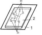
图1为本发明的建立神经元单细胞水平联系的装置的结构示意图。Fig. 1 is a schematic structural diagram of a device for establishing horizontal connections between neurons and single cells according to the present invention.
图2为基底的制备流程图;Fig. 2 is the preparation flowchart of substrate;
图3为正常生长的神经元与用本装置生长的神经元的示意图Figure 3 is a schematic diagram of normal growing neurons and neurons grown with this device
具体实施方式Detailed ways
图1为本发明的建立神经元之间单细胞水平连接的装置的结构示意图,由图1可知,本发明提供的建立神经元之间单细胞水平连接的装置,包括:Fig. 1 is a schematic structural diagram of a device for establishing a single-cell horizontal connection between neurons according to the present invention. It can be seen from Fig. 1 that the device for establishing a single-cell horizontal connection between neurons provided by the present invention includes:
—基底1,所述基底1上表面上附着有促进神经细胞黏附的宽度为5-10微米的蛋白条带;两相邻蛋白条带间间距为20-100微米;所述基板1上表面上蛋白条带之外的其它区域涂覆有抗拒细胞黏附的聚醚F127层;—
—紧密覆于所述基底上表面上的下表面上具有至少一组微凹槽单元的聚二甲基硅氧烷印章2;- a
所述微凹槽单元包括:The micro groove unit comprises:
一条直线型中间凹槽21;a linear
设置于所述直线型中间凹槽左侧或/和右侧的至少一条直线型侧凹槽;所述直线型侧凹槽的中间段与所述直线型中间凹槽不相交,所述直线型侧凹槽中间段之外的两端段分别向远离直线型中间凹槽的方向倾斜;所述直线型中间凹槽、和所述直线型侧凹槽的槽端处分别设有与相应凹槽相通的垂直孔道;所述直线型中间凹槽和所述直线型侧凹槽的长度均在1.5-2厘米范围内,宽度均为40微米;两相邻凹槽槽壁间间距为500微米-1厘米;At least one linear side groove arranged on the left side or/and right side of the linear middle groove; the middle section of the straight side groove does not intersect with the linear middle groove, and the straight line The two end sections other than the middle section of the side groove are respectively inclined to the direction away from the linear middle groove; the groove ends of the straight middle groove and the straight side groove are respectively provided with corresponding grooves Connected vertical channels; the length of the linear middle groove and the linear side groove are both in the range of 1.5-2 cm, and the width is 40 microns; the distance between the walls of two adjacent grooves is 500 microns- 1 cm;
所述基底上表面上附着的蛋白条带与所述聚二甲基硅氧烷印章的直线型中间凹槽和直线型侧凹槽相交不重合。The protein strips attached to the upper surface of the base do not coincide with the intersection of the linear middle groove and the linear side groove of the polydimethylsiloxane stamp.
所述蛋白条带宽度为5微米。The protein band width is 5 microns.
所述的聚醚F127为(H(OCH2CH2)x(OCH2CHCH3)y(OCH2CH2)zOH);其中,x≥1,代表-OCH2CH2-的个数;y≥1,代表-OCH2CHCH3-的个数;z≥1,代表-OCH2CH2-的个数。The polyether F127 is (H(OCH2CH2)x (OCH2CHCH3)y (OCH2CH2)z OH); wherein, x≥1, represents the number of -OCH2CH2-; y≥1, represents the number of -OCH2CHCH3-; z≥1, representing the number of -OCH2CH2-.
图2为在基底表面附着多条平行排列促进神经细胞黏附蛋白条带,其它区域为抗拒细胞黏附的聚醚F127的基底的制备流程示意图;由图2可知,可分为三个步骤:Figure 2 is a schematic diagram of the preparation process of a substrate with multiple parallel arrays of nerve cell adhesion-promoting protein strips attached to the surface of the substrate, and the other regions are polyether F127 that resists cell adhesion; as can be seen from Figure 2, it can be divided into three steps:
步骤1:表面有凸型条带状结构的聚二甲基硅氧烷印章4有微结构面蘸上促进神经细胞黏附的蛋白溶液,朝下垂直置于细胞培养皿3表面;Step 1: The
步骤2:揭去聚二甲基硅氧烷印章4,蛋白就会从聚二甲基硅氧烷印章4的凸型条带状结构表面转移到细胞培养皿3表面对应的部位,在细胞培养皿3表面形成多组平行排列的蛋白条带11;Step 2: Peel off the
步骤3:将表面吸附有蛋白条带11的细胞培养皿3用聚醚F127孵育,聚醚F127吸附在细胞培养皿3表面没有蛋白质吸附的区域;制备成表面附着有多条平行排列蛋白条带11,其它区域为抗拒细胞黏附区域12的基底1。Step 3: Incubate the
所述聚醚F127的分子式为H(OCH2CH2)x(OCH2CHCH3)y(OCH2CH2)zOH;其中,x≥1,代表-OCH2CH2-的个数;y≥1,代表-OCH2CHCH3-的个数;z≥1,代表-OCH2CH2-的个数。The molecular formula of the polyether F127 is H(OCH2CH2)x (OCH2CHCH3)y (OCH2CH2)z OH; wherein, x≥1, represents the number of -OCH2CH2-; y≥1, represents the number of -OCH2CHCH3-; z ≥1, representing the number of -OCH2CH2-.
实施例1Example 1
1)光刻:使用光刻技术,分别制备有微结构单元一和微结构单元二的硅片。首先用作图软件L-edit设计出所需要的两种图型,微结构单元一包括平行排列的直线型凹槽,其宽度为5微米(5-10微米均可),间距为50微米(20-100微米均可)。1) Photolithography: use photolithography technology to prepare silicon wafers with
所述微结构单元二具有至少一组凸型线型微结构单元,该凸形线型微结构单元包括:一条直线型中间凸型线;位于所述直线型中间凸型线左侧或右侧的至少一条直线型侧凸型线;所述直线型侧凸型线与所述直线型中间凸型线不相交;所述直线型侧凸型线的中间段之外的两端段分别向远离直线型中间凸型线的方向倾斜;所述直线型中间凸型线和所述直线型侧凸型线长度均为1.5厘米(1.5-2厘米均可),宽度40微米;两相邻凸型线间间距为500微米(200微米-1000微米均可);The second microstructure unit has at least one set of convex linear microstructure units, and the convex linear microstructure unit includes: a straight middle convex line; located on the left or right side of the straight middle convex line at least one linear side convex line; the straight side convex line does not intersect with the straight middle convex line; The direction of the linear convex line is inclined; the length of the straight central convex line and the linear side convex line is 1.5 cm (1.5-2 cm) and the width is 40 microns; two adjacent convex lines The spacing between lines is 500 microns (200 microns-1000 microns are acceptable);
打印分辨率微3600dip的胶片,接着涂胶(SU-8系列负胶),利用甩胶机均匀的将光刻胶涂在硅片上,80℃烘烤3小时后硬化,把胶片垂直放在涂有光刻胶的基板上,显影曝光后就再涂有光刻胶的硅片上制成了所需的微型结构单元一和微型结构单元二;Print a film with a resolution of micro 3600dip, then apply glue (SU-8 series negative glue), use a glue machine to evenly coat the photoresist on the silicon wafer, bake at 80°C for 3 hours and harden, put the film vertically on the On the substrate coated with photoresist, the required
2)印章制备:用聚二甲基硅氧烷对步骤1)得到的两种微结构单元进行翻模,得到与这两种微结构单元相对应的聚二甲基硅氧烷印章一和印章二;2) Stamp preparation: use polydimethylsiloxane to mold the two microstructure units obtained in step 1) to obtain
把聚二甲基硅氧烷印章一有微结构面朝上,在等离子清洗器中氧化3分钟,制备成具有亲水表面的聚二甲基硅氧烷印章一;Put the
3)基底制备:聚二甲基硅氧烷印章一有微结构单元的面蘸上促进神经细胞黏附的蛋白溶液(层粘连蛋白,laminin),有微结构单元面朝下垂直置于细胞细胞培养皿表面;5分钟后揭去聚二甲基硅氧烷印章一,层粘连蛋白就会从聚二甲基硅氧烷印章一的凸型条带状结构表面转移到细胞培养皿表面对应的部位,在细胞培养皿表面形成平行排列的层粘连蛋白条带;3) Substrate preparation: The surface of polydimethylsiloxane stamp with microstructural units is dipped in a protein solution (laminin, laminin) that promotes nerve cell adhesion, and the surface with microstructural units is placed vertically in cell culture Peel off the
将表面吸附有层粘连蛋白条带的细胞培养皿用聚醚F127孵育45分钟后,聚醚F127吸附在细胞培养皿表面的蛋白质条带之外区域;After incubating the cell culture dish with the laminin bands adsorbed on the surface with polyether F127 for 45 minutes, the polyether F127 was adsorbed on the surface of the cell culture dish outside the protein bands;
用双蒸水去除细胞培养皿表面多余的聚醚F127溶液;制备成表面附着有平行排列层粘连蛋白条带,其它区域为抗拒细胞黏附的聚醚F127的基底;Use double distilled water to remove excess polyether F127 solution on the surface of the cell culture dish; prepare laminin strips arranged in parallel on the surface, and the other areas are substrates of polyether F127 that resist cell adhesion;
4)加载微流通道:将步骤2)制备的聚二甲基硅氧烷印章二取出,把具有微凹型单元的面朝下,置于步骤3)中制备的基底上表面与其进行接触性连接,形成封闭的流通管腔;4) Loading the microfluidic channel: Take out the
5)种植细胞:从乳鼠(出生后12小时内)大脑海马区提取海马神经元,制备海马神经元细胞悬浮溶液,细胞密度为106/ml,把神经细胞经由与凹槽相通的垂直孔道送入流通管腔中,放入细胞培养箱(37℃,5%CO2)中培养;5) Planting cells: extract hippocampal neurons from the hippocampus of suckling mice (within 12 hours after birth), prepare a suspension solution of hippocampal neuron cells with a cell density of 106 /ml, and pass the neurons through the vertical channel communicating with the groove Send it into the flow-through lumen, and put it into a cell culture incubator (37°C, 5% CO2 ) for cultivation;
6)神经元间单线联系的建立:步骤5)中的装置中的神经元在细胞培养箱中培养30-60分钟后,单个的神经元黏附在流通管腔内的培养皿表面多组平行排列的蛋白条带上,形成纵向单个排列的一列神经元,每个蛋白条带横行上生长两个神经元;揭去聚二甲基硅氧烷印章二,神经元在神经细胞培养液中生长,单根神经突起将会沿着神经胞体黏附的蛋白条带生长;几天后,在同一条蛋白条带上定向生长的两条神经突起相互接触,形成单细胞水平的两个神经元之间的连接。6) Establishment of a single-line connection between neurons: After the neurons in the device in step 5) are cultured in the cell culture incubator for 30-60 minutes, individual neurons adhere to the surface of the culture dish in the flow tube and are arranged in parallel in multiple groups On the protein strips, a single column of neurons is formed in the vertical direction, and two neurons are grown on each protein strip horizontally; the polydimethylsiloxane seal is removed, and the neurons grow in the nerve cell culture medium. A single neurite will grow along the protein strip attached to the nerve cell body; after a few days, two neurites growing oriented on the same protein strip will contact each other, forming a gap between two neurons at the single-cell level. connect.
实施例2Example 2
1)光刻:使用光刻技术,分别制备有微结构单元一和微结构单元二的硅片。首先用作图软件L-edit设计出所需要的两种图型,微结构单元一包括多组平行排列的直线型凹槽,其宽度为5微米(5-10微米均可),间距为50微米(20-100微米均可)。微结构单元二包括至少一组凸型线型微结构单元,该凸形线型微结构单元包括:一个直线型凸型线(凸型线一),一个斜线形凸型线,(凸型线二);所述凸型线一和凸型线二中间段不相交,最短间距为500微米,最长间距为1000微米;所述凸型线二的中间段之外的两端部分向远离中间凸型线的方向倾斜;所述凸型线的长度在1.5厘米(1.5-2厘米均可),宽度40微米;1) Photolithography: use photolithography technology to prepare silicon wafers with
打印分辨率微3600dip的胶片,接着涂胶(SU-8系列负胶),利用甩胶机均匀的将光刻胶涂在硅片基底上,80℃烘烤3小时后硬化,把胶片垂直放在涂有光刻胶的基板上,显影曝光后就再涂有光刻胶的硅片上制成了所需的微型结构单元一和微型结构单元二;Print a film with a resolution of micro 3600dip, then apply glue (SU-8 series negative glue), use a glue machine to evenly coat the photoresist on the silicon wafer substrate, bake at 80°C for 3 hours and harden, put the film vertically On the substrate coated with photoresist, the required
2)印章制备:用聚二甲基硅氧烷对步骤1)得到的两种微结构单元进行翻模,得到与这两种微结构单元相对应的聚二甲基硅氧烷印章一和印章二;2) Stamp preparation: use polydimethylsiloxane to mold the two microstructure units obtained in step 1) to obtain
把聚二甲基硅氧烷印章一有微结构面朝上,在等离子清洗器中氧化3分钟,制备成具有亲水表面的聚二甲基硅氧烷印章一;Put the
3)基底制备:聚二甲基硅氧烷印章一有微结构面蘸上促进神经细胞黏附的蛋白溶液(层粘连蛋白,laminin),有微结构面朝下垂直置于细胞细胞培养皿表面;5分钟后揭去聚二甲基硅氧烷印章一,层粘连蛋白就会从聚二甲基硅氧烷印章一的凸型条带状结构表面转移到细胞培养皿表面对应的部位,在细胞培养皿表面形成多组平行排列的层粘连蛋白条带;3) Substrate preparation: dip the polydimethylsiloxane stamp with a microstructure surface into a protein solution (laminin, laminin) that promotes nerve cell adhesion, and place the microstructure surface vertically on the surface of the cell culture dish; Remove the polydimethylsiloxane stamp one after 5 minutes, and laminin will transfer from the surface of the convex strip structure of the polydimethylsiloxane stamp one to the corresponding position on the surface of the cell culture dish. Multiple groups of laminin bands arranged in parallel formed on the surface of the culture dish;
将表面吸附有层粘连蛋白条带的细胞培养皿用聚醚F127孵育45分钟后,聚醚F127吸附在细胞培养皿表面蛋白质条带之外的区域;After incubating the cell culture dish with laminin bands adsorbed on the surface with polyether F127 for 45 minutes, polyether F127 was adsorbed on the area outside the protein bands on the surface of the cell culture dish;
用双蒸水去除细胞培养皿表面多余的聚醚F127溶液;制备成表面附着有多条平行排列层粘连蛋白条带,其它区域为抗拒细胞黏附的聚醚F127的基底;Use double distilled water to remove excess polyether F127 solution on the surface of the cell culture dish; prepare the surface with multiple parallel laminin strips attached to the surface, and the other areas are the substrate of polyether F127 that resists cell adhesion;
4)加载微流通道:将步骤2)制备的聚二甲基硅氧烷印章二取出,把具有微凹型单元的面朝下,置于步骤3)中制备的基底上表面与其进行接触性连接,形成封闭的流通管腔;4) Loading the microfluidic channel: Take out the
5)种植细胞:从乳鼠(出生后12小时内)大脑海马区提取海马神经元,制备海马神经元细胞悬浮溶液,细胞密度为106/ml,把神经细胞经由与凹槽相通的垂直孔道送入流通管腔中,放入细胞培养箱(37℃,5%CO2)中培养;5) Planting cells: extract hippocampal neurons from the hippocampus of suckling mice (within 12 hours after birth), prepare hippocampal neuron cell suspension solution with a cell density of 106 /ml, and pass the neurons through the vertical channel communicating with the groove Send it into the flow-through lumen, and put it into a cell culture incubator (37°C, 5% CO2 ) for cultivation;
6)神经元间单线联系的建立:步骤5)中的装置中的神经元在细胞培养箱中培养30-60分钟后,单个的神经元黏附在流通管腔内的培养皿表面多组平行排列的蛋白条带上,形成纵向单个排列的一列神经元,每个蛋白条带横行上生长两个神经元;揭去聚二甲基硅氧烷印章二,神经元在神经细胞培养液中生长,单根神经突起将会沿着神经胞体黏附的蛋白条带生长;几天后,由于距离不同,在同一条蛋白条带上定向生长的两条神经突起陆续相互接触,形成单细胞水平的两个神经元之间的连接。6) Establishment of a single-line connection between neurons: After the neurons in the device in step 5) are cultured in the cell culture incubator for 30-60 minutes, individual neurons adhere to the surface of the culture dish in the flow tube and are arranged in parallel in multiple groups On the protein strips, a single column of neurons is formed in the vertical direction, and two neurons are grown on each protein strip horizontally; the polydimethylsiloxane seal is removed, and the neurons grow in the nerve cell culture medium. A single neurite will grow along the protein strip attached to the nerve cell body; after a few days, due to the different distances, two neurites growing oriented on the same protein strip will contact each other one after another, forming two neurons at the single-cell level. Connections between neurons.
实施例3Example 3
1)光刻:使用光刻技术,分别制备有微结构单元一和微结构单元二的硅片。首先用作图软件L-edit设计出所需要的两种图型,微结构单元一包括多组平行排列的直线型凹槽,其宽度为5微米(5-10微米均可),间距为50微米(20-100微米均可)。微结构单元二包括至少一组凸型线型微结构单元,该凸形线型微结构单元包括三个直线型凸型线(凸型线一、凸型线二、凸型线三);所述凸型线一位于凸型线二和凸型线三中间,凸型线一、凸型线二和凸型线三中间段平行,间距为500微米;所述凸型线二、凸型线三的中间段之外的两端部分向远离中间凸型线的方向倾斜;所述凸型线的长度在1.5厘米(1.5-2厘米均可),宽度40微米;1) Photolithography: use photolithography technology to prepare silicon wafers with
打印分辨率微3600dip的胶片,接着涂胶(SU-8系列负胶),利用甩胶机均匀的将光刻胶涂在硅片基底上,80℃烘烤3小时后硬化,把胶片垂直放在涂有光刻胶的基板上,显影曝光后就再涂有光刻胶的硅片上制成了所需的微型结构单元一和微型结构单元二;Print a film with a resolution of micro 3600dip, then apply glue (SU-8 series negative glue), use a glue machine to evenly coat the photoresist on the silicon wafer substrate, bake at 80°C for 3 hours and harden, put the film vertically On the substrate coated with photoresist, the required
2)印章制备:用聚二甲基硅氧烷对步骤1)得到的两种微结构单元进行翻模,得到与这两种微结构单元相对应的聚二甲基硅氧烷印章一和印章二;2) Stamp preparation: use polydimethylsiloxane to mold the two microstructure units obtained in step 1) to obtain
把聚二甲基硅氧烷印章一有微结构面朝上,在等离子清洗器中氧化3分钟,制备成具有亲水表面的聚二甲基硅氧烷印章一;Put the
3)基底制备:聚二甲基硅氧烷印章一有微结构面蘸上促进神经细胞黏附的蛋白溶液(层粘连蛋白,laminin),有结构面朝下垂直置于细胞细胞培养皿表面;5分钟后揭去聚二甲基硅氧烷印章一,层粘连蛋白就会从聚二甲基硅氧烷印章一的凸型条带状结构表面转移到细胞培养皿表面对应的部位,在细胞培养皿表面形成多组平行排列的层粘连蛋白条带;3) Substrate preparation: dip the polydimethylsiloxane stamp with a microstructured surface into a protein solution (laminin, laminin) that promotes nerve cell adhesion, and place it vertically on the surface of the cell culture dish with the structured surface facing down; 5 After a few minutes, the polydimethylsiloxane seal is removed, and the laminin will be transferred from the surface of the convex strip structure of the polydimethylsiloxane seal to the corresponding position on the surface of the cell culture dish. Multiple sets of parallel laminin bands were formed on the surface of the dish;
将表面吸附有层粘连蛋白条带的细胞培养皿用聚醚F127孵育45分钟后,聚醚F127吸附在细胞培养皿表面没有蛋白质吸附的区域;After incubating the cell culture dish with laminin bands adsorbed on the surface with polyether F127 for 45 minutes, polyether F127 was adsorbed on the surface of the cell culture dish without protein adsorption;
用双蒸水去除细胞培养皿表面多余的聚醚F127溶液;制备成表面附着有多条平行排列层粘连蛋白条带,其它区域为抗拒细胞黏附的聚醚F127的基底;Use double distilled water to remove excess polyether F127 solution on the surface of the cell culture dish; prepare the surface with multiple parallel laminin strips attached to the surface, and the other areas are the substrate of polyether F127 that resists cell adhesion;
4)加载微流通道:将步骤2)制备的聚二甲基硅氧烷印章二取出,把具有微凹型单元的面朝下,置于步骤3)中制备的基底上表面与其进行接触性连接,形成封闭的流通管腔;4) Loading the microfluidic channel: Take out the
5)种植细胞:从乳鼠(出生后12小时内)大脑海马区提取海马神经元,制备海马神经元细胞悬浮溶液,细胞密度为106/ml,把神经细胞经由与凹槽相通的垂直孔道送入流通管腔中,放入细胞培养箱(37℃,5%CO2)中培养;5) Planting cells: extract hippocampal neurons from the hippocampus of suckling mice (within 12 hours after birth), prepare hippocampal neuron cell suspension solution with a cell density of 106 /ml, and pass the neurons through the vertical channel communicating with the groove Send it into the flow-through lumen and put it into the cell culture incubator (37°C, 5% CO2 ) for cultivation;
6)神经元间单线联系的建立:步骤5)中的装置中的神经元在细胞培养箱中培养30-60分钟后,单个的神经元黏附在流通管腔内的培养皿表面多组平行排列的蛋白条带上,形成纵向单个排列的一列神经元,每个蛋白条带横行上生长三个神经元;揭去聚二甲基硅氧烷印章二,神经元在神经细胞培养液中生长,单根神经突起将会沿着神经胞体黏附的蛋白条带生长;几天后,在同一条蛋白条带上沿着蛋白条带定向生长的几根神经突起相互接触,形成单细胞水平的三个神经元之间的连接。6) Establishment of a single-line connection between neurons: After the neurons in the device in step 5) are cultured in the cell culture incubator for 30-60 minutes, individual neurons adhere to the surface of the culture dish in the flow tube and are arranged in parallel in multiple groups On the protein strips, a single column of neurons is formed in the vertical direction, and three neurons are grown on each protein strip in horizontal rows; the polydimethylsiloxane seal is removed, and the neurons grow in the nerve cell culture medium. A single neurite will grow along the protein strip attached to the nerve cell body; after a few days, several neurites that grow directional along the protein strip on the same protein strip will contact each other, forming three single-cell levels. Connections between neurons.
Claims (6)
Priority Applications (1)
| Application Number | Priority Date | Filing Date | Title |
|---|---|---|---|
| CN2008102399237ACN101748061B (en) | 2008-12-15 | 2008-12-15 | Device and method for growing connections between neurons at the single-cell level |
Applications Claiming Priority (1)
| Application Number | Priority Date | Filing Date | Title |
|---|---|---|---|
| CN2008102399237ACN101748061B (en) | 2008-12-15 | 2008-12-15 | Device and method for growing connections between neurons at the single-cell level |
Publications (2)
| Publication Number | Publication Date |
|---|---|
| CN101748061A CN101748061A (en) | 2010-06-23 |
| CN101748061Btrue CN101748061B (en) | 2012-07-11 |
Family
ID=42475677
Family Applications (1)
| Application Number | Title | Priority Date | Filing Date |
|---|---|---|---|
| CN2008102399237AExpired - Fee RelatedCN101748061B (en) | 2008-12-15 | 2008-12-15 | Device and method for growing connections between neurons at the single-cell level |
Country Status (1)
| Country | Link |
|---|---|
| CN (1) | CN101748061B (en) |
Families Citing this family (8)
| Publication number | Priority date | Publication date | Assignee | Title |
|---|---|---|---|---|
| CN102586103B (en)* | 2011-01-13 | 2014-12-10 | 国家纳米科学中心 | Device capable of controlling neurite branch site and manufacture method as well as application of device |
| CN102337211B (en)* | 2011-08-25 | 2013-04-17 | 中国科学院深圳先进技术研究院 | cell culture device |
| CN102337213B (en)* | 2011-10-13 | 2013-06-05 | 西北工业大学 | Polydimethylsiloxane (PDMS)-based three-dimensional single cell culture chip and controllable preparation method thereof |
| CN103357072B (en)* | 2012-03-28 | 2015-02-04 | 国家纳米科学中心 | Hydrogel with micro-flow passage, as well as preparation method and application thereof |
| CN103065038B (en)* | 2012-11-27 | 2015-12-23 | 大连理工大学 | A Combined Cell-Level Set Simulation Method for Plasma Etching |
| CN104130943B (en)* | 2014-07-28 | 2016-06-29 | 民航总医院 | Neuron and the orderly co-culture device of neurogliocyte, preparation method and neuron and the orderly co-culture method of neurogliocyte |
| CN108057132A (en)* | 2018-01-26 | 2018-05-22 | 浙江大学 | A kind of striping polylactide-caprolactone film of surface retention gelatin and preparation method thereof |
| CN109055315A (en)* | 2018-07-26 | 2018-12-21 | 长春理工大学 | A kind of nerve cell localized cell culture method |
- 2008
- 2008-12-15CNCN2008102399237Apatent/CN101748061B/ennot_activeExpired - Fee Related
Non-Patent Citations (4)
| Title |
|---|
| Anne M Taylor等.A microfluidic culture platform for CNS axonal injury, regeneration and transport.《Nature Methods》.2005,第2卷(第8期),599-605.* |
| AnneMTaylor等.AmicrofluidiccultureplatformforCNSaxonalinjury regeneration and transport.《Nature Methods》.2005 |
| E. Detrait等.Orientation of cell adhesion and growth on patterned heterogeneous polystyrene surface.《Journal of Neuroscience Methods》.1998,第84卷193-204.* |
| Roy Biran等.Surfactant-immobilized fibronectin enhances bioactivity and regulates sensory neurite outgrowth.《Journal of Biomedical Materials Research》.2001,第55卷(第1期),1-12.* |
Also Published As
| Publication number | Publication date |
|---|---|
| CN101748061A (en) | 2010-06-23 |
Similar Documents
| Publication | Publication Date | Title |
|---|---|---|
| CN101748061B (en) | Device and method for growing connections between neurons at the single-cell level | |
| Morin et al. | Constraining the connectivity of neuronal networks cultured on microelectrode arrays with microfluidic techniques: a step towards neuron-based functional chips | |
| Claverol-Tinture et al. | Multielectrode arrays with elastomeric microstructured overlays for extracellular recordings from patterned neurons | |
| US20060141617A1 (en) | Multilayered microcultures | |
| Musick et al. | Three-dimensional micro-electrode array for recording dissociated neuronal cultures | |
| CN102156158B (en) | Device for culturing and measuring microfluidic chip by using topological diagram type nerve cell network | |
| CN109234163B (en) | A high-throughput tumor-targeted drug concentration screening microfluidic device | |
| CN104611224A (en) | Cell co-culture micro-fluidic chip and application thereof | |
| Kang et al. | Agarose microwell based neuronal micro-circuit arrays on microelectrode arrays for high throughput drug testing | |
| CN109894163B (en) | High-flux and high-content drug screening micro-fluidic chip and preparation method thereof | |
| CN115216405A (en) | Multilayer cascade neural network micro-fluidic chip and preparation method thereof | |
| Hong et al. | Neurons-on-a-chip: in vitro neurotools | |
| CN112662554B (en) | Microfluidic platform, preparation method and application thereof | |
| CN106479893B (en) | Device and method for patterning co-culture of multiple cells | |
| WO2008019573A1 (en) | Devices and methods for the adhesion of multiple types of cells onto the same substrate | |
| CN101333522A (en) | Device and method for orderly adhering multiple cells to the same substrate | |
| CN101629945B (en) | Device for detecting electrical signal of nerve cell | |
| CN101624569B (en) | Device and method for operating multiple adherent cells on same substrate | |
| CN101748060B (en) | Device and method for arranging multiple cells on the same plane and manipulating them | |
| CN117363481A (en) | Microfluidic neural chip for realizing single-cell electrical interconnection, preparation method and inspection method | |
| CN102586103B (en) | Device capable of controlling neurite branch site and manufacture method as well as application of device | |
| CN113337451B (en) | A method for precise patterning of single cells in a multi-shear microfluidic chip | |
| CN118222398B (en) | A microfluidic biointerconnected neural network chip and its preparation method | |
| CN116144495A (en) | A microfluidic co-culture chip and a method for real-time monitoring of cell migration based on the microfluidic chip | |
| CN115678778A (en) | Micro-fluidic chip device for culturing three-dimensional cell clusters |
Legal Events
| Date | Code | Title | Description |
|---|---|---|---|
| C06 | Publication | ||
| PB01 | Publication | ||
| C10 | Entry into substantive examination | ||
| SE01 | Entry into force of request for substantive examination | ||
| C14 | Grant of patent or utility model | ||
| GR01 | Patent grant | ||
| CF01 | Termination of patent right due to non-payment of annual fee | Granted publication date:20120711 Termination date:20211215 | |
| CF01 | Termination of patent right due to non-payment of annual fee |