CN101684454B - Method for preparing and separating definitive endoderm - Google Patents
Method for preparing and separating definitive endodermDownload PDFInfo
- Publication number
- CN101684454B CN101684454BCN2008102004834ACN200810200483ACN101684454BCN 101684454 BCN101684454 BCN 101684454BCN 2008102004834 ACN2008102004834 ACN 2008102004834ACN 200810200483 ACN200810200483 ACN 200810200483ACN 101684454 BCN101684454 BCN 101684454B
- Authority
- CN
- China
- Prior art keywords
- cells
- definitive endoderm
- cell
- population
- cell population
- Prior art date
- Legal status (The legal status is an assumption and is not a legal conclusion. Google has not performed a legal analysis and makes no representation as to the accuracy of the status listed.)
- Active
Links
Images


Landscapes
- Micro-Organisms Or Cultivation Processes Thereof (AREA)
Abstract
Translated fromChineseDescription
Translated fromChinese技术领域technical field
本发明属于生物技术领域,更具体地,本发明涉及一种定型内胚层细胞(Definitive Endoderm,DE)的制备和分离方法。The invention belongs to the field of biotechnology, more specifically, the invention relates to a preparation and separation method of definitive endoderm cells (Definitive Endoderm, DE).
背景技术Background technique
源于胚泡期内细胞团的胚胎干细胞(Embryonic Stem Cells,ES)具有多全能性,在合适的培养条件下,可以使其维持近似均一且理论上无限的自我更新能力;而在特定的诱导条件下,它们能够向几乎所有体细胞分化。胚胎干细胞为现代生命科学和药物科学研究提供了良好条件,是研究哺乳动物早期胚胎发育的理想模型,也在基于细胞的生物治疗(Cell-Based Therapy)和药物筛选、毒理测试等研究中起到重要作用。The embryonic stem cells (Embryonic Stem Cells, ES) derived from the cell clusters in the blastocyst stage are pluripotent, and under appropriate culture conditions, they can maintain approximately uniform and theoretically unlimited self-renewal capabilities; Under certain conditions, they can differentiate into almost all somatic cells. Embryonic stem cells provide good conditions for modern life science and pharmaceutical science research. They are an ideal model for studying the early embryonic development of mammals. to an important role.
近年来,对胚胎干细胞自我更新及哺乳动物胚胎发育的分子机制的进一步了解使得干细胞向体细胞定向分化已成为干细胞生物学研究的热点,也在再生医学领域越发受到重视。之前的研究工作表明,小鼠ES细胞向包括神经元、心肌细胞、肝脏细胞、内分泌胰腺细胞等在内的体细胞的定向分化已取得了一定进展;人类胚胎干细胞向神经元、激素分泌胰腺细胞分化已取得一定进步,但关于人类胚胎干细胞向肝脏细胞的分化研究少有报道。In recent years, the further understanding of the molecular mechanism of embryonic stem cell self-renewal and mammalian embryonic development has made the directed differentiation of stem cells into somatic cells become a hot spot in stem cell biology research, and has also attracted more and more attention in the field of regenerative medicine. Previous research work has shown that the directional differentiation of mouse ES cells to somatic cells including neurons, cardiomyocytes, liver cells, endocrine pancreatic cells, etc. has made some progress; Differentiation has made some progress, but there are few reports on the differentiation of human embryonic stem cells into liver cells.
中国肝脏疾病的发病率高,影响面广,对国人健康和社会经济的影响巨大。特别是乙肝病毒等嗜肝性病毒感染在临床上可以引起肝炎、肝硬化和肝癌的发生,而目前又缺乏真正有效的治疗方法。所以,发展全新的肝脏疾病的治疗方法不仅具有重要的医学意义,而且更加具有特殊的社会影响和潜在的经济效益。特别令人鼓舞的是,在体外实验中已有可能使胚胎干细胞和来源于肝组织的肝干细胞分化为肝前体细胞、成熟的肝细胞及胆管细胞,并在活体动物的移植实验中也观察到外源的干细胞对损伤肝脏的再殖(Repopulate),使损伤肝脏在结构上和功能上得到有效的恢复。这些进展的取得,充分显示了利用干细胞对人类肝脏疾病进行临床治疗的可能性。The incidence of liver disease in China is high and affects a wide range of people, which has a huge impact on the health of Chinese people and social economy. In particular, hepatotropic virus infection such as hepatitis B virus can cause hepatitis, liver cirrhosis and liver cancer clinically, but there is no real effective treatment method at present. Therefore, the development of new treatment methods for liver diseases is not only of great medical significance, but also has special social impact and potential economic benefits. It is particularly encouraging that it has been possible to differentiate embryonic stem cells and hepatic stem cells derived from liver tissue into hepatic precursors, mature hepatocytes, and cholangiocytes in vitro, and has also been observed in transplantation experiments in living animals. Repopulate the damaged liver with exogenous stem cells, so that the structure and function of the damaged liver can be effectively restored. The achievement of these advances has fully demonstrated the possibility of clinical treatment of human liver diseases using stem cells.
小鼠和人ES细胞体外分化的研究表明,ES细胞的肝向分化能在一定程度模拟在体发育过程,使得体外分化具有较好的理论依据。因此,对哺乳动物肝脏发育的分子机制的了解,有助于指导胚胎干细胞肝向分化。然而,哺乳动物肝脏发育是一个在时间和空间上受到精确调控的生物学事件,这就决定了肝向分化的复杂性。干细胞肝向分化尚有许多问题待于解决,如诱导效率低、重复性差、细胞成分混杂(各个胚层基因表达具有相似性,难于区分特定的细胞群)、分化细胞的真实性等。因此,探索高效可重复的胚胎干细胞肝向诱导的方法势在必行。胚胎干细胞肝向分化的第一步是获得高比例且较纯的内胚层细胞。综合本技术领域以往的研究,胚胎干细胞向定型内胚层分化有三个技术问题还需要解决:Studies on the in vitro differentiation of mouse and human ES cells have shown that the hepatic differentiation of ES cells can simulate the in vivo development process to a certain extent, which makes the in vitro differentiation have a better theoretical basis. Therefore, an understanding of the molecular mechanisms underlying mammalian liver development can help guide embryonic stem cell hepatic differentiation. However, mammalian liver development is a biological event that is precisely regulated in time and space, which dictates the complexity of hepatic differentiation. There are still many problems to be solved in the hepatic differentiation of stem cells, such as low induction efficiency, poor reproducibility, mixed cell components (the gene expression of each germ layer is similar, it is difficult to distinguish specific cell groups), the authenticity of differentiated cells, etc. Therefore, it is imperative to explore an efficient and reproducible method for hepatic induction of embryonic stem cells. The first step in hepatic differentiation of embryonic stem cells is to obtain a high proportion of pure endoderm cells. Based on the previous research in this technical field, there are three technical problems that need to be solved in the differentiation of embryonic stem cells to definitive endoderm:
首先需要解决的问题是如何提高诱导效率。研究已经发现,通过单层培养诱导(Monolayer culture),Activin A(激动素)能够模拟Tgf-b信号通路的作用,促进ES细胞分化成Gsc+中内胚层(Mesoendoderm),进而分化成内胚层样细胞;而通过形成拟胚体(Embryonic Body,EB),Activin A也能促使ES细胞分化成Brachury+原条样细胞,进一步形成内胚层细胞;同时Activin A的诱导作用也在人胚胎干细胞向内胚层分化中被证明。然而,尽管如此,这些诱导方法的效率都不高,低于50%。The first problem to be solved is how to improve the induction efficiency. Studies have found that through monolayer culture induction (Monolayer culture), Activin A (kinesin) can mimic the role of the Tgf-b signaling pathway, and promote the differentiation of ES cells into Gsc+ Mesoendoderm, and then differentiate into endoderm-like cells ; and through the formation of embryoid bodies (Embryonic Body, EB), Activin A can also promote ES cells to differentiate into Brachury+ primitive streak-like cells, and further form endoderm cells; at the same time, the induction of Activin A can also induce human embryonic stem cells to differentiate into endoderm proved in. However, despite this, the efficiency of these induction methods is not high, below 50%.
其次需要解决的问题是如何区分定型内胚层和内脏内胚层(Vecialendoderm,VE)。在哺乳动物发育过程中,VE来自原始内胚层(Primitiveendoderm,PE),不参与器官发育,但VE和DE共同表达很多相同的基因,而且VE很容易被诱导出来,之前的诱导方法因无法较好地区分VE和DE,得到的往往是一个混合的细胞群。The second problem to be solved is how to distinguish definitive endoderm from visceral endoderm (Vecialendoderm, VE). During mammalian development, VE comes from primitive endoderm (Primitive endoderm, PE) and does not participate in organ development, but VE and DE co-express many of the same genes, and VE is easily induced, and the previous induction methods are not good. Distinguishing between VE and DE often results in a mixed cell population.
再次需要解决的问题是如何将DE从分化的细胞群中挑选出来。利用荧光激活的细胞分选技术(Fluorescence Activated Cell Sorting,FACS),只要选定合适的表面抗原,就可以将特定细胞群标记并分离。现在所知的DE特异表达的表面抗原还很少,因此有必要选定能够正确区分胚内和胚外细胞,以及中胚层(Mesoderm)和定型内胚层的表面标记。Again, the question to be addressed is how to sort DE out of differentiated cell populations. Using fluorescence activated cell sorting (Fluorescence Activated Cell Sorting, FACS), as long as the appropriate surface antigen is selected, specific cell populations can be labeled and separated. There are few known surface antigens specifically expressed by DE, so it is necessary to select surface markers that can correctly distinguish intraembryonic and extraembryonic cells, as well as mesoderm and definitive endoderm.
发明内容Contents of the invention
本发明的目的在于提供一种定型内胚层细胞(DE)的制备和分离方法。The purpose of the present invention is to provide a preparation and isolation method of definitive endoderm cells (DE).
本发明的另一目的在于提供一种用于促进定型内胚层细胞向分化的分化培养基。Another object of the present invention is to provide a differentiation medium for promoting the differentiation of definitive endoderm cells.
本发明的另一目的在于提供一种可用于分选内胚层细胞的表面标记。Another object of the present invention is to provide a surface marker that can be used for sorting endoderm cells.
在本发明的第一方面,提供一种制备含定型内胚层细胞的细胞群的方法,所述方法包括:In a first aspect of the present invention, there is provided a method of preparing a cell population containing definitive endoderm cells, the method comprising:
(1)培养胚胎干细胞(Embryonic Stem cells,ES),形成拟胚体;(1) Culture embryonic stem cells (Embryonic Stem cells, ES) to form embryoid bodies;
(2)用含活化素A(Activin A)和Wnt信号通路激动剂的分化培养基处理拟胚体,诱导拟胚体分化,从而获得含有定型内胚层细胞的细胞群,所述细胞群中定型内胚层细胞占60%以上;较佳的70%以上;更佳的80%以上。(2) Treat the embryoid body with a differentiation medium containing Activin A (Activin A) and a Wnt signaling pathway agonist to induce differentiation of the embryoid body, thereby obtaining a cell population containing definitive endoderm cells. Endoderm cells account for more than 60%; preferably more than 70%; more preferably more than 80%.
在另一优选例中,所述的胚胎干细胞是非人哺乳动物胚胎干细胞,如兔、鼠、羊、猪、猴。较佳的,所述的胚胎干细胞是鼠胚胎干细胞。In another preferred embodiment, the embryonic stem cells are non-human mammalian embryonic stem cells, such as rabbits, mice, sheep, pigs, and monkeys. Preferably, the embryonic stem cells are mouse embryonic stem cells.
在另一优选例中,在步骤(2)中,在用Wnt信号通路激动剂处理的同时,还包括:用BMP4信号通路抑制剂处理拟胚体。In another preferred embodiment, in step (2), while treating with an agonist of the Wnt signaling pathway, it also includes: treating the embryoid body with an inhibitor of the BMP4 signaling pathway.
在另一优选例中,所述的Wnt信号通路激动剂选自:(2’Z,3’E)-6-Bromoindirubin-3′-肟(BIO)或LiCl;或In another preferred example, the Wnt signaling pathway agonist is selected from: (2'Z, 3'E)-6-Bromoindirubin-3'-oxime (BIO) or LiCl; or
所述的BMP4信号通路抑制剂是:Noggin。The BMP4 signaling pathway inhibitor is: Noggin.
在另一优选例中,活化素A的用量是:20-100ng/ml培养基;较佳的是30-80ng/ml培养基;更佳的是50-80ng/ml培养基。In another preferred example, the dosage of activin A is: 20-100ng/ml culture medium; preferably 30-80ng/ml culture medium; more preferably 50-80ng/ml culture medium.
在另一优选例中,LiCl的用量是0.5-10mM;较佳的是1-8mM;更佳的是2-6mM。In another preferred example, the dosage of LiCl is 0.5-10 mM; preferably 1-8 mM; more preferably 2-6 mM.
在另一优选例中,(2’Z,3’E)-6-Bromoindirubin-3′-肟的用量是0.5-5μM;较佳的是0.8-4μM;更佳的是2μM。In another preferred example, the dosage of (2'Z,3'E)-6-Bromoindirubin-3'-oxime is 0.5-5 μM; preferably 0.8-4 μM; more preferably 2 μM.
在另一优选例中,:Noggin的用量是100-600ng/m;较佳的是150-500ng/ml;更佳的是200-400ng/ml。In another preferred example, the dosage of Noggin is 100-600ng/m; preferably 150-500ng/ml; more preferably 200-400ng/ml.
在另一优选例中,所述方法还包括:(3)从(2)获得的细胞群中分离出定型内胚层细胞群。In another preferred example, the method further includes: (3) isolating definitive endoderm cell populations from the cell populations obtained in (2).
在另一优选例中,采用流式细胞分选方法分离出定型内胚层细胞群。In another preferred embodiment, the definitive endoderm cell population is isolated by flow cytometry.
在另一优选例中,所述的从(2)获得的细胞群中分离出定型内胚层细胞群的方法是:In another preferred example, the method for isolating the definitive endoderm cell population from the cell population obtained in (2) is:
分选细胞表面Cxcr4分子阳性且c-Kit分子阳性的细胞群,所述的细胞群是定型内胚层细胞群;或Sorting cell populations positive for Cxcr4 molecules and positive for c-Kit molecules on the cell surface, said cell populations being definitive endoderm cell populations; or
分选细胞表面Cxcr4分子阳性且CD140α分子阴性的细胞群,所述的细胞群是定型内胚层细胞群;或Sorting cell populations that are positive for Cxcr4 molecules on the cell surface and negative for CD140α molecules, and the cell populations are definitive endoderm cell populations; or
分选细胞表面Cxcr4分子阳性且EpCam分子阳性的细胞群,所述的细胞群是定型内胚层细胞群。The cell populations positive for Cxcr4 molecules and EpCam molecules on the cell surface are sorted, and the cell populations are definitive endoderm cell populations.
在本发明的第二方面,提供一种含定型内胚层细胞的细胞群,所述细胞群中定型内胚层细胞占60%以上,所述的细胞群采用以下方法获得:In the second aspect of the present invention, a cell group containing definitive endoderm cells is provided, wherein definitive endoderm cells account for more than 60% of the cell group, and the cell group is obtained by the following method:
(1)培养胚胎干细胞,形成拟胚体;(1) culturing embryonic stem cells to form embryoid bodies;
(2)用含活化素A和Wnt信号通路激动剂的分化培养基处理拟胚体,诱导拟胚体分化,从而获得含有定型内胚层细胞的细胞群。(2) Treat the embryoid body with a differentiation medium containing activin A and a Wnt signaling pathway agonist to induce differentiation of the embryoid body, thereby obtaining a cell population containing definitive endoderm cells.
在另一优选例中,在步骤(2)之后,还包括:In another preferred example, after step (2), it also includes:
从所述含有定型内胚层细胞的细胞群中分选细胞表面Cxcr4分子阳性且c-Kit分子阳性的细胞群,得到定型内胚层细胞群;或Sorting cell populations positive for Cxcr4 molecules and c-Kit molecules on the cell surface from the cell populations containing definitive endoderm cells to obtain definitive endoderm cell populations; or
从所述含有定型内胚层细胞的细胞群中分选细胞表面Cxcr4分子阳性且CD140α分子阴性的细胞群,得到定型内胚层细胞群;或Sorting cell populations positive for Cxcr4 molecules and negative for CD140α molecules on the cell surface from the cell populations containing definitive endoderm cells to obtain definitive endoderm cell populations; or
从所述含有定型内胚层细胞的细胞群中分选细胞表面Cxcr4分子阳性且EpCam分子阳性的细胞群,得到定型内胚层细胞群。Sorting cell populations positive for Cxcr4 molecules and EpCam molecules on the cell surface from the cell populations containing definitive endoderm cells to obtain definitive endoderm cell populations.
在本发明的第三方面,提供一种用于制备定型内胚层细胞的分化培养基,所述的分化培养基含有(较佳地由以下组分组成):In a third aspect of the present invention, there is provided a differentiation medium for preparing definitive endoderm cells, said differentiation medium containing (preferably consisting of the following components):
无血清胚胎干细胞分化培养基,以及:Serum-free embryonic stem cell differentiation medium, and:
20-100ng/ml活化素A;和20-100ng/ml Activin A; and
0.5-10mM LiCl,或0.5-5μM(2’Z,3’E)-6-Bromoindirubin-3′-肟。0.5-10 mM LiCl, or 0.5-5 μM (2'Z, 3'E)-6-Bromoindirubin-3'-oxime.
在另一优选例中,含有30-80ng/ml活化素A;较佳的含有50-80ng/ml活化素A。In another preferred embodiment, it contains 30-80ng/ml activin A; preferably contains 50-80ng/ml activin A.
在另一优选例中,含有1-8mM LiCl;较佳的含有2-6mM LiCl。In another preferred embodiment, it contains 1-8mM LiCl; preferably contains 2-6mM LiCl.
在另一优选例中,含有0.8-4μM(2’Z,3’E)-6-Bromoindirubin-3′-肟;较佳的含有2μM(2’Z,3’E)-6-Bromoindirubin-3′-肟。In another preferred embodiment, it contains 0.8-4 μM (2'Z, 3'E)-6-Bromoindirubin-3'-oxime; preferably contains 2 μM (2'Z, 3'E)-6-Bromoindirubin-3 '-oxime.
在另一优选例中,所述的分化培养基还含有:100-600ng/ml的Noggin;较佳地含有150-500ng/ml的Noggin;更佳地含有200-400ng/ml的Noggin。In another preferred example, the differentiation medium further contains: 100-600 ng/ml Noggin; preferably 150-500 ng/ml Noggin; more preferably 200-400 ng/ml Noggin.
在另一优选例中,所述的无血清胚胎干细胞培养基含有:70-80%体积比的IMDM;20-30%体积比的F12培养基;其中还添加:In another preferred example, the serum-free embryonic stem cell medium contains: 70-80% volume ratio of IMDM; 20-30% volume ratio of F12 medium; wherein:
按照体积0.2-2%(较佳地0.3-1.5%)N2添加物;0.2-2% (preferably 0.3-1.5%) N2 addition by volume;
按照体积0.5-2%(较佳地0.7-1.5%)B27添加物;0.5-2% (preferably 0.7-1.5%) B27 additive by volume;
0.2-1(较佳地0.4-0.8)mg/ml的BSA;0.2-1 (preferably 0.4-0.8) mg/ml of BSA;
0.2-1(较佳地0.4-0.8)mM的维生素C;和0.2-1 (preferably 0.4-0.8) mM vitamin C; and
0.2-0.8(较佳地0.3-0.6)mM的3-巯基硫代甘油(MTG)。0.2-0.8 (preferably 0.3-0.6) mM 3-mercaptothioglycerol (MTG).
在本发明的第四方面,提供所述的分化培养基的用途,用于制备含定型内胚层细胞的细胞群,所述细胞群中定型内胚层细胞占60%以上;较佳的70%以上;更佳的80%以上。In the fourth aspect of the present invention, the use of the differentiation medium is provided for preparing a cell group containing definitive endoderm cells, and in the cell group, definitive endoderm cells account for more than 60%; preferably more than 70% ; More than 80% better.
在本发明的第五方面,提供一种CD140α分子的特异性抗体的用途,用于鉴定Cxcr4分子阳性的细胞或细胞群是否属于定型内胚层细胞或细胞群。In the fifth aspect of the present invention, the use of a CD140α molecule-specific antibody is provided for identifying whether the Cxcr4 molecule-positive cells or cell groups belong to definitive endoderm cells or cell groups.
在本发明的第六方面,提供一种EpCam分子的特异性抗体的用途,用于鉴定Cxcr4分子阳性的细胞或细胞群是否属于定型内胚层细胞或细胞群,或用于分离定型内胚层细胞或细胞群。In the sixth aspect of the present invention, there is provided a use of a specific antibody for EpCam molecules, for identifying whether Cxcr4 molecule-positive cells or cell groups belong to definitive endoderm cells or cell groups, or for isolating definitive endoderm cells or cell population.
本发明的其它方面由于本文的公开内容,对本领域的技术人员而言是显而易见的。Other aspects of the invention will be apparent to those skilled in the art from the disclosure herein.
附图说明Description of drawings
图1显示了以Cxcr4+/c-Kit+作为分选的标记,利用FACS技术分析定型内胚层细胞的分化情况。Figure 1 shows the differentiation of definitive endoderm cells analyzed by FACS technology using Cxcr4+/c-Kit+ as a sorting marker.
图2A显示了以Cxcr4+/c-Kit+作为分选的标记,利用FACS技术分析定型内胚层细胞的分化情况。Figure 2A shows the differentiation of definitive endoderm cells analyzed by FACS technology using Cxcr4+/c-Kit+ as a sorting marker.
图2B显示了以Cxcr4+/Cd140α-作为分选的标记,利用FACS技术分析定型内胚层细胞的分化情况。Figure 2B shows the differentiation of definitive endoderm cells analyzed by FACS technology using Cxcr4+/Cd140α- as a sorting marker.
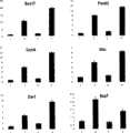
图3显示了Real time PCR比较4种条件下DE相关基因的表达。其中,Figure 3 shows the expression of DE-related genes under the four conditions compared by Real time PCR. in,
1.采用分化培养基6进行诱导的基因表达情况;1. Gene expression induced by
2.采用分化培养基8进行诱导的基因表达情况;2. The expression of genes induced by
3.采用分化培养基7进行诱导的基因表达情况;3. The expression of genes induced by
4.采用分化培养基2进行诱导的基因表达情况。4. Gene expression induced by
图4显示了RT-PCR对分选的细胞进行基因表达分析,其中,Fig. 4 shows that RT-PCR carries out gene expression analysis to sorted cells, wherein,
ES表示胚胎干细胞中相关基因RT-PCR产物的电泳结果;ES represents the electrophoresis results of RT-PCR products of related genes in embryonic stem cells;
EB表示拟胚体中相关基因RT-PCR产物的电泳结果;EB represents the electrophoresis results of RT-PCR products of related genes in embryoid bodies;
“-”表示分选出的Cxcr4+/EpCam-细胞中相关基因RT-PCR产物电泳结果;"-" indicates the electrophoresis results of RT-PCR products of related genes in the sorted Cxcr4+/EpCam- cells;
“+”表示分选出的Cxcr4+/EpCam+细胞中相关基因RT-PCR产物的电泳结果;"+" indicates the electrophoresis results of RT-PCR products of related genes in the sorted Cxcr4+/EpCam+ cells;
H2O为阴性对照的电泳结果。H2 O is the electrophoresis result of the negative control.
具体实施方式Detailed ways
本发明人经过长期而广泛的研究,首次揭示一种可显著提高胚胎干细胞向定型内胚层细胞分化的效率的方法,所述方法包括采用Wnt信号通路激动剂诱导拟胚体使之分化为定型内胚层细胞,更佳地还采用BMP4信号通路抑制剂来诱导。本发明的方法简单高效,可显著地将哺乳动物胚胎干细胞向定型内胚层分化的效率提高。本发明人还找到了可与Cxcr4相结合用于分选或富集定型内胚层细胞群的新的表面标志物Cd140α或EpCam,发现并验证了Cxcr4+/Cd140α-细胞群或Cxcr4+/EpCam+细胞群是富含定型内胚层细胞的细胞群。After long-term and extensive research, the present inventors revealed for the first time a method that can significantly improve the differentiation efficiency of embryonic stem cells into definitive endoderm cells. The method includes using Wnt signaling pathway agonists to induce embryoid bodies to differentiate into definitive endoderm cells. The germ layer cells are preferably also induced with a BMP4 signaling pathway inhibitor. The method of the invention is simple and efficient, and can significantly improve the differentiation efficiency of mammalian embryonic stem cells to definitive endoderm. The present inventors have also found a new surface marker Cd140α or EpCam that can be combined with Cxcr4 for sorting or enriching definitive endoderm cell populations, and found and verified that Cxcr4+/Cd140α- cell populations or Cxcr4+/EpCam+ cell populations are A cell population enriched for definitive endoderm cells.
胚胎干细胞的定向分化的难点在于:①定向分化效率低;②分化的目的细胞纯度低,难于富集;③分化的细胞的真实性。本发明人针对这三个难题,对哺乳动物胚胎干细胞向定型内胚层细胞的分化进行了深入探索和优化,使分化效率大大提高,通过活细胞分析/分选技术富集目的细胞群,并通过基因表达分析验证了富集到的细胞群的真实性。Difficulties in directed differentiation of embryonic stem cells lie in: ① low efficiency of directed differentiation; ② low purity of differentiated target cells, difficult to enrich; ③ authenticity of differentiated cells. Aiming at these three problems, the present inventors conducted in-depth exploration and optimization on the differentiation of mammalian embryonic stem cells to definitive endoderm cells, which greatly improved the differentiation efficiency, enriched the target cell population through live cell analysis/sorting technology, and passed Gene expression analysis verified the authenticity of the enriched cell population.
制备含定型内胚层细胞的细胞群的方法Method for preparing cell populations comprising definitive endoderm cells
本发明提供一种制备含定型内胚层细胞的细胞群的方法,所述方法包括:(1)培养胚胎干细胞,形成拟胚体;和(2)用含Activin A和Wnt信号通路激动剂的分化培养基处理拟胚体,诱导拟胚体分化,从而获得含有定型内胚层细胞的细胞群。The invention provides a method for preparing a cell population containing definitive endoderm cells, the method comprising: (1) culturing embryonic stem cells to form embryoid bodies; and (2) differentiation with activators containing Activin A and Wnt signaling pathways The culture medium is used to treat the embryoid body to induce differentiation of the embryoid body, thereby obtaining a cell population containing definitive endoderm cells.
在制备过程中,首先需要使未分化的胚胎干细胞形成拟胚体,拟胚体的培养是本领域人员已知的技术。作为本发明的优选方式,采用悬浮培养的方式制备拟胚体。In the preparation process, it is first necessary to form embryoid bodies from undifferentiated embryonic stem cells, and the cultivation of embryoid bodies is a technique known to those skilled in the art. As a preferred mode of the present invention, the embryoid body is prepared by means of suspension culture.
以往,已经采用Activin A来诱导拟胚体定向分化为内胚层细胞,但是诱导效率低下,无法获得令人满意的分化细胞群。本发明人在深入研究中意外地发现,Wnt信号通路激动剂可与Activin A协同发挥作用,非常有效地诱导胚胎干细胞定向分化为定型内胚层细胞,获得定型内胚层细胞占优势;更佳地,还同时用BMP4信号通路抑制剂进行诱导,这将使得细胞群中含有更多的定型内胚层细胞。In the past, Activin A has been used to induce embryoid bodies to differentiate into endoderm cells, but the induction efficiency is low and satisfactory differentiated cell populations cannot be obtained. The inventors unexpectedly discovered during in-depth research that the Wnt signaling pathway agonist can work synergistically with Activin A to effectively induce the directional differentiation of embryonic stem cells into definitive endoderm cells, and obtain the dominance of definitive endoderm cells; more preferably, Simultaneous induction with inhibitors of the BMP4 signaling pathway will result in more definitive endoderm cells in the population.
多种对于Wnt信号通路具有促进作用的Wnt信号通路激动剂均可用于本发明。作为本发明的优选方式,所述的Wnt信号通路激动剂选自:BIO或LiCl。作为本发明的优选方式,LlCl的用量是0.5-10mM;较佳的是1-8mM;更佳的是2-6mM。作为本发明的优选方式,BIO的用量是0.5-5μM;较佳的是0.8-4μM;更佳的是1-3μM,如2μM。本发明人在研究中发现,过高含量的LiCl或BIO可导致细胞发生明显的死亡。A variety of Wnt signaling pathway agonists that can promote the Wnt signaling pathway can be used in the present invention. As a preferred mode of the present invention, the Wnt signaling pathway agonist is selected from: BIO or LiCl. As a preferred mode of the present invention, the dosage of LlCl is 0.5-10mM; preferably 1-8mM; more preferably 2-6mM. As a preferred mode of the present invention, the dosage of BIO is 0.5-5 μM; preferably 0.8-4 μM; more preferably 1-3 μM, such as 2 μM. The present inventors found in the research that too high content of LiCl or BIO can lead to obvious cell death.
多种对于BMP4信号通路具有抑制作用的BMP4信号通路抑制剂均可用于本发明。作为本发明的优选方式,所述的BMP4信号通路抑制剂是:Noggin。作为本发明的优选方式,Noggin的用量是100-600ng/ml;较佳的是150-500ng/ml;更佳的200-400ng/ml。Noggin是一种本领域已知的蛋白,其基因序列例如可以与Gene ID:18121所示的序列基本上相同。A variety of BMP4 signaling pathway inhibitors that have an inhibitory effect on the BMP4 signaling pathway can be used in the present invention. As a preferred mode of the present invention, the BMP4 signaling pathway inhibitor is: Noggin. As a preferred mode of the present invention, the dosage of Noggin is 100-600ng/ml; preferably 150-500ng/ml; more preferably 200-400ng/ml. Noggin is a protein known in the art, and its gene sequence, for example, can be substantially identical to the sequence shown in Gene ID: 18121.
Activin A的用量可以根据本领域人员已知的适合用量。作为本发明的优选方式,Activin A的用量是:20-100ng/ml培养基;较佳的30-80ng/ml培养基;更佳的是50-80ng/ml培养基。The dosage of Activin A can be according to the suitable dosage known by those skilled in the art. As a preferred mode of the present invention, the dosage of Activin A is: 20-100ng/ml medium; preferably 30-80ng/ml medium; more preferably 50-80ng/ml medium.
步骤(1)和步骤(2)的培养或诱导时间可以根据本领域人员的经验来确定。作为本发明的优选方式,步骤(1)诱导拟胚体的时间在36-60小时,如48小时左右;步骤(2)诱导分化为定型内胚层细胞的时间在36-150小时或更长。本领域人员也可以通过在诱导过程中分析细胞群中多个细胞的性质来确定合适的时间。The culture or induction time of step (1) and step (2) can be determined according to the experience of those skilled in the art. As a preferred mode of the present invention, the time for step (1) to induce embryoid bodies is 36-60 hours, such as about 48 hours; the time for step (2) to induce differentiation into definitive endoderm cells is 36-150 hours or longer. The appropriate time can also be determined by one skilled in the art by analyzing the properties of multiple cells in the cell population during the induction process.
本发明还包括采用前述的制备方法获得的含定型内胚层细胞的细胞的细胞群。所述的细胞群中,定型内胚层细胞数量占细胞整体数量的60%以上,较佳地占细胞整体数量的70%以上;更佳地占细胞整体数量的80%以上。The present invention also includes the cell group containing definitive endoderm cells obtained by the aforementioned preparation method. In the cell population, the number of definitive endoderm cells accounts for more than 60% of the total number of cells, preferably more than 70% of the total number of cells; more preferably more than 80% of the total number of cells.
利用FACS和表面标记技术,能够将定型内胚层细胞富集至Cxcr4+/Cd140α-细胞群或Cxcr4+/EpCam+细胞群中;利用RT-PCR以及其它基因表达分析技术,证明富集到的内胚层细胞纯度较高,很少有其它胚层细胞的污染。Using FACS and surface marker technology, it is possible to enrich definitive endoderm cells into Cxcr4+/Cd140α- cell population or Cxcr4+/EpCam+ cell population; use RT-PCR and other gene expression analysis techniques to prove the purity of the enriched endoderm cells Higher, less contamination from other germ layer cells.
因此,本发明还包括经过分选技术分离出来的定型内胚层细胞含量更高的细胞群。优选地,所述的细胞群是细胞表面Cxcr4分子阳性且EpCam分子阳性的细胞群;或所述的细胞群是细胞表面Cxcr4分子阳性且Cd140α阴性的细胞群;或所述的细胞群是细胞表面Cxcr4分子阳性且c-Kit阳性的细胞群。Therefore, the present invention also includes cell populations with higher content of definitive endoderm cells separated by sorting techniques. Preferably, the cell population is a cell population positive for Cxcr4 molecules on the cell surface and positive for EpCam molecules; or the population of cells is positive for Cxcr4 molecules on the cell surface and negative for Cd140α; or the population of cells is Cell population positive for Cxcr4 molecule and positive for c-Kit.
利用本发明的方法,可建立哺乳动物胚胎干细胞向定型内胚层分化的系统,为研究哺乳动物胚胎发育分子机制提供了研究模型。胚胎干细胞向内胚层的分化为细胞进一步向肝脏细胞或胰脏细胞分化奠定基础,最终可实现胚胎干细胞向功能性肝脏细胞和/或胰脏细胞的高效分化,从而为细胞移植的临床研究和基础研究提供良好途径。Using the method of the invention, a system for differentiation of mammalian embryonic stem cells to definitive endoderm can be established, providing a research model for studying the molecular mechanism of mammalian embryonic development. The differentiation of embryonic stem cells to endoderm lays the foundation for the further differentiation of cells into liver cells or pancreatic cells, and finally the efficient differentiation of embryonic stem cells into functional liver cells and/or pancreatic cells can be achieved, thus laying a foundation for the clinical research and foundation of cell transplantation Research offers good avenues.
本发明涉及的胚胎干细胞是非人哺乳动物的胚胎干细胞,或是通过商业途径购买的,而并非通过破坏胚胎的方式获得的胚胎干细胞。所述的胚胎干细胞优选的是非人哺乳动物胚胎干细胞,如兔、鼠、羊、猪、猴。较佳的,所述的胚胎干细胞是鼠胚胎干细胞。The embryonic stem cells involved in the present invention are the embryonic stem cells of non-human mammals, or the embryonic stem cells obtained through commercial channels rather than destroying embryos. The embryonic stem cells are preferably non-human mammalian embryonic stem cells, such as rabbits, mice, sheep, pigs, and monkeys. Preferably, the embryonic stem cells are mouse embryonic stem cells.
早在1998年人胚胎干细胞和胚胎生殖细胞就已经建系成功,例如1998年Thomson领导的小组从14个囊胚中最终建立起5个人类ES细胞系:H1、H13、H14、H7和H19;Gearhart领导的小组从5-9周龄的流产胎儿的性腺嵴及肠系膜中分离原始的干细胞,以期避免因直接利用胚胎所造成的伦理学上的麻烦。见晁岚等,“人胚胎干细胞的研究进展”,《现代妇产科进展》,2003年7月第12卷第4期。基于上述工作,在2000年的2月份,Wisconsin Alumni ResearchFoundation(WARF)建立了WiCell,WiCell是一家非官方的、非盈利性的附属机构,它以低费用向合格的科学家分配人胚胎干细胞。此外,提供这种现成的人胚胎干细胞的机构还包括NSCB(National Stem Cell Bank)、ES CELLINTERNATIONAL、nov cell、TECHNION-HOME TO ISRAEL’S NOBELSCIENTISTS、UCSF(University of California San Fransisco)等机构。As early as 1998, human embryonic stem cells and embryonic germ cells had been successfully established. For example, in 1998, the team led by Thomson finally established 5 human ES cell lines from 14 blastocysts: H1, H13, H14, H7 and H19; The group led by Gearhart isolated primitive stem cells from the gonadal ridge and mesentery of aborted fetuses at the age of 5-9 weeks, in order to avoid the ethical troubles caused by the direct use of embryos. See Chao Lan et al., "Research Progress of Human Embryonic Stem Cells", "Progress in Modern Obstetrics and Gynecology", July 2003,
表面标志物surface markers
作为本发明的优选方式,在获得了定型内胚层细胞占优势的细胞群后,还包括步骤:从获得的细胞群中分离出定型内胚层细胞群。As a preferred mode of the present invention, after obtaining a cell group in which definitive endoderm cells dominate, a step is further included: separating the definitive endoderm cell group from the obtained cell group.
本领域人员已知的多种定型内胚层细胞特定表面分子均可用于本发明,作为鉴定或分离定型内胚层细胞有用的分子。所述的细胞表面分子较佳的是Cxcr4分子和c-Kit分子,当待测细胞表面Cxcr4分子和c-Kit分子均是阳性(+)时,表示该待测细胞是定型内胚层细胞。A variety of definitive endoderm cell-specific surface molecules known to those skilled in the art can be used in the present invention as molecules useful for identifying or isolating definitive endoderm cells. The cell surface molecules are preferably Cxcr4 molecules and c-Kit molecules. When the Cxcr4 molecules and c-Kit molecules on the surface of the cell to be tested are both positive (+), it means that the cell to be tested is a definitive endoderm cell.
Cxcr4是表达于胚胎发育早其中胚层和内胚层的膜蛋白,是趋化因子cxc12的受体。该基因不表达于为分化的ES细胞和胚外组织,因此可以区分定型内胚层(DE)和内脏内胚层(VE)。已知该基因参与调节胚胎发育合成体细胞的迁移。Cxcr4的基因序列是本领域已知的,例如与Gene ID:12767所示的序列基本上相同。Cxcr4 is a membrane protein expressed in the early mesoderm and endoderm of embryonic development, and is the receptor of the chemokine cxc12. This gene is not expressed in undifferentiated ES cells and extraembryonic tissues, thus distinguishing definitive endoderm (DE) from visceral endoderm (VE). This gene is known to be involved in the regulation of embryonic development and migration of synthetic somatic cells. The gene sequence of Cxcr4 is known in the art, for example, it is substantially identical to the sequence shown in Gene ID: 12767.
c-kit,即kit oncogene,表达于很多proginator细胞表面的膜受体,作为SCF(stem cell factor)受体,c-Kit主要介导scf信号通路,调节细胞的增值等。内胚层细胞表达c-kit。c-kit的基因序列是本领域已知的,例如与Gene ID:16590所示的序列基本上相同。c-kit, that is, kit oncogene, is expressed on the membrane receptors on the surface of many proginator cells. As an SCF (stem cell factor) receptor, c-Kit mainly mediates the SCF signaling pathway and regulates cell proliferation. Endoderm cells express c-kit. The gene sequence of c-kit is known in the art, for example, it is substantially identical to the sequence shown in Gene ID: 16590.
本发明人在研究中还着重于开发新的定型内胚层细胞特定的表面分子,找到了新的可用于鉴定或分离定型内胚层细胞的表面分子。因此,作为本发明的一种优选方式,采用Cxcr4分子和CD140α分子作为判断定型内胚层细胞的表面分子,分选细胞表面Cxcr4分子阳性且CD140α分子阴性(-)的细胞群,所述的细胞群是定型内胚层细胞群。The inventors also focused on developing new surface molecules specific to definitive endoderm cells in their research, and found new surface molecules that can be used to identify or isolate definitive endoderm cells. Therefore, as a preferred mode of the present invention, Cxcr4 molecules and CD140α molecules are used as surface molecules for judging definitive endoderm cells, and the cell populations with Cxcr4 molecule positive and CD140α molecule negative (-) on the cell surface are sorted, and the cell populations are Is the definitive endoderm cell population.
CD140α,即pdgfra(platelet derived growth factor receptor alpha),是表达于早期中胚层以及中胚层衍生细胞表面的膜受体,介导pdgf信号通路。本发明人发现内胚层细胞不表达CD140α。CD140α的基因序列是本领域已知的,例如与GeneID:18595所示的序列基本上相同。CD140α, or PDGfra (platelet derived growth factor receptor alpha), is a membrane receptor expressed on the surface of early mesoderm and mesoderm-derived cells, mediating the PDGF signaling pathway. The inventors found that endoderm cells do not express CD140α. The gene sequence of CD140α is known in the art, for example, it is substantially identical to the sequence shown in GeneID:18595.
作为本发明的另一种优选方式,采用Cxcr4分子和EpCam分子作为判断定型内胚层细胞的表面分子,分选细胞表面Cxcr4分子阳性且EpCam分子阳性的细胞群,所述的细胞群是定型内胚层细胞群。As another preferred mode of the present invention, Cxcr4 molecules and EpCam molecules are used as surface molecules for judging definitive endoderm cells, and cell populations positive for Cxcr4 molecules and EpCam molecules on the cell surface are selected, and the cell populations are definitive endoderm cell population.
EpCam,又名tumor-associated calcium signal transducer 1(tacstd1),是特异表达于上皮细胞表面的粘连分子,主要介导细胞与细胞之间的连接。中胚层细胞不表达epcam,而内胚层和ES细胞表达epcam。c-kit的基因序列是本领域已知的,例如与GeneID:17075所示的序列基本上相同。EpCam, also known as tumor-associated calcium signal transducer 1 (tacstd1), is an adhesion molecule specifically expressed on the surface of epithelial cells, mainly mediating the connection between cells. Mesoderm cells do not express epcam, whereas endoderm and ES cells express epcam. The gene sequence of c-kit is known in the art, for example, it is substantially identical to the sequence shown in GeneID:17075.
可以采用本领域技术人员已知的方法来鉴定或分离定型内胚层细胞群。较为经典的方法如荧光激活的细胞分选技术(FACS)或磁性细胞分选法,也即利用抗定型内胚层细胞特定表面分子的抗体从总的细胞群中分离表达内胚层细胞特定表面分子的细胞群。优选的方法例如是FACS法。各种细胞都有相应的表面分子表达方式,通过免疫荧光标记技术将细胞表面分子同标记有特异染料或荧光的一种或以上特异性抗体结合后,在流式细胞仪同一激光光源激发下可产生一种或集中特异荧光色,由荧光检测器对每个细胞的体积、结构、荧光种类及性质等多种参数同时测定,从而可分析得到细胞群体的多种表面分子的表达情况,或分选出特定细胞。FACS法是一种可以对细胞或亚细胞结构进行快速测量的细胞分析或分选技术,利用合适的细胞表面分子,通过FACS法来分析或分选细胞是本领域人员熟知的技术。Definitive endoderm cell populations can be identified or isolated using methods known to those skilled in the art. More classical methods such as fluorescence-activated cell sorting (FACS) or magnetic cell sorting, that is, using antibodies against specific surface molecules of definitive endoderm cells to separate cells expressing specific surface molecules of endoderm cells from the total cell population. cell population. A preferred method is, for example, the FACS method. All kinds of cells have corresponding expression methods of surface molecules. After the cell surface molecules are combined with one or more specific antibodies labeled with specific dyes or fluorescence through immunofluorescence labeling technology, they can be excited by the same laser light source in the flow cytometer. One or concentrated specific fluorescent colors are generated, and multiple parameters such as the volume, structure, fluorescent type and properties of each cell are simultaneously measured by the fluorescence detector, so that the expression of various surface molecules of the cell population can be analyzed, or analyzed. Select specific cells. The FACS method is a cell analysis or sorting technique that can rapidly measure cells or subcellular structures. Using appropriate cell surface molecules to analyze or sort cells through the FACS method is well known to those skilled in the art.
培养基culture medium
本发明还提供了一种用于制备定型内胚层细胞的分化培养基,所述的分化培养基含有:无血清胚胎干细胞分化培养基,Activin A,以及LiCl或BIO(或LiCl与BIO的组合)。The present invention also provides a differentiation medium for preparing definitive endoderm cells, wherein the differentiation medium contains: serum-free embryonic stem cell differentiation medium, Activin A, and LiCl or BIO (or a combination of LiCl and BIO) .
所述的无血清胚胎干细胞分化培养基是本领域人员已知的培养基,其可以是多种配方的,任何可诱导胚胎干细胞形成拟胚体的胚胎干细胞分化培养基均可用于本发明,作为普通的胚胎干细胞分化培养基。例如,在Nat Biotechnol,2006,24(11):1402-1411中,记载了无血清胚胎干细胞分化培养基。应理解,本领域人员均熟悉如何利用无血清胚胎干细胞分化培养基将胚胎干细胞诱导分化为拟胚体。作为本发明的优选方式,所述的无血清胚胎干细胞分化培养基含有:70-80%体积比的IMDM;20-30%体积比的F12培养基;其中还添加:按照体积0.2-2%的N2添加物;按照体积0.5-2%的B27添加物;0.2-1mg/ml的BSA;0.2-1mM的维生素C;和0.2-0.8mM的MTG。The serum-free embryonic stem cell differentiation medium is a medium known to those skilled in the art, and it can be of various formulations. Any embryonic stem cell differentiation medium that can induce embryonic stem cells to form embryoid bodies can be used in the present invention, as Ordinary embryonic stem cell differentiation medium. For example, in Nat Biotechnol, 2006, 24(11):1402-1411, a serum-free embryonic stem cell differentiation medium is described. It should be understood that those skilled in the art are familiar with how to use serum-free embryonic stem cell differentiation medium to induce differentiation of embryonic stem cells into embryoid bodies. As a preferred mode of the present invention, the serum-free embryonic stem cell differentiation medium contains: 70-80% volume ratio of IMDM; 20-30% volume ratio of F12 medium; wherein: 0.2-2% volume N2 supplement; B27 supplement 0.5-2% by volume; BSA 0.2-1 mg/ml; vitamin C 0.2-1 mM; and MTG 0.2-0.8 mM.
作为本发明的优选方式,所述的培养基中含有20-100ng/ml(较佳的30-80ng/ml Activin A;更佳的40-70ng/ml)Activin A,和0.5-10mM(较佳的1-8mM;更佳的2-6ng/ml)LiCl,或0.5-5μM(较佳的0.8-4μM;更佳的1-3μM)BIO。As a preferred mode of the present invention, the medium contains 20-100ng/ml (better 30-80ng/ml Activin A; better 40-70ng/ml) Activin A, and 0.5-10mM (better 1-8mM; more preferably 2-6ng/ml) LiCl, or 0.5-5μM (preferably 0.8-4μM; more preferably 1-3μM) BIO.
作为本发明的更优选方式,所述的分化培养基还含有:100-600ng/ml的Noggin;较佳地,所述的分化培养基含有150-500ng/ml的Noggin;更佳的含有200-400ng/ml的Noggin。As a more preferred mode of the present invention, the differentiation medium also contains: Noggin of 100-600ng/ml; preferably, the differentiation medium contains Noggin of 150-500ng/ml; more preferably contains 200- Noggin at 400ng/ml.
本发明所述的分化培养基中,各组分均可以通过商购的途径获得。因此,根据本发明人提供的配方,可以方便地配制出所述的分化培养基。In the differentiation medium of the present invention, each component can be obtained through commercial channels. Therefore, according to the formula provided by the inventors, the differentiation medium can be conveniently formulated.
本发明的主要优点在于:The main advantages of the present invention are:
(1)本发明首次揭示一种无血清悬浮培养诱导胚胎干细胞高效分化为定型内胚层细胞并通过流式细胞术富集的新方法,该方法具有诱导条件简单,重复性高;诱导效率高的优点。(1) The present invention discloses for the first time a new method for inducing embryonic stem cells to efficiently differentiate into definitive endoderm cells by serum-free suspension culture and enriching them by flow cytometry. This method has the advantages of simple induction conditions, high reproducibility, and high induction efficiency advantage.
(2)本发明的方法富集到的内胚层细胞纯度较高,很少有其它胚层细胞的污染。(2) The endoderm cells enriched by the method of the present invention have high purity, and there is little pollution from other germ layer cells.
(3)本发明提供了新的表面标记,可良好地用于富集目的细胞群。(3) The present invention provides new surface markers, which can be well used to enrich target cell populations.
(4)采用本发明的方法,不仅可为早期尤其是内胚层发育体外研究提供有用的模型,而且为定向分化研究创造性地建立了一套方法学,使得胚胎干细胞向定型内胚层分化简单易行,为进一步向内胚层衍生的器官,如肝脏细胞,胰腺内分泌细胞的定向分化奠定了重要基础。本发明所涉及的分化方案以动物内胚层发育的分子机制为理论依据,通过优化几条关键信号通路的协同作用,最终达到高效分化的目的;这充分说明,体外分化必须依赖合理的理论依据,而这一理论依据源于早期胚胎发育的体内机制。(4) Adopting the method of the present invention can not only provide a useful model for the in vitro research of early stage especially endoderm development, but also creatively establish a set of methodology for the research of directed differentiation, so that the differentiation of embryonic stem cells to definitive endoderm is simple and easy , which laid an important foundation for further directed differentiation to endoderm-derived organs, such as liver cells and pancreatic endocrine cells. The differentiation scheme involved in the present invention is based on the molecular mechanism of animal endoderm development, and finally achieves the goal of efficient differentiation by optimizing the synergy of several key signaling pathways; this fully demonstrates that in vitro differentiation must rely on reasonable theoretical basis, This theory is based on the in vivo mechanism of early embryonic development.
下面结合具体实施例,进一步阐述本发明。应理解,这些实施例仅用于说明本发明而不用于限制本发明的范围。下列实施例中未注明具体条件的实验方法,通常按照常规条件如Sambrook等人,分子克隆:实验室指南(New York:Cold Spring Harbor Laboratory Press,1989)中所述的条件,或按照制造厂商所建议的条件。除非另外说明,否则百分比和份数按重量计算。Below in conjunction with specific embodiment, further illustrate the present invention. It should be understood that these examples are only used to illustrate the present invention and are not intended to limit the scope of the present invention. The experimental methods not indicating specific conditions in the following examples are usually according to conventional conditions such as Sambrook et al., molecular cloning: the conditions described in the laboratory guide (New York: Cold Spring Harbor Laboratory Press, 1989), or according to the manufacturer's suggested conditions. Percentages and parts are by weight unless otherwise indicated.
实施例1.培养基的配制
1.无血清培养基的配制1. Preparation of serum-free medium
将IMDM与F12混合物按照如表1用量配制:Prepare the mixture of IMDM and F12 according to the dosage shown in Table 1:
表1Table 1
在前述配制的混合物中,加入表2配方的成分:In the previously prepared mixture, add the ingredients of the formula in Table 2:
表2Table 2
2.低血清培养的培养基2. Low serum culture medium
在GMEM培养基(购自Invitrogen)中,加入表3配方的成分。In GMEM medium (purchased from Invitrogen), the ingredients of the formula in Table 3 were added.
表3table 3
3.本发明的分化培养基的配制3. Preparation of differentiation medium of the present invention
在无血清培养基中加入Activin A、LiCl/BIO或Noggin,得到分化培养基,如表4。Add Activin A, LiCl/BIO or Noggin to serum-free medium to obtain differentiation medium, as shown in Table 4.
表4Table 4
3.用于对照的分化培养基的配制3. Preparation of differentiation medium for control
在前述的无血清培养基中加或不加入Activin A、LiCl/BIO或Noggin,得到用于对照的分化培养基,如表5。Add or not add Activin A, LiCl/BIO or Noggin to the aforementioned serum-free medium to obtain a differentiation medium for control, as shown in Table 5.
表5table 5
实施例2.诱导小鼠胚胎干细胞向定型内胚层细胞分化及采用Cxcr4+/c-Kit+标记的筛选Example 2. Inducing mouse embryonic stem cells to differentiate into definitive endoderm cells and screening using Cxcr4+/c-Kit+ markers
利用上述配制的培养基诱导小鼠胚胎干细胞向定型内胚层细胞(DE)分化的方法,包括:The method for inducing mouse embryonic stem cells to differentiate into definitive endoderm cells (DE) using the medium prepared above comprises:
1)低血清培养小鼠ES细胞(购自ATCC,CRL-1821),保持未分化状态;1) Mouse ES cells (purchased from ATCC, CRL-1821) were cultured in low serum to maintain an undifferentiated state;
2)将小鼠ES细胞用0.25%Trypin-EDTA(Gibco)消化,计数,按2×10⁴个/ml、总体积15ml种于10cm低贴壁细菌培养皿中,采用前述配制的无血清培养基于正常条件下培养48小时,使拟胚体形成;2) Digest mouse ES cells with 0.25% Trypin-EDTA (Gibco), count them, plant them in a 10cm low-adherence bacterial culture dish at 2×10⁴ cells/ml, with a total volume of 15 ml, and culture them with the previously prepared serum-free Based on culturing for 48 hours under normal conditions, the embryoid bodies are formed;
3)将拟胚体收集,用分化培养基6继续诱导48小时。3) Collect the embryoid bodies and continue to induce them with
4)将诱导的拟胚体消化成单细胞,用特异抗体标记,在不同的时间点(分别是采用分化培养基诱导后的第3天,第4天,第5天和第6天)采用FACS法进行分选和分析。4) The induced embryoid bodies were digested into single cells, labeled with specific antibodies, and used at different time points (3 days, 4 days, 5 days and 6 days after induction with differentiation medium respectively). FACS method for sorting and analysis.
FACS分析或分选方法具体如下:FACS analysis or sorting methods are as follows:
将细胞调整至1×10⁶个/ml,取100μl细胞,按1μg/1×106个细胞分别加入荧光素标记的抗Cxcr4抗体和抗c-Kit抗体(均购自R&D),室温放置30分钟后用PBS离心洗涤2次,重悬细胞于PBS中,在流式细胞仪上检测Cxcr4和c-Kit标志的阳性率。Adjust the cells to 1×10⁶ cells/ml, take 100 μl cells, add fluorescein-labeled anti-Cxcr4 antibody and anti-c-Kit antibody (both purchased from R&D) at 1 μg/1×106 cells, and place at room temperature for 30 Minutes later, centrifuge and wash twice with PBS, resuspend the cells in PBS, and detect the positive rates of Cxcr4 and c-Kit markers on a flow cytometer.
结果发现,在诱导后第4天,获得Cxcr4+/c-Kit+(已知Cxcr4和c-Kit阳性的细胞为定型内胚层细胞)的细胞占整体细胞数的30%左右;随着诱导时间的增加,Cxcr4+/c-Kit+的细胞比例发生下降,如图1所示。It was found that on the 4th day after induction, the cells that obtained Cxcr4+/c-Kit+ (known Cxcr4 and c-Kit positive cells are definitive endoderm cells) accounted for about 30% of the total number of cells; with the increase of induction time , the ratio of Cxcr4+/c-Kit+ cells decreased, as shown in Figure 1.
实施例3.诱导小鼠胚胎干细胞向定型内胚层细胞分化Example 3. Inducing mouse embryonic stem cells to differentiate into definitive endoderm cells
1.采用Cxcr4+/c-Kit+标记的筛选1. Screening using Cxcr4+/c-Kit+ markers
利用上述配制的培养基诱导小鼠胚胎干细胞向定型内胚层细胞分化的方法,包括:The method for inducing the differentiation of mouse embryonic stem cells to definitive endoderm cells using the above-mentioned prepared medium comprises:
1)低血清培养小鼠ES细胞,保持未分化状态;1) Mouse ES cells were cultured in low serum to maintain an undifferentiated state;
2)将小鼠ES细胞用0.25%Trypin-EDTA(Gibco)消化,计数,按2×10⁴个/ml、总体积15ml种于10cm低贴壁细菌培养皿中,采用前述配制的无血清培养基于正常条件下培养48小时,使拟胚体形成;2) Digest mouse ES cells with 0.25% Trypin-EDTA (Gibco), count them, plant them in a 10cm low-adherence bacterial culture dish at 2×10⁴ cells/ml, with a total volume of 15 ml, and culture them with the previously prepared serum-free Based on culturing for 48 hours under normal conditions, the embryoid bodies are formed;
3)将拟胚体收集,用50ng/ml Activin A和不同浓度(分别是0,1,2,5,10mM)LiCl的分化培养基继续诱导48小时;3) Collect the embryoid bodies, and continue to induce for 48 hours with 50ng/ml Activin A and different concentrations (0, 1, 2, 5, 10mM) LiCl differentiation medium;
4)将诱导的拟胚体消化成单细胞,用抗Cxcr4抗体和抗c-Kit抗体标记,采用FACS法进行分选和分析。4) The induced embryoid bodies were digested into single cells, labeled with anti-Cxcr4 antibody and anti-c-Kit antibody, sorted and analyzed by FACS method.
结果发现,在诱导后的4天内,获得Cxcr4+/c-Kit+的细胞占整体细胞数的比例随LiCl的浓度逐渐上升,由23.3%增加到80.8%;随着LiCl浓度的继续增加,Cxcr4+/c-Kit+的细胞占整体细胞的比例基本不变,如图2所示。The results found that within 4 days after induction, the proportion of cells that acquired Cxcr4+/c-Kit+ in the total number of cells gradually increased with the concentration of LiCl, from 23.3% to 80.8%; The ratio of -Kit+ cells to the overall cells was basically unchanged, as shown in Figure 2.
因此可见,加入LiCl活化经典Wnt信号通路,对Acitivin A有明显的促进作用;如图2A所示,LiCl以浓度依赖的形式,协同Activin A诱导DE的分化,最高比例可达80%以上。Therefore, it can be seen that adding LiCl to activate the canonical Wnt signaling pathway can significantly promote Acitivin A; as shown in Figure 2A, LiCl cooperates with Activin A to induce the differentiation of DE in a concentration-dependent manner, and the highest ratio can reach more than 80%.
2.采用Cxcr4+/CD140α1-标记的筛选2. Screening using Cxcr4+/CD140α1- marker
基本方法同前述1,不同点在于采用Cxcr4+/CD140α1-作为细胞筛选的标记。即进行FACS时,采用荧光标记的抗Cxcr4抗体和抗CD140α1抗体(购自eBioscience)标记细胞。The basic method is the same as the above 1, the difference is that Cxcr4+/CD140α1- is used as a marker for cell selection. That is, when performing FACS, the cells were labeled with fluorescently labeled anti-Cxcr4 antibody and anti-CD140α1 antibody (purchased from eBioscience).
结果发现,以Cxcr4+/Cd140α-作为表面标记,Cxcr4+/Cd140α-的细胞群占整体细胞群的比例达到约70%,也即筛选结果基本上接近于采用Cxcr4+/c-Kit+筛选的结果,见图2B。It was found that with Cxcr4+/Cd140α- as the surface marker, the cell population of Cxcr4+/Cd140α- accounted for about 70% of the total cell population, that is, the screening results were basically close to the results of screening using Cxcr4+/c-Kit+, see Fig. 2B.
3.采用Cxcr4+/EpCam+标记的筛选3. Screening using Cxcr4+/EpCam+ markers
基本方法同前述1,不同点在于:The basic method is the same as the above 1, the difference lies in:
(1)在步骤3)中,采用分化培养基1诱导。(1) In step 3),
(2)采用Cxcr4+/EpCam+作为细胞筛选的标记。即进行FACS时,采用荧光标记的抗Cxcr4抗体和抗EpCam抗体(购自BD Pharmingen)标记细胞。(2) Using Cxcr4+/EpCam+ as a marker for cell selection. That is, when performing FACS, the cells were labeled with fluorescently labeled anti-Cxcr4 antibody and anti-EpCam antibody (purchased from BD Pharmingen).
结果发现,以Cxcr4+/EpCam+作为表面标记,Cxcr4+/EpCam+的细胞群占整体细胞群的比例也达到约70%,也即筛选结果基本上接近于采用Cxcr4+/c-Kit+筛选的结果。It was found that with Cxcr4+/EpCam+ as the surface marker, the proportion of Cxcr4+/EpCam+ cell population to the overall cell population also reached about 70%, that is, the screening results were basically close to the screening results using Cxcr4+/c-Kit+.
4.诱导小鼠胚胎干细胞向定型内胚层细胞分化并以Cxcr4+/c-Kit+标记筛选4. Inducing mouse embryonic stem cells to differentiate into definitive endoderm cells and screening with Cxcr4+/c-Kit+ markers
基本方法同前述1,不同点在于:在步骤3)中,采用分化培养基3或4或5进行诱导。The basic method is the same as the above 1, the difference is that in step 3),
结果发现,在诱导后的0-5天内,获得Cxcr4+/c-Kit+的细胞占整体细胞数的比例逐渐上升,在第5天Cxcr4+/c-Kit+的细胞约75%。It was found that within 0-5 days after induction, the proportion of Cxcr4+/c-Kit+ cells to the total number of cells gradually increased, and the number of Cxcr4+/c-Kit+ cells was about 75% on
实施例4.对诱导分化后的细胞群进行基因表达分析Example 4. Gene expression analysis of cell populations after induction of differentiation
1.Real time PCR1. Real time PCR
通过Real time PCR,对采用不同的诱导分化培养基所制备的细胞群进行基因表达分析。用于分析的基因是经典用于指示和区分定型内胚层细胞的基因标志,包括Sox17、FoxA2、Cxcr4、Gsc、Cer1和Sox7,这些基因的高表达可表明所分析的细胞群是定型内胚层细胞群(或细胞群中定型内胚层细胞占据优势)。Gene expression analysis was performed on cell populations prepared with different differentiation-inducing media by Real time PCR. The genes analyzed were genetic markers classically used to indicate and distinguish definitive endoderm cells, including Sox17, FoxA2, Cxcr4, Gsc, Cer1, and Sox7, the high expression of these genes can indicate that the analyzed cell population is definitive endoderm cells group (or cell group in which definitive endoderm cells predominate).
当进行PCR分析时,用于PCR扩增的引物分别如表6。When performing PCR analysis, the primers used for PCR amplification are shown in Table 6, respectively.
表6Table 6
采用Real time PCR,检测加入Activin A,加入或不加入LiCl、Noggin条件下DE相关各基因的表达情况;设置如下:Real time PCR was used to detect the expression of DE-related genes under the conditions of adding Activin A, adding or not adding LiCl, and Noggin; the settings were as follows:
1.如实施例2的方法,采用分化培养基6进行诱导;1. As in the method of Example 2, the
2.如实施例2的方法,采用分化培养基8进行诱导;2. As in the method of Example 2, the
3.如实施例2的方法,采用分化培养基7进行诱导;3. As in the method of Example 2, the
4.如实施例2的方法,采用分化培养基2进行诱导。4. As in the method of Example 2,
结果发现,在加入Activin A和LiCl条件下,可显著地提高分化为内胚层细胞的数量。进一步加入Noggin抑制BMP4信号,能够促使内胚层细胞的数量进一步提高。但是,单独加入Noggin并没有明显上调DE相关基因的表达,需要在Activin A,LiCl同时存在的条件下才可实现。见图3。It was found that the number of differentiated endoderm cells could be significantly increased under the condition of adding Activin A and LiCl. Further adding Noggin to inhibit BMP4 signaling can further increase the number of endoderm cells. However, the addition of Noggin alone did not significantly up-regulate the expression of DE-related genes, which can only be achieved under the conditions of the simultaneous presence of Activin A and LiCl. See Figure 3.
因此,基因表达分析表明,分选出的该细胞群表达定型内胚层特异的基因,很少有其它胚层细胞的污染。Thus, gene expression analysis indicated that the sorted cell population expressed definitive endoderm-specific genes with little contamination from other germ layer cells.
2.RT-PCR2. RT-PCR
为了进一步证实分选得到的Cxcr4+/EpCam+目的细胞群能够代表定型内胚层,本发明人进一步采用RT-PCR对分选的细胞群进行基因表达分析。表达分析的部分基因及其对于的引物如表7。In order to further confirm that the sorted Cxcr4+/EpCam+ target cell population can represent definitive endoderm, the inventors further used RT-PCR to analyze the gene expression of the sorted cell population. Some genes and their corresponding primers for expression analysis are shown in Table 7.
表7Table 7

RT-PCR分析的结果见图4,表明:定型内胚层的标志基因,如Sox17,FoxA2,Cxcr4,Hhex等,在Cxcr4+/EpCam+细胞(“+”)中高表达;而其它胚层的基因虽有表达,但相比分选前细胞(“EB”)以及Cxcr4+/EpCam-(“-”)细胞群,明显变弱。说明,定型内胚层细胞群通过FACS富集于Cxcr4+/EpCam+细胞群中,定型内胚层特异的标志基因也在该细胞群中高表达,验证了这一细胞群代表定型内胚层的真实性。The results of RT-PCR analysis are shown in Figure 4, which shows that the marker genes of definitive endoderm, such as Sox17, FoxA2, Cxcr4, Hhex, etc., are highly expressed in Cxcr4+/EpCam+ cells ("+"); while the genes of other germ layers are expressed , but significantly weaker compared to the pre-sort ("EB") and Cxcr4+/EpCam- ("-") cell populations. It shows that the definitive endoderm cell population is enriched in the Cxcr4+/EpCam+ cell population by FACS, and the definitive endoderm-specific marker genes are also highly expressed in this cell population, which verifies the authenticity of this cell population representing definitive endoderm.
结果表明,无论是Cxcr4+/EpCam+还是Cxcr4+/Cd140a-代表的定型内层,都高表达内胚层相关的标志性基因,如Sox17,Cxcr4,FoxA2,Hhex等。而其它胚层的基因,如中胚层基因,Flkl,Pdgfra等,虽有表达,但与分选前细胞相比,表达明显降低。这说明,分选得到的细胞能够在很大程度上代表定型内胚层。The results showed that both Cxcr4+/EpCam+ and Cxcr4+/Cd140a-represented definitive inner layers highly expressed endoderm-related marker genes, such as Sox17, Cxcr4, FoxA2, Hhex, etc. The genes of other germ layers, such as mesoderm genes, Flkl, Pdgfra, etc., were expressed, but compared with the cells before sorting, the expression was significantly lower. This indicates that the sorted cells can largely represent definitive endoderm.
在本发明提及的所有文献都在本申请中引用作为参考,就如同每一篇文献被单独引用作为参考那样。此外应理解,在阅读了本发明的上述讲授内容之后,本领域技术人员可以对本发明作各种改动或修改,这些等价形式同样落于本申请所附权利要求书所限定的范围。All documents mentioned in this application are incorporated by reference in this application as if each were individually incorporated by reference. In addition, it should be understood that after reading the above teaching content of the present invention, those skilled in the art can make various changes or modifications to the present invention, and these equivalent forms also fall within the scope defined by the appended claims of the present application.
序列表sequence listing
<110>中国科学院上海生命科学研究院<110> Shanghai Institutes for Biological Sciences, Chinese Academy of Sciences
<120>一种定型内胚层细胞的制备和分离方法<120> A preparation and isolation method of definitive endoderm cells
<130>085114<130>085114
<160>28<160>28
<170>PatentIn version 3.3<170>PatentIn version 3.3
<210>1<210>1
<211>19<211>19
<212>DNA<212>DNA
<213>人工序列<213> Artificial sequence
<221>misc_feature<221>misc_feature
<223>引物<223> Primer
<400>1<400>1

<210>2<210>2
<211>19<211>19
<212>DNA<212>DNA
<213>人工序列<213> Artificial sequence
<221>misc_feature<221>misc_feature
<223>引物<223> Primer
<400>2<400>2

<210>3<210>3
<211>18<211>18
<212>DNA<212>DNA
<213>人工序列<213> Artificial sequence
<221>misc_feature<221>misc_feature
<223>引物<223> Primer
<400>3<400>3

<210>4<210>4
<211>18<211>18
<212>DNA<212>DNA
<213>人工序列<213> Artificial sequence
<221>misc_feature<221>misc_feature
<223>引物<223> Primer
<400>4<400>4

<210>5<210>5
<211>20<211>20
<212>DNA<212>DNA
<213>人工序列<213> Artificial sequence
<221>misc_feature<221>misc_feature
<223>引物<223> Primer
<400>5<400>5

<210>6<210>6
<211>21<211>21
<212>DNA<212>DNA
<213>人工序列<213> Artificial sequence
<221>misc_feature<221>misc_feature
<223>引物<223> Primer
<400>6<400>6

<210>7<210>7
<211>18<211>18
<212>DNA<212>DNA
<213>人工序列<213> Artificial sequence
<221>misc_feature<221>misc_feature
<223>引物<223> Primer
<400>7<400>7

<210>8<210>8
<211>20<211>20
<212>DNA<212>DNA
<213>人工序列<213> Artificial sequence
<221>misc_feature<221>misc_feature
<223>引物<223> Primer
<400>8<400>8

<210>9<210>9
<211>21<211>21
<212>DNA<212>DNA
<213>人工序列<213> Artificial sequence
<221>misc_feature<221>misc_feature
<223>引物<223> Primer
<400>9<400>9

<210>10<210>10
<211>19<211>19
<212>DNA<212>DNA
<213>人工序列<213> Artificial sequence
<221>misc_feature<221>misc_feature
<223>引物<223> Primer
<400>10<400>10

<210>11<210>11
<211>20<211>20
<212>DNA<212>DNA
<213>人工序列<213> Artificial sequence
<221>misc_feature<221>misc_feature
<223>引物<223> Primer
<400>11<400>11

<210>12<210>12
<211>20<211>20
<212>DNA<212>DNA
<213>人工序列<213> Artificial sequence
<221>misc_feature<221>misc_feature
<223>引物<223> Primer
<400>12<400>12

<210>13<210>13
<211>25<211>25
<212>DNA<212>DNA
<213>人工序列<213> Artificial sequence
<221>misc_feature<221>misc_feature
<223>引物<223> Primer
<400>13<400>13

<210>14<210>14
<211>25<211>25
<212>DNA<212>DNA
<213>人工序列<213> Artificial sequence
<221>misc_feature<221>misc_feature
<223>引物<223> Primer
<400>14<400>14

<210>15<210>15
<211>20<211>20
<212>DNA<212>DNA
<213>人工序列<213> Artificial sequence
<221>misc_feature<221>misc_feature
<223>引物<223> Primer
<400>15<400>15

<210>16<210>16
<211>20<211>20
<212>DNA<212>DNA
<213>人工序列<213> Artificial sequence
<221>misc_feature<221>misc_feature
<223>引物<223> Primer
<400>16<400>16

<210>17<210>17
<211>20<211>20
<212>DNA<212>DNA
<213>人工序列<213> Artificial sequence
<221>misc_feature<221>misc_feature
<223>引物<223> Primer
<400>17<400>17

<210>18<210>18
<211>20<211>20
<212>DNA<212>DNA
<213>人工序列<213> Artificial sequence
<221>misc_feature<221>misc_feature
<223>引物<223> Primer
<400>18<400>18

<210>19<210>19
<211>23<211>23
<212>DNA<212>DNA
<213>人工序列<213> Artificial sequence
<221>misc_feature<221>misc_feature
<223>引物<223> Primer
<400>19<400>19

<210>20<210>20
<211>23<211>23
<212>DNA<212>DNA
<213>人工序列<213> Artificial sequence
<221>misc_feature<221>misc_feature
<223>引物<223> Primer
<400>20<400>20

<210>21<210>21
<211>23<211>23
<212>DNA<212>DNA
<213>人工序列<213> Artificial sequence
<221>misc_feature<221>misc_feature
<223>引物<223> Primer
<400>21<400>21

<210>22<210>22
<211>23<211>23
<212>DNA<212>DNA
<213>人工序列<213> Artificial sequence
<221>misc_feature<221>misc_feature
<223>引物<223> Primer
<400>22<400>22

<210>23<210>23
<211>24<211>24
<212>DNA<212>DNA
<213>人工序列<213> Artificial sequence
<221>misc_feature<221>misc_feature
<223>引物<223> Primer
<400>23<400>23

<210>24<210>24
<211>29<211>29
<212>DNA<212>DNA
<213>人工序列<213> Artificial sequence
<221>misc_feature<221>misc_feature
<223>引物<223> Primer
<400>24<400>24

<210>25<210>25
<211>21<211>21
<212>DNA<212>DNA
<213>人工序列<213> Artificial sequence
<221>misc_feature<221>misc_feature
<223>引物<223> Primer
<400>25<400>25

<210>26<210>26
<211>21<211>21
<212>DNA<212>DNA
<213>人工序列<213> Artificial sequence
<221>misc_feature<221>misc_feature
<223>引物<223> Primer
<400>26<400>26

<210>27<210>27
<211>20<211>20
<212>DNA<212>DNA
<213>人工序列<213> Artificial sequence
<220><220>
<221>misc_feature<221>misc_feature
<223>引物<223> Primer
<400>27<400>27

<210>28<210>28
<211>20<211>20
<212>DNA<212>DNA
<213>人工序列<213> Artificial sequence
<221>misc_feature<221>misc_feature
<223>引物<223> Primer
<400>28<400>28

Claims (11)
Priority Applications (1)
| Application Number | Priority Date | Filing Date | Title |
|---|---|---|---|
| CN2008102004834ACN101684454B (en) | 2008-09-25 | 2008-09-25 | Method for preparing and separating definitive endoderm |
Applications Claiming Priority (1)
| Application Number | Priority Date | Filing Date | Title |
|---|---|---|---|
| CN2008102004834ACN101684454B (en) | 2008-09-25 | 2008-09-25 | Method for preparing and separating definitive endoderm |
Publications (2)
| Publication Number | Publication Date |
|---|---|
| CN101684454A CN101684454A (en) | 2010-03-31 |
| CN101684454Btrue CN101684454B (en) | 2012-08-15 |
Family
ID=42047831
Family Applications (1)
| Application Number | Title | Priority Date | Filing Date |
|---|---|---|---|
| CN2008102004834AActiveCN101684454B (en) | 2008-09-25 | 2008-09-25 | Method for preparing and separating definitive endoderm |
Country Status (1)
| Country | Link |
|---|---|
| CN (1) | CN101684454B (en) |
Families Citing this family (7)
| Publication number | Priority date | Publication date | Assignee | Title |
|---|---|---|---|---|
| CN103890167A (en)* | 2011-06-21 | 2014-06-25 | 诺沃—诺迪斯克有限公司 | Efficient induction of definitive endoderm from pluripotent stem cells |
| CN103194424A (en)* | 2013-03-28 | 2013-07-10 | 于涛 | Method for inducing embryonic stem cell into pancreatic tissue-like cells |
| CN104497144B (en)* | 2014-11-27 | 2017-11-07 | 同济大学 | A kind of mesoporous silicon dioxide nano microsphere compound and preparation method and application |
| CN108865969B (en)* | 2017-05-11 | 2022-04-01 | 北京大学 | MAPK/PKC signaling pathway activators to promote human cholangiocyte differentiation and maturation |
| CN113201480B (en)* | 2021-03-30 | 2023-03-24 | 弗元(上海)生物科技有限公司 | Method for inducing stem cell differentiation into liver cells |
| CN113234664B (en)* | 2021-05-11 | 2024-05-10 | 澳门大学 | Preparation method and application of pancreatic progenitor cells |
| CN115404200B (en)* | 2021-05-28 | 2024-08-23 | 深圳市人民医院 | Method for promoting uniform differentiation of human embryonic stem cells into definitive endoderm cells |
Citations (1)
| Publication number | Priority date | Publication date | Assignee | Title |
|---|---|---|---|---|
| CN101023163A (en)* | 2004-07-09 | 2007-08-22 | 赛瑟拉公司 | Methods for identifying factors for differentiating definitive endoderm |
- 2008
- 2008-09-25CNCN2008102004834Apatent/CN101684454B/enactiveActive
Patent Citations (1)
| Publication number | Priority date | Publication date | Assignee | Title |
|---|---|---|---|---|
| CN101023163A (en)* | 2004-07-09 | 2007-08-22 | 赛瑟拉公司 | Methods for identifying factors for differentiating definitive endoderm |
Non-Patent Citations (3)
| Title |
|---|
| Fuming Li等.Significant induction of embryonic stem(ES)cells differentiating into functional hepatocytes.《Cell Research》.2008,摘要.* |
| Kubo A等.Development of definitive endoderm from embryonic stem cells in culture.《Development》.2004,第131卷(第7期),1651-1662.* |
| Takenaga M等.Regulated Nodal signaling promotes differentiation of the definitive endoderm and mesoderm from ES cells.《J Cell Sci》.2007,第120卷2078-2090.* |
Also Published As
| Publication number | Publication date |
|---|---|
| CN101684454A (en) | 2010-03-31 |
Similar Documents
| Publication | Publication Date | Title |
|---|---|---|
| US20220228118A1 (en) | Methods and materials for hematoendothelial differentiation of human pluripotent stem cells under defined conditions | |
| JP6718431B2 (en) | Production of midbrain dopaminergic neurons and methods for their use | |
| CN101684454B (en) | Method for preparing and separating definitive endoderm | |
| KR20080030039A (en) | Suspension Culture of Human Embryonic Stem Cells | |
| WO2009116893A1 (en) | Method for producing endothelial cells (variants) | |
| CN103834613B (en) | The method for preparing multipotency angiocarpy precursor and maintaining its cardiovascular differentiation capability | |
| US20240240149A1 (en) | A method for producing blood progenitor and progenitor t cells, resulting cells and methods and uses thereof | |
| CN103540566A (en) | Methods and compositions for long term hematopoietic repopulation | |
| KR101760239B1 (en) | Method for isolating primary mesenchymal stem cells derived from human embryonic stem cells using cell insert culture system | |
| CN101735975B (en) | Method for preparing and separating stereotyped endoderm cells by using monolayer culture technology | |
| CN120153063A (en) | Method for differentiating pluripotent stem cells into hematopoietic precursor cells and stem cells | |
| CN120025979B (en) | Method for Differentiation of Neural Progenitor Cells from the Medial Ganglionic Eminence | |
| JP2007238473A (en) | Factor for promoting maintenance and amplification of hematopoietic stem cell and method for amplification of hematopoietic stem cell | |
| WO2024182860A1 (en) | Methods and compositions for in vitro haematopoiesis and lymphopoiesis | |
| CN117887658A (en) | Method for differentiating human pluripotent stem cells into natural killer cells and application thereof | |
| WO2025060030A1 (en) | Method for culturing pluripotent stem cells and use thereof | |
| WO2025188853A1 (en) | Method of differentiation of pluripotent stem cells to natural killer cells | |
| CN118667753A (en) | Method for preparing mature oocytes and its application | |
| CN119899800A (en) | Method for inducing NK cell differentiation from iPSCs | |
| Aghami et al. | ESC cardiac differentiation and applications | |
| Hierlihy | Identification and characterization of stem cell-like SP cells in the post-natal myocardium. | |
| HK1218927B (en) | Methods and materials for hematoendothelial differentiation of human pluripotent stem cells under defined conditions |
Legal Events
| Date | Code | Title | Description |
|---|---|---|---|
| C06 | Publication | ||
| PB01 | Publication | ||
| C10 | Entry into substantive examination | ||
| SE01 | Entry into force of request for substantive examination | ||
| C14 | Grant of patent or utility model | ||
| GR01 | Patent grant | ||
| TR01 | Transfer of patent right | ||
| TR01 | Transfer of patent right | Effective date of registration:20200706 Address after:200031 building 35, No. 320, Yueyang Road, Xuhui District, Shanghai Patentee after:Center for excellence and innovation of molecular cell science, Chinese Academy of Sciences Address before:200031 No. 320, Yueyang Road, Shanghai Patentee before:SHANGHAI INSTITUTES FOR BIOLOGICAL SCIENCES, CHINESE ACADEMY OF SCIENCES | |
| TR01 | Transfer of patent right | ||
| TR01 | Transfer of patent right | Effective date of registration:20210727 Address after:215127 Room 501, B2 building, 218 Xinghu street, Suzhou Industrial Park, Suzhou area, China (Jiangsu) pilot Free Trade Zone, Suzhou, Jiangsu Patentee after:Yinjia (Suzhou) Biomedical Technology Co.,Ltd. Address before:Building 35, NO.320, Yueyang Road, Xuhui District, Shanghai, 200031 Patentee before:Center for excellence and innovation of molecular cell science, Chinese Academy of Sciences |