CN101681520B - PET local tomography - Google Patents
PET local tomographyDownload PDFInfo
- Publication number
- CN101681520B CN101681520BCN2008800178971ACN200880017897ACN101681520BCN 101681520 BCN101681520 BCN 101681520BCN 2008800178971 ACN2008800178971 ACN 2008800178971ACN 200880017897 ACN200880017897 ACN 200880017897ACN 101681520 BCN101681520 BCN 101681520B
- Authority
- CN
- China
- Prior art keywords
- data
- projection
- image
- space
- described object
- Prior art date
- Legal status (The legal status is an assumption and is not a legal conclusion. Google has not performed a legal analysis and makes no representation as to the accuracy of the status listed.)
- Expired - Fee Related
Links
Images


Classifications
- G—PHYSICS
- G06—COMPUTING OR CALCULATING; COUNTING
- G06T—IMAGE DATA PROCESSING OR GENERATION, IN GENERAL
- G06T11/00—2D [Two Dimensional] image generation
- G06T11/003—Reconstruction from projections, e.g. tomography
- G06T11/006—Inverse problem, transformation from projection-space into object-space, e.g. transform methods, back-projection, algebraic methods
- A—HUMAN NECESSITIES
- A61—MEDICAL OR VETERINARY SCIENCE; HYGIENE
- A61B—DIAGNOSIS; SURGERY; IDENTIFICATION
- A61B6/00—Apparatus or devices for radiation diagnosis; Apparatus or devices for radiation diagnosis combined with radiation therapy equipment
- A61B6/02—Arrangements for diagnosis sequentially in different planes; Stereoscopic radiation diagnosis
- A61B6/03—Computed tomography [CT]
- A61B6/037—Emission tomography
- A—HUMAN NECESSITIES
- A61—MEDICAL OR VETERINARY SCIENCE; HYGIENE
- A61B—DIAGNOSIS; SURGERY; IDENTIFICATION
- A61B6/00—Apparatus or devices for radiation diagnosis; Apparatus or devices for radiation diagnosis combined with radiation therapy equipment
- A61B6/58—Testing, adjusting or calibrating thereof
- A61B6/582—Calibration
- A61B6/583—Calibration using calibration phantoms
- G—PHYSICS
- G06—COMPUTING OR CALCULATING; COUNTING
- G06T—IMAGE DATA PROCESSING OR GENERATION, IN GENERAL
- G06T2211/00—Image generation
- G06T2211/40—Computed tomography
- G06T2211/412—Dynamic
- G—PHYSICS
- G06—COMPUTING OR CALCULATING; COUNTING
- G06T—IMAGE DATA PROCESSING OR GENERATION, IN GENERAL
- G06T2211/00—Image generation
- G06T2211/40—Computed tomography
- G06T2211/424—Iterative
- G—PHYSICS
- G06—COMPUTING OR CALCULATING; COUNTING
- G06T—IMAGE DATA PROCESSING OR GENERATION, IN GENERAL
- G06T2211/00—Image generation
- G06T2211/40—Computed tomography
- G06T2211/432—Truncation
Landscapes
- Engineering & Computer Science (AREA)
- Physics & Mathematics (AREA)
- Health & Medical Sciences (AREA)
- Life Sciences & Earth Sciences (AREA)
- Theoretical Computer Science (AREA)
- General Physics & Mathematics (AREA)
- Medical Informatics (AREA)
- Surgery (AREA)
- Veterinary Medicine (AREA)
- Pathology (AREA)
- Radiology & Medical Imaging (AREA)
- Biomedical Technology (AREA)
- Heart & Thoracic Surgery (AREA)
- Molecular Biology (AREA)
- Nuclear Medicine, Radiotherapy & Molecular Imaging (AREA)
- Animal Behavior & Ethology (AREA)
- General Health & Medical Sciences (AREA)
- Public Health (AREA)
- Optics & Photonics (AREA)
- Algebra (AREA)
- High Energy & Nuclear Physics (AREA)
- Mathematical Analysis (AREA)
- Mathematical Optimization (AREA)
- Mathematical Physics (AREA)
- Pure & Applied Mathematics (AREA)
- Biophysics (AREA)
- Nuclear Medicine (AREA)
- Apparatus For Radiation Diagnosis (AREA)
- Image Processing (AREA)
Abstract
Translated fromChinese
Description
Translated fromChinese以下内容涉及正电子成像领域,并且更特别地涉及在正电子发射断层摄影(PET)中采集的数据的重建。它特别适用于期望生成指示受检查对象的局部感兴趣区域的图像数据的医学和其他应用。The following relates to the field of positron emission tomography, and more particularly to the reconstruction of data acquired in positron emission tomography (PET). It is particularly suitable for medical and other applications where it is desired to generate image data indicative of a local region of interest of an object under examination.
正电子发射断层摄影(PET)是核医学的分支,其中将诸如18F-氟代脱氧葡萄糖(FDG)的发射正电子的放射性药物注射到患者的身体内。随着放射性药物衰减,生成正电子。更具体地说,在所谓的正电子湮灭事件中多个正电子中的每一个与电子反应,由此生成重合成对的511keV的伽马射线,其沿着响应线(LOR)在相反的方向上传播。通常将由PET扫描器在同一时间内检测到的伽马射线对记录为湮灭事件。Positron emission tomography (PET) is a branch of nuclear medicine in which positron-emitting radiopharmaceuticals suchas18F -fluorodeoxyglucose (FDG) are injected into the body of a patient. As the radiopharmaceutical decays, positrons are generated. More specifically, each of the plurality of positrons reacts with an electron in a so-called positron annihilation event, thereby generating coincident pairs of 511 keV gamma rays in opposite directions along the line of response (LOR) on spread. Gamma-ray pairs detected at the same time by the PET scanner are usually recorded as annihilation events.
检测器技术的发展已经导致飞行时间(TOF)PET扫描器的可用性,其中也采集了重合伽马射线对的到达时间差。TOF信息预测了沿着LOR的湮灭的最可能位置。由于实际检测器系统的特征在于有限的定时分辨率,因此通常根据Gaussian概率分布来对湮灭位置进行建模。Advances in detector technology have led to the availability of time-of-flight (TOF) PET scanners, where the time difference of arrival of coincident gamma-ray pairs is also acquired. The TOF information predicted the most likely location of annihilation along the LOR. Since practical detector systems are characterized by finite timing resolution, the annihilation locations are usually modeled according to a Gaussian probability distribution.
已经认识到TOF PET改善了更逼近对象外围的噪声变化,并且因此对较大对象来说具有比常规非TOF PET而言改进的病变检测能力。它还具有使用较小投影角来提供与非TOF PET可比的图像分辨率的好处。与非TOFPET相比,它还对检测器标准化和不完美散射修正更不敏感。It has been recognized that TOF PET improves noise variations closer to the periphery of the object, and thus has improved lesion detection capabilities for larger objects than conventional non-TOF PET. It also has the benefit of using a smaller projection angle to provide image resolution comparable to non-TOF PET. It is also less sensitive to detector normalization and imperfect scatter correction than non-TOF PET.
来自扫描的数据用于重建指示对象中的放射性核素的分布的体积或图像空间数据,通常利用迭代重建技术。迭代重建技术的示例包括最大似然期望值最大化(ML-EM)技术、有序子集最大期望值(OS-EM)技术、可调子块迭代最大期望值(RBI-EM)技术、行处理最大化似然(RAMLA)技术,共轭梯度(CG)以及有限内存拟牛顿(LMQN)技术。参见Shepp andVardi,Maximum Likelihood Reconstruction for Emission Tomography,IEEETrans.Med.Imaging vol.MI-2,pp 113-122(1982);Hudson and Larkin,Accelerated Image Reconstruction Using Ordered Subsets of Projection Data,IEEE Trans.Med.Imaging vol.13,no.4,pp 601-609(1994);Byrne,Accelerating the EMML Algorithm and Related Iterative Algorithms byRescaled Block-Iterative Methods,IEEE Trans.Image Processing,vol.7,no.1pp.100-109(1998);Brown and DePierro,A Row-Action Alternative to the EMAlgorithm for Maximizing Likelihoods in Emission Tomography,IEEE Trans.Med.Imaging vol.15,no.5,pp 687-699(1996);Mumcuoglu,E.U.;Leahy,R.;Cherry,S.R.;Zhenyu Zhou,Fast gradient-based methods for Bayesianreconstruction of transmission and emission PET images,IEEE Trans.Med.Imag.,13(4):687-701(1994);C.Goldstein,W.Wang and G.Gindi,Limited-Memory Quasi-Newton Iterative Reconstruction in Emission ComputedTomography,46th Annual Meeting of the Society of Nuclear Medicine,California,(1999);J.M.Bardsley,Alimited-memory,quasi-Newtonpreconditioner for nonnegatively constrained image reconstruction,J.Opt.Soc.Am.A 21,724-731(2004)。Data from the scans are used to reconstruct volume or image space data indicative of the distribution of radionuclides in the object, typically using iterative reconstruction techniques. Examples of iterative reconstruction techniques include Maximum Likelihood Expectation Maximization (ML-EM), Ordered Subset Expectation Maximization (OS-EM), Adjustable Subblock Iterative Expectation Maximization (RBI-EM), Row Processing Maximization Ran (RAMLA) technology, conjugate gradient (CG) and limited memory quasi-Newton (LMQN) technology. See Shepp and Vardi, Maximum Likelihood Reconstruction for Emission Tomography, IEEE Trans. Med. Imaging vol. MI-2, pp 113-122 (1982); Hudson and Larkin, Accelerated Image Reconstruction Using Ordered Subsets of Projection Data, IEEE Trans. Med. Imaging vol.13, no.4, pp 601-609 (1994); Byrne, Accelerating the EMML Algorithm and Related Iterative Algorithms by Rescaled Block-Iterative Methods, IEEE Trans.Image Processing, vol.7, no.1pp.100-109( 1998); Brown and DePierro, A Row-Action Alternative to the EMAlgorithm for Maximizing Likelihoods in Emission Tomography, IEEE Trans.Med.Imaging vol.15, no.5, pp 687-699(1996); Mumcuoglu, E.U.; Leahy, R.; Cherry, S.R.; Zhenyu Zhou, Fast gradient-based methods for Bayesian reconstruction of transmission and emission PET images, IEEE Trans.Med.Imag., 13(4):687-701(1994); C.Goldstein, W. Wang and G.Gindi, Limited-Memory Quasi-Newton Iterative Reconstruction in Emission Computed Tomography, 46th Annual Meeting of the Society of Nuclear Medicine, California, (1999); J.M.Bardsley, Limited-memory, quasi- Newton preconditioner for nonnegatively constrained image reconstruction, J. Opt. Soc. Am. A 21, 724-731 (2004).
基于解析算法的局部断层摄影重建技术已经使用投影数据的截断投影数据来重建对象的局部感兴趣区域(ROI)。基于滤波反投影(FBP)的解析算法已经用于查找ROI中的氡的不连续性以及指数氡变换。参见Ramm,等人的The Radon Transform and Local Tomography(CRC Press,1996);Katsevich等人的美国专利No.5,539,800,题为Pseudolocal Tomography;Katsevich等人的美国专利No.5,550,892,题为EnhancedLocal Tomography;Katsevich等人的美国专利No.5,717,211,题为Generalized Local EmissionTomography。基于小波的解析方法也已经用于X射线局部断层摄影。参见Walnut等人的美国专利No.5,953,388,题为Method and Apparatus forProcessing Data from Tomographic Imaging Systems;Bilgot等人的Wavelets,Local Tomography and Interventional X-Ray Imaging,IEEE Nuclear ScienceSymposium 2004Conference Record,vol.6,pp.3505-3509(Oct.2004);还参见Holschneider,Inverse Radon Transforms Through Inverse WaveletTransforms,Inverse Problems,vol.7pp.853-861(1999)。迭代共轭梯度算法也已经用在针对单光子发射计算机断层摄影(SPECT)应用中利用旋转带状检测器生成的平面积分数据的局部ROI重建中。参见Zeng等LocalTomography Property of Residual Minimization Reconstruction with PlanarIntegral Data,IEEE Transactions on Nuclear Science,vol.50,no.5,pp.1590-1594(2003)。一般来说,在SPECT中,迭代算法胜过用于局部断层摄影的解析方法。Analytical algorithm-based local tomographic reconstruction techniques have used truncated projection data of the projection data to reconstruct a local region of interest (ROI) of an object. Analytical algorithms based on filtered back projection (FBP) have been used to find Radon discontinuities in ROIs as well as exponential Radon transformations. See Ramm, et al., The Radon Transform and Local Tomography (CRC Press, 1996); Katsevich et al., U.S. Patent No. 5,539,800, entitled Pseudolocal Tomography; Katsevich et al., U.S. Patent No. 5,550,892, entitled Enhanced Local Tomography; Katsevich et al. U.S. Patent No. 5,717,211 to et al., entitled Generalized Local Emission Tomography. Wavelet-based analytical methods have also been used in X-ray local tomography. See U.S. Patent No. 5,953,388 by Walnut et al., entitled Method and Apparatus for Processing Data from Tomographic Imaging Systems; Wavelets by Bilgot et al., Local Tomography and Interventional X-Ray Imaging, IEEE Nuclear Science Symposium 2004 Conference Record, ppv. 3505-3509 (Oct. 2004); see also Holschneider, Inverse Radon Transforms Through Inverse Wavelet Transforms, Inverse Problems, vol. 7pp.853-861 (1999). Iterative conjugate gradient algorithms have also been used in local ROI reconstruction for single-photon emission computed tomography (SPECT) applications from planar integral data generated with rotating ribbon detectors. See Zeng et al Local Tomography Property of Residual Minimization Reconstruction with Planar Integral Data, IEEE Transactions on Nuclear Science, vol.50, no.5, pp.1590-1594 (2003). In general, iterative algorithms outperform analytical methods for partial tomography in SPECT.
本申请的各方面解决上述问题和其他问题。Aspects of the present application address the above-referenced problems and others.
根据第一方面,一种设备包括:投影数据空间截断器,其在空间上截断在对象的正电子发射检查中采集的正电子发射投影数据;以及迭代重建器,其重建所截断的投影数据以生成指示所述对象的第一图像空间数据。According to a first aspect, an apparatus comprising: a projection data spatial truncater that spatially truncates positron emission projection data acquired in a positron emission inspection of a subject; and an iterative reconstructor that reconstructs the truncated projection data to First image space data indicative of the object is generated.
根据另一个方面,一种正电子发射局部断层摄影方法包括迭代地重建空间上截断的投影数据以生成指示对象的第一图像空间数据,所述投影数据指示在所述对象中出现的正电子湮灭并且是使用正电子发射扫描器采集的。该方法还包括以人类可感知的形式展示所述第一图像空间数据。According to another aspect, a method of positron emission tomography includes iteratively reconstructing spatially truncated projection data to generate first image space data indicative of an object, the projection data indicative of positron annihilation occurring in the object and was acquired using a positron emission scanner. The method also includes presenting the first image space data in a human perceivable form.
根据另一个方面,一种正电子发射局部断层摄影设备包括用于迭代地重建空间上截断的投影数据以生成指示对象的第一图像空间数据的装置,所述投影数据指示在所述对象中出现的正电子湮灭并且是使用正电子发射扫描器采集的。该设备还包括用于以人类可感知的形式展示所述第一图像空间数据的装置。According to another aspect, a positron emission tomography apparatus comprises means for iteratively reconstructing spatially truncated projection data to generate first image space data indicative of an object in which a The positron annihilations are collected using a positron emission scanner. The apparatus also includes means for presenting said first image space data in a human perceivable form.
根据另一个方面,一种设备包括正电子发射扫描器和操作地与所述扫描器通信的迭代局部重建器。所述重建器重建沿着与所述对象的横向子区域相交的响应线采集的空间上截断的投影数据以生成指示所述对象的第一图像空间数据。所述设备还包括补偿所述子区域的运动的运动补偿器。According to another aspect, an apparatus includes a positron emission scanner and an iterative local reconstructor in operative communication with the scanner. The reconstructor reconstructs spatially truncated projection data acquired along a line of response intersecting a lateral sub-region of the object to generate first image space data indicative of the object. The apparatus also includes a motion compensator that compensates for motion of the sub-regions.
通过阅读和理解随附的说明书,本领域技术人员将认识到本发明更多其他的方面。Still other aspects of the present invention will be appreciated to those skilled in the art upon reading and understanding the accompanying specification.
本发明可以表现为各种部件和部件的布置,以及各种步骤和步骤的布置。附图仅作为说明优选实施例的目的,而不应解释为限制本发明。The invention can take form in various components and arrangements of components, and in various steps and arrangements of steps. The drawings are only for purposes of illustrating the preferred embodiments and are not to be construed as limiting the invention.
图1示出了一种组合的PET/CT系统;Figure 1 shows a combined PET/CT system;
图2示出了投影数据空间截断器的操作;Figure 2 illustrates the operation of the projection data space truncater;
图3示出了一种仿真人体模型;Fig. 3 shows a kind of simulation human body model;
图4示出了一种均方根误差;Figure 4 shows a root mean square error;
图5示出了一种均方根误差;Figure 5 shows a root mean square error;
图6示出了一种局部重建器;Figure 6 shows a local reconstructor;
图7示出了一种方法。Figure 7 illustrates one method.
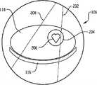
参考图1,一种组合的PET/CT系统100包括PET扫描架部分102和CT扫描架部分104。所述PET扫描架部分102包括围绕检查区域108呈环形设置的伽马辐射敏感探测器106。该探测器106探测发生在PET检查区域108内的正电子湮灭事件的伽马辐射特性。依赖于诸如探测器系统的几何形状和设计等因素,所述PET系统可能具有比检查区域108的横向尺寸更小的有效横向视场(FOV)。Referring to FIG. 1 , a combined PET/
CT部分104包括辐射源110,例如围绕CT检查区域112旋转的X射线管。辐射敏感探测器114探测由所述X射线源发射的、横穿检查区域112的辐射。CT部分104的横向FOV是诸如X射线源110和探测器114的几何形状及设计等因素的函数,并且可能在一些情况下小于或者不同于所述PET部分102的横向FOV。The
将PET扫描架部分102和CT扫描架部分104优选地定位在靠近它们各自的检查区域108、112,这些检查区域沿着公共纵轴或z轴设置。对象支架116支撑要被成像的对象118,诸如人类患者。所述对象支架116优选地可协同PET/CT系统100的操作在纵向上移动,从而可以由所述PET扫描架部分102和所述CT扫描架部分104在多个纵向位置扫描所述对象118。The
CT数据采集系统122处理来自CT探测器114的信号以生成CT投影数据,该CT投影数据指示沿着穿过检查区域112的多条直线或射线的辐射衰减。CT重建器126利用适当的重建算法来重建CT投影数据,以生成指示对象118的空间变化辐射衰减的图像数据。The CT
PET数据采集系统120提供PET投影数据,诸如由探测器106探测到的湮灭事件列表。更具体地,所述投影数据提供针对每个事件的LOR上的信息,诸如LOR的横向和纵向位置、其横向角和方位角以及在具有飞行时间性能的系统的情况下的TOF信息。或者,可以将该数据重新收集到一个或多个正弦面元或投影面元中。PET
局部感兴趣区域(ROI)识别器140识别局部ROI,该局部ROI通常是较大的受检查对象的子集。在一种技术中,利用关于对象的先验信息来确定所述ROI。例如,在人类患者的情况下,可以利用已知的形态特征来估计包括诸如心脏的器官的ROI的位置。在另一种实现方式中,计算机处理器在CT或PET系统数据的低分辨率重建或者其他重建中自动地或半自动地识别ROI的位置,以例如定位病变、活动中心或其他感兴趣场。所述ROI还可以通过用户使用低分辨率或其他图像来手动描绘。在特别适用于对象的一部分位于PET和/或CT系统中之一或二者的有效横向FOV之外的情况的另一实施方式中,ROI可以确立为对象位于相关视场之内的那部分。注意前述技术可以进行组合;也可以使用其他适当的技术。A local region of interest (ROI)
PET投影数据空间截断器138在空间上截断投影数据,例如通过识别沿着穿过所识别的ROI的LOR采集的投影数据或者剔除那些不是沿着穿过所识别的ROI的LOR采集的投影数据。作为示例,图2示出了穿过包括心脏206的ROI 204的第一LOR 202以及不穿过所述ROI的第二LOR 208。注意所述空间截断器138可以被省略,特别是在对象的一部分位于PET成像系统102的横向FOV之外的情况下,在这种情况下所采集的投影数据在空间上被截断。The PET projection data
现在转回图1,在包括运动补偿的系统的情况下,局部运动补偿器142补偿ROI的运动。可以利用适当的运动监控器,诸如在人类患者情况下的呼吸、心脏或其他生理监控器来测量对象的运动。也可以通过分析投影空间或图像空间数据来探测运动。类似地,该运动补偿可以在重建之前应用于空间截断的投影数据或者在重建之后应用于图像空间域。局部运动探测和补偿技术的示例还在2007年2月5日提交的专利申请号PCT/US2007/61597的题为Local Motion Compensation Based on List ModeData以及2007年2月7日提交的美国临时申请号为60/888560的题为Motion Estimation in Treatment Planning中被描述,这两个申请与当前申请为共同拥有并以引用的方式清楚地并入本文。Turning now to FIG. 1 , in the case of a system that includes motion compensation, the
重建器144利用迭代重建技术生成指示对象118中放射性核素的分布的图像空间数据。如将在下面更详细描述的,所述重建器144包括对所截断的投影数据进行重建的局部ROI重建器146。所述重建器144还可以使用非截断的投影数据(即,包括沿着不穿过所述ROI的LOR探测到的那些事件的投影数据)以重建较大的对象。
该系统还可以包括图像组合器148。在这种情况下,较大对象的组合重建图像(例如由CT扫描器104或PET部分102中的一个或两个采集的(多个)图像)可以与局部ROI的图像合并或集成。当图像的特征在于不同的坐标系、空间分辨率等时,配准处理器可以用于配准图像或者提供其他必要的校正。图像组合器148的使用在局部运动补偿或有益于在较大对象的背景下展现ROI的其他应用中是特别有用的。The system may also include an
工作站计算机用作操作者控制台128。该控制台128包括人类可读的输出装置,诸如监视器或显示器,以及输入装置,诸如键盘和鼠标。驻留在控制台128上的软件允许操作者执行一些功能,诸如与ROI识别器140和图像组合器148互动、观察或者操纵由PET和CT重建器144、126生成的图像数据、建立期望的扫描协议、开始和结束扫描等。A workstation computer serves as
系统100上的变型也是可能的。例如,扫描器的CT部分可以省略、远离PET扫描架部分102定位或者替换为另一个成像装置,诸如磁共振(MR)扫描器。作为另一个示例,可以由与PET扫描架部分102相关的传输源提供衰减信息或解剖信息。Variations on
现在将进一步描述局部ROI重建器146。虽然为了解释的清楚,以下讨论将关注于二维(2D)重建,但本领域技术人员将认识到所描述的技术同样适用于且易于扩展到三维(3D)重建。The
给定在二维(2D)空间的发射对象f(x,y)及其衰减系数μ(x,y),则在逆时针旋转一角度


等式1Equation 1

在PET成像中,在去除随机性和散射、校正探测器效率偏差以及适当插值之后针对TOF性能扫描器的所测量的投影数据的平均值可以表示成

等式2Equation 2

等式3Equation 3

其中
等式4Equation 4

项hTOF(t)是TOF卷积核,其常被建模为具有已知的半高宽(FWHM)以及±nσ的核宽度(σ=FWHM/2.355)的高斯分布。注意到通过沿着TOF投影

由于PET中的有限的光子计数统计,所测量的投影数据或
等式5Equation 5
等式6Equation 6

给定等式5和6,并假定衰减系数μ(x,y)是已知的,则PET图像重建的目标是重建所述发射对象f(x,y)。Given Equations 5 and 6, and assuming that the attenuation coefficient μ(x,y) is known, the goal of PET image reconstruction is to reconstruct the emitting object f(x,y).
对于高计数统计,非TOF 2D投影


在局部断层摄影中,假定我们对以坐标(x0,y0)为中心的对象的较小或局部ROI(表示为ε{x0,y0})感兴趣。这一局部对象ROI可以退化成单一像素(x0,y0)。利用等式1,穿过局部ROI的所有LOR可以被定义成其中给定
基于等式5和6,在截断的TOF和非TOF投影数据上形成对数似然值:Based on Equations 5 and 6, log-likelihood values were formed on the truncated TOF and non-TOF projection data:
等式7Equation 7

在这里,使用了所测量的投影


等式7非常类似于非截断投影数据的对数似然值。但是,所述投影数据在穿过局部ROI的局部LOR上被累加而非完整投影。应该注意,尽管我们仅对对象的局部ROI感兴趣,但在所有对象元件上执行前向投影。类似于完整投影,截断投影的对数似然值的Hessian矩阵也是全局凸起的。因此存在唯一的最大值解。现在问题是这个解是否与局部ROI内的原始发射对象完全相同。当局部ROI的尺寸较大时,更有可能得到正确的ROI对象估计。对于小的局部ROI而言,TOF比非TOF更有可能得到正确的对象ROI估计,因为TOF高斯核比非TOF均匀核具有更好的局部化特性。Equation 7 is very similar to the log-likelihood for non-truncated projected data. However, the projection data is accumulated rather than fully projected over the local LORs across the local ROI. It should be noted that although we are only interested in the local ROI of the object, forward projection is performed on all object elements. Similar to the full projection, the Hessian matrix of the log-likelihood values of the truncated projection is also globally convex. Therefore there is a unique maximum solution. The question now is whether this solution is exactly the same as the original emitted object inside the local ROI. When the size of the local ROI is larger, it is more likely to get the correct ROI object estimation. For small local ROIs, TOF is more likely than non-TOF to get the correct object ROI estimation, because TOF Gaussian kernel has better localization properties than non-TOF uniform kernel.
为了优化等式7,使用一种期望最大化(EM)算法。更新等式可以被表述如下,其中k为迭代次数:To optimize Equation 7, an Expectation-Maximization (EM) algorithm is used. The update equation can be formulated as follows, where k is the number of iterations:
等式8Equation 8

为了加速收敛速率,使用一种基于
将前向投影实现为针对TOF的旋转-卷积算子以及针对非TOF的旋转-累加算子。二者均与衰减因子相乘。将反投影首先实现为衰减的乘法,然后作为针对TOF的卷积-反旋转算子和作为针对非TOF的均匀散布-反旋转算子。将对象的初始估计设定为在整个图像视场(FOV)上是均匀的。Implement forward projection as a rotation-convolution operator for TOF and a rotation-accumulation operator for non-TOF. Both are multiplied by the decay factor. The backprojection is implemented first as a multiplication of decay, then as a convolution-derotation operator for TOF and as a uniform spread-derotation operator for non-TOF. The initial estimate of the object is set to be uniform across the entire image field of view (FOV).
误差函数可以用于评估ROI重建器146的性能,特别是结合体模或发射对象已知的其他研究。可以将评估ROI中对象估计与发射对象f之间的误差的均方根误差(RMSE)函数定义为如下:The error function can be used to evaluate the performance of the
等式9Equation 9
其中nROI是局部ROI中的像素的数量。where nROI is the number of pixels in the local ROI.
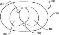
现在将描述一种计算机模拟,其使用一般在图3中示出的2D 420毫米(mm)×300mm胸腔体模302以及32mm直径的肺部病变304。该对象具有144×144像素FOV,4mm像素尺寸。A computer simulation will now be described using a 2D 420 millimeter (mm) x 300
在所模拟的发射对象中,将病变304对背景的对比率设定为8∶1,皮肤306对背景的比率设定为1.3∶1,且骨骼308对背景的比率设定为1.2∶1。在衰减图中,骨骼308对水的比率设定为1.2∶1;将肺区域310模拟为具有空气衰减。In the simulated emission subjects, the
将投影模拟为具有衰减效应,但是没有任何检测器效率偏差、散射或随机性。在
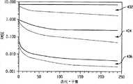
选择三种尺寸的ROI(1个像素、36mm直径和144mm直径)并使其居中在病变像素(57,79)上。在无噪声投影中,具有12个子集及高达20次迭代的ROI-OS-EM算法被用于TOF和非TOF重建。图4示出了分别针对1个像素的局部ROI 402、36mm的局部ROI 404和144mm的局部ROI406的TOF及非TOF的RMSE与迭代次数及子集数之间的关系,其中虚线表示TOF的情况而实线则表示非TOF的情况。TOF算法和非TOF算法对于中等和较大尺寸的ROI都是收敛的,但是TOF的收敛比非TOF的收敛更靠近原始体模。当所述ROI退化到1个像素时,虽然处于较低的对比度水平,TOF仍然收敛到一个解,而非TOF则完全不收敛。Three sizes of ROIs (1 pixel, 36 mm diameter and 144 mm diameter) were selected and centered on the lesion pixel (57, 79). In noise-free projection, the ROI-OS-EM algorithm with 12 subsets and up to 20 iterations was used for TOF and non-TOF reconstructions. Fig. 4 shows the relationship between the RMSE of TOF and non-TOF, the number of iterations and the number of subsets for a
表I显示出针对三种尺寸的ROI,在第10次迭代后TOF和非TOF ROI的最大绝对偏差和平均绝对偏差:Table I shows the maximum and mean absolute deviations for TOF and non-TOF ROIs after the 10th iteration for three sizes of ROIs:
表ITable I
TOF ROI-OS-EM在所有情况下都胜过非TOF ROI-OS-EM。TOF ROI-OS-EM outperforms non-TOF ROI-OS-EM in all cases.
对于有噪声的投影,在没有任何截断的情况下针对TOF和非TOF生成400K的总计数。这一噪声水平类似于在去除随机抽样和散射之后的全身临床PET。根据分别具有16K、62K和160K的总计数的噪声完整投影生成三种尺寸的ROI截断投影(中心在病变上的1个像素、36mm和144mm)。图5示出针对1个像素的局部ROI 402、36mm的局部ROI 404和144mm的局部ROI 406的TOF及非TOF的RMSE与迭代次数及子集数之间的关系,其中12个子集和8次迭代被用于ROI-OS-EM。其趋势类似于无噪声的情况。TOF达到比非TOF更小的ROI RMSE。针对144mm的ROI 406,进行大量迭代后,TOF和非TOF的RMSE均由于噪声放大而增大,这很类似于完整数据OS-EM。For noisy projections, 400K total counts were generated for TOF and non-TOF without any truncation. This noise level is similar to whole-body clinical PET after removal of random sampling and scatter. Three sizes of ROI truncated projections (1 pixel centered on the lesion, 36 mm and 144 mm) were generated from the noisy full projections with total counts of 16K, 62K and 160K, respectively. Fig. 5 shows the relationship between the RMSE of TOF and non-TOF for a
为了视觉比较,在8个子集和2次迭代的条件下,针对144mm的ROI截断投影数据生成TOF重建和非TOF重建,并且与根据完整投影数据生成的TOF重建和TOF重建相比较。通过用来自截断数据的对象ROI替换来自完整数据的对象ROI,TOF重建和非TOF重建与完整数据组合在一起。相对于非TOF截断ROI,TOF截断ROI更好地与完整数据图像混合,而非TOF截断ROI在与完整数据图像组合时具有显著的伪影。For visual comparison, TOF and non-TOF reconstructions were generated for ROI truncated projection data at 144 mm, conditioned on 8 subsets and 2 iterations, and compared to TOF and TOF reconstructions generated from full projection data. TOF and non-TOF reconstructions were combined with the full data by replacing the object ROIs from the full data with object ROIs from the truncated data. TOF truncated ROIs blend better with full data images relative to non-TOF truncated ROIs, which have significant artifacts when combined with full data images.
对于大的局部ROI,TOF ROI-OS-EM以合理的精确性重建ROI之外的区域,而非TOF ROI-OS-EM重建则不是这样。For large local ROIs, TOF ROI-OS-EM reconstructs regions outside the ROI with reasonable accuracy, while non-TOF ROI-OS-EM reconstruction does not.
虽然TOF和非TOF重建均可以提供有用的信息,但TOF重建的性能一般优于非TOF的性能。例如,TOF重建在无噪声和有噪声的情况下均比非TOF重建具有更好的ROI RMSE。在模拟条件下,TOF重建可以重建单一像素的ROI,而非TOF重建则不然。来自截断的TOF重建的重建ROI也比非TOF更好地组合成完整数据图像。While both TOF and non-TOF reconstructions can provide useful information, the performance of TOF reconstructions is generally better than that of non-TOF. For example, TOF reconstruction has better ROI RMSE than non-TOF reconstruction in both noise-free and noisy situations. Under simulated conditions, TOF reconstruction can reconstruct single-pixel ROIs, while non-TOF reconstruction does not. Reconstructed ROIs from truncated TOF reconstructions also assembled into full data images better than non-TOF.
局部断层摄影,特别是在提供TOF数据的系统的情况下的TOF局部断层摄影,可以在很多应用中使用。这些应用的示例包括患者局部运动补偿,其中可以根据局部运动校正的投影数据生成小的ROI图像而非通过重建完整数据集。另一个示例包括在部分对象处于扫描器的有效FOV之外的情况下恢复患者数据。Tomography, especially TOF tomography in the case of systems providing TOF data, can be used in many applications. Examples of these applications include patient local motion compensation, where small ROI images can be generated from local motion corrected projection data rather than by reconstructing the full dataset. Another example includes recovering patient data where part of the object is outside the effective FOV of the scanner.
现在将参考图6来描述局部重建器146的一种实施方式。One embodiment of the
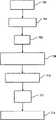
初始对象估计602用于建立初始当前对象估计604。前向投影器606对当前对象估计604进行前向投影以产生针对与ROI相交的那些LOR的对象估计投影。前向投影器606还可以应用其他期望模型和/或校正如用于探测器标准化、衰减、随机抽样和散射的模型。The
比较器616比较针对沿着与局部ROI相交的LOR采集的投影数据的对象估计投影和空间截断的测量投影数据,例如通过确定二者之间的比率或差值来进行比较。A
反投影器624对经比较的投影进行反投影。
图像更新器626使用经反投影的数据来生成新的图像估计。注意对象灵敏度校正是使用与ROI相交的那些投影执行的。
更新后的图像估计变成当前图像估计,且重复该程序直到满足终止条件,例如优化诸如等式7的目标函数。注意该优化是在被截断的投影上执行的。The updated image estimate becomes the current image estimate, and the procedure is repeated until a termination condition is met, eg optimizing an objective function such as Equation 7. Note that this optimization is performed on truncated projections.
当重建器146根据有序子集方法来执行重建时,针对每个子集更新一次对象估计,并且在访问子集中的每一个之后完成一次迭代。When reconstructor 146 performs reconstruction according to the ordered subset method, object estimates are updated once for each subset, and an iteration is completed after each of the subsets is visited.
应该注意到所述重建并不局限于OS-EM技术且可以利用其他适当技术,诸如ML-EM、RBI-EM或其他最大似然方法、RAMLA、CG或LMQN,来执行。还可以使用具有先验信息的最大后验法。还可以使用最小二乘方或其他优化函数。It should be noted that the reconstruction is not limited to the OS-EM technique and can be performed using other suitable techniques, such as ML-EM, RBI-EM or other maximum likelihood methods, RAMLA, CG or LMQN. A maximum a posteriori method with prior information can also be used. Least squares or other optimization functions can also be used.
还应该注意到上面描述的各种函数,特别是由PET投影截断器138、局部ROI识别器140、运动补偿器142、重建器144、146、图像组合器148和配准处理器执行的那些函数,通常是使用一个或多个计算机处理器执行的。导致(多个)处理器执行重建的计算机可读指令被承载在一个或多个计算机可读介质上,诸如计算机磁盘、易失性或非易失性存储器,或其它处理器可访问的介质等。所述指令还可以通过适当的通信网络,诸如互联网,传输到处理器可访问的存储介质。It should also be noted that the various functions described above, particularly those performed by the
现在将参考图7进一步描述操作。Operation will now be further described with reference to FIG. 7 .
在702实施对象的成像检查。当利用组合PET/CT或其他混合模态扫描器执行该检查时,通常基本同时执行所述检查的PET和混合模态部分。然而,应该认识到,所述扫描可以在时间和/或空间上分离开。也可以省略第二模态检查。注意可以在缺少对象的情况下执行对所采集的投影数据的进一步处理。An imaging examination of a subject is performed at 702 . When the examination is performed using a combination PET/CT or other mixed modality scanner, the PET and mixed modality portions of the examination are typically performed substantially simultaneously. However, it should be appreciated that the scans may be separated in time and/or space. The second modal check can also be omitted. Note that further processing of the acquired projection data can be performed in the absence of objects.
在704重建来自第二模态的数据,例如生成在空间上变化的对象衰减数据。Data from the second modality is reconstructed at 704, eg, generating spatially varying object attenuation data.
在706识别局部ROI。当使用来自成像检查的PET或混合部分的信息来识别所述局部ROI时,应该在识别ROI之前重建相关的投影数据。At 706 a local ROI is identified. When using information from a PET or hybrid portion of an imaging exam to identify the local ROI, the associated projection data should be reconstructed prior to identifying the ROI.
在708识别沿着与所识别的ROI相交的LOR采集的投影数据。Projection data acquired along a LOR that intersects the identified ROI is identified at 708 .
在710重建所识别的投影数据以生成指示所识别的ROI和/或较大对象的图像空间数据。The identified projection data is reconstructed at 710 to generate image space data indicative of the identified ROI and/or larger object.
在712执行所识别的ROI的运动补偿。当在投影空间中执行运动补偿时,通常在重建之前执行该运动补偿。Motion compensation of the identified ROI is performed at 712 . When performing motion compensation in projected space, it is usually performed before reconstruction.
在714,以人类可读的形式展示所重建的图像。如果需要,所识别的ROI的图像可以与对象的其他(多个)图像组合起来,例如通过将它们叠加或显示在它们相对于由PET或第二模态扫描中一者或二者产生的图像的正确的位置上。At 714, the reconstructed image is displayed in human readable form. If desired, images of the identified ROIs can be combined with other image(s) of the subject, for example by superimposing them or displaying them relative to the image produced by either or both of the PET or second modality scan on the correct position.
应该理解可以适当改变各个步骤的执行顺序。It should be understood that the order of execution of the respective steps can be appropriately changed.
已经通过参考优选实施例描述了本发明。当然,通过阅读和理解前面的描述,其他人员可以进行修改和改变。意在将本发明解释为包括所有这些修改和改变,只要它们落入所附的权利要求书的范围内。The invention has been described with reference to the preferred embodiments. Of course, modifications and alterations will occur to others upon reading and understanding the preceding description. It is intended that the present invention be construed as including all such modifications and changes as long as they fall within the scope of the appended claims.
Claims (15)
Applications Claiming Priority (3)
| Application Number | Priority Date | Filing Date | Title |
|---|---|---|---|
| US94072207P | 2007-05-30 | 2007-05-30 | |
| US60/940,722 | 2007-05-30 | ||
| PCT/IB2008/051764WO2008146186A2 (en) | 2007-05-30 | 2008-05-06 | Pet local tomography |
Publications (2)
| Publication Number | Publication Date |
|---|---|
| CN101681520A CN101681520A (en) | 2010-03-24 |
| CN101681520Btrue CN101681520B (en) | 2013-09-25 |
Family
ID=39846999
Family Applications (1)
| Application Number | Title | Priority Date | Filing Date |
|---|---|---|---|
| CN2008800178971AExpired - Fee RelatedCN101681520B (en) | 2007-05-30 | 2008-05-06 | PET local tomography |
Country Status (6)
| Country | Link |
|---|---|
| US (1) | US8457380B2 (en) |
| EP (1) | EP2156408B1 (en) |
| JP (1) | JP2010528312A (en) |
| CN (1) | CN101681520B (en) |
| RU (1) | RU2471204C2 (en) |
| WO (1) | WO2008146186A2 (en) |
Families Citing this family (28)
| Publication number | Priority date | Publication date | Assignee | Title |
|---|---|---|---|---|
| CN101933046B (en)* | 2008-01-25 | 2012-10-03 | 模拟逻辑有限公司 | image combination |
| US8218848B2 (en)* | 2008-07-23 | 2012-07-10 | Siemens Aktiengesellschaft | System and method for the generation of attenuation correction maps from MR images |
| US8660636B2 (en) | 2009-01-19 | 2014-02-25 | Koninklijke Philips N.V. | Regional reconstruction and quantitative assessment in list mode PET imaging |
| EP2398390B1 (en)* | 2009-02-17 | 2015-04-15 | Koninklijke Philips N.V. | Model-based extension of field-of-view in nuclear imaging |
| CN102483852B (en) | 2009-06-08 | 2016-08-03 | 皇家飞利浦电子股份有限公司 | Time-of-flight positron emission tomography reconstruction using image content generated event-by-event based on time-of-flight information |
| US8299438B2 (en)* | 2009-07-16 | 2012-10-30 | Siemens Medical Solutions Usa, Inc. | Model based estimation of a complete or partial positron emission tomography attenuation map using maximum likelihood expectation maximization |
| US8987674B2 (en)* | 2009-09-04 | 2015-03-24 | Shimadzu Corporation | Data processing method for nuclear medicine, and a nuclear medicine diagnostic apparatus |
| WO2011070465A2 (en)* | 2009-12-10 | 2011-06-16 | Koninklijke Philips Electronics, N.V. | Method and apparatus for using time of flight information to detect and correct for motion in imaging scans |
| DE102010019016B4 (en)* | 2010-05-03 | 2017-03-02 | Siemens Healthcare Gmbh | Method for reconstructing image data of a moving examination object from measured data together with associated objects |
| US8625869B2 (en)* | 2010-05-21 | 2014-01-07 | Siemens Medical Solutions Usa, Inc. | Visualization of medical image data with localized enhancement |
| WO2012017345A1 (en)* | 2010-08-04 | 2012-02-09 | Koninklijke Philips Electronics N.V. | Method and system for iterative image reconstruction |
| US20120078089A1 (en)* | 2010-09-23 | 2012-03-29 | General Electric Company | Method and apparatus for generating medical images |
| EP2883084B1 (en)* | 2012-08-10 | 2017-09-27 | Koninklijke Philips N.V. | Virtual frames for distributed list-mode time-of-light reconstruction with continuous bed movement |
| CN105103194B (en) | 2013-04-10 | 2019-01-29 | 皇家飞利浦有限公司 | Reconstructed image data visualization |
| CN105144241B (en) | 2013-04-10 | 2020-09-01 | 皇家飞利浦有限公司 | Image quality index and/or imaging parameter recommendation based thereon |
| US9684973B2 (en)* | 2014-12-08 | 2017-06-20 | General Electric Company | Systems and methods for selecting imaging data for principle components analysis |
| US9990741B2 (en)* | 2015-09-28 | 2018-06-05 | Siemens Medical Solutions Usa, Inc. | Motion correction in a projection domain in time of flight positron emission tomography |
| US20190133542A1 (en)* | 2016-04-19 | 2019-05-09 | The General Hospital Corporation | Systems and methods for data-driven respiratory gating in positron emission tomography |
| US10531275B2 (en) | 2016-12-12 | 2020-01-07 | Commscope Technologies Llc | Cluster neighbor discovery in centralized radio access network using transport network layer (TNL) address discovery |
| WO2018127470A1 (en)* | 2017-01-06 | 2018-07-12 | Koninklijke Philips N.V. | Using time-of-flight to detect and correct misalignment in pet/ct imaging |
| WO2018172566A1 (en)* | 2017-03-24 | 2018-09-27 | Koninklijke Philips N.V. | Noise-robust real-time extraction of the respiratory motion signal from pet list-data |
| WO2018202648A1 (en)* | 2017-05-01 | 2018-11-08 | Koninklijke Philips N.V. | Generation of accurate hybrid datasets for quantitative molecular imaging |
| US10282871B2 (en) | 2017-07-10 | 2019-05-07 | Shanghai United Imaging Healthcare Co., Ltd. | Systems and methods for pet image reconstruction |
| CN109350099A (en)* | 2018-09-13 | 2019-02-19 | 中山市明峰医疗器械有限公司 | A Random Event Removal Processing Method Applied in Clinical PET System |
| US10743830B2 (en)* | 2018-12-04 | 2020-08-18 | Canon Medical Systems Corporation | Method and apparatus for scatter correction in position emission tomography (PET) imaging by performing a short PET scan in an extended region to estimate scatter coming from outside of the field of view (FOV) |
| CN110544233B (en)* | 2019-07-30 | 2022-03-08 | 北京的卢深视科技有限公司 | Depth image quality evaluation method based on face recognition application |
| WO2021102614A1 (en)* | 2019-11-25 | 2021-06-03 | 中国科学院深圳先进技术研究院 | Method and terminal for processing positron emission tomography (pet) data |
| US12159330B2 (en)* | 2022-02-17 | 2024-12-03 | Canon Medical Systems Corporation | Event property-dependent point spread function modeling and image reconstruction for PET |
Citations (4)
| Publication number | Priority date | Publication date | Assignee | Title |
|---|---|---|---|---|
| US5953388A (en)* | 1997-08-18 | 1999-09-14 | George Mason University | Method and apparatus for processing data from a tomographic imaging system |
| CN1504960A (en)* | 2002-10-04 | 2004-06-16 | GEҽҩϵͳ����Ƽ���˾ | Method and apparatus for truncation and compensation |
| US20050249432A1 (en)* | 2004-02-10 | 2005-11-10 | Yu Zou | Imaging system |
| US20070076933A1 (en)* | 2005-09-30 | 2007-04-05 | Jared Starman | Estimating the 0th and 1st moments in C-arm CT data for extrapolating truncated projections |
Family Cites Families (20)
| Publication number | Priority date | Publication date | Assignee | Title |
|---|---|---|---|---|
| GB2164230A (en) | 1984-08-29 | 1986-03-12 | Clayton Found Res | Three-dimensional time-of-flight positron emission camera system |
| US5539800A (en) | 1995-03-24 | 1996-07-23 | The Regents Of The University Of California, Office Of Technology Transfer | Pseudolocal tomography |
| US5550892A (en) | 1995-03-24 | 1996-08-27 | The Regents Of The University Of California | Enhanced local tomography |
| WO1997005574A1 (en)* | 1995-07-27 | 1997-02-13 | Imperial Cancer Research Technology Limited | Raw data segmentation and analysis in image tomography |
| US5717211A (en) | 1996-07-22 | 1998-02-10 | The Regents Of The University Of California | Generalized local emission tomography |
| US6147353A (en)* | 1997-05-30 | 2000-11-14 | Picker International, Inc. | Image shift for gamma camera |
| NZ509667A (en)* | 1998-08-06 | 2003-08-29 | Wisconsin Alumni Res Found | Delivery modification system for radiation therapy |
| US6473634B1 (en)* | 2000-11-22 | 2002-10-29 | Koninklijke Philips Electronics N.V. | Medical imaging at two temporal resolutions for tumor treatment planning |
| US6771732B2 (en)* | 2002-02-28 | 2004-08-03 | The Board Of Trustees Of The University Of Illinois | Methods and apparatus for fast divergent beam tomography |
| CN1809841B (en)* | 2003-06-18 | 2010-05-12 | 皇家飞利浦电子股份有限公司 | Reconstruction method, device and system for motion compensation |
| US7254209B2 (en)* | 2003-11-17 | 2007-08-07 | General Electric Company | Iterative CT reconstruction method using multi-modal edge information |
| JP5021489B2 (en) | 2004-12-22 | 2012-09-05 | コーニンクレッカ フィリップス エレクトロニクス エヌ ヴィ | Real-time list mode reconstruction |
| EP1875437B1 (en) | 2005-04-14 | 2010-03-10 | Koninklijke Philips Electronics N.V. | Three-dimensional time-of-flight pet with course angular and slice rebinning |
| WO2007008532A1 (en)* | 2005-07-08 | 2007-01-18 | Wisconsin Alumni Research Foundation | Constrained backprojection reconstruction method for undersampled mri |
| US7381959B2 (en) | 2005-08-17 | 2008-06-03 | General Electric Company | Technique for reconstructing PET scan images |
| CN101305297B (en) | 2005-11-10 | 2012-01-04 | 皇家飞利浦电子股份有限公司 | PET Imaging Using Anatomy List Pattern Masks |
| JP5254810B2 (en)* | 2006-02-28 | 2013-08-07 | コーニンクレッカ フィリップス エレクトロニクス エヌ ヴィ | Local motion compensation based on list mode data |
| US7848559B2 (en)* | 2006-05-17 | 2010-12-07 | Siemens Medical Solutions Usa, Inc. | Discrete axial re-binning of time-of-flight positron emission tomography data |
| GB2450073B (en)* | 2006-08-25 | 2009-11-04 | Siemens Molecular Imaging Ltd | Regional reconstruction of spatially distributed functions |
| US7680240B2 (en)* | 2007-03-30 | 2010-03-16 | General Electric Company | Iterative reconstruction of tomographic image data method and system |
- 2008
- 2008-05-06RURU2009149481/28Apatent/RU2471204C2/ennot_activeIP Right Cessation
- 2008-05-06CNCN2008800178971Apatent/CN101681520B/ennot_activeExpired - Fee Related
- 2008-05-06USUS12/600,666patent/US8457380B2/ennot_activeExpired - Fee Related
- 2008-05-06EPEP08763067.9Apatent/EP2156408B1/ennot_activeNot-in-force
- 2008-05-06WOPCT/IB2008/051764patent/WO2008146186A2/enactiveApplication Filing
- 2008-05-06JPJP2010509922Apatent/JP2010528312A/enactivePending
Patent Citations (4)
| Publication number | Priority date | Publication date | Assignee | Title |
|---|---|---|---|---|
| US5953388A (en)* | 1997-08-18 | 1999-09-14 | George Mason University | Method and apparatus for processing data from a tomographic imaging system |
| CN1504960A (en)* | 2002-10-04 | 2004-06-16 | GEҽҩϵͳ����Ƽ���˾ | Method and apparatus for truncation and compensation |
| US20050249432A1 (en)* | 2004-02-10 | 2005-11-10 | Yu Zou | Imaging system |
| US20070076933A1 (en)* | 2005-09-30 | 2007-04-05 | Jared Starman | Estimating the 0th and 1st moments in C-arm CT data for extrapolating truncated projections |
Also Published As
| Publication number | Publication date |
|---|---|
| US20100303319A1 (en) | 2010-12-02 |
| RU2471204C2 (en) | 2012-12-27 |
| JP2010528312A (en) | 2010-08-19 |
| US8457380B2 (en) | 2013-06-04 |
| CN101681520A (en) | 2010-03-24 |
| RU2009149481A (en) | 2011-07-10 |
| EP2156408B1 (en) | 2021-03-17 |
| WO2008146186A2 (en) | 2008-12-04 |
| WO2008146186A3 (en) | 2009-01-29 |
| EP2156408A2 (en) | 2010-02-24 |
Similar Documents
| Publication | Publication Date | Title |
|---|---|---|
| CN101681520B (en) | PET local tomography | |
| RU2413245C2 (en) | Processing positron emission tomography images using anatomic list mode mask | |
| Lewitt et al. | Overview of methods for image reconstruction from projections in emission computed tomography | |
| JP5734664B2 (en) | Image Restoration Method Using Dilution Constraint Correction | |
| JP4965575B2 (en) | Distributed iterative image reconstruction | |
| US9155514B2 (en) | Reconstruction with partially known attenuation information in time of flight positron emission tomography | |
| EP3067864B1 (en) | Iterative reconstruction with enhanced noise control filtering | |
| EP2210238B1 (en) | Apparatus and method for generation of attenuation map | |
| CN103329168B (en) | Truncation Compensation for Iterative Cone Beam CT Reconstruction for SPECT/CT Systems | |
| Levkovilz et al. | The design and implementation of COSEN, an iterative algorithm for fully 3-D listmode data | |
| CN101765865A (en) | Motion Correction in Nuclear Imaging | |
| Melot et al. | Some proximal methods for Poisson intensity CBCT and PET | |
| US20220375038A1 (en) | Systems and methods for computed tomography image denoising with a bias-reducing loss function | |
| EP2715671A1 (en) | Fast computation of the sensitivity matrix in iterative algorithms | |
| Bai et al. | PET image reconstruction: methodology and quantitative accuracy | |
| Li et al. | Lesion quantification in dual-modality mammography using expectation maximization with attenuation correction | |
| Zou et al. | Image Reconstruction for Source Trajectories with Kinks |
Legal Events
| Date | Code | Title | Description |
|---|---|---|---|
| C06 | Publication | ||
| PB01 | Publication | ||
| C10 | Entry into substantive examination | ||
| SE01 | Entry into force of request for substantive examination | ||
| C14 | Grant of patent or utility model | ||
| GR01 | Patent grant | ||
| CF01 | Termination of patent right due to non-payment of annual fee | Granted publication date:20130925 | |
| CF01 | Termination of patent right due to non-payment of annual fee |