CN101663054B - Drug delivery intravascular stent and method of use thereof - Google Patents
Drug delivery intravascular stent and method of use thereofDownload PDFInfo
- Publication number
- CN101663054B CN101663054BCN200780046647.6ACN200780046647ACN101663054BCN 101663054 BCN101663054 BCN 101663054BCN 200780046647 ACN200780046647 ACN 200780046647ACN 101663054 BCN101663054 BCN 101663054B
- Authority
- CN
- China
- Prior art keywords
- stent
- scaffold
- roughening
- stents
- abrading
- Prior art date
- Legal status (The legal status is an assumption and is not a legal conclusion. Google has not performed a legal analysis and makes no representation as to the accuracy of the status listed.)
- Active
Links
Images


Landscapes
- Media Introduction/Drainage Providing Device (AREA)
- Materials For Medical Uses (AREA)
Abstract
Translated fromChineseDescription
Translated fromChinese背景技术Background technique
并发症(例如再狭窄)是在接受了医学手术(例如经皮冠状动脉腔内成形术(percutaneous transluminal coronary angioplasty,PTCA)形式的动脉粥样硬化治疗的患者中复发的问题。通常通过称为支架术的方法来治疗再狭窄,其中,通过外科手术将医疗装置植入患病的动脉中以防止其在术后发生堵塞。Complications, such as restenosis, are recurring problems in patients treated for atherosclerosis in the form of medical procedures such as percutaneous transluminal coronary angioplasty (PTCA). Restenosis is treated as a surgical procedure in which a medical device is surgically implanted in the diseased artery to prevent it from becoming clogged after surgery.
支架一般为柱形,并且通常由生物相容性的金属(例如钛或外科用钢(surgical steel))制成。大多数支架是可收缩的,并且通过经腔导管(transluminal catheter)递送到发生堵塞的动脉处。支架固定在导管上,并且可以自身扩张或者通过该支架内的囊体的膨胀而扩张,当所述装置已经位于合适的位置后移除囊体和导管。通常类型的导管的实例在Palmaz的题为“Complaint Implantable Medical Devices and Methods of MakingSame(相容的可植入医疗装置及其制备方法)”的美国专利申请No.US 6,936,066公布中公开。Stents are generally cylindrical and are usually made of a biocompatible metal such as titanium or surgical steel. Most stents are collapsible and delivered to the blocked artery via a transluminal catheter. The stent is secured to the catheter and can expand by itself or by expansion of a balloon within the stent, which is removed when the device has been in place. An example of a general type of catheter is disclosed in Palmaz's U.S. Patent Application No. US 6,936,066 publication entitled "Complaint Implantable Medical Devices and Methods of Making Same."
再狭窄和血栓形成是由支架疗法所导致的并发症。在为了克服这些并发症的做出的努力中,支架通常被开发为具有额外的受控制的药物洗脱(drug elution)的特征。为了实现该特征,如Hunter的题为anti-angiogenicCompositions and Methods of Use(抗血管生成组合物及其使用方法)的美国专利No.5,716,981中所公开的,用与聚合物混合的API对金属支架进行涂敷。以该方式递送的典型的治疗剂的实例有抗增殖剂、抗凝剂、抗炎剂以及免疫抑制剂,但是还有许多其它可以使用的化学和生物学物质。通常在涂层上应用生物可降解材料的多孔层,以对药物向体内的受控制的释放进行调节。涂教有聚合物的药物洗脱支架的通常类型在Ragheb的题为“Coated Implantable Medical Device(涂敷的可植入医疗装置)”的美国专利No.6,774,278和6,730,064中公开。Restenosis and thrombosis are complications resulting from stent therapy. In an effort to overcome these complications, stents are often developed with the additional feature of controlled drug elution. To achieve this feature, metal stents were treated with an API mixed with a polymer as disclosed in Hunter, U.S. Patent No. 5,716,981 entitled anti-angiogenic Compositions and Methods of Use. Apply. Typical examples of therapeutic agents delivered in this manner are antiproliferative agents, anticoagulants, antiinflammatory agents and immunosuppressants, but there are many other chemical and biological substances that could be used. A porous layer of biodegradable material is usually applied over the coating to regulate the controlled release of the drug into the body. A general type of polymer-coated drug-eluting stent is disclosed in US Patent Nos. 6,774,278 and 6,730,064 to Ragheb, entitled "Coated Implantable Medical Device."
已经有假设认为由于分层这种聚合物的存在导致了血栓形成。人们认为,随着时间的流逝,保护性聚合物可能与裸金属或基质分离,产生的锋利的突起或边缘与红血细胞直接接触进而导致血栓形成,引起患者的严重疾病或死亡。已经设计了不含有永久性聚合物(permanent polymer)的支架,如BSI生物可降解PLLA(BSI biodegradable PLLA)。一些支架的设计朝向完全无聚合物的方向发展,例如表面改质的支架或生物相容性的油涂层。It has been hypothesized that thrombus formation is caused by the presence of such polymers as a layer. It is thought that over time, the protective polymer may detach from the bare metal or matrix, creating sharp protrusions or edges that come into direct contact with red blood cells and cause blood clots to form, causing serious illness or death in the patient. Scaffolds have been designed that do not contain permanent polymers, such as BSI biodegradable PLLA (BSI biodegradable PLLA). Some stent designs are moving towards completely polymer-free, such as surface-modified stents or biocompatible oil coatings.
为了增加药物负载能力,可以将支架制造为具有粗糙化的表面。支架上的粗糙表面提供了增加支架的总表面积的突起和凹陷,从而增加了可能与支架有关的API的量。可以通过许多方法对支架的表面进行粗糙化,例如烧结,如Yan的题为“Porous Medical Stent(多孔医疗支架)”的美国专利No.5,843,172中所公开的。还可以通过磨蚀技术来对表面进行粗糙化,例如喷砂和还原酸蚀刻,如在Leitao的题为“Device For Incorporation andRelease of Biologically Active Agents(用于掺入和释放生物活性剂的装置)”的欧洲专利No.0,806,212中公开的那样。还可以通过直接在装置上冲压凹槽来对支架表面进行粗糙化,如在Thomas的题为“Method ofForming a Drug Eluting Stent(制备药物洗脱支架的方法)”的美国专利No.7,055,237中所公开的那样,或使用弹丸喷射(shot peening)或激光喷射(laser peening)的常规金属加工技术来对支架表面进行粗糙化。具有机械锚固层的支架在共有的美国公布No.2006/0069427中进行了描述。然而,支架和导管横截面是涂层厚度的物理限制。粗糙的支架遇到的问题是必须将支架和导管的横截面的厚度限定得足够窄,以能够穿过堵塞的动脉。To increase drug loading capacity, stents can be fabricated with roughened surfaces. The rough surface on the scaffold provides protrusions and depressions that increase the overall surface area of the scaffold, thereby increasing the amount of API that may be associated with the scaffold. The surface of the stent can be roughened by a number of methods, such as sintering, as disclosed in Yan, US Patent No. 5,843,172 entitled "Porous Medical Stent." The surface can also be roughened by abrasive techniques, such as sandblasting and reducing acid etching, as described in Leitao entitled "Device For Incorporation and Release of Biologically Active Agents (for the incorporation and release of biologically active agents)" as disclosed in European Patent No. 0,806,212. The surface of the stent can also be roughened by stamping grooves directly on the device, as disclosed in Thomas, U.S. Patent No. 7,055,237 entitled "Method of Forming a Drug Eluting Stent" The surface of the stent is roughened, as in the previous study, or using conventional metalworking techniques such as shot peening or laser peening. Stents with mechanical anchoring layers are described in commonly-owned US Publication No. 2006/0069427. However, stent and catheter cross-sections are physical limitations on coating thickness. The problem encountered with rough stents is that the thickness of the cross-section of the stent and catheter must be defined narrow enough to be able to pass through the blocked artery.
考虑到与支架疗法相关的并发症,需要开发具有粗糙支架的增加的表面积的支架,其可以以使结构完整性和载药能力最大化的方式制备。进一步地,需要开发无聚合物的支架,该支架能递送API以降低或消除后期的支架血栓形成的风险。最后,需要开发使药物负载能力最大化、同时使支架-导管横截面的总厚度最小化的支架。Given the complications associated with stent therapy, there is a need to develop scaffolds with increased surface area of rough scaffolds that can be fabricated in a manner that maximizes structural integrity and drug loading capacity. Further, there is a need to develop polymer-free stents that can deliver APIs to reduce or eliminate the risk of late stent thrombosis. Finally, there is a need to develop stents that maximize drug loading capacity while minimizing the overall thickness of the stent-catheter cross-section.
发明概述Summary of the invention
本发明的实施方案通过提供一种医疗装置例如支架和减少横截面(crossing profile)厚度且不损害结构完整性的处理药物洗脱支架的方法解决了上述的一个或多个问题。所公开的实施方案还使药物负载能力最大化和/或增加了耐疲劳性。Embodiments of the present invention address one or more of the above-mentioned problems by providing a medical device, such as a stent, and a method of processing a drug-eluting stent that reduces cross-sectional thickness without compromising structural integrity. The disclosed embodiments also maximize drug loading capacity and/or increase fatigue resistance.
一方面,提供了一种非聚合物的治疗剂洗脱支架。优选地,所述支架包括其中至少一部分具有被改质或磨蚀的微结构的至少一个表面。在一种优选实施方案中,所述治疗剂至少被应用在所述支架表面的被改质的部分上。In one aspect, a non-polymer therapeutic agent eluting stent is provided. Preferably, the scaffold comprises at least one surface at least a portion of which has a modified or abraded microstructure. In a preferred embodiment, the therapeutic agent is applied to at least the modified portion of the stent surface.
另一方面,公开了制备具有至少一个“粗糙化”表面的支架的方法。在一种实施方案中,该方法包括将裸金属支架卷入到烃膜层中,所述膜层为支架的内层和侧面中的至少一个提供了掩蔽物。然后用磨料处理外表面,随后将掩蔽层溶解或以其它方式移除。优选地,然后根据ASTM标准将所述支架进行声波处理、清洁和钝化。In another aspect, methods of making scaffolds having at least one "roughened" surface are disclosed. In one embodiment, the method includes embedding a bare metal stent into a hydrocarbon film layer that provides a mask for at least one of an inner layer and sides of the stent. The outer surface is then treated with an abrasive and the masking layer is subsequently dissolved or otherwise removed. Preferably, the scaffold is then sonicated, cleaned and passivated according to ASTM standards.
在另一种实施方案中,用喷射的方法处理支架表面。将未经处理的支架在心轴上夹紧,并且使用板或辊通过在支架表面上冲压被称为弹丸的金属颗粒来进行粗糙化。还可以通过在支架表面上进行喷丸(blasting shot)、例如通过射流洗吹(jet blasting)来获得粗糙度。在另一种实施方案中,还可以使用激光而不是弹丸以类似的方法得到表面粗糙度。In another embodiment, the stent surface is treated by spraying. Untreated stents are clamped on a mandrel and roughened using plates or rollers by stamping metal particles called shot on the surface of the stent. Roughness can also be achieved by blasting shots on the surface of the stent, for example by jet blasting. In another embodiment, the surface roughness can also be similarly obtained using a laser instead of a shot.
在另一种实施方案中,用气动压力机(pneumatic press)或水压机(hydraulic press)处理支架,以产生粗糙化的表面。将未处理的支架固定到心轴上,并通过气动压力机或水压机将模式(pattern)压印在所述支架的表面上。在一种实施方案中,所述模式为已预设的。在另一种实施方案中,所述气动压力机或水压机由计算机控制。这种冲压方法的优势是其破坏支架体中的微晶结构以增加支架体的耐疲劳性的能力。In another embodiment, the stent is treated with a pneumatic press or hydraulic press to produce a roughened surface. Untreated stents are fixed to a mandrel and a pattern is imprinted on the surface of the stent by a pneumatic or hydraulic press. In one embodiment, the mode is preset. In another embodiment, the pneumatic press or hydraulic press is computer controlled. An advantage of this stamping method is its ability to disrupt the microcrystalline structure in the scaffold body to increase the fatigue resistance of the scaffold body.
在另一种实施方案中,在将其切割成所需的支架长度前,对全部长度(例如2.5M)的支架进行处理。将全部的支架长度固定到心轴上,然后通过所公开的方法之一进行处理。In another embodiment, the full length (eg, 2.5M) scaffold is processed before cutting it into the desired scaffold length. The entire stent length is secured to the mandrel and then processed by one of the disclosed methods.
在另一种实施方案中,用活性药物成分(active pharmaceuticalingredient,API)或API和聚合物的组合对支架进行涂敷。可以通过直接将所需的药物或药物/聚合物的组合喷涂在所述支架的表面上得到涂层。此外,可以通过将所述支架浸渍到所需API涂层的溶液中来实现用所需的化学药品对所述支架的涂敷。在另一种实施方案中,通过自动移液器(automated pipetting)用API或API/聚合物的组合近腔涂敷支架。In another embodiment, the stent is coated with an active pharmaceutical ingredient (API) or a combination of an API and a polymer. Coatings can be obtained by spraying the desired drug or drug/polymer combination directly onto the surface of the stent. In addition, coating the stent with desired chemicals can be accomplished by dipping the stent into a solution of the desired API coating. In another embodiment, the stent is abluminally coated with API or API/polymer combination by automated pipetting.
本发明的上述和其它方面以及实施方案将通过以下详细的描述并结合附图而变得更显而易见。The above and other aspects and embodiments of the present invention will become more apparent from the following detailed description when taken in conjunction with the accompanying drawings.
附图说明Description of drawings
通过发明详述和附图能更充分地理解本发明,其中:The present invention can be more fully understood from the detailed description of the invention and the accompanying drawings, in which:
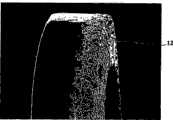
图1为具有金属丝体的血管内支架的扫描图;Fig. 1 is a scanning diagram of a stent in a blood vessel with a wire body;
图2A为被磨蚀的支架表面的扫描电子显微图;Figure 2A is a scanning electron micrograph of an abraded stent surface;
图2B为图2A的表面的扫描电子显微图,显示了磨蚀后在支架表面上产生的突起的数量;Figure 2B is a scanning electron micrograph of the surface of Figure 2A showing the number of protrusions produced on the scaffold surface after abrasion;
图2C为图2A的表面的扫描电子显微图,显示了磨蚀后在支架表面上产生的凹陷的数量;Figure 2C is a scanning electron micrograph of the surface of Figure 2A showing the number of depressions produced on the surface of the scaffold after abrasion;
图3A是气动压力机对支架表面进行处理的示意图;Fig. 3A is a schematic diagram of the treatment of the surface of the bracket by the pneumatic press;
图3B为图3A的顶端固定的冲床组件的近距离的主视图,显示了具有多个冲头(peener)的气动压力机;3B is a close-up front view of the fixed-top punch assembly of FIG. 3A showing a pneumatic press with multiple peeners;
图3C为图3B的顶端固定的冲床组件的近距离的侧视图;Figure 3C is a close-up side view of the top-fixed punch assembly of Figure 3B;
图3D为用于图3A的气动压力机的冲床组件的顶端固定附件的近距离的主视图,显示了示例性的模式;3D is a close-up front view of a top end attachment attachment for the punch assembly of the pneumatic press of FIG. 3A , showing an exemplary model;
图4为涂敷有药物的经处理的支架的扫描电子显微图;Figure 4 is a scanning electron micrograph of a drug-coated stent;
图5为历经以小时计的累积时间以释放的药物总量百分数测量的来自本发明的支架和

图6为表示分别在三个月和两个月在猪植入模型中从本发明的支架和

图7为表示用质谱测得的在猪植入模型中从本发明的支架和

图8为表示不含药物的支架和含有Biolimus药物的支架的堵塞的面积百分数的图;Figure 8 is a representation of a drug-free stent and a Biolimus-containing Plot of area percent occlusion of drug-stented stent;
图9A至图9F为植入裸金属支架(图9A-9B)、具有含有Biolimus

图10A至图10K为微结构支架的组织形态测量(histomorphometry)的图。10A-10K are graphs of histomorphometry of microstructured scaffolds.
发明详述Detailed description of the invention
I.血管内支架I. Intravascular stent
图1显示了在收缩状态的根据本发明构建的支架。该支架包括具有至少一个被至少部分粗糙化或磨蚀的表面的结构构件或结构体,其至少用来容纳和释放抗再狭窄化合物,这将在下文进行进一步的描述。Figure 1 shows a stent constructed according to the present invention in a collapsed state. The stent includes a structural member or structure having at least one at least partially roughened or abraded surface at least for containing and releasing an anti-restenotic compound, as further described below.
在所示实施方案中,所述支架体是由通过称为接头4的丝彼此相连的一系列的被称为支杆3的管状构件形成的。各支杆3具有可扩张的Z形、锯齿形、螺旋形带状线圈或正弦波结构,并且与各个接头4的连接用于增加整体支架柔韧性。收缩状态的支架的直径为约0.5-2.0mm,优选0.71-1.65mm,长度为5-100mm。扩张的支架的直径为该支架收缩状态的至少2倍并且最高为8-9倍,例如,收缩状态直径为0.7-1.5mm的支架可以径向扩张到所选择的2.0-8.0mm或更大的扩张状态。具有连接的、可扩张的管状构件的这种一般的支架体结构的支架是已知的,例如在PCT公布No.WO99/07308中有描述,其被本申请共同具有并以引用方式合并入本文。In the embodiment shown, the stent body is formed by a series of tubular members called
优选地,支架结构是由生物相容性材料如不锈钢制备的。通常用于支架结构的生物相容性材料的另外的实例有:钽、钛、镍钛金属互化物(nitinol)、金、铂、铬镍铁合金(inconel)、铱、银、钨或其它生物相容性材料,或它们的任意的合金;碳或碳纤维;醋酸纤维素、硝酸纤维素、聚硅氧烷、聚对苯二甲酸乙二酯(polyethylene teraphthalate)、聚氨基甲酸酯、聚酰胺、聚酯、聚原酸酯、聚酐(polyanhydride)、聚醚砜、聚碳酸酯、聚丙烯、高分子量聚乙烯、聚四氟乙烯或其它生物相容性共聚物材料,或它们的混合物或共聚物;聚-L-乳酸、聚羟基乙酸或它们的共聚物、聚酐、聚己内酯、聚羟基丁酸戊酯(polyhydroxybutyrate valerate)或其它生物可降解的聚合物,或它们的混合物或共聚物;蛋白质、细胞外基质组分、胶原蛋白、纤维蛋白或其它生物学物质(biologic agent);或上述这些物质的任意的合适的混合物。典型的支架的实例在美国专利No.6,730,064中已有描述。各个支架的尺寸将根据其所要递送于其中的体腔(body lumen)而不同。例如,支架可以具有从约0.5mm至约25.0mm范围的直径和从约4mm至约100mm或更长的长度。支架测量的例子已经在Shulze的美国专利No.6,939,376中进行了描述,其被本申请共同具有并以引用方式合并入本文。Preferably, the stent structure is fabricated from a biocompatible material such as stainless steel. Additional examples of biocompatible materials commonly used in stent structures are: tantalum, titanium, nitinol, gold, platinum, inconel, iridium, silver, tungsten, or other biophase Capacitive material, or any alloy thereof; carbon or carbon fiber; cellulose acetate, nitrocellulose, polysiloxane, polyethylene terephthalate (polyethylene teraphthalate), polyurethane, polyamide, Polyester, polyorthoester, polyanhydride, polyethersulfone, polycarbonate, polypropylene, high molecular weight polyethylene, polytetrafluoroethylene, or other biocompatible copolymer materials, or mixtures or copolymers thereof substances; poly-L-lactic acid, polyglycolic acid or their copolymers, polyanhydrides, polycaprolactone, polyhydroxybutyrate valerate (polyhydroxybutyrate valerate) or other biodegradable polymers, or their mixtures or copolymers substances; proteins, extracellular matrix components, collagen, fibrin or other biological substances (biologic agents); or any suitable mixture of these substances. An example of a typical stent is described in US Patent No. 6,730,064. The size of each stent will vary depending on the body lumen into which it is to be delivered. For example, the stent can have a diameter ranging from about 0.5 mm to about 25.0 mm and a length of from about 4 mm to about 100 mm or more. Examples of stent measurements have been described in US Patent No. 6,939,376 to Shulze, which is commonly owned by this application and is incorporated herein by reference.
如图2A所示,所述支架的至少一个表面的至少一部分具有粗糙化的或磨蚀的微结构或改质的表面。这种微结构可以包括至少一种从所述微结构洗脱的治疗剂。如从图2B-2C所见,所述粗糙化的或改质的表面提供了缝隙或垂直凸起表面特征和/或咬边(undercut)或凹槽(recess)区域。可以理解的是,治疗剂的溶液或含有治疗剂的溶液可以例如通过毛细力而被吸入这些凹槽,并对凸起表面进行涂敷。以此方式可以增加用于涂敷支架的表面积。这些层的厚度指的是层的平均厚度,例如,层的可注入部分的平均深度。优选地,如图2A所示,支架的近腔表面的至少一部分包括微结构表面加工。As shown in Figure 2A, at least a portion of at least one surface of the scaffold has a roughened or abrasive microstructure or modified surface. Such microstructures may include at least one therapeutic agent eluted from said microstructures. As seen from Figures 2B-2C, the roughened or modified surface provides crevices or vertically raised surface features and/or undercut or recess areas. It will be appreciated that a solution of or containing a therapeutic agent may be drawn into these grooves and coat the raised surfaces, for example by capillary forces. In this way the surface area available for coating the stent can be increased. The thickness of these layers refers to the average thickness of the layer, eg the average depth of the injectable portion of the layer. Preferably, at least a portion of the abluminal surface of the stent includes a microstructured surface finish, as shown in Figure 2A.
II.制备改质表面的方法II. Methods for preparing modified surfaces
在一种实施方案中,该方法包括使用掩蔽物以防止至少一部分的支架被磨蚀。优选地,所述掩蔽物为烃膜,例如,
在一种实施方案中,然后使用微喷(microblasting)系统例如Comco,Inc.的MICROBL
在另一种实施方案中,通过任何合适的方法(例如通过超声波清洗)来移除所述掩蔽物。通常超声清洗机中盛有加热到45℃的去离子水。将一样品瓶HPLC级的氯仿在热板上加热至50-60℃。将具有经处理的支架的玻璃毛细管心轴在一瓶40℃和50℃的HPLC级的氯仿中温育5-10分钟。随后将含有氯仿和心轴的瓶在45℃的去离子水中进行声波处理2分钟。In another embodiment, the mask is removed by any suitable method, such as by ultrasonic cleaning. Usually the ultrasonic cleaner contains deionized water heated to 45°C. A vial of HPLC grade chloroform was heated to 50-60°C on a hot plate. Glass capillary mandrels with treated scaffolds were incubated in a bottle of HPLC grade chloroform at 40°C and 50°C for 5-10 minutes. The bottle containing the chloroform and mandrel was then sonicated in deionized water at 45°C for 2 minutes.
由于支架表面5的粗糙化,在金属表面上暴露出不同的元素(element),这能增加对腐蚀的易感性。作为结果,根据ASTM标准对处理的支架进行一般钝化,并将其在一系列溶剂例如氯仿、丙酮和/或异丙醇中进行清洗。在一种实施方案中,掩蔽物被移除并将处理的支架进行声波处理后,将其从氯仿瓶中取出。用丙酮漂洗样品瓶然后再填充以丙酮。将处理的支架置于该瓶中,并在超声清洗机中声波处理2分钟。用异丙醇漂洗所述瓶并随后再填充以异丙醇。将支架在超声清洗机中再进行声波处理2分钟。随后,将处理的支架在60℃±3℃的20%(以体积计)的硝酸浴中进行钝化30分钟。随后将支架用大量的去离子水漂洗10次。随后将支架置于600ml溶剂例如异丙醇中,并在超声清洗机中进行声波处理5分钟,并使其风干。Due to the roughening of the
在另一种实施方案中,支架的表面通过弹丸喷射以受控制的方式被均匀地磨蚀。使用被称为弹丸的金属颗粒(尺寸为约1-5μm,由原子量至少为43g/mol的元素制得)对支架表面5进行粗糙化。例如,弹丸可以为颗粒钽、颗粒钨、颗粒铂、颗粒铱、颗粒金、颗粒铋、颗粒钡、颗粒锆和它们的合金的形式。合适的合金的例子包括铂/镍合金和铂/铱合金。In another embodiment, the surface of the scaffold is uniformly abraded by shot blasting in a controlled manner. The
可以通过将所需量的弹丸置于支架表面5的预定部分上和以需要的模式来对支架表面5进行处理。使用板或辊对颗粒施加压力以在支架表面5中产生凹痕。还可以通过以足以产生凹痕的速度在支架表面5上射流吹洗所述颗粒来达到粗糙度。对金属表面进行弹丸喷射的实例在美国专利No.6,911,100中已有描述。The
在另外的实施方案中,可以通过使用激光而不是使用弹丸来达到与上述相似的均匀的、受控制的表面粗糙度。对外部或内部支架表面5的所需部分进行一系列的放电。所述放电使表面与足够的能量接触以使支架表面上的材料蒸发,从而产生坑窝(有时被称为孔洞),这种结合的效果是具有增加了的表面积的粗糙表面。该方法的实例描述于美国专利No.6,913,617中。In other embodiments, a uniform, controlled surface roughness similar to that described above can be achieved by using a laser instead of a shot. A series of discharges are applied to the desired portion of the outer or
在另一种实施方案中,通过压缩对支架的表面进行均匀的处理。将支架固定到心轴上,所述心轴被插入配备有预成型的凸出部分的模具中,所述预成型的凸出部分在支架表面5上以所需的量、形状、大小和模式形成凹痕。可以以许多方法形成凹痕,例如将它们焊接到支架表面5上或通过喷砂形成。然后围绕支架闭合模具,从而形成所需深度的凹痕并覆盖所需的表面积。在支架的整个表面或表面的一部分上对支架进行处理,这取决于模具的制造。该方法的实例在美国专利No.7,055,237中进行了描述。In another embodiment, the surface of the scaffold is uniformly treated by compression. The bracket is secured to a mandrel which is inserted into a mold equipped with pre-formed projections in the desired amount, shape, size and pattern on the
在另一种实施方案中,用气动压力机或水压机处理支架表面5。气动压力机是本领域公知的,如在美国专利No.4,079,617中所描述的。水压机也是本领域已知的,如美国专利No.7,033,155中所描述的。如图3A-3D中所示,将支架置于静止或旋转的心轴1上。配置由计算机控制的气动压力机或水压机8以数种预定方式之一(例如,随机或以所需的模式)对支架表面进行处理。压力机的冲床组件9可以被配置成含有一个或多个冲头10、11,其在此被定义为产生凹痕的机械装置。在优选的实施方案中,所述冲床组件含有多个冲头。可以理解的是,所述冲头可以为同一的或不同的长度以形成表面微结构。各个冲头10、11保持在缩进位置直到计算机编程处理支架表面5。根据所选择的程序,冲头10、11将以足够产生凹痕的力对支架表面5进行冲压。通常,冲床组件9被配置为不超过所需支架的宽度,例如,如果支架支杆3为15μm,则多个冲头10、11的总宽度也不超过15μm。在给定的冲床组件9上的冲头10、11的数量可以根据支架的宽度而不同。类似地,所述冲床组件9可以被配置为固定于压力机的预成型的顶端,所述顶端根据所需的模式可以互换。而且,所述顶端可以为静止的而支架进行转动,或者作为替代选择,顶端可以是可移动的,这体现在固定到冲床的单个冲头10、11,其能随机在支架表面5上产生压痕。In another embodiment, the
在另一种实施方案中,在将其用激光切割成多个所需的支架长度前,对整个长度的支架(例如约2.5m)进行处理。支架被水平地或垂直地固定在一个或多个心轴1上,并使用本申请中所公开的方法之一进行磨蚀。依照磨蚀技术,随机地、均匀地或以所需模式处理支架。此外,对支架的长和侧面进行纵向地、垂直地或螺旋地处理。而且通过将其在静止的粗糙化机械装置上移动对支架表面5进行处理,或作为替代选择,将整个支架管的长静止,而粗糙化机械装置可以以所公开的方式之一沿管的长移动,例如水平地、垂直地、螺旋地移动。In another embodiment, the entire length of the stent (eg, about 2.5 m) is processed before it is laser cut into desired stent lengths. The stent is fixed horizontally or vertically on one or
在处理后的支架上进行动电位腐蚀试验,以证实钝化步骤的需要性及其有效性。数据显示处理后的、钝化的支架的击穿电位良好地位于ASTM规定的电压水平标准内。因此,在粗糙化过程和钝化后,与未处理的对照支架相比较,经处理的支架并未表现出更大的腐蚀可能性,并且粗糙化过程未增加再狭窄和血栓形成的可能性。Potentiodynamic corrosion tests were performed on the treated stents to demonstrate the need for and effectiveness of the passivation step. The data show that the breakdown potential of the treated, passivated stent is well within the voltage level standards specified by ASTM. Thus, after the roughening process and passivation, the treated stents did not exhibit a greater likelihood of corrosion compared to the untreated control stents, and the roughening process did not increase the likelihood of restenosis and thrombosis.
未处理的支架的壁的大约厚度通常为0.05mm左右。如图2B-2C中所示,以所公开的方式对支架表面5进行处理产生具有约1.30μm的平均突起6高度和2.08μm的平均凹陷7深度的处理的支架表面5。为了测量粗糙化过程对支架结构完整性的影响(如果有影响的话),对经处理的支架进行轴向疲劳测试和俄歇分析(auger analysis)。轴向疲劳测试集中在支架最易于断裂的部分,即在支架支杆3之间的接头4。在模拟的生理条件下超过3百万次的循环后,未处理的支架对照和粗糙化的支架均保持完整。由于经处理的支架的一部分在粗糙化过程中被移除,并且经处理的支架能与具有更多表面积能承受的未处理的完整支架承受相同的条件,应该理解该粗糙化过程确实由于破坏了支架体的微晶结构而增加了支架的耐疲劳性。最后,对经处理的支架进行俄歇分析,以表征表面化学,其显示了在钝化的未处理的支架和钝化的经处理的支架中相同元素具有相似的量。这证实了以所公开的方式对未处理的对照支架进行钝化的过程对支架的表面化学不具有有害影响。The approximate thickness of the walls of untreated stents is typically around 0.05 mm. As shown in Figures 2B-2C, treating the
优选地,将API(例如抗增殖的Biolimus


优选地,所述API物质通过自动移液法被应用到支架的近腔部分,如在共有的美国专利No.6,939,376中所描述的。通过将所需的API溶解在适当的溶剂例如乙酸乙酯中制得浓度范围为约25mg/ml至约100mg/ml的溶液。将该溶液置于储液器中,该储液器具有被设计用于以预定速率递送溶液的泵。通过微型控制器控制所述泵,例如,可从I&J Fisnar Inc获得的4-Axis Dispensing Robot Model。将用于将溶剂混合物递送到支架表面5的溶液递送管固定在储液器的底部。所述储液器和递送管被安放在可移动的支撑物上,该支撑物能连续地或以小步幅移动溶剂递送管,例如,沿支架的纵轴方向每步0.2mm移动溶剂递送管。Preferably, the API substance is applied to the abluminal portion of the stent by automated pipetting, as described in commonly-owned US Patent No. 6,939,376. Solutions are prepared at concentrations ranging from about 25 mg/ml to about 100 mg/ml by dissolving the desired API in a suitable solvent such as ethyl acetate. The solution is placed in a reservoir with a pump designed to deliver the solution at a predetermined rate. The pump is controlled by a microcontroller, for example, the 4-Axis Dispensing Robot Model available from I&J Fisnar Inc. A solution delivery tube for delivering the solvent mixture to the
用旋转的与支架的至少一端的内表面接触的卡头(chuck)将未涂敷的支架夹住。通过连续地或以小度数步幅(例如每步0.5度)旋转来实现支架的轴向旋转。作为替代选择,递送管被保持在固定的位置,并且除旋转运动以外支架还沿其纵向移动以实现涂敷过程。The uncoated stent is clamped with a rotating chuck in contact with the inner surface of at least one end of the stent. Axial rotation of the stent is accomplished by rotation either continuously or in small degree steps (eg, 0.5 degree steps). Alternatively, the delivery tube is held in a fixed position and the carriage is moved longitudinally along it in addition to the rotational movement to effectuate the coating process.
为了准确应用药物/溶剂混合物,在本生灯(Bunsen burner)下进一步牵拉管,其根据需要因支架的长度和侧面而异。在本发明的范围内使用互相配合的多于一种的流体分配管类型以形成涂层,或者作为替代选择,使用多于一种的装配有不同尖的或含有不同粘度的溶液或多种溶液的不同化学混配物的可移动的溶液储液器以相同的方法形成涂层。在另一种实施方案中,将聚对二甲苯、聚对二甲苯衍生物或其它生物相容性聚合物的无孔层应用在经处理的支架表面上,并且所需的API在所述无孔层上形成层。任选地,在API上直接应用另外一层轻微无孔的聚合物,这有助于随时间发生受控制的释放。根据本发明,所述支架包括至少一个位于其表面上的API层,并且其它表面将不含有API或含有一种或多种不同的API。以此方式,可将一种或多种API从支架的腔表面递送到血流,并且在支架的血管损伤部位表面递送用于不同病症的不同治疗。For accurate application of the drug/solvent mixture, the tubing is further pulled under a Bunsen burner, which varies as needed depending on the length and side of the stent. It is within the scope of the present invention to use more than one type of fluid distribution tube mated to form the coating, or alternatively, to use more than one solution or solutions fitted with different tips or containing different viscosities Removable solution reservoirs of different chemical compounds are coated in the same way. In another embodiment, a non-porous layer of parylene, parylene derivatives, or other biocompatible polymers is applied to the surface of the treated scaffold, and the desired API A layer is formed on the porous layer. Optionally, an additional layer of lightly non-porous polymer is applied directly on top of the API, which facilitates controlled release over time. According to the invention, said scaffold comprises at least one API layer on its surface, and the other surfaces will contain no API or one or more different APIs. In this way, one or more APIs can be delivered from the luminal surface of the stent to the bloodstream, and delivered at the surface of the stent at the vascular injury site for different treatments for different conditions.
在另一种实施方案中,所述支架能在不需要聚合物的情况下被API分子所涂敷。如图4所示,以上文所公开的方法之一对支架的全部或一部分进行粗糙化的过程允许API直接粘附到经处理的支架的表面14上。在一些实施方案中,API被吸到粗糙化的表面的凹陷和空腔内。通常应用到经处理的支架上的API分子为抗血小板剂或抗血栓形成剂,或地塞米松、地塞米松醋酸酯、地塞米松磷酸钠或其它地塞米松衍生物或抗炎类固醇。所述支架还能用来递送其它类型的API分子例如溶血栓剂、血管扩张剂、抗高血压剂、杀菌剂或抗生素、抗有丝分裂剂、抗增殖剂、抗分泌剂、非甾体抗炎药、免疫抑制剂、生长因子和生长因子拮抗药、抗肿瘤剂和/或化疗药、抗聚合酶剂、抗病毒剂、光动力治疗剂、抗体靶向治疗剂、前体药物、性激素、自由基清除剂、抗氧化剂、生物学物质、放疗药(radiotherapeutic agents)、造影剂和放射性标记物质。In another embodiment, the scaffold can be coated with API molecules without the need for polymers. As shown in FIG. 4, the process of roughening all or a portion of the scaffold in one of the methods disclosed above allows the API to adhere directly to the
在本发明的实施方案中可以使用各种抗再狭窄化合物,包括抗增殖剂,例如紫杉酚(taxol)(紫杉醇(paclitaxel))、反义化合物、阿霉素,并且最特别地是具有以下所示的通用结构、也被统称为“莫司”化合物的大环三烯免疫抑制化合物。后一类别的一些化合物及其合成在例如美国专利No.4,650,803、5,288,711、5,516,781、5,665,772和6,153,252中、在PCT公布No.WO 97/35575中、在美国专利No.6,273,913B1中和在美国专利申请No.60/176086、2000/021217A1和200I/002935A1中进行了记载。Various anti-restenotic compounds may be used in embodiments of the invention, including anti-proliferative agents such as taxol (paclitaxel), antisense compounds, doxorubicin, and most particularly with General structure shown, macrocyclic triene immunosuppressive compounds also known collectively as "limus" compounds. Some compounds of the latter class and their syntheses are described, for example, in U.S. Pat. It is described in No. 60/176086, 2000/021217A1 and 200I/002935A1.
所述支架可以包括在由支架体组成的组件中,所述支架体环绕固定在导管上的泄气的囊体,其用于将支架展开至血管损伤部位。通过肱动脉或股动脉将支架引入患者的心血管系统。导管组件通过冠状动脉血管系统向前推进,直到泄气的囊体和支架的组合横越血管损伤部位被放置。随后使囊体膨胀到预定尺寸,以将支架扩张到足够大以与管腔连续接触的直径。随后将囊体放气至较小的形状以允许导管从患者的血管系统中撤回,从而将支架留在原地。典型的支架植入操作的实例在美国专利No 6,913,617中已有描述。The stent may be included in an assembly consisting of a stent body surrounding a deflated balloon affixed to a catheter for deployment of the stent to the site of vascular injury. The stent is introduced into the patient's cardiovascular system through the brachial or femoral artery. The catheter assembly is advanced through the coronary vasculature until the deflated balloon and stent combination is placed across the vascular lesion. The balloon is then inflated to a predetermined size to expand the stent to a diameter large enough to make continuous contact with the lumen. The balloon is then deflated to a smaller shape to allow the catheter to be withdrawn from the patient's vasculature, leaving the stent in place. An example of a typical stenting procedure is described in U.S. Patent No 6,913,617.
III.使用方法III. How to use
该部分描述了根据本发明的血管治疗方法以及根据本发明构建的支架的性能特征。This section describes the vascular treatment method according to the present invention and the performance characteristics of the stent constructed according to the present invention.
本发明的方法被设计用于使具有局部血管损伤的患者或者具有血管堵塞风险的患者发生再狭窄的风险和/或程度最小化。通常,血管损伤是在血管造影过程中为了打开部分堵塞的血管(例如冠状动脉或外周血管动脉)而产生的。在该血管造影过程中,将囊体导管置于堵塞部位,并且将远侧末端的囊体充气并放气一次或多次,以迫使堵塞的血管打开。这种血管扩张、特别是涉及血管壁处的表面创伤的这种血管扩张(其中血小板可被移除)通常产生足够的局部损伤,使得血管随时间推移通过细胞增殖和再堵塞进行应答。不足为奇的是,再狭窄的发生或严重度通常与血管造影过程中所涉及的血管拉伸程度有关。特别是在过拉伸35%或更多的情况下,再狭窄具有高发生频率,并且通常相当严重,即,血管堵塞。The methods of the present invention are designed to minimize the risk and/or extent of restenosis in patients with localized vascular injury or in patients at risk of vascular occlusion. Typically, vascular injury is created during angiography to open a partially blocked vessel, such as a coronary artery or a peripheral vascular artery. During this angiographic procedure, a balloon catheter is placed at the site of the blockage, and the balloon at the distal tip is inflated and deflated one or more times to force the blocked vessel open. This vasodilation, especially such vasodilation involving superficial trauma at the vessel wall where platelets can be removed, generally produces enough local damage that the vessel responds over time by cellular proliferation and reocclusion. Not surprisingly, the occurrence or severity of restenosis is often related to the degree of vessel stretching involved in the angiographic procedure. Especially in cases of overstretching of 35% or more, restenosis has a high frequency of occurrence and is usually quite severe, ie vascular blockage.
在实施本发明时,通常将所述支架以其收缩状态放置在导管的远侧末端或者在导管腔内,或以收缩状态放置在远侧末端的囊体上。随后将远侧导管末端导入损伤部位或潜在发生堵塞的部位,然后从导管中释放出来,例如,如果支架是自扩张的,使用拉线将支架释放到该部位,或者通过囊体膨胀使囊体上的支架扩张,直到支架与血管壁实际接触,从而将所述支架植入该部位的组织壁。In practicing the invention, the stent is typically placed in its collapsed state on the distal tip of the catheter or within the lumen of the catheter, or in its collapsed state on the balloon of the distal tip. The distal catheter tip is then introduced into the lesion or potentially occluded site and released from the catheter, for example, using a pull wire to release the stent to the site if the stent is self-expanding, or balloon inflation to place the stent on the stent. The stent expands until the stent actually makes contact with the vessel wall, thereby implanting the stent into the tissue wall at that site.
一旦在所述部位展开,所述支架开始向血管部位内层的细胞释放活性化合物,以抑制细胞增殖。图5显示了Biolimus


图6显示了Biolimus
图7显示了用质谱仪测量的涂敷有聚合物的


图9A-9F以横切面显示了血管区域,所述血管区域具有植入的裸金属支架(图9A-9B)、具有225μg PLA和225μg Biolimus


Schwartz等(“Restenosis After Balloon Angioplasty-A PracticalProliferative Model in Porcine Coronary Arteries”,Circulation 82:(6)2190-2200,Dec 1990.)中概括描述的猪再狭窄动物模型实验证实了本发明的支架限制再狭窄程度的能力,并证实了本发明的支架比目前提出的和所测试的支架相比具有的优势。该研究总结在实施例2中。("Restenosis After Balloon Angioplasty-A Practical Proliferative Model in Porcine Coronary Arteries", Circulation 82: (6) 2190-2200, Dec 1990.) Experiments in the porcine restenosis animal model generally describe the stent-limited regeneration of the present invention. stenosis and demonstrated the advantages of the stent of the present invention over currently proposed and tested stents. This study is summarized in Example 2.
简而言之,该研究比较了支架植入(裸金属支架、涂敷有聚合物的支架和改质的支架)后28天再狭窄的程度。Briefly, the study compared the degree of restenosis 28 days after stent implantation (bare metal stents, polymer-coated stents, and modified stents).
图9A-9F显示了涂敷有聚合物的支架和改质的支架均极大地降低了再狭窄的水平。概括而言,用聚合物药物涂敷的和改质的支架治疗的血管显示出了良好的愈合,具有良好建立的内皮层,这是在植入后28天完全愈合和血管内稳定的证据。Figures 9A-9F show that both the polymer coated stent and the modified stent greatly reduced the level of restenosis. In summary, vessels treated with polymer drug-coated and modified stents showed excellent healing with well-established endothelial layers, evidence of complete healing and intravascular stabilization at 28 days post-implantation.
图片显示了改质的支架至少与具有洗脱药物的聚合物涂层的支架相当。The pictures show that the modified stent is at least comparable to the stent with the drug-eluting polymer coating.
以下的实施例将阐述制造和使用本发明的支架的各个方面。它们不是对本发明的范围的限制。The following examples illustrate various aspects of making and using the stents of the present invention. They are not limitations on the scope of the invention.
实施例1Example 1
Biolimus在体外从支架中的药物释放BiolimusDrug release from stents in vitro
根据已知的方法,在37℃PBS pH 7.4/Tween介质中对用含有Biolimus


实施例2Example 2
动物植入实验Animal Implantation Experiment
将具有和不具有Biolimus
28天后,根据批准的方案对动物实施安乐死,从动物中取出心脏和周边组织。After 28 days, the animal was euthanized according to an approved protocol, and the heart and surrounding tissues were removed from the animal.
使用含有数码相机的显微镜得到已经被固定成切片的血管横切面的高分辨率图像,其结果示于图9A-9F中。按以下操作对该图像进行组织形态测量分析:High resolution images of cross-sections of blood vessels that had been fixed into sections were obtained using a microscope with a digital camera, the results of which are shown in Figures 9A-9F. Perform histomorphometric analysis on this image as follows:
将支架和动脉进行解剖,并由组织学家切片。就多种生长信号、细胞增殖和其它细胞碎片对样品染色。组织形态测量按以下进行:Stents and arteries were dissected and sectioned by a histologist. Samples were stained for various growth signals, cell proliferation, and other cellular debris. Histomorphometric measurements were performed as follows:
以mm2计的动脉面积(图10A)、IEL(图10B)、以mm2计的内膜面积(图10C)、以mm2计的腔面积(图10D)、以微米计的内膜厚度(图10E)、狭窄面积%(图10F)、基于损伤和炎症的组织学分级(图10G)、基于内膜细胞外基质和EB/GC反应的组织学分级(图10H)、基于内皮化和内膜纤维蛋白的组织学分级(图10I)、基于中层(medial)炎症、坏死和纤维化的组织学分级(图10J)、基于外膜炎症和纤维化的组织学分级(图10K)。Arterial Area in mm (Fig. 10A), IEL (Fig. 10B), Intimal Area in mm (Fig. 10C), Luminal Area inmm (Fig. 10D), Intimal Thickness in Microns (Fig. 10E), % stenosis area (Fig. 10F), histological grade based on injury and inflammation (Fig. 10G), histological grade based on intimal extracellular matrix and EB/GC reaction (Fig. 10H), endothelialization and Histological grading of intimal fibrin ( FIG. 10I ), histological grading based on medial inflammation, necrosis and fibrosis ( FIG. 10J ), histological grading based on adventitial inflammation and fibrosis ( FIG. 10K ).
下表显示了在28天随访时治疗效果的结果。下表中题为“腔面积mm2”的栏中的数据报告了在28天随访时从猪中取出的支架和血管的形态测量分析的结果(f/u):The table below shows the results of the treatment effect at the 28-day follow-up. The data in the column entitled "Luminal Areamm2 " in the table below report the results of the morphometric analysis (f/u) of the stents and vessels removed from the pigs at the 28-day follow-up:
表1:组织形态测量结果Table 1: Results of Histomorphometric Measurements

图8显示了具有改质的表面的支架和具有改质的表面以及μgBiolimus
本发明的描述仅为示例性说明,因此,不背离本发明的主旨的改变包括在本发明的范围内。这种改变不应该理解为背离了本发明的主旨和范围。The description of the present invention is illustrative only, and therefore, changes that do not depart from the gist of the present invention are included in the scope of the present invention. Such changes should not be interpreted as a departure from the spirit and scope of the invention.
Claims (8)
Translated fromChineseApplications Claiming Priority (5)
| Application Number | Priority Date | Filing Date | Title |
|---|---|---|---|
| US85307706P | 2006-10-20 | 2006-10-20 | |
| US60/853,077 | 2006-10-20 | ||
| US11/690,768 | 2007-03-23 | ||
| US11/690,768US8067055B2 (en) | 2006-10-20 | 2007-03-23 | Drug-delivery endovascular stent and method of use |
| PCT/US2007/022285WO2008048679A2 (en) | 2006-10-20 | 2007-10-19 | Drug-delivery endovascular stent and method of use |
Related Child Applications (1)
| Application Number | Title | Priority Date | Filing Date |
|---|---|---|---|
| CN201210347762XADivisionCN102885664A (en) | 2006-10-20 | 2007-10-19 | Drug-delivery endovascular stent and method of use |
Publications (2)
| Publication Number | Publication Date |
|---|---|
| CN101663054A CN101663054A (en) | 2010-03-03 |
| CN101663054Btrue CN101663054B (en) | 2013-08-07 |
Family
ID=41421510
Family Applications (2)
| Application Number | Title | Priority Date | Filing Date |
|---|---|---|---|
| CN200780046647.6AActiveCN101663054B (en) | 2006-10-20 | 2007-10-19 | Drug delivery intravascular stent and method of use thereof |
| CN200780046686.6AActiveCN101600463B (en) | 2006-10-20 | 2007-10-19 | Drug-delivery endovascular stent and method of use |
Family Applications After (1)
| Application Number | Title | Priority Date | Filing Date |
|---|---|---|---|
| CN200780046686.6AActiveCN101600463B (en) | 2006-10-20 | 2007-10-19 | Drug-delivery endovascular stent and method of use |
Country Status (1)
| Country | Link |
|---|---|
| CN (2) | CN101663054B (en) |
Families Citing this family (10)
| Publication number | Priority date | Publication date | Assignee | Title |
|---|---|---|---|---|
| US20080097591A1 (en) | 2006-10-20 | 2008-04-24 | Biosensors International Group | Drug-delivery endovascular stent and method of use |
| US20110146361A1 (en)* | 2009-12-22 | 2011-06-23 | Edwards Lifesciences Corporation | Method of Peening Metal Heart Valve Stents |
| CN102114273A (en)* | 2009-12-30 | 2011-07-06 | 微创医疗器械(上海)有限公司 | Self-expanding medicament stent and preparation method thereof |
| WO2012018836A2 (en)* | 2010-08-02 | 2012-02-09 | Cordis Corporation | Flexible helical stent having intermediate structural feature |
| US8632847B2 (en)* | 2011-07-13 | 2014-01-21 | Abbott Cardiovascular Systems Inc. | Methods of manufacture of bioresorbable and durable stents with grooved lumenal surfaces for enhanced re-endothelialization |
| WO2019033342A1 (en)* | 2017-08-17 | 2019-02-21 | 鼎科医疗技术(苏州)有限公司 | Degradable metal stent |
| CN109223267A (en)* | 2018-11-22 | 2019-01-18 | 山东吉威医疗制品有限公司 | A kind of non-polymer coating drug stent system and manufacture craft |
| CN109374558A (en)* | 2018-12-11 | 2019-02-22 | 山东吉威医疗制品有限公司 | A kind of measuring method of non-polymer medication coat FirebirdTM vitro release |
| CN111939330A (en)* | 2020-07-22 | 2020-11-17 | 苏州晶俊新材料科技有限公司 | Zinc alloy anastomosis nail and preparation method thereof |
| CN112386377A (en)* | 2020-11-30 | 2021-02-23 | 山东瑞安泰医疗技术有限公司 | Medicine-carrying type punctiform stent |
Citations (2)
| Publication number | Priority date | Publication date | Assignee | Title |
|---|---|---|---|---|
| US20040191404A1 (en)* | 2001-04-12 | 2004-09-30 | Syed Hossainy | Method of making a variable surface area stent |
| US6805898B1 (en)* | 2000-09-28 | 2004-10-19 | Advanced Cardiovascular Systems, Inc. | Surface features of an implantable medical device |
Family Cites Families (2)
| Publication number | Priority date | Publication date | Assignee | Title |
|---|---|---|---|---|
| DK1521603T3 (en)* | 2002-07-12 | 2011-04-18 | Cook Inc | Coated medical device |
| US7901451B2 (en)* | 2004-09-24 | 2011-03-08 | Biosensors International Group, Ltd. | Drug-delivery endovascular stent and method for treating restenosis |
- 2007
- 2007-10-19CNCN200780046647.6Apatent/CN101663054B/enactiveActive
- 2007-10-19CNCN200780046686.6Apatent/CN101600463B/enactiveActive
Patent Citations (2)
| Publication number | Priority date | Publication date | Assignee | Title |
|---|---|---|---|---|
| US6805898B1 (en)* | 2000-09-28 | 2004-10-19 | Advanced Cardiovascular Systems, Inc. | Surface features of an implantable medical device |
| US20040191404A1 (en)* | 2001-04-12 | 2004-09-30 | Syed Hossainy | Method of making a variable surface area stent |
Also Published As
| Publication number | Publication date |
|---|---|
| CN101600463A (en) | 2009-12-09 |
| CN101663054A (en) | 2010-03-03 |
| CN101600463B (en) | 2015-01-07 |
Similar Documents
| Publication | Publication Date | Title |
|---|---|---|
| AU2007313160B2 (en) | Drug-delivery endovascular stent and method of use | |
| US10456508B2 (en) | Drug delivery endovascular stent and method of use | |
| CN101663054B (en) | Drug delivery intravascular stent and method of use thereof | |
| US7575593B2 (en) | Implantable device with reservoirs for increased drug loading | |
| US7861570B2 (en) | Stent with improved drug loading capacity | |
| AU2007256720A1 (en) | Use of plasma in formation of biodegradable stent coating | |
| US8114153B2 (en) | Endoprostheses | |
| HK1134792B (en) | Drug-delivery endovascular stent and method of use | |
| HK1133604B (en) | Drug-delivery endovascular stent and method of use |
Legal Events
| Date | Code | Title | Description |
|---|---|---|---|
| C06 | Publication | ||
| PB01 | Publication | ||
| C10 | Entry into substantive examination | ||
| SE01 | Entry into force of request for substantive examination | ||
| C14 | Grant of patent or utility model | ||
| GR01 | Patent grant |