CN101622195B - Metal oxide fibers and nanofibers, method for making same, and uses thereof - Google Patents
Metal oxide fibers and nanofibers, method for making same, and uses thereofDownload PDFInfo
- Publication number
- CN101622195B CN101622195BCN2007800442984ACN200780044298ACN101622195BCN 101622195 BCN101622195 BCN 101622195BCN 2007800442984 ACN2007800442984 ACN 2007800442984ACN 200780044298 ACN200780044298 ACN 200780044298ACN 101622195 BCN101622195 BCN 101622195B
- Authority
- CN
- China
- Prior art keywords
- metal oxide
- nanofibers
- electrospinning
- alumina
- fiber
- Prior art date
- Legal status (The legal status is an assumption and is not a legal conclusion. Google has not performed a legal analysis and makes no representation as to the accuracy of the status listed.)
- Expired - Fee Related
Links
- 239000002121nanofiberSubstances0.000titleclaimsabstractdescription356
- 239000000835fiberSubstances0.000titleclaimsabstractdescription164
- 238000000034methodMethods0.000titleclaimsabstractdescription136
- 150000004706metal oxidesChemical class0.000titleclaimsabstractdescription130
- 229910044991metal oxideInorganic materials0.000titleclaimsabstractdescription129
- GWEVSGVZZGPLCZ-UHFFFAOYSA-NTitan oxideChemical compoundO=[Ti]=OGWEVSGVZZGPLCZ-UHFFFAOYSA-N0.000claimsabstractdescription134
- PNEYBMLMFCGWSK-UHFFFAOYSA-Naluminium oxideInorganic materials[O-2].[O-2].[O-2].[Al+3].[Al+3]PNEYBMLMFCGWSK-UHFFFAOYSA-N0.000claimsabstractdescription134
- CPLXHLVBOLITMK-UHFFFAOYSA-NMagnesium oxideChemical compound[Mg]=OCPLXHLVBOLITMK-UHFFFAOYSA-N0.000claimsabstractdescription25
- 239000000395magnesium oxideSubstances0.000claimsabstractdescription15
- 229920000642polymerPolymers0.000claimsdescription72
- 238000001523electrospinningMethods0.000claimsdescription67
- 238000010438heat treatmentMethods0.000claimsdescription48
- 239000000126substanceSubstances0.000claimsdescription43
- 239000000203mixtureSubstances0.000claimsdescription40
- 239000001267polyvinylpyrrolidoneSubstances0.000claimsdescription40
- 229920000036polyvinylpyrrolidonePolymers0.000claimsdescription40
- 235000013855polyvinylpyrrolidoneNutrition0.000claimsdescription40
- HDYRYUINDGQKMC-UHFFFAOYSA-Macetyloxyaluminum;dihydrateChemical compoundO.O.CC(=O)O[Al]HDYRYUINDGQKMC-UHFFFAOYSA-M0.000claimsdescription36
- 229940009827aluminum acetateDrugs0.000claimsdescription36
- 229910052782aluminiumInorganic materials0.000claimsdescription29
- 239000002105nanoparticleSubstances0.000claimsdescription28
- 239000012702metal oxide precursorSubstances0.000claimsdescription25
- -1aluminium alkoxideChemical class0.000claimsdescription24
- VXUYXOFXAQZZMF-UHFFFAOYSA-Ntitanium(IV) isopropoxideChemical compoundCC(C)O[Ti](OC(C)C)(OC(C)C)OC(C)CVXUYXOFXAQZZMF-UHFFFAOYSA-N0.000claimsdescription24
- 229920003171Poly (ethylene oxide)Polymers0.000claimsdescription19
- 238000000576coating methodMethods0.000claimsdescription18
- 239000011248coating agentSubstances0.000claimsdescription17
- 238000004519manufacturing processMethods0.000claimsdescription17
- 239000002904solventSubstances0.000claimsdescription16
- 239000004408titanium dioxideSubstances0.000claimsdescription15
- LFQSCWFLJHTTHZ-UHFFFAOYSA-NEthanolChemical compoundCCOLFQSCWFLJHTTHZ-UHFFFAOYSA-N0.000claimsdescription13
- 239000012298atmosphereSubstances0.000claimsdescription13
- XLYOFNOQVPJJNP-UHFFFAOYSA-NwaterSubstancesOXLYOFNOQVPJJNP-UHFFFAOYSA-N0.000claimsdescription12
- KGBXLFKZBHKPEV-UHFFFAOYSA-Nboric acidChemical compoundOB(O)OKGBXLFKZBHKPEV-UHFFFAOYSA-N0.000claimsdescription11
- 239000004327boric acidSubstances0.000claimsdescription11
- 229910052751metalInorganic materials0.000claimsdescription10
- 239000002184metalSubstances0.000claimsdescription10
- 238000007669thermal treatmentMethods0.000claimsdescription10
- QTBSBXVTEAMEQO-UHFFFAOYSA-NAcetic acidChemical compoundCC(O)=OQTBSBXVTEAMEQO-UHFFFAOYSA-N0.000claimsdescription9
- 239000004793PolystyreneSubstances0.000claimsdescription9
- 229920001432poly(L-lactide)Polymers0.000claimsdescription9
- 229920002451polyvinyl alcoholPolymers0.000claimsdescription9
- 238000000197pyrolysisMethods0.000claimsdescription9
- 239000004372Polyvinyl alcoholSubstances0.000claimsdescription8
- 239000002131composite materialSubstances0.000claimsdescription8
- BDAGIHXWWSANSR-UHFFFAOYSA-Nmethanoic acidNatural productsOC=OBDAGIHXWWSANSR-UHFFFAOYSA-N0.000claimsdescription8
- 229920001200poly(ethylene-vinyl acetate)Polymers0.000claimsdescription8
- 229920000139polyethylene terephthalatePolymers0.000claimsdescription8
- 239000005020polyethylene terephthalateSubstances0.000claimsdescription8
- 238000009987spinningMethods0.000claimsdescription8
- 229920002292Nylon 6Polymers0.000claimsdescription7
- 150000001875compoundsChemical class0.000claimsdescription7
- 229920000747poly(lactic acid)Polymers0.000claimsdescription7
- OKKJLVBELUTLKV-UHFFFAOYSA-NMethanolChemical compoundOCOKKJLVBELUTLKV-UHFFFAOYSA-N0.000claimsdescription6
- 239000004693PolybenzimidazoleSubstances0.000claimsdescription6
- FACXGONDLDSNOE-UHFFFAOYSA-Nbuta-1,3-diene;styreneChemical compoundC=CC=C.C=CC1=CC=CC=C1.C=CC1=CC=CC=C1FACXGONDLDSNOE-UHFFFAOYSA-N0.000claimsdescription6
- 229920002480polybenzimidazolePolymers0.000claimsdescription6
- 229920002223polystyrenePolymers0.000claimsdescription6
- 229920000468styrene butadiene styrene block copolymerPolymers0.000claimsdescription6
- AXZKOIWUVFPNLO-UHFFFAOYSA-Nmagnesium;oxygen(2-)Chemical compound[O-2].[Mg+2]AXZKOIWUVFPNLO-UHFFFAOYSA-N0.000claimsdescription5
- TWNQGVIAIRXVLR-UHFFFAOYSA-Noxo(oxoalumanyloxy)alumaneChemical compoundO=[Al]O[Al]=OTWNQGVIAIRXVLR-UHFFFAOYSA-N0.000claimsdescription5
- 229920003366poly(p-phenylene terephthalamide)Polymers0.000claimsdescription5
- OSWFIVFLDKOXQC-UHFFFAOYSA-N4-(3-methoxyphenyl)anilineChemical compoundCOC1=CC=CC(C=2C=CC(N)=CC=2)=C1OSWFIVFLDKOXQC-UHFFFAOYSA-N0.000claimsdescription4
- 238000006555catalytic reactionMethods0.000claimsdescription4
- 229920002301cellulose acetatePolymers0.000claimsdescription4
- 235000019253formic acidNutrition0.000claimsdescription4
- 239000004417polycarbonateSubstances0.000claimsdescription4
- 229920000515polycarbonatePolymers0.000claimsdescription4
- 239000004925Acrylic resinSubstances0.000claimsdescription3
- 229920000178Acrylic resinPolymers0.000claimsdescription3
- 102000016359FibronectinsHuman genes0.000claimsdescription3
- 108010067306FibronectinsProteins0.000claimsdescription3
- JVTAAEKCZFNVCJ-REOHCLBHSA-NL-lactic acidChemical compoundC[C@H](O)C(O)=OJVTAAEKCZFNVCJ-REOHCLBHSA-N0.000claimsdescription3
- 239000004698PolyethyleneSubstances0.000claimsdescription3
- GUJOJGAPFQRJSV-UHFFFAOYSA-Ndialuminum;dioxosilane;oxygen(2-);hydrateChemical compoundO.[O-2].[O-2].[O-2].[Al+3].[Al+3].O=[Si]=O.O=[Si]=O.O=[Si]=O.O=[Si]=OGUJOJGAPFQRJSV-UHFFFAOYSA-N0.000claimsdescription3
- 238000000608laser ablationMethods0.000claimsdescription3
- 229910052901montmorilloniteInorganic materials0.000claimsdescription3
- 229920003227poly(N-vinyl carbazole)Polymers0.000claimsdescription3
- 229920000767polyanilinePolymers0.000claimsdescription3
- 229920001610polycaprolactonePolymers0.000claimsdescription3
- 239000004632polycaprolactoneSubstances0.000claimsdescription3
- 229920002338polyhydroxyethylmethacrylatePolymers0.000claimsdescription3
- 229920000428triblock copolymerPolymers0.000claimsdescription3
- 239000004411aluminiumSubstances0.000claims4
- 238000007740vapor depositionMethods0.000claims4
- JOYRKODLDBILNP-UHFFFAOYSA-NEthyl urethaneChemical compoundCCOC(N)=OJOYRKODLDBILNP-UHFFFAOYSA-N0.000claims2
- 150000001732carboxylic acid derivativesChemical class0.000claims2
- RTZKZFJDLAIYFH-UHFFFAOYSA-NetherSubstancesCCOCCRTZKZFJDLAIYFH-UHFFFAOYSA-N0.000claims2
- 239000004800polyvinyl chlorideSubstances0.000claims2
- 229920000915polyvinyl chloridePolymers0.000claims2
- WHNWPMSKXPGLAX-UHFFFAOYSA-NN-Vinyl-2-pyrrolidoneChemical compoundC=CN1CCCC1=OWHNWPMSKXPGLAX-UHFFFAOYSA-N0.000claims1
- BVKZGUZCCUSVTD-UHFFFAOYSA-Ncarbonic acidChemical compoundOC(O)=OBVKZGUZCCUSVTD-UHFFFAOYSA-N0.000claims1
- ZXGIFJXRQHZCGJ-UHFFFAOYSA-Nerbium(3+);oxygen(2-)Chemical compound[O-2].[O-2].[O-2].[Er+3].[Er+3]ZXGIFJXRQHZCGJ-UHFFFAOYSA-N0.000claims1
- 238000009434installationMethods0.000claims1
- 239000013528metallic particleSubstances0.000claims1
- 239000002114nanocompositeSubstances0.000claims1
- 238000009941weavingMethods0.000claims1
- 239000000463materialSubstances0.000abstractdescription73
- 230000008569processEffects0.000abstractdescription30
- 229910001416lithium ionInorganic materials0.000abstractdescription16
- HBBGRARXTFLTSG-UHFFFAOYSA-NLithium ionChemical compound[Li+]HBBGRARXTFLTSG-UHFFFAOYSA-N0.000abstractdescription10
- 238000002360preparation methodMethods0.000abstractdescription9
- 230000001681protective effectEffects0.000abstractdescription9
- 230000029058respiratory gaseous exchangeEffects0.000abstractdescription9
- 231100000481chemical toxicantToxicity0.000abstractdescription5
- 239000002575chemical warfare agentSubstances0.000abstractdescription5
- 239000003440toxic substanceSubstances0.000abstractdescription5
- 239000000523sampleSubstances0.000description41
- 239000003795chemical substances by applicationSubstances0.000description29
- 238000012360testing methodMethods0.000description26
- XAGFODPZIPBFFR-UHFFFAOYSA-NaluminiumChemical compound[Al]XAGFODPZIPBFFR-UHFFFAOYSA-N0.000description22
- 230000015572biosynthetic processEffects0.000description22
- 210000005036nerveAnatomy0.000description22
- 238000000137annealingMethods0.000description18
- 238000012512characterization methodMethods0.000description18
- 238000000354decomposition reactionMethods0.000description17
- 238000006243chemical reactionMethods0.000description16
- 239000002245particleSubstances0.000description16
- 238000001228spectrumMethods0.000description16
- 238000005259measurementMethods0.000description15
- 239000003153chemical reaction reagentSubstances0.000description14
- VQCBHWLJZDBHOS-UHFFFAOYSA-Nerbium(iii) oxideChemical compoundO=[Er]O[Er]=OVQCBHWLJZDBHOS-UHFFFAOYSA-N0.000description14
- UEZVMMHDMIWARA-UHFFFAOYSA-MphosphonateChemical compound[O-]P(=O)=OUEZVMMHDMIWARA-UHFFFAOYSA-M0.000description14
- 230000009257reactivityEffects0.000description14
- 238000004833X-ray photoelectron spectroscopyMethods0.000description13
- 238000001878scanning electron micrographMethods0.000description13
- WYURNTSHIVDZCO-UHFFFAOYSA-NTetrahydrofuranChemical compoundC1CCOC1WYURNTSHIVDZCO-UHFFFAOYSA-N0.000description12
- 230000003197catalytic effectEffects0.000description12
- 229910003002lithium saltInorganic materials0.000description12
- 238000005481NMR spectroscopyMethods0.000description11
- 230000001965increasing effectEffects0.000description11
- 238000003786synthesis reactionMethods0.000description11
- 230000000694effectsEffects0.000description10
- 159000000002lithium saltsChemical class0.000description10
- 239000000843powderSubstances0.000description10
- 238000009826distributionMethods0.000description9
- 238000002474experimental methodMethods0.000description9
- 125000002887hydroxy groupChemical group[H]O*0.000description9
- 238000010348incorporationMethods0.000description9
- 239000002581neurotoxinSubstances0.000description9
- 239000003921oilSubstances0.000description9
- 241000894007speciesSpecies0.000description9
- 239000010936titaniumSubstances0.000description9
- ZMXDDKWLCZADIW-UHFFFAOYSA-NN,N-DimethylformamideChemical compoundCN(C)C=OZMXDDKWLCZADIW-UHFFFAOYSA-N0.000description8
- 238000002441X-ray diffractionMethods0.000description8
- 239000000443aerosolSubstances0.000description8
- 238000013459approachMethods0.000description8
- 230000007423decreaseEffects0.000description8
- 239000003792electrolyteSubstances0.000description8
- 239000011734sodiumSubstances0.000description8
- 229910052719titaniumInorganic materials0.000description8
- 238000005388cross polarizationMethods0.000description7
- 239000013078crystalSubstances0.000description7
- 230000007547defectEffects0.000description7
- 230000005855radiationEffects0.000description7
- 238000001004secondary ion mass spectrometryMethods0.000description7
- 229910052708sodiumInorganic materials0.000description7
- 238000001179sorption measurementMethods0.000description7
- ZOXJGFHDIHLPTG-UHFFFAOYSA-NBoronChemical compound[B]ZOXJGFHDIHLPTG-UHFFFAOYSA-N0.000description6
- HEDRZPFGACZZDS-UHFFFAOYSA-NChloroformChemical compoundClC(Cl)ClHEDRZPFGACZZDS-UHFFFAOYSA-N0.000description6
- 238000005033Fourier transform infrared spectroscopyMethods0.000description6
- OAICVXFJPJFONN-UHFFFAOYSA-NPhosphorusChemical compound[P]OAICVXFJPJFONN-UHFFFAOYSA-N0.000description6
- RTAQQCXQSZGOHL-UHFFFAOYSA-NTitaniumChemical compound[Ti]RTAQQCXQSZGOHL-UHFFFAOYSA-N0.000description6
- 229910052796boronInorganic materials0.000description6
- 230000008859changeEffects0.000description6
- 239000003086colorantSubstances0.000description6
- 238000001784detoxificationMethods0.000description6
- 238000006460hydrolysis reactionMethods0.000description6
- 230000007246mechanismEffects0.000description6
- PXHVJJICTQNCMI-UHFFFAOYSA-NnickelSubstances[Ni]PXHVJJICTQNCMI-UHFFFAOYSA-N0.000description6
- 230000000241respiratory effectEffects0.000description6
- YMWUJEATGCHHMB-UHFFFAOYSA-NDichloromethaneChemical compoundClCClYMWUJEATGCHHMB-UHFFFAOYSA-N0.000description5
- DGAQECJNVWCQMB-PUAWFVPOSA-MIlexoside XXIXChemical compoundC[C@@H]1CC[C@@]2(CC[C@@]3(C(=CC[C@H]4[C@]3(CC[C@@H]5[C@@]4(CC[C@@H](C5(C)C)OS(=O)(=O)[O-])C)C)[C@@H]2[C@]1(C)O)C)C(=O)O[C@H]6[C@@H]([C@H]([C@@H]([C@H](O6)CO)O)O)O.[Na+]DGAQECJNVWCQMB-PUAWFVPOSA-M0.000description5
- 229910052799carbonInorganic materials0.000description5
- 238000004090dissolutionMethods0.000description5
- 230000005684electric fieldEffects0.000description5
- 238000001704evaporationMethods0.000description5
- 230000008020evaporationEffects0.000description5
- 239000004744fabricSubstances0.000description5
- 239000007789gasSubstances0.000description5
- 239000011521glassSubstances0.000description5
- 238000011068loading methodMethods0.000description5
- 238000000465mouldingMethods0.000description5
- 238000000655nuclear magnetic resonance spectrumMethods0.000description5
- 229910052698phosphorusInorganic materials0.000description5
- 239000011574phosphorusSubstances0.000description5
- 239000004065semiconductorSubstances0.000description5
- 238000000371solid-state nuclear magnetic resonance spectroscopyMethods0.000description5
- 239000000758substrateSubstances0.000description5
- 230000000007visual effectEffects0.000description5
- IJGRMHOSHXDMSA-UHFFFAOYSA-NAtomic nitrogenChemical compoundN#NIJGRMHOSHXDMSA-UHFFFAOYSA-N0.000description4
- KFZMGEQAYNKOFK-UHFFFAOYSA-NIsopropanolChemical compoundCC(C)OKFZMGEQAYNKOFK-UHFFFAOYSA-N0.000description4
- 239000003570airSubstances0.000description4
- 125000000217alkyl groupChemical group0.000description4
- 239000002585baseSubstances0.000description4
- 229910001593boehmiteInorganic materials0.000description4
- 230000015556catabolic processEffects0.000description4
- 229910052593corundumInorganic materials0.000description4
- 238000001914filtrationMethods0.000description4
- 239000011888foilSubstances0.000description4
- 239000003365glass fiberSubstances0.000description4
- 230000007062hydrolysisEffects0.000description4
- FAHBNUUHRFUEAI-UHFFFAOYSA-MhydroxidooxidoaluminiumChemical compoundO[Al]=OFAHBNUUHRFUEAI-UHFFFAOYSA-M0.000description4
- 239000012535impuritySubstances0.000description4
- 230000004048modificationEffects0.000description4
- 238000012986modificationMethods0.000description4
- 229910052760oxygenInorganic materials0.000description4
- WYMSBXTXOHUIGT-UHFFFAOYSA-NparaoxonChemical compoundCCOP(=O)(OCC)OC1=CC=C([N+]([O-])=O)C=C1WYMSBXTXOHUIGT-UHFFFAOYSA-N0.000description4
- 230000036961partial effectEffects0.000description4
- 239000004814polyurethaneSubstances0.000description4
- 239000002243precursorSubstances0.000description4
- 239000012703sol-gel precursorSubstances0.000description4
- 229910018072Al 2 O 3Inorganic materials0.000description3
- 229910018626Al(OH)Inorganic materials0.000description3
- BTBUEUYNUDRHOZ-UHFFFAOYSA-NBorateChemical compound[O-]B([O-])[O-]BTBUEUYNUDRHOZ-UHFFFAOYSA-N0.000description3
- OKTJSMMVPCPJKN-UHFFFAOYSA-NCarbonChemical compound[C]OKTJSMMVPCPJKN-UHFFFAOYSA-N0.000description3
- CURLTUGMZLYLDI-UHFFFAOYSA-NCarbon dioxideChemical compoundO=C=OCURLTUGMZLYLDI-UHFFFAOYSA-N0.000description3
- WHXSMMKQMYFTQS-UHFFFAOYSA-NLithiumChemical compound[Li]WHXSMMKQMYFTQS-UHFFFAOYSA-N0.000description3
- 238000005004MAS NMR spectroscopyMethods0.000description3
- YXFVVABEGXRONW-UHFFFAOYSA-NTolueneChemical compoundCC1=CC=CC=C1YXFVVABEGXRONW-UHFFFAOYSA-N0.000description3
- 238000002835absorbanceMethods0.000description3
- NIXOWILDQLNWCW-UHFFFAOYSA-Nacrylic acid groupChemical groupC(C=C)(=O)ONIXOWILDQLNWCW-UHFFFAOYSA-N0.000description3
- 239000003463adsorbentSubstances0.000description3
- 239000012080ambient airSubstances0.000description3
- QVGXLLKOCUKJST-UHFFFAOYSA-Natomic oxygenChemical compound[O]QVGXLLKOCUKJST-UHFFFAOYSA-N0.000description3
- 230000008901benefitEffects0.000description3
- 238000005229chemical vapour depositionMethods0.000description3
- 238000013461designMethods0.000description3
- 238000011161developmentMethods0.000description3
- 238000009792diffusion processMethods0.000description3
- MUCZHBLJLSDCSD-UHFFFAOYSA-Ndiisopropyl fluorophosphateChemical compoundCC(C)OP(F)(=O)OC(C)CMUCZHBLJLSDCSD-UHFFFAOYSA-N0.000description3
- 229960005051fluostigmineDrugs0.000description3
- 238000007306functionalization reactionMethods0.000description3
- 229910052744lithiumInorganic materials0.000description3
- 239000011777magnesiumSubstances0.000description3
- 230000003278mimic effectEffects0.000description3
- 238000002156mixingMethods0.000description3
- 239000003960organic solventSubstances0.000description3
- 230000003647oxidationEffects0.000description3
- 238000007254oxidation reactionMethods0.000description3
- 239000001301oxygenSubstances0.000description3
- 229960004623paraoxonDrugs0.000description3
- 238000005240physical vapour depositionMethods0.000description3
- 229920005594polymer fiberPolymers0.000description3
- 229920002635polyurethanePolymers0.000description3
- 230000002829reductive effectEffects0.000description3
- 230000004044responseEffects0.000description3
- 150000003839saltsChemical class0.000description3
- 239000007921spraySubstances0.000description3
- 238000002411thermogravimetryMethods0.000description3
- 230000007704transitionEffects0.000description3
- BYEAHWXPCBROCE-UHFFFAOYSA-N1,1,1,3,3,3-hexafluoropropan-2-olChemical compoundFC(F)(F)C(O)C(F)(F)FBYEAHWXPCBROCE-UHFFFAOYSA-N0.000description2
- CSCPPACGZOOCGX-UHFFFAOYSA-NAcetoneChemical compoundCC(C)=OCSCPPACGZOOCGX-UHFFFAOYSA-N0.000description2
- 239000004215Carbon black (E152)Substances0.000description2
- RYGMFSIKBFXOCR-UHFFFAOYSA-NCopperChemical compound[Cu]RYGMFSIKBFXOCR-UHFFFAOYSA-N0.000description2
- 229910052691ErbiumInorganic materials0.000description2
- FXHOOIRPVKKKFG-UHFFFAOYSA-NN,N-DimethylacetamideChemical compoundCN(C)C(C)=OFXHOOIRPVKKKFG-UHFFFAOYSA-N0.000description2
- 229910001252Pd alloyInorganic materials0.000description2
- OFBQJSOFQDEBGM-UHFFFAOYSA-NPentaneChemical compoundCCCCCOFBQJSOFQDEBGM-UHFFFAOYSA-N0.000description2
- 229910052581Si3N4Inorganic materials0.000description2
- VYPSYNLAJGMNEJ-UHFFFAOYSA-NSilicium dioxideChemical compoundO=[Si]=OVYPSYNLAJGMNEJ-UHFFFAOYSA-N0.000description2
- NINIDFKCEFEMDL-UHFFFAOYSA-NSulfurChemical compound[S]NINIDFKCEFEMDL-UHFFFAOYSA-N0.000description2
- QAOWNCQODCNURD-UHFFFAOYSA-NSulfuric acidChemical compoundOS(O)(=O)=OQAOWNCQODCNURD-UHFFFAOYSA-N0.000description2
- 238000000026X-ray photoelectron spectrumMethods0.000description2
- VBWZBVILWFLXTF-UHFFFAOYSA-M[O-2].O[Er+2]Chemical compound[O-2].O[Er+2]VBWZBVILWFLXTF-UHFFFAOYSA-M0.000description2
- 238000010521absorption reactionMethods0.000description2
- 239000002253acidSubstances0.000description2
- 229920005822acrylic binderPolymers0.000description2
- 239000000853adhesiveSubstances0.000description2
- 230000001070adhesive effectEffects0.000description2
- AZDRQVAHHNSJOQ-UHFFFAOYSA-NalumaneChemical group[AlH3]AZDRQVAHHNSJOQ-UHFFFAOYSA-N0.000description2
- 238000004458analytical methodMethods0.000description2
- 229910001680bayeriteInorganic materials0.000description2
- 238000005452bendingMethods0.000description2
- 239000003054catalystSubstances0.000description2
- 238000001311chemical methods and processMethods0.000description2
- 239000013626chemical specieSubstances0.000description2
- 229920001688coating polymerPolymers0.000description2
- 238000002485combustion reactionMethods0.000description2
- 239000004020conductorSubstances0.000description2
- PMHQVHHXPFUNSP-UHFFFAOYSA-Mcopper(1+);methylsulfanylmethane;bromideChemical compoundBr[Cu].CSCPMHQVHHXPFUNSP-UHFFFAOYSA-M0.000description2
- 239000010431corundumSubstances0.000description2
- 238000005336crackingMethods0.000description2
- 238000005384cross polarization magic-angle spinningMethods0.000description2
- 238000002425crystallisationMethods0.000description2
- 230000008025crystallizationEffects0.000description2
- 230000006378damageEffects0.000description2
- 238000006731degradation reactionMethods0.000description2
- 239000004053dental implantSubstances0.000description2
- SNVCRNWSNUUGEA-UHFFFAOYSA-NdichlorophosphoryloxymethaneChemical compoundCOP(Cl)(Cl)=OSNVCRNWSNUUGEA-UHFFFAOYSA-N0.000description2
- VONWDASPFIQPDY-UHFFFAOYSA-Ndimethyl methylphosphonateChemical compoundCOP(C)(=O)OCVONWDASPFIQPDY-UHFFFAOYSA-N0.000description2
- 230000009977dual effectEffects0.000description2
- 239000002001electrolyte materialSubstances0.000description2
- 230000007613environmental effectEffects0.000description2
- 239000003344environmental pollutantSubstances0.000description2
- UYAHIZSMUZPPFV-UHFFFAOYSA-NerbiumChemical compound[Er]UYAHIZSMUZPPFV-UHFFFAOYSA-N0.000description2
- 239000005038ethylene vinyl acetateSubstances0.000description2
- 239000012467final productSubstances0.000description2
- 239000012530fluidSubstances0.000description2
- 229910001679gibbsiteInorganic materials0.000description2
- 230000036571hydrationEffects0.000description2
- 238000006703hydration reactionMethods0.000description2
- 229930195733hydrocarbonNatural products0.000description2
- 150000002430hydrocarbonsChemical class0.000description2
- 150000004679hydroxidesChemical class0.000description2
- 230000033444hydroxylationEffects0.000description2
- 238000005805hydroxylation reactionMethods0.000description2
- 230000006872improvementEffects0.000description2
- 238000002329infrared spectrumMethods0.000description2
- 239000007788liquidSubstances0.000description2
- FUJCRWPEOMXPAD-UHFFFAOYSA-Nlithium oxideChemical compound[Li+].[Li+].[O-2]FUJCRWPEOMXPAD-UHFFFAOYSA-N0.000description2
- 229910001947lithium oxideInorganic materials0.000description2
- 230000007774longtermEffects0.000description2
- 229910052749magnesiumInorganic materials0.000description2
- 239000002073nanorodSubstances0.000description2
- 231100000618neurotoxinToxicity0.000description2
- 229910052759nickelInorganic materials0.000description2
- 229910052757nitrogenInorganic materials0.000description2
- 238000005457optimizationMethods0.000description2
- 239000011368organic materialSubstances0.000description2
- 150000003018phosphorus compoundsChemical class0.000description2
- 230000001699photocatalysisEffects0.000description2
- BASFCYQUMIYNBI-UHFFFAOYSA-NplatinumSubstances[Pt]BASFCYQUMIYNBI-UHFFFAOYSA-N0.000description2
- 230000010287polarizationEffects0.000description2
- 231100000719pollutantToxicity0.000description2
- 239000004626polylactic acidSubstances0.000description2
- 238000000746purificationMethods0.000description2
- HQVNEWCFYHHQES-UHFFFAOYSA-Nsilicon nitrideChemical compoundN12[Si]34N5[Si]62N3[Si]51N64HQVNEWCFYHHQES-UHFFFAOYSA-N0.000description2
- 238000005245sinteringMethods0.000description2
- 238000003980solgel methodMethods0.000description2
- 239000007787solidSubstances0.000description2
- 239000002910solid wasteSubstances0.000description2
- 230000003595spectral effectEffects0.000description2
- 238000004544sputter depositionMethods0.000description2
- 229910052717sulfurInorganic materials0.000description2
- 239000011593sulfurSubstances0.000description2
- 238000010998test methodMethods0.000description2
- 230000008646thermal stressEffects0.000description2
- OGIDPMRJRNCKJF-UHFFFAOYSA-Ntitanium oxideInorganic materials[Ti]=OOGIDPMRJRNCKJF-UHFFFAOYSA-N0.000description2
- 238000012546transferMethods0.000description2
- 230000001131transforming effectEffects0.000description2
- 239000002351wastewaterSubstances0.000description2
- 229910001845yogo sapphireInorganic materials0.000description2
- 102000012440AcetylcholinesteraseHuman genes0.000description1
- 108010022752AcetylcholinesteraseProteins0.000description1
- NLHHRLWOUZZQLW-UHFFFAOYSA-NAcrylonitrileChemical compoundC=CC#NNLHHRLWOUZZQLW-UHFFFAOYSA-N0.000description1
- 229910002706AlOOHInorganic materials0.000description1
- 208000033116Asbestos intoxicationDiseases0.000description1
- 208000035143Bacterial infectionDiseases0.000description1
- IVHVNMLJNASKHW-UHFFFAOYSA-MChlorphonium chlorideChemical compound[Cl-].CCCC[P+](CCCC)(CCCC)CC1=CC=C(Cl)C=C1ClIVHVNMLJNASKHW-UHFFFAOYSA-M0.000description1
- 238000004566IR spectroscopyMethods0.000description1
- 229920004459Kel-F® PCTFEPolymers0.000description1
- 229920003369Kevlar® 49Polymers0.000description1
- 229910025794LaB6Inorganic materials0.000description1
- 229910032387LiCoO2Inorganic materials0.000description1
- 229910002097Lithium manganese(III,IV) oxideInorganic materials0.000description1
- FYYHWMGAXLPEAU-UHFFFAOYSA-NMagnesiumChemical compound[Mg]FYYHWMGAXLPEAU-UHFFFAOYSA-N0.000description1
- 241000258241MantisSpecies0.000description1
- 229910000661Mercury cadmium tellurideInorganic materials0.000description1
- 229920001410MicrofiberPolymers0.000description1
- SUAKHGWARZSWIH-UHFFFAOYSA-NN,N‐diethylformamideChemical compoundCCN(CC)C=OSUAKHGWARZSWIH-UHFFFAOYSA-N0.000description1
- 101710138657NeurotoxinProteins0.000description1
- XKIBROFIMNVGKX-UHFFFAOYSA-NOP(O)(=O)P(=O)=OChemical compoundOP(O)(=O)P(=O)=OXKIBROFIMNVGKX-UHFFFAOYSA-N0.000description1
- MDBVZFGSKMWJFD-UHFFFAOYSA-NOP(O)=O.OP(O)(O)=OChemical compoundOP(O)=O.OP(O)(O)=OMDBVZFGSKMWJFD-UHFFFAOYSA-N0.000description1
- 206010048685Oral infectionDiseases0.000description1
- 229910019142PO4Inorganic materials0.000description1
- 239000004952PolyamideSubstances0.000description1
- 239000004721Polyphenylene oxideSubstances0.000description1
- DYAHQFWOVKZOOW-UHFFFAOYSA-NSarinChemical compoundCC(C)OP(C)(F)=ODYAHQFWOVKZOOW-UHFFFAOYSA-N0.000description1
- 206010040880Skin irritationDiseases0.000description1
- GRXKLBBBQUKJJZ-UHFFFAOYSA-NSomanChemical compoundCC(C)(C)C(C)OP(C)(F)=OGRXKLBBBQUKJJZ-UHFFFAOYSA-N0.000description1
- 239000004809TeflonSubstances0.000description1
- 229920006362Teflon®Polymers0.000description1
- 229910010413TiO 2Inorganic materials0.000description1
- ATJFFYVFTNAWJD-UHFFFAOYSA-NTinChemical compound[Sn]ATJFFYVFTNAWJD-UHFFFAOYSA-N0.000description1
- BZHJMEDXRYGGRV-UHFFFAOYSA-NVinyl chlorideChemical compoundClC=CBZHJMEDXRYGGRV-UHFFFAOYSA-N0.000description1
- 239000002250absorbentSubstances0.000description1
- 230000002745absorbentEffects0.000description1
- KSZVHVUMUSIKTC-UHFFFAOYSA-Nacetic acid;propan-2-oneChemical compoundCC(C)=O.CC(O)=OKSZVHVUMUSIKTC-UHFFFAOYSA-N0.000description1
- 229940022698acetylcholinesteraseDrugs0.000description1
- 150000007513acidsChemical class0.000description1
- 230000000274adsorptive effectEffects0.000description1
- 239000003513alkaliSubstances0.000description1
- JPUHCPXFQIXLMW-UHFFFAOYSA-Naluminium triethoxideChemical compoundCCO[Al](OCC)OCCJPUHCPXFQIXLMW-UHFFFAOYSA-N0.000description1
- 150000001450anionsChemical group0.000description1
- 206010003441asbestosisDiseases0.000description1
- 238000003556assayMethods0.000description1
- 125000004429atomChemical group0.000description1
- 208000022362bacterial infectious diseaseDiseases0.000description1
- 230000008033biological extinctionEffects0.000description1
- 239000012620biological materialSubstances0.000description1
- 230000005540biological transmissionEffects0.000description1
- 239000013590bulk materialSubstances0.000description1
- 239000006227byproductSubstances0.000description1
- OJIJEKBXJYRIBZ-UHFFFAOYSA-Ncadmium nickelChemical compound[Ni].[Cd]OJIJEKBXJYRIBZ-UHFFFAOYSA-N0.000description1
- MCMSPRNYOJJPIZ-UHFFFAOYSA-Ncadmium;mercury;telluriumChemical compound[Cd]=[Te]=[Hg]MCMSPRNYOJJPIZ-UHFFFAOYSA-N0.000description1
- 238000001354calcinationMethods0.000description1
- 239000003990capacitorSubstances0.000description1
- 239000001569carbon dioxideSubstances0.000description1
- 229910002092carbon dioxideInorganic materials0.000description1
- 150000004649carbonic acid derivativesChemical class0.000description1
- 150000001735carboxylic acidsChemical class0.000description1
- 210000003169central nervous systemAnatomy0.000description1
- 239000000919ceramicSubstances0.000description1
- 229910021525ceramic electrolyteInorganic materials0.000description1
- 239000013043chemical agentSubstances0.000description1
- 239000007795chemical reaction productSubstances0.000description1
- UUAGAQFQZIEFAH-UHFFFAOYSA-NchlorotrifluoroethyleneChemical compoundFC(F)=C(F)ClUUAGAQFQZIEFAH-UHFFFAOYSA-N0.000description1
- 238000003776cleavage reactionMethods0.000description1
- 238000007796conventional methodMethods0.000description1
- 239000013256coordination polymerSubstances0.000description1
- 229920001577copolymerPolymers0.000description1
- 230000003247decreasing effectEffects0.000description1
- 238000007257deesterification reactionMethods0.000description1
- 230000002950deficientEffects0.000description1
- 238000005695dehalogenation reactionMethods0.000description1
- 230000032798delaminationEffects0.000description1
- 230000001419dependent effectEffects0.000description1
- 238000000151depositionMethods0.000description1
- 230000008021depositionEffects0.000description1
- 238000010586diagramMethods0.000description1
- 229910001648diasporeInorganic materials0.000description1
- 238000001965diffuse reflectance infrared spectroscopyMethods0.000description1
- 238000010036direct spinningMethods0.000description1
- 230000008034disappearanceEffects0.000description1
- 239000006185dispersionSubstances0.000description1
- 238000006073displacement reactionMethods0.000description1
- 238000005516engineering processMethods0.000description1
- 230000002708enhancing effectEffects0.000description1
- 150000002148estersChemical class0.000description1
- XEKOWRVHYACXOJ-UHFFFAOYSA-Nethyl acetateSubstancesCCOC(C)=OXEKOWRVHYACXOJ-UHFFFAOYSA-N0.000description1
- 230000001747exhibiting effectEffects0.000description1
- 238000013401experimental designMethods0.000description1
- 238000001125extrusionMethods0.000description1
- 239000003733fiber-reinforced compositeSubstances0.000description1
- 239000011152fibreglassSubstances0.000description1
- 239000002657fibrous materialSubstances0.000description1
- 239000003292glueSubstances0.000description1
- PCHJSUWPFVWCPO-UHFFFAOYSA-NgoldChemical compound[Au]PCHJSUWPFVWCPO-UHFFFAOYSA-N0.000description1
- 229910052737goldInorganic materials0.000description1
- 239000010931goldSubstances0.000description1
- 229910052736halogenInorganic materials0.000description1
- 150000002367halogensChemical class0.000description1
- 229920006130high-performance polyamidePolymers0.000description1
- 229910052739hydrogenInorganic materials0.000description1
- 239000001257hydrogenSubstances0.000description1
- 150000002431hydrogenChemical class0.000description1
- 125000004435hydrogen atomChemical group[H]*0.000description1
- 230000003301hydrolyzing effectEffects0.000description1
- 238000005286illuminationMethods0.000description1
- 230000003100immobilizing effectEffects0.000description1
- 239000007943implantSubstances0.000description1
- 230000002401inhibitory effectEffects0.000description1
- 230000010354integrationEffects0.000description1
- 238000010884ion-beam techniqueMethods0.000description1
- 150000002500ionsChemical class0.000description1
- 231100001231less toxicToxicity0.000description1
- 230000031700light absorptionEffects0.000description1
- 229910001386lithium phosphateInorganic materials0.000description1
- GLNWILHOFOBOFD-UHFFFAOYSA-Nlithium sulfideChemical compound[Li+].[Li+].[S-2]GLNWILHOFOBOFD-UHFFFAOYSA-N0.000description1
- VXAAFQCAJIHDOO-UHFFFAOYSA-Nlithium;sulfur monoxideChemical compound[Li].S=OVXAAFQCAJIHDOO-UHFFFAOYSA-N0.000description1
- 230000014759maintenance of locationEffects0.000description1
- 239000011159matrix materialSubstances0.000description1
- 229910052987metal hydrideInorganic materials0.000description1
- 150000004681metal hydridesChemical class0.000description1
- 239000002923metal particleSubstances0.000description1
- 150000002739metalsChemical class0.000description1
- 244000005700microbiomeSpecies0.000description1
- 239000003658microfiberSubstances0.000description1
- 239000011859microparticleSubstances0.000description1
- 239000012046mixed solventSubstances0.000description1
- 230000000877morphologic effectEffects0.000description1
- 230000004660morphological changeEffects0.000description1
- JNSFBADDTAVJTP-UHFFFAOYSA-Nn,n-dimethylacetamide;ethyl carbamateChemical compoundCCOC(N)=O.CN(C)C(C)=OJNSFBADDTAVJTP-UHFFFAOYSA-N0.000description1
- 239000011858nanopowderSubstances0.000description1
- 239000003958nerve gasSubstances0.000description1
- 230000007935neutral effectEffects0.000description1
- 229910000652nickel hydrideInorganic materials0.000description1
- 150000004767nitridesChemical class0.000description1
- 231100000252nontoxicToxicity0.000description1
- 230000003000nontoxic effectEffects0.000description1
- YVPOTNAPPSUMJX-UHFFFAOYSA-Noctadecanoic acid;phosphoric acidChemical compoundOP(O)(O)=O.CCCCCCCCCCCCCCCCCC(O)=OYVPOTNAPPSUMJX-UHFFFAOYSA-N0.000description1
- 239000007800oxidant agentSubstances0.000description1
- 125000004430oxygen atomChemical groupO*0.000description1
- 229910052763palladiumInorganic materials0.000description1
- 230000000737periodic effectEffects0.000description1
- 239000002957persistent organic pollutantSubstances0.000description1
- NBIIXXVUZAFLBC-UHFFFAOYSA-KphosphateChemical compound[O-]P([O-])([O-])=ONBIIXXVUZAFLBC-UHFFFAOYSA-K0.000description1
- 239000010452phosphateSubstances0.000description1
- 238000007146photocatalysisMethods0.000description1
- 230000000704physical effectEffects0.000description1
- 229910052697platinumInorganic materials0.000description1
- 231100000572poisoningToxicity0.000description1
- 230000000607poisoning effectEffects0.000description1
- 229920002647polyamidePolymers0.000description1
- 125000003367polycyclic groupChemical group0.000description1
- 229920000570polyetherPolymers0.000description1
- 239000011112polyethylene naphthalateSubstances0.000description1
- 229920000307polymer substratePolymers0.000description1
- 239000011148porous materialSubstances0.000description1
- 238000001144powder X-ray diffraction dataMethods0.000description1
- 238000004886process controlMethods0.000description1
- 238000012545processingMethods0.000description1
- 239000000047productSubstances0.000description1
- 238000004451qualitative analysisMethods0.000description1
- 238000004445quantitative analysisMethods0.000description1
- 230000009467reductionEffects0.000description1
- 238000009877renderingMethods0.000description1
- 238000011160researchMethods0.000description1
- 239000013557residual solventSubstances0.000description1
- 239000011347resinSubstances0.000description1
- 229920005989resinPolymers0.000description1
- 230000025600response to UVEffects0.000description1
- 230000002441reversible effectEffects0.000description1
- 238000013341scale-upMethods0.000description1
- 238000004626scanning electron microscopyMethods0.000description1
- 230000007017scissionEffects0.000description1
- 238000005204segregationMethods0.000description1
- 230000035945sensitivityEffects0.000description1
- 238000000926separation methodMethods0.000description1
- 150000004756silanesChemical class0.000description1
- 239000000377silicon dioxideSubstances0.000description1
- 235000012239silicon dioxideNutrition0.000description1
- 230000036556skin irritationEffects0.000description1
- 231100000475skin irritationToxicity0.000description1
- 239000002002slurrySubstances0.000description1
- 239000000344soapSubstances0.000description1
- 238000010996solid-state NMR spectroscopyMethods0.000description1
- 238000000935solvent evaporationMethods0.000description1
- 125000006850spacer groupChemical group0.000description1
- 238000004611spectroscopical analysisMethods0.000description1
- 230000001954sterilising effectEffects0.000description1
- 238000004659sterilization and disinfectionMethods0.000description1
- 238000003860storageMethods0.000description1
- 238000003887surface segregationMethods0.000description1
- 230000002123temporal effectEffects0.000description1
- YLQBMQCUIZJEEH-UHFFFAOYSA-NtetrahydrofuranNatural productsC=1C=COC=1YLQBMQCUIZJEEH-UHFFFAOYSA-N0.000description1
- 239000004753textileSubstances0.000description1
- 238000005979thermal decomposition reactionMethods0.000description1
- 231100000331toxicToxicity0.000description1
- 231100000167toxic agentToxicity0.000description1
- 230000002588toxic effectEffects0.000description1
- 230000009466transformationEffects0.000description1
- 238000004627transmission electron microscopyMethods0.000description1
- WOZZOSDBXABUFO-UHFFFAOYSA-Ntri(butan-2-yloxy)alumaneChemical compound[Al+3].CCC(C)[O-].CCC(C)[O-].CCC(C)[O-]WOZZOSDBXABUFO-UHFFFAOYSA-N0.000description1
- TWQULNDIKKJZPH-UHFFFAOYSA-Ktrilithium;phosphateChemical compound[Li+].[Li+].[Li+].[O-]P([O-])([O-])=OTWQULNDIKKJZPH-UHFFFAOYSA-K0.000description1
- UAEJRRZPRZCUBE-UHFFFAOYSA-NtrimethoxyalumaneChemical compound[Al+3].[O-]C.[O-]C.[O-]CUAEJRRZPRZCUBE-UHFFFAOYSA-N0.000description1
- OBROYCQXICMORW-UHFFFAOYSA-NtripropoxyalumaneChemical compound[Al+3].CCC[O-].CCC[O-].CCC[O-]OBROYCQXICMORW-UHFFFAOYSA-N0.000description1
- 238000002371ultraviolet--visible spectrumMethods0.000description1
- 238000011144upstream manufacturingMethods0.000description1
- 238000004065wastewater treatmentMethods0.000description1
- 230000004580weight lossEffects0.000description1
- 229910006636γ-AlOOHInorganic materials0.000description1
Images


Classifications
- C—CHEMISTRY; METALLURGY
- C30—CRYSTAL GROWTH
- C30B—SINGLE-CRYSTAL GROWTH; UNIDIRECTIONAL SOLIDIFICATION OF EUTECTIC MATERIAL OR UNIDIRECTIONAL DEMIXING OF EUTECTOID MATERIAL; REFINING BY ZONE-MELTING OF MATERIAL; PRODUCTION OF A HOMOGENEOUS POLYCRYSTALLINE MATERIAL WITH DEFINED STRUCTURE; SINGLE CRYSTALS OR HOMOGENEOUS POLYCRYSTALLINE MATERIAL WITH DEFINED STRUCTURE; AFTER-TREATMENT OF SINGLE CRYSTALS OR A HOMOGENEOUS POLYCRYSTALLINE MATERIAL WITH DEFINED STRUCTURE; APPARATUS THEREFOR
- C30B29/00—Single crystals or homogeneous polycrystalline material with defined structure characterised by the material or by their shape
- C30B29/10—Inorganic compounds or compositions
- C30B29/16—Oxides
- B—PERFORMING OPERATIONS; TRANSPORTING
- B01—PHYSICAL OR CHEMICAL PROCESSES OR APPARATUS IN GENERAL
- B01D—SEPARATION
- B01D39/00—Filtering material for liquid or gaseous fluids
- B01D39/14—Other self-supporting filtering material ; Other filtering material
- B01D39/20—Other self-supporting filtering material ; Other filtering material of inorganic material, e.g. asbestos paper, metallic filtering material of non-woven wires
- B01D39/2068—Other inorganic materials, e.g. ceramics
- B01D39/2082—Other inorganic materials, e.g. ceramics the material being filamentary or fibrous
- C—CHEMISTRY; METALLURGY
- C04—CEMENTS; CONCRETE; ARTIFICIAL STONE; CERAMICS; REFRACTORIES
- C04B—LIME, MAGNESIA; SLAG; CEMENTS; COMPOSITIONS THEREOF, e.g. MORTARS, CONCRETE OR LIKE BUILDING MATERIALS; ARTIFICIAL STONE; CERAMICS; REFRACTORIES; TREATMENT OF NATURAL STONE
- C04B35/00—Shaped ceramic products characterised by their composition; Ceramics compositions; Processing powders of inorganic compounds preparatory to the manufacturing of ceramic products
- C04B35/622—Forming processes; Processing powders of inorganic compounds preparatory to the manufacturing of ceramic products
- C04B35/62227—Forming processes; Processing powders of inorganic compounds preparatory to the manufacturing of ceramic products obtaining fibres
- C04B35/62231—Forming processes; Processing powders of inorganic compounds preparatory to the manufacturing of ceramic products obtaining fibres based on oxide ceramics
- C04B35/62236—Fibres based on aluminium oxide
- C—CHEMISTRY; METALLURGY
- C04—CEMENTS; CONCRETE; ARTIFICIAL STONE; CERAMICS; REFRACTORIES
- C04B—LIME, MAGNESIA; SLAG; CEMENTS; COMPOSITIONS THEREOF, e.g. MORTARS, CONCRETE OR LIKE BUILDING MATERIALS; ARTIFICIAL STONE; CERAMICS; REFRACTORIES; TREATMENT OF NATURAL STONE
- C04B35/00—Shaped ceramic products characterised by their composition; Ceramics compositions; Processing powders of inorganic compounds preparatory to the manufacturing of ceramic products
- C04B35/622—Forming processes; Processing powders of inorganic compounds preparatory to the manufacturing of ceramic products
- C04B35/62227—Forming processes; Processing powders of inorganic compounds preparatory to the manufacturing of ceramic products obtaining fibres
- C04B35/62231—Forming processes; Processing powders of inorganic compounds preparatory to the manufacturing of ceramic products obtaining fibres based on oxide ceramics
- C04B35/62259—Fibres based on titanium oxide
- C—CHEMISTRY; METALLURGY
- C04—CEMENTS; CONCRETE; ARTIFICIAL STONE; CERAMICS; REFRACTORIES
- C04B—LIME, MAGNESIA; SLAG; CEMENTS; COMPOSITIONS THEREOF, e.g. MORTARS, CONCRETE OR LIKE BUILDING MATERIALS; ARTIFICIAL STONE; CERAMICS; REFRACTORIES; TREATMENT OF NATURAL STONE
- C04B35/00—Shaped ceramic products characterised by their composition; Ceramics compositions; Processing powders of inorganic compounds preparatory to the manufacturing of ceramic products
- C04B35/622—Forming processes; Processing powders of inorganic compounds preparatory to the manufacturing of ceramic products
- C04B35/626—Preparing or treating the powders individually or as batches ; preparing or treating macroscopic reinforcing agents for ceramic products, e.g. fibres; mechanical aspects section B
- C04B35/62605—Treating the starting powders individually or as mixtures
- C04B35/62625—Wet mixtures
- C04B35/6264—Mixing media, e.g. organic solvents
- C—CHEMISTRY; METALLURGY
- C04—CEMENTS; CONCRETE; ARTIFICIAL STONE; CERAMICS; REFRACTORIES
- C04B—LIME, MAGNESIA; SLAG; CEMENTS; COMPOSITIONS THEREOF, e.g. MORTARS, CONCRETE OR LIKE BUILDING MATERIALS; ARTIFICIAL STONE; CERAMICS; REFRACTORIES; TREATMENT OF NATURAL STONE
- C04B35/00—Shaped ceramic products characterised by their composition; Ceramics compositions; Processing powders of inorganic compounds preparatory to the manufacturing of ceramic products
- C04B35/622—Forming processes; Processing powders of inorganic compounds preparatory to the manufacturing of ceramic products
- C04B35/626—Preparing or treating the powders individually or as batches ; preparing or treating macroscopic reinforcing agents for ceramic products, e.g. fibres; mechanical aspects section B
- C04B35/628—Coating the powders or the macroscopic reinforcing agents
- C04B35/62844—Coating fibres
- C04B35/62847—Coating fibres with oxide ceramics
- C—CHEMISTRY; METALLURGY
- C04—CEMENTS; CONCRETE; ARTIFICIAL STONE; CERAMICS; REFRACTORIES
- C04B—LIME, MAGNESIA; SLAG; CEMENTS; COMPOSITIONS THEREOF, e.g. MORTARS, CONCRETE OR LIKE BUILDING MATERIALS; ARTIFICIAL STONE; CERAMICS; REFRACTORIES; TREATMENT OF NATURAL STONE
- C04B35/00—Shaped ceramic products characterised by their composition; Ceramics compositions; Processing powders of inorganic compounds preparatory to the manufacturing of ceramic products
- C04B35/622—Forming processes; Processing powders of inorganic compounds preparatory to the manufacturing of ceramic products
- C04B35/626—Preparing or treating the powders individually or as batches ; preparing or treating macroscopic reinforcing agents for ceramic products, e.g. fibres; mechanical aspects section B
- C04B35/63—Preparing or treating the powders individually or as batches ; preparing or treating macroscopic reinforcing agents for ceramic products, e.g. fibres; mechanical aspects section B using additives specially adapted for forming the products, e.g.. binder binders
- C04B35/632—Organic additives
- C04B35/634—Polymers
- C—CHEMISTRY; METALLURGY
- C30—CRYSTAL GROWTH
- C30B—SINGLE-CRYSTAL GROWTH; UNIDIRECTIONAL SOLIDIFICATION OF EUTECTIC MATERIAL OR UNIDIRECTIONAL DEMIXING OF EUTECTOID MATERIAL; REFINING BY ZONE-MELTING OF MATERIAL; PRODUCTION OF A HOMOGENEOUS POLYCRYSTALLINE MATERIAL WITH DEFINED STRUCTURE; SINGLE CRYSTALS OR HOMOGENEOUS POLYCRYSTALLINE MATERIAL WITH DEFINED STRUCTURE; AFTER-TREATMENT OF SINGLE CRYSTALS OR A HOMOGENEOUS POLYCRYSTALLINE MATERIAL WITH DEFINED STRUCTURE; APPARATUS THEREFOR
- C30B29/00—Single crystals or homogeneous polycrystalline material with defined structure characterised by the material or by their shape
- C30B29/60—Single crystals or homogeneous polycrystalline material with defined structure characterised by the material or by their shape characterised by shape
- D—TEXTILES; PAPER
- D01—NATURAL OR MAN-MADE THREADS OR FIBRES; SPINNING
- D01D—MECHANICAL METHODS OR APPARATUS IN THE MANUFACTURE OF ARTIFICIAL FILAMENTS, THREADS, FIBRES, BRISTLES OR RIBBONS
- D01D5/00—Formation of filaments, threads, or the like
- D01D5/0007—Electro-spinning
- D01D5/0015—Electro-spinning characterised by the initial state of the material
- D01D5/003—Electro-spinning characterised by the initial state of the material the material being a polymer solution or dispersion
- D01D5/0038—Electro-spinning characterised by the initial state of the material the material being a polymer solution or dispersion the fibre formed by solvent evaporation, i.e. dry electro-spinning
- D—TEXTILES; PAPER
- D01—NATURAL OR MAN-MADE THREADS OR FIBRES; SPINNING
- D01F—CHEMICAL FEATURES IN THE MANUFACTURE OF ARTIFICIAL FILAMENTS, THREADS, FIBRES, BRISTLES OR RIBBONS; APPARATUS SPECIALLY ADAPTED FOR THE MANUFACTURE OF CARBON FILAMENTS
- D01F9/00—Artificial filaments or the like of other substances; Manufacture thereof; Apparatus specially adapted for the manufacture of carbon filaments
- D01F9/08—Artificial filaments or the like of other substances; Manufacture thereof; Apparatus specially adapted for the manufacture of carbon filaments of inorganic material
- H—ELECTRICITY
- H01—ELECTRIC ELEMENTS
- H01M—PROCESSES OR MEANS, e.g. BATTERIES, FOR THE DIRECT CONVERSION OF CHEMICAL ENERGY INTO ELECTRICAL ENERGY
- H01M10/00—Secondary cells; Manufacture thereof
- H01M10/05—Accumulators with non-aqueous electrolyte
- H01M10/052—Li-accumulators
- H—ELECTRICITY
- H01—ELECTRIC ELEMENTS
- H01M—PROCESSES OR MEANS, e.g. BATTERIES, FOR THE DIRECT CONVERSION OF CHEMICAL ENERGY INTO ELECTRICAL ENERGY
- H01M10/00—Secondary cells; Manufacture thereof
- H01M10/05—Accumulators with non-aqueous electrolyte
- H01M10/056—Accumulators with non-aqueous electrolyte characterised by the materials used as electrolytes, e.g. mixed inorganic/organic electrolytes
- B—PERFORMING OPERATIONS; TRANSPORTING
- B01—PHYSICAL OR CHEMICAL PROCESSES OR APPARATUS IN GENERAL
- B01D—SEPARATION
- B01D2239/00—Aspects relating to filtering material for liquid or gaseous fluids
- B01D2239/02—Types of fibres, filaments or particles, self-supporting or supported materials
- B01D2239/025—Types of fibres, filaments or particles, self-supporting or supported materials comprising nanofibres
- C—CHEMISTRY; METALLURGY
- C04—CEMENTS; CONCRETE; ARTIFICIAL STONE; CERAMICS; REFRACTORIES
- C04B—LIME, MAGNESIA; SLAG; CEMENTS; COMPOSITIONS THEREOF, e.g. MORTARS, CONCRETE OR LIKE BUILDING MATERIALS; ARTIFICIAL STONE; CERAMICS; REFRACTORIES; TREATMENT OF NATURAL STONE
- C04B2235/00—Aspects relating to ceramic starting mixtures or sintered ceramic products
- C04B2235/02—Composition of constituents of the starting material or of secondary phases of the final product
- C04B2235/30—Constituents and secondary phases not being of a fibrous nature
- C04B2235/32—Metal oxides, mixed metal oxides, or oxide-forming salts thereof, e.g. carbonates, nitrates, (oxy)hydroxides, chlorides
- C04B2235/3201—Alkali metal oxides or oxide-forming salts thereof
- C04B2235/3203—Lithium oxide or oxide-forming salts thereof
- C—CHEMISTRY; METALLURGY
- C04—CEMENTS; CONCRETE; ARTIFICIAL STONE; CERAMICS; REFRACTORIES
- C04B—LIME, MAGNESIA; SLAG; CEMENTS; COMPOSITIONS THEREOF, e.g. MORTARS, CONCRETE OR LIKE BUILDING MATERIALS; ARTIFICIAL STONE; CERAMICS; REFRACTORIES; TREATMENT OF NATURAL STONE
- C04B2235/00—Aspects relating to ceramic starting mixtures or sintered ceramic products
- C04B2235/02—Composition of constituents of the starting material or of secondary phases of the final product
- C04B2235/30—Constituents and secondary phases not being of a fibrous nature
- C04B2235/32—Metal oxides, mixed metal oxides, or oxide-forming salts thereof, e.g. carbonates, nitrates, (oxy)hydroxides, chlorides
- C04B2235/3205—Alkaline earth oxides or oxide forming salts thereof, e.g. beryllium oxide
- C04B2235/3206—Magnesium oxides or oxide-forming salts thereof
- C—CHEMISTRY; METALLURGY
- C04—CEMENTS; CONCRETE; ARTIFICIAL STONE; CERAMICS; REFRACTORIES
- C04B—LIME, MAGNESIA; SLAG; CEMENTS; COMPOSITIONS THEREOF, e.g. MORTARS, CONCRETE OR LIKE BUILDING MATERIALS; ARTIFICIAL STONE; CERAMICS; REFRACTORIES; TREATMENT OF NATURAL STONE
- C04B2235/00—Aspects relating to ceramic starting mixtures or sintered ceramic products
- C04B2235/02—Composition of constituents of the starting material or of secondary phases of the final product
- C04B2235/30—Constituents and secondary phases not being of a fibrous nature
- C04B2235/32—Metal oxides, mixed metal oxides, or oxide-forming salts thereof, e.g. carbonates, nitrates, (oxy)hydroxides, chlorides
- C04B2235/3217—Aluminum oxide or oxide forming salts thereof, e.g. bauxite, alpha-alumina
- C—CHEMISTRY; METALLURGY
- C04—CEMENTS; CONCRETE; ARTIFICIAL STONE; CERAMICS; REFRACTORIES
- C04B—LIME, MAGNESIA; SLAG; CEMENTS; COMPOSITIONS THEREOF, e.g. MORTARS, CONCRETE OR LIKE BUILDING MATERIALS; ARTIFICIAL STONE; CERAMICS; REFRACTORIES; TREATMENT OF NATURAL STONE
- C04B2235/00—Aspects relating to ceramic starting mixtures or sintered ceramic products
- C04B2235/02—Composition of constituents of the starting material or of secondary phases of the final product
- C04B2235/30—Constituents and secondary phases not being of a fibrous nature
- C04B2235/32—Metal oxides, mixed metal oxides, or oxide-forming salts thereof, e.g. carbonates, nitrates, (oxy)hydroxides, chlorides
- C04B2235/3224—Rare earth oxide or oxide forming salts thereof, e.g. scandium oxide
- C—CHEMISTRY; METALLURGY
- C04—CEMENTS; CONCRETE; ARTIFICIAL STONE; CERAMICS; REFRACTORIES
- C04B—LIME, MAGNESIA; SLAG; CEMENTS; COMPOSITIONS THEREOF, e.g. MORTARS, CONCRETE OR LIKE BUILDING MATERIALS; ARTIFICIAL STONE; CERAMICS; REFRACTORIES; TREATMENT OF NATURAL STONE
- C04B2235/00—Aspects relating to ceramic starting mixtures or sintered ceramic products
- C04B2235/02—Composition of constituents of the starting material or of secondary phases of the final product
- C04B2235/30—Constituents and secondary phases not being of a fibrous nature
- C04B2235/32—Metal oxides, mixed metal oxides, or oxide-forming salts thereof, e.g. carbonates, nitrates, (oxy)hydroxides, chlorides
- C04B2235/3231—Refractory metal oxides, their mixed metal oxides, or oxide-forming salts thereof
- C04B2235/3232—Titanium oxides or titanates, e.g. rutile or anatase
- C—CHEMISTRY; METALLURGY
- C04—CEMENTS; CONCRETE; ARTIFICIAL STONE; CERAMICS; REFRACTORIES
- C04B—LIME, MAGNESIA; SLAG; CEMENTS; COMPOSITIONS THEREOF, e.g. MORTARS, CONCRETE OR LIKE BUILDING MATERIALS; ARTIFICIAL STONE; CERAMICS; REFRACTORIES; TREATMENT OF NATURAL STONE
- C04B2235/00—Aspects relating to ceramic starting mixtures or sintered ceramic products
- C04B2235/02—Composition of constituents of the starting material or of secondary phases of the final product
- C04B2235/30—Constituents and secondary phases not being of a fibrous nature
- C04B2235/34—Non-metal oxides, non-metal mixed oxides, or salts thereof that form the non-metal oxides upon heating, e.g. carbonates, nitrates, (oxy)hydroxides, chlorides
- C04B2235/3409—Boron oxide, borates, boric acids, or oxide forming salts thereof, e.g. borax
- C—CHEMISTRY; METALLURGY
- C04—CEMENTS; CONCRETE; ARTIFICIAL STONE; CERAMICS; REFRACTORIES
- C04B—LIME, MAGNESIA; SLAG; CEMENTS; COMPOSITIONS THEREOF, e.g. MORTARS, CONCRETE OR LIKE BUILDING MATERIALS; ARTIFICIAL STONE; CERAMICS; REFRACTORIES; TREATMENT OF NATURAL STONE
- C04B2235/00—Aspects relating to ceramic starting mixtures or sintered ceramic products
- C04B2235/02—Composition of constituents of the starting material or of secondary phases of the final product
- C04B2235/30—Constituents and secondary phases not being of a fibrous nature
- C04B2235/44—Metal salt constituents or additives chosen for the nature of the anions, e.g. hydrides or acetylacetonate
- C04B2235/441—Alkoxides, e.g. methoxide, tert-butoxide
- C—CHEMISTRY; METALLURGY
- C04—CEMENTS; CONCRETE; ARTIFICIAL STONE; CERAMICS; REFRACTORIES
- C04B—LIME, MAGNESIA; SLAG; CEMENTS; COMPOSITIONS THEREOF, e.g. MORTARS, CONCRETE OR LIKE BUILDING MATERIALS; ARTIFICIAL STONE; CERAMICS; REFRACTORIES; TREATMENT OF NATURAL STONE
- C04B2235/00—Aspects relating to ceramic starting mixtures or sintered ceramic products
- C04B2235/02—Composition of constituents of the starting material or of secondary phases of the final product
- C04B2235/30—Constituents and secondary phases not being of a fibrous nature
- C04B2235/44—Metal salt constituents or additives chosen for the nature of the anions, e.g. hydrides or acetylacetonate
- C04B2235/449—Organic acids, e.g. EDTA, citrate, acetate, oxalate
- C—CHEMISTRY; METALLURGY
- C04—CEMENTS; CONCRETE; ARTIFICIAL STONE; CERAMICS; REFRACTORIES
- C04B—LIME, MAGNESIA; SLAG; CEMENTS; COMPOSITIONS THEREOF, e.g. MORTARS, CONCRETE OR LIKE BUILDING MATERIALS; ARTIFICIAL STONE; CERAMICS; REFRACTORIES; TREATMENT OF NATURAL STONE
- C04B2235/00—Aspects relating to ceramic starting mixtures or sintered ceramic products
- C04B2235/02—Composition of constituents of the starting material or of secondary phases of the final product
- C04B2235/50—Constituents or additives of the starting mixture chosen for their shape or used because of their shape or their physical appearance
- C04B2235/52—Constituents or additives characterised by their shapes
- C04B2235/5208—Fibers
- C04B2235/5216—Inorganic
- C04B2235/522—Oxidic
- C04B2235/5224—Alumina or aluminates
- C—CHEMISTRY; METALLURGY
- C04—CEMENTS; CONCRETE; ARTIFICIAL STONE; CERAMICS; REFRACTORIES
- C04B—LIME, MAGNESIA; SLAG; CEMENTS; COMPOSITIONS THEREOF, e.g. MORTARS, CONCRETE OR LIKE BUILDING MATERIALS; ARTIFICIAL STONE; CERAMICS; REFRACTORIES; TREATMENT OF NATURAL STONE
- C04B2235/00—Aspects relating to ceramic starting mixtures or sintered ceramic products
- C04B2235/02—Composition of constituents of the starting material or of secondary phases of the final product
- C04B2235/50—Constituents or additives of the starting mixture chosen for their shape or used because of their shape or their physical appearance
- C04B2235/52—Constituents or additives characterised by their shapes
- C04B2235/5208—Fibers
- C04B2235/5252—Fibers having a specific pre-form
- C04B2235/5256—Two-dimensional, e.g. woven structures
- C—CHEMISTRY; METALLURGY
- C04—CEMENTS; CONCRETE; ARTIFICIAL STONE; CERAMICS; REFRACTORIES
- C04B—LIME, MAGNESIA; SLAG; CEMENTS; COMPOSITIONS THEREOF, e.g. MORTARS, CONCRETE OR LIKE BUILDING MATERIALS; ARTIFICIAL STONE; CERAMICS; REFRACTORIES; TREATMENT OF NATURAL STONE
- C04B2235/00—Aspects relating to ceramic starting mixtures or sintered ceramic products
- C04B2235/02—Composition of constituents of the starting material or of secondary phases of the final product
- C04B2235/50—Constituents or additives of the starting mixture chosen for their shape or used because of their shape or their physical appearance
- C04B2235/52—Constituents or additives characterised by their shapes
- C04B2235/5208—Fibers
- C04B2235/5268—Orientation of the fibers
- Y—GENERAL TAGGING OF NEW TECHNOLOGICAL DEVELOPMENTS; GENERAL TAGGING OF CROSS-SECTIONAL TECHNOLOGIES SPANNING OVER SEVERAL SECTIONS OF THE IPC; TECHNICAL SUBJECTS COVERED BY FORMER USPC CROSS-REFERENCE ART COLLECTIONS [XRACs] AND DIGESTS
- Y02—TECHNOLOGIES OR APPLICATIONS FOR MITIGATION OR ADAPTATION AGAINST CLIMATE CHANGE
- Y02E—REDUCTION OF GREENHOUSE GAS [GHG] EMISSIONS, RELATED TO ENERGY GENERATION, TRANSMISSION OR DISTRIBUTION
- Y02E60/00—Enabling technologies; Technologies with a potential or indirect contribution to GHG emissions mitigation
- Y02E60/10—Energy storage using batteries
- Y—GENERAL TAGGING OF NEW TECHNOLOGICAL DEVELOPMENTS; GENERAL TAGGING OF CROSS-SECTIONAL TECHNOLOGIES SPANNING OVER SEVERAL SECTIONS OF THE IPC; TECHNICAL SUBJECTS COVERED BY FORMER USPC CROSS-REFERENCE ART COLLECTIONS [XRACs] AND DIGESTS
- Y10—TECHNICAL SUBJECTS COVERED BY FORMER USPC
- Y10T—TECHNICAL SUBJECTS COVERED BY FORMER US CLASSIFICATION
- Y10T428/00—Stock material or miscellaneous articles
- Y10T428/249921—Web or sheet containing structurally defined element or component
- Y10T428/249953—Composite having voids in a component [e.g., porous, cellular, etc.]
- Y10T428/249986—Void-containing component contains also a solid fiber or solid particle
- Y—GENERAL TAGGING OF NEW TECHNOLOGICAL DEVELOPMENTS; GENERAL TAGGING OF CROSS-SECTIONAL TECHNOLOGIES SPANNING OVER SEVERAL SECTIONS OF THE IPC; TECHNICAL SUBJECTS COVERED BY FORMER USPC CROSS-REFERENCE ART COLLECTIONS [XRACs] AND DIGESTS
- Y10—TECHNICAL SUBJECTS COVERED BY FORMER USPC
- Y10T—TECHNICAL SUBJECTS COVERED BY FORMER US CLASSIFICATION
- Y10T428/00—Stock material or miscellaneous articles
- Y10T428/29—Coated or structually defined flake, particle, cell, strand, strand portion, rod, filament, macroscopic fiber or mass thereof
- Y10T428/2913—Rod, strand, filament or fiber
- Y10T428/2933—Coated or with bond, impregnation or core
- Y10T428/2938—Coating on discrete and individual rods, strands or filaments
- Y—GENERAL TAGGING OF NEW TECHNOLOGICAL DEVELOPMENTS; GENERAL TAGGING OF CROSS-SECTIONAL TECHNOLOGIES SPANNING OVER SEVERAL SECTIONS OF THE IPC; TECHNICAL SUBJECTS COVERED BY FORMER USPC CROSS-REFERENCE ART COLLECTIONS [XRACs] AND DIGESTS
- Y10—TECHNICAL SUBJECTS COVERED BY FORMER USPC
- Y10T—TECHNICAL SUBJECTS COVERED BY FORMER US CLASSIFICATION
- Y10T442/00—Fabric [woven, knitted, or nonwoven textile or cloth, etc.]
- Y10T442/30—Woven fabric [i.e., woven strand or strip material]
Landscapes
- Chemical & Material Sciences (AREA)
- Engineering & Computer Science (AREA)
- Manufacturing & Machinery (AREA)
- Ceramic Engineering (AREA)
- Inorganic Chemistry (AREA)
- Organic Chemistry (AREA)
- Materials Engineering (AREA)
- Structural Engineering (AREA)
- Chemical Kinetics & Catalysis (AREA)
- General Chemical & Material Sciences (AREA)
- Metallurgy (AREA)
- Crystallography & Structural Chemistry (AREA)
- Electrochemistry (AREA)
- Textile Engineering (AREA)
- Mechanical Engineering (AREA)
- Geology (AREA)
- Dispersion Chemistry (AREA)
- Life Sciences & Earth Sciences (AREA)
- Artificial Filaments (AREA)
- Spinning Methods And Devices For Manufacturing Artificial Fibers (AREA)
- Chemical Or Physical Treatment Of Fibers (AREA)
- Nonwoven Fabrics (AREA)
- Compounds Of Alkaline-Earth Elements, Aluminum Or Rare-Earth Metals (AREA)
- Catalysts (AREA)
Abstract
Translated fromChineseDescription
Translated fromChinese相关申请资料Related application materials
本申请与下列申请相关:在审美国临时申请第60/848,189号,其标题为“金属氧化物纤维和纳米纤维、其制备方法和其用途(MetalOxide Fibers and Nanofibers,Method for Making Same and UsesThereof)”,2006年9月29日提交;美国临时申请第60/919,453号,其标题为“金属氧化物纤维和纳米纤维、其制备方法和其用途”,2007年3月22日提交;以及美国临时申请第60/939,498号,其标题为“在锂离子电池中被用作隔膜/电解质材料的含有锂盐的氧化铝纳米纤维的制备(Fabrication of Alumina Nanofibers Containing Lithium Salts for Use asSeparator/Electrolyte Materials in Lithium-Ion Batteries)”,2007年5月22日提交;它们的每一个通过引用以其全部并入本文。This application is related to the following application: Pending U.S. Provisional Application No. 60/848,189, entitled "Metal Oxide Fibers and Nanofibers, Method for Making Same and Uses Thereof" , filed September 29, 2006; U.S. Provisional Application No. 60/919,453, entitled "Metal Oxide Fibers and Nanofibers, Methods of Making The Same, and Uses Thereof," filed March 22, 2007; and U.S. Provisional Application No. 60/939,498, entitled "Fabrication of Alumina Nanofibers Containing Lithium Salts for Use as Separator/Electrolyte Materials in Lithium- Ion Batteries), filed May 22, 2007; each of which is incorporated herein by reference in its entirety.
技术领域technical field
本发明一般涉及金属氧化物纤维和纳米纤维、其制备方法及其用途。这样的金属氧化物纳米纤维具有吸收和分解化学战剂和其他有毒化学物质的能力。这些纳米纤维可以被并入到防护衣和用于呼吸的装置中,或者在另外的实例中,可以被用于锂离子电池中。在一个实施方式中,本发明涉及二氧化钛、氧化铝和/或氧化镁纤维和纳米纤维,以及涉及它们的制备方法。在另一情况中,α-相氧化铝被用作纳米纤维中的一种材料。The present invention generally relates to metal oxide fibers and nanofibers, methods for their preparation and uses thereof. Such metal oxide nanofibers have the ability to absorb and decompose chemical warfare agents and other toxic chemicals. These nanofibers can be incorporated into protective clothing and devices for breathing, or in another example, can be used in lithium-ion batteries. In one embodiment, the present invention relates to titania, alumina and/or magnesia fibers and nanofibers, and to methods of their preparation. In another case, alpha-phase alumina was used as a material in the nanofibers.
背景技术Background technique
第一次世界大战以来研发的许多化学战神经性毒剂是有机磷酯。这些种类在通常的环境条件下是液体且在战场上意图被分散为气溶胶。人暴露在这些化合物下引发涉及乙酰胆碱酯酶的反应,不可逆地抑制对中枢神经系统的控制。g-试剂(g-agent)GB(sarin)、GD(soman)和GF具有结构(RO)PO(CH3)(F),其中R分别从直链烷基变化到支链烷基、到环状烷基。有毒得多的和化学多相的V-系列试剂(例如VX)包含连接到异丙基氨基官能部分的P-S键。正是这些神经性毒剂通过卤素、酯或硫碱断裂引起的去官能化使它们毒性小得多或者无毒。Many chemical warfare nerve agents developed since World War I are organophosphates. These species are liquids under normal environmental conditions and are intended to be dispersed as aerosols on the battlefield. Human exposure to these compounds triggers a reaction involving acetylcholinesterase, irreversibly inhibiting the control of the central nervous system. g-agent (g-agent) GB(sarin), GD(soman) and GF have the structure (RO)PO(CH3 )(F), where R varies from straight-chain alkyl to branched-chain alkyl, to ring like alkyl. The much more toxic and chemically heterogeneous V-series reagents (such as VX) contain a PS bond attached to an isopropylamino functional moiety. It is the defunctionalization of these neurotoxins by halogen, ester or thio-alkali cleavage that renders them much less toxic or non-toxic.
氧化铝、氧化钛、氧化镁和其他氧化物可以吸收且在一些情况下分解神经性毒剂和相关的化合物。最高的表面活性已经显示在纳米级的晶体颗粒上发生,该颗粒具有许多边缘和缺陷位。这样的材料,其性质受控于大的表面与体积比,展示了用于分解神经性毒剂的活性性质。Aluminum oxide, titanium oxide, magnesium oxide, and other oxides can absorb and in some cases break down nerve agents and related compounds. The highest surface activity has been shown to occur on nanoscale crystalline particles with many edges and defect sites. Such materials, whose properties are controlled by large surface-to-volume ratios, exhibit active properties for decomposing nerve agents.
将纳米颗粒并入到意图用于现场服务的结构——例如衣服或过滤器——中是困难的,因为包封或限制它们的方法必须不能导致表面积或活性性质降低。将具有纳米级表面结构的材料用于磷(膦)酸盐分解是重要的。这些材料为纳米纤维形式,具有便于并入到衣服和过滤器中的宏观长度。Incorporating nanoparticles into structures intended for field service, such as clothing or filters, is difficult because methods of encapsulating or confining them must not result in a reduction in surface area or active properties. It is important to use materials with nanoscale surface structures for phosphonate decomposition. These materials are in the form of nanofibers with macroscopic lengths for incorporation into clothing and filters.
已知金属氧化物纳米颗粒分解神经性毒剂,但是它们不能并入到衣服和呼吸过滤器中。纳米纤维可以被并入到衣服中以及被用于呼吸过滤器中,同时仍然具有用于使神经性毒剂去毒所需的纳米级表面活性。这些纳米纤维的大表面积产生了高表面活性,同时纳米纤维的长度使它们适合于并入到防护衣和用于呼吸的装置中。纳米纤维材料的性质,例如强度、孔隙率、吸收化学种类的能力等等,可以通过将不同的纤维结合成复合材料而进行控制。该复合材料对于保护性呼吸装置和衣服允许湿气交换和减轻穿用者身上的热应力是理想的。Metal oxide nanoparticles are known to break down nerve agents, but they cannot be incorporated into clothing and respiratory filters. Nanofibers can be incorporated into clothing and used in respiratory filters while still possessing the nanoscale surface activity required for detoxifying nerve agents. The large surface area of these nanofibers results in high surface activity, while the length of the nanofibers makes them suitable for incorporation into protective clothing and devices for breathing. The properties of nanofibrous materials, such as strength, porosity, ability to absorb chemical species, etc., can be controlled by combining different fibers into composite materials. The composite is ideal for protective breathing apparatus and clothing to allow moisture exchange and reduce thermal stress on the wearer.
蓄电池(镍-镉、镍/金属氢化物)和锂离子可充电电池占全部电池市场近10%。可充电锂离子电池占据市场的明显数量,其每年都有增长。可充电电池和蓄电池的这种增长是由于便携式装备例如手机和笔记本电脑的销售增长引起的。Storage batteries (nickel-cadmium, nickel/metal hydride) and lithium-ion rechargeable batteries account for nearly 10 percent of the total battery market. Rechargeable lithium-ion batteries occupy a significant amount of the market, which is growing every year. This growth in rechargeable batteries and accumulators is due to the increasing sales of portable equipment such as cell phones and notebook computers.
因此在本领域需要适合的材料,其可以作为纳米纤维被并入以及被用于衣服和/或呼吸过滤器,以抵抗化学试剂。在本领域还需要在电池中使用的适合材料。金属氧化物纳米纤维的使用提供了提供这样的适合材料的手段。There is therefore a need in the art for suitable materials that can be incorporated as nanofibers and used in clothing and/or respiratory filters to resist chemical agents. There is also a need in the art for suitable materials for use in batteries. The use of metal oxide nanofibers provides a means of providing such suitable materials.
发明内容Contents of the invention
本发明一般涉及金属氧化物纤维和纳米纤维、其制备方法及其用途。这样的金属氧化物纳米纤维具有吸收和分解化学战剂和其他有毒化学物质的能力。这些纳米纤维可以被并入到防护衣和用于呼吸的装置中,或者在另外的实例中,可以被用于锂离子电池中。在一个实施方式中,本发明涉及二氧化钛、氧化铝和/或氧化镁纤维和纳米纤维,以及涉及其制备方法。在另一情况中,α-相氧化铝被用作纳米纤维中的一种材料。【0009a】在一个实施方式中,本发明涉及产生具有提高的催化、传导、表面或结构功能的金属氧化物纳米纤维的方法,包括以下步骤:(I)选择和提供至少一种金属氧化物前体和至少一种聚合物到至少一种纳米纤维生产装置,(II)混合所述至少一种金属氧化物前体、所述至少一种聚合物和任选的溶剂,(III)电纺得到的混合物,以产生含有所述至少一种金属氧化物前体和所述至少一种聚合物的纤维,(IV)热处理该纤维,以形成具有提高功能的金属氧化物纳米纤维,和(V)收集该金属氧化物纳米纤维。The present invention generally relates to metal oxide fibers and nanofibers, methods for their preparation and uses thereof. Such metal oxide nanofibers have the ability to absorb and decompose chemical warfare agents and other toxic chemicals. These nanofibers can be incorporated into protective clothing and devices for breathing, or in another example, can be used in lithium-ion batteries. In one embodiment, the present invention relates to titania, alumina and/or magnesia fibers and nanofibers, and to methods of making them. In another case, alpha-phase alumina was used as a material in the nanofibers. [0009a] In one embodiment, the present invention relates to a method of producing metal oxide nanofibers with enhanced catalytic, conductive, surface or structural functionality, comprising the steps of: (1) selecting and providing at least one metal oxide precursor body and at least one polymer to at least one nanofiber production device, (II) mixing said at least one metal oxide precursor, said at least one polymer and optionally a solvent, (III) electrospinning to obtain to produce fibers comprising said at least one metal oxide precursor and said at least one polymer, (IV) thermally treating the fibers to form metal oxide nanofibers with enhanced functionality, and (v) The metal oxide nanofibers were collected.
在另一个实施方式中,本发明涉及产生具有提高的催化、传导、表面或结构功能的金属氧化物纳米纤维的方法,包括以下步骤:(a)选择和提供一种或多种聚合物纳米纤维,(b)用溶胶-凝胶前体涂层所述一种或多种聚合物纳米纤维,(c)热处理所述一种或多种聚合物纳米纤维和溶胶-凝胶的混合物,以将得到的片材转化成金属氧化物,(d)通过真空模塑,将该聚合物纳米纤维并入到相容的微米级纤维过滤器介质中,和(e)任选地,使用丙烯酸粘合剂,将金属氧化物纳米纤维结合在一起。In another embodiment, the present invention is directed to a method of producing metal oxide nanofibers with enhanced catalytic, conductive, surface or structural functionality comprising the steps of: (a) selecting and providing one or more polymeric nanofibers , (b) coating the one or more polymer nanofibers with a sol-gel precursor, (c) heat treating the mixture of the one or more polymer nanofibers and the sol-gel to incorporate The resulting sheet is converted to a metal oxide, (d) incorporating the polymeric nanofibers into a compatible micron-scale fiber filter media by vacuum molding, and (e) optionally, bonded using acrylic agent to bind the metal oxide nanofibers together.
在又一个实施方式中,本发明涉及产生具有提高的催化、传导、表面或结构功能的金属氧化物纳米纤维的方法,包括以下步骤:(i)选择和提供一种或多种聚合物纳米纤维,(ii)用溶胶-凝胶前体涂布所述一种或多种聚合物纳米纤维,(iii)通过真空模塑,将该聚合物纳米纤维并入到相容的微米级纤维过滤器介质,(iv)热处理所述一种或多种聚合物纳米纤维和溶胶-凝胶的混合物,以将得到的片材转化成金属氧化物,和(v)任选地,使用丙烯酸粘合剂,将金属氧化物纳米纤维结合在一起。In yet another embodiment, the present invention is directed to a method of producing metal oxide nanofibers with enhanced catalytic, conductive, surface or structural functionality comprising the steps of: (i) selecting and providing one or more polymeric nanofibers , (ii) coating the one or more polymeric nanofibers with a sol-gel precursor, (iii) incorporating the polymeric nanofibers into a compatible micron-scale fiber filter by vacuum molding medium, (iv) heat treating the one or more polymeric nanofibers and the sol-gel mixture to convert the resulting sheet into a metal oxide, and (v) optionally, using an acrylic binder , binding metal oxide nanofibers together.
在再一个实施方式中,本发明涉及产生复合的金属氧化物纳米纤维和聚合物纳米纤维的方法,其具有提高的催化、传导、表面或结构功能,包括以下步骤:(A)选择和提供至少一种金属氧化物前体和/或金属氧化物纳米颗粒到至少一种纳米纤维生产装置,(B)选择和提供至少一种聚合物到所述至少一种纳米纤维生产装置,(C)混合所述至少一种金属氧化物前体和/或金属氧化物纳米颗粒、所述至少一种聚合物和任选的溶剂,(D)电纺得到的混合物,以产生纤维,其含有所述至少一种金属氧化物前体和/或金属氧化物纳米颗粒和所述至少一种聚合物,(E)热处理该纤维,以形成复合的金属氧化物纳米纤维和聚合物纳米纤维,其具有提高的功能,和(F)收集该复合的金属氧化物纳米纤维和聚合物纳米纤维。In yet another embodiment, the present invention is directed to a method of producing composite metal oxide nanofibers and polymer nanofibers with enhanced catalytic, conductive, surface or structural functionality comprising the steps of: (A) selecting and providing at least a metal oxide precursor and/or metal oxide nanoparticles to at least one nanofiber production device, (B) selecting and providing at least one polymer to said at least one nanofiber production device, (C) mixing The at least one metal oxide precursor and/or metal oxide nanoparticles, the at least one polymer and optionally a solvent, (D) electrospun the resulting mixture to produce fibers containing the at least A metal oxide precursor and/or metal oxide nanoparticles and said at least one polymer, (E) heat treating the fibers to form composite metal oxide nanofibers and polymer nanofibers having increased function, and (F) collecting the composite metal oxide nanofibers and polymer nanofibers.
在另一个实施方式中,本发明涉及电纺的金属氧化物纳米纤维,其具有提高的催化、传导、表面或结构功能,其包括:一种或多种金属氧化物前体、一种或多种聚合物并且选择所述一种或多种金属氧化物前体和一种或多种聚合物以便彼此相容。In another embodiment, the present invention relates to electrospun metal oxide nanofibers having enhanced catalytic, conductive, surface or structural functionality comprising: one or more metal oxide precursors, one or more a polymer and the one or more metal oxide precursors and the one or more polymers are selected so as to be compatible with each other.
在又一个实施方式中,本发明涉及防止化学暴露的装置,其包括一种或多种金属氧化物纳米纤维。In yet another embodiment, the present invention is directed to a device for preventing chemical exposure comprising one or more metal oxide nanofibers.
在另一个实施方式中,本发明涉及在锂离子电池中使用的电解质,其包括从锂盐制备的氧化铝-锂盐纳米纤维,该锂盐被并入到氧化铝纳米纤维中或其上。In another embodiment, the present invention is directed to an electrolyte for use in a lithium ion battery comprising alumina-lithium salt nanofibers prepared from a lithium salt incorporated into or onto the alumina nanofibers.
附图说明Description of drawings
图1详解了钛-紫外光-锐钛矿试验设置的视图。Figure 1 details a view of the titanium-UV-anatase experimental setup.
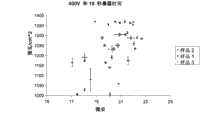
图2是在400V以及5秒暴露时间下的UV光强度对电流图。Figure 2 is a graph of UV light intensity versus current at 400V and 5 second exposure time.
图3是在400V以及10秒暴露时间下的UV光强度对电流图。Figure 3 is a graph of UV light intensity versus current at 400V and 10 seconds exposure time.
图4是在400V以及15秒暴露时间下的UV光强度对电流图。Figure 4 is a graph of UV light intensity versus current at 400V and 15 second exposure time.
图5是在700V以及5秒暴露时间下的UV光强度对电流图。Figure 5 is a graph of UV light intensity versus current at 700V and a 5 second exposure time.
图6是在1000V以及5秒暴露时间下的UV光强度对电流图。Figure 6 is a graph of UV light intensity versus current at 1000V and 5 second exposure time.
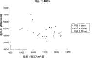
图7是对于样品1在400V下电阻率对强度的图——(趋势详述了电阻率随暴露时间而增加)。Figure 7 is a graph of resistivity versus intensity at 400 V for Sample 1 - (Trend details resistivity increase with exposure time).
图8是对于样品1在700V下电阻率对强度的图——(趋势详述了电阻率随暴露时间而增加)。Figure 8 is a graph of resistivity versus intensity at 700 V for Sample 1 - (Trend details resistivity increase with exposure time).
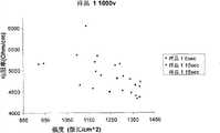
图9是对于样品1在1000V下电阻率对强度的图——(趋势详述了电阻率随暴露时间而降低)。Figure 9 is a graph of resistivity versus intensity at 1000 V for Sample 1 - (the trend details the decrease in resistivity with exposure time).
图10是照片,图解了视觉观察电纺丝动力学的一种技术。Figure 10 is a photograph illustrating one technique for visual observation of electrospinning kinetics.
图11是高速照片,显示了在聚合物喷射内的颜色变化,其可以被用于确定它的厚度。Figure 11 is a high speed photograph showing the color change within a polymer jet which can be used to determine its thickness.
图12是照片,详解了纳米纤维对过滤介质捕获液滴的影响的微观证据。Figure 12 is a photograph detailing microscopic evidence of the effect of nanofibers on the capture of droplets by filter media.
图13是聚合物纳米纤维连续片材的照片,其通过旋转的固体转筒装置收集。Figure 13 is a photograph of a continuous sheet of polymeric nanofibers collected by a rotating solid drum apparatus.
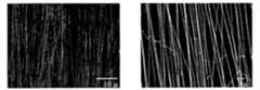
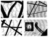
图14详解了在分段的转筒装置上进行5分钟电纺之后,部分排齐的尼龙-6纳米纤维的SEM图像。【0029a】图15是混合了5微米玻璃纤维的聚酰胺纳米纤维的SEM图像(左)和由玻璃纤维和聚合物纳米纤维制成的圆盘过滤器(右)。该过滤器通过用丙烯酸粘合剂真空模塑纤维的含水浆液而形成。Figure 14 details a SEM image of partially aligned nylon-6 nanofibers after 5 minutes of electrospinning on a segmented drum setup. [0029a] Figure 15 is an SEM image of polyamide nanofibers mixed with 5 micron glass fibers (left) and a disc filter made of glass fibers and polymer nanofibers (right). The filter is formed by vacuum molding an aqueous slurry of fibers with an acrylic binder.
图16是在热解后,自-支撑且连续的二氧化钛纳米纤维的SEM图像。Figure 16 is a SEM image of self-supporting and continuous titania nanofibers after pyrolysis.
图17是在773K(下图案)和1173K(上图案)热解后,二氧化钛纳米纤维的X射线衍射图案的图。Figure 17 is a graph of the X-ray diffraction pattern of titania nanofibers after pyrolysis at 773K (lower pattern) and 1173K (upper pattern).
图18显示了用氧化铒颗粒掺杂的二氧化钛纳米纤维的SEM图像。Figure 18 shows a SEM image of titania nanofibers doped with erbium oxide particles.
图19是热解的二氧化钛纳米纤维和并入氧化铒颗粒的热解的二氧化钛纳米纤维的XPS测量扫描图。Figure 19 is an XPS measurement scan of pyrolyzed titania nanofibers and pyrolyzed titania nanofibers infused with erbium oxide particles.
图20是溶液浓度和间隙距离对电纺纳米纤维的直径的影响图。Figure 20 is a graph of the effect of solution concentration and gap distance on the diameter of electrospun nanofibers.
图21是照片,详解了用不同氮化铝膜涂布的纳米纤维,聚合物的热解显示该涂层完全覆盖了纳米纤维。Figure 21 is a photograph detailing nanofibers coated with different aluminum nitride films, pyrolysis of the polymers shows that the coating completely covers the nanofibers.
图22是初纺乙酸铝/PVP纳米纤维的SEM图像。Figure 22 is a SEM image of as-spun aluminum acetate/PVP nanofibers.
图23详解了乙酸铝(A)、聚乙烯吡咯烷酮(PVP)(B)、电纺纤维(C)、加热到1200℃的氧化铝纳米纤维(D)的51MHz 13CCP/MASNMR图谱,所有的样品都在5kHZ下纺丝。Figure 23 details the 51MHz13 CCP/MASNMR spectra of aluminum acetate (A), polyvinylpyrrolidone (PVP) (B), electrospun fibers (C), alumina nanofibers heated to 1200°C (D), all samples Both were spun at 5kHZ.
图24是初纺PVP/乙酸铝纳米纤维的热重分析(TGA)数据图。Figure 24 is a graph of thermogravimetric analysis (TGA) data of as-spun PVP/aluminum acetate nanofibers.
图25详解了乙酸铝(A)、电纺纤维(B)、加热到1200℃的氧化铝纳米纤维(C)、α-氧化铝粉末(D)的195MHz 27AI MAS NMR图谱,所有的样品都在13kHZ下被纺丝。Figure 25 details the 195 MHz27 AI MAS NMR spectra of aluminum acetate (A), electrospun fibers (B), alumina nanofibers heated to 1200 °C (C), α-alumina powder (D), all samples Spinned at 13kHZ.
图26详解了在1200℃退火之后α-氧化铝纳米纤维的XRD;α-相氧化铝(α)衍射峰占优势,但是少量百分数的硼酸盐(B)被检测到;用星号标注的特征是仪器假象。Figure 26 details the XRD of α-alumina nanofibers after annealing at 1200°C; the α-phase alumina (α) diffraction peaks dominate, but a small percentage of borate (B) is detected; marked with asterisks Characterized by instrument artifacts.
图27是SIMS表面灵敏技术图,详解了含有Na的纳米纤维和杂质的影响。Figure 27 is a SIMS surface sensitive technique diagram detailing the effect of Na-containing nanofibers and impurities.
图28是初纺纳米纤维和在1200℃退火之后α-氧化铝纳米纤维的XPS谱图;注意,在退火之后在表面的钠和硼的水平比从起始混合物的组成预期的高得多;证明在结晶过程期间发生了表面离析。Figure 28 is the XPS spectrum of as-spun nanofibers and alpha-alumina nanofibers after annealing at 1200°C; note that the levels of sodium and boron at the surface after annealing are much higher than expected from the composition of the starting mixture; It was demonstrated that surface segregation occurred during the crystallization process.
图29详解了初合成的纳米纤维、加热到1200℃的纳米纤维和Al2O3粉末(商业的)的DRIFTS图谱,显示硼酸盐和羟基模式存在于加热的纳米纤维中,但是不在粉末中;该图谱的羟基区域显示,纳米纤维可以在它们的表面存在不同于在粉末上发现的酸和碱位,以及因此可以导致增强的或独特的表面化学性质。Figure 29 details the DRIFTS spectra of as-synthesized nanofibers, nanofibers heated to 1200°C, andAl2O3 powder (commercial), showing that borate andhydroxyl modes are present in the heated nanofibers, but not in the powder ; the hydroxyl region of the map shows that nanofibers can have acid and base sites on their surface that differ from those found on powders, and thus can lead to enhanced or unique surface chemistry.
图30是电纺PVP/乙酸铝(Al(ac))纤维的SEM图像。Figure 30 is a SEM image of electrospun PVP/aluminum acetate (Al(ac)) fibers.
图31是在多种温度下退火的PVP/乙酸铝(AlAc)电纺纤维的SEM图像。Figure 31 is an SEM image of PVP/aluminum acetate (AlAc) electrospun fibers annealed at various temperatures.
图32是在1200℃退火的氧化铝纤维的X-射线光电子图谱。Figure 32 is an X-ray photoelectron spectrum of an alumina fiber annealed at 1200°C.
图33是在湿烘焙条件下,在多种温度下退火的氧化铝纤维的27Al直接极化NMR图谱。Figure 33 is a27 Al direct polarization NMR spectrum of alumina fibers annealed at various temperatures under wet bake conditions.
图34是在干烘焙条件下,在多种温度下退火的氧化铝纤维的27Al直接极化NMR图谱。Figure 34 is a27 Al direct polarization NMR spectrum of alumina fibers annealed at various temperatures under dry bake conditions.
图35是在干烘焙条件下,在多种温度下退火后,氧化铝纤维的粉末X-射线衍射数据图。Figure 35 is a graph of powder X-ray diffraction data of alumina fibers after annealing at various temperatures under dry baking conditions.
图36是在干烘焙条件下退火的氧化铝纤维的1H→27Al交叉极化NMR图谱。Fig. 36 is a1 H→27 Al cross-polarization NMR spectrum of alumina fibers annealed under dry baking conditions.
图37是在湿烘焙条件下退火的氧化铝纤维的DRIFTS图谱。Figure 37 is a DRIFTS pattern of alumina fibers annealed under wet bake conditions.
图38是用于加热氧化铝纤维垫的构造的示意图。Figure 38 is a schematic illustration of a configuration for heating an alumina fiber mat.
图39是乙酸铝(底部)和在525℃退火30分钟的乙酸铝(顶部)的27Al SSNMR谱图。Figure 39 is a27 Al SSNMR spectrum of aluminum acetate (bottom) and aluminum acetate annealed at 525°C for 30 minutes (top).
图40是锂离子电池的设计图,其中隔膜/电解质从氧化铝/LiX纳米纤维制备;和Figure 40 is a design drawing of a Li-ion battery in which the separator/electrolyte is fabricated from alumina/LiX nanofibers; and
图41是在400℃退火之后,PVP/乙酸铝/Lil的电纺纳米纤维的SEM显微图。Figure 41 is a SEM micrograph of electrospun nanofibers of PVP/aluminum acetate/Lil after annealing at 400°C.
具体实施方式Detailed ways
本发明一般涉及金属氧化物纤维和纳米纤维,其制备方法和其用途。这样的金属氧化物纳米纤维具有吸收和分解化学战剂和其他有毒化学物质的能力。这些纳米纤维可以被并入到防护衣和用于呼吸的装置中,或者在另外的实例中,可以被用于锂离子电池中。在一个实施方式中,本发明涉及二氧化钛、氧化铝和/或氧化镁纤维和纳米纤维,以及涉及它们的制备方法。在另一情况中,α-相氧化铝被用作纳米纤维中的一种材料。The present invention generally relates to metal oxide fibers and nanofibers, processes for their preparation and uses thereof. Such metal oxide nanofibers have the ability to absorb and decompose chemical warfare agents and other toxic chemicals. These nanofibers can be incorporated into protective clothing and devices for breathing, or in another example, can be used in lithium-ion batteries. In one embodiment, the present invention relates to titania, alumina and/or magnesia fibers and nanofibers, and to methods of their preparation. In another case, alpha-phase alumina was used as a material in the nanofibers.
对于本发明的目的,热处理包括在规定的气氛中,将初纺纳米纤维暴露到升高的温度下。在最简单的热处理应用中,纳米纤维被加热,以去除残留的溶剂和/或水。在更普遍的情况下,样品被加热,以热解有机材料(聚合物、和金属氧化物前体的任何有机部分)、煅烧金属-氧化物和烧结颗粒,以产生基于金属-氧化物或掺杂金属氧化物的纳米纤维。温度范围可以从室温/21℃到1200℃。样品加热时间可以在几分钟和几小时之间变化以及样品加热温度可以是恒定的、以线性方式变化或在复杂加热曲线下变化。缓慢加热(几小时内将温度从21℃缓升到700℃)允许聚合物脱离纤维结构并使金属氧化物替代它,以形成金属氧化物纤维,同时保留大部分纤维结构。进一步加热到更高的温度对于使金属氧化物改变晶态(晶相)是有用的。纳米纤维的热处理可以导致存在于纳米纤维中的有机材料部分或全部热解、金属氧化物部分或全部煅烧和颗粒烧结。热处理发生的气氛可以是环境气氛、富含或缺乏多种气体(氧气、氮气、二氧化碳等)的气氛,和/或具有在0和100%之间的相对湿度。For the purposes of the present invention, thermal treatment includes exposing as-spun nanofibers to elevated temperatures in a defined atmosphere. In the simplest heat treatment application, nanofibers are heated to remove residual solvent and/or water. In the more general case, the sample is heated to pyrolyze the organic material (polymer, and any organic part of the metal oxide precursor), calcinate the metal-oxide, and sinter the particles to produce metal-oxide-based or doped Nanofibers of heterometallic oxides. The temperature range can be from room temperature/21°C to 1200°C. Sample heating times can vary between minutes and hours and sample heating temperatures can be constant, varied in a linear fashion, or varied under complex heating curves. Slow heating (ramp from 21°C to 700°C over several hours) allows the polymer to break free from the fiber structure and allow the metal oxide to replace it, forming metal oxide fibers while retaining most of the fiber structure. Further heating to higher temperatures is useful for changing the crystalline state (crystal phase) of the metal oxide. Thermal treatment of the nanofibers can result in partial or total pyrolysis of organic materials present in the nanofibers, partial or total calcination of metal oxides and sintering of the particles. The atmosphere in which the heat treatment takes place may be ambient, an atmosphere enriched or deficient in various gases (oxygen, nitrogen, carbon dioxide, etc.), and/or have a relative humidity between 0 and 100%.
关于金属氧化物纳米纤维,功能化指通过改变组成、改变结构或通过改变纤维的形态或纤维结构而改变材料性质。功能化可以通过前面定义的“热处理”过程而取得。这个过程控制了在纳米纤维中的金属原子位的氧化态和配位数以及表面羟基存在的数目。某些金属氧化态和配位数是更具活性的,而增强这些位的数目增加了纳米纤维的反应性。表面羟基在许多反应中也是重要的,包括神经性毒剂的去毒,并且增加它们的数目增加了纳米纤维在神经性毒剂分解中的效率。功能化还通过加入化学反应的催化剂而实现。催化剂性质可以通过加入纳米或微米金属颗粒例如Pd或Pt、通过将表面涂层施加到纤维上或者通过改变金属氧化物纤维的晶体结构,而发展到纤维中。通过使在纤维和/或纤维结构中产生光子灵敏性的不同金属氧化物材料的纳米纤维接触,功能性可以被发展。例如PN结可以通过接触NiO和SnO纤维而形成。功能性还可以通过用其他化合物掺杂纤维而在纤维中形成,以便改变它们的电或热传导性。可替代地,功能性可以以构造纤维垫的方式发展到纤维结构中。例如,彼此平行取向的纤维将偏振光。最后,功能性可以通过涂布纤维的表面被并入到纤维结构中,以改变它们的性质。例如,硅烷可以被涂布到金属氧化物纤维的表面,以改变它们的湿润性。With respect to metal oxide nanofibers, functionalization refers to changing the material properties by changing the composition, changing the structure, or by changing the morphology or structure of the fibers. Functionalization can be achieved by the "heat treatment" process defined above. This process controls the oxidation state and coordination number of the metal atomic sites in the nanofibers and the number of surface hydroxyl groups present. Certain metal oxidation states and coordination numbers are more reactive, and enhancing the number of these sites increases the reactivity of the nanofibers. Surface hydroxyl groups are also important in many reactions, including the detoxification of nerve agents, and increasing their number increases the efficiency of the nanofibers in the breakdown of nerve agents. Functionalization is also achieved by adding catalysts for chemical reactions. Catalytic properties can be developed into the fibers by adding nano or micro metal particles such as Pd or Pt, by applying surface coatings to the fibers or by changing the crystal structure of the metal oxide fibers. Functionality can be developed by contacting nanofibers of different metal oxide materials that produce photon sensitivity in the fiber and/or fiber structure. For example, a PN junction can be formed by contacting NiO and SnO fibers. Functionality can also be created in fibers by doping them with other compounds in order to modify their electrical or thermal conductivity. Alternatively, functionality can be developed into the fibrous structure in such a way as to construct the fibrous mat. For example, fibers oriented parallel to each other will polarize light. Finally, functionality can be incorporated into the fiber structure by coating the surface of the fibers to modify their properties. For example, silanes can be coated onto the surface of metal oxide fibers to alter their wettability.
钛纳米纤维暴露于紫外光Titanium nanofibers exposed to UV light
二氧化钛纳米纤维已经被发现在紫外光中是活性的。UV辐射入射之后,二氧化钛纳米纤维使基本是电阻的转化成宽带隙半导体。当被暴露到变化强度的UV光下时,二氧化钛纳米纤维的电阻率/电导性变化。二氧化钛电阻率随UV强度、暴露时间和电压的变化在进行改变这些参数的试验中被详细地研究。假设是,当光强度和暴露时间增加时,电阻率将降低。为了完成这个试验,在三种电压和三个暴露时间下,测试3个锐钛矿样品,以及在每个点数据被取三次。Titanium dioxide nanofibers have been found to be active in ultraviolet light. Upon incident UV radiation, titanium dioxide nanofibers convert the substantially resistive into a wide bandgap semiconductor. The resistivity/conductivity of the titania nanofibers changed when exposed to varying intensities of UV light. The variation of titanium dioxide resistivity with UV intensity, exposure time and voltage was studied in detail in experiments performed varying these parameters. The hypothesis was that the resistivity would decrease as the light intensity and exposure time increased. To complete this experiment, three anatase samples were tested at three voltages and three exposure times, and data was taken three times at each point.
钛纳米纤维/紫外光试验的结果Results of Titanium Nanofiber/UV Light Test
对于所有的电压(400、700、1000),电阻率随光强度降低。在固定的电压和暴露时间,电流随UV强度增加而增加。对于400V(伏特)样品,电阻率随暴露时间延长而增加,但是对于700和1000V样品,电阻率随暴露时间降低。对于在1000μW/cm2数量级的UV强度,可以预期看见在5000和20000Ohm/cm之间的电阻率。For all voltages (400, 700, 1000), resistivity decreases with light intensity. At a fixed voltage and exposure time, the current increases with increasing UV intensity. For the 400V (volts) sample, the resistivity increases with exposure time, but for the 700 and 1000V samples, the resistivity decreases with exposure time. For UV intensities on the order of 1000 μW/cm2 one would expect to see resistivities between 5000 and 20000 Ohm/cm.
在400V,锐钛矿在5和10秒UV暴露之间达到最小的电阻率。在700V,最小的电阻率位于15秒的暴露阈值,而在1000V,样品在大于15秒的暴露时间处具有最小电阻率。电阻率随紫外光强度增加而降低,在固定的电压和暴露时间,电流随紫外线强度增加而增加。At 400V, anatase reached a minimum resistivity between 5 and 10 seconds of UV exposure. At 700V, the minimum resistivity was located at an exposure threshold of 15 seconds, while at 1000V, the samples had minimum resistivities at exposure times greater than 15 seconds. Resistivity decreases with increasing UV intensity, and current increases with increasing UV intensity at a fixed voltage and exposure time.
涉及二氧化钛纳米纤维的前期工作已显示,通过从电阻材料转化成半导体材料,二氧化钛是光催化的且对UV光响应。涉及电压、暴露时间和光强度变化的具体工作在本领域还没有完成。对电阻率相对于这些数值的变化幅度的掌握增加了知识库,以便将二氧化钛成功用于长远的工业、医药和化学可能情况。本讨论通过试验-经验图形技术来解决这些问题,以证明这些树脂的变化如何影响二氧化钛对UV辐射的响应。Previous work involving titania nanofibers has shown that titania is photocatalytic and responsive to UV light by converting from a resistive material to a semiconducting material. Specific work involving changes in voltage, exposure time, and light intensity has not been done in the field. Knowledge of how much resistivity varies relative to these values adds to the knowledge base for the successful use of titanium dioxide for long-term industrial, medical and chemical possibilities. This discussion addresses these issues through an experimental-empirical graphical technique to demonstrate how changes in these resins affect the response of titania to UV radiation.
光催化作用被定义为涉及由催化剂或由基质(substrate)进行光吸收的催化反应。UV辐射入射之后,通过将电阻材料转化为半导体材料,二氧化钛纳米纤维展示了这个行为。这个现象在几种不同领域中具有重要性。Photocatalysis is defined as a catalytic reaction involving light absorption by a catalyst or by a substrate. Titania nanofibers exhibit this behavior by converting the resistive material into a semiconducting material after UV radiation incidence. This phenomenon is of importance in several different fields.
也许,二氧化钛纳米纤维的应用中最重要的领域是废水工业领域。二氧化钛纳米纤维可以被用于处理废水,因为当具有能量hv的光子撞击二氧化钛半导体材料时,电子从材料的价带被激发到材料的导带,因此在价带中产生电子空穴。这个电子空穴(hvb)是强氧化剂,其随后分解有机污染物和对人体有害的微生物。这些材料被分解,主要是由于羟基的形成,其以下面的化学方程式的顺序形成:(1)TiO2+hv→ecb+hvb(2)hvb+Ti--OH→Ti+OH基Perhaps the most important field of application of titania nanofibers is the wastewater industry. Titanium dioxide nanofibers can be used to treat wastewater because when a photon with energy hv strikes a titanium dioxide semiconductor material, electrons are excited from the valence band of the material to the conduction band of the material, thus generating electron holes in the valence band. This electron hole (hvb ) is a strong oxidizing agent, which then decomposes organic pollutants and microorganisms harmful to humans. These materials are decomposed mainly due to the formation of hydroxyl groups, which are formed in the order of the following chemical equation: (1) TiO2 +hv→ecb +hvb (2)hvb +Ti--OH→Ti+OHgroups
应用的另一个领域涉及PVA(聚乙烯醇)的降解,其是广泛使用的聚合材料。PVA在几种电容中被使用,但是它的降解是非常缓慢的,因此PVA分解的更快速更有效的方法是理想的。最近的研究表明在二氧化钛载体上的铂金属可以快速地降解PVA固体废物。对二氧化钛响应于UV辐射的更全面理解加速了这样方法整合到固体废物材料的处理中。Another field of application concerns the degradation of PVA (polyvinyl alcohol), which is a widely used polymeric material. PVA is used in several capacitors, but its degradation is very slow, so a faster and more efficient method of PVA decomposition is ideal. Recent studies have shown that platinum metal on titania supports can rapidly degrade PVA solid waste. A more complete understanding of titania's response to UV radiation hastened the integration of such methods into the treatment of solid waste materials.
二氧化钛纳米纤维的另一种应用包括对牙齿移植物表面的杀菌。二氧化钛在这里被用作粗糙的牙齿移植物,提供更好的表面,以安装到口腔中,但当暴露到嘴的内部时,更粗糙的表面伴随着对细菌感染更高的亲合性。使用二氧化钛大大地降低了——如果没有完全消除的话——在牙齿移植中口腔感染的危险。Another application of titanium dioxide nanofibers includes the sterilization of dental implant surfaces. Titanium dioxide is used here as a rough tooth implant, providing a better surface to fit into the mouth, but when exposed to the interior of the mouth, the rougher surface comes with a higher affinity for bacterial infection. The use of titanium dioxide greatly reduces - if not eliminates - the risk of oral infection in dental implants.
与TiO2不同应用相伴的,还有几种制造二氧化钛纳米纤维的方法。这些方法包括电纺、真空电弧等离子蒸发和溶胶-凝胶法。电纺技术包括TiO2微粒在聚合物水溶液中分散以及随后在高电压环境中将该纤维纺在旋转的轮式装置上。在这个技术中自动推进注射器也被用于纳米纤维的制造。真空电弧等离子蒸发使用充满氧气的真空室以及从LaB6阴极聚集到阳极磁棒上的等离子束。二氧化钛沉积在与样品源相对的室末端的玻璃基底固定器上。溶胶-凝胶方法包括以特定摩尔比制备凝胶,随后通过手用玻璃棒挤出纤维。尽管繁琐和缓慢,但是这个方法允许非常少的杂质。Along with the different applications ofTiO2 , there are also several methods for fabricating TiO2 nanofibers. These methods include electrospinning, vacuum arc plasma evaporation, and sol-gel methods. The electrospinning technique involves dispersion ofTiO2 microparticles in an aqueous polymer solution and subsequent spinning of the fibers on a rotating wheel device in a high voltage environment. Self-propelled injectors are also used in the fabrication of nanofibers in this technique. Vacuum arc plasma evaporation uses a vacuum chamber filled with oxygen and a plasma beam focused from theLaB6 cathode onto the anode magnet bar. Titanium dioxide was deposited on a glass substrate holder at the end of the chamber opposite the sample source. The sol-gel method involves preparing a gel at a specific molar ratio followed by extrusion of fibers by hand with a glass rod. Although tedious and slow, this method allows very few impurities.
钛纳米纤维/紫外光的试验方法Test method for titanium nanofiber/ultraviolet light
存在多种类型的二氧化钛纳米纤维,以及基于它的上等质量,锐钛矿被选为试验样品。使用的锐钛矿样品通过电纺技术被加工。适合的电纺技术在多个专利中被公开,包括但不限于美国专利第6,753,454号,在此通过引用并入它的全部,因为其教导与电纺相关。There are many types of titania nanofibers, and based on its superior quality, anatase was chosen as the test sample. The anatase samples used were processed by electrospinning technique. Suitable electrospinning techniques are disclosed in various patents, including but not limited to US Patent No. 6,753,454, which is hereby incorporated by reference in its entirety for its teachings pertaining to electrospinning.
这个试验使用三件关键设备:UV光仪表、Keithley电压源/电流测量设备以及UV光源。使用的光源是BLAK RAY长波UV灯B 100AP,115V 60Hz和2.5Amp,1991年5月生产。使用的光在这个试验中是重要的因素,因为它影响试验的路径。该UV光仪表测量光强度,以μW/cm2为单位,具有0-2000和0-20000两种可行的范围。电流测量设备测量电流为μAmps,对于这个试验这是适当的范围。This test uses three key pieces of equipment: a UV light meter, a Keithley voltage source/current measurement device, and a UV light source. The light source used is BLAK RAY long-wave UV lamp B 100AP, 115V 60Hz and 2.5Amp, produced in May 1991. The light used is an important factor in this test as it affects the path of the test. This UV light meter measures light intensity in μW/cm2 with two possible ranges of 0-2000 and 0-20000. The current measuring device measures the current in μAmps, which is the appropriate range for this test.
试验设计的最初意图包括就距离校准紫外光强度,随后改变二氧化钛样品与光源的距离,以有效地改变光强度。在几次试验之后,这种校准被发现无效。光强度随时间变化,以及校准没有精确地表示数据。因此,需要对试验过程进行再次评估。The original intent of the experimental design involved calibrating the UV light intensity for distance and then varying the distance of the titania sample from the light source to effectively vary the light intensity. After several trials, this calibration was found to be ineffective. Light intensity varies with time, and the calibration does not accurately represent the data. Therefore, a reassessment of the testing process is required.
建立“表样(table-like)”框架来支持该试验。在这种支持下,37cm金属导轨装备有载重小车,自由地沿着导轨的距离移动。锐钛矿样品被放置在玻璃载片上,玻璃载片随后通过镀金金属触头被固定到Teflon底部上。该样品装置与UV光仪表一起被放置在载重小车上的纤维玻璃基底。在图1中显示的该装置利用了已知的事实:UV-A辐射随在到仪表与样品的线和UV源的中心线之间的角度余弦改变。在距离UV光源中心线相等的垂直距离,放置样品和仪表,以便同时测量光强度和到样品上的分散入射辐射。A "table-like" framework was established to support the experiment. With this support, a 37cm metal rail is equipped with a trolley, which moves freely along the distance of the rail. Anatase samples were placed on glass slides, which were then affixed to Teflon bottoms with gold-plated metal contacts. The sample device was placed on a fiberglass substrate on a trolley along with a UV light meter. The setup shown in Figure 1 takes advantage of the known fact that UV-A radiation varies with the cosine of the angle between the line to the meter and the sample and the centerline of the UV source. At an equal vertical distance from the centerline of the UV light source, place the sample and meter so that both the light intensity and the scattered incident radiation onto the sample are measured.
三个样品在三个电压设置(400V、700V和1000V)和三个暴露时间(5、10和15秒)下被测量。每个样品点获取数据三次。暴露时间通过将不透明UV隔离物直接放在光源的前面和随后将该样品暴露在UV源下预定的时间段而产生。光强度通过沿37cm轨道上的12个等距离间隔获取数据而改变。该技术允许测量暴露时间、电压和光强度对二氧化钛样品的影响。Three samples were measured at three voltage settings (400V, 700V and 1000V) and three exposure times (5, 10 and 15 seconds). Data were acquired three times for each sample point. Exposure times were generated by placing an opaque UV spacer directly in front of the light source and subsequently exposing the sample to the UV source for a predetermined period of time. Light intensity was varied by acquiring data at 12 equidistant intervals along a 37 cm track. This technique allows the measurement of the effects of exposure time, voltage and light intensity on titania samples.
钛纳米纤维/紫外光试验的数据和结果Data and Results of Titanium Nanofiber/UV Experiments
用于这个试验的三个样品的大小、重量和厚度在下面表1中列出。厚度从锐钛矿的密度(4.26g/cc)确定。表1
如在该试验方法中暗示的,该试验存在许多“原始”数据,因此,对于该数据,图形输出代表最好的形式。图2到6以图表示每个图标题标明的电压以及暴露时间下光强度和电流的平均值。误差小线段代表三次重复数据点的标准偏差。As implied in the test method, there is a lot of "raw" data for this test, so the graphical output represents the best form for this data. Figures 2 to 6 graphically represent the mean values of light intensity and current at the voltage indicated in the title of each figure and the exposure time. Small error bars represent the standard deviation of triplicate data points.
为了确定电阻和电阻率,下面的关系被使用:ρ=R*A/L (3)e=iR (4)其中R是电阻,欧姆,ρ是电阻率,欧姆/厘米,i是电流,安培,e是电压,A是样品的截面积以及L是样品的长度。To determine resistance and resistivity, the following relationship is used: ρ=R*A/L (3)e=iR (4) where R is resistance in ohms, ρ is resistivity in ohms/cm, and i is current in amperes , e is the voltage, A is the cross-sectional area of the sample and L is the length of the sample.
以这种方法,在试验条件下的锐钛矿的电阻率的范围被计算且在表2中示出。这些范围用于在900和1450μW/cm2之间的光强度。In this way, the range of resistivity of anatase under test conditions was calculated and shown in Table 2. These ranges are for light intensities between 900 and 1450 μW/cm2 .
钛纳米纤维/紫外光试验的讨论/分析Discussion/Analysis of Titanium Nanofiber/UV Test
在该数据中测定了几个点。在表2中,可以看到电阻率的范围从样品到样品显著变化,从样品1和2的5000欧姆/厘米到样品3的25000欧姆/厘米。样品1和2与样品3之间的不同是对于1和2来说在电极之间的距离是1.7厘米而对于样品3来说电极之间距离是1厘米。Several points were determined in this data. In Table 2, it can be seen that the range of resistivity varies significantly from sample to sample, from 5000 ohm/cm for
在每个试验中电阻率随UV强度的增加而降低。如预期的,通过查看附图7-9和向图右下斜的图示,可以领会这种行为。支持该现象的解释陈述为,当更多的UV光撞击锐钛矿样品时,更多的电子从价带释放到导带。该信息的使用使得污水处理设备处理更多的生物污染物,因为额外的电子空穴可以氧化这些污染物。
下一个观察到的关系将电阻率和暴露时间相关起来。对于400V样品,每个样品展现与最初期望的相反的活性。一种理论是当暴露时间增加时,电流将增加并且因此电阻率将降低。对于400V样品来说相反现象发生,如该趋势在图7中显示。对于700V样品1,也可以看见这种“相反的行为”。但是在样品2和3,电阻率随暴露时间更长而降低,对于1000v样品,该趋势非常强烈。这引导试验人员相信,对于400V样品来说,最大电流(或最小电阻率)在5和10秒UV暴露时间之间可以得到。该最大的阈值可以在10到15秒之内,或对于700V样品为稍微长一些的范围,对于1000V甚至更长。The next observed relationship relates resistivity to exposure time. For the 400V samples, each sample exhibited the opposite activity than originally expected. One theory is that as the exposure time increases, the current will increase and thus the resistivity will decrease. The opposite phenomenon occurs for the 400V sample, as the trend is shown in FIG. 7 . This "opposite behavior" can also be seen for
还能得出结论:对于在900和1450μW/cm2之间的紫外光强度,预期电阻率在5000-20000欧姆/厘米的数量级。It can also be concluded that for UV light intensities between 900 and 1450 μW/cm2 , resistivities of the order of 5000-20000 ohms/cm are expected.
在这个试验研究中收集的数据对锐钛矿光电导性确定了主要因素之间的一般关系,以及提供给试验人员这里所示变量的工作范围。需要附加的工作来提供足够精确的结果,以确定理论模型或数学上定义的“趋势”,用于将来应用。这可以通过测量强度的方法需要改进这一事实进行解释。如前面所描述,“角度的余弦”方法对于最初的研究是满意的,但是,更精确的测量方法是必需的,以能够精确地预测在紫外线辐射下锐钛矿的行为。The data collected in this experimental study determined the general relationship between the main factors for anatase photoconductivity, and provided experimenters with working ranges for the variables shown here. Additional work is required to provide results precise enough to identify theoretical models or mathematically defined "trends" for future applications. This can be explained by the fact that the method of measuring intensity needs improvement. As previously described, the "cosine of angles" method was satisfactory for initial studies, however, more precise measurements are required to be able to accurately predict the behavior of anatase under UV radiation.
防护以免于基于磷和硫的神经性毒剂Protection against phosphorus and sulfur based nerve agents
金属氧化物纳米纤维作为基质,在其上化学战剂和其他有毒化学物质可以被吸附或吸收和分解。这些纳米纤维的大的单位质量表面积产生高表面反应性,同时纳米纤维的长度使它们适合于并入到防护衣和用于呼吸的设备中。已知金属氧化物纳米颗粒分解神经性毒剂,但是不能容易地并入到衣服和呼吸过滤器中。纳米纤维可以被并入到衣服中以及可以被用于呼吸过滤器中,同时仍然展示神经性毒剂去毒所需要的纳米级表面反应性。纳米纤维材料的性质,例如强度、孔隙率、吸附化学种类的能力等等,可以通过将不同的纤维结合到复合材料中进行控制。例如,跨过神经性毒剂过滤介质的压降可以通过将氧化物纳米纤维与更大的纤维结合而被最小化。该复合材料对于保护性呼吸装置和衣服是理想的,以允许湿气交换以及减轻在人员上的热应力。Metal oxide nanofibers serve as substrates on which chemical warfare agents and other toxic chemicals can be adsorbed or absorbed and decomposed. The large surface area per unit mass of these nanofibers yields high surface reactivity, while the length of the nanofibers makes them suitable for incorporation into protective clothing and devices for breathing. Metal oxide nanoparticles are known to break down nerve agents, but cannot be easily incorporated into clothing and respiratory filters. Nanofibers can be incorporated into clothing and can be used in respiratory filters, while still exhibiting the nanoscale surface reactivity required for the detoxification of nerve agents. The properties of nanofibrous materials, such as strength, porosity, ability to adsorb chemical species, etc., can be controlled by incorporating different fibers into composite materials. For example, the pressure drop across a nerve agent filter media can be minimized by combining oxide nanofibers with larger fibers. The composite is ideal for protective breathing apparatus and clothing to allow moisture exchange and reduce thermal stress on personnel.
在金属氧化物纳米纤维的产生和使用中,三种方法是重要的:(1)直接电纺含有聚合物和金属氧化物前体的溶液,(2)涂布聚合物纳米纤维,和(3)将金属氧化物颗粒和纳米纤维并入吸收性聚合物纳米纤维内。前两个方法包括热解步骤,以形成金属氧化物纳米纤维,而第三个方法在最后的产物中存留聚合种类。该材料系统包括二氧化钛、氧化铝和氧化镁-系统,当以纳米颗粒形式时,已知对神经性毒剂的分解展示高催化表面行为。电纺对于产生纳米纤维的非纺织垫和片材来说,是简单和可扩大规模的方法。聚合物、溶剂、金属氧化物前体的混合物和电纺条件控制得到的纳米纤维性质。这些材料可以克服对基于磷和/或硫的神经性毒剂的个人防护的科技问题。In the production and use of metal oxide nanofibers, three approaches are important: (1) direct electrospinning of solutions containing polymer and metal oxide precursors, (2) coating of polymer nanofibers, and (3) ) incorporation of metal oxide particles and nanofibers into absorbent polymer nanofibers. The first two methods include a pyrolysis step to form metal oxide nanofibers, while the third method leaves the polymeric species in the final product. The material system comprises titania, alumina and magnesia-systems which, when in nanoparticle form, are known to exhibit highly catalytic surface behavior for the decomposition of neurotoxic agents. Electrospinning is a simple and scalable method for producing nanofiber nonwoven mats and sheets. The mixture of polymers, solvents, metal oxide precursors, and electrospinning conditions control the properties of the resulting nanofibers. These materials can overcome the technical problems of personal protection against phosphorus and/or sulfur based nerve agents.
由金属氧化物纳米纤维组成的实验室规模片材通过使用转筒式装置产生,以收集以长连续片材形式存在的非纺织电纺纳米纤维。在这些片材中的纤维是无规取向的,产生非纺织似织物材料,其是柔软的且有一些弹性。在一个实施方式中,这是宏观上大片材料,全部由纳米级的小(直径)聚合物纳米纤维组成。这种方法通过使用由等距离的导线组组成的分段筒被扩展,导线被接地,以产生对于喷丝头顶端的大电位差。产生的材料经过较短的时间,获得片材内纳米纤维的部分定向排列。对于更长的时间,以无规模式和排列开始纺丝的纤维逐渐地失去。这些排列的垫足够厚,以切割和处理。Laboratory-scale sheets composed of metal oxide nanofibers were produced by using a drum-type device to collect nonwoven electrospun nanofibers in the form of long continuous sheets. The fibers in these sheets are randomly oriented, resulting in a nonwoven fabric-like material that is soft and somewhat elastic. In one embodiment, this is a macroscopically large sheet of material, all composed of nanoscale small (diameter) polymer nanofibers. This approach is extended by using a segmented barrel consisting of equidistant sets of wires which are grounded to create a large potential difference to the spinneret tip. The resulting material obtains a partial orientation of the nanofibers within the sheet over a short period of time. For longer times, fibers spun initially in a random pattern and arrangement are gradually lost. These lined pads are thick enough to cut and handle.
由排列的纳米纤维组成的结构(例如织物和过滤器)将比由无纺布材料形成的那些结构具有优越的性质(热、机械、传质等等)。采用排列的和无规区域的组合,可以使纳米纤维片材适合于具体的应用。在这种情况下,目标是能够使磷(膦)酸盐气溶胶捕获和分解同时允许水蒸汽交换的材料。在过滤介质的情况下,纳米纤维还可以需要额外的结构支撑。二氧化钛纳米纤维通过如下形成:用溶胶-凝胶前体涂布聚合物纳米纤维,随后热处理,然后取聚合纳米纤维片材并将它们转化成金属氧化物。下一步使用真空模塑方法,将长聚合物纳米纤维并入到微米级的玻璃纤维过滤介质中。该混合的纤维容易被真空模塑成多孔圆盘,其厚1厘米以及直径6厘米。对于结构强度来说,丙烯酸粘合剂被用于将纤维结合在一起。通过电纺和后来的涂布或热处理从材料产生纳米纤维的能力,与自有的专门技术(in-house expertise)相结合,以从得到的材料形成过滤介质,对于基于磷的神经性毒剂的分解提供了巨大的潜力。Structures (such as fabrics and filters) composed of aligned nanofibers will have superior properties (thermal, mechanical, mass transfer, etc.) over those formed from non-woven materials. Using a combination of aligned and random domains, nanofibrous sheets can be tailored to specific applications. In this case, the target is a material that enables the capture and decomposition of phosphonate aerosols while allowing water vapor exchange. In the case of filter media, nanofibers may also require additional structural support. Titanium dioxide nanofibers are formed by coating polymeric nanofibers with a sol-gel precursor, followed by heat treatment, and then taking sheets of polymeric nanofibers and converting them into metal oxides. The next step uses vacuum molding to infuse long polymer nanofibers into micron-sized glass fiber filter media. The mixed fibers were easily vacuum molded into porous discs, 1 cm thick and 6 cm in diameter. For structural strength, acrylic adhesives are used to hold the fibers together. The ability to generate nanofibers from the material by electrospinning and subsequent coating or heat treatment, combined with in-house expertise to form filter media from the resulting material, is critical for phosphorus-based nerve agents. Breakdown offers great potential.
产生的纳米纤维的长度和直径取决于产生纤维的条件。聚合物的浓度、金属氧化物前体浓度、摩尔比、聚合物分子量、施加的电势、在样品和收集盘之间的距离、溶液流动速率、湿度和温度的试验参数影响最后的产物。这些参数的每一个被研究且被最优化,以产生希望的纳米纤维特性。The length and diameter of the nanofibers produced depend on the conditions under which the fibers were produced. Experimental parameters of polymer concentration, metal oxide precursor concentration, molar ratio, polymer molecular weight, applied potential, distance between sample and collection pan, solution flow rate, humidity and temperature affect the final product. Each of these parameters was studied and optimized to produce the desired nanofiber properties.
纳米纤维的应用Applications of nanofibers
吸附或吸收和随后分解磷(膦)酸盐种类的衣服和呼吸过滤器现在是可能的。由含有纳米纤维或纳米颗粒的薄聚合物片材制备的轻质雨布可以折叠且方便携带和存放。这种保护的立即可用性对于在涉及民众的恐怖袭击事件中降低恐慌的传播提供了放心。将金属氧化物纳米纤维和纳米颗粒并入到聚合物纳米纤维结构中是重要的。聚合物纳米纤维将金属氧化物材料结合到一起,形成柔软的系统,其没有带来皮肤刺激或石棉沉着病的威胁。磷(膦)酸盐种类的溶解性使一些聚合物通过吸收溶胀,以高速率捕获有毒化合物,随后通过与聚合物密切接触的金属氧化物表面允许化学去毒。Clothing and breathing filters that adsorb or absorb and subsequently decompose phosphonate species are now possible. Lightweight raincloths prepared from thin polymer sheets containing nanofibers or nanoparticles can be folded and easily carried and stored. The immediate availability of this protection provides reassurance in reducing the spread of panic in the event of a terrorist attack involving the population. Incorporation of metal oxide nanofibers and nanoparticles into polymer nanofibrous structures is important. Polymer nanofibers bond metal oxide materials together to form a soft system that poses no threat of skin irritation or asbestosis. The solubility of phosphonate species allows some polymers to swell by absorption, trapping toxic compounds at high rates and subsequently allowing chemical detoxification by the metal oxide surface in intimate contact with the polymer.
电纺发展Electrospinning Development
电纺是电流体动力学过程,用于制备薄聚合物纤维,其直径在从大约1纳米到几千纳米的范围内。制备聚合物纳米纤维所需的设备的低投资费用和简单性使科学家和工程师在领域例如过滤、生物材料、生物医学设备、化学分析、催化、航空、纤维增强复合材料、能量转化、防护衣、农业和其他领域能够从许多种聚合物生产实验数量的纳米纤维。几乎任何材料都可以被并入到纳米纤维中。本发明建立在现有技术状态电纺过程的现有工作以及与吸附在金属氧化物上的磷化合物相关的材料表征的基础上。Electrospinning is an electrohydrodynamic process for the preparation of thin polymer fibers, with diameters ranging from about 1 nanometer to several thousand nanometers. The low capital cost and simplicity of the equipment required to make polymer nanofibers has enabled scientists and engineers in fields such as filtration, biomaterials, biomedical devices, chemical analysis, catalysis, aviation, fiber-reinforced composites, energy conversion, protective clothing, Agriculture and other fields are able to produce experimental quantities of nanofibers from many types of polymers. Almost any material can be incorporated into nanofibers. The present invention builds on existing work on state-of-the-art electrospinning processes and material characterizations related to phosphorus compounds adsorbed on metal oxides.
电纺动力学的视觉观察和模型化Visual observation and modeling of electrospinning dynamics
因为神经性毒剂的可靠吸附和分解取决于具有良好控制性质的纳米纤维的生产,因此生产大量的具有明确和均一性质的纳米纤维是聚合物纳米纤维新兴用途所面临的问题。控制电纺过程是关键。改进的视觉观察技术包括观察在喷射内的干涉色,喷射具有范围从光波长到大于10微米的直径。这是关键的范围,其通常包括电驱动弯曲稳定性的开始。直径的这些观察在喷射的直段和延长并弯曲的喷射的移动卷曲部分上均进行,这通过用数码照相机记录颜色而进行。Because reliable adsorption and decomposition of neurotoxic agents depends on the production of nanofibers with well-controlled properties, producing large quantities of nanofibers with well-defined and uniform properties is a problem facing emerging uses of polymeric nanofibers. Controlling the electrospinning process is key. Improved visual observation techniques include observation of interference colors within jets having diameters ranging from the wavelength of light to greater than 10 microns. This is the critical range, which generally includes the onset of electrically driven bending stability. These observations of diameter were made both on the straight segment of the jet and on the moving curled portion of the jet that was extended and curved, by recording the color with a digital camera.
用汇聚于样品上的锥形体白光照射喷射,如图10所示。观察轴在垂直于喷射直段方向的平面上偏离照射锥形体轴几度。在角度几乎5度的范围内观看颜色,但是颜色和喷射直径之间的准确关系决定于这个角度。The jet is illuminated with a cone of white light focused on the sample, as shown in Figure 10. The viewing axis is offset by a few degrees from the axis of the illumination cone in a plane perpendicular to the direction of the spray straight section. Color is viewed within an angle of almost 5 degrees, but the exact relationship between color and jet diameter is determined by this angle.
图10是详解光路径的实验布置的比例图。从光源到照相机的距离是大约2米,因此照相设备不影响喷射附近的电场。颜色不是用棱镜看见的光谱颜色,而是干涉色的复合混合色,类似于肥皂泡中看见的柔和色彩。照相机聚焦在喷射上。Figure 10 is a scaled view of the experimental setup detailing the light path. The distance from the light source to the camera is about 2 meters, so the camera equipment does not affect the electric field near the jet. The colors are not spectral colors seen with a prism, but complex mixtures of interference colors, similar to the pastel colors seen in soap bubbles. The camera focuses on the jet.
图11显示了来自喷射视频片段的一系列图像,该喷射在电压突然降低到喷射不再流动的值后被“染色”。通过观察激光的衍射图案测量具有可识别颜色的每一段的直径建立相应于多种颜色的直径。对于照相机的具体布置以及白光的光谱分布,仅仅需要一次校准。在包络线圆锥体内的蓝带详解了喷射直径比光波长短的位置,其在300到400纳米之间。Figure 11 shows a series of images from a video clip of a jet that was "tinted" after a sudden drop in voltage to a value at which the jet no longer flows. The diameters corresponding to the various colors are established by measuring the diameter of each segment with a recognizable color by observing the diffraction pattern of the laser light. Only one calibration is required for the specific arrangement of the cameras and the spectral distribution of the white light. The blue bands within the envelope cone detail where the jet diameter is shorter than the wavelength of light, which is between 300 and 400 nanometers.
这一系列图像说明,快速测量的喷射直径和锥度比以及有经验的观察者可以例如解释当电压或其他试验参数变化时“在飞行中”的直径变化。该照相记录含有大量有关喷射直径作为时间的函数而变化的详细信息。通过快速将玻璃棒穿过喷射扰乱的喷射视频图像可以被分析,以显示整个喷射的直径变化。这种新能力提供了比较喷射的计算和试验行为所需要的信息,所述行为响应于大约16ms的时间尺度上的工艺参数变化。This series of images illustrates that quickly measured jet diameter and taper ratios and an experienced observer can, for example, interpret diameter changes "on the fly" as voltage or other test parameters are varied. This photographic record contains a wealth of detailed information on the change in jet diameter as a function of time. Video images of jet disturbances by rapidly passing a glass rod through the jet can be analyzed to show diameter variations across the jet. This new capability provides the information needed to compare the computational and experimental behavior of jetting in response to process parameter changes on a time scale of approximately 16 ms.
多种立体照相的、高帧频、短暴露时间照相设置还可以用于提供对喷射路径的详细三维测量以及路径的变化。关于多段喷射的位置、轨迹和形状的时间演变的信息被收集、加工和数字化存储。该信息显示了纺丝喷射对在用于描述该过程的大约半打相互关联的参数中具体工艺参数值变化的响应。产生信息的这些努力在产生的纳米纤维均匀性方面产生巨大的提高。对纳米纤维性质的控制对于可靠地吸附或吸收并随后分解神经性毒剂的系统是关键的。A variety of stereographic, high frame rate, short exposure time camera setups can also be used to provide detailed three-dimensional measurements of jet paths and changes in paths. Information about the temporal evolution of the position, trajectory and shape of the multi-segment jets is collected, processed and digitally stored. This information shows the response of the spin jet to changes in the values of specific process parameters among the half dozen or so interrelated parameters used to describe the process. These efforts to generate information have yielded enormous improvements in the uniformity of the nanofibers produced. Control over the properties of the nanofibers is critical for systems that reliably adsorb or absorb and subsequently decompose neurotoxic agents.
当流体喷射的环交叉、接触且在交叉点结合在一起时,形成“花环”。在某一重复的工艺条件范围下,这些环的长的复杂链从聚内酯和其他聚合物形成。与喷射上形成侧枝相关的相似观察被注意到。这些详细的观察支持这样的理论——尽管电纺喷射的行为相当复杂,但是它比目前应用的更容易进行程度高得多的控制。A "rosette" is formed when the rings of fluid jets intersect, touch and join together at the point of intersection. Long complex chains of these rings are formed from polylactones and other polymers under a certain repeated range of process conditions. Similar observations were noted related to the formation of side branches on jets. These detailed observations support the theory that, although the behavior of the electrospinning jet is rather complex, it is much easier to control than is currently applied.
作为时间函数,在三维上成功进行喷射路径的详细测量,允许控制喷射行为更多方面的方式,因此,当数学模型将与电驱动弯曲不稳定性开始的详细测量相比较时,更复杂的纳米纤维结构可以以成本效益的方式被生产。Successful detailed measurements of the jet path in three dimensions as a function of time allow more facet-wise ways of controlling jet behavior, and thus more complex nanometer Fiber structures can be produced in a cost-effective manner.
纳米纤维过滤和排列试验Nanofiber Filtration and Alignment Tests
在凝聚过滤领域中的活动目前集中在观察空气中的油滴与聚合物纳米纤维接触并粘附到聚合物纳米纤维上的速率。该油滴有时比纳米纤维的直径更小,但是,该油滴典型地比微纤维(直径5微米)小、而比纳米纤维(直径100纳米)大。下面的图12来自一组试验,其中纳米纤维上的油滴直径的增长被用于测量粘附速率。这些数据被用于理解小油滴如何与纳米纤维相互作用——通过扩散或撞击。该信息与气溶胶神经性毒剂种类的捕捉有关。Activities in the field of coalescing filtration are currently focused on observing the rate at which airborne oil droplets come into contact with and adhere to polymer nanofibers. The oil droplets are sometimes smaller than the diameter of the nanofibers, however, the oil droplets are typically smaller than the microfibers (5 microns in diameter) and larger than the nanofibers (100 nanometers in diameter). Figure 12 below is from a set of experiments in which the increase in diameter of oil droplets on nanofibers was used to measure the rate of adhesion. These data were used to understand how small oil droplets interact with the nanofibers -- either by diffusion or impact. This information is relevant to the capture of aerosol nerve agent species.
为了产生大的织物且能够生产抗g-试剂的衣服和过滤器,需要研发扩大用于金属氧化物纳米纤维的电纺工艺的技术。为了证明这个概念,转筒被设计且被构造,用于以长连续片材的形式收集无纺电纺纳米纤维。In order to produce large fabrics and be able to produce g-agent resistant clothing and filters, techniques to scale up the electrospinning process for metal oxide nanofibers need to be developed. To demonstrate this concept, a drum was designed and constructed to collect nonwoven electrospun nanofibers in the form of long continuous sheets.
图13显示,这样的方法可以被用于生产足够数量的材料,用于测试磷(膦)酸盐吸附。在该片材中的纤维是随机定向的,产生类似无纺织物的材料,其是柔软的且有些弹性。注意,这是宏观上大片的材料,全部由纳米级的小(直径)聚合物纳米纤维组成。使用分段式转筒,这个方法可以被延伸,结果显示,在片材内成功取得纳米纤维的部分定向排列。该分段式转筒由接地的等距离导线组组成,以产生相对于喷丝头头部的大电势差。随着聚合物纺丝,它跨过铜线进行排列。Figure 13 shows that such a method can be used to produce sufficient quantities of material for testing phosphonate adsorption. The fibers in the sheet are randomly oriented, resulting in a nonwoven-like material that is soft and somewhat elastic. Note that this is a macroscopically large sheet of material, all composed of nanoscale small (diameter) polymer nanofibers. Using a segmented drum, this method can be extended and it was shown that partial orientation of the nanofibers within the sheet was successfully achieved. The segmented drum consists of groups of equidistant wires that are grounded to create a large potential difference with respect to the spinneret head. As the polymer is spun, it aligns across the copper wire.
用于这些试验的电纺在多种时间增量下进行,以便研究分层以及任何可注意到的极限对在缠结被注意到之前能够纺丝的纤维数量的影响。如在图14中的扫描电子显微镜(SEM)中所见,良好的排列存在,直到纺丝大约15分钟。这些排列的垫足够厚,以切割和处理。对于更长时间,纤维开始以无规图样纺丝并且排列渐渐地丧失。Electrospinning for these trials was performed at various time increments in order to study the effect of delamination and any noticeable limit on the number of fibers that could be spun before entanglement was noticed. As seen in the scanning electron microscope (SEM) in Figure 14, good alignment exists until approximately 15 minutes of spinning. These lined pads are thick enough to cut and handle. For longer times, the fibers begin to spin in a random pattern and the alignment is gradually lost.
一种假设包括由排列的纳米纤维组成的结构(例如织物和过滤器)比由非织物材料形成的材料具有更优良的性质(热的、机械的、传质等等)。使用排列和无规区域的结合,可以调节纳米纤维片材,用于具体应用。目标是能够使磷(膦)酸盐气溶胶捕捉且分解同时允许水蒸汽交换的材料。One hypothesis involves that structures composed of aligned nanofibers (such as fabrics and filters) have superior properties (thermal, mechanical, mass transfer, etc.) than materials formed from non-woven materials. Using a combination of aligned and random domains, nanofibrous sheets can be tuned for specific applications. The goal is a material capable of trapping and decomposing phosphonate aerosols while allowing water vapor exchange.
另一个障碍包括在过滤介质中如何结构上支撑金属氧化物纳米纤维。在本发明领域已知如何通过用溶胶-凝胶前体涂布聚合物纳米纤维、随后进行热处理来形成二氧化钛纳米纤维。因此,可以采用类似图13和14的聚合材料的片材且将它们转化成成金属氧化物。将长聚合物纳米纤维并入到微米大小的玻璃纤维过滤介质中的在前研究使用真空模塑方法。混合的纤维容易真空模塑为1厘米厚以及直径6厘米的多孔圆盘(见图15)。对于结构强度,丙烯酸粘合剂被用于将纤维粘合在一起。Another hurdle includes how to structurally support metal oxide nanofibers in filter media. It is known in the field of the invention how to form titania nanofibers by coating polymer nanofibers with a sol-gel precursor followed by heat treatment. Thus, sheets of polymeric material like those of Figures 13 and 14 can be taken and converted into metal oxides. Previous studies incorporating long polymer nanofibers into micron-sized glass fiber filter media used vacuum molding methods. The blended fibers were easily vacuum molded into
相关金属氧化物试验Related Metal Oxide Tests
如前面所述,纳米级金属氧化物对于神经性毒剂的化学去毒提供了优点。描述了二氧化钛纳米纤维和用氧化铒改性的二氧化钛纳米纤维的近来合成。这个工作证明能够形成金属氧化物纳米纤维以及如需要能够改性它们。用于神经性毒剂去毒的氧化铝和氧化镁的生产遵循下述的相同加工步骤和化学,其被应用于附加的金属氧化物前体。还证明了金属氧化物颗粒并入到金属氧化物纳米纤维基质中。这表明,可以将金属氧化物颗粒包括在其他氧化物或聚合物的纳米纤维中。这产生了用于神经性毒剂个人防护的独特织物和过滤器。As previously mentioned, nanoscale metal oxides offer advantages for the chemical detoxification of nerve agents. A recent synthesis of titania nanofibers and titania nanofibers modified with erbium oxide is described. This work demonstrates the ability to form metal oxide nanofibers and modify them if desired. The production of alumina and magnesia for nerve agent detoxification follows the same processing steps and chemistry described below, applied to additional metal oxide precursors. The incorporation of metal oxide particles into the metal oxide nanofiber matrix was also demonstrated. This suggests that metal oxide particles can be included in nanofibers of other oxides or polymers. This results in unique fabrics and filters for nerve agent personal protection.
二氧化钛纳米纤维的合成和表征Synthesis and Characterization of TiO Nanofibers
对于本文所描述的纤维,钛酸四异丙酯(TPT)(TYZOR TPT,源自Dupont)与10%的聚乙烯吡咯烷酮(PVP)(平均分子量360,000,源自PolySciences,Inc.)溶液在无水乙醇中混合。为了最优化纳米纤维生产的电纺过程,TPT与PVP溶液的混合比率按重量计从0.008变化到1.38。金属垫圈或铝箔在纺丝期间被用作收集器。纤维在空气中被水解12小时,随后在空气中被加热以热解PVP。对于掺杂氧化铒的二氧化钛纳米纤维,0.4 TPT∶PVP重量比率与氧化铒(III)颗粒(StremChemical,Inc.)——在电纺丝之前加入到溶液中——一起使用。使用按重量计为1.0的氧化铒∶TPT比制备的纤维被生产且在下面的试验中被使用。For the fibers described herein, tetraisopropyl titanate (TPT) (TYZOR TPT, derived from Dupont) was mixed with 10% polyvinylpyrrolidone (PVP) (average molecular weight 360,000, derived from PolySciences, Inc.) in anhydrous mixed in ethanol. To optimize the electrospinning process for nanofiber production, the mixing ratio of TPT to PVP solution was varied from 0.008 to 1.38 by weight. Metal washers or aluminum foils are used as collectors during spinning. The fibers were hydrolyzed in air for 12 hours and then heated in air to pyrolyze PVP. For erbium oxide-doped titania nanofibers, a 0.4 TPT:PVP weight ratio was used with erbium(III) oxide particles (StremChemical, Inc.), added to the solution prior to electrospinning. Fibers prepared using an erbium oxide:TPT ratio of 1.0 by weight were produced and used in the following experiments.
在热解之后二氧化钛纤维的SEM图像在图16中被显示,TPT∶PVP前体重量比率为0.8。该比率在决定得到纤维的直径方面起着重要作用。在非常低(0.008)的TPT∶PVP比率,连续的二氧化钛纤维不能通过电纺产生。0.02的TPT∶PVP比率对于形成能在本文所讨论的条件下免于热分解的二氧化钛纳米纤维是最优的比率。TPT∶PVP重量比率的增加导致二氧化钛纳米纤维的直径和结构均匀性增加,如图16中对于0.8重量比率所见的。增加TPT含量进一步导致纤维具有微米范围内的直径。现有工作证明技术人员使用的对电纺条件的控制,以调节得到的纳米纤维的性质,达到神经性毒剂去毒纳米纤维系统的目标。The SEM image of titania fibers after pyrolysis is shown in Figure 16 with a TPT:PVP precursor weight ratio of 0.8. This ratio plays an important role in determining the diameter of the resulting fiber. At very low (0.008) TPT:PVP ratios, continuous titania fibers could not be produced by electrospinning. A TPT:PVP ratio of 0.02 is an optimal ratio for forming titania nanofibers that are immune to thermal decomposition under the conditions discussed herein. An increase in the TPT:PVP weight ratio resulted in an increase in the diameter and structural uniformity of titania nanofibers, as seen in FIG. 16 for the 0.8 weight ratio. Increasing the TPT content further results in fibers having diameters in the micron range. Existing work demonstrates the artisan's use of control over electrospinning conditions to tune the properties of the resulting nanofibers with the goal of neurotoxin-detoxifying nanofiber systems.
二氧化钛具有板钛矿、锐钛矿和金红石晶体结构。为了确定这些相存在,二氧化钛纳米纤维被磨成粉末,且进行X射线衍射(XRD)测量。如在图17中所显示,得到的Bragg峰是强的。在烧结炉中在773K下退火,使PVP热解,留下锐钛矿相二氧化钛纳米纤维,如通过下衍射图所显示。增加退火温度达到1173K,产生金红石相二氧化钛纳米纤维,如通过上衍射图所显示。主要的衍射峰使用标注A(hkl)和R(hkl)标记,其中A和R分别代表锐钛矿和金红石,以及整数h、k和l是米勒指数。这证明了能够控制产生的金属氧化物纳米纤维的晶体结构。Titanium dioxide has brookite, anatase and rutile crystal structures. To confirm the presence of these phases, titania nanofibers were pulverized and subjected to X-ray diffraction (XRD) measurements. As shown in Figure 17, the resulting Bragg peak is strong. Annealing at 773 K in a sintering furnace pyrolyzes the PVP, leaving titania nanofibers in the anatase phase, as shown by the lower diffractogram. Increasing the annealing temperature up to 1173 K produces titania nanofibers in the rutile phase, as shown by the upper diffractogram. The main diffraction peaks are marked using the notations A(hkl) and R(hkl), where A and R stand for anatase and rutile, respectively, and the integers h, k and l are the Miller indices. This demonstrates the ability to control the crystal structure of the resulting metal oxide nanofibers.
如在图18中热解的掺杂二氧化铒的二氧化钛纳米纤维的SEM图像所示,该纤维支撑较大的二氧化铒颗粒(左)且包封较小的二氧化铒颗粒(右)。这在纳米纤维研发方面提供了明显的灵活性。在图18中,二氧化钛纳米纤维具有非常大的长度与直径比且形成无规非织物垫,其具有许多孔,以用于气体流过。将金属氧化物颗粒并入到其他金属氧化物纳米纤维基质中的能力被用于研发用于衣服和呼吸装备的材料的尝试中,所述衣服和呼吸装备分解基于磷的神经性毒剂。As shown in the SEM image of pyrolyzed erbium dioxide-doped titania nanofibers in Figure 18, the fibers support larger erbium dioxide particles (left) and encapsulate smaller erbium dioxide particles (right). This provides significant flexibility in nanofiber development. In Figure 18, titania nanofibers have a very large length to diameter ratio and form a random non-woven mat with many pores for gas flow through. The ability to incorporate metal oxide particles into other metal oxide nanofiber matrices has been used in attempts to develop materials for clothing and respiratory gear that break down phosphorus-based nerve agents.
X射线光电子能谱(XPS)测量表征了我们的二氧化钛纳米纤维表面的化学组成。热解的二氧化钛纳米纤维和那些并入氧化铒颗粒的二氧化钛纳米纤维的测量扫描呈现在图19中。为了清晰起见,这些谱已经被垂直地分离,但是没有进行背景拟合(background fitting)。如预期的,所有纤维含有Ti、O和C,以及上图谱也显示氧化铒的存在。X-ray photoelectron spectroscopy (XPS) measurements characterize the chemical composition of the surface of our titania nanofibers. Measurement scans of pyrolyzed titania nanofibers and those incorporating erbium oxide particles are presented in FIG. 19 . The spectra have been separated vertically for clarity, but no background fitting has been performed. As expected, all fibers contained Ti, O, and C, and the upper profile also showed the presence of erbium oxide.
金属氧化物纤维的合成Synthesis of metal oxide fibers
生产金属氧化物纳米纤维的两者主要技术被广泛地使用。一种是溶液的直接电纺,该溶液包括聚合物和金属氧化物前体,随后进行热处理,另一种包括仅仅电纺聚合物,随后用金属氧化物通过物理或化学蒸汽沉积或溶胶-凝胶技术进行涂层。最近得以发展且如前描述用于神经性毒剂去毒化的第三种方法将金属氧化物纳米颗粒以及纳米纤维并入到具有吸附性聚合物纳米纤维的组分中。Two main techniques for producing metal oxide nanofibers are widely used. One is the direct electrospinning of a solution comprising polymer and metal oxide precursors followed by thermal treatment, and the other involves electrospinning only the polymer followed by physical or chemical vapor deposition or sol-gel with metal oxide. Glue technology for coating. A third approach recently developed and described previously for neurotoxic agent detoxification incorporates metal oxide nanoparticles as well as nanofibers into components with adsorbent polymer nanofibers.
对于所有的三种方法,理解电纺条件和得到的纤维特征之间的关系是重要的。如前面所描述,彻底理解电纺过程使用复杂的视觉观察技术。将这些视觉观察试验的结果与得到的纤维的表征相结合,以便更全面理解如何控制纳米纤维尺寸和形状。例如,图20提供了试验结果,显示随着间隙距离(在高电压喷丝头和接地的集电板之间的距离)增加,纤维直径降低。此外,随着聚合物浓度增加,纤维直径增加。间隙距离影响电场以及用于喷射延长和溶剂蒸发的可用时间。溶剂浓度影响流体喷射的粘度、蒸发速率、喷射的弹性模量以及可能影响喷射的电荷量。通过所有这些参数的结合影响,溶剂浓度影响最终的纤维尺寸和形状。For all three methods, it is important to understand the relationship between electrospinning conditions and the resulting fiber characteristics. As previously described, a thorough understanding of the electrospinning process uses sophisticated visual observation techniques. The results of these visual observation experiments were combined with the characterization of the resulting fibers for a more complete understanding of how to control nanofiber size and shape. For example, Figure 20 provides experimental results showing that as the gap distance (distance between the high voltage spinneret and the grounded collector plate) increases, the fiber diameter decreases. Furthermore, as the polymer concentration increases, the fiber diameter increases. The gap distance affects the electric field and the time available for spray extension and solvent evaporation. The solvent concentration affects the viscosity of the fluid jet, the rate of evaporation, the modulus of elasticity of the jet, and possibly the amount of charge on the jet. Through the combined influence of all these parameters, the solvent concentration affects the final fiber size and shape.
如前所述,电纺过程的变化影响得到的纳米纤维的特征,包括表面结构。这对于直接纺丝金属氧化物纳米纤维以及对于通过涂布聚合物纳米纤维形成的那些纤维是重要的。进行了广泛的工作,用纳米纤维作为涂布许多材料的基底。例如,MPD-1的纳米纤维,一种高性能聚酰胺,具有100纳米的平均直径,使用化学和物理蒸汽沉积技术进行涂层。同样,纤维已经用金属、氧化物和氮化物涂层。作为实例,图21显示纤维使用氮化铝(宽带隙半导体)、通过反应性喷射物理气相沉积技术进行涂层。As mentioned previously, variations in the electrospinning process affect the characteristics of the resulting nanofibers, including surface structure. This is important for direct spinning metal oxide nanofibers as well as for those formed by coating polymer nanofibers. Extensive work has been done using nanofibers as substrates for coating many materials. For example, nanofibers of MPD-1, a high-performance polyamide with an average diameter of 100 nm, were coated using chemical and physical vapor deposition techniques. Likewise, fibers have been coated with metals, oxides and nitrides. As an example, Figure 21 shows fibers coated with aluminum nitride (a wide bandgap semiconductor) by reactive spray physical vapor deposition techniques.
压力、功率和基底与目标的距离被控制,使得在每个情况下,沉积物质的输送通过扩散实现。如在图21中所显示,不同组的纤维具有不同的表面形态,这是由于控制了沉积条件。聚合物核的热解显示涂层在纤维周围是连续的。不同的表面形态源于在涂层内不同类型的缺陷。这些相同类型的缺陷影响磷(膦)酸盐的吸附和分解。对核纳米纤维表面形态和组成进行改动,使得已涂层表面的粗糙度、化学稳定性、可湿性、传导性和其他物理性质可以被控制。另外的工作继续沿着这些方向对金属氧化物材料进行,以便发展对于磷(膦)酸盐分解有效的高表面积纳米纤维结构。Pressure, power and substrate-to-target distance are controlled such that in each case the transport of the deposited species is achieved by diffusion. As shown in Figure 21, different groups of fibers had different surface morphologies due to the controlled deposition conditions. Pyrolysis of the polymer core revealed that the coating was continuous around the fiber. The different surface morphologies result from different types of defects within the coating. These same types of defects affect the adsorption and decomposition of phosphonate. Modifications to the surface morphology and composition of the core nanofibers allow the roughness, chemical stability, wettability, conductivity, and other physical properties of the coated surface to be controlled. Additional work continues along these lines on metal oxide materials in order to develop high surface area nanofibrous structures effective for phosphonate decomposition.
金属氧化物纳米纤维片材的合成Synthesis of Metal Oxide Nanofiber Sheets
形成大的纳米纤维片材或垫的吸附系统的一个实施方式是二氧化钛。电纺二氧化钛纳米纤维的直接方法和它们的表征在前面讨论。另一种方法使用聚合物纳米纤维片材涂层,随后热解聚合物基底,如前所述。大约15厘米乘以50厘米的二氧化钛纳米纤维片材从多个喷丝头被电纺且在转筒上被收集。这提供了足够数量的材料,用于纤维结构、化学计量和对磷(膦)酸盐的反应性的平行研究。One embodiment of an adsorption system that forms large nanofibrous sheets or mats is titanium dioxide. Direct methods for electrospinning titania nanofibers and their characterization are discussed previously. Another approach uses polymer nanofiber sheet coating followed by pyrolysis of the polymer substrate, as previously described. Titanium dioxide nanofiber sheets approximately 15 centimeters by 50 centimeters were electrospun from multiple spinnerets and collected on a drum. This provided a sufficient number of materials for parallel studies of fiber structure, stoichiometry and reactivity to phosphonate.
氧化镁和氧化铝的纳米级颗粒被并入到二氧化钛纳米纤维,且表征其表面反应性和形态结构。此外,将金属氧化物纳米颗粒并入到由通过暴露于磷(膦)酸盐而膨胀的聚合物制成的纳米纤维中。在这个第三种方法中,聚合物吸收了磷(膦)酸盐,其随后通过近纳米级的金属氧化物材料被去毒。可能的是,纳米纤维与纳米颗粒的某些结合协同地提供了比可单独提供任一种材料系统甚至更高的对磷(膦)酸盐分解的表面反应性。文献显示,氧化铝和氧化镁的纳米颗粒分解目标磷化合物,但是不知道用纳米纤维包封或支撑它们是增强还是减弱它们在这方面的性能。另外的工作遵照类似于在二氧化钛纳米纤维合成研发中使用的方法,以便产生用于测试的氧化镁和氧化铝的连续纳米纤维。Nanoscale particles of magnesium oxide and aluminum oxide were incorporated into titania nanofibers, and their surface reactivity and morphological structure were characterized. In addition, metal oxide nanoparticles were incorporated into nanofibers made from polymers swollen by exposure to phosphonate. In this third approach, the polymer absorbs phosphonate, which is subsequently detoxified by near nanoscale metal oxide materials. It is possible that certain combinations of nanofibers and nanoparticles synergistically provide even higher surface reactivity to phosphonate decomposition than can be provided by either material system alone. The literature shows that nanoparticles of alumina and magnesia decompose target phosphorus compounds, but it is not known whether encapsulating or supporting them with nanofibers enhances or weakens their performance in this regard. Additional work followed a method similar to that used in the development of titania nanofiber synthesis in order to produce continuous nanofibers of magnesia and alumina for testing.
金属氧化物纳米纤维的表征Characterization of metal oxide nanofibers
产生用于分解神经性毒剂及其模拟物的最优金属氧化物纳米纤维的关键是详细了解纳米纤维的组成和结构、分解机理和神经性毒剂模拟物的分解速率和暴露于模拟物的纳米纤维的载荷能力。这些参数取决于纳米纤维合成工艺和条件。纤维,如前所述在不同的条件下被加工,使用SEM(形态)、XRD和TEM(结构)、XPS(表面组成)和固态NMR(SSNMR,活性位结构和密度)进行表征。此外,纤维和神经毒气模拟物反应后,反应产物由SSNMR和FTIR监测。在这个试验中所研究的神经性毒剂模拟物是磷酸二乙基4-硝基苯酯(对氧磷)、氟磷酸二异丙酯(DFP)、甲基膦酸二甲酯(DMMP)和二氯磷酸甲酯(MDCP)。The key to generating optimal metal oxide nanofibers for decomposing nerve agents and their simulants is a detailed understanding of the composition and structure of the nanofibers, the mechanism of disintegration, and the rate of decomposition of neuroagent simulants and nanofibers exposed to the simulants load capacity. These parameters depend on the nanofiber synthesis process and conditions. Fibers, processed under different conditions as previously described, were characterized using SEM (morphology), XRD and TEM (structure), XPS (surface composition) and solid-state NMR (SSNMR, active site structure and density). In addition, after the fibers reacted with the nerve gas simulants, the reaction products were monitored by SSNMR and FTIR. The neurotoxin simulants studied in this test were diethyl 4-nitrophenyl phosphate (paraoxon), diisopropyl fluorophosphate (DFP), dimethyl methylphosphonate (DMMP) and Methyl dichlorophosphate (MDCP).
表面表征Surface representation
纳米颗粒金属氧化物的高反应性被主张是由表面上存在的大量缺陷位所引起。这些缺陷位可以通过控制工艺条件而主要在具有高表面积和小直径的纳米纤维上形成。表面金属位的表征提供了与模拟物反应的位置的化学细节。下面的金属氧化物纳米纤维的NMR表征的讨论集中在氧化铝,但是相似的研究也对二氧化钛和氧化镁纤维进行。应用交叉极化、幻角自旋、高电场和多量子技术产生高分辨光谱。氧化铝纳米粉末的NMR(同位素27Al)研究揭示了在体相材料中四面体和八面体氧化铝位置的存在。只有表面氧化铝位置被选择用于使用1H-27Al交叉极化进行表征。使用这个技术,只有靠近质子(表面氢)的那些氧化铝位置在NMR光谱中产生信号。氧化铝纳米颗粒的研究显示八面体位置在表面占优势且对g-试剂和VX具有反应性。The high reactivity of nanoparticulate metal oxides is claimed to be caused by the presence of a large number of defect sites on the surface. These defect sites can be mainly formed on nanofibers with high surface area and small diameter by controlling the process conditions. Characterization of surface metal sites provides chemical details of the sites that react with mimics. The following discussion of NMR characterization of metal oxide nanofibers focuses on alumina, but similar studies have also been performed on titania and magnesia fibers. Apply cross-polarization, magic-angle spin, high electric field, and multiple quantum techniques to generate high-resolution spectra. NMR (isotope27 Al) studies of alumina nanopowders revealed the presence of tetrahedral and octahedral alumina sites in the bulk material. Only surface alumina sites were selected for characterization using1 H-27 Al cross-polarization. Using this technique, only those alumina sites close to protons (surface hydrogens) produce signals in the NMR spectrum. Studies of alumina nanoparticles show that octahedral sites are dominant on the surface and are reactive towards g-reagents and VX.
纳米纤维的表面组成通过使用1H-27Al交叉极化进行表征,以便在吸附模拟物之前,确定表面氧化铝位置的结构和分布。这种表征被延伸以监测直接与模拟物相互作用的氧化铝位置。暴露之后,与模拟物反应的氧化铝位置使用31P-27Al交叉极化技术进行表征。这个方法允许确定与模拟物直接反应的金属氧化物位置的类型。27Al NMR数据与31P数据相结合,以便将纳米纤维表面和模拟物水解活性相关联。这个信息被用于确定以迭代方式进行的最优合成程序(合成、表征、优化)。The surface composition of the nanofibers was characterized by using1 H-27 Al cross-polarization to determine the structure and distribution of surface alumina sites prior to adsorption of simulants. This characterization was extended to monitor the alumina sites that directly interact with the mimic. After exposure, the alumina sites that reacted with the simulant were characterized using the31 P-27 Al cross-polarization technique. This method allows the determination of the type of metal oxide site that directly reacts with the simulant.27 Al NMR data were combined with31 P data in order to correlate the nanofiber surface and simulant hydrolytic activity. This information is used to determine the optimal synthesis procedure (synthesis, characterization, optimization) performed in an iterative fashion.
纳米纤维的表面通过XPS进行表征,其被用于确定在金属氧化物纳米纤维系统中的金属原子的氧化态,以及磷(膦)酸盐种类的结合。当如需要,使用Al、Mg和Ti阴极作为X射线源以帮助区分未知特征时,测量扫描的半定性分析和高分辨率图谱的定量分析以补充SSNMR试验的方式进行。这些表面表征试验的结果被用于改进迭代方式的纳米纤维合成程序。The surface of the nanofibers was characterized by XPS, which was used to determine the oxidation state of the metal atoms in the metal oxide nanofiber system, as well as the incorporation of phospho(phosphonate) species. Semi-qualitative analysis of measurement scans and quantitative analysis of high-resolution spectra were performed in a manner that complemented SSNMR experiments when Al, Mg, and Ti cathodes were used as X-ray sources to help distinguish unknown features, as required. The results of these surface characterization experiments were used to refine the nanofiber synthesis procedure in an iterative manner.
磷(膦)酸盐摄取能力Phosphate (phosphonate) uptake capacity
研究的金属氧化物纳米纤维的另一个重要性质是它们对神经性毒剂和模拟物的摄取能力或神经性毒剂和模拟物的最大载荷水平。载荷水平使用UV/vis或IR确定,取决于模拟物的吸收值。对于在UV/vis区域中具有大吸收度的模拟物(即对氧磷),纳米纤维被加入到一定体积的戊烷(或其他合适的溶剂)和模拟物中。以周期的时间间隔,该溶液的样品被移除且它的UV/vis光谱被收集。保留在溶液中的模拟物的吸收度被用于确定浓度,其产生被吸附到纳米纤维的模拟物的数量。具有小消光系数的模拟物将通过气相IR进行表征。纳米纤维被放入到气相IR测定池中,随后模拟物被加入到测定池中。IR谱在24小时期间被收集,且基于与未结合模拟物种类相关的特征的强度,确定每克纳米纤维结合的模拟物的数量。进行使用液体测定池IR,与减弱的总反射系数相结合的相似摄取试验,以及扩散反射技术。Another important property of the investigated metal oxide nanofibers is their uptake capacity or maximum loading level of neurotoxic agents and simulants. Loading levels are determined using UV/vis or IR, depending on the absorbance value of the simulant. For simulants with large absorbance in the UV/vis region (ie, paraoxon), nanofibers were added to a volume of pentane (or other suitable solvent) and simulants. At periodic time intervals, a sample of the solution was removed and its UV/vis spectrum collected. The absorbance of the simulant remaining in solution is used to determine the concentration, which yields the amount of simulant adsorbed to the nanofibers. Mimicters with small extinction coefficients will be characterized by gas phase IR. The nanofibers were placed in a gas-phase IR measurement cell, and the simulants were then added to the measurement cell. IR spectra were collected over a 24 hour period, and the amount of simulant bound per gram of nanofiber was determined based on the intensity of the signature associated with the unbound simulant species. Similar uptake tests using liquid measuring cell IR, combined with reduced total reflectance, and diffuse reflectance techniques were performed.
纤维的实际载荷水平可以明显地比最初的载荷水平更大,如果结合的有机磷(膦)酸盐导致金属氧化物溶解的话。吸附在氧化铝和VX上的单硬脂酸磷酸盐、吸附在氧化铝纳米颗粒上的GB和GD的NMR研究表明形成了大量有机磷酸铝盐。在这些材料中,在与有机磷酸盐反应之后,反应性表面位置得以再生且金属氧化物对有机磷酸盐分解具有活性,直到消耗完。纳米纤维被进行光谱表征,以便确定是否发生溶解以及溶解发生的速率。还进行溶液的研究,以确定纳米纤维的总长期载荷水平。The actual loading level of the fiber can be significantly greater than the initial loading level if the bound organophosphonate causes dissolution of the metal oxide. NMR studies of phosphate monostearate adsorbed on alumina and VX, GB and GD adsorbed on alumina nanoparticles revealed the formation of abundant aluminum organophosphates. In these materials, after reaction with the organophosphate, the reactive surface sites are regenerated and the metal oxide is active for organophosphate decomposition until consumed. The nanofibers were spectroscopically characterized in order to determine whether dissolution occurred and the rate at which dissolution occurred. Solution studies were also performed to determine the overall long-term loading level of the nanofibers.
在金属氧化物纳米纤维上的磷(膦)酸盐分解机理Phosphate Decomposition Mechanism on Metal Oxide Nanofibers
磷(膦)酸盐分解的机理包括几步,其在这里通过光谱阐明。具体而言,寻找由于吸附、中毒或污染机理——其导致纳米纤维表面反应性降低——以及反应性金属氧化物位置的结构引起的去卤化以及从磷部分进行酯分裂的证据。尽管在这个研究中使用的模拟物的表面化学没有确切地模仿实际神经性毒剂的表面化学,但是这些相似的化合物允许我们确定金属氧化物纳米纤维材料是否具有解离地吸附g-试剂和VX的潜能。The mechanism of phospho(phosphon)ate decomposition consists of several steps, which are elucidated here by spectroscopy. Specifically, evidence was sought for dehalogenation and ester cleavage from phosphorus moieties due to adsorption, poisoning or fouling mechanisms that lead to reduced reactivity of the nanofiber surface, as well as the structure of the reactive metal oxide sites. Although the surface chemistry of the simulants used in this study did not exactly mimic that of actual neurotoxic agents, these similar compounds allowed us to determine whether metal oxide nanofibrous materials have the ability to dissociately adsorb g-agent and VX. potential.
在金属氧化物表面上发生的模拟物的水解反应通过SSNMR被监测,如前所报道。例如,已经显示在金属氧化物粉末上的神经性毒剂和模拟物的反应是多相的。在MgO上,GD和VX分别进行P-F和P-S键的完全水解。对于两种有机磷酸盐化合物来说,P-O键的水解仅仅部分完成。当结合到氧化铝时,仅仅观察到P-F和P-S键的水解。模拟物对氧磷与DFP反应的研究再次显示反应性依赖于金属氧化物。在氧化铝上,P-F键被完全水解,但是,在MgO上,模拟物完全水解为PO4-3。31P化学位移容易辨别结合2、3或4个氧原子的磷,并将被用于确定一个、两个或三个水解步骤是否已经发生以及每个反应的程度。水解反应的副产物将通过13C和19F NMR监测。The hydrolysis reaction of the simulant occurring on the metal oxide surface was monitored by SSNMR, as previously reported. For example, the reaction of nerve agents and simulants on metal oxide powders has been shown to be heterogeneous. On MgO, GD and VX undergo complete hydrolysis of PF and PS bonds, respectively. For both organophosphate compounds, the hydrolysis of the PO bond is only partially complete. When bound to alumina, only hydrolysis of the PF and PS bonds was observed. Studies of the reaction of the simulant paraoxon with DFP again show that the reactivity is dependent on the metal oxide. On alumina, the PF bond is completely hydrolyzed, however, on MgO, the mimic is completely hydrolyzed to PO4-3 .The 31 P chemical shift readily discriminates phosphorus bound to 2, 3 or 4 oxygen atoms and will be used to determine whether one, two or three hydrolysis steps have occurred and the extent of each reaction. By-products of the hydrolysis reaction will be monitoredby13Cand19F NMR.
在纳米纤维材料上模拟物的分解速率Decomposition rates of simulants on nanofibrous materials
在纳米纤维上神经性毒剂模拟物的分解速率由两种不同的方法确定。反应的溶解速率从上面讨论(摄取能力)的UV/vis和IR数据确定。该反应速率从溶液对时间图中的模拟物吸收系数变化计算。第二种方法是在不存在溶剂下,当模拟物直接与纳米纤维反应时,确定分解的速率。神经性毒剂模拟物被直接加入到纳米纤维中,固态31P和13C NMR被用于跟踪该反应。用这种方法测定的反应速率是复杂的,因为它们是纳米纤维表面上发生的分解反应速率以及模拟物达到表面的扩散速率的函数。但是,不同材料的比较提供了在纳米纤维优化方面所需的见识。The decomposition rate of neurotoxic agent simulants on nanofibers was determined by two different methods. The dissolution rate of the reaction was determined from the UV/vis and IR data discussed above (uptake capacity). The reaction rate is calculated from the change in the absorption coefficient of the simulant in the solution versus time plot. The second approach was to determine the rate of decomposition when the simulant reacted directly with the nanofibers in the absence of solvent. Nerve agent simulants were added directly to the nanofibers, and solid-state31Pand13C NMR were used to follow the reaction. The reaction rates measured in this way are complex because they are a function of the rate of decomposition reactions occurring on the nanofiber surface and the rate of diffusion of the simulants to the surface. However, a comparison of different materials provides needed insight in nanofiber optimization.
过滤研究filter research
用于分解神经性毒剂并最终保护免于神经性毒剂的氧化物纳米纤维的成功应用需要机械稳定且具有足够多孔的织物,以便允许士兵或平民容易呼吸。典型的过滤器结构从最有希望的纳米纤维制备且进行测试,以确定流动阻力和水蒸汽交换性质。目前,中试凝聚过滤器测试装置具有Laskin Nozzle,用于产生油滴气溶胶,其具有主要在100到300纳米范围内的油滴。该气溶胶被用于挑战在过滤器固定器中装载的过滤器样品(直径6厘米圆盘)。过滤器上游和下游的小滴浓度通过TSI SMPS3934测量,其能够测量直径从60到1000纳米范围的小滴。压降跨过滤器样品测量,以及流动速率使用转子流量计控制。该装置被改进且被用于以概念验证的方法(proof-of-concept approach)进行适度的或稀释的磷(膦)酸盐气溶胶的初步研究。Successful application of oxide nanofibers for breaking down and ultimately protecting against nerve agents requires fabrics that are mechanically stable and sufficiently porous to allow soldiers or civilians to breathe easily. Typical filter structures were fabricated from the most promising nanofibers and tested to determine flow resistance and water vapor exchange properties. Currently, a pilot coalescing filter test rig with a Laskin Nozzle is used to generate an oil droplet aerosol with oil droplets predominantly in the 100 to 300 nm range. This aerosol was used to challenge filter samples (6 cm diameter discs) loaded in filter holders. The droplet concentration upstream and downstream of the filter is measured by the TSI SMPS3934, which is capable of measuring droplets ranging in diameter from 60 to 1000 nm. Pressure drop was measured across the filter sample, and flow rate was controlled using a rotameter. The device was modified and used in a proof-of-concept approach for preliminary studies of moderate or diluted phosphonate aerosols.
典型的试验将过滤器介质暴露于稀释的气溶胶,且测量跨过过滤器介质的压降和小滴浓度,随后将这些测量值与出口流的特征结合,以确定存在的种类。取决于这些概念验证测试的结果,对原来设备进行修改,以提高性能。例如,如果压降太高,增加在过滤器介质中用于支撑金属氧化物纳米纤维的大玻璃纤维的百分比。如果在测定池下游侧的小滴浓度太高,则改变纤维尺寸的分布,这取决于观察到的小滴尺寸。典型地,对于捕获小滴所需要的纤维尺寸与要捕获的小滴的尺寸正相关。如果太多小的小滴(<70纳米)穿过过滤器介质,则增加在过滤器介质中的小氧化物纤维(<70纳米)的比率。最后,如果磷(膦)酸盐在出口流中被发现,则增加在介质中的金属氧化物纳米纤维的比率或者改变纳米纤维的类型。A typical test exposes the filter media to a dilute aerosol and measures the pressure drop and droplet concentration across the filter media, these measurements are then combined with the characteristics of the outlet flow to determine the species present. Depending on the results of these proof-of-concept tests, modifications were made to the original device to improve performance. For example, if the pressure drop is too high, increase the percentage of macroglass fibers used to support the metal oxide nanofibers in the filter media. If the droplet concentration is too high on the downstream side of the assay cell, the fiber size distribution is altered, depending on the observed droplet size. Typically, the fiber size required to capture a droplet is directly related to the size of the droplet to be captured. If too many small droplets (<70 nm) pass through the filter media, increase the ratio of small oxide fibers (<70 nm) in the filter media. Finally, if phosphonate is found in the outlet stream, increase the ratio of metal oxide nanofibers in the media or change the type of nanofibers.
α-相氧化铝纳米纤维的合成和表征Synthesis and Characterization of α-Phase Alumina Nanofibers
氧化铝、氢氧化物和羟基氧化物,一旦加热,在转变成热力学稳定的刚玉相(α-A2O3)之前,经过几个亚稳的中间相(过渡型氧化铝)。因为它们的高表面积、酸碱性质的混合和表面水合作用/羟基化作用机理,这些过渡型氧化铝在工业上作为烃裂化、瓦斯油馏分的净化、控制燃烧排放、化学武器的摧毁和其他化学过程中的吸附剂和催化材料是重要的。由于通过简单地将水软铝石相加热到400-450℃就容易形成,以及由于它的大表面积和反应性,在这些应用中使用的主要氧化铝相是γ-氧化铝。在增强氧化铝的化学性质的尝试中,中间结构的和纳米级氧化铝已经通过多种技术合成。氧化铝纳米颗粒典型地具有<10纳米的直径以及几百纳米的长度,以及中等高的表面积。在一种情况下,具有大约5μm的长度以及高宽比为25-250的氧化铝纳米棒被产生。Alumina, hydroxides and oxyhydroxides, upon heating, pass through several metastableintermediate phases (transition aluminas) before transforming into the thermodynamically stable corundum phase (α-A2O3 ). Because of their high surface area, mixed acid-base properties, and surface hydration/hydroxylation mechanism, these transitional aluminas are used industrially for hydrocarbon cracking, purification of gas oil fractions, control of combustion emissions, chemical weapons destruction, and other Adsorbents and catalytic materials are important in chemical processes. The main alumina phase used in these applications is gamma alumina due to its ease of formation by simply heating the boehmite phase to 400-450°C, and due to its large surface area and reactivity. In an attempt to enhance the chemical properties of alumina, mesostructured and nanoscale aluminas have been synthesized by various techniques. Alumina nanoparticles typically have diameters < 10 nanometers and lengths of several hundred nanometers, and moderately high surface areas. In one instance, alumina nanorods with a length of approximately 5 μm and an aspect ratio of 25-250 were produced.
电纺技术在产生具有小直径(即高表面积)以及大高宽比(容易处理的大体积材料)的金属氧化物纳米纤维的尝试中已被使用。在这种技术中,在吸液管中的聚合物前体溶液被保持在相对于附近接地板的高(kV)电势上。由于施加了电场,前体溶液的带电喷射从吸液管中被喷射出。溶剂的快速蒸发导致具有直径小于2μm的聚合物纤维的收集。Electrospinning techniques have been used in attempts to produce metal oxide nanofibers with small diameters (ie high surface areas) and high aspect ratios (easily handled bulky materials). In this technique, the polymer precursor solution in the pipette is held at a high (kV) potential relative to a nearby ground plate. Due to the applied electric field, a charged jet of precursor solution is ejected from the pipette. Fast evaporation of the solvent resulted in the collection of polymer fibers with a diameter less than 2 μm.
使用前面提到的电纺方法,随后是加热处理,氧化铝纳米纤维被产生,产生具有大于一千的高高宽比的纳米纤维。这些纤维将高表面积与宏观处理能力相结合。当与其他氧化铝结构相比较时,这些纤维的合成方法具有提供给氧化铝纤维感兴趣的和增强的催化和吸附性质的潜能。该报告描述了通过电纺生产的金属氧化物纳米纤维的结构、组成和表面反应性的最初详细表征。Alumina nanofibers were produced using the aforementioned electrospinning method followed by heat treatment, resulting in nanofibers with aspect ratios greater than one thousand. These fibers combine high surface area with macroscopic handling capabilities. The synthesis of these fibers has the potential to provide interesting and enhanced catalytic and adsorption properties to alumina fibers when compared to other alumina structures. This report describes the first detailed characterization of the structure, composition, and surface reactivity of metal oxide nanofibers produced by electrospinning.
试验装置和设计:使用的化学物质Test setup and design: Chemicals used
用硼酸稳定的乙酸铝从STREM化学公司购买。聚乙烯吡咯烷酮(PVP)从Sigma-Aldrich购买。200标准强度的无水乙醇从pharmcoproducts,inc购买,在纳米纤维形成中用作溶剂。氧化铝粉末(α-Al2O3,<10μm)也从Sigma-Aldrich购买。所有的材料以接收状态使用。Aluminum acetate stabilized with boric acid was purchased from STREM Chemical Company. Polyvinylpyrrolidone (PVP) was purchased from Sigma-Aldrich. 200 proof absolute ethanol was purchased from pharmcoproducts, inc and used as a solvent in nanofiber formation. Alumina powder (α-Al2 O3 , <10 μm) was also purchased from Sigma-Aldrich. All materials are used as received.
氧化铝纳米纤维的形成Formation of alumina nanofibers
用硼酸稳定的乙酸铝与10%的聚乙烯吡咯烷酮(PVP)在乙醇中混合。乙酸铝与聚合物溶液的比率在固定的纺丝条件下变化。该混合物被放置在吸液管中且使用1kv/cm的电场电纺。在纺丝过程期间,接地的铝箔被用作收集器。纤维随后在空气中在多种温度下加热并进行相应表征。Aluminum acetate stabilized with boric acid was mixed with 10% polyvinylpyrrolidone (PVP) in ethanol. The ratio of aluminum acetate to polymer solution was varied under fixed spinning conditions. The mixture was placed in a pipette and electrospun using an electric field of 1 kv/cm. A grounded aluminum foil was used as a collector during the spinning process. The fibers were then heated in air at various temperatures and characterized accordingly.
乙酸铝混合物用5克∶10毫升∶13毫升比率的乙酸铝、水、和乙醇制备,且保持过夜,以使混合物完全溶解。聚合物溶液用1克∶10毫升比率的聚乙烯吡咯烷酮(PVP)与乙醇制备。不同比率的乙酸铝与聚合物溶液被混合,发现1∶1溶液提供最优的纳米纤维形成。An aluminum acetate mixture was prepared using a ratio of 5 g: 10 mL: 13 mL of aluminum acetate, water, and ethanol and left overnight to allow complete dissolution of the mixture. Polymer solutions were prepared with polyvinylpyrrolidone (PVP) and ethanol in a ratio of 1 g:10 ml. Different ratios of aluminum acetate to polymer solutions were mixed and it was found that a 1:1 solution provided optimal nanofiber formation.
电纺装备包括铝箔片,其包裹在传导材料四周,传导材料具有连接到箔的接地线,以使收集区域接地。纺丝溶液被放入到吸液管中,以及铜线(通过供电电缆连接)被放入到吸液管的宽端。为了使该溶液带电且产生纤维,将电源转到25到30V的电压。收集的纤维随后被加热,以从材料中去除引导聚合物。聚合物在大约400℃开始热解,留下氧化铝纳米纤维。The electrospinning setup consisted of a sheet of aluminum foil wrapped around a conductive material with a ground wire connected to the foil to ground the collection area. The spinning solution was put into the pipette and the copper wire (connected by the power cable) was put into the wide end of the pipette. To charge the solution and create fibers, the power supply was turned to 25 to 30V. The collected fibers are then heated to remove the guiding polymer from the material. The polymer starts to pyrolyze at about 400°C, leaving alumina nanofibers.
扫描电子显微镜(SEM)Scanning Electron Microscope (SEM)
SEM在JOEL JEM-5310仪器上进行,且在25kV和60mA下操作。纤维样品被放置在双边传导碳带上,其连接到样品固定器。随后,通过用非常薄层的Pd合金溅射而进行涂层,使得表面导电,同时保持未改变样品的特征。SEM was performed on a JOEL JEM-5310 instrument operating at 25 kV and 60 mA. Fiber samples are placed on double-sided conductive carbon tape, which is attached to a sample holder. Subsequently, coating was performed by sputtering with a very thin layer of Pd alloy, rendering the surface conductive while leaving the characteristics of the sample unchanged.
室被排空且样品被放入该室中。随后该室被放入在低真空模式下,以及将高达25V的电压穿过它,以溅射Pd合金。接下来,为了显示纤维的表面特征,SEM图像在变化的放大倍率下取得。所有的数据被保存。结束时,系统被排空,样品被移除以及将系统保持在高压模式下。The chamber is evacuated and a sample is placed in the chamber. The chamber was then put in low vacuum mode and a voltage of up to 25V was passed across it to sputter the Pd alloy. Next, SEM images were taken at varying magnifications in order to reveal the surface features of the fibers. All data is saved. At the end, the system is vented, the sample is removed and the system is kept in high pressure mode.
红外光谱IR
使用Harrick“prying mantis”漫反射附件,在Bruker IFS 66v/s光谱仪上进行傅立叶变换红外光谱。使用宽带汞镉碲检测器、SiC源和KBr分束器。光谱使用2cm-1分辨度记载以及1024个扫描被平均。所有的试验在真空下,小于5mbar的压力下进行。Fourier transform infrared spectroscopy was performed on a Bruker IFS 66 V/s spectrometer using a Harrick "prying mantis" diffuse reflectance accessory. A broadband mercury cadmium telluride detector, SiC source and KBr beamsplitter were used. Spectra were recorded using 2 cm−1 resolution and 1024 scans were averaged. All tests were carried out under vacuum at a pressure of less than 5 mbar.
X射线光电子谱(XPS)和次级离子质谱(SIMS)X-ray Photoelectron Spectroscopy (XPS) and Secondary Ion Mass Spectrometry (SIMS)
XPS和SIMS测量在高真空条件下、在VG ESCALAB Mk II系统中进行。在双阳极(Mg/Al)X射线源上的铝阳极被用于XPS,具有180W功率。分析器具有固定的透射能量,50eV用于测量扫描,和20eV用于大部分仔细扫描。正和负离子SIMS用3keV Ar+离子束和MM12-12四极质谱仪进行。XPS and SIMS measurements were performed in a VG ESCALAB Mk II system under high vacuum conditions. Aluminum anodes on a dual anode (Mg/Al) X-ray source were used for XPS with 180W power. The analyzer has a fixed transmission energy, 50eV for survey scans, and 20eV for most careful scans. Positive and negative ion SIMS were performed with a 3keV Ar+ ion beam and an MM12-12 quadrupole mass spectrometer.
固态核磁共振谱Solid State NMR Spectroscopy
27Al谱在Varian Unityplus 750MHz(17.61T)光谱仪上、使用Doty Scientific DSI-971 Narrow Bore交叉极化-幻角自旋(CP/MAS)探针进行收集。样品用金端盖装填在4mm氮化硅转子中。简易的一个脉冲(布洛赫衰变)试验用旋转速度13kHz进行。13C谱在VarianUnityplus-200(4.7T)光谱仪上、使用Doty Scientific变化温度幻角自旋(VTMAS)探针收集。交叉极化和幻角自旋被用于得到13C谱,使用2ms的交叉极化时间和5kHz的旋转速度。13C化学位移被用于TMS参考以及样品使用Kel-F端盖被装填到7mm氮化硅转子中。27 Al spectra were collected on a Varian Unityplus 750MHz (17.61T) spectrometer using a Doty Scientific DSI-971 Narrow Bore cross-polarization-magic-angle spinning (CP/MAS) probe. Samples were packed in 4 mm silicon nitride rotors with gold end caps. A simple one-pulse (Bloch decay) test was performed with a rotation speed of 13 kHz.13 C spectra were collected on a VarianUnityplus-200 (4.7T) spectrometer using a Doty Scientific varying temperature magic angle spinning (VTMAS) probe. Cross-polarization and magic-angle spinning were used to obtain13C spectra using a cross-polarization time of 2 ms and a
试验结果test results
聚乙烯吡咯烷酮(PVP)的纳米纤维——其具有500纳米的平均直径,使用先前提到的电纺技术制备——在图22中的扫描电子显微照片(SEM)中被显示。当纤维从含有乙酸铝的溶液中被纺丝时,聚合物纤维浸渍了乙酸铝。Nanofibers of polyvinylpyrrolidone (PVP), with an average diameter of 500 nm, prepared using the previously mentioned electrospinning technique, are shown in the scanning electron micrograph (SEM) in FIG. 22 . The polymer fibers are impregnated with aluminum acetate when the fibers are spun from a solution containing aluminum acetate.
这些纤维的13C固态NMR谱,图23,显示聚合物和乙酸铝在纤维形成过程期间都没有进行任何反应。去除部分或所有牺牲聚合物(PVP)通过将纤维在高达1200℃的温度下退火而完成。在这个温度,聚合物完全分解,如通过它的13C NMR谱的消失所证明,参考图23D。The13C solid state NMR spectrum of these fibers, Figure 23, shows that neither the polymer nor the aluminum acetate underwent any reaction during the fiber formation process. Removal of some or all of the sacrificial polymer (PVP) is accomplished by annealing the fibers at temperatures up to 1200°C. At this temperature, the polymer decomposes completely, as evidenced by the disappearance ofits13C NMR spectrum, see Figure 23D.
NMR发现与在图24中的TGA测量一致,其中在525℃以上温度退火后,观察到明显的重量损失。The NMR findings are consistent with the TGA measurements in Figure 24, where a significant weight loss was observed after annealing at temperatures above 525°C.
在图25中显示的高场27Al NMR数据也显示,在电纺期间铝位置没有变化。将纤维加热到1200℃后,光谱包括在15ppm处源自八面体(Oh)位置的峰和70ppm处源自四面体(Td)位置的峰(图25D),并且与对于α-氧化铝颗粒观察到的几乎一样(图25A)。热处理后,在光谱上表现的变化显示,氧化铝纳米纤维已经产生以及它们的体相结构类似于α-氧化铝粉末。The high field27 Al NMR data shown in Figure 25 also showed no change in aluminum position during electrospinning. After heating the fiber to 1200 °C, the spectrum included a peak at 15 ppm originating from the octahedral (Oh ) position and a peak at 70 ppm originating from the tetrahedral (Td ) position (Fig. 25D), and was consistent with that for α-alumina The particles were almost identical to those observed (Fig. 25A). After heat treatment, changes in the spectra showed that alumina nanofibers had been produced and their bulk phase structure was similar to that of α-alumina powder.
通过加热到1200℃而产生的纤维直径与初纺纤维比变化很小,如从SEM显微照片所确定的。但是,非常大的高宽比被降低,以及纤维材料比初纺乙酸铝/PVP纤维显著地更易碎。There was little change in fiber diameter to as-spun fiber ratio by heating to 1200°C, as determined from SEM micrographs. However, the very large aspect ratio is reduced, and the fiber material is significantly more brittle than as-spun aluminum acetate/PVP fibers.
XRD测量还确认纳米纤维样品在退火之后主要是α相氧化铝,但是检测到小微量的硼,如图26所见。硼的存在并不奇怪,是从存在于乙酸铝中的2.7wt.%硼酸所产生。XRD measurements also confirmed that the nanofiber samples were predominantly alpha-phase alumina after annealing, but small traces of boron were detected, as seen in FIG. 26 . The presence of boron is not surprising, being produced from 2.7 wt.% boric acid present in aluminum acetate.
到此描述的数据提供了氧化铝纳米纤维体相结构的见识;但是,这些纳米纤维表面的组成在确定它们的化学反应性方面才是关键的。使用1H-27Al交叉极化NMR,仅仅使接近质子即在纳米纤维表面的铝原子得以描述。CP光谱显示弱信号,表明低量表面羟基,其与具有低表面积的α-氧化铝材料的在前研究一致。The data described thus far provide insight into the bulk phase structure of alumina nanofibers; however, the composition of the surface of these nanofibers is critical in determining their chemical reactivity. Using1 H-27 Al cross-polarized NMR, only the aluminum atoms close to the protons, ie at the surface of the nanofibers, were described. The CP spectra showed a weak signal, indicating a low amount of surface hydroxyl groups, consistent with previous studies of α-alumina materials with low surface areas.
进行SIMS后,显然这些纳米纤维包含钠,这是由于在工艺中使用的乙酸铝的组成(包含大约2wt%的Na)。但是,SIMS是表面敏感技术,以及尽管在初始溶液中钠的总量按重量计是少的,但在结晶期间的离析现象可以使纳米纤维表面富集杂质,如在图27中所证明。After performing SIMS, it was clear that these nanofibers contained sodium due to the composition of the aluminum acetate used in the process (containing approximately 2 wt% Na). However, SIMS is a surface-sensitive technique, and although the total amount of sodium in the initial solution is low by weight, the segregation phenomenon during crystallization can enrich the nanofiber surface with impurities, as demonstrated in FIG. 27 .
这样的发现通过初纺纳米纤维和加热到1200℃的那些纳米纤维的XPS测量扫描被确证(图28)。对这些结果的合理解释包括加热热解了PVP以及因此氮和碳信号的相对强度基本降低这一事实。在退火之后,由XPS确定的原子%组成是10.8%Al和6.3%Na,表明表面充分富含钠。此外,纤维表面的3.2at.%由硼组成,其比从在乙酸铝中以2.7%存在的硼酸加成物所预测的具有更大的百分数。这表明,为了确保它们的表面展现期望的化学反应性,必须小心来对从金属-有机前体电纺的陶瓷纳米纤维进行光谱表征。Such findings were corroborated by XPS measurement scans of as-spun nanofibers and those nanofibers heated to 1200°C (Figure 28). Reasonable explanations for these results include the fact that heating pyrolyzes PVP and thus substantially reduces the relative intensities of the nitrogen and carbon signals. After annealing, the atomic % composition determined by XPS was 10.8% Al and 6.3% Na, indicating that the surface was sufficiently rich in sodium. Furthermore, 3.2 at.% of the fiber surface consisted of boron, a larger percentage than predicted from the boric acid adduct present at 2.7% in aluminum acetate. This suggests that care must be taken in the spectroscopic characterization of ceramic nanofibers electrospun from metal-organic precursors in order to ensure that their surfaces exhibit the desired chemical reactivity.
但是,源自漫反射IR光谱(DRIFTS)的结果清楚地将氧化铝纳米纤维限定为唯一。在图29中,初纺纳米纤维展现了PVP和乙酸铝的IR带,与图23和25的NMR结果一致,表明在电纺期间没有化学反应发生。加热后,纳米纤维产生IR光谱,其不同于α-氧化铝粉末。由于硼酸盐,在1300-1500cm-1区域中的模式,以及明确的羟基带——其在粉末中不存在——在光谱中显现。这些后面的羟基带表明,纳米纤维的表面位置分布区别于粉末的表面位置分布。确信这是由于在形态变化期间——其在PVP加热和去除期间发生——产生的缺陷、占据纳米纤维表面的钠和硼杂质、或在纳米纤维内氧化铝晶体的限制几何形状所引起。However, results derived from diffuse reflectance IR spectroscopy (DRIFTS) clearly define alumina nanofibers as unique. In Figure 29, the as-spun nanofibers exhibited the IR bands of PVP and aluminum acetate, consistent with the NMR results of Figures 23 and 25, indicating that no chemical reaction occurred during electrospinning. After heating, the nanofibers produce an IR spectrum, which is different from that of α-alumina powder. A mode in the region of 1300-1500 cm−1 due to borate, and a well-defined hydroxyl band, which is absent in the powder, appear in the spectrum. These latter hydroxyl bands indicate that the surface site distribution of the nanofibers differs from that of the powder. This is believed to be due to defects created during the morphological change, which occurs during PVP heating and removal, sodium and boron impurities occupying the nanofiber surface, or the confined geometry of the alumina crystals within the nanofibers.
具有独特结构和组成的氧化铝纤维的制造Fabrication of alumina fibers with unique structure and composition
如前面提到,氧化铝、氢氧化物和羟基氢氧化物,例如三水铝石(γ-Al(OH)3)、三羟铝石(α-Al(OH)3)、诺三水铝石(Nordstandite)(Al(OH)3)、勃姆石(γ-AlOOH)、水铝石(α-AlOOH),加热后,在转化成热力学稳定钢玉相(α-Al2O3)之前,经过几种亚稳中间相(过渡态氧化铝)。几种这些相转化路线如下表示:230℃ 850℃ 1200℃三羟铝石→η→θ→α-Al2O3450℃ 750℃ 1000℃ 1200℃勃姆石→γ→δ→θ+α→α-Al2O3方程式(5)As mentioned earlier, aluminum oxides, hydroxides and oxyhydroxides such as gibbsite (γ-Al(OH)3 ), bayerite (α-Al(OH)3 ), gibbsite Nordstandite (Al(OH)3 ), boehmite (γ-AlOOH), diaspore (α-AlOOH), after heating, before transforming into thermodynamically stable corundum phase (α-Al2 O3 ) , through several metastable mesophases (transition state alumina). Several of these phase transformation routes are represented as follows: 230°C 850°
这些材料具有不同的晶体结构以及在4、5和6-配位铝位的分数上也不同。因为它们的高表面积、酸碱性质的混合以及表面水合作用/羟基化作用机理,这些过渡态氧化铝在工业上作为烃裂化、瓦斯油馏分的净化、控制燃烧排放、化学武器的摧毁和其他化学过程中的吸附剂和催化材料是重要的。在这些应用中使用的主要氧化铝相是γ-氧化铝,这是由于它的高表面积和反应性,以及这个相在将勃姆石相加热到400-450℃后容易产生。在增强氧化铝的化学性质的尝试中,中间结构的和纳米级的氧化铝已经通过多种技术合成。这些纳米颗粒典型地具有<10纳米的直径以及几百纳米的长度,但在一种情况下,具有大约5μm的长度以及高宽比为25-250范围的氧化铝纳米棒被产生。这些材料具有中等高的表面积。These materials have different crystal structures and differ in the fraction of 4, 5 and 6-coordinated aluminum sites. Because of their high surface area, mixed acid-base properties, and surface hydration/hydroxylation mechanism, these transition state aluminas are used industrially for hydrocarbon cracking, purification of gas oil fractions, control of combustion emissions, chemical weapons destruction, and others. Adsorbents and catalytic materials are important in chemical processes. The main alumina phase used in these applications is gamma alumina due to its high surface area and reactivity, and the ease with which this phase is produced after heating the boehmite phase to 400-450°C. In an attempt to enhance the chemical properties of alumina, mesostructured and nanoscale aluminas have been synthesized by various techniques. These nanoparticles typically have a diameter of <10 nanometers and a length of several hundred nanometers, but in one case alumina nanorods with a length of approximately 5 μm and an aspect ratio in the range of 25-250 were produced. These materials have moderately high surface areas.
虽然氧化铝的纳米颗粒的确提供高表面积,但它们遭受处理的困难。小颗粒尺寸使得通过方法例如过滤来回收纳米颗粒很困难。而且,将颗粒并入和保持在其他材料例如纺织物中,由于很难将纳米颗粒固定而受到阻碍。纳米直径氧化铝纤维的形成——其可以被容易处理且提供希望的催化和结构性质——是理想的。While nanoparticles of alumina do provide high surface area, they suffer from handling difficulties. The small particle size makes it difficult to recover nanoparticles by methods such as filtration. Furthermore, the incorporation and retention of particles in other materials, such as textiles, is hampered by the difficulty of immobilizing nanoparticles. The formation of nanometer diameter alumina fibers, which can be easily processed and provide desirable catalytic and structural properties, is ideal.
作为在电纺新材料中努力的延伸,近来发展了生产氧化铝纳米纤维的方法。这个方法在单一电纺步骤中产生氧化铝纳米纤维,随后加热处理,产生高宽比超过10的纳米纤维。这些纤维结合了高表面积与宏观处理能力(>100μm的长纤维)。对这些纤维的独特合成方法产生与其他氧化铝结构相比具有不同表面和体相结构以及组成的材料,其可以产生具有感兴趣的和增强的催化和吸附性质的氧化铝纤维。As an extension of efforts in electrospinning new materials, methods for producing alumina nanofibers have recently been developed. This method produces alumina nanofibers in a single electrospinning step, followed by heat treatment, resulting in nanofibers with aspect ratios exceeding 10. These fibers combine high surface area with macroscopic handling capabilities (>100 μm long fibers). The unique synthetic approach to these fibers yields materials with different surface and bulk structures and compositions compared to other alumina structures, which can lead to alumina fibers with interesting and enhanced catalytic and adsorptive properties.
除了确定制造氧化铝纳米纤维的方法外,我们的该材料的分子水平表征已经产生了纤维组成和结构的详细了解。这些研究包括纤维内部以及表面部分的表征。这些研究允许确定生产程序如何影响氧化铝纳米纤维的结构和组成。表征包括应用固态核磁共振(SSNMR)、傅立叶变换红外光谱(FTIR)、X光光电子能谱(XPS)、扫描及透射电子显微镜(分别为SEM和TEM)、X光衍射(XRD)、次级离子质谱法(SIMS)和其他表征技术。这些研究允许人们确定纤维直径和形态、晶体结构和结晶度、表面元素组成、表面活性位结构和数目、存在的氧化铝相以及表面积的定性测量。In addition to identifying methods for fabricating alumina nanofibers, our molecular-level characterization of this material has yielded a detailed understanding of fiber composition and structure. These studies include characterization of the fiber interior as well as the surface portion. These studies allowed to determine how the production procedure affects the structure and composition of alumina nanofibers. Characterization includes the application of solid-state nuclear magnetic resonance (SSNMR), Fourier transform infrared spectroscopy (FTIR), X-ray photoelectron spectroscopy (XPS), scanning and transmission electron microscopy (SEM and TEM, respectively), X-ray diffraction (XRD), secondary ion Mass spectrometry (SIMS) and other characterization techniques. These studies allow one to determine fiber diameter and morphology, crystal structure and crystallinity, surface elemental composition, surface active site structure and number, presence of alumina phases, and qualitative measurements of surface area.
制备程序preparation procedure
电纺Electrospinning
应用电纺来形成聚乙烯吡咯烷酮/氧化铝-硼酸盐的纳米纤维先前已经报告。这些纤维随后被加热(煅烧)到1000-1400℃的温度,以产生氧化铝-硼酸盐氧化物纤维。虽然先前使用电纺随后加热的方法,以形成氧化铝,但我们发展了加热方法,以生产具有独特结构、组成以及机械性质的纳米纤维。The application of electrospinning to form polyvinylpyrrolidone/alumina-borate nanofibers has been previously reported. These fibers are then heated (calcined) to temperatures of 1000-1400°C to produce alumina-borate oxide fibers. While electrospinning followed by heating was previously used to form alumina, we developed a heating method to produce nanofibers with unique structures, compositions, and mechanical properties.
含有用硼酸稳定的乙酸铝(Al(ac))、具有<5微米直径的聚乙烯吡咯烷酮(PVP)的纤维通过电纺方法生产,参考图30。在这个方法中,处于负电势的电极被放置在容纳在吸液管中的PVP和Al(ac)的溶液中。在吸液管尖部附近是收集器,其或者接地或者保持正电势。在溶液和收集器之间的电势差导致聚合物和铝试剂的喷射形成,其被收集在收集器中。通过这个技术产生的纤维在收集器表面形成了非纺织垫。纤维的直径是温度、湿度和吸液管尖部和收集器之间距离的函数。Fibers containing aluminum acetate (Al(ac)) stabilized with boric acid, polyvinylpyrrolidone (PVP) with a diameter < 5 microns were produced by electrospinning, cf. FIG. 30 . In this method, an electrode at a negative potential is placed in a solution of PVP and Al(ac) contained in a pipette. Near the pipette tip is the collector, which is either grounded or held at a positive potential. The potential difference between the solution and the collector results in the formation of a jet of polymer and aluminum reagent, which is collected in the collector. The fibers produced by this technique form a non-woven mat on the surface of the collector. The diameter of the fiber is a function of temperature, humidity and the distance between the pipette tip and the collector.
(I)铝纳米纤维(I) aluminum nanofiber
在电纺过程中使用的聚合物可以是但不限于选自下面列出的那些聚合物(以及相关的溶剂):聚合物 溶剂1 醋酸纤维素 丙酮2 丙烯酸树脂(96%丙烯腈) DMF3 a)聚环氧乙烷 a)水/氯仿b)聚乙烯醇b) 水c)醋酸纤维素 c)丙酮4a)聚(甲基丙烯酸2-羟乙酯) a)甲酸b)聚苯乙烯 b)二甲基甲酰胺和二乙基甲酰胺c)聚(醚酰胺) c)六氟2-丙醇5 聚环氧乙烷(PEO) 水6 聚对苯二甲酸乙二酯 二氯甲烷和三氟乙酸的混合物7 聚苯胺/PEO掺合物 氯仿8 聚醚氨酯 二甲基乙酰胺9 聚-L-乳酸(PLLA) 二氯甲烷聚碳酸酯(PC)聚乙烯基咔唑10 聚苯乙烯 四氢呋喃(THF)11 聚苯并咪唑(PBI) N,N-二甲基乙酰胺(DMAc)12 尼龙6和尼龙6+蒙脱石(NLS) 1,1,1,3,3,3-六氟-2-丙醇(HFIP)和DMF13 a)聚环氧乙烷(PE) a)异丙醇(IPA)b) 聚碳酸酯(PC) b)DMF和THFc) 聚氨酯(PU) c)DMF14 聚氯乙烯 THF,DMF15 聚氨酯 DMF16 聚己酸内酯 丙酮17 苯乙烯-丁二烯-苯乙烯(SBS)三嵌段共聚物 75%THF和25%DMF18 聚-L-乳酸 二氯甲烷19 聚(甲基丙烯酸甲酯-无规) 甲苯和DMF混合溶剂PMMA-r-TAN20 聚乙烯-乙酸乙酯共聚物(PEVA), 氯仿聚乳酸(PLA)以及PEVA和PLA的掺合物21 聚(对-亚苯基对苯二酰)(PPTA) 95-98wt%硫酸(来自Dupont的Kevlar 49)22 聚对苯二甲酸乙二酯(PET)和聚萘二甲酸乙二醇酯(PEN)23 丝样聚合物,其具有 甲酸/六氟异丙醇纤连蛋白功能(SLPF)24 聚氨酯和PEO 四氢呋喃和二甲基甲酰胺The polymer used in the electrospinning process may be, but not limited to, selected from those polymers (and associated solvents) listed below:PolymerSolvent 1 Cellulose Acetate Acetone 2 Acrylic Resin (96% Acrylonitrile) DMF3 a) Polyethylene oxide a) water/chloroform b) polyvinyl alcohol b) water c) cellulose acetate c) acetone 4 a) poly(2-hydroxyethyl methacrylate) a) formic acid b) polystyrene b) di Methylformamide and diethylformamide c) Poly(etheramide) c) Hexafluoro-2-propanol5 Polyethylene oxide (PEO) Water6 Polyethylene terephthalate Dichloromethane and trifluoro Mixture of acetic acid 7 Polyaniline/PEO blend Chloroform 8 Polyether urethane dimethylacetamide 9 Poly-L-lactic acid (PLLA) Dichloromethane polycarbonate (PC) Polyvinyl carbazole 10 Polystyrene tetrahydrofuran (THF)11 polybenzimidazole (PBI) N,N-dimethylacetamide (DMAc)12 nylon 6 and nylon 6+montmorillonite (NLS) 1,1,1,3,3,3-hexafluoro -2-Propanol (HFIP) and DMF13 a) Polyethylene oxide (PE) a) Isopropanol (IPA) b) Polycarbonate (PC) b) DMF and THF c) Polyurethane (PU) c) DMF14 Polyurethane Vinyl chloride THF, DMF15 Polyurethane DMF16 Polycaprolactone acetone17 Styrene-butadiene-styrene (SBS) triblock copolymer 75% THF and 25% DMF18 Poly-L-lactide dichloromethane19 Poly(methylene chloride methyl acrylate-random) toluene and DMF mixed solvent PMMA-r-TAN20 polyethylene-ethyl acetate co- Polymer (PEVA), chloroform polylactic acid (PLA) and blends of PEVA and PLA 21 Poly(p-phenylene terephthaloyl) (PPTA) 95-98 wt% sulfuric acid (Kevlar 49 from Dupont) 22 Polymer Polyethylene terephthalate (PET) and polyethylene naphthalate (PEN)23 Silk-like polymers with formic acid/hexafluoroisopropanol fibronectin function (SLPF)24 Polyurethane and PEO THF and dimethylformamide
铝试剂可以是但不限于带电或中性的铝化合物。铝试剂的实例包括乙酸铝、用硼酸稳定的乙酸铝、烷醇铝(甲醇铝、乙醇铝、丁醇铝、丙醇铝等)。另外的非-铝实施方式将产生多种其他金属氧化物,并包括但不限于钛、二氧化硅、镍、镁、钛酸烷基酯以及最后是锡/镍组合物,其产生PN结。Aluminum reagents can be, but are not limited to, charged or neutral aluminum compounds. Examples of aluminum reagents include aluminum acetate, aluminum acetate stabilized with boric acid, aluminum alkoxides (aluminum methoxide, aluminum ethoxide, aluminum butoxide, aluminum propoxide, etc.). Additional non-aluminum embodiments will result in a variety of other metal oxides, and include, but are not limited to, titanium, silicon dioxide, nickel, magnesium, alkyl titanates, and finally tin/nickel compositions, which create a PN junction.
溶剂可以是,但不限于,水、乙醇和/或甲醇。The solvent can be, but is not limited to, water, ethanol and/or methanol.
溶剂还可以包括酸的存在(碳酸、甲酸、乙酸、可溶性羧酸)。The solvent may also include the presence of acids (carbonic acid, formic acid, acetic acid, soluble carboxylic acids).
在电纺期间的最适环境湿度是小于50%相对湿度。The optimum ambient humidity during electrospinning is less than 50% relative humidity.
金属氧化物纳米纤维的热处理:Heat treatment of metal oxide nanofibers:
使用电纺产生的纤维随后被加热处理,以去除所有的、或部分的聚合物以及将铝试剂转变成氧化铝。在热处理期间的温度是500℃到1200℃,时间从10分钟到6小时。在本文中,术语氧化铝指主要由铝、氧和氢组成的材料,铝在材料中主要占据4、5或6配位位置。纤维还可含有其他元素,这些元素来源于纤维被电纺的溶液或来源于热处理过程。在样品加热之后产生的纤维直径<5微米,参见图31。在纤维内的元素分布可以不是非均质的,因为在一些氧化铝纤维中存在的硼在纤维表面上以更高水平存在,参见图32。Fibers produced using electrospinning are then heat-treated to remove all, or some, of the polymer and convert the aluminum reagent to alumina. The temperature during the heat treatment is 500°C to 1200°C, and the time is from 10 minutes to 6 hours. In this context, the term alumina refers to a material mainly composed of aluminum, oxygen and hydrogen, with aluminum occupying mainly 4, 5 or 6 coordination positions in the material. The fibers may also contain other elements, either from the solution in which the fibers are electrospun or from the heat treatment process. The resulting fibers were <5 microns in diameter after sample heating, see FIG. 31 . The elemental distribution within the fibers may not be heterogeneous, as the boron present in some alumina fibers is present at higher levels on the fiber surface, see FIG. 32 .
纤维的热处理或者在对环境大气敞开的烘箱中进行,即“湿-烘焙”方法,或者在对大气关闭的烘箱中进行,即“干-烘焙”方法。在湿烘焙方法的情况下,烘箱包含开口(烟囱),其允许在加热期间环境空气循环穿过烘箱,而在干烘焙方法中,烘箱不含有开口。在热处理过程期间湿度的水平可以通过加入额外的水到烘箱中而改变。Thermal treatment of the fibers is carried out either in an oven open to the ambient atmosphere, the "wet-cure" process, or in an oven closed to the atmosphere, the "dry-cure" process. In the case of wet roasting methods, the oven contains openings (chimneys) which allow ambient air to circulate through the oven during heating, whereas in dry roasting methods the oven contains no openings. The level of humidity can be changed during the heat treatment process by adding additional water to the oven.
在湿烘焙方法中,当样品被加热到525℃时,产生的氧化铝纤维类似于γ-氧化铝相,以及当加热到1200℃时,为α-氧化铝相。在中间的温度被加热的样品具有类似于η、δ和θ相的结构。在所有低于1200℃的温度下,纤维可以由氧化铝相的混合物组成。在氧化铝纳米纤维中存在的相通过Al固态NMR(图33)和X射线衍射技术(图34)确定。在加热到1200℃之后,纤维具有低拉伸强度且易脆。在纳米纤维中4、5和6-配位氧化铝位置的分数不同于所报道的加热到相似温度的体相氧化铝中的分数。铝的不同配位位置从Al SSNMR确定,其中对于6-配位位置的Al化学位移是0-20ppm,5-配位位置的Al化学位移是35-60ppm,以及4-配位位置的Al化学位移是65-80ppm。In the wet baking method, the resulting alumina fibers resembled the gamma-alumina phase when the sample was heated to 525°C, and the alpha-alumina phase when heated to 1200°C. Samples heated at intermediate temperatures have structures resembling eta, delta, and theta phases. At all temperatures below 1200°C, the fibers may consist of a mixture of alumina phases. The phases present in the alumina nanofibers were determined by Al solid-state NMR (Figure 33) and X-ray diffraction techniques (Figure 34). After heating to 1200°C, the fibers have low tensile strength and are brittle. The fraction of 4, 5 and 6-coordinated alumina sites in the nanofibers is different from that reported for bulk alumina heated to a similar temperature. The different coordination positions of aluminum were determined from Al SSNMR, where the Al chemical shifts for the 6-coordination position were 0-20 ppm, the Al chemical shifts for the 5-coordination position were 35-60 ppm, and the Al chemical shifts for the 4-coordination position The displacement is 65-80ppm.
在加热电纺纳米纤维之后产生的材料非常不同于通过简单加热Al(ac)而产生的材料。当Al(ac)被加热时,Al固态NMR数据显示得到的材料以4-和5-配位铝位置为主,参考图38。而当PVP/Al(ac)初纺纤维被加热时,材料具有非常不同的氧化铝位置分布。该纤维的形成产生了不同形式的氧化铝。The material produced after heating the electrospun nanofibers is very different from that produced by simply heating Al(ac). When Al(ac) is heated, Al solid-state NMR data show that the resulting material is dominated by 4- and 5-coordinated aluminum sites, see Figure 38. Whereas when the PVP/Al(ac) as-spun fiber was heated, the material had a very different alumina site distribution. The formation of this fiber produces different forms of alumina.
在干烘焙方法中,在纳米纤维中4、5和6-配位氧化铝位置的分布不同于使用湿烘焙方法生产的纤维中观察的分布。在纤维中存在的氧化铝相通过Al固态NMR(图34)和X射线衍射技术(图35)确定确定。干烘焙方法产生的氧化铝纳米纤维具有较低的结晶度,而且比湿烘焙方法产生的更加具有柔性。通过在开始热处理过程之前,与电纺纤维一起,加入已知量的水到烘箱中,干烘焙方法产生的氧化铝纳米纤维可以被改进,以在结构上更接近于湿烘焙方法产生的结构。湿烘焙和干烘焙氧化铝纳米纤维的表面结构相互不同,如从Al SSNMR和傅里叶变换红外(FTIR)光谱所测定的,并且不同于通过传统方法形成的氧化铝,参见图36和37。In the dry baking method, the distribution of 4, 5 and 6-coordinated alumina sites in the nanofibers differs from that observed in fibers produced using the wet baking method. The alumina phase present in the fibers was confirmed by Al solid state NMR (Figure 34) and X-ray diffraction techniques (Figure 35). Alumina nanofibers produced by the dry-baking method have lower crystallinity and are more flexible than those produced by the wet-baking method. By adding a known amount of water to the oven along with the electrospun fibers before starting the heat treatment process, the alumina nanofibers produced by the dry roasting method can be modified to more closely resemble those produced by the wet roasting process in structure. The surface structures of wet-baked and dry-baked alumina nanofibers differ from each other, as determined from Al SSNMR and Fourier transform infrared (FTIR) spectroscopy, and from alumina formed by conventional methods, see FIGS. 36 and 37 .
在加热聚合物/氧化铝试剂纳米纤维样品以便产生氧化铝纤维期间,材料在样品的边缘处卷曲。为了使纤维垫保持平坦,在样品加热期间,它可以被放在两片铝之间。在加热期间,聚合物和铝试剂的分解产物需要排出,以及水分需要接触到纳米纤维。接近纳米纤维或者通过沿铝块长度的通道实现,或者通过整个穿过铝块、正交于含有纳米纤维的表面的通道实现。加热设备的示意图在图38中显示。During heating of the polymer/alumina reagent nanofiber samples to produce alumina fibers, the material crimped at the edges of the samples. To keep the fiber mat flat, it can be placed between two sheets of aluminum during sample heating. During heating, the decomposition products of the polymer and the aluminum reagent need to be expelled, and moisture needs to come into contact with the nanofibers. Access to the nanofibers is achieved either through channels along the length of the aluminum block, or through channels all the way through the aluminum block, normal to the surface containing the nanofibers. A schematic of the heating apparatus is shown in FIG. 38 .
铝纳米纤维Aluminum nanofiber
氧化铝纤维的结构和组成取决于用于形成纤维的退火过程。此外,纤维表面的结构和组成取决于用于制备纤维的退火/热处理过程。The structure and composition of alumina fibers depends on the annealing process used to form the fibers. Furthermore, the structure and composition of the fiber surface depend on the annealing/heat treatment process used to prepare the fiber.
氧化铝纤维的组成和结构不同于在不存在聚合物纳米纤维下通过使氧化铝试剂退火而产生的材料。The composition and structure of the alumina fibers is different from the material produced by annealing the alumina reagent in the absence of polymer nanofibers.
退火/热处理有限暴露于环境大气中的初纺纤维产生具有增加柔性的氧化铝纤维。Annealing/heat treating as-spun fibers with limited exposure to ambient atmosphere produces alumina fibers with increased flexibility.
在存在环境大气下将电纺纤维加热到525℃,产生类似于但不同于γ-氧化铝的氧化铝纳米纤维。在存在环境大气下将电纺纤维加热到1200℃,产生类似于但不同于α-氧化铝的氧化铝纳米纤维。在存在环境大气下将电纺纤维加热到525℃和1200℃之间,产生含有类似于γ、η、δ、θ和α-相氧化铝中一种或多种的结构的氧化铝纳米纤维。在有限摄取环境空气的烘箱中加热电纺纤维产生具有不同于在环境空气中加热所观察的4、5和6配位氧化铝位置分布的氧化铝纳米纤维。Heating the electrospun fibers to 525°C in the presence of ambient atmosphere produced alumina nanofibers similar to, but distinct from, gamma-alumina. Heating the electrospun fibers to 1200°C in the presence of ambient atmosphere produced alumina nanofibers similar to, but distinct from, α-alumina. Heating the electrospun fibers to between 525°C and 1200°C in the presence of ambient atmosphere produces alumina nanofibers containing structures resembling one or more of gamma, eta, delta, theta, and alpha-phase alumina. Heating the electrospun fibers in an oven with limited intake of ambient air produced alumina nanofibers with a different distribution of 4, 5, and 6 coordinate alumina sites than that observed for heating in ambient air.
在纳米纤维样品加热期间,将水加入到烘箱中改变了在氧化铝纳米纤维中4、5和6配位氧化铝位置的分布。Adding water to the oven changed the distribution of 4, 5 and 6 coordinate alumina sites in the alumina nanofibers during heating of the nanofiber samples.
氧化铝纳米纤维的表面结构不同于体相氧化铝并且该结构可以通过退火温度、时间和空气进行控制。The surface structure of alumina nanofibers is different from bulk alumina and this structure can be controlled by annealing temperature, time and air.
双纳米纤维结构的使用Use of dual nanofiber structures
关于选择和/或结构,通过本发明生产的金属氧化物纳米纤维可用于多种实施方式。在一个实施方式中,两种不同的金属氧化物纳米纤维以螺旋方式被织在一起,如同纱。在另一个实施方式中,3种不同、4种不同或者甚至5种不同的纳米纤维被用于产生具有不同应用用途的纳米纤维。例如,一种金属氧化物纳米纤维的性质可以最佳用于抗击试剂A,而另一种纳米纤维可以最佳用于抗击试剂Y。二者在纱中结合或通过其他方法纺在一起给使用者提供了两种纳米纤维的最佳性质。多种纳米纤维的使用不限于纺在一起或缠绕在一起。纳米纤维可以端部对端部连接,得到具有交替性质的长度。此外,交替性质允许纤维具有更宽范围的应用。With regard to selection and/or structure, the metal oxide nanofibers produced by the present invention can be used in various embodiments. In one embodiment, two different metal oxide nanofibers are woven together in a helical fashion like a yarn. In another embodiment, 3 different, 4 different or even 5 different nanofibers are used to generate nanofibers with different application uses. For example, the properties of one metal oxide nanofiber may be optimal for combating Agent A, while another nanofiber may be optimal for combating Agent Y. Combining the two in a yarn or spinning them together by other means provides the user with the best properties of both nanofibers. The use of multiple nanofibers is not limited to being spun together or entangled together. Nanofibers can be joined end-to-end to give lengths of alternating nature. Furthermore, the alternating nature allows the fibers to have a wider range of applications.
含锂盐的氧化铝纳米纤维的生产,其用作锂离子电池中的隔膜/电解质Production of alumina nanofibers containing lithium salts for use as separators/electrolytes in lithium-ion batteries
锂离子电池由三个主要部分组成:阳极、电解质-隔膜和阴极,参考图40。在放电期间在电池内的反应包括:阳极:碳/Li→Li++e-隔膜/电解质:Li+从阳极传导到阴极阴极:LiCoO2或LiMn2O4+Li++e-在电池的充电期间,这些反应逆向发生。A Li-ion battery consists of three main parts: anode, electrolyte-separator, and cathode, see Figure 40. Reactions within the battery during discharge include: Anode: Carbon / Li → Li+ +e- Separator / Electrolyte: Li+ conduction from anode to cathodeCathode :LiCoO2 orLiMn2O4 + Li+ +e- in the battery During charging, these reactions happen in reverse.
隔膜/电解质具有两个作用:(1)在两个电极之间传导Li离子,和(2)防止电极接触。用于锂离子电池的典型电解质/隔膜具有下面性质:高离子导电性、高化学稳定性、对超过4伏特电势的稳定性、在宽温度范围内保留高离子导电性的能力和生产不昂贵,同时是环境友好的。对于电解质,商业可得的锂离子电池具有三种基本设计的一种:(1)有机溶剂,典型地是碳酸酯,和LiX(X为阴离子)盐,(2)聚合物,典型地聚环氧乙烷,和LiX盐,以及(3)一些有机试剂存在的聚合物,和LiX盐。但是,这些构造具有许多问题。含有有机溶剂的系统必须被包装,因此,没有溶剂的泄露或蒸发。在聚合物系统的情况下,它们遭受低离子导电性。此外,有机溶剂的存在导致锂离子电池可能着火。克服这些缺点的一种方法是使用固体、无机、玻璃和陶瓷电解质。实例包括氧化锂、磷酸锂、氧氮化锂(LIPON)、硫化锂、氧硫化锂以及大量其他系统。The separator/electrolyte has two roles: (1) conduct Li ions between the two electrodes, and (2) prevent the electrodes from contacting. Typical electrolytes/separators for Li-ion batteries have the following properties: high ionic conductivity, high chemical stability, stability to potentials in excess of 4 volts, ability to retain high ionic conductivity over a wide temperature range and are inexpensive to produce, At the same time, it is environmentally friendly. For electrolytes, commercially available Li-ion cells have one of three basic designs: (1) organic solvents, typically carbonates, and LiX (X is an anion) salts, (2) polymers, typically polycyclic Oxyethane, and LiX salts, and (3) polymers in the presence of some organic reagents, and LiX salts. However, these configurations have a number of problems. Systems containing organic solvents must be packaged so that there is no leakage or evaporation of the solvent. In the case of polymer systems, they suffer from low ionic conductivity. In addition, the presence of organic solvents causes lithium-ion batteries to potentially catch fire. One way to overcome these disadvantages is to use solid, inorganic, glass and ceramic electrolytes. Examples include lithium oxide, lithium phosphate, lithium oxynitride (LIPON), lithium sulfide, lithium oxysulfide, and numerous other systems.
氧化铝纳米纤维,其使用电纺过程产生且含有锂盐,作为隔膜/电解质具有最优的性质。这些材料受益于具有相对高的离子导电性(>0.17mS/cm)、化学和电化学稳定、轻质、生产不昂贵以及环境友好。直径小于1微米的纳米纤维具有高表面积,在低质量纳米纤维时提供与电极大的接触面积。氧化铝纳米纤维被这样生产,使得它们是柔性的,提供了用于柔性锂离子电池中的隔膜/电解质材料。这种柔性帮助在电池弯曲期间,降低氧化锂纳米纤维和电极之间的分离,或者在电池放电和再充电期间,改变电极尺寸。纳米纤维生产参数被控制,以产生具有不同的结晶度百分比、缺陷位置密度、氧化铝组成和结构的材料,使得纤维形态、组成和结构优化,以便实现最大的离子导电性。纤维被生产且随后在电池生产中被用于电极,或纤维可以被直接电纺到电极表面上。Alumina nanofibers, produced using an electrospinning process and containing lithium salts, have optimal properties as separators/electrolytes. These materials benefit from having relatively high ionic conductivity (>0.17 mS/cm), chemical and electrochemical stability, light weight, inexpensive production, and environmental friendliness. Nanofibers with a diameter of less than 1 micron have a high surface area, providing a large contact area with the electrode at low mass nanofibers. Alumina nanofibers are produced such that they are flexible, providing a separator/electrolyte material for use in flexible Li-ion batteries. This flexibility helps reduce the separation between the lithium oxide nanofibers and the electrodes during battery bending, or changing the electrode dimensions during battery discharge and recharge. Nanofiber production parameters were controlled to produce materials with different crystallinity percentages, defect site densities, alumina compositions, and structures, enabling fiber morphology, composition, and structure to be optimized for maximum ionic conductivity. The fibers are produced and subsequently used in electrodes in battery production, or the fibers can be electrospun directly onto the electrode surface.
为了使纳米纤维有效地用作锂阳离子导电材料,它们必须含有锂盐。将锂盐并入到氧化铝纳米纤维上或氧化铝纳米纤维中可以使用多种实施方式完成,其中三种在下面详细描述:In order for nanofibers to be effective as lithium cation-conducting materials, they must contain lithium salts. Incorporation of lithium salts onto or into alumina nanofibers can be accomplished using a variety of embodiments, three of which are described in detail below:
在一个实施方式中,氧化铝纳米纤维通过电纺溶液产生,该溶液含有聚合物,例如,但不限于,聚乙烯吡咯烷酮(PVP),以及氧化铝试剂,例如但不限于用硼酸稳定的乙酸铝。随后在200到1200℃之间的温度下退火该纳米纤维,以便形成氧化铝的纳米纤维。任选加入到溶液中的是锂盐,随后电纺该溶液,以便产生含有氧化铝和锂试剂的聚合物纳米纤维。该纤维随后退火,以便产生氧化铝-锂盐纳米纤维,参考图41。In one embodiment, alumina nanofibers are produced by electrospinning a solution containing a polymer such as, but not limited to, polyvinylpyrrolidone (PVP), and an alumina reagent such as, but not limited to, aluminum acetate stabilized with boric acid . The nanofibers are subsequently annealed at a temperature between 200 and 1200° C. in order to form nanofibers of alumina. Optionally added to the solution is a lithium salt, which is then electrospun to produce polymeric nanofibers containing alumina and lithium reagents. The fibers were then annealed to produce alumina-lithium salt nanofibers, see FIG. 41 .
在另一个实施方式中,PVP和氧化铝试剂的电纺纳米纤维,在退火之前或之后,被浸入到锂盐的溶液中。这用锂盐涂布和/或浸渍纤维,随后该纤维可以被就此使用,或在升高的温度下退火。In another embodiment, electrospun nanofibers of PVP and alumina reagents are immersed in a solution of a lithium salt, either before or after annealing. This coats and/or impregnates the fiber with the lithium salt, which can then be used as is, or annealed at elevated temperature.
在又一实施方式中,PVP和氧化铝试剂的电纺纳米纤维,在退火之前或之后,通过使用化学蒸汽沉积、溅射或激光烧蚀,被暴露于锂盐蒸汽。纤维随后被就此使用或在升高的温度下退火。In yet another embodiment, electrospun nanofibers of PVP and alumina reagents are exposed to lithium salt vapor either before or after annealing by using chemical vapor deposition, sputtering or laser ablation. The fibers are then used as is or annealed at elevated temperature.
尽管本发明具体参考本文所详解的一些实施方式详细地被描述,但是,其他实施方式可以取得相同的结果。本发明的改变和修改对于本领域的技术人员来说是显而易见的,并且本发明意图在所附权利要求中覆盖所有这样的修改和等价物。Although the invention has been described in detail with specific reference to some of the embodiments illustrated herein, other embodiments can achieve the same result. Variations and modifications of the present invention will be apparent to those skilled in the art, and the present invention is intended to cover in the appended claims all such modifications and equivalents.
Claims (35)
Applications Claiming Priority (7)
| Application Number | Priority Date | Filing Date | Title |
|---|---|---|---|
| US84818906P | 2006-09-29 | 2006-09-29 | |
| US60/848,189 | 2006-09-29 | ||
| US91945307P | 2007-03-22 | 2007-03-22 | |
| US60/919,453 | 2007-03-22 | ||
| US93949807P | 2007-05-22 | 2007-05-22 | |
| US60/939,498 | 2007-05-22 | ||
| PCT/US2007/020948WO2008111960A2 (en) | 2006-09-29 | 2007-09-28 | Metal oxide fibers and nanofibers, method for making same, and uses thereof |
Publications (2)
| Publication Number | Publication Date |
|---|---|
| CN101622195A CN101622195A (en) | 2010-01-06 |
| CN101622195Btrue CN101622195B (en) | 2013-08-07 |
Family
ID=39760247
Family Applications (1)
| Application Number | Title | Priority Date | Filing Date |
|---|---|---|---|
| CN2007800442984AExpired - Fee RelatedCN101622195B (en) | 2006-09-29 | 2007-09-28 | Metal oxide fibers and nanofibers, method for making same, and uses thereof |
Country Status (5)
| Country | Link |
|---|---|
| US (1) | US20100009267A1 (en) |
| EP (2) | EP2267199A3 (en) |
| CN (1) | CN101622195B (en) |
| CA (1) | CA2664837A1 (en) |
| WO (1) | WO2008111960A2 (en) |
Families Citing this family (148)
| Publication number | Priority date | Publication date | Assignee | Title |
|---|---|---|---|---|
| DE202004021953U1 (en) | 2003-09-12 | 2013-06-19 | Vessix Vascular, Inc. | Selectable eccentric remodeling and / or ablation of atherosclerotic material |
| US8396548B2 (en) | 2008-11-14 | 2013-03-12 | Vessix Vascular, Inc. | Selective drug delivery in a lumen |
| US9713730B2 (en) | 2004-09-10 | 2017-07-25 | Boston Scientific Scimed, Inc. | Apparatus and method for treatment of in-stent restenosis |
| US8019435B2 (en) | 2006-05-02 | 2011-09-13 | Boston Scientific Scimed, Inc. | Control of arterial smooth muscle tone |
| EP2076198A4 (en) | 2006-10-18 | 2009-12-09 | Minnow Medical Inc | Inducing desirable temperature effects on body tissue |
| JP5559539B2 (en) | 2006-10-18 | 2014-07-23 | べシックス・バスキュラー・インコーポレイテッド | System that induces desirable temperature effects on body tissue |
| EP2455036B1 (en) | 2006-10-18 | 2015-07-15 | Vessix Vascular, Inc. | Tuned RF energy and electrical tissue characterization for selective treatment of target tissues |
| EP2355737B1 (en) | 2008-11-17 | 2021-08-11 | Boston Scientific Scimed, Inc. | Selective accumulation of energy without knowledge of tissue topography |
| WO2010120964A2 (en)* | 2009-04-16 | 2010-10-21 | University Of Delaware | Fibers, tubes and porous structures of metal and metal oxide |
| EP2422001A1 (en)* | 2009-04-21 | 2012-02-29 | Basf Se | Water-based production of metal-oxide and metal nanofibers |
| EP2461395B1 (en)* | 2009-06-30 | 2019-01-23 | LG Chem, Ltd. | Method for manufacturing an electrode having a porous coating layer |
| US20110206928A1 (en)* | 2009-08-24 | 2011-08-25 | Maranchi Jeffrey P | Reinforced Fibers and Related Processes |
| KR20110024179A (en)* | 2009-09-01 | 2011-03-09 | 현대자동차주식회사 | Vehicle roof headliner and manufacturing method thereof |
| CA2716144A1 (en) | 2009-10-02 | 2011-04-02 | University Of Windsor | Method of surface treatment of aluminum foil and its alloy and method of producing immobilized nanocatalyst of transition metal oxides and their alloys |
| US8409768B2 (en)* | 2009-10-12 | 2013-04-02 | Board Of Regents, The University Of Texas Systems | Tuning of Fe catalysts for growth of spin-capable carbon nanotubes |
| CN102695824A (en)* | 2009-11-04 | 2012-09-26 | 巴斯夫欧洲公司 | Process for producing nanofibres |
| CN101899725B (en)* | 2010-03-31 | 2014-06-11 | 清华大学 | Nano fiber of metal oxide and preparation method thereof |
| WO2011126580A2 (en) | 2010-04-09 | 2011-10-13 | Minnow Medical, Inc. | Power generating and control apparatus for the treatment of tissue |
| US9192790B2 (en) | 2010-04-14 | 2015-11-24 | Boston Scientific Scimed, Inc. | Focused ultrasonic renal denervation |
| KR101117126B1 (en)* | 2010-04-19 | 2012-02-24 | 한국과학기술연구원 | Metal oxide ultrafine fiber-based composite separator with heat resistance and secondary battery using same |
| US8473067B2 (en) | 2010-06-11 | 2013-06-25 | Boston Scientific Scimed, Inc. | Renal denervation and stimulation employing wireless vascular energy transfer arrangement |
| US9084609B2 (en) | 2010-07-30 | 2015-07-21 | Boston Scientific Scime, Inc. | Spiral balloon catheter for renal nerve ablation |
| US9155589B2 (en) | 2010-07-30 | 2015-10-13 | Boston Scientific Scimed, Inc. | Sequential activation RF electrode set for renal nerve ablation |
| US9358365B2 (en) | 2010-07-30 | 2016-06-07 | Boston Scientific Scimed, Inc. | Precision electrode movement control for renal nerve ablation |
| US9463062B2 (en) | 2010-07-30 | 2016-10-11 | Boston Scientific Scimed, Inc. | Cooled conductive balloon RF catheter for renal nerve ablation |
| US9408661B2 (en) | 2010-07-30 | 2016-08-09 | Patrick A. Haverkost | RF electrodes on multiple flexible wires for renal nerve ablation |
| US10535853B2 (en) | 2010-09-21 | 2020-01-14 | Hollingsworth & Vose Company | Glass compositions with leachable metal oxides and ions |
| US20120070728A1 (en)* | 2010-09-21 | 2012-03-22 | Hollingsworth & Vose Company | Compositions and delivery systems with leachable metal ions |
| EP2439253A1 (en)* | 2010-10-05 | 2012-04-11 | Research Institute of Petroleum Industry (RIPI) | Hydrodesulfurization nanocatalyst comprising a support of nanotube / nanorod alumina, method of preparation and application |
| US8974451B2 (en) | 2010-10-25 | 2015-03-10 | Boston Scientific Scimed, Inc. | Renal nerve ablation using conductive fluid jet and RF energy |
| US9220558B2 (en) | 2010-10-27 | 2015-12-29 | Boston Scientific Scimed, Inc. | RF renal denervation catheter with multiple independent electrodes |
| US9028485B2 (en) | 2010-11-15 | 2015-05-12 | Boston Scientific Scimed, Inc. | Self-expanding cooling electrode for renal nerve ablation |
| US9668811B2 (en) | 2010-11-16 | 2017-06-06 | Boston Scientific Scimed, Inc. | Minimally invasive access for renal nerve ablation |
| US9089350B2 (en) | 2010-11-16 | 2015-07-28 | Boston Scientific Scimed, Inc. | Renal denervation catheter with RF electrode and integral contrast dye injection arrangement |
| US9326751B2 (en) | 2010-11-17 | 2016-05-03 | Boston Scientific Scimed, Inc. | Catheter guidance of external energy for renal denervation |
| CN101982581B (en)* | 2010-11-18 | 2013-01-16 | 东华大学 | Method for preparing aluminum oxide nanofiber by electrostatic spinning |
| US9060761B2 (en) | 2010-11-18 | 2015-06-23 | Boston Scientific Scime, Inc. | Catheter-focused magnetic field induced renal nerve ablation |
| US9023034B2 (en) | 2010-11-22 | 2015-05-05 | Boston Scientific Scimed, Inc. | Renal ablation electrode with force-activatable conduction apparatus |
| US9192435B2 (en) | 2010-11-22 | 2015-11-24 | Boston Scientific Scimed, Inc. | Renal denervation catheter with cooled RF electrode |
| US20120157993A1 (en) | 2010-12-15 | 2012-06-21 | Jenson Mark L | Bipolar Off-Wall Electrode Device for Renal Nerve Ablation |
| US9220561B2 (en) | 2011-01-19 | 2015-12-29 | Boston Scientific Scimed, Inc. | Guide-compatible large-electrode catheter for renal nerve ablation with reduced arterial injury |
| CN102633275A (en)* | 2011-02-15 | 2012-08-15 | 长春理工大学 | Bead chain manganese borate Mn2OBO3 one-dimensional nanofiber and preparation method thereof |
| US8778447B2 (en)* | 2011-03-09 | 2014-07-15 | Electronics And Telecommunications Research Institute | Hybrid compositions containing organic material and inorganic material, and hybrid dielectric layers including the same |
| US9102570B2 (en) | 2011-04-22 | 2015-08-11 | Cornell University | Process of making metal and ceramic nanofibers |
| EP2723926A1 (en)* | 2011-06-27 | 2014-04-30 | The Research Foundation Of State University Of New York | Fiber mats coated with nanogrid visible spectrum photocatalysts |
| CN103813745B (en) | 2011-07-20 | 2016-06-29 | 波士顿科学西美德公司 | In order to visualize, be directed at and to melt transcutaneous device and the method for nerve |
| EP2734264B1 (en) | 2011-07-22 | 2018-11-21 | Boston Scientific Scimed, Inc. | Nerve modulation system with a nerve modulation element positionable in a helical guide |
| CN102340002B (en)* | 2011-08-29 | 2014-03-12 | 长春理工大学 | Lithium iron phosphate nanofiber as lithium ion battery cathode material and preparation method thereof |
| CN103975100A (en)* | 2011-08-30 | 2014-08-06 | 康奈尔大学 | Metal and ceramic nanofibers |
| WO2013055826A1 (en) | 2011-10-10 | 2013-04-18 | Boston Scientific Scimed, Inc. | Medical devices including ablation electrodes |
| EP2765940B1 (en) | 2011-10-11 | 2015-08-26 | Boston Scientific Scimed, Inc. | Off-wall electrode device for nerve modulation |
| US9420955B2 (en) | 2011-10-11 | 2016-08-23 | Boston Scientific Scimed, Inc. | Intravascular temperature monitoring system and method |
| JP2013082596A (en)* | 2011-10-12 | 2013-05-09 | Kawaken Fine Chem Co Ltd | Alumina multilayer porous body and method for producing the same |
| US9364284B2 (en) | 2011-10-12 | 2016-06-14 | Boston Scientific Scimed, Inc. | Method of making an off-wall spacer cage |
| EP2768568B1 (en) | 2011-10-18 | 2020-05-06 | Boston Scientific Scimed, Inc. | Integrated crossing balloon catheter |
| US9162046B2 (en) | 2011-10-18 | 2015-10-20 | Boston Scientific Scimed, Inc. | Deflectable medical devices |
| DE102011085511A1 (en)* | 2011-10-31 | 2013-05-02 | Siemens Medical Instruments Pte. Ltd. | Membrane for covering an opening in a hearing aid |
| US8951251B2 (en) | 2011-11-08 | 2015-02-10 | Boston Scientific Scimed, Inc. | Ostial renal nerve ablation |
| WO2013074813A1 (en) | 2011-11-15 | 2013-05-23 | Boston Scientific Scimed, Inc. | Device and methods for renal nerve modulation monitoring |
| US9119632B2 (en) | 2011-11-21 | 2015-09-01 | Boston Scientific Scimed, Inc. | Deflectable renal nerve ablation catheter |
| US9265969B2 (en) | 2011-12-21 | 2016-02-23 | Cardiac Pacemakers, Inc. | Methods for modulating cell function |
| US9028472B2 (en) | 2011-12-23 | 2015-05-12 | Vessix Vascular, Inc. | Methods and apparatuses for remodeling tissue of or adjacent to a body passage |
| EP2797534A1 (en) | 2011-12-28 | 2014-11-05 | Boston Scientific Scimed, Inc. | Device and methods for nerve modulation using a novel ablation catheter with polymeric ablative elements |
| US9050106B2 (en) | 2011-12-29 | 2015-06-09 | Boston Scientific Scimed, Inc. | Off-wall electrode device and methods for nerve modulation |
| US9421293B2 (en) | 2012-04-17 | 2016-08-23 | Ngee Ann Polytechnic | Filtration medium with electrospun metal oxide nanofiber layer |
| US9136526B2 (en)* | 2012-04-20 | 2015-09-15 | Korea Institute Of Energy Research | Method of manufacturing anode active material, and anode and lithium battery using the anode active material |
| US10660703B2 (en) | 2012-05-08 | 2020-05-26 | Boston Scientific Scimed, Inc. | Renal nerve modulation devices |
| TWM439064U (en)* | 2012-06-29 | 2012-10-11 | Tex Ray Ind Co Ltd | Hollow heat- absorbing and heat-generating plied yarn structure |
| KR101880603B1 (en)* | 2012-08-22 | 2018-07-23 | 동국대학교 산학협력단 | Method for preparing of silicon oxide-carbon composite for negative electrode of lithium secondary battery |
| US10321946B2 (en) | 2012-08-24 | 2019-06-18 | Boston Scientific Scimed, Inc. | Renal nerve modulation devices with weeping RF ablation balloons |
| CN104780859B (en) | 2012-09-17 | 2017-07-25 | 波士顿科学西美德公司 | Self-positioning electrode systems and methods for renal neuromodulation |
| US10549127B2 (en) | 2012-09-21 | 2020-02-04 | Boston Scientific Scimed, Inc. | Self-cooling ultrasound ablation catheter |
| US10398464B2 (en) | 2012-09-21 | 2019-09-03 | Boston Scientific Scimed, Inc. | System for nerve modulation and innocuous thermal gradient nerve block |
| CN104869930B (en) | 2012-10-10 | 2020-12-25 | 波士顿科学国际有限公司 | Renal neuromodulation apparatus and methods |
| US20140205971A1 (en)* | 2013-01-18 | 2014-07-24 | The Trustees Of The Stevens Institute Of Technology | Modification of root form dental implants for accelerated tissue regeneration |
| WO2014123910A1 (en)* | 2013-02-05 | 2014-08-14 | A123 Systems, Inc. | Electrode materials with a synthetic solid electrolyte interface |
| US20140234394A1 (en)* | 2013-02-19 | 2014-08-21 | William A. McDonough | Benefical contact surfaces for functional objects |
| WO2014163987A1 (en) | 2013-03-11 | 2014-10-09 | Boston Scientific Scimed, Inc. | Medical devices for modulating nerves |
| WO2014143571A1 (en) | 2013-03-11 | 2014-09-18 | Boston Scientific Scimed, Inc. | Medical devices for modulating nerves |
| US9808311B2 (en) | 2013-03-13 | 2017-11-07 | Boston Scientific Scimed, Inc. | Deflectable medical devices |
| CN105228546B (en) | 2013-03-15 | 2017-11-14 | 波士顿科学国际有限公司 | Medical devices and methods for treating hypertension utilizing impedance compensation |
| EP2967734B1 (en) | 2013-03-15 | 2019-05-15 | Boston Scientific Scimed, Inc. | Methods and apparatuses for remodeling tissue of or adjacent to a body passage |
| US10265122B2 (en) | 2013-03-15 | 2019-04-23 | Boston Scientific Scimed, Inc. | Nerve ablation devices and related methods of use |
| CN105473092B (en) | 2013-06-21 | 2019-05-17 | 波士顿科学国际有限公司 | The medical instrument for renal nerve ablation with rotatable shaft |
| CN105473091B (en) | 2013-06-21 | 2020-01-21 | 波士顿科学国际有限公司 | Renal denervation balloon catheter with co-movable electrode supports |
| US9707036B2 (en) | 2013-06-25 | 2017-07-18 | Boston Scientific Scimed, Inc. | Devices and methods for nerve modulation using localized indifferent electrodes |
| CN105358084B (en) | 2013-07-01 | 2018-11-09 | 波士顿科学国际有限公司 | Medical instrument for renal nerve ablation |
| CN105377169B (en) | 2013-07-11 | 2019-04-19 | 波士顿科学国际有限公司 | Devices and methods for neuromodulation |
| US10413357B2 (en) | 2013-07-11 | 2019-09-17 | Boston Scientific Scimed, Inc. | Medical device with stretchable electrode assemblies |
| JP2015017344A (en)* | 2013-07-12 | 2015-01-29 | イビデン株式会社 | Inorganic fiber, method for producing inorganic fiber assembly, holding seal material, and exhaust gas purification apparatus |
| US9925001B2 (en) | 2013-07-19 | 2018-03-27 | Boston Scientific Scimed, Inc. | Spiral bipolar electrode renal denervation balloon |
| US10342609B2 (en) | 2013-07-22 | 2019-07-09 | Boston Scientific Scimed, Inc. | Medical devices for renal nerve ablation |
| US10695124B2 (en) | 2013-07-22 | 2020-06-30 | Boston Scientific Scimed, Inc. | Renal nerve ablation catheter having twist balloon |
| CN103409848B (en)* | 2013-08-11 | 2016-06-29 | 吉林大学 | A kind of preparation method with core shell heterojunction structure metal-oxide composite nano fiber |
| CN105473093B (en) | 2013-08-22 | 2019-02-05 | 波士顿科学国际有限公司 | Flexible circuit with improved adhesion to renal neuromodulation balloon |
| CN103484974B (en)* | 2013-08-22 | 2015-07-08 | 浙江理工大学 | Method for preparing indium tin oxide with nanofiber and nano-octahedral dual-level structure |
| US9895194B2 (en) | 2013-09-04 | 2018-02-20 | Boston Scientific Scimed, Inc. | Radio frequency (RF) balloon catheter having flushing and cooling capability |
| EP3043733A1 (en) | 2013-09-13 | 2016-07-20 | Boston Scientific Scimed, Inc. | Ablation balloon with vapor deposited cover layer |
| US11246654B2 (en) | 2013-10-14 | 2022-02-15 | Boston Scientific Scimed, Inc. | Flexible renal nerve ablation devices and related methods of use and manufacture |
| EP3057488B1 (en) | 2013-10-14 | 2018-05-16 | Boston Scientific Scimed, Inc. | High resolution cardiac mapping electrode array catheter |
| US9962223B2 (en) | 2013-10-15 | 2018-05-08 | Boston Scientific Scimed, Inc. | Medical device balloon |
| US9770606B2 (en) | 2013-10-15 | 2017-09-26 | Boston Scientific Scimed, Inc. | Ultrasound ablation catheter with cooling infusion and centering basket |
| EP3057521B1 (en) | 2013-10-18 | 2020-03-25 | Boston Scientific Scimed, Inc. | Balloon catheters with flexible conducting wires |
| CN105658163B (en) | 2013-10-25 | 2020-08-18 | 波士顿科学国际有限公司 | Embedded thermocouple in denervation flexible circuit |
| CN103628181B (en)* | 2013-11-06 | 2015-08-19 | 华东师范大学 | A kind of preparation method of magnesium oxide composite fiber |
| CN106861289B (en)* | 2013-12-09 | 2019-02-01 | 纳米及先进材料研发院有限公司 | Interwoven filtering barrier |
| US10294129B2 (en) | 2013-12-09 | 2019-05-21 | General Electric Company | Polymeric-metal composite electrode-based electrochemical device for generating oxidants |
| CN103726131A (en)* | 2013-12-12 | 2014-04-16 | 中国铝业股份有限公司 | Preparation method for polycrystalline alumina fiber |
| EP3091922B1 (en) | 2014-01-06 | 2018-10-17 | Boston Scientific Scimed, Inc. | Tear resistant flex circuit assembly |
| CN106572881B (en) | 2014-02-04 | 2019-07-26 | 波士顿科学国际有限公司 | Alternative placement of thermal sensors on bipolar electrodes |
| US11000679B2 (en) | 2014-02-04 | 2021-05-11 | Boston Scientific Scimed, Inc. | Balloon protection and rewrapping devices and related methods of use |
| FR3019563B1 (en)* | 2014-04-03 | 2016-04-29 | Centre Nat Rech Scient | PROCESS FOR PREPARING MACROSCOPIC FIBERS OF TITANIUM DIOXIDE BY CONTINUOUS UNIDIRECTIONAL EXTRUSION, FIBERS OBTAINED AND APPLICATIONS |
| CZ308566B6 (en)* | 2014-06-27 | 2020-12-09 | Ústav fyzikální chemie J. Heyrovského AV ČR, v. v. i. | Process for the preparation of inorganic nanofibers, in particular for use as heterogeneous catalysts, and inorganic nanofibers |
| US11058521B2 (en) | 2014-08-18 | 2021-07-13 | University of Central Oklahoma | Method and apparatus for improving osseointegration, functional load, and overall strength of intraosseous implants |
| US10932910B2 (en) | 2014-08-18 | 2021-03-02 | University of Central Oklahoma | Nanofiber coating to improve biological and mechanical performance of joint prosthesis |
| US10661261B2 (en)* | 2015-03-13 | 2020-05-26 | The Research Foundation For The State University Of New York | Metal oxide nanofibrous materials for photodegradation of environmental toxins |
| CN105063804A (en)* | 2015-07-21 | 2015-11-18 | 苏州明动新材料科技有限公司 | Preparation technology of metal nanometer fiber |
| JP6704229B2 (en) | 2015-09-14 | 2020-06-03 | リンテック オブ アメリカ インコーポレーテッドLintec of America, Inc. | Flexible sheet, heat conductive member, conductive member, antistatic member, heating element, electromagnetic wave shield, and method for manufacturing flexible sheet |
| EP4190753A1 (en)* | 2016-01-14 | 2023-06-07 | Folia Water, Inc. | Substrates with metal nanoparticles, related articles, and a continuous process for making same |
| US9994715B2 (en)* | 2016-02-16 | 2018-06-12 | Sila Nanotechnologies Inc. | Formation and modifications of ceramic nanowires and their use in functional materials |
| CA3055171C (en)* | 2016-03-23 | 2021-07-27 | University of Central Oklahoma | Method and apparatus to coat a metal implant with electrospun nanofiber matrix |
| WO2017205732A1 (en)* | 2016-05-26 | 2017-11-30 | The Regents Of The University Of California | Electrospun nanofiber composites for water treatment applications |
| US20170362740A1 (en)* | 2016-06-16 | 2017-12-21 | Eurekite Holding BV | Flexible ceramic fibers and polymer composite and method of making the same |
| KR101777975B1 (en)* | 2016-08-30 | 2017-09-26 | 고려대학교 산학협력단 | Nanofiber-Nanowire Composite and The Manufacturing Method of The Same |
| KR101736456B1 (en)* | 2016-10-31 | 2017-05-16 | (주)오스테오닉 | Medical biodegradable composites with fibrous ceramic reinforcements and preparing thereof |
| IT201700064613A1 (en)* | 2017-06-12 | 2018-12-12 | Univ Bologna Alma Mater Studiorum | ELECTROFILED MULTISCAL SCAFFOLD FOR THE REGENERATION AND / OR REPLACEMENT OF THE TENDINE / BINDING FABRIC AND THE PRODUCTION METHOD |
| CN107237009A (en)* | 2017-06-30 | 2017-10-10 | 长兴华悦耐火材料厂 | A kind of environment-friendly type refractory fibre and preparation method thereof |
| US11031657B2 (en) | 2017-11-28 | 2021-06-08 | Massachusetts Institute Of Technology | Separators comprising elongated nanostructures and associated devices and methods, including devices and methods for energy storage and/or use |
| KR102791831B1 (en)* | 2017-12-22 | 2025-04-03 | 실라 나노테크놀로지스 인코포레이티드 | Separator having a ceramic-containing separator layer |
| WO2019133011A1 (en)* | 2017-12-30 | 2019-07-04 | University Of Cincinnati | Composite |
| CN108610535B (en)* | 2018-03-30 | 2020-11-06 | 东南大学 | A kind of cross-linked polyethylene composite material for ultra-high voltage direct current and preparation method thereof |
| CN108550770B (en)* | 2018-05-24 | 2020-12-18 | 江西华昊新能源有限公司 | Composite diaphragm for lithium ion battery and preparation method thereof |
| KR102043423B1 (en)* | 2018-06-12 | 2019-11-11 | 경북대학교 산학협력단 | Composite metal oxide coated, electrically conductivite membrane filters for water treatment and method of fabricating the same |
| CN109755642B (en)* | 2019-03-19 | 2020-12-11 | 北京化工大学 | A kind of preparation method of inorganic solid electrolyte film with three-dimensional network structure |
| CN110190335B (en)* | 2019-06-03 | 2021-06-25 | 哈尔滨工业大学 | A kind of solid electrolyte interface modification method and its application |
| CN110690445B (en)* | 2019-10-12 | 2022-06-07 | 福建师范大学 | Preparation method of polyaluminium coated nickel cobalt lithium manganate material |
| KR102363555B1 (en)* | 2020-04-07 | 2022-02-17 | 주식회사 김일두연구소 | Submicron fiber membrane and manufacturing method thereof |
| CN112408949B (en)* | 2020-10-28 | 2021-12-28 | 山东大学 | Alumina microfiber preparation device, method and application using microfluidic chip |
| KR102641219B1 (en)* | 2020-12-16 | 2024-02-29 | 고려대학교 산학협력단 | Super-amphiphilic fiber-nanowire composite and application |
| CN113151933A (en)* | 2021-05-21 | 2021-07-23 | 北京邮电大学 | Method for preparing alumina nano-fiber by utilizing electrostatic spinning |
| CN113502596B (en)* | 2021-06-28 | 2022-09-02 | 南通大学 | Self-supporting MgTiO 3 Nano fiber chemical warfare agent degradation material and preparation method thereof |
| CN113737393B (en)* | 2021-09-15 | 2022-06-07 | 陕西环保产业研究院有限公司 | Electrostatic spinning nanofiber membrane and preparation method thereof |
| CN114214832B (en)* | 2021-11-19 | 2024-01-26 | 东华大学 | Nanofiber material for protecting chemical weapon and preparation method and application thereof |
| CN116446107A (en)* | 2022-01-10 | 2023-07-18 | 北京化工大学 | Multistage hollow titanate-based spinning fiber film material and preparation method and application thereof |
| US12221721B2 (en)* | 2022-08-12 | 2025-02-11 | City University Of Hong Kong | Electrospun radiative cooling textile |
| CN116407960A (en)* | 2023-03-22 | 2023-07-11 | 南京理工大学 | Titanium dioxide flexible composite film with Kevlar fiber as substrate and preparation method thereof |
| CN117535840A (en)* | 2023-11-13 | 2024-02-09 | 中铝郑州有色金属研究院有限公司 | Alumina nanofiber yarn and preparation method thereof |
| CN119465076B (en)* | 2024-11-08 | 2025-09-26 | 上海交通大学 | A method for preparing anode titanium-based porous transmission layer |
Citations (1)
| Publication number | Priority date | Publication date | Assignee | Title |
|---|---|---|---|---|
| US6106913A (en)* | 1997-10-10 | 2000-08-22 | Quantum Group, Inc | Fibrous structures containing nanofibrils and other textile fibers |
Family Cites Families (14)
| Publication number | Priority date | Publication date | Assignee | Title |
|---|---|---|---|---|
| US6110590A (en)* | 1998-04-15 | 2000-08-29 | The University Of Akron | Synthetically spun silk nanofibers and a process for making the same |
| AU2705600A (en)* | 1998-10-01 | 2000-05-01 | University Of Akron, The | Process and apparatus for the production of nanofibers |
| US6753454B1 (en) | 1999-10-08 | 2004-06-22 | The University Of Akron | Electrospun fibers and an apparatus therefor |
| US6503432B1 (en)* | 2000-03-02 | 2003-01-07 | E. I. Du Pont De Nemours And Company | Process for forming multilayer articles by melt extrusion |
| US6520425B1 (en)* | 2001-08-21 | 2003-02-18 | The University Of Akron | Process and apparatus for the production of nanofibers |
| US6787230B2 (en)* | 2001-12-13 | 2004-09-07 | Hag-Yong Kim | Ultrafine inorganic fiber, and a process of preparing for the same |
| US7390452B2 (en)* | 2002-03-08 | 2008-06-24 | Board Of Regents, The University Of Texas System | Electrospinning of polymer and mesoporous composite fibers |
| AU2003262818A1 (en)* | 2002-08-22 | 2004-03-11 | George G. Chase | Nanofibers with modified optical properties |
| WO2004080217A1 (en)* | 2003-03-07 | 2004-09-23 | Virginia Commonweath University | Electroprocessed phenolic materials and methods |
| EP1709219A4 (en)* | 2003-09-05 | 2008-03-05 | Univ Louisiana State | NANO FIBERS AND DEVICE AND METHOD FOR MANUFACTURING NANO FIBERS BY REACTIVE ELECTROSPINS |
| US7482297B2 (en)* | 2004-09-29 | 2009-01-27 | The University Of Akron | Size controlled fibers, tubes and channels synthesized by heterogeneous deposition via sol-gel processing |
| US20060182670A1 (en)* | 2005-01-26 | 2006-08-17 | Allen Susan D | Light-mediated air purification system and method |
| US7466539B2 (en)* | 2005-09-30 | 2008-12-16 | Wisconsin Alumni Research Foundation | Electrochemical double-layer capacitor using organosilicon electrolytes |
| US9267220B2 (en)* | 2006-03-31 | 2016-02-23 | Cornell University | Nanofibers, nanotubes and nanofiber mats comprising crystaline metal oxides and methods of making the same |
- 2007
- 2007-09-28CNCN2007800442984Apatent/CN101622195B/ennot_activeExpired - Fee Related
- 2007-09-28EPEP20100185802patent/EP2267199A3/ennot_activeWithdrawn
- 2007-09-28CACA002664837Apatent/CA2664837A1/ennot_activeAbandoned
- 2007-09-28EPEP07873978Apatent/EP2086877A4/ennot_activeWithdrawn
- 2007-09-28USUS12/443,059patent/US20100009267A1/ennot_activeAbandoned
- 2007-09-28WOPCT/US2007/020948patent/WO2008111960A2/enactiveApplication Filing
Patent Citations (1)
| Publication number | Priority date | Publication date | Assignee | Title |
|---|---|---|---|---|
| US6106913A (en)* | 1997-10-10 | 2000-08-22 | Quantum Group, Inc | Fibrous structures containing nanofibrils and other textile fibers |
Also Published As
| Publication number | Publication date |
|---|---|
| CA2664837A1 (en) | 2008-09-18 |
| EP2086877A4 (en) | 2011-01-05 |
| WO2008111960A3 (en) | 2008-12-31 |
| EP2267199A3 (en) | 2012-09-12 |
| EP2086877A2 (en) | 2009-08-12 |
| WO2008111960A2 (en) | 2008-09-18 |
| US20100009267A1 (en) | 2010-01-14 |
| EP2267199A2 (en) | 2010-12-29 |
| CN101622195A (en) | 2010-01-06 |
Similar Documents
| Publication | Publication Date | Title |
|---|---|---|
| CN101622195B (en) | Metal oxide fibers and nanofibers, method for making same, and uses thereof | |
| Pokhrel et al. | Flame-made particles for sensors, catalysis, and energy storage applications | |
| US12064753B2 (en) | Metal oxide nanofibrous materials for photodegradation of environmental toxins | |
| Modafferi et al. | Highly sensitive ammonia resistive sensor based on electrospun V2O5 fibers | |
| Aghasiloo et al. | Highly porous TiO2 nanofibers by humid-electrospinning with enhanced photocatalytic properties | |
| Einert et al. | Mesoporous high-entropy oxide thin films: electrocatalytic water oxidation on high-surface-area spinel (Cr0. 2Mn0. 2Fe0. 2Co0. 2Ni0. 2) 3O4 electrodes | |
| Reddy et al. | Preparation and electrochemical studies of electrospun TiO2 nanofibers and molten salt method nanoparticles | |
| Mahapatra et al. | Synthesis of ultra-fine α-Al2O3 fibers via electrospinning method | |
| Dai et al. | Ceramic nanofibers fabricated by electrospinning and their applications in catalysis, environmental science, and energy technology | |
| Shao et al. | Highly crystalline and ordered nanoporous SnO 2 thin films with enhanced acetone sensing property at room temperature | |
| US20090068466A1 (en) | Titania fiber and method for manufacturing titania fiber | |
| KR101562254B1 (en) | Macromolecular nanofiber having photocatalystic activity in visible light region and manufacturing method thereof | |
| KR20150028529A (en) | Nb-TiO2 CATALYST SUPPORTS AND METHOD FOR SYNTHESIS OF THE SAME USING ELECTROSPINNING | |
| Kim et al. | Sunlight-driven self-cleaning ultrafine particulate matter filter with antibacterial activity | |
| Aykut et al. | Synthesis and characterization of silver/lithium cobalt oxide (Ag/LiCoO2) nanofibers via sol–gel electrospinning | |
| Zhang et al. | Bismuth oxychloride/carbon nanofiber heterostructures for the degradation of 4-nitrophenol | |
| Xu et al. | One stone two birds: a sinter-resistant TiO 2 nanofiber-based unbroken mat enables PM capture and in situ elimination | |
| KR101684738B1 (en) | Gas Sensor Fabrication method of catalyst-loaded porous metal oxide nanofiber metal oxide nanofiber networks prepared by transferring of catalyst-coated polymeric sacrificial colloid template, and gas sensors using the same | |
| Esposito et al. | Metal Oxide-Based Nanofibers and Their Applications | |
| KR101289284B1 (en) | Preparation method of ball-type porous ceramic granule | |
| Koçyiğit | Boron and praseodymium doped bismuth oxide nanocomposites: Preparation and sintering effects | |
| Tony et al. | Hydrogen gas production from waste water by TiO2 and nitrogen-doped TiO2 nano fibers with simultaneous pollutants removal | |
| CN115520942B (en) | Biological neural network-shaped electrocatalytic film and preparation method and application thereof | |
| Sahar et al. | Electrospun Materials | |
| Choi et al. | Growth kinetics of nanograins in Co3O4 fibers |
Legal Events
| Date | Code | Title | Description |
|---|---|---|---|
| C06 | Publication | ||
| PB01 | Publication | ||
| C10 | Entry into substantive examination | ||
| SE01 | Entry into force of request for substantive examination | ||
| C14 | Grant of patent or utility model | ||
| GR01 | Patent grant | ||
| CF01 | Termination of patent right due to non-payment of annual fee | Granted publication date:20130807 Termination date:20160928 | |
| CF01 | Termination of patent right due to non-payment of annual fee |