CN101564558A - Alginate-barium sulfate microsphere, preparation method and application thereof - Google Patents
Alginate-barium sulfate microsphere, preparation method and application thereofDownload PDFInfo
- Publication number
- CN101564558A CN101564558ACNA200910051456XACN200910051456ACN101564558ACN 101564558 ACN101564558 ACN 101564558ACN A200910051456X ACNA200910051456X ACN A200910051456XACN 200910051456 ACN200910051456 ACN 200910051456ACN 101564558 ACN101564558 ACN 101564558A
- Authority
- CN
- China
- Prior art keywords
- barium sulfate
- sodium alginate
- microspheres
- microsphere
- sulfate sodium
- Prior art date
- Legal status (The legal status is an assumption and is not a legal conclusion. Google has not performed a legal analysis and makes no representation as to the accuracy of the status listed.)
- Pending
Links
Images


Landscapes
- Medicinal Preparation (AREA)
Abstract
Description
Translated fromChinese技术领域technical field
本发明涉及聚合物微球及其制备方法,具体的涉及硫酸钡海藻酸钠微球、其制备方法及用途。The invention relates to a polymer microsphere and a preparation method thereof, in particular to a barium sulfate sodium alginate microsphere, a preparation method and an application thereof.
背景技术Background technique
介入疗法是20世纪70年代发展起来的一种治疗方法,在肝癌的综合治疗中具有举足轻重的作用(王为,栓塞剂在肝癌介入治疗中的应用[J],人民军医,2001,44(5):268-270)。正常肝脏终末小动脉的内径约为20-50μm,肝窦宽7-15μm,毛细血管宽1-8μm,肝癌组织中的微血管直径7-400μm,肿瘤边缘新生动脉血管直径在25-75μm,理论上讲,只有将直径小于20μm的细小动脉全部栓塞,才能达到有效的栓塞效果,并不至于进入肝窦和毛细血管造成异位栓塞(孔晓龙,刘滔滔,黄文静,化疗栓塞性微球治疗肝癌的概况[J],中国临床药学杂志,1999,8(5):325-326)。因此,栓塞剂的选择和开发至关重要。固体栓塞剂是人们努力的一个方向,目前临床应用的固体栓塞剂主要有明胶颗粒,聚乙烯醇微球等,但其常需与造影剂混合使用来示踪,而造影剂的快速流失容易导致误栓塞(李维新,高国栋,梁秦川,显影固体栓塞剂含钡明胶微球的研制[J],第四军医大学学报,2001,22(19):1812-1814)。Interventional therapy is a treatment method developed in the 1970s, which plays a pivotal role in the comprehensive treatment of liver cancer (Wang Wei, The application of embolism in interventional therapy of liver cancer [J], People's Military Medical, 2001, 44 (5 ): 268-270). The inner diameter of normal liver terminal arterioles is about 20-50 μm, the width of hepatic sinusoids is 7-15 μm, the width of capillaries is 1-8 μm, the diameter of microvessels in liver cancer tissue is 7-400 μm, and the diameter of new arteries at the edge of tumors is 25-75 μm. Generally speaking, only by embolizing all small arteries with a diameter of less than 20 μm can an effective embolization effect be achieved, and it will not enter the hepatic sinusoids and capillaries to cause ectopic embolism (Kong Xiaolong, Liu Taotao, Huang Wenjing, Chemoembolization microspheres in the treatment of liver cancer General situation [J], Chinese Journal of Clinical Pharmacy, 1999, 8(5): 325-326). Therefore, the selection and development of embolic agents is crucial. Solid embolic agents are a direction that people are striving for. Currently, the clinically used solid embolic agents mainly include gelatin particles, polyvinyl alcohol microspheres, etc., but they often need to be mixed with contrast agents for tracing, and the rapid loss of contrast agents can easily lead to Misembolization (Li Weixin, Gao Guodong, Liang Qinchuan, Development of solid embolic agent barium-containing gelatin microspheres [J], Journal of Fourth Military Medical University, 2001, 22(19): 1812-1814).
肝癌的介入治疗中,目前最常用的栓塞剂是碘化油。碘化油是目前使用的栓塞剂中唯一真正意义上的阳性栓塞剂(X线透视下可视),而其余各种栓塞剂,如明胶海绵、颗粒性栓塞剂(如微球等)、中药类栓塞剂等均为阴性栓塞剂。碘化油是粘滞性液体栓塞剂,普通碘化油粘度较高,使用很不方便,超液化碘油常不能充分、全部积聚于肝癌病灶内,且容易被清除,影响栓塞效果;另外,碘化油虽然可以选择性滞留在肝癌病灶内使肿瘤细胞坏死,但它也可能滞留在肝硬化组织及增生结节内,加重肝功能的损害。In the interventional therapy of liver cancer, lipiodol is the most commonly used embolic agent at present. Lipiodol is the only truly positive embolic agent (visible under X-ray fluoroscopy) among the currently used embolic agents, while other embolic agents, such as gelatin sponge, granular embolic agents (such as microspheres, etc.), traditional Chinese medicine Embolism-like agents are all negative embolism agents. Iodized oil is a viscous liquid embolic agent. Ordinary iodized oil has a high viscosity and is inconvenient to use. Super liquefied iodized oil often cannot be fully accumulated in liver cancer lesions and is easily removed, which affects the embolization effect; in addition, Although iodized oil can selectively stay in liver cancer lesions to cause tumor cell necrosis, it may also stay in liver cirrhosis and hyperplasia nodules, aggravating the damage of liver function.
颗粒性栓塞剂除明胶海绵外,已见报道的颗粒性栓塞剂还有聚乙烯醇(polyvinylalcohol,PVA)、冻干硬脑膜、多丙交酯微球(polylactide microsphere)、自体毛发、炭素纤维颗粒等。这些颗粒性栓塞剂易注射,但不显影(需借助造影剂显影,容易误栓)。In addition to gelatin sponge, granular embolic agents that have been reported include polyvinylalcohol (PVA), freeze-dried dura mater, polylactide microspheres, autologous hair, and carbon fiber particles. wait. These granular embolic agents are easy to inject, but they are not visualized (contrast agent is needed for visualization, which is easy to misembolize).
临床上常用的是明胶海绵,通常需要临用前剪碎,1-3mm3大小只能用来堵塞较粗的2~3级肝动脉血管,侧支循环容易形成,加之易于吸收再通,所以它不能阻断肿瘤边缘生长活跃的浸润性病灶的丰富血供来源,必然为这些病灶的残存提供滋生条件。PVA、冻干硬脑膜等形态不规则,大小不均一,易栓塞血管近端,产生周围间隙,难以闭塞血管全截面,均不能达到理想的栓塞。其余几种栓塞剂目前应用很少。Gelatin sponge is commonly used clinically, and usually needs to be cut into pieces before use. The size of1-3mm3 can only be used to block the thicker grade 2-3 hepatic arteries. Collateral circulation is easy to form, and it is easy to absorb and recanalize, so It cannot block the rich blood supply source of the actively growing infiltrating lesions at the edge of the tumor, and it will inevitably provide breeding conditions for the survival of these lesions. PVA, freeze-dried dura mater, etc. are irregular in shape and size, and are easy to embolize the proximal end of the blood vessel, resulting in surrounding gaps, and it is difficult to occlude the entire cross-section of the blood vessel. The remaining embolic agents are rarely used at present.
近年来,随着天然或人工合成的高分子化合物在医学领域应用研究的深入,人们的思路逐渐转向了将颗粒性栓塞剂微粒化,做成微球制剂来栓塞微血管。当微球直径为50~150μm,可栓塞到与之直径相等的管径的微动脉内,栓塞位点在肝内动脉吻合支水平以远,至肝窦前小动脉水平,侧支循环不易形成。已见文献报道的微球有乙基纤维素微球、丝裂霉素清蛋白微球、阿霉素羧甲基化葡聚糖微球(ADM carboxymethyldextran microsphere)等。王杰等运用葡聚糖微球肝栓塞治疗肝癌,微球能栓塞到直径100μm的微动脉水平,被栓血管191天微球仍未被吸收,也未见再通现象,门静脉内未见微球栓子存在。In recent years, with the in-depth application of natural or synthetic polymer compounds in the medical field, people's thinking has gradually turned to micronizing granular embolic agents and making them into microsphere preparations to embolize microvessels. When the diameter of the microsphere is 50-150 μm, it can be embolized into the arteriole with the same diameter, and the embolization site is far from the level of the anastomotic branch of the intrahepatic artery, to the level of the anterior sinusoidal artery, and the collateral circulation is not easy to form . Microspheres that have been reported in the literature include ethyl cellulose microspheres, mitomycin albumin microspheres, and adriamycin carboxymethyldextran microspheres (ADM carboxymethyldextran microspheres). Wang Jie et al. used dextran microspheres for hepatic embolization to treat liver cancer. The microspheres can be embolized to the level of the arterioles with a diameter of 100 μm. Ball emboli are present.
近年来开发的海藻酸钠是从海洋的褐藻中提取的直链阴离子多糖,是一新型无毒、生物相容性好、可生物降解的天然高分子材料,具有较好的成膜及成塑性,近年来已被应用于细胞、药物载体领域。如1980年Lin与Sun首创用海藻酸钠-聚赖氨酸-海藻酸钠(A-P-A)制成的微球包裹胰岛进行同种鼠胰岛移植获得成功,但直到最近才被真正运用作为微球化免疫隔离方法,受到国内外研究者关注,微球化人工细胞治疗疾病的研究,为解决组织细胞移植中的免疫排斥反应和移植来源稀少的难题提供了新的途径。Sodium alginate developed in recent years is a linear anionic polysaccharide extracted from marine brown algae. It is a new type of non-toxic, biocompatible and biodegradable natural polymer material with good film-forming and plasticity properties. , has been applied in the fields of cells and drug carriers in recent years. For example, in 1980, Lin and Sun pioneered the use of microspheres made of sodium alginate-polylysine-sodium alginate (A-P-A) to encapsulate islets for allogenic mouse islet transplantation, but it was not really used as a microsphere until recently. The immune isolation method has attracted the attention of researchers at home and abroad. The research on microspherical artificial cells to treat diseases has provided a new way to solve the problem of immune rejection in tissue cell transplantation and the scarcity of transplantation sources.
微球制剂具有易注射、易控制、栓塞作用彻底持久、不易形成侧支循环、无选择性的特点。但作为肿瘤血管栓塞剂,与其他阴性栓塞剂一样,最大缺点就是缺乏X线透视的可视性,栓塞过程中因不能直视栓塞剂到达的部位,仍有一定的盲目性,可能导致意外栓塞。The microsphere preparation has the characteristics of easy injection, easy control, thorough and long-lasting embolization effect, not easy to form collateral circulation, and non-selective. However, as a tumor vascular embolic agent, like other negative embolic agents, the biggest disadvantage is the lack of X-ray visibility. During the embolization process, there is still a certain degree of blindness due to the inability to directly see the site where the embolic agent reaches, which may lead to accidental embolism. .
因此,本领域迫切需要开发一种具有X线可视性的微球。Therefore, there is an urgent need in the art to develop a microsphere with X-ray visibility.
发明内容Contents of the invention
本发明的第一方面提供一种硫酸钡海藻酸钠微球,所述微球的组分和重量百分比为:The first aspect of the present invention provides a kind of barium sulfate sodium alginate microsphere, and the component and weight percent of described microsphere are:
海藻酸钠 2%-10%;Sodium alginate 2%-10%;
氯化钙 10%-40%;Calcium chloride 10%-40%;
W/O型乳化剂 2.5%-7.5%;W/O type emulsifier 2.5%-7.5%;
壳聚糖 0.5%-2.5%;Chitosan 0.5%-2.5%;
硫酸钡 40%-85%。Barium sulfate 40%-85%.
上述海藻酸钠的重量百分比优选5.1%-6.2%;上述氯化钙的重量百分比优选23%-28%;上述W/O型乳化剂的重量百分比优选4.5%-5.5%;壳聚糖的重量百分比优选1.3%-1.6%;硫酸钡的重量百分比优选61%-74%。The weight percentage of above-mentioned sodium alginate is preferably 5.1%-6.2%; The weight percentage of above-mentioned calcium chloride is preferably 23%-28%; The weight percentage of above-mentioned W/O type emulsifier is preferably 4.5%-5.5%; The weight percentage of chitosan The percentage is preferably 1.3%-1.6%; the weight percentage of barium sulfate is preferably 61%-74%.
上述W/O型乳化剂的HLB值在3-6之间。The HLB value of the above-mentioned W/O emulsifier is between 3-6.
上述W/O型乳化剂为司盘、吐温、或它们的混合物。The above-mentioned W/O emulsifier is Span, Tween, or a mixture thereof.
上述W/O型乳化剂为司盘-80、吐温-80、或它们的混合物。The above-mentioned W/O emulsifier is Span-80, Tween-80, or a mixture thereof.
上述W/O型乳化剂为司盘-80与吐温-80的混合物。The above-mentioned W/O emulsifier is a mixture of Span-80 and Tween-80.
上述司盘-80与吐温-80的质量比为5∶1-15∶1。The mass ratio of Span-80 to Tween-80 is 5:1-15:1.
上述硫酸钡海藻酸钠微球的直径为50-400μm。The above-mentioned barium sulfate sodium alginate microspheres have a diameter of 50-400 μm.
上述硫酸钡海藻酸钠微球的直径为50-200μm。优选50-150μm。The above-mentioned barium sulfate sodium alginate microspheres have a diameter of 50-200 μm. Preferably 50-150 μm.
上述硫酸钡海藻酸钠微球的直径为50-100μm。The above-mentioned barium sulfate sodium alginate microspheres have a diameter of 50-100 μm.
本发明的第二方面提供一种硫酸钡海藻酸钠微球的制备方法,所述制备方法中以海藻酸钠、壳聚糖为膜材,以硫酸钡为芯材,以氯化钙为交联剂,采取乳化-离子交联法制备。The second aspect of the present invention provides a method for preparing barium sulfate sodium alginate microspheres. In the preparation method, sodium alginate and chitosan are used as film materials, barium sulfate is used as core material, and calcium chloride is used as crosslinking material. The linking agent is prepared by emulsification-ion cross-linking method.
上述乳化-离子交联法中使用的乳化剂为W/O型。The emulsifier used in the above-mentioned emulsification-ion crosslinking method is W/O type.
上述W/O型乳化剂的HLB值在3-6之间。The HLB value of the above-mentioned W/O emulsifier is between 3-6.
上述W/O型乳化剂为司盘、吐温、或它们的混合物。The above-mentioned W/O emulsifier is Span, Tween, or a mixture thereof.
上述W/O型乳化剂为司盘-80、吐温-80、或它们的混合物。The above-mentioned W/O emulsifier is Span-80, Tween-80, or a mixture thereof.
上述W/O型乳化剂为司盘-80与吐温-80的混合物。The above-mentioned W/O emulsifier is a mixture of Span-80 and Tween-80.
上述司盘-80与吐温-80的质量比为5∶1-15∶1。The mass ratio of Span-80 to Tween-80 is 5:1-15:1.
上述硫酸钡海藻酸钠微球的制备方法,所述反应物的重量百分比为:The preparation method of above-mentioned barium sulfate sodium alginate microsphere, the weight percent of described reactant is:
海藻酸钠 2%-10%;Sodium alginate 2%-10%;
氯化钙 10%-40%;Calcium chloride 10%-40%;
W/O型乳化剂 2.5%-7.5%;W/O type emulsifier 2.5%-7.5%;
壳聚糖 0.5%-2.5%;Chitosan 0.5%-2.5%;
硫酸钡 40%-85%。Barium sulfate 40%-85%.
较佳的,上述反应物的重量百分比为:Preferably, the weight percent of above-mentioned reactant is:
海藻酸钠 5.1%-6.2%;Sodium alginate 5.1%-6.2%;
氯化钙 23%-28%;Calcium chloride 23%-28%;
W/O型乳化剂 4.5%-5.5%;W/O type emulsifier 4.5%-5.5%;
壳聚糖 1.3%-1.6%;Chitosan 1.3%-1.6%;
硫酸钡 61%-74%。Barium sulfate 61%-74%.
上述硫酸钡海藻酸钠微球的制备方法,其制备步骤如下:The preparation method of above-mentioned barium sulfate sodium alginate microspheres, its preparation steps are as follows:
(a)粉碎硫酸钡分散于1-5%海藻酸钠溶液,2.5-5%第一乳化剂,搅拌均匀,成为水相;(a) Dispersing pulverized barium sulfate in 1-5% sodium alginate solution, 2.5-5% first emulsifier, stirring evenly to become the water phase;
(b)按照水相与有机相体积比为1∶1-1∶4量取有机相,倒入容器,加入2.5-5%第二乳化剂水浴搅拌,逐滴注入水相,获得乳液;(b) Measure the organic phase according to the volume ratio of the water phase to the organic phase of 1:1-1:4, pour it into a container, add 2.5-5% of the second emulsifier in a water bath and stir it, and inject it into the water phase drop by drop to obtain an emulsion;
(c)加入2-5%氯化钙溶液固化乳液;(c) adding 2-5% calcium chloride solution to solidify the emulsion;
(d)乳液静置分层,分离出微球并水洗;(d) the emulsion is allowed to stand for stratification, and the microspheres are separated and washed with water;
(e)将步骤(d)中微球分散在水中,然后加入壳聚糖溶液和2-5%氯化钙溶液,搅拌分离获得硫酸钡海藻酸钠微球。本制备方法中百分比均为重量百分比。(e) Disperse the microspheres in step (d) in water, then add chitosan solution and 2-5% calcium chloride solution, stir and separate to obtain barium sulfate sodium alginate microspheres. Percentages in this preparation method are percentages by weight.
上述第一乳化剂为吐温-80。The above-mentioned first emulsifier is Tween-80.
上述第二乳化剂为司盘-80。The above-mentioned second emulsifier is Span-80.
上述第一乳化剂与第二乳化剂的质量比为5∶1至15∶1。The mass ratio of the first emulsifier to the second emulsifier is 5:1 to 15:1.
上述司盘-80与上述吐温-80的质量比为5∶1至15∶1。The mass ratio of the above-mentioned Span-80 to the above-mentioned Tween-80 is 5:1 to 15:1.
上述有机相为玉米油、豆油、花生油或葵花籽油等食用油。The above-mentioned organic phase is edible oils such as corn oil, soybean oil, peanut oil or sunflower oil.
上述水相与上述有机相的体积比为1∶2。The volume ratio of the above-mentioned aqueous phase to the above-mentioned organic phase is 1:2.
上述氯化钙溶液重量百分比浓度为3%。The weight percentage concentration of the above-mentioned calcium chloride solution is 3%.
壳聚糖溶液重量百分比浓度为0.5-1%,优选0.5%。The weight percent concentration of chitosan solution is 0.5-1%, preferably 0.5%.
上述硫酸钡海藻酸钠微球的制备方法,其具体步骤为:The preparation method of above-mentioned barium sulfate sodium alginate microspheres, its concrete steps are:
称取硫酸钡2.0g置乳钵中研磨,用水10ml分3次转移至100ml小烧杯中,超声5min,加入1%海藻酸钠溶液10ml、吐温-80 0.5ml,继续超声5min,将混匀后的硫酸钡海藻酸钠溶液抽入20ml针筒,作为水相;量取玉米油40ml置250ml烧杯内,加入司盘-80 6g,置50-60℃水浴搅拌10min,逐滴注入水相,乳化20min,水浴温度降至40℃,调节RCF至58g,滴加3%氯化钙溶液10ml,固化15min;固化后的乳液移入分液漏斗,静置12h分层,取下层溶液,离心分取下层微球,用水洗涤3次;水洗后的微球分散于20ml水中,加0.5%壳聚糖溶液5ml,3%氯化钙溶液5ml搅拌15min,离心,水洗3次,获得硫酸钡海藻酸钠微球。Weigh 2.0g of barium sulfate, grind it in a mortar, transfer 10ml of water to a 100ml small beaker in 3 times, ultrasonicate for 5min, add 10ml of 1% sodium alginate solution, Tween-80 0.5ml, continue ultrasonicating for 5min, mix well The final barium sulfate and sodium alginate solution was pumped into a 20ml syringe as the water phase; measure 40ml of corn oil and put it in a 250ml beaker, add 6g of Span-80, put it in a water bath at 50-60°C and stir for 10min, and pour it into the water phase drop by drop. Emulsify for 20 minutes, lower the temperature of the water bath to 40°C, adjust the RCF to 58g, add 10ml of 3% calcium chloride solution dropwise, and solidify for 15 minutes; transfer the cured emulsion into a separatory funnel, let it stand for 12 hours to separate layers, take the lower layer solution, and centrifuge The lower layer of microspheres was washed with water for 3 times; the washed microspheres were dispersed in 20ml of water, added with 5ml of 0.5% chitosan solution and 5ml of 3% calcium chloride solution, stirred for 15min, centrifuged, and washed with water for 3 times to obtain barium sulfate sodium alginate Microspheres.
上述硫酸钡海藻酸钠微球分散于0.25%海藻酸钠溶液中置冰箱4℃冷藏室保存。The above-mentioned barium sulfate sodium alginate microspheres are dispersed in 0.25% sodium alginate solution and stored in a refrigerator at 4°C.
本发明的第三方面提供了上述硫酸钡海藻酸钠微球在制备注射剂中的应用。The third aspect of the present invention provides the application of the above-mentioned barium sulfate sodium alginate microspheres in the preparation of injections.
衡量乳化性能最常用的指标是亲水亲油平衡值(HLB值)。HLB值低表示乳化剂的亲油性强,易形成油包水(W/O)型体系。本发明中采用的乳化剂为W/O型,其HLB值在3-6之间。The most commonly used index to measure the emulsifying performance is the hydrophilic-lipophilic balance value (HLB value). A low HLB value indicates that the emulsifier has strong lipophilicity and is easy to form a water-in-oil (W/O) system. The emulsifier used in the present invention is W/O type, and its HLB value is between 3-6.
本研究拟以具有生物相容和生物降解特点的天然多糖海藻酸钠(李沙,侯新朴,海藻酸钠-壳聚糖微囊成型机理及其对大分子药物的载药、释药研究[J],药学学报,2003,38(5):380-383;;聂淑芳,吴学明,刘宏飞,姜华威,潘卫三,海藻酸钠骨架材料中药物释放的影响因素[J],药学学报,2004,39(7):561-565;S.K.Bajpai,Shubhra Sharma,Investigation of swelling/degradation behaviour of alginate beads crosslinked withCa2+and Ba2+ions[J],Reactive & Functional Polymers,2004,59:129-140;Haque T,ChenH,Ouyang W,Martoni C,Lawuyi B,Urbanska AM.et al,In vitro study of alginate-chitosanmicrocapsules:an alternative to liver cell transplants for the treatment of liverfailure[J],Biotechnol Lett,2005,27(5):317-322)、壳聚糖(S.K.Bajpai,S.Sharma,Investigation of swelling/degradation behavior of alginate beads crosslinked withCa2+and Ba2+ions,Relative & Functional Polymers 2004,59:129-140)为膜材,以硫酸钡为芯材,氯化钙为交联剂,采取乳化-离子交联法制备可以在X线下直接显影的颗粒型固体栓塞剂,使操作者能够准确追踪栓塞剂的栓塞部位。This study intends to study the formation mechanism of sodium alginate-chitosan microcapsules and their drug loading and release for macromolecular drugs with the natural polysaccharide sodium alginate (Li Sha, Hou Xinpu) with biocompatibility and biodegradation[J]. ], Acta Pharmaceutica Sinica, 2003, 38(5): 380-383; Nie Shufang, Wu Xueming, Liu Hongfei, Jiang Huawei, Pan Weisan, Influencing Factors of Drug Release in Sodium Alginate Framework Materials [J], Acta Pharmacological Sinica, 2004, 39 (7): 561-565; SK Bajpai, Shubhra Sharma, Investigation of swelling/degradation behavior of alginate beads crosslinked withCa 2+ and Ba2+ ions[J], Reactive & Functional Polymers, 2004, 59: 129-140; Haque T , ChenH, Ouyang W, Martoni C, Lawuyi B, Urbanska AM. et al, In vitro study of alginate-chitosan microcapsules: an alternative to liver cell transplants for the treatment of liver failure[J], Biotechnol Lett, 2005, 27(5) : 317-322), chitosan (SKBajpai, S.Sharma, Investigation of swelling/degradation behavior of alginate beads crosslinked with Ca2+and Ba2+ions, Relative & Functional Polymers 2004, 59: 129-140) as membrane material, with Barium sulfate is used as the core material, calcium chloride is used as the cross-linking agent, and the emulsification-ion cross-linking method is used to prepare the granular solid embolic agent that can be directly developed under X-ray, so that the operator can accurately track the embolism site of the embolic agent.
本研究拟采用高分子材料海藻酸钠来制备微球,严格制备工艺和质量标准,控制微球粒径,并用此微球包覆BaSO4,解决微球制剂不具有X线可视性的缺陷,最后加以动物模型验证,以期为肝癌介入治疗提供新的阳性颗粒性栓塞剂。目前尚未见任何有关包覆BaSO4海藻酸钠微球用于肿瘤栓塞治疗的报道,该新型阳性颗粒性微球栓塞剂将对肿瘤的介入治疗产生重大影响,并将产生巨大的经济效益。This research intends to use the polymer material sodium alginate to prepare microspheres, strictly control the preparation process and quality standards, control the particle size of the microspheres, and use the microspheres to coat BaSO4 to solve the defect that the microsphere preparations do not have X-ray visibility Finally, it will be verified by animal models, in order to provide a new positive particle embolism for interventional therapy of liver cancer. So far, there has not been any report about coating BaSO4 sodium alginate microspheres for tumor embolization therapy. This new positive particle microsphere embolization agent will have a major impact on tumor interventional therapy and will generate huge economic benefits.
本发明微球有以下优点:The microspheres of the present invention have the following advantages:
1、微球呈球形单体,圆形,表面光滑,分布均匀,分散性好。1. Microspheres are spherical monomers, round, with smooth surface, uniform distribution and good dispersibility.
2、微球的稳定性好。2. The microspheres have good stability.
3、微球在透视下良好显影。3. The microspheres are well developed under perspective.
4、微球具有良好的生物相容性4. Microspheres have good biocompatibility
5、微球对肝癌肿瘤微血管有明显的栓塞作用,经动物实验证明使用本发明微球能抑制肿瘤生长,延长动物生存期。5. The microspheres have an obvious embolism effect on the microvessels of liver cancer tumors. Animal experiments have proved that the use of the microspheres of the present invention can inhibit tumor growth and prolong the survival period of animals.
以下结合附图对本发明进行详细说明。The present invention will be described in detail below in conjunction with the accompanying drawings.
附图说明Description of drawings
图1是显微镜下微球的形态(×100)。Figure 1 is the morphology of the microspheres under the microscope (×100).
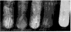
图2是不同硫酸钡用量的海藻酸钠微球的显影效果。Figure 2 is the development effect of sodium alginate microspheres with different barium sulfate dosages.
从左到右硫酸钡用量分别1.5g,0.5g,2.0g,1.0g和阳性对照(复方泛影葡胺注射液)。The amount of barium sulfate from left to right is 1.5g, 0.5g, 2.0g, 1.0g and the positive control (compound diatrizoate meglumine injection).
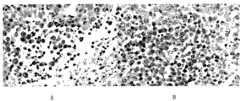
图3腹腔灌洗液中微球的形态变化。Figure 3 Morphological changes of microspheres in peritoneal lavage fluid.
A.空白腹液;B.低剂量组1周时散在微球;C.高剂量组1周时散在微球;D.低剂量组2周时吞噬微球;E.高剂量组2周时吞噬微球;F.低剂量组4周时吞噬微球;G.高剂量组4周时吞噬微球A. Blank peritoneal fluid; B. Microspheres dispersed in low dose group at 1 week; C. Microspheres dispersed in high dose group at 1 week; D. Microspheres phagocytized in low dose group at 2 weeks; E. High dose group at 2 weeks Phagocytosis of microspheres; F. Phagocytosis of microspheres in the low-dose group at 4 weeks; G. Phagocytosis of microspheres in the high-dose group at 4 weeks
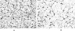
图4试验兔介入治疗HE染色切片(×200)Figure 4 HE-stained slices of rabbits undergoing interventional therapy (×200)
A.正常对照组;B.肿瘤对照组;C.介入治疗组;D.介入治疗组。A. normal control group; B. tumor control group; C. interventional therapy group; D. interventional therapy group.
图5.抗CD34免疫组化染色切片(×400)Figure 5. Anti-CD34 immunohistochemical staining section (×400)
A.肿瘤对照组;B.肿瘤刘照组;C.介入治疗组;D.介入治疗组。A. Tumor control group; B. Tumor Liu Zhao group; C. Interventional therapy group; D. Interventional therapy group.
图6.抗VEGF免疫组化染色切片(×400)Figure 6. Anti-VEGF immunohistochemical staining section (×400)
A.介入治疗组;B.肿瘤对照组。A. Interventional therapy group; B. Tumor control group.
具体实施方式Detailed ways
下面结合具体实施例,进一步阐述本发明。应理解,这些实施例仅用于说明本发明而不用于限制本发明的范围。下列实施例中未注明具体条件的实验方法,通常按照常规条件如Sambrook等人,分子克隆:实验室指南(New York:Cold Spring Harbor Laboratory Press,1989)中所述的条件,或按照制造厂商所建议的条件。除非另外说明,否则百分比和份数按重量计算。Below in conjunction with specific embodiment, further illustrate the present invention. It should be understood that these examples are only used to illustrate the present invention and are not intended to limit the scope of the present invention. The experimental method that does not indicate specific conditions in the following examples, usually according to conventional conditions such as Sambrook et al., molecular cloning: the conditions described in the laboratory guide (New York: Cold Spring Harbor Laboratory Press, 1989), or according to the manufacturer suggested conditions. Percentages and parts are by weight unless otherwise indicated.
实施例1微球制备Embodiment 1 microsphere preparation
1材料与方法1 Materials and methods
1.1仪器与试药 电动搅拌器(德国IKA RW20 digital);显微镜(上海上光实业有限公司,XSP-2XC);超声仪(上海Branson公司,SB3200-T);旋转黏度计(上海天平仪器厂,NDJ-1型);LS激光粒度分析仪(美国Beckman Coulter仪器公司)。海藻酸钠(青岛胶南明月海藻有限公司,药用级,010509014);壳聚糖(冰岛普利美公司,医用级,06042005);微粉化硫酸钡II型(山东长清制药厂,药用级,200620216);复方泛影葡胺注射液(先灵(广州)药业有限公司,200607014);司盘-80、吐温-80(化学纯,上海化学试剂商店);玉米油(食品级,市售);氯化钙、冰醋酸、柠檬酸钠等均为分析纯。1.1 Instruments and reagents Electric stirrer (Germany IKA RW20 digital); Microscope (Shanghai Shangguang Industrial Co., Ltd., XSP-2XC); Ultrasonic instrument (Shanghai Branson Company, SB3200-T); Rotational viscometer (Shanghai Tianping Instrument Factory, NDJ-1 type); LS laser particle size analyzer (Beckman Coulter Instrument Company, USA). Sodium alginate (Qingdao Jiaonan Mingyue Seaweed Co., Ltd., pharmaceutical grade, 010509014); chitosan (Iceland Primo company, medical grade, 06042005); micronized barium sulfate type II (Shandong Changqing Pharmaceutical Factory, pharmaceutical grade , 200620216); Compound Diatrizoate meglumine injection (Schering (Guangzhou) Pharmaceutical Co., Ltd., 200607014); Span-80, Tween-80 (chemically pure, Shanghai Chemical Reagent Store); corn oil (food grade, Commercially available); calcium chloride, glacial acetic acid, sodium citrate, etc. were all analytically pure.
1.2工艺优选1.2 Process optimization
考虑到诸多因素对微球成形的影响,参照有关文献报道(Vandenberg GW,Drolet C et al,Factors affecting protein release from alginate-chitosan coacervate microcapsulesduring production and gastric/intestinal simulation[J],J ControlRelease,2001,77(3):297-307;王康,何志敏,海藻酸微胶囊的制备及在药物控释中的研究进展[J],化学工程,2002,30(1):48-53;刘善奎,高申,钟延强,张洪英,孙树汉,DNA疫苗海藻酸钠微球的制备及体外释药[J],第二军医大学学报,2004,25(1):58-60;孔璐,张阳德,5-FU纳米微粒的制备及其释药特性研究[J],解放军医学杂志,2007,32(1):32-34;张彦青,张明春,解军波,戚务勤,韩淑珍,阿司匹林壳聚糖-海藻酸钠微囊处方优选与释药机制研究[J],中国药房,2007,18(4):278-280;石晓丽,徐军,张雪梅,程晓耕,干扰素壳聚糖/海藻酸钠微囊控释制剂载体的初步研究[J],中国生物制品学杂志,2007,20(2):117-118;吕慧侠,周建平,戴影秋,邓瑾,刘馨,海藻酸钙掩味微囊的制备[J],中国药科大学学报,2007,38(2):125-128;刘伟,王莹,王士斌,吴文果,蓝琪,王新刚,乳化-凝胶化法制备药用载体海藻酸钙微球的研究[J],生物医学工程研究,2007,26(2):155-158。)在预试验的基础上,选取影响微球性状较显著的4个因素作为考察对象,即海藻酸钠浓度、CaCl2浓度、司盘-80与吐温-80比例、壳聚糖浓度,通过L9(34)正交设计试验优选工艺条件。取粒径分布、微球形态、分散度和均匀度4个指标进行综合评分。评判标准:粒径大小占综合评分的40%,其余三项各占20%;以显微镜下观察所得来进行评分;粒径、形态、分散度及均匀度都分为3个级别,各赋予一定的分值;综合评分以各子项目的分值之和计算。Considering the influence of many factors on the formation of microspheres, refer to relevant literature reports (Vandenberg GW, Drolet C et al, Factors affecting protein release from alginate-chitosan coacervate microcapsules during production and gastric/intestinal simulation[J], J ControlRelease, 2001, 77 (3): 297-307; Wang Kang, He Zhimin, Preparation of alginic acid microcapsules and research progress in drug release[J], Chemical Engineering, 2002, 30(1): 48-53; Liu Shankui, Gao Shen , Zhong Yanqiang, Zhang Hongying, Sun Shuhan, Preparation and in vitro drug release of DNA vaccine sodium alginate microspheres [J], Journal of Second Military Medical University, 2004, 25(1): 58-60; Kong Lu, Zhang Yangde, 5-FU nano Preparation of microparticles and research on their drug release properties[J], PLA Medical Journal, 2007, 32(1): 32-34; Zhang Yanqing, Zhang Mingchun, Xie Junbo, Qi Wuqin, Han Shuzhen, Prescription optimization of aspirin chitosan-sodium alginate microcapsules Research on drug release mechanism and release mechanism [J], Chinese Pharmacy, 2007, 18(4): 278-280; Shi Xiaoli, Xu Jun, Zhang Xuemei, Cheng Xiaogeng, Preliminary study on the carrier of interferon chitosan/sodium alginate microcapsule controlled release preparation [J], Chinese Journal of Biological Products, 2007, 20(2): 117-118; Lu Huixia, Zhou Jianping, Dai Yingqiu, Deng Jin, Liu Xin, Preparation of calcium alginate taste-masking microcapsules [J], Chinese Medicine Journal of University of Science and Technology of China, 2007, 38(2): 125-128; Liu Wei, Wang Ying, Wang Shibin, Wu Wenguo, Lan Qi, Wang Xingang, Preparation of pharmaceutical carrier calcium alginate microspheres by emulsification-gelation[J] , Biomedical Engineering Research, 2007, 26(2): 155-158.) On the basis of preliminary experiments, four factors that significantly affect the properties of microspheres were selected as the investigation objects, namely sodium alginate concentration, CaCl2 concentration, The ratio of Span-80 and Tween-80, the concentration of chitosan, and the optimal process conditions were tested by L9(34) orthogonal design. The four indicators of particle size distribution, microsphere shape, dispersion and uniformity were used for comprehensive scoring. Judging criteria: particle size accounts for 40% of the comprehensive score, and the remaining three items each account for 20%; the score is based on observations under a microscope; particle size, shape, dispersion and uniformity are divided into 3 levels, each given a certain level. The overall score is calculated by the sum of the scores of each sub-item.
1.3微球的形态、粒径和分布 将微球配成混悬液,摇匀后取样制片,显微镜下观察微球的外观、形态、分散度,采用血细胞计数板显微计数法初步考察微球的粒径、分布,每次计数不少于500粒。用LS激光粒度分析仪测定粒径大小和分布状态。1.3 Morphology, particle size and distribution of microspheres Prepare the microspheres into a suspension, shake well and take samples to make slices. Observe the appearance, shape and degree of dispersion of the microspheres under a microscope. The particle size and distribution of the balls should not be less than 500 particles per count. The particle size and distribution state were determined by LS laser particle size analyzer.
1.4微球的稳定性 分别取硫酸钡2.0g投料量制备的微球混悬液5ml,考查100℃水浴加热0.5h、1h、2h,-4℃冰冻24h,37℃振摇1.5h、3h、6h、12h、24h,钴60(10kGy)照射0.5h等条件对微球稳定性的影响。处理结束后,放置至室温,摇匀后取样制片,置显微镜下观察破损情况,用血细胞计数板计数,调整视野重复6次计算结果。破损率=(破损微球数量/观察微球总数)×100%。1.4 Stability of microspheres Take 5ml of microsphere suspension prepared by 2.0g of barium sulfate, heat in 100°C water bath for 0.5h, 1h, 2h, freeze at -4°C for 24h, shake at 37°C for 1.5h, 3h, 6h, 12h, 24h, cobalt 60 (10kGy) irradiation for 0.5h and other conditions on the stability of microspheres. After the treatment, place it at room temperature, shake well, take a sample and make a slice, observe the damage under a microscope, count with a hemocytometer, adjust the field of view and repeat 6 times to calculate the result. Breakage rate = (number of damaged microspheres/total number of observed microspheres) × 100%.
1.5微球混悬液的悬浮性 取硫酸钡2.0g投料量制备的微球,使用水和不同浓度的海藻酸钠溶液配制混悬液,按药典(国家药典委员会,中华人民共和国药典(二部)[M],北京:化学工业出版社,2005:附录40)方法测定沉降体积比和黏度。1.5 Suspension of microsphere suspension Get the microspheres prepared by barium sulfate 2.0g feeding amount, use water and sodium alginate solution of different concentrations to prepare suspension, according to Pharmacopoeia (National Pharmacopoeia Committee, Pharmacopoeia of the People's Republic of China (Part Two) )[M], Beijing: Chemical Industry Press, 2005: Appendix 40) to determine sedimentation volume ratio and viscosity.
1.6硫酸钡用量与包封率 取各投料量下制备微球,调节最终体积为30ml,摇匀后取微球混悬液10ml,加入柠檬酸钠使终浓度0.055mol/L(李朝霞,朱建良,制备海藻酸钠-壳聚糖生物微胶囊的技术研究[J],盐城工学院学报(自然科学版),2005,18(2):58-62),液化1h,冰浴超声30min,离心(RCF 90g)5min,水洗3次,将粉术转移至干燥恒重的称量瓶中,105℃干燥至恒重,测得其质量。包封率=(干燥硫酸钡粉末质量×3/实际硫酸钡投料量)×100%。1.6 Dosage of barium sulfate and encapsulation efficiency The microspheres were prepared according to the dosages, and the final volume was adjusted to 30ml. After shaking well, 10ml of the microsphere suspension was taken, and sodium citrate was added to make the final concentration 0.055mol/L (Li Zhaoxia, Zhu Jianliang, Technical research on preparing sodium alginate-chitosan biomicrocapsules [J], Yancheng Institute of Technology (Natural Science Edition), 2005, 18 (2): 58-62), liquefaction 1h, ice bath ultrasonic 30min, centrifugation ( RCF 90g) for 5 minutes, washed with water for 3 times, transferred the powder to a dry weighing bottle with constant weight, dried at 105°C to constant weight, and measured its mass. Encapsulation rate=(dried barium sulfate powder mass×3/actual barium sulfate charging amount)×100%.
1.7微球显影效果 取1.6中微球混悬液10ml,复方泛影葡胺注射液作为阳性对照,在X光机下显影,定性观察显影效果。1.7 Microsphere development effect Take 10ml of the microsphere suspension in 1.6, compound diatrizoate meglumine injection as a positive control, develop under X-ray machine, and observe the development effect qualitatively.
2结果2 results
2.1工艺优选结果 设计的4个因素中,CaCl2浓度对微球形态影响较小,其次是壳聚糖浓度,海藻酸钠浓度、司盘-80和吐温-80的用量和配比对微球粒径和形态的影响较大,详见表1、2、3。微球制备工艺为:称取硫酸钡2.0g置乳钵中研磨,用水10ml分3次转移至100ml小烧杯中,超声5min,加入1%海藻酸钠溶液10ml、吐温-80 0.5ml,继续超声5min,将混匀后的硫酸钡海藻酸钠溶液抽入20ml针筒,作为水相;量取玉米油40ml置250ml烧杯内,加入司盘-80 6g,置50-60℃水浴搅拌(RCF 90g)10min,逐滴注入水相,乳化20min,水浴温度降至40℃,调节RCF至58g,滴加3%氯化钙溶液10ml,固化15min;固化后的乳液移入分液漏斗,静置12h分层,取下层溶液,离心(RCF 90g)分取下层微球,用水洗涤3次;水洗后的微球分散于20ml水中,加0.5%壳聚糖溶液5ml,3%氯化钙溶液5ml搅拌(RCF 22g)15min,离心,水洗3次,分散于0.25%海藻酸钠溶液置冰箱4℃冷藏室保存。2.1 Process optimization results Among the 4 factors designed, the concentration of CaCl2 had little effect on the morphology of microspheres, followed by the concentration of chitosan, the concentration of sodium alginate, the amount and ratio of Span-80 and Tween-80. The particle size and shape of the ball have a great influence, see Table 1, 2, 3 for details. The microsphere preparation process is as follows: Weigh 2.0 g of barium sulfate, grind it in a mortar, transfer 10 ml of water to a 100 ml small beaker in 3 times, ultrasonicate for 5 minutes, add 10 ml of 1% sodium alginate solution, 0.5 ml of Tween-80, continue Ultrasound for 5 minutes, draw the mixed barium sulfate sodium alginate solution into a 20ml syringe as the water phase; measure 40ml of corn oil into a 250ml beaker, add 6g of Span-80, and stir in a 50-60℃ water bath (RCF 90g) for 10min, pour into the water phase drop by drop, emulsify for 20min, lower the temperature of the water bath to 40°C, adjust the RCF to 58g, add 10ml of 3% calcium chloride solution dropwise, and solidify for 15min; transfer the solidified emulsion into a separatory funnel and let it stand for 12h Separate layers, take off the lower layer solution, centrifuge (RCF 90g) to separate the lower layer of microspheres, wash with water 3 times; the washed microspheres are dispersed in 20ml of water, add 5ml of 0.5% chitosan solution, 5ml of 3% calcium chloride solution and stir (RCF 22g) for 15min, centrifuge, wash 3 times with water, disperse in 0.25% sodium alginate solution and store in refrigerator at 4°C.
表1正交试验设计的因素及水平Table 1 Factors and levels of orthogonal experimental design
表2正交试验设计的结果及评判Table 2 The results and judgment of the orthogonal experimental design
表3F检验结果Table 3F test results

2.2微球的形态、粒径和分布2.2 Morphology, particle size and distribution of microspheres
观察结果,硫酸钡海藻酸钠微球大部分为球形单体,圆形,表面光滑,不粘连,分散性较好,所有微球均呈现均匀分散的黑色不透光状态(见图1),平均粒径为53.08±32.72μm。Observation results, barium sulfate sodium alginate microspheres are mostly spherical monomers, round, smooth surface, non-adhesive, good dispersion, all microspheres are uniformly dispersed black opaque state (see Figure 1), The average particle size is 53.08±32.72 μm.
2.3稳定性考查2.3 Stability test
硫酸钡海藻酸钠微球热稳定性较好,100℃水浴加热2h破损率仅为(2.0±1.1)%;-4℃冰冻24h后微球严重破损,其破损率达(86.0±19.2)%;37℃振摇条件下,前6h破损率升高幅度明显比后18h大;钴60(10kGy)照射0.5h破损率达(10.3±3.2)%。因此加热灭菌是制备微球注射用混悬液的有效方法。Barium sulfate sodium alginate microspheres have good thermal stability, and the damage rate is only (2.0±1.1)% when heated in a water bath at 100°C for 2 hours; the microspheres are severely damaged after being frozen at -4°C for 24 hours, and the damage rate reaches (86.0±19.2)% ; Under shaking conditions at 37°C, the damage rate increased significantly in the first 6 hours than in the last 18 hours; the damage rate reached (10.3±3.2)% when cobalt 60 (10kGy) was irradiated for 0.5 hours. Therefore heat sterilization is an effective method for preparing microsphere injection suspension.
2.4悬浮性考查2.4 Suspension test
结果见表4,0.25%海藻酸钠作为助悬剂时,其沉降体积比可以达到0.92±0.018,符合药典标准,而0.50%海藻酸钠微球混悬液黏度较大,不利于今后的临床给药,故选择0.25%海藻酸钠作为助悬剂的最佳浓度。The results are shown in Table 4. When 0.25% sodium alginate is used as a suspending agent, its sedimentation volume ratio can reach 0.92±0.018, which meets the pharmacopoeia standard. However, the viscosity of 0.50% sodium alginate microsphere suspension is relatively high, which is not conducive to future clinical practice. Administration, so choose 0.25% sodium alginate as the best concentration of suspending agent.
表4 4种不同混悬条件下微球的沉降体积比和动力黏度(n=6,x±s)Table 4 Sedimentation volume ratio and dynamic viscosity of microspheres under 4 different suspension conditions (n=6, x±s)
2.5包封率2.5 encapsulation rate
硫酸钡用量为1.0、1.5、2.0g时,其包封率的变化不大。结合定性显影结果,选择2.0g为硫酸钡的最佳投料量。When the dosage of barium sulfate is 1.0, 1.5, 2.0g, the encapsulation efficiency has little change. Combined with the results of qualitative development, 2.0g was selected as the optimal dosage of barium sulfate.
2.5微球显影效果2.5 microsphere development effect
图2显示,同阳性对照相比,投料量为0.5g、1g的硫酸钡海藻酸钠微球X光吸收效果不明显,而投料量为1.5g、2g的硫酸钡海藻酸钠微球可见明显X线吸收,但由于仪器条件所限,无法测量其具体吸收值,只能做定性比较。Figure 2 shows that compared with the positive control, the X-ray absorption effect of the barium sulfate sodium alginate microspheres with a feeding amount of 0.5g and 1g is not obvious, but that of the barium sulfate sodium alginate microspheres with a feeding amount of 1.5g and 2g is obvious X-ray absorption, but due to the limitation of instrument conditions, its specific absorption value cannot be measured, and only qualitative comparison can be made.
3讨论3 Discussion
3.1海藻酸钠浓度对微球的影响3.1 Effect of sodium alginate concentration on microspheres
由于芯材硫酸钡是不溶固体,故在相同的条件下,海藻酸钠的浓度增大,整个水相的黏度也相应增大,乳化时就不如海藻酸钠低浓度情况下的分散度好,故成品的分散度、形态的圆整性都不如人意。实验中海藻酸钠浓度如果太低也不利于形成所需粒径大小的微球;海藻酸钠浓度较高条件下制得的微囊粒径较大,但形态较差。Since barium sulfate, the core material, is an insoluble solid, under the same conditions, as the concentration of sodium alginate increases, the viscosity of the entire water phase also increases accordingly, and the emulsification is not as good as the dispersion of sodium alginate at a low concentration. Therefore, the degree of dispersion and the roundness of the finished product are not satisfactory. In the experiment, if the concentration of sodium alginate is too low, it is not conducive to the formation of microspheres with the required particle size; the microcapsules prepared under the condition of high concentration of sodium alginate are larger in particle size, but the shape is poor.
3.2乳化剂用量对微球的影响3.2 Effect of emulsifier dosage on microspheres
制备工艺包括形成W/O乳剂的乳化过程。液液两相乳化形成微球较容易,而固液两相乳化,要想得到的微球圆整,则要求较高。司盘-80和吐温-80的用量对微球圆整性及分散度的影响较大。理论上乳化剂HLB值应。使用一定比例司盘-80和吐温-80混合液作为乳化剂,其HLB值在5.72,落在<6的HLB值内,保证了微球圆整性及分散度。The preparation process includes the emulsification process of forming W/O emulsion. Liquid-liquid two-phase emulsification is easier to form microspheres, while solid-liquid two-phase emulsification requires higher requirements to obtain round microspheres. The amount of Span-80 and Tween-80 had a great influence on the roundness and dispersion of microspheres. Theoretically the emulsifier HLB value should be. Using a certain proportion of Span-80 and Tween-80 mixture as emulsifier, its HLB value is 5.72, falling within the HLB value of <6, which ensures the roundness and dispersion of the microspheres.
3.3壳聚糖溶液的影响3.3 Effect of chitosan solution
pH较高或浓度较大滴加时微球存在粘连情况。壳聚糖溶液用2%醋酸调节pH值在5.0左右,再滴加到微球悬浮液中进行孵化,这样可改善微球粘连情况,但不影响孵化。微球外层膜的牢固度与壳聚糖浓度有一定关系,海藻酸钠与壳聚糖利用正负静电相互作用成膜,一旦壳聚糖的浓度降低就可能使其与海藻酸钠的交联不够充分,膜的强度会受到影响,但是壳聚糖浓度太高,会导致微球粘连,分散度下降,故可以考虑用低浓度壳聚糖分两次充分交联。When the pH is high or the concentration is high, there is adhesion of the microspheres. The chitosan solution is adjusted to a pH value of about 5.0 with 2% acetic acid, and then added dropwise to the microsphere suspension for incubation, which can improve the adhesion of the microspheres, but does not affect the incubation. The firmness of the outer film of microspheres has a certain relationship with the concentration of chitosan. Sodium alginate and chitosan use positive and negative electrostatic interactions to form a film. Once the concentration of chitosan decreases, the interaction with sodium alginate may occur. If the linkage is not sufficient, the strength of the membrane will be affected, but if the chitosan concentration is too high, the microspheres will stick together and the dispersion will decrease, so it can be considered to fully cross-link twice with low concentration chitosan.
3.4关于微球的粒径 实验发现RCF与微球粒径呈负相关。在保证微球形态的情况下,降低RCF能增大微球粒径。当乳化RCF降为33g时,粒径大都在100μm左右;当RCF降至8g时,粒径大都在120μm左右,且形态非常不好。另外,此次实验所制得微球粒径差异较大(53.08±32.72μm),在下一步的栓塞应用时,对肝窦等有造成异位栓塞的可能。如何解决这一问题呢,一是对工艺做进一步的优化,二是应用筛网来进行截留,去除粒径在30μm以下的微球,以降低异位栓塞的可能性。总体上,微球粒径在50~150μm的占总数的65%以上。3.4 About the particle size of microspheres It was found that RCF was negatively correlated with the particle size of microspheres. In the case of ensuring the shape of the microspheres, reducing the RCF can increase the particle size of the microspheres. When the emulsified RCF is reduced to 33g, the particle size is mostly around 100μm; when the RCF is reduced to 8g, the particle size is mostly around 120μm, and the shape is very bad. In addition, the particle size of the microspheres produced in this experiment varies greatly (53.08±32.72 μm), which may cause ectopic embolism to liver sinusoids in the next embolization application. How to solve this problem, one is to further optimize the process, and the other is to use a screen to intercept and remove microspheres with a particle size below 30 μm to reduce the possibility of ectopic embolism. In general, microspheres with a particle size of 50-150 μm account for more than 65% of the total.
3.5稳定性考查时发现微球冷冻后会变形,而且是不可逆的变形,这可能与芯材是硫酸钡固体有关系。3.5 During the stability test, it was found that the microspheres will deform after freezing, and the deformation is irreversible, which may be related to the fact that the core material is barium sulfate solid.
总之,我们的实验结果表明,应用海藻酸钠、壳聚糖为膜材,硫酸钡为芯材,能够制备出符合一定质量标准的硫酸钡海藻酸钠微球。In conclusion, our experimental results show that using sodium alginate and chitosan as membrane materials and barium sulfate as core material, barium sulfate sodium alginate microspheres meeting certain quality standards can be prepared.
实施例2生物相容性研究Embodiment 2 biocompatibility research
取体重200~250g的Wistar健康雄性大鼠(第二军医大学实验动物中心)10只,分为2个剂量组,每组5只。制备每毫升含100000个微球的悬浮液,各取0.5、0.1毫升即50000、10000个微球稀释到1毫升,大鼠腹腔注射。1周后,2个剂量组各取1只大鼠,处死后,用生理盐水1ml,灌洗腹腔,取腹腔灌洗液1ml置显微镜下观察。2周后,2个剂量组各取2只大鼠,处死后,用生理盐水1ml,灌洗腹腔,取腹腔灌洗液1ml置显微镜下观察。4周后,同2周做同样的处理。灌洗液镜检计数微球并观察微球的形态变化,并观察囊壁厚度、有无纤维化,以了解微球在受体体内的稳定性,即生物相容性。Take 10 Wistar healthy male rats (Experimental Animal Center, Second Military Medical University) weighing 200-250 g, and divide them into 2 dose groups, 5 in each group. A suspension containing 100,000 microspheres per milliliter was prepared, and 0.5 and 0.1 milliliters, that is, 50,000 and 10,000 microspheres were diluted to 1 milliliter, and intraperitoneally injected into rats. One week later, one rat was taken from each of the two dosage groups, and after being sacrificed, the peritoneal cavity was lavaged with 1 ml of normal saline, and 1 ml of the peritoneal lavage fluid was taken and observed under a microscope. After 2 weeks, 2 rats in each of the 2 dose groups were sacrificed, and the peritoneal cavity was lavaged with 1 ml of normal saline, and 1 ml of the peritoneal lavage fluid was taken and observed under a microscope. After 4 weeks, do the same treatment with 2 weeks. Microscopic examination of the lavage fluid counted the microspheres and observed the morphological changes of the microspheres, and observed the thickness of the capsule wall and whether there was fibrosis, so as to understand the stability of the microspheres in the recipient body, that is, biocompatibility.
结果显示:整体动物没有观察到明显毒性反应。剂量组之间有明显的剂量差异,高剂量组散在微球明显比低剂量组多;第2周以后能观察到微球被吞噬的现象;微球能被包裹、吞噬,见图3A-G。The results showed that no obvious toxic reaction was observed in the whole animal. There is a significant dose difference between the dose groups. The high-dose group has more scattered microspheres than the low-dose group; after the second week, the microspheres can be observed to be phagocytized; the microspheres can be encapsulated and phagocytized, as shown in Figure 3A-G .
实施例3试验兔介入治疗观察Example 3 Experimental Rabbit Interventional Therapy Observation
VX2肿瘤细胞株(来源)按细胞培养技术进行复苏,肿瘤细胞悬液0.5ml接种于兔后腿外侧肌肉内,2周后即制成荷瘤兔。取瘤块,用无菌眼科剪剪成约1.0mm大小的瘤组织块,置于无菌生理盐水中备用。实验兔用4%戊巴比妥钠1ml/kg静脉麻醉后仰卧位固定。常规腹部备毛,消毒铺巾,上腹部正中切口打开腹腔,充分暴露肝脏,将1mm3肿瘤块用镊子夹住直接穿刺入肝实质内,穿刺道用Arista止血淀粉封堵。观察肿瘤生长情况。CT检查时各实验组荷瘤兔所用扫描参数相同。CT扫描参数:80KV,200mA,层厚2.5mm,Pitch3,FOV 25cm,所有实验兔均在深度麻醉下采集图像以保证图像的清晰。The VX2 tumor cell line (source) was resuscitated according to the cell culture technique, and 0.5ml of the tumor cell suspension was inoculated into the lateral muscle of the rabbit's hind leg, and two weeks later, the tumor-bearing rabbit was made. Tumor pieces were taken, cut into tumor tissue pieces about 1.0 mm in size with sterile ophthalmic scissors, and placed in sterile saline for later use. The experimental rabbits were anesthetized intravenously with 4% pentobarbital sodium 1ml/kg and fixed in the supine position. Routine abdominal hair preparation, disinfection and draping, midline upper abdominal incision was made to open the abdominal cavity to fully expose the liver, the 1 mm3 tumor mass was clamped with forceps and directly punctured into the liver parenchyma, and the puncture track was blocked with Arista hemostatic starch. Observe tumor growth. The scanning parameters used in the CT examination of the tumor-bearing rabbits in each experimental group were the same. CT scanning parameters: 80KV, 200mA, slice thickness 2.5mm, Pitch3, FOV 25cm, all experimental rabbits were collected images under deep anesthesia to ensure the clarity of images.
介入治疗时间选择在接种后2周进行,插管方法模拟人肝动脉插管介入治疗的方法。麻醉后,切开皮肤,分离右股动脉,穿刺置入3F微导管和微导丝。导管于T12水平造影(总量4ml,速率1ml/s),发现腹腔干动脉开口,导丝引导微导管至肝总动脉再次造影(总量3ml,速率1ml/s)。The time of interventional treatment was selected 2 weeks after inoculation, and the intubation method simulated the method of human hepatic artery cannulation interventional treatment. After anesthesia, the skin was incised, the right femoral artery was separated, and a 3F microcatheter and microguidewire were inserted through puncture. Angiography was performed on the catheter at T12 level (total volume 4ml, rate 1ml/s), celiac artery opening was found, and the guide wire guided the microcatheter to the common hepatic artery for another imaging (total volume 3ml, rate 1ml/s).
①实验兔分别于介入术前、术后2周行肝脏螺旋CT扫描,观察肿瘤生长,观察微球在肿瘤内积聚情况。①Hepatic helical CT scanning was performed on experimental rabbits before and 2 weeks after interventional surgery to observe tumor growth and the accumulation of microspheres in the tumor.
②实验兔分别于介入术前、术后7天行肝功能(ALT、AST、TB等)检查。②The experimental rabbits underwent liver function (ALT, AST, TB, etc.) examinations before and 7 days after the intervention.
③介入术后2周,处死实验兔,取出肝脏。③Two weeks after the intervention, the experimental rabbits were sacrificed and the livers were taken out.
病理:大体观察肿瘤大小、坏死情况、包膜情况。进行常规病理学检查,抗CD34免疫组化染色,抗VEGF免疫组化染色。抗VEGF免疫组化染色中阳性细胞为细胞浆出现浅黄色、棕黄色或棕褐色颗粒,>10%为阳性表达。Pathology: Grossly observe the tumor size, necrosis, and capsule. Routine pathological examination, anti-CD34 immunohistochemical staining, anti-VEGF immunohistochemical staining. Positive cells in anti-VEGF immunohistochemical staining were pale yellow, brownish yellow or brown granules in the cytoplasm, and >10% were positive expression.
①植瘤成功率:25只实验兔VX2肿瘤接种后21只可见肝肿瘤生长,移植成功率为84%(21/25)。① Tumor implantation success rate: 21 of 25 experimental rabbits were inoculated with VX2 tumors, and liver tumors grew, and the implantation success rate was 84% (21/25).
②动物分组:② Animal grouping:
1.正常对照组5只。1. 5 rats in the normal control group.
2.肿瘤对照组10只:观察肿瘤重量、体积5只;观察生存期5只。2. Tumor control group: 10 rats: 5 rats were observed for tumor weight and volume; 5 rats were observed for survival time.
3.介入治疗组11只(9只有效):观察重量、体积5只(14天);观察生存期4只。3. 11 rats in the interventional treatment group (9 rats are effective): 5 rats were observed for weight and volume (14 days); 4 rats were observed for survival period.
③介入治疗成功率:11只实验兔行介入治疗,9只成功,成功率81.8%。另外,1只实验兔介入治疗手术失败,1只实验兔介入治疗后3天死亡(均从治疗组中剔除)。③Success rate of interventional therapy: 11 experimental rabbits underwent interventional therapy, 9 of them were successful, the success rate was 81.8%. In addition, 1 experimental rabbit failed the interventional therapy operation, and 1 experimental rabbit died 3 days after the interventional therapy (all were excluded from the treatment group).
表5对肿瘤重量和体积的影响(±S,n=5)Effect of Table 5 on tumor weight and volume (±S, n=5)
与对照组比*p<0.01Compared with the control group *p<0.01
介入治疗组肿瘤重量2.434±0.992克,肿瘤对照组肿瘤重量4.696±1.246克,p<0.01。介入治疗组肿瘤体积2.126±0.929cm3,肿瘤对照组肿瘤体积3.962±1.101cm3,p<0.01。结果表明:介入治疗对肿瘤生长有明显抑制作用。The tumor weight of the interventional therapy group was 2.434±0.992 grams, and that of the tumor control group was 4.696±1.246 grams, p<0.01. The tumor volume of the interventional therapy group was 2.126±0.929cm3 , and that of the tumor control group was 3.962±1.101cm3 , p<0.01. The results showed that the interventional therapy had a significant inhibitory effect on tumor growth.
表6对肝功能指标的影响(±S,n=5)The influence of table 6 on liver function index (±S, n=5)
与正常对照组比△△p<0.01,与肿瘤对照组比**p<0.01Compared with normal control group, △△p<0.01, compared with tumor control group, **p<0.01
介入治疗后7大,肿瘤对照组与正常对照组相比,p<0.01,显示了肿瘤生长对肝功能的损害;而介入治疗组明显低于肿瘤对照组,与肿瘤对照组比,p<0.01,说明介入治疗不仅控制肿瘤生长,而且通过控制肿瘤生长保护了肝功能。Seven days after interventional treatment, the tumor control group was compared with the normal control group, p<0.01, showing the damage of tumor growth to liver function; while the interventional treatment group was significantly lower than the tumor control group, compared with the tumor control group, p<0.01 , indicating that interventional therapy not only controls tumor growth, but also protects liver function by controlling tumor growth.
在苏木精-伊红染色(HE)染色切片上,肿瘤对照组:细胞丰富,排列紧密,显著异型,并可见较多病理性核分裂,见图4B。介入治疗组:细胞排列明显松散,癌细胞大片坏死,走形与血管相一致,癌细胞核固缩和核碎裂现象明显增多,见图4C,图4D。On the hematoxylin-eosin (HE) stained sections, the tumor control group: the cells were abundant, tightly arranged, significantly atypia, and more pathological mitotic figures were seen, as shown in Figure 4B. Interventional treatment group: the arrangement of cells was obviously loose, large pieces of cancer cells were necrotic, and the shape was consistent with blood vessels, and the phenomenon of nuclear pyknosis and nuclear fragmentation of cancer cells increased significantly, as shown in Figure 4C and Figure 4D.
被抗CD34单克隆抗体染成棕黄色的血管内皮细胞代表着微血管,微血管被肿瘤细胞分割,呈点状、线状分布。Vascular endothelial cells stained brownish yellow by anti-CD34 monoclonal antibody represent microvessels, which are divided by tumor cells and distributed in dots and lines.
肿瘤对照组:癌灶区微血管数目较多,癌组织间可见丰富的新生血管,见图5A,图5B。CD34,EnVision染色(×400)。Tumor control group: the number of microvessels in the cancer focus area is large, and abundant new blood vessels can be seen between cancer tissues, as shown in Figure 5A and Figure 5B. CD34, EnVision staining (×400).
介入治疗组:癌组织大片坏死,残存的癌组织内新生血管明显减少,见图5C,图5D。CD34,EnVision染色(×400)。Interventional therapy group: Large areas of cancer tissue were necrotic, and new blood vessels in the remaining cancer tissue were significantly reduced, as shown in Figure 5C and Figure 5D. CD34, EnVision staining (×400).
VEGF定位于肿瘤细胞浆及胞膜,阳性表达为胞浆或胞膜染为棕黄色(图中显示淡灰色)。VEGF is localized in the tumor cell plasma and membrane, and the positive expression means that the cytoplasm or membrane is stained brownish yellow (shown in light gray in the figure).
介入治疗组:癌细胞大片坏死,坏死较彻底的肿瘤细胞无VEGF的表达,残存癌细胞胞质可见弱阳性的VEGF表达,见图6A。Interventional therapy group: large areas of cancer cells were necrotic, tumor cells with complete necrosis had no expression of VEGF, and weak positive expression of VEGF could be seen in the cytoplasm of remaining cancer cells, as shown in Figure 6A.
肿瘤对照组:癌细胞胞质可见丰富的VEGF表达,呈细颗粒状,见图6B。Tumor control group: abundant expression of VEGF can be seen in the cytoplasm of cancer cells, which is in the form of fine granules, as shown in Figure 6B.
对照组5只:生存期分别为39d、43d、46d、53d、55d;5 rats in the control group: the survival periods were 39d, 43d, 46d, 53d, and 55d;
介入治疗组4只:生存期分别为60d、68d,2只70大时仍存活。4 rats in the interventional treatment group: the survival periods were 60d and 68d respectively, and 2 rats were still alive when they were 70 years old.
动物死亡后尸检发现肝肿瘤直径均大于4cm,但大部分肿瘤组织已经坏死、液化。动物死亡前不进饮食,体重减轻,无活力。After the death of the animals, the autopsy found that the diameters of the liver tumors were all greater than 4 cm, but most of the tumor tissues had been necrotic and liquefied. Animals were deprived of food and drink prior to death, losing weight and lacking vigor.
以上对本发明的描述并不用于限制本发明的范围。对于本领域的普通技术人员来说,可以根据本发明是供的技术方案做出各种相应的改变,这些改变都应属于本发明的保护范围。The above description of the present invention is not intended to limit the scope of the present invention. For those skilled in the art, various corresponding changes can be made according to the technical solutions provided by the present invention, and these changes should all belong to the protection scope of the present invention.
Claims (18)
Translated fromChinesePriority Applications (1)
| Application Number | Priority Date | Filing Date | Title |
|---|---|---|---|
| CNA200910051456XACN101564558A (en) | 2009-05-18 | 2009-05-18 | Alginate-barium sulfate microsphere, preparation method and application thereof |
Applications Claiming Priority (1)
| Application Number | Priority Date | Filing Date | Title |
|---|---|---|---|
| CNA200910051456XACN101564558A (en) | 2009-05-18 | 2009-05-18 | Alginate-barium sulfate microsphere, preparation method and application thereof |
Publications (1)
| Publication Number | Publication Date |
|---|---|
| CN101564558Atrue CN101564558A (en) | 2009-10-28 |
Family
ID=41281033
Family Applications (1)
| Application Number | Title | Priority Date | Filing Date |
|---|---|---|---|
| CNA200910051456XAPendingCN101564558A (en) | 2009-05-18 | 2009-05-18 | Alginate-barium sulfate microsphere, preparation method and application thereof |
Country Status (1)
| Country | Link |
|---|---|
| CN (1) | CN101564558A (en) |
Cited By (9)
| Publication number | Priority date | Publication date | Assignee | Title |
|---|---|---|---|---|
| CN102532564A (en)* | 2012-01-16 | 2012-07-04 | 孙珊 | Hydrogel and preparation method thereof |
| CN103224247A (en)* | 2013-04-10 | 2013-07-31 | 中国地质大学(武汉) | Method for preparing nanometer barium sulfate by adopting alginate as template |
| CN104861946A (en)* | 2015-03-30 | 2015-08-26 | 山东大学 | Flexible controlled-release microsphere disperse system formed by natural water-soluble polymer and preparation thereof and application of system in reinforcement of oil extraction |
| CN107335091A (en)* | 2017-08-31 | 2017-11-10 | 河南汇博医疗股份有限公司 | A kind of long acting antibiotic hydrogel and preparation method thereof |
| CN107418872A (en)* | 2016-08-31 | 2017-12-01 | 四川蓝光英诺生物科技股份有限公司 | Prepare the device of microballoon and prepare the method for microballoon |
| CN108578790A (en)* | 2018-07-27 | 2018-09-28 | 宁波迪创医疗科技有限公司 | One kind can developable seaweed acidic group biomaterial and preparation method thereof |
| CN110038137A (en)* | 2019-03-04 | 2019-07-23 | 天益健康科学研究院(镇江)有限公司 | A kind of CT visualization and the difunctional microcapsules of mucoadhesive type and preparation method and applications |
| CN113116856A (en)* | 2019-12-31 | 2021-07-16 | 北京远大九和药业有限公司 | Eucalyptus and pinene enteric-coated microspheres and preparation method thereof |
| CN114524636A (en)* | 2022-03-30 | 2022-05-24 | 郑州大学 | Barium-containing super absorbent resin microsphere and preparation and application thereof |
- 2009
- 2009-05-18CNCNA200910051456XApatent/CN101564558A/enactivePending
Cited By (13)
| Publication number | Priority date | Publication date | Assignee | Title |
|---|---|---|---|---|
| CN102532564B (en)* | 2012-01-16 | 2013-09-25 | 孙珊 | Hydrogel and preparation method thereof |
| CN102532564A (en)* | 2012-01-16 | 2012-07-04 | 孙珊 | Hydrogel and preparation method thereof |
| CN103224247A (en)* | 2013-04-10 | 2013-07-31 | 中国地质大学(武汉) | Method for preparing nanometer barium sulfate by adopting alginate as template |
| CN103224247B (en)* | 2013-04-10 | 2015-02-04 | 中国地质大学(武汉) | Method for preparing nanometer barium sulfate by adopting alginate as template |
| CN104861946A (en)* | 2015-03-30 | 2015-08-26 | 山东大学 | Flexible controlled-release microsphere disperse system formed by natural water-soluble polymer and preparation thereof and application of system in reinforcement of oil extraction |
| CN104861946B (en)* | 2015-03-30 | 2017-06-23 | 山东大学 | A kind of flexible control-release microsphere dispersion formed by natural water-soluble copolymer and its preparation and the application in intensified oil reduction |
| CN107418872A (en)* | 2016-08-31 | 2017-12-01 | 四川蓝光英诺生物科技股份有限公司 | Prepare the device of microballoon and prepare the method for microballoon |
| CN107335091A (en)* | 2017-08-31 | 2017-11-10 | 河南汇博医疗股份有限公司 | A kind of long acting antibiotic hydrogel and preparation method thereof |
| CN108578790A (en)* | 2018-07-27 | 2018-09-28 | 宁波迪创医疗科技有限公司 | One kind can developable seaweed acidic group biomaterial and preparation method thereof |
| CN110038137A (en)* | 2019-03-04 | 2019-07-23 | 天益健康科学研究院(镇江)有限公司 | A kind of CT visualization and the difunctional microcapsules of mucoadhesive type and preparation method and applications |
| CN113116856A (en)* | 2019-12-31 | 2021-07-16 | 北京远大九和药业有限公司 | Eucalyptus and pinene enteric-coated microspheres and preparation method thereof |
| CN114524636A (en)* | 2022-03-30 | 2022-05-24 | 郑州大学 | Barium-containing super absorbent resin microsphere and preparation and application thereof |
| CN114524636B (en)* | 2022-03-30 | 2022-12-23 | 郑州大学 | Barium-containing super absorbent resin microsphere and preparation and application thereof |
Similar Documents
| Publication | Publication Date | Title |
|---|---|---|
| CN101564558A (en) | Alginate-barium sulfate microsphere, preparation method and application thereof | |
| Le Renard et al. | The in vivo performance of magnetic particle-loaded injectable, in situ gelling, carriers for the delivery of local hyperthermia | |
| US10456235B2 (en) | Embolization particulates for occluding a blood vessel | |
| Rong et al. | Alginate-calcium microsphere loaded with thrombin: a new composite biomaterial for hemostatic embolization | |
| Jay et al. | Engineering of multifunctional gels integrating highly efficient growth factor delivery with endothelial cell transplantation | |
| JP2009533527A (en) | Method for preparing swellable and deformable microspheres | |
| CN103239730B (en) | Medical sodium alginate gel microsphere and preparation method and application thereof | |
| WO2015179997A1 (en) | Polyhydroxyl polymer embolic microsphere and preparation process therefor | |
| Zhu et al. | Liquid metal‐enabled microspheres with high drug loading and multimodal imaging for artery embolization | |
| JP2008513381A (en) | Paclitaxel-sodium alginate microsphere vascular embolic agent and method for producing the same | |
| US11607388B2 (en) | Drug-loaded microbead compositions, embolization compositions and associated methods | |
| KR20160055810A (en) | Oily compositions | |
| CN110522946B (en) | rhBMP-2-loaded bone repair material microsphere and preparation method thereof | |
| CN110327310A (en) | A kind of multicore is total to shell composite drug carried microsphere and its preparation method and application | |
| US12428467B2 (en) | Gelatin particles, method for producing gelatin particles, gelatin particle-containing cell, and method for producing gelatin particle-containing cell | |
| CN108403663A (en) | GO-PEG gel micro-balls with nucleocapsid and its preparation method and application | |
| CN101011601A (en) | Medicament-carrying complex calcium phosphate bone cement for spinal column reconstruction during tumour operation | |
| CN107550879A (en) | A kind of preparation method of gelfoam drug bearing microsphere | |
| Shen et al. | Microfluidic fabrication of X-ray-visible sodium hyaluronate microspheres for embolization | |
| Chen et al. | Dual-modality imaging particle size monodisperse poly (ethylene glycol) diacrylate drug-loaded embolic microspheres for tumor therapy | |
| Liu et al. | A novel coacervate embolic agent for tumor chemoembolization | |
| Luo et al. | Embolic effects of Bletilla striata microspheres in renal artery and transplanted VX2 liver tumor model in rabbits | |
| CN103751856B (en) | A kind of polylactic acid embolism microsphere with good dispersion | |
| KR100962105B1 (en) | Lyophilized formulation | |
| CN118718089A (en) | Injectable fat decellularized biomaterial and preparation method thereof |
Legal Events
| Date | Code | Title | Description |
|---|---|---|---|
| C06 | Publication | ||
| PB01 | Publication | ||
| C10 | Entry into substantive examination | ||
| SE01 | Entry into force of request for substantive examination | ||
| C02 | Deemed withdrawal of patent application after publication (patent law 2001) | ||
| WD01 | Invention patent application deemed withdrawn after publication | Application publication date:20091028 |