CN101495049B - Thorascopic heart valve repair method and apparatus - Google Patents
Thorascopic heart valve repair method and apparatusDownload PDFInfo
- Publication number
- CN101495049B CN101495049BCN2006800026452ACN200680002645ACN101495049BCN 101495049 BCN101495049 BCN 101495049BCN 2006800026452 ACN2006800026452 ACN 2006800026452ACN 200680002645 ACN200680002645 ACN 200680002645ACN 101495049 BCN101495049 BCN 101495049B
- Authority
- CN
- China
- Prior art keywords
- shaft
- suture
- tip
- valve
- needle
- Prior art date
- Legal status (The legal status is an assumption and is not a legal conclusion. Google has not performed a legal analysis and makes no representation as to the accuracy of the status listed.)
- Active
Links
- 210000003709heart valveAnatomy0.000titleclaimsabstractdescription12
- 230000008439repair processEffects0.000titleabstractdescription12
- 238000000034methodMethods0.000titledescription19
- 210000002216heartAnatomy0.000claimsabstractdescription32
- 239000000835fiberSubstances0.000claimsdescription25
- 230000007246mechanismEffects0.000claimsdescription22
- 238000005286illuminationMethods0.000claimsdescription6
- 210000000779thoracic wallAnatomy0.000claimsdescription4
- 238000003780insertionMethods0.000claimsdescription2
- 230000037431insertionEffects0.000claimsdescription2
- 210000000115thoracic cavityAnatomy0.000abstractdescription9
- 210000005242cardiac chamberAnatomy0.000abstractdescription6
- 238000003384imaging methodMethods0.000abstractdescription4
- 210000004115mitral valveAnatomy0.000description13
- 210000001519tissueAnatomy0.000description10
- 238000001356surgical procedureMethods0.000description7
- 230000001746atrial effectEffects0.000description6
- 210000005246left atriumAnatomy0.000description6
- 210000005240left ventricleAnatomy0.000description6
- 238000012800visualizationMethods0.000description5
- 210000000038chestAnatomy0.000description4
- 230000000007visual effectEffects0.000description4
- 241001248697AlaudidaeSpecies0.000description3
- 238000010009beatingMethods0.000description3
- 239000008280bloodSubstances0.000description3
- 210000004369bloodAnatomy0.000description3
- 210000004351coronary vesselAnatomy0.000description3
- 230000003601intercostal effectEffects0.000description3
- 210000003540papillary muscleAnatomy0.000description3
- 238000002604ultrasonographyMethods0.000description3
- 229920000544Gore-TexPolymers0.000description2
- 210000000709aortaAnatomy0.000description2
- 210000001765aortic valveAnatomy0.000description2
- 230000001101cardioplegic effectEffects0.000description2
- 230000002612cardiopulmonary effectEffects0.000description2
- 239000012530fluidSubstances0.000description2
- 230000006870functionEffects0.000description2
- 230000023597hemostasisEffects0.000description2
- 208000014674injuryDiseases0.000description2
- 210000004165myocardiumAnatomy0.000description2
- 210000000056organAnatomy0.000description2
- 238000010992refluxMethods0.000description2
- 230000008733traumaEffects0.000description2
- 210000000591tricuspid valveAnatomy0.000description2
- 206010002329AneurysmDiseases0.000description1
- 208000027896Aortic valve diseaseDiseases0.000description1
- 208000035478Interatrial communicationDiseases0.000description1
- 208000011682Mitral valve diseaseDiseases0.000description1
- 206010027727Mitral valve incompetenceDiseases0.000description1
- 206010033799ParalysisDiseases0.000description1
- 208000012287ProlapseDiseases0.000description1
- 206010067171RegurgitationDiseases0.000description1
- 208000001910Ventricular Heart Septal DefectsDiseases0.000description1
- 230000003187abdominal effectEffects0.000description1
- 238000002679ablationMethods0.000description1
- 210000001367arteryAnatomy0.000description1
- 208000013914atrial heart septal defectDiseases0.000description1
- 206010003664atrial septal defectDiseases0.000description1
- 230000009286beneficial effectEffects0.000description1
- 230000008901benefitEffects0.000description1
- 230000002146bilateral effectEffects0.000description1
- 210000004204blood vesselAnatomy0.000description1
- 230000000747cardiac effectEffects0.000description1
- 238000007675cardiac surgeryMethods0.000description1
- 210000003748coronary sinusAnatomy0.000description1
- 238000012937correctionMethods0.000description1
- QTCANKDTWWSCMR-UHFFFAOYSA-Ncostic aldehydeNatural productsC1CCC(=C)C2CC(C(=C)C=O)CCC21CQTCANKDTWWSCMR-UHFFFAOYSA-N0.000description1
- 230000008034disappearanceEffects0.000description1
- 201000010099diseaseDiseases0.000description1
- 208000037265diseases, disorders, signs and symptomsDiseases0.000description1
- 238000002592echocardiographyMethods0.000description1
- 238000005516engineering processMethods0.000description1
- 238000002695general anesthesiaMethods0.000description1
- 230000004217heart functionEffects0.000description1
- 208000018578heart valve diseaseDiseases0.000description1
- 230000002439hemostatic effectEffects0.000description1
- 239000007943implantSubstances0.000description1
- 238000007689inspectionMethods0.000description1
- ISTFUJWTQAMRGA-UHFFFAOYSA-Niso-beta-costalNatural productsC1C(C(=C)C=O)CCC2(C)CCCC(C)=C21ISTFUJWTQAMRGA-UHFFFAOYSA-N0.000description1
- 210000004072lungAnatomy0.000description1
- 238000013507mappingMethods0.000description1
- 239000002184metalSubstances0.000description1
- 230000003387muscularEffects0.000description1
- 230000002107myocardial effectEffects0.000description1
- 239000013307optical fiberSubstances0.000description1
- 210000003516pericardiumAnatomy0.000description1
- 230000003836peripheral circulationEffects0.000description1
- 230000002035prolonged effectEffects0.000description1
- 230000002685pulmonary effectEffects0.000description1
- 210000003492pulmonary veinAnatomy0.000description1
- 238000011084recoveryMethods0.000description1
- 230000009467reductionEffects0.000description1
- 238000002271resectionMethods0.000description1
- 238000012552reviewMethods0.000description1
- 238000000926separation methodMethods0.000description1
- 238000004904shorteningMethods0.000description1
- 210000001562sternumAnatomy0.000description1
- 238000011477surgical interventionMethods0.000description1
- 238000013151thrombectomyMethods0.000description1
- 238000012285ultrasound imagingMethods0.000description1
- 238000009423ventilationMethods0.000description1
- 230000002861ventricularEffects0.000description1
- 201000003130ventricular septal defectDiseases0.000description1
Images


Classifications
- A—HUMAN NECESSITIES
- A61—MEDICAL OR VETERINARY SCIENCE; HYGIENE
- A61B—DIAGNOSIS; SURGERY; IDENTIFICATION
- A61B17/00—Surgical instruments, devices or methods
- A61B17/04—Surgical instruments, devices or methods for suturing wounds; Holders or packages for needles or suture materials
- A61B17/0469—Suturing instruments for use in minimally invasive surgery, e.g. endoscopic surgery
- A—HUMAN NECESSITIES
- A61—MEDICAL OR VETERINARY SCIENCE; HYGIENE
- A61B—DIAGNOSIS; SURGERY; IDENTIFICATION
- A61B1/00—Instruments for performing medical examinations of the interior of cavities or tubes of the body by visual or photographical inspection, e.g. endoscopes; Illuminating arrangements therefor
- A61B1/06—Instruments for performing medical examinations of the interior of cavities or tubes of the body by visual or photographical inspection, e.g. endoscopes; Illuminating arrangements therefor with illuminating arrangements
- A61B1/07—Instruments for performing medical examinations of the interior of cavities or tubes of the body by visual or photographical inspection, e.g. endoscopes; Illuminating arrangements therefor with illuminating arrangements using light-conductive means, e.g. optical fibres
- A—HUMAN NECESSITIES
- A61—MEDICAL OR VETERINARY SCIENCE; HYGIENE
- A61B—DIAGNOSIS; SURGERY; IDENTIFICATION
- A61B17/00—Surgical instruments, devices or methods
- A61B17/04—Surgical instruments, devices or methods for suturing wounds; Holders or packages for needles or suture materials
- A61B17/0482—Needle or suture guides
- A—HUMAN NECESSITIES
- A61—MEDICAL OR VETERINARY SCIENCE; HYGIENE
- A61B—DIAGNOSIS; SURGERY; IDENTIFICATION
- A61B17/00—Surgical instruments, devices or methods
- A61B17/04—Surgical instruments, devices or methods for suturing wounds; Holders or packages for needles or suture materials
- A61B17/0483—Hand-held instruments for holding sutures
- A—HUMAN NECESSITIES
- A61—MEDICAL OR VETERINARY SCIENCE; HYGIENE
- A61B—DIAGNOSIS; SURGERY; IDENTIFICATION
- A61B17/00—Surgical instruments, devices or methods
- A61B17/04—Surgical instruments, devices or methods for suturing wounds; Holders or packages for needles or suture materials
- A61B17/0491—Sewing machines for surgery
- A—HUMAN NECESSITIES
- A61—MEDICAL OR VETERINARY SCIENCE; HYGIENE
- A61B—DIAGNOSIS; SURGERY; IDENTIFICATION
- A61B17/00—Surgical instruments, devices or methods
- A61B17/04—Surgical instruments, devices or methods for suturing wounds; Holders or packages for needles or suture materials
- A61B17/06—Needles ; Sutures; Needle-suture combinations; Holders or packages for needles or suture materials
- A61B17/062—Needle manipulators
- A61B17/0625—Needle manipulators the needle being specially adapted to interact with the manipulator, e.g. being ridged to snap fit in a hole of the manipulator
- A—HUMAN NECESSITIES
- A61—MEDICAL OR VETERINARY SCIENCE; HYGIENE
- A61B—DIAGNOSIS; SURGERY; IDENTIFICATION
- A61B17/00—Surgical instruments, devices or methods
- A61B17/28—Surgical forceps
- A61B17/29—Forceps for use in minimally invasive surgery
- A—HUMAN NECESSITIES
- A61—MEDICAL OR VETERINARY SCIENCE; HYGIENE
- A61B—DIAGNOSIS; SURGERY; IDENTIFICATION
- A61B5/00—Measuring for diagnostic purposes; Identification of persons
- A61B5/0033—Features or image-related aspects of imaging apparatus, e.g. for MRI, optical tomography or impedance tomography apparatus; Arrangements of imaging apparatus in a room
- A61B5/0036—Features or image-related aspects of imaging apparatus, e.g. for MRI, optical tomography or impedance tomography apparatus; Arrangements of imaging apparatus in a room including treatment, e.g., using an implantable medical device, ablating, ventilating
- A—HUMAN NECESSITIES
- A61—MEDICAL OR VETERINARY SCIENCE; HYGIENE
- A61B—DIAGNOSIS; SURGERY; IDENTIFICATION
- A61B5/00—Measuring for diagnostic purposes; Identification of persons
- A61B5/0059—Measuring for diagnostic purposes; Identification of persons using light, e.g. diagnosis by transillumination, diascopy, fluorescence
- A61B5/0082—Measuring for diagnostic purposes; Identification of persons using light, e.g. diagnosis by transillumination, diascopy, fluorescence adapted for particular medical purposes
- A61B5/0084—Measuring for diagnostic purposes; Identification of persons using light, e.g. diagnosis by transillumination, diascopy, fluorescence adapted for particular medical purposes for introduction into the body, e.g. by catheters
- A—HUMAN NECESSITIES
- A61—MEDICAL OR VETERINARY SCIENCE; HYGIENE
- A61B—DIAGNOSIS; SURGERY; IDENTIFICATION
- A61B5/00—Measuring for diagnostic purposes; Identification of persons
- A61B5/02—Detecting, measuring or recording for evaluating the cardiovascular system, e.g. pulse, heart rate, blood pressure or blood flow
- A61B5/02028—Determining haemodynamic parameters not otherwise provided for, e.g. cardiac contractility or left ventricular ejection fraction
- A—HUMAN NECESSITIES
- A61—MEDICAL OR VETERINARY SCIENCE; HYGIENE
- A61B—DIAGNOSIS; SURGERY; IDENTIFICATION
- A61B5/00—Measuring for diagnostic purposes; Identification of persons
- A61B5/48—Other medical applications
- A61B5/4836—Diagnosis combined with treatment in closed-loop systems or methods
- A—HUMAN NECESSITIES
- A61—MEDICAL OR VETERINARY SCIENCE; HYGIENE
- A61B—DIAGNOSIS; SURGERY; IDENTIFICATION
- A61B1/00—Instruments for performing medical examinations of the interior of cavities or tubes of the body by visual or photographical inspection, e.g. endoscopes; Illuminating arrangements therefor
- A61B1/00163—Optical arrangements
- A61B1/00165—Optical arrangements with light-conductive means, e.g. fibre optics
- A61B1/00167—Details of optical fibre bundles, e.g. shape or fibre distribution
- A—HUMAN NECESSITIES
- A61—MEDICAL OR VETERINARY SCIENCE; HYGIENE
- A61B—DIAGNOSIS; SURGERY; IDENTIFICATION
- A61B17/00—Surgical instruments, devices or methods
- A61B17/28—Surgical forceps
- A61B17/29—Forceps for use in minimally invasive surgery
- A61B17/295—Forceps for use in minimally invasive surgery combined with cutting implements
- A—HUMAN NECESSITIES
- A61—MEDICAL OR VETERINARY SCIENCE; HYGIENE
- A61B—DIAGNOSIS; SURGERY; IDENTIFICATION
- A61B17/00—Surgical instruments, devices or methods
- A61B2017/00017—Electrical control of surgical instruments
- A61B2017/00022—Sensing or detecting at the treatment site
- A61B2017/00057—Light
- A—HUMAN NECESSITIES
- A61—MEDICAL OR VETERINARY SCIENCE; HYGIENE
- A61B—DIAGNOSIS; SURGERY; IDENTIFICATION
- A61B17/00—Surgical instruments, devices or methods
- A61B17/00234—Surgical instruments, devices or methods for minimally invasive surgery
- A61B2017/00238—Type of minimally invasive operation
- A61B2017/00243—Type of minimally invasive operation cardiac
- A—HUMAN NECESSITIES
- A61—MEDICAL OR VETERINARY SCIENCE; HYGIENE
- A61B—DIAGNOSIS; SURGERY; IDENTIFICATION
- A61B17/00—Surgical instruments, devices or methods
- A61B2017/00743—Type of operation; Specification of treatment sites
- A61B2017/00778—Operations on blood vessels
- A61B2017/00783—Valvuloplasty
- A—HUMAN NECESSITIES
- A61—MEDICAL OR VETERINARY SCIENCE; HYGIENE
- A61B—DIAGNOSIS; SURGERY; IDENTIFICATION
- A61B17/00—Surgical instruments, devices or methods
- A61B17/04—Surgical instruments, devices or methods for suturing wounds; Holders or packages for needles or suture materials
- A61B17/06—Needles ; Sutures; Needle-suture combinations; Holders or packages for needles or suture materials
- A61B17/06066—Needles, e.g. needle tip configurations
- A61B2017/0608—J-shaped
- A—HUMAN NECESSITIES
- A61—MEDICAL OR VETERINARY SCIENCE; HYGIENE
- A61B—DIAGNOSIS; SURGERY; IDENTIFICATION
- A61B90/00—Instruments, implements or accessories specially adapted for surgery or diagnosis and not covered by any of the groups A61B1/00 - A61B50/00, e.g. for luxation treatment or for protecting wound edges
- A61B90/08—Accessories or related features not otherwise provided for
- A61B2090/0807—Indication means
- A—HUMAN NECESSITIES
- A61—MEDICAL OR VETERINARY SCIENCE; HYGIENE
- A61B—DIAGNOSIS; SURGERY; IDENTIFICATION
- A61B90/00—Instruments, implements or accessories specially adapted for surgery or diagnosis and not covered by any of the groups A61B1/00 - A61B50/00, e.g. for luxation treatment or for protecting wound edges
- A61B90/30—Devices for illuminating a surgical field, the devices having an interrelation with other surgical devices or with a surgical procedure
- A61B2090/306—Devices for illuminating a surgical field, the devices having an interrelation with other surgical devices or with a surgical procedure using optical fibres
- G—PHYSICS
- G02—OPTICS
- G02B—OPTICAL ELEMENTS, SYSTEMS OR APPARATUS
- G02B23/00—Telescopes, e.g. binoculars; Periscopes; Instruments for viewing the inside of hollow bodies; Viewfinders; Optical aiming or sighting devices
- G02B23/24—Instruments or systems for viewing the inside of hollow bodies, e.g. fibrescopes
- G02B23/2407—Optical details
- G02B23/2461—Illumination
- G02B23/2469—Illumination using optical fibres
Landscapes
- Health & Medical Sciences (AREA)
- Life Sciences & Earth Sciences (AREA)
- Surgery (AREA)
- Veterinary Medicine (AREA)
- Engineering & Computer Science (AREA)
- Biomedical Technology (AREA)
- Heart & Thoracic Surgery (AREA)
- Medical Informatics (AREA)
- Molecular Biology (AREA)
- Animal Behavior & Ethology (AREA)
- General Health & Medical Sciences (AREA)
- Public Health (AREA)
- Nuclear Medicine, Radiotherapy & Molecular Imaging (AREA)
- Biophysics (AREA)
- Physics & Mathematics (AREA)
- Pathology (AREA)
- Radiology & Medical Imaging (AREA)
- Cardiology (AREA)
- Ophthalmology & Optometry (AREA)
- Physiology (AREA)
- Optics & Photonics (AREA)
- Surgical Instruments (AREA)
- Prostheses (AREA)
- Ultra Sonic Daignosis Equipment (AREA)
- External Artificial Organs (AREA)
- Pipeline Systems (AREA)
Abstract
Description
Translated fromChinese相关申请的交叉引用Cross References to Related Applications
本申请建立在2005年1月21日提交的题为“胸镜(thorascopic)心瓣膜修复方法与装置”的美国临时专利申请第60/645,677号的基础上。This application builds upon US Provisional Patent Application No. 60/645,677, filed January 21, 2005, entitled "Thorascopic Heart Valve Repair Method and Apparatus."
发明背景Background of the invention
目前,进行各种类型的外科手术来研究、诊断和治疗心脏和胸部大血管疾病。这些手术包括:二尖瓣、主动脉和其它心瓣膜的修复与置换,房间隔缺损和室间隔缺损的修复,肺部血栓切除术,动脉瘤的治疗、电生理映射和心肌膜消融,以及需要将介入设备引入心脏或大血管内部的其它手术。Currently, various types of surgical procedures are performed to study, diagnose and treat diseases of the heart and thoracic great vessels. These procedures include: mitral valve, aortic and other heart valve repair and replacement, atrial septal defect and ventricular septal defect repair, pulmonary thrombectomy, treatment of aneurysms, electrophysiological mapping and myocardial ablation, and Other procedures in which an interventional device is introduced inside the heart or great blood vessels.
采用目前的技术,这些手术多数需要大体开胸术,通常是正中胸骨切开术的形式,从而获取通往患者胸腔的通路。使用锯件或其它切割器械纵向切开胸骨,使相对的两半肋架的前部或腹部分离开来。从而形成通往胸腔的大开口,外科手术人员通过该开口直接肉眼观察并操作心脏及其它内含物。With current technology, most of these procedures require a gross thoracotomy, usually in the form of a median sternotomy, to gain access to the patient's chest. The sternum is cut longitudinally using a saw or other cutting instrument to separate the anterior or abdominal portions of the opposing costal halves. This creates a large opening to the chest cavity through which the surgeon can directly visually view and manipulate the heart and other contents.
外科介入心脏通常需要将心脏和冠状血管从动脉系统的其它部分分离开,并停止心脏功能。通常,通过胸骨切开术引入外部主动脉十字钳闭并将其应用于头肱动脉和冠状动脉口(coronary ostia)之间的主动脉,从而将心脏从动脉系统分离。然后通过直接注入冠状动脉口或通过主动脉根部穿刺以将心脏麻痹液注入冠状动脉,从而停止心脏功能。在一些情况下,将心脏麻痹液注入冠状窦从而逆行灌注心肌膜。患者置于心肺转流设备上以维持氧合血的外周循环。Surgical intervention of the heart usually requires separation of the heart and coronary vessels from the rest of the arterial system and cessation of heart function. Typically, the heart is separated from the arterial system by introducing an external aortic cross clamp via sternotomy and applying it to the aorta between the brachiocephalic artery and the coronary ostia. The heart is then stopped by injecting directly into the ostia of the coronary arteries or by puncturing through the aortic root to inject cardioplegic fluid into the coronary arteries. In some cases, cardioplegic fluid is injected into the coronary sinus to perfuse the myocardium retrogradely. The patient is placed on cardiopulmonary bypass to maintain peripheral circulation of oxygenated blood.
本发明尤其感兴趣的是外科治疗心瓣膜的心内手术,尤其是二尖瓣和主动脉瓣。根据近期评估,美国医院里每年有79,000以上的患者被诊断患有主动脉和二尖瓣疾病。美国每年要进行49,000次以上的二尖瓣或主动脉瓣置换术以及大量心瓣膜修复手术。Of particular interest to the present invention are endocardiac procedures for the surgical treatment of heart valves, especially the mitral and aortic valves. According to recent estimates, more than 79,000 patients in US hospitals are diagnosed with aortic and mitral valve disease each year. More than 49,000 mitral or aortic valve replacements and numerous heart valve repairs are performed in the United States each year.
可使用多种外科技术来修复患病或受损瓣膜,包括:瓣膜成形术(收缩瓣膜环)、四角切除术(使瓣膜小叶狭窄)、口角开大术(切开瓣膜连合以分离瓣膜小叶)、缩短二尖瓣或三尖瓣腱索、复置已分离的二尖瓣或三尖瓣腱索或乳头肌组织、以及瓣膜和环组织的脱钙作用。或者,可通过切除天然瓣膜的瓣膜小叶来置换瓣膜,并且通常通过使置换瓣缝合于天然瓣膜环而使置换瓣膜固定在瓣膜位置中。目前可使用多种类型的置换瓣膜,包括机械和生物假体、自体移植物和同种异体移植物,如Bodnar和Frater在Replacement Cardiac Valves 1-357(1991)中所述,其内容通过引用包括在此。心瓣膜疾病及其外科治疗的综述参见Kirklin和Barratt-Boyes,Cardiac Surgery 323-459(1986),其全部内容通过引用包括在此。A variety of surgical techniques can be used to repair diseased or damaged valves, including: valvuloplasty (shrinking the valve annulus), quadrangular resection (narrowing the valve leaflets), keratotomy (cutting the valve commissures to separate the valve leaflets ), shortening of mitral or tricuspid chordae, repositioning of detached mitral or tricuspid chordae or papillary muscle tissue, and decalcification of valve and annulus tissue. Alternatively, the valve can be replaced by excising the valve leaflets of the native valve, and the replacement valve is usually secured in the valve position by suturing the replacement valve to the native valve annulus. Several types of replacement valves are currently available, including mechanical and biological prostheses, autografts, and allografts, as described by Bodnar and Frater in Replacement Cardiac Valves 1-357 (1991), the contents of which are included by reference here. For a review of heart valve disease and its surgical treatment see Kirklin and Barratt-Boyes, Cardiac Surgery 323-459 (1986), the entire contents of which are hereby incorporated by reference.
通过左心房壁最容易到达位于左心房和左心室之间的二尖瓣,该左心房壁一般位于心脏后侧,即通过正中胸骨切开术暴露的心脏相对侧。因此,为了通过胸骨切开术到达二尖瓣,需翻转心脏使得左心房进入经胸骨切开术可到达的位置。然后在右侧肺静脉的前面、在左心房中制备开口或心房切口。通过缝合或回缩器械回缩心房切口,暴露出直接位于心房切口后侧的二尖瓣。然后,采用上文提到的技术之一来修复或置换瓣膜。The mitral valve, located between the left atrium and left ventricle, is most easily accessed through the left atrium wall, which is generally located on the posterior side of the heart, ie, the opposite side of the heart that was exposed by the median sternotomy. Therefore, in order to access the mitral valve via sternotomy, the heart needs to be turned so that the left atrium is brought into a position accessible via sternotomy. An opening or atrial incision is then made in the left atrium, anterior to the right pulmonary vein. The atrial incision is retracted by suturing or retracting instruments, exposing the mitral valve directly posterior to the atrial incision. Then, the valve is repaired or replaced using one of the techniques mentioned above.
如果不希望正中胸骨切开术和/或心脏翻转操作,则可采用另一种到达二尖瓣的技术。在该技术中,在胸腔右后侧、通常是第五个肋间隙的区域中制备大切口。切除患者一个或多个肋,向外回缩切口附近的其它肋以形成通往胸腔的大开口。然后,左心房暴露在心脏后侧,在左心房壁中形成心房切口,通过心房切口可到达二尖瓣以进行修复或置换。If a median sternotomy and/or cardiac inversion maneuver is not desired, another technique for reaching the mitral valve can be used. In this technique, a large incision is made in the right posterior side of the ribcage, usually in the area of the fifth intercostal space. One or more of the patient's ribs are resected, and the other ribs near the incision are retracted outward to create a large opening to the chest cavity. The left atrium is then exposed at the back of the heart, and an atrial incision is made in the left atrium wall through which the mitral valve can be accessed for repair or replacement.
采用上述开胸技术,由正中胸骨切开术或右侧开胸术所提供的大开口使外科医生能够直接通过左心房切口看到二尖瓣,并且可将手置于胸腔中的心脏外部附近以操纵外科器械、去除离体组织、和/或通过心房切口引入用于附连在心脏内的置换瓣膜。然而,这些侵入性开胸术造成高程度创伤、并发症的风险大、住院时间延长,以及患者痛苦的恢复过程。而且,虽然心瓣膜外科手术对许多患者产生有益结果,但是可能受益于该手术的许多其它患者不能或不愿承受该技术的创伤或风险。Using the thoracotomy technique described above, the large opening provided by the median sternotomy or right thoracotomy allows the surgeon to see the mitral valve directly through the left atrial incision and to place the hand near the outside of the heart in the chest cavity To manipulate surgical instruments, remove ex vivo tissue, and/or introduce a replacement valve for attachment within the heart through an atrial incision. However, these invasive thoracotomies result in a high degree of trauma, a high risk of complications, prolonged hospital stays, and a painful recovery process for the patient. Furthermore, while heart valve surgery produces beneficial outcomes for many patients, many other patients who might benefit from the procedure are unable or unwilling to suffer the trauma or risks of the technique.
人体心脏内的二尖瓣和三尖瓣包括:孔口(环孔)、两个(对于二尖瓣而言)或三个(对于三尖瓣而言)小叶以及瓣膜下器官。瓣膜下器官包括多个腱索,它们将运动瓣膜小叶连接于心室内的肌肉组织(乳头肌)。腱索断裂或拉长将导致部分或全部小叶下垂,引起二尖瓣(或三尖瓣)反流。外科手术校正二尖瓣反流的常用技术是在瓣膜下垂区段和乳头肌之间植入人工腱索(通常4-0或5-0Gore-Tex缝线)。该操作通常通过正中胸骨切开术进行,并且需要用主动脉十字钳闭以实现心肺转流和麻痹停止心脏。The mitral and tricuspid valves in the human heart consist of an orifice (annulus), two (for the mitral valve) or three (for the tricuspid valve) leaflets, and subvalvular organs. The subvalvular organs include multiple chordae that connect the moving valve leaflets to the muscular tissue (papillary muscles) within the ventricle. Rupture or elongation of the chordae will cause some or all of the leaflets to prolapse, causing mitral (or tricuspid) regurgitation. A common technique for surgical correction of mitral regurgitation is to implant an artificial chordae (usually 4-0 or 5-0 Gore-Tex sutures) between the prolapsed segment of the valve and the papillary muscle. This procedure is usually performed through a median sternotomy and requires cross-clamping of the aorta to achieve cardiopulmonary bypass and paralysis to stop the heart.
发明概述Summary of the invention
本发明是在心脏搏动的同时进行侵入性最小的心脏瓣膜胸镜修复的方法和装置。更具体地说,该方法包括将器械插入穿过患者胸腔壁和心壁。器械远端带有可移动元件,操纵该元件来抓持并保持瓣膜小叶,同时针机构刺穿瓣膜小叶并使缝线环绕瓣膜小叶的一部分。将缝线与器械一起取出心脏,根据超声成像系统的观察,调节张力以实现最佳瓣膜功能之后,缝线在心尖打结。The present invention is a method and apparatus for minimally invasive thoracoscopic repair of heart valves while the heart is beating. More specifically, the method includes inserting the instrument through the patient's chest wall and heart wall. The distal end of the device has a movable member that is manipulated to grasp and hold the valve leaflet while the needle mechanism pierces the valve leaflet and wraps the suture around a portion of the valve leaflet. The suture is taken out of the heart together with the instrument, and after the tension is adjusted to achieve optimal valve function according to the observation of the ultrasound imaging system, the suture is tied at the apex of the heart.
除抓持和针机构之外,器械还包括光纤,提供瓣膜小叶是否适当抓持的直观指示。一组照明光纤在一组感应光纤附近围绕针机构终止于器械远端。感应光纤从器械传递来自远端的光,形成操作者观察到的图像。如果瓣膜小叶被适当抓持,来自照明光纤的光通过感应光纤从小叶瓣膜反射回去。另一方面,如果瓣膜小叶未被适当抓持,则感应光纤将见到血。In addition to the grip and needle mechanism, the device includes optical fibers that provide a visual indication of proper grip of the valve leaflets. A set of illumination fibers terminates at the distal end of the instrument around the needle mechanism adjacent a set of sensing fibers. Sensing fibers transmit light from the distal end of the instrument to form the image seen by the operator. If the valve leaflets are properly grasped, light from the illumination fiber is reflected back from the leaflet through the sensing fiber. On the other hand, if the valve leaflets are not properly grasped, the sensing fibers will see blood.
本发明的总的目的是提供能够修复心脏瓣膜而无需开心手术的器械和方法。在心脏搏动的同时将器械穿过胸腔壁开口进入心脏腔室。该器械能够修复心瓣膜,然后从心脏和胸腔退出。A general object of the present invention is to provide devices and methods that enable the repair of heart valves without the need for open heart surgery. The device is passed through an opening in the chest wall and into a chamber of the heart while the heart is beating. The device repairs heart valves and then exits the heart and chest cavity.
优选实施方式的详细描述Detailed description of the preferred embodiment
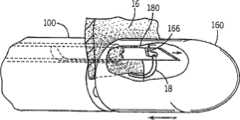
在全身麻醉和双腔通气的情况下,安排并用布帘遮挡患者,从而实现进入右外侧、前侧和左外侧胸腔壁的足够大的外科手术通路(从一侧的腋后线到另一侧的腋后线)。如图1所示,将一个或多个胸腔镜检口经肋间隙插入左胸,并经上述口将器械10插入胸腔。或者,在腋前线的第五个或第六个肋间隙中进行左胸开胸小手术(3-5cm)。患者完全肝素化。左肺下陷之后,打开覆盖左心室14的叶尖12的心包膜并使其边缘悬吊于皮肤切割线。这提供了心尖的闭合通路。通过经食道或血管内超声心动图显象(图中未示出)的组合,并且使用构建在器械10内的光纤系统直接观察,提供对心内手术的指导,如下详细所述。将双侧纱布荷包缝线置于左心室的叶尖12上并在该位置制备穿刺切口。经该切口将外科手术器械10插入搏动心脏的左心室14中。Under general anesthesia and double-chamber ventilation, arrange and drape the patient to allow adequate surgical access to the right lateral, anterior, and left lateral chest walls (from the posterior axillary line on one side to the other) posterior axillary line). As shown in FIG. 1 , one or more thoracoscopic inspection ports are inserted into the left chest through the intercostal space, and an
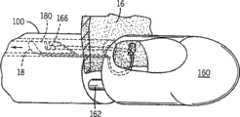
具体参考图2,可用器械10抓住二尖瓣16的脱垂区段,并将人工腱索18固定于其游离边缘。通过超声(echo)和直接光纤观察确保被植入的人工腱索18的准确定位,如下详细所述。然后,将器械10从左心室14中收回,将未附连端与它一起拉出。器械10和腱索18退出后,通过将荷包缝线系在左心室叶尖12中的切口周围以实现止血。如图3所示,新植入腱索18在直接超声多普勒观察下适当张紧并固定于心尖12的外部。就是说,在新植入的腱索18上施加张力,并在超声图像上观察修复瓣膜16的操作。调节张力直到反流最小。Referring specifically to FIG. 2, the prolapsed segment of the
虽然在上文的描述中植入了单个腱索18,还可植入其它腱索或缝线并使其以最佳张力附连于心尖12。在这种情况下,调节所有新植入的腱索18的张力直到实现最佳瓣膜功能。Although a
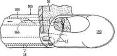
如图4和5所示,用于实现上述操作的器械10包括刚性金属轴100,在该金属轴的胸腔外末端(近端)具有手柄120,使得可以操纵器械并将其导入适当位置。用于控制位于器械远端140的抓持机构和针机构的驱动机构也被安装在手柄120附近。如下所述,通过夹紧剪刀样抓持手柄120操纵抓持机构,通过移动上翻控制轴122操纵针机构。As shown in Figures 4 and 5, the
位于器械10远端即胸腔内末端140的是抓持机构,可操纵该机构来握持脱垂的瓣膜小叶。如图6和7所示,在优选的实施方式中,该机构是在轴100的远端由一组杆162支持的尖端160。杆162在轴100内滑动,以在操纵剪刀样抓持手柄120时,尖端160在图6B和7所示的开放位置与图6A所示的闭合位置间运动。如下所述,二尖瓣膜小叶位于开放尖端160和轴100远端之间的间隙中,通过闭合尖端160以将瓣膜小叶夹紧于其间而俘获该二尖瓣膜小叶。Located at the distal,
在形成于轴100中的针腔164内设置针180,针180连接于轴100近端的控制轴122。针机构180在内缩位置与延伸位置间滑动,在内缩位置时,它被容纳在轴100远端附近的管腔164中,在延伸位置时,在尖端处于闭合位置的情况下它可延伸进入滑动尖端160。结果,当瓣膜小叶被俘获在尖端160和轴100的远端之间时,通过移动控制轴122可使针从管腔164延伸以刺穿俘获的小叶并完全穿过其中。A
轴100的远端还包括将要配置在患者心脏中的人工腱索或缝线18。缝线18通常是由例如Gore-Tex等公司制造的4-0或5-0缝线。通过操纵抓持机构和针机构180来设置缝线18,如下更详细所述。The distal end of the
轴100的尺寸和形状适合插入患者胸腔,穿过左心室壁,与心肌形成不透水密封。它具有圆形或椭圆形截面,可容纳器械的手柄端与心脏内末端之间的控制链接以及光纤观察系统,如下更详细所述。
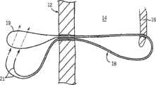
如图8A-8F所示,将器械10远端优选实施方式的缝线配置系统设置在待修复的瓣膜小叶16周围,如图8A所示。缝线18中间折叠以形成位于尖端160中的环19。缝线18的两端被设置在杆162下方的轴100中形成的缝线管腔165内。如图8B所示,通过闭合尖端160抓持瓣膜小叶16,延伸针180以刺穿小叶16并延伸进入尖端160。针180一侧上形成的凹口166钩住缝线环19。然后,针180回缩穿过小叶16以将缝线环19牵拉通过穿刺开口,如图8C所示。然后,释放小叶16,器械10从心脏退出,如图8D所示,用器械10牵拉缝线18的两端和中点。如图8E所示,器械10释放缝线18,外科医生将缝线两端21穿过其中点处的环19。然后,牵拉两端21,环19沿缝线18滑入心脏腔室14,在心脏腔室中形成围绕瓣膜小叶边缘的Larks(拉克司)头结,如图8F所示。As shown in Figures 8A-8F, the preferred embodiment suture deployment system at the distal end of the
可以上述方式植入多条缝线18直到获得令人满意的结果。配置好缝线18之后,通过预先定位的荷包缝线或通过任何类型的适当止血装置或技术来修复心壁切口。检查止血,设置并固定合适的胸腔引流管,并闭合所有切口。
如图9A-9D所示,器械10远端处的缝线配置系统的第二个实施方式是设置在待修复瓣膜小叶16的周围,如图9A所示。在该实施方式中,缝线18是一闭合环,环的一端位于尖端160内,另一端位于管腔164中且包裹围绕针180。针180延伸穿过被抓持的瓣膜小叶16进入器械尖端160,在尖端160中环状缝线18的一端钩住在针的一侧上形成的凹口166,如图9B所示。然后,针180回缩以使环状缝线18通过小叶16中的穿刺开口。然后,通过使尖端160滑入其开放位置而释放小叶,如图9C所示。然后,器械10退出,如图9D所示,使得环状缝线18的未钩住端沿针的长度向小叶16滑动,在小叶16中形成围绕小叶边缘的Larks头结。As shown in Figures 9A-9D, a second embodiment of the suture deployment system at the distal end of the
然后,器械10从心脏腔室14退出,将缝线18的钩住端牵拉穿过心壁。缝线18固定于心尖的外部。The
如图10A-10D所示,器械10远端处的缝线配置系统的第三个实施方式是被设置在待修复瓣膜小叶16的周围,如图10A所示。缝线18的中点17环绕管腔164,其松弛的两端20卷绕在尖端160中。闭合尖端160以俘获瓣膜小叶16之后,针180延伸穿过被抓持的瓣膜小叶160进入器械尖端160。缝线18的游离两端20位于尖端160内以形成环19,在针的一侧上形成的凹口166延伸穿过环19并“钩住”缝线18的游离两端,如图10B所示。然后,针180回缩入管腔164,将缝线18的钩住端牵拉穿过小叶16中的穿刺开口。然后,通过使尖端160滑入其开放位置而释放小叶,如图10C所示。然后,器械10从心脏退出,如图10D所示,将游离两端20牵拉穿过小叶16,缝线18的中点17形成围绕小叶边缘的Larks头结。As shown in FIGS. 10A-10D, a third embodiment of a suture deployment system at the distal end of the
然后,器械10从心脏腔室14退出,将缝线18的游离端20牵拉穿过心壁。缝线18的游离端20固定于心尖的外部。The
其它缝线配置系统也是可能的,例如,针可穿过小叶,与附连于器械尖端160中环状缝线18一端的褡扣配合装置连接。然后,针退出,将装置和环状缝线向后牵拉穿过小叶中的穿刺开口,如上所述。Other suture deployment systems are also possible, for example, a needle may pass through the leaflet to connect with a snap fit attached to one end of the looped
如图7所示,为促进手术期间的观察,四个光纤信道170沿器械轴100的长度延伸并在其远端终止。每个信道170包含至少一根照明光纤,其胸腔外末端连接于白光源(图中未示出)。每个信道170还包含至少一根感应光纤,将来自远端的反射光传递回连接于胸腔外末端的可视监视器(图中未示出)。在优选的实施方式中,每个信道170包含两根照明光纤和两根感应光纤。As shown in Figure 7, to facilitate visualization during surgery, four
四个光纤信道170围绕针腔164设置,使得在适当抓持瓣膜小叶16时,瓣膜小叶组织16靠着所有光纤170的远端。结果,光从组织反射回到感应光纤,在可视监视器上显示四个白色圆环。当小叶16不恰当地按压在信道170的远端时,光不会从小叶16反射,可视监视器显示从血液反射的红色。如果没有俘获瓣膜组织,监视器显示四个红点,而在俘获瓣膜组织时,对应于与组织接触的光纤信道170的点变为白色。如果监视器显示所有四个点都是白色的,则表示瓣膜组织俘获是最佳的。如果只有上面两个点变为白色,下面的点仍然是红色,则表示瓣膜小叶16“咬合”太浅而不能适当附连缝线18。Four
除了确保瓣膜小叶被适当俘获的光纤可视化系统之外,也可采用其它实时可视化系统以便将器械10引导至瓣膜小叶16。优选地,可使用经食道或血管内彩色多普勒超声心动图显象系统来实现上述目的。如上所述,也可采用该成像系统,根据经食道或血管内彩色多普勒超声心动图显象观察反流减少或消失来实时确定新植入的人工腱索的长度。In addition to fiber optic visualization systems to ensure that the valve leaflets are properly captured, other real-time visualization systems may also be employed to guide the
Claims (10)
Translated fromChineseApplications Claiming Priority (3)
| Application Number | Priority Date | Filing Date | Title |
|---|---|---|---|
| US64567705P | 2005-01-21 | 2005-01-21 | |
| US60/645,677 | 2005-01-21 | ||
| PCT/US2006/001699WO2006078694A2 (en) | 2005-01-21 | 2006-01-19 | Thorascopic heart valve repair method and apparatus |
Publications (2)
| Publication Number | Publication Date |
|---|---|
| CN101495049A CN101495049A (en) | 2009-07-29 |
| CN101495049Btrue CN101495049B (en) | 2010-12-15 |
Family
ID=36692807
Family Applications (1)
| Application Number | Title | Priority Date | Filing Date |
|---|---|---|---|
| CN2006800026452AActiveCN101495049B (en) | 2005-01-21 | 2006-01-19 | Thorascopic heart valve repair method and apparatus |
Country Status (8)
| Country | Link |
|---|---|
| US (6) | US8465500B2 (en) |
| EP (1) | EP1845861B1 (en) |
| JP (1) | JP4960262B2 (en) |
| CN (1) | CN101495049B (en) |
| AT (1) | ATE513516T1 (en) |
| AU (1) | AU2006206591B2 (en) |
| CA (1) | CA2595459C (en) |
| WO (1) | WO2006078694A2 (en) |
Cited By (3)
| Publication number | Priority date | Publication date | Assignee | Title |
|---|---|---|---|---|
| US9131938B2 (en) | 2007-03-29 | 2015-09-15 | Nobles Medical Technologies, Inc. | Suturing devices and methods for closing a patent foramen ovale |
| US9326764B2 (en) | 2008-05-09 | 2016-05-03 | Nobles Medical Technologies Inc. | Suturing devices and methods for suturing an anatomic valve |
| US9398907B2 (en) | 1999-07-02 | 2016-07-26 | Quickpass, Inc. | Suturing device |
Families Citing this family (87)
| Publication number | Priority date | Publication date | Assignee | Title |
|---|---|---|---|---|
| ITPC20000013A1 (en) | 2000-04-13 | 2000-07-13 | Paolo Ferrazzi | INTROVENTRICULAR DEVICE AND RELATED METHOD FOR THE TREATMENT AND CORRECTION OF MYOCARDIOPATHIES. |
| EP1845861B1 (en) | 2005-01-21 | 2011-06-22 | Mayo Foundation for Medical Education and Research | Thorascopic heart valve repair apparatus |
| EP1909655A2 (en) | 2005-06-20 | 2008-04-16 | Sutura, Inc. | Method and apparatus for applying a knot to a suture |
| US20070049952A1 (en)* | 2005-08-30 | 2007-03-01 | Weiss Steven J | Apparatus and method for mitral valve repair without cardiopulmonary bypass, including transmural techniques |
| US8764820B2 (en) | 2005-11-16 | 2014-07-01 | Edwards Lifesciences Corporation | Transapical heart valve delivery system and method |
| US7635386B1 (en) | 2006-03-07 | 2009-12-22 | University Of Maryland, Baltimore | Methods and devices for performing cardiac valve repair |
| US8303622B2 (en)* | 2007-03-14 | 2012-11-06 | St. Jude Medical, Inc. | Heart valve chordae replacement methods and apparatus |
| CA2698388C (en) | 2007-09-07 | 2015-11-24 | Edwards Lifesciences Corporation | Active holder for annuloplasty ring delivery |
| CA2703129C (en)* | 2007-10-18 | 2016-02-16 | Neochord Inc. | Minimially invasive repair of a valve leaflet in a beating heart |
| US8337390B2 (en) | 2008-07-30 | 2012-12-25 | Cube S.R.L. | Intracardiac device for restoring the functional elasticity of the cardiac structures, holding tool for the intracardiac device, and method for implantation of the intracardiac device in the heart |
| US20100094334A1 (en)* | 2008-10-10 | 2010-04-15 | Matthew Krever | Plication device with formable linear fastener for use in the direct plication annuloplasty treatment of mitral valve regurgitation |
| US8439970B2 (en) | 2009-07-14 | 2013-05-14 | Edwards Lifesciences Corporation | Transapical delivery system for heart valves |
| US8500757B2 (en)* | 2009-07-28 | 2013-08-06 | Edwards Lifesciences Corporation | Surgical puncture cinch and closure system |
| US20110190793A1 (en)* | 2010-01-29 | 2011-08-04 | Med-Venture Investments, Llc | Methods and apparatuses for suturing of cardiac openings |
| US8496671B1 (en)* | 2010-06-16 | 2013-07-30 | Cardica, Inc. | Mitral valve treatment |
| JP5745837B2 (en)* | 2010-12-24 | 2015-07-08 | オリンパス株式会社 | Treatment tool |
| EP2658480B1 (en)* | 2010-12-29 | 2017-11-01 | Neochord Inc. | Exchangeable system for minimally invasive beating heart repair of heart valve leaflets |
| EP3644194B1 (en) | 2011-04-15 | 2022-12-07 | Heartstitch, Inc. | Suturing devices for suturing an anatomic valve |
| US9381082B2 (en) | 2011-04-22 | 2016-07-05 | Edwards Lifesciences Corporation | Devices, systems and methods for accurate positioning of a prosthetic valve |
| CN103813757A (en) | 2011-06-01 | 2014-05-21 | 尼奥绰德有限公司 | Minimally Invasive Repair of heart valve leaflets |
| WO2013003228A1 (en) | 2011-06-27 | 2013-01-03 | University Of Maryland, Baltimore | Transapical mitral valve repair device |
| US8992550B2 (en)* | 2011-07-20 | 2015-03-31 | Coloplast A/S | Suture system with capsule eyelet providing multiple suture tissue fixation |
| CN104023646A (en)* | 2011-11-23 | 2014-09-03 | 奥特鲁泰克控股有限公司 | Medical occlusion device |
| US8938283B2 (en)* | 2011-12-01 | 2015-01-20 | Neochord, Inc. | Surgical navigation for repair of heart valve leaflets |
| US10076414B2 (en) | 2012-02-13 | 2018-09-18 | Mitraspan, Inc. | Method and apparatus for repairing a mitral valve |
| CA2900930A1 (en) | 2012-02-13 | 2013-08-22 | Mitraspan, Inc. | Method and apparatus for repairing a mitral valve |
| US9706988B2 (en) | 2012-05-11 | 2017-07-18 | Heartstitch, Inc. | Suturing devices and methods for suturing an anatomic structure |
| US20150289867A1 (en)* | 2012-05-23 | 2015-10-15 | Laprotech Ab | Needle for a laproscopic instrument |
| CN104000594B (en)* | 2013-02-27 | 2015-09-30 | 金仕生物科技(常熟)有限公司 | The involutory height measuring rule of valve |
| US10149757B2 (en) | 2013-03-15 | 2018-12-11 | Edwards Lifesciences Corporation | System and method for transaortic delivery of a prosthetic heart valve |
| CN103300938B (en)* | 2013-06-27 | 2015-05-20 | 中国医学科学院阜外心血管病医院 | Trans-atrial pathway atrioventricular valve regurgitation animal modeling device |
| WO2015002815A1 (en) | 2013-07-02 | 2015-01-08 | Med-Venture Investments, Llc | Suturing devices and methods for suturing an anatomic structure |
| JP6469109B2 (en) | 2013-12-06 | 2019-02-13 | メッド − ベンチャー インベストメンツ、エルエルシー | Suture method and apparatus |
| CN103708376B (en)* | 2013-12-17 | 2015-09-23 | 天津大学 | Large-scale deck plate Large travel range synchronization of jacking up control structure |
| US9681864B1 (en) | 2014-01-03 | 2017-06-20 | Harpoon Medical, Inc. | Method and apparatus for transapical procedures on a mitral valve |
| WO2015184138A1 (en) | 2014-05-29 | 2015-12-03 | Cardiaq Valve Technologies, Inc. | Prosthesis, delivery device and methods of use |
| US10178993B2 (en) | 2014-07-11 | 2019-01-15 | Cardio Medical Solutions, Inc. | Device and method for assisting end-to-side anastomosis |
| CN104248457B (en)* | 2014-09-03 | 2016-10-05 | 郭文彬 | An artificial chord device, threading element and kit |
| GB2536538B (en) | 2014-09-17 | 2018-07-18 | Cardiomech As | Anchor for implantation in body tissue |
| CN105615931B (en)* | 2014-10-29 | 2017-12-19 | 上海理工大学 | The Minimally Invasive Surgery apparatus of mitral regurgitation is repaired through apex of the heart implantable artificial chordae tendineae |
| WO2017059406A1 (en) | 2015-10-01 | 2017-04-06 | Neochord, Inc. | Ringless web for repair of heart valves |
| EP3355804B1 (en) | 2015-10-02 | 2020-07-15 | Harpoon Medical, Inc. | Distal anchor apparatus for mitral valve repair |
| CN109152573B (en) | 2015-10-02 | 2021-04-13 | 哈珀恩医疗有限公司 | Distal anchor device and method for mitral valve repair |
| CN108778187A (en) | 2015-12-30 | 2018-11-09 | 管道医疗技术股份有限公司 | tethered mitral valve leaflets |
| WO2017180092A1 (en) | 2016-04-11 | 2017-10-19 | Nobles Medical Technologies Ii, Inc. | Suture spools for tissue suturing device |
| US10624743B2 (en) | 2016-04-22 | 2020-04-21 | Edwards Lifesciences Corporation | Beating-heart mitral valve chordae replacement |
| US10925731B2 (en) | 2016-12-30 | 2021-02-23 | Pipeline Medical Technologies, Inc. | Method and apparatus for transvascular implantation of neo chordae tendinae |
| US11083580B2 (en) | 2016-12-30 | 2021-08-10 | Pipeline Medical Technologies, Inc. | Method of securing a leaflet anchor to a mitral valve leaflet |
| US9877833B1 (en)* | 2016-12-30 | 2018-01-30 | Pipeline Medical Technologies, Inc. | Method and apparatus for transvascular implantation of neo chordae tendinae |
| DE102017002974B4 (en) | 2017-03-28 | 2024-08-08 | Immanuel Albertinen Diakonie Ggmbh | Heart valve implant, suitable for use in minimally invasive surgery to repair a heart valve and/or a heart valve leaflet on the beating heart and heart valve implant system |
| US10213306B2 (en) | 2017-03-31 | 2019-02-26 | Neochord, Inc. | Minimally invasive heart valve repair in a beating heart |
| US10765515B2 (en) | 2017-04-06 | 2020-09-08 | University Of Maryland, Baltimore | Distal anchor apparatus and methods for mitral valve repair |
| EP4115818A3 (en) | 2017-06-19 | 2023-04-05 | Heartstitch, Inc. | Suturing systems and methods for suturing body tissue |
| CA3065223C (en) | 2017-06-19 | 2024-05-07 | Harpoon Medical, Inc. | Method and apparatus for cardiac procedures |
| EP3641660B1 (en) | 2017-06-19 | 2024-11-20 | Heartstitch, Inc. | Suturing devices for suturing an opening in the apex of the heart |
| CN107456253A (en)* | 2017-07-20 | 2017-12-12 | 西安康拓医疗技术有限公司 | The knotting device and its knotting method of suture in a kind of heart valve surgery |
| EP3668415B1 (en) | 2017-08-18 | 2023-10-25 | Nobles Medical Technologies II, Inc. | Apparatus for applying a knot to a suture |
| WO2019051379A1 (en) | 2017-09-11 | 2019-03-14 | Heartstitch, Inc. | Methods and devices for papillary suturing |
| WO2019083894A1 (en) | 2017-10-24 | 2019-05-02 | University Of Maryland, Baltimore | Method and apparatus for cardiac procedures |
| CN108186163B (en) | 2017-11-07 | 2023-07-28 | 杭州德晋医疗科技有限公司 | Artificial tendon implantation system with detection device |
| CN109771094B (en)* | 2017-11-14 | 2020-12-29 | 杭州德晋医疗科技有限公司 | Artificial chordae tendineae implantation system with position detection device |
| CA3094990C (en) | 2018-03-23 | 2023-01-03 | Neochord, Inc. | Device for suture attachment for minimally invasive heart valve repair |
| CN110313947B (en)* | 2018-03-28 | 2024-07-02 | 杭州德晋医疗科技有限公司 | Heart valve repair system |
| US11517435B2 (en) | 2018-05-04 | 2022-12-06 | Edwards Lifesciences Corporation | Ring-based prosthetic cardiac valve |
| US11253360B2 (en) | 2018-05-09 | 2022-02-22 | Neochord, Inc. | Low profile tissue anchor for minimally invasive heart valve repair |
| US11173030B2 (en) | 2018-05-09 | 2021-11-16 | Neochord, Inc. | Suture length adjustment for minimally invasive heart valve repair |
| EP3790509A4 (en) | 2018-05-09 | 2022-03-09 | NeoChord, Inc. | SYSTEMS AND METHODS FOR TRANSCATHETER VALVE REPAIR |
| AU2019336254B2 (en) | 2018-09-07 | 2021-12-09 | Neochord, Inc. | Device for suture attachment for minimally invasive heart valve repair |
| US20220015906A1 (en) | 2018-11-29 | 2022-01-20 | Cardiomech As | Device for Heart Repair |
| JP2022513793A (en) | 2018-12-12 | 2022-02-09 | パイプライン メディカル テクノロジーズ, インコーポレイテッド | Methods and equipment for mitral valve chordae tendineae repair |
| CN109700490B (en)* | 2018-12-29 | 2020-09-11 | 先健科技(深圳)有限公司 | Suturing device |
| CA3126935A1 (en)* | 2019-01-16 | 2020-07-23 | Neochord, Inc. | Transcatheter methods for heart valve repair |
| WO2020163852A1 (en)* | 2019-02-08 | 2020-08-13 | Children's Medical Center Corporation | Optical delivery and insertion of artificial chordae tendineae |
| EP3941397B1 (en)* | 2019-03-19 | 2025-09-03 | Coremedic GmbH | Instrument for repairing an atrioventricular heart valve |
| WO2020214818A1 (en) | 2019-04-16 | 2020-10-22 | Neochord, Inc. | Transverse helical cardiac anchor for minimally invasive heart valve repair |
| CN113729885B (en)* | 2019-12-02 | 2023-01-20 | 北京领健医疗科技有限公司 | Puncture needle, coupler, guide device and repair instrument |
| CN111110400B (en)* | 2019-12-09 | 2022-02-22 | 先健科技(深圳)有限公司 | Heart valve tether and have its heart valve subassembly |
| CN113116422B (en)* | 2019-12-31 | 2024-11-19 | 杭州德晋医疗科技有限公司 | Valve suture device and valve suture system |
| WO2021146757A2 (en) | 2020-01-16 | 2021-07-22 | Neochord, Inc. | Helical cardiac anchors for minimally invasive heart valve repair |
| CA3173298A1 (en)* | 2020-02-27 | 2021-09-02 | Altyx Surgical Inc. | Tissue surface piercing systems and methods |
| CA3178711A1 (en) | 2020-04-22 | 2021-10-28 | Edwards Lifesciences Corporation | Controlled suture tensioning |
| JP7701390B2 (en) | 2020-06-17 | 2025-07-01 | パイプライン メディカル テクノロジーズ, インコーポレイテッド | Methods and devices for mitral valve chordae repair |
| WO2022055923A1 (en) | 2020-09-10 | 2022-03-17 | Edwards Lifesciences Corporation | Closing tissue openings |
| CN115399917B (en)* | 2021-11-05 | 2025-09-23 | 瀚芯医疗科技(深圳)有限公司 | Artificial chord tendon implant device |
| EP4432933A1 (en)* | 2021-11-15 | 2024-09-25 | Boston Scientific Scimed Inc. | Devices, systems, and methods for positioning a leaflet clip |
| CN114869376B (en)* | 2022-05-09 | 2023-03-10 | 南京鼓楼医院 | Atria stitching instrument under thoracoscope |
| CN115919509A (en)* | 2023-02-22 | 2023-04-07 | 上海汇禾医疗器械有限公司 | Heart valve steady-state flow testing system |
Citations (1)
| Publication number | Priority date | Publication date | Assignee | Title |
|---|---|---|---|---|
| US5667472A (en)* | 1994-03-18 | 1997-09-16 | Clarus Medical Systems, Inc. | Surgical instrument and method for use with a viewing system |
Family Cites Families (217)
| Publication number | Priority date | Publication date | Assignee | Title |
|---|---|---|---|---|
| US652388A (en)* | 1900-02-19 | 1900-06-26 | Lewis W Driskell | Churn. |
| US2751908A (en) | 1953-03-19 | 1956-06-26 | American Cystoscope Makers Inc | Surgical instrument |
| US3664660A (en)* | 1967-12-20 | 1972-05-23 | Ruenzi Kurt | Device for feeding flat objects to a processing machine |
| US3664330A (en)* | 1969-09-12 | 1972-05-23 | Harold L Deutsch | Fiber optic medical tool |
| US3667474A (en)* | 1970-01-05 | 1972-06-06 | Konstantin Vasilievich Lapkin | Dilator for performing mitral and tricuspidal commissurotomy per atrium cordis |
| US3842840A (en)* | 1973-05-07 | 1974-10-22 | E Schweizer | Suture applicator |
| US4258716A (en) | 1978-02-06 | 1981-03-31 | The University Of Melbourne | Microsurgical instruments |
| US4351345A (en)* | 1978-10-10 | 1982-09-28 | Carney Andrew L | Methods of securing electrodes to the heart |
| US4759348A (en)* | 1981-09-28 | 1988-07-26 | Cawood Charles David | Endoscope assembly and surgical instrument for use therewith |
| US6631247B1 (en) | 1999-09-29 | 2003-10-07 | Ricoh Co., Ltd. | Method and system for remote diagnostic, control and information collection based on various communication modes for sending messages to a resource manager |
| US4957498A (en)* | 1987-11-05 | 1990-09-18 | Concept, Inc. | Suturing instrument |
| US4960424A (en)* | 1988-06-30 | 1990-10-02 | Grooters Ronald K | Method of replacing a defective atrio-ventricular valve with a total atrio-ventricular valve bioprosthesis |
| GB8829044D0 (en)* | 1988-12-13 | 1989-01-25 | Fullcharge Ltd | Diaphragm valves |
| US4967498A (en) | 1989-01-19 | 1990-11-06 | Kao Pei Chin | Picture frame |
| US5290300A (en) | 1989-07-31 | 1994-03-01 | Baxter International Inc. | Flexible suture guide and holder |
| US4935027A (en)* | 1989-08-21 | 1990-06-19 | Inbae Yoon | Surgical suture instrument with remotely controllable suture material advancement |
| US5059201A (en)* | 1989-11-03 | 1991-10-22 | Asnis Stanley E | Suture threading, stitching and wrapping device for use in open and closed surgical procedures |
| US5984939A (en) | 1989-12-05 | 1999-11-16 | Yoon; Inbae | Multifunctional grasping instrument with cutting member and operating channel for use in endoscopic and non-endoscopic procedures |
| US5665100A (en) | 1989-12-05 | 1997-09-09 | Yoon; Inbae | Multifunctional instrument with interchangeable operating units for performing endoscopic procedures |
| US5259846A (en)* | 1991-01-07 | 1993-11-09 | United States Surgical Corporation | Loop threaded combined surgical needle-suture device |
| US5211650A (en) | 1991-01-07 | 1993-05-18 | Laparomed Corporation | Dual function suturing device and method |
| US5383877A (en) | 1991-05-01 | 1995-01-24 | Clarke; Henry C. | Instruments and method for suturing and ligation |
| US5452733A (en)* | 1993-02-22 | 1995-09-26 | Stanford Surgical Technologies, Inc. | Methods for performing thoracoscopic coronary artery bypass |
| US5571215A (en)* | 1993-02-22 | 1996-11-05 | Heartport, Inc. | Devices and methods for intracardiac procedures |
| US5676651A (en)* | 1992-08-06 | 1997-10-14 | Electric Boat Corporation | Surgically implantable pump arrangement and method for pumping body fluids |
| US5762458A (en)* | 1996-02-20 | 1998-06-09 | Computer Motion, Inc. | Method and apparatus for performing minimally invasive cardiac procedures |
| US5297536A (en)* | 1992-08-25 | 1994-03-29 | Wilk Peter J | Method for use in intra-abdominal surgery |
| CA2143825A1 (en) | 1992-08-28 | 1994-03-17 | Donald L. Murphy | Endoscopic suturing device |
| US5772597A (en)* | 1992-09-14 | 1998-06-30 | Sextant Medical Corporation | Surgical tool end effector |
| US5312423A (en)* | 1992-10-01 | 1994-05-17 | Advanced Surgical Intervention, Inc. | Apparatus and method for laparaoscopic ligation |
| JPH06142114A (en)* | 1992-10-30 | 1994-05-24 | Olympus Optical Co Ltd | In-celom treating device |
| US5304185A (en)* | 1992-11-04 | 1994-04-19 | Unisurge, Inc. | Needle holder |
| US5667478A (en)* | 1992-11-06 | 1997-09-16 | Clarus Medical Systems, Inc. | Surgical instrument with stick-on fiber-optic viewing system and method of using |
| US6355050B1 (en) | 1992-12-10 | 2002-03-12 | Abbott Laboratories | Device and method for suturing tissue |
| US5336229A (en)* | 1993-02-09 | 1994-08-09 | Laparomed Corporation | Dual ligating and dividing apparatus |
| US5797960A (en) | 1993-02-22 | 1998-08-25 | Stevens; John H. | Method and apparatus for thoracoscopic intracardiac procedures |
| US5972030A (en)* | 1993-02-22 | 1999-10-26 | Heartport, Inc. | Less-invasive devices and methods for treatment of cardiac valves |
| US6629984B1 (en) | 1998-07-07 | 2003-10-07 | Kwan-Ho Chan | Surgical repair kit and its method of use |
| US5618290A (en)* | 1993-10-19 | 1997-04-08 | W.L. Gore & Associates, Inc. | Endoscopic suture passer and method |
| US5431666A (en)* | 1994-02-24 | 1995-07-11 | Lasersurge, Inc. | Surgical suture instrument |
| US5547455A (en)* | 1994-03-30 | 1996-08-20 | Medical Media Systems | Electronically steerable endoscope |
| US5474519A (en)* | 1994-05-10 | 1995-12-12 | Bloomer; William E. | Method for obtaining stereoscopic imagery from a pair of endoscopes |
| CA2157744C (en) | 1994-10-07 | 2005-08-23 | Charles R. Sherts | Endoscopic vascular suturing apparatus |
| US5653716A (en)* | 1994-12-29 | 1997-08-05 | Acufex Microsurgical, Inc. | Suture manipulating instrument with grasping members |
| US5626607A (en) | 1995-04-03 | 1997-05-06 | Heartport, Inc. | Clamp assembly and method of use |
| US5857961A (en)* | 1995-06-07 | 1999-01-12 | Clarus Medical Systems, Inc. | Surgical instrument for use with a viewing system |
| US5839639A (en)* | 1995-08-17 | 1998-11-24 | Lasersurge, Inc. | Collapsible anvil assembly and applicator instrument |
| US6562052B2 (en) | 1995-08-24 | 2003-05-13 | Sutura, Inc. | Suturing device and method |
| US6117144A (en) | 1995-08-24 | 2000-09-12 | Sutura, Inc. | Suturing device and method for sealing an opening in a blood vessel or other biological structure |
| US6436107B1 (en) | 1996-02-20 | 2002-08-20 | Computer Motion, Inc. | Method and apparatus for performing minimally invasive surgical procedures |
| US5972004A (en) | 1996-02-23 | 1999-10-26 | Cardiovascular Technologies, Llc. | Wire fasteners for use in minimally invasive surgery and apparatus and methods for handling those fasteners |
| US6162233A (en) | 1996-02-23 | 2000-12-19 | Cardiovascular Technologies, Llc | Wire fasteners for use in minimally invasive surgery and means and methods for handling those fasteners |
| US6149660A (en)* | 1996-04-22 | 2000-11-21 | Vnus Medical Technologies, Inc. | Method and apparatus for delivery of an appliance in a vessel |
| DE19616984A1 (en)* | 1996-04-27 | 1997-10-30 | Basf Lacke & Farben | Binders and their use in radiation-curable coating compositions |
| US5762613A (en)* | 1996-05-07 | 1998-06-09 | Spectrascience, Inc. | Optical biopsy forceps |
| DE19632298B4 (en) | 1996-08-10 | 2004-09-23 | Deutsches Zentrum für Luft- und Raumfahrt e.V. | Gripping device for use in minimally invasive surgery |
| US5993466A (en) | 1997-06-17 | 1999-11-30 | Yoon; Inbae | Suturing instrument with multiple rotatably mounted spreadable needle holders |
| US5993467A (en) | 1996-11-27 | 1999-11-30 | Yoon; Inbae | Suturing instrument with rotatably mounted spreadable needle holder |
| US6050936A (en) | 1997-01-02 | 2000-04-18 | Myocor, Inc. | Heart wall tension reduction apparatus |
| US6183411B1 (en) | 1998-09-21 | 2001-02-06 | Myocor, Inc. | External stress reduction device and method |
| US7883539B2 (en) | 1997-01-02 | 2011-02-08 | Edwards Lifesciences Llc | Heart wall tension reduction apparatus and method |
| US6077214A (en) | 1998-07-29 | 2000-06-20 | Myocor, Inc. | Stress reduction apparatus and method |
| US5961440A (en)* | 1997-01-02 | 1999-10-05 | Myocor, Inc. | Heart wall tension reduction apparatus and method |
| US6406420B1 (en) | 1997-01-02 | 2002-06-18 | Myocor, Inc. | Methods and devices for improving cardiac function in hearts |
| US6045497A (en)* | 1997-01-02 | 2000-04-04 | Myocor, Inc. | Heart wall tension reduction apparatus and method |
| US5957879A (en) | 1997-01-24 | 1999-09-28 | Heartport, Inc. | Methods and devices for maintaining cardiopulmonary bypass and arresting a patient's heart |
| US5830231A (en)* | 1997-03-19 | 1998-11-03 | Geiges, Jr.; John J. | Handle and actuating mechanism for surgical instruments |
| US5897564A (en)* | 1997-04-08 | 1999-04-27 | Ethicon Endo-Surgery, Inc. | Endoscopic instrument assembly for fastening tissue |
| US5957936A (en)* | 1997-05-01 | 1999-09-28 | Inbae Yoon | Instrument assemblies for performing anatomical tissue ligation |
| US5908429A (en)* | 1997-05-01 | 1999-06-01 | Yoon; Inbae | Methods of anatomical tissue ligation |
| US5908428A (en)* | 1997-05-27 | 1999-06-01 | United States Surgical Corporation | Stitching devices for heart valve replacement surgery |
| US5919128A (en)* | 1997-06-18 | 1999-07-06 | The Regents Of The University Of California | Sparse aperture endoscope |
| EP2133030A1 (en)* | 1997-06-27 | 2009-12-16 | The Trustees of Columbia University of the City of New York | Method and apparatus for circulatory valve repair |
| US5910148A (en)* | 1997-08-06 | 1999-06-08 | Mitek Surgical Products, Inc. | Suture retrograder |
| AU9225598A (en)* | 1997-09-04 | 1999-03-22 | Endocore, Inc. | Artificial chordae replacement |
| FR2768324B1 (en) | 1997-09-12 | 1999-12-10 | Jacques Seguin | SURGICAL INSTRUMENT FOR PERCUTANEOUSLY FIXING TWO AREAS OF SOFT TISSUE, NORMALLY MUTUALLY REMOTE, TO ONE ANOTHER |
| US6695810B2 (en) | 1997-11-21 | 2004-02-24 | Advanced Interventional Technologies, Inc. | Endolumenal aortic isolation assembly and method |
| US6234995B1 (en)* | 1998-11-12 | 2001-05-22 | Advanced Interventional Technologies, Inc. | Apparatus and method for selectively isolating a proximal anastomosis site from blood in an aorta |
| US5928181A (en) | 1997-11-21 | 1999-07-27 | Advanced International Technologies, Inc. | Cardiac bypass catheter system and method of use |
| US6332893B1 (en) | 1997-12-17 | 2001-12-25 | Myocor, Inc. | Valve to myocardium tension members device and method |
| US6190357B1 (en)* | 1998-04-21 | 2001-02-20 | Cardiothoracic Systems, Inc. | Expandable cannula for performing cardiopulmonary bypass and method for using same |
| US6508777B1 (en) | 1998-05-08 | 2003-01-21 | Cardeon Corporation | Circulatory support system and method of use for isolated segmental perfusion |
| US6585144B2 (en) | 1998-06-19 | 2003-07-01 | Acimed Life Systems, Inc. | Integrated surgical staple retainer for a full thickness resectioning device |
| US6165183A (en) | 1998-07-15 | 2000-12-26 | St. Jude Medical, Inc. | Mitral and tricuspid valve repair |
| US6260552B1 (en) | 1998-07-29 | 2001-07-17 | Myocor, Inc. | Transventricular implant tools and devices |
| AU5273599A (en) | 1998-08-10 | 2000-03-06 | Coroneo Inc. | Surgical suture and associated anchoring mechanism for tissue retraction |
| US6419626B1 (en)* | 1998-08-12 | 2002-07-16 | Inbae Yoon | Surgical instrument endoscope with CMOS image sensor and physical parameter sensor |
| US6178346B1 (en)* | 1998-10-23 | 2001-01-23 | David C. Amundson | Infrared endoscopic imaging in a liquid with suspended particles: method and apparatus |
| US6270508B1 (en)* | 1998-10-26 | 2001-08-07 | Charles H. Klieman | End effector and instrument for endoscopic and general surgery needle control |
| US6468265B1 (en) | 1998-11-20 | 2002-10-22 | Intuitive Surgical, Inc. | Performing cardiac surgery without cardioplegia |
| US7811296B2 (en) | 1999-04-09 | 2010-10-12 | Evalve, Inc. | Fixation devices for variation in engagement of tissue |
| US6752813B2 (en) | 1999-04-09 | 2004-06-22 | Evalve, Inc. | Methods and devices for capturing and fixing leaflets in valve repair |
| US8216256B2 (en) | 1999-04-09 | 2012-07-10 | Evalve, Inc. | Detachment mechanism for implantable fixation devices |
| US20040044350A1 (en) | 1999-04-09 | 2004-03-04 | Evalve, Inc. | Steerable access sheath and methods of use |
| AU770243B2 (en) | 1999-04-09 | 2004-02-19 | Evalve, Inc. | Methods and apparatus for cardiac valve repair |
| US7226467B2 (en) | 1999-04-09 | 2007-06-05 | Evalve, Inc. | Fixation device delivery catheter, systems and methods of use |
| US6152934A (en)* | 1999-06-14 | 2000-11-28 | Ethicon Endo-Surgery, Inc. | Surgical knot tying instrument |
| US7744613B2 (en)* | 1999-06-25 | 2010-06-29 | Usgi Medical, Inc. | Apparatus and methods for forming and securing gastrointestinal tissue folds |
| US7217240B2 (en) | 1999-10-01 | 2007-05-15 | Intuitive Surgical, Inc. | Heart stabilizer |
| US6312447B1 (en) | 1999-10-13 | 2001-11-06 | The General Hospital Corporation | Devices and methods for percutaneous mitral valve repair |
| US6626930B1 (en) | 1999-10-21 | 2003-09-30 | Edwards Lifesciences Corporation | Minimally invasive mitral valve repair method and apparatus |
| US6585727B1 (en) | 1999-10-22 | 2003-07-01 | Genzyme Corporation | Surgical instrument light source and surgical illumination method |
| US6626917B1 (en)* | 1999-10-26 | 2003-09-30 | H. Randall Craig | Helical suture instrument |
| US6926730B1 (en) | 2000-10-10 | 2005-08-09 | Medtronic, Inc. | Minimally invasive valve repair procedure and apparatus |
| US6458153B1 (en) | 1999-12-31 | 2002-10-01 | Abps Venture One, Ltd. | Endoluminal cardiac and venous valve prostheses and methods of manufacture and delivery thereof |
| US6989028B2 (en) | 2000-01-31 | 2006-01-24 | Edwards Lifesciences Ag | Medical system and method for remodeling an extravascular tissue structure |
| US6402781B1 (en) | 2000-01-31 | 2002-06-11 | Mitralife | Percutaneous mitral annuloplasty and cardiac reinforcement |
| JP3679674B2 (en)* | 2000-02-03 | 2005-08-03 | オリンパス株式会社 | Endoscope |
| EP1261282B1 (en)* | 2000-03-03 | 2013-09-25 | C. R. Bard, Inc. | Endoscopic tissue apposition device with multiple suction ports |
| IL138632A (en)* | 2000-09-21 | 2008-06-05 | Minelu Zonnenschein | Multiple view endoscopes |
| US6537198B1 (en) | 2000-03-21 | 2003-03-25 | Myocor, Inc. | Splint assembly for improving cardiac function in hearts, and method for implanting the splint assembly |
| US20020107514A1 (en) | 2000-04-27 | 2002-08-08 | Hooven Michael D. | Transmural ablation device with parallel jaws |
| US7083628B2 (en)* | 2002-09-03 | 2006-08-01 | Edwards Lifesciences Corporation | Single catheter mitral valve repair device and method for use |
| US6869444B2 (en) | 2000-05-22 | 2005-03-22 | Shlomo Gabbay | Low invasive implantable cardiac prosthesis and method for helping improve operation of a heart valve |
| WO2001095809A1 (en) | 2000-06-14 | 2001-12-20 | Sterilis, Inc. | Suturing method and apparatus |
| US6840246B2 (en)* | 2000-06-20 | 2005-01-11 | University Of Maryland, Baltimore | Apparatuses and methods for performing minimally invasive diagnostic and surgical procedures inside of a beating heart |
| SE0002878D0 (en) | 2000-08-11 | 2000-08-11 | Kimblad Ola | Device and method of treatment of atrioventricular regurgitation |
| WO2002015795A2 (en) | 2000-08-25 | 2002-02-28 | Sutura, Inc. | Suture cutter |
| US20080091264A1 (en) | 2002-11-26 | 2008-04-17 | Ample Medical, Inc. | Devices, systems, and methods for reshaping a heart valve annulus, including the use of magnetic tools |
| US6602288B1 (en) | 2000-10-05 | 2003-08-05 | Edwards Lifesciences Corporation | Minimally-invasive annuloplasty repair segment delivery template, system and method of use |
| US6723038B1 (en) | 2000-10-06 | 2004-04-20 | Myocor, Inc. | Methods and devices for improving mitral valve function |
| US6616684B1 (en) | 2000-10-06 | 2003-09-09 | Myocor, Inc. | Endovascular splinting devices and methods |
| US6918917B1 (en) | 2000-10-10 | 2005-07-19 | Medtronic, Inc. | Minimally invasive annuloplasty procedure and apparatus |
| US6533796B1 (en)* | 2000-10-11 | 2003-03-18 | Lsi Solutions, Inc. | Loader for surgical suturing instrument |
| WO2002043569A2 (en) | 2000-11-28 | 2002-06-06 | Intuitive Surgical, Inc. | Endoscopic beating-heart stabilizer and vessel occlusion fastener |
| US6810882B2 (en) | 2001-01-30 | 2004-11-02 | Ev3 Santa Rosa, Inc. | Transluminal mitral annuloplasty |
| US6997931B2 (en)* | 2001-02-02 | 2006-02-14 | Lsi Solutions, Inc. | System for endoscopic suturing |
| US7083638B2 (en) | 2001-02-12 | 2006-08-01 | Arthrocare Corporation | Method and apparatus for attaching connective tissues to bone using a knotless suture anchoring device |
| US6649196B2 (en) | 2001-03-12 | 2003-11-18 | Mayo Foundation For Medical Education And Research | Methods of reducing β-amyloid polypeptides |
| ATE415869T1 (en)* | 2001-03-23 | 2008-12-15 | Arthrex Inc | INSTRUMENT FOR PULLING A THREAD DURING ARTHROSCOPIC SEWING OF TISSUE |
| US7186264B2 (en) | 2001-03-29 | 2007-03-06 | Viacor, Inc. | Method and apparatus for improving mitral valve function |
| US6622730B2 (en) | 2001-03-30 | 2003-09-23 | Myocor, Inc. | Device for marking and aligning positions on the heart |
| US20050125011A1 (en) | 2001-04-24 | 2005-06-09 | Spence Paul A. | Tissue fastening systems and methods utilizing magnetic guidance |
| US6619291B2 (en) | 2001-04-24 | 2003-09-16 | Edwin J. Hlavka | Method and apparatus for catheter-based annuloplasty |
| US7935145B2 (en) | 2001-05-17 | 2011-05-03 | Edwards Lifesciences Corporation | Annuloplasty ring for ischemic mitral valve insuffuciency |
| US7122040B2 (en)* | 2001-06-15 | 2006-10-17 | J. Donald Hill | Suture placement apparatus |
| US6702847B2 (en)* | 2001-06-29 | 2004-03-09 | Scimed Life Systems, Inc. | Endoluminal device with indicator member for remote detection of endoleaks and/or changes in device morphology |
| US6645205B2 (en) | 2001-08-15 | 2003-11-11 | Core Medical, Inc. | Apparatus and methods for reducing lung volume |
| US20030050693A1 (en) | 2001-09-10 | 2003-03-13 | Quijano Rodolfo C. | Minimally invasive delivery system for annuloplasty rings |
| JP3921681B2 (en) | 2001-10-01 | 2007-05-30 | ニプロ株式会社 | Intracardiac suture device |
| US6692605B2 (en) | 2001-10-15 | 2004-02-17 | Eastman Kodak Company | Method for laminating an overlay to verify a pattern or as a pattern |
| ATE326906T1 (en)* | 2001-10-23 | 2006-06-15 | Arthrex Inc | SURGICAL DEVICE FOR SEWING TISSUE |
| US7118583B2 (en)* | 2001-10-23 | 2006-10-10 | Arthrex, Inc. | Meniscal suturing instrument and method |
| GB0125925D0 (en) | 2001-10-29 | 2001-12-19 | Univ Glasgow | Mitral valve prosthesis |
| US6575971B2 (en) | 2001-11-15 | 2003-06-10 | Quantum Cor, Inc. | Cardiac valve leaflet stapler device and methods thereof |
| US6978176B2 (en)* | 2001-12-08 | 2005-12-20 | Lattouf Omar M | Treatment for patient with congestive heart failure |
| US6740107B2 (en) | 2001-12-19 | 2004-05-25 | Trimedyne, Inc. | Device for treatment of atrioventricular valve regurgitation |
| US20030120341A1 (en)* | 2001-12-21 | 2003-06-26 | Hani Shennib | Devices and methods of repairing cardiac valves |
| US6764510B2 (en) | 2002-01-09 | 2004-07-20 | Myocor, Inc. | Devices and methods for heart valve treatment |
| WO2003105670A2 (en) | 2002-01-10 | 2003-12-24 | Guided Delivery Systems, Inc. | Devices and methods for heart valve repair |
| SE520622C2 (en) | 2002-01-25 | 2003-08-05 | Btg Kaelle Inventing Ab | Method and apparatus for measuring concentrations |
| US7048754B2 (en) | 2002-03-01 | 2006-05-23 | Evalve, Inc. | Suture fasteners and methods of use |
| US7261728B2 (en)* | 2002-03-15 | 2007-08-28 | Ethicon Endo-Surgery, Inc. | Biopsy forceps device and method |
| US6719786B2 (en)* | 2002-03-18 | 2004-04-13 | Medtronic, Inc. | Flexible annuloplasty prosthesis and holder |
| US7094244B2 (en) | 2002-03-26 | 2006-08-22 | Edwards Lifesciences Corporation | Sequential heart valve leaflet repair device and method of use |
| US20030187457A1 (en) | 2002-04-02 | 2003-10-02 | Weber John A. | Apparatus and method for removing an object from a body |
| JP3890589B2 (en)* | 2002-04-15 | 2007-03-07 | ニプロ株式会社 | Intracardiac suture device |
| US6770084B1 (en) | 2002-06-26 | 2004-08-03 | Opus Medical, Inc. | Suture capture device |
| US6936054B2 (en)* | 2002-07-22 | 2005-08-30 | Boston Scientific Scimed, Inc. | Placing sutures |
| EP1534146B1 (en)* | 2002-08-13 | 2008-01-23 | The General Hospital Corporation | Cardiac devices for percutaneous repair of atrioventricular valves |
| US20040087978A1 (en)* | 2002-08-27 | 2004-05-06 | Velez Juan Manuel | Surgical fascia closure instrument, guide and method |
| US20050021055A1 (en)* | 2002-08-27 | 2005-01-27 | Souhail Toubia | Surgical closure instrument and methods |
| AU2003295380A1 (en) | 2002-11-12 | 2004-06-03 | Myocor, Inc. | Devices and methods for heart valve treatment |
| US7112219B2 (en) | 2002-11-12 | 2006-09-26 | Myocor, Inc. | Devices and methods for heart valve treatment |
| US7247134B2 (en) | 2002-11-12 | 2007-07-24 | Myocor, Inc. | Devices and methods for heart valve treatment |
| AU2003290979A1 (en) | 2002-11-15 | 2004-06-15 | The Government Of The United States Of America As Represented By The Secretary Of Health And Human Services | Method and device for catheter-based repair of cardiac valves |
| US6997950B2 (en)* | 2003-01-16 | 2006-02-14 | Chawla Surendra K | Valve repair device |
| US7381210B2 (en)* | 2003-03-14 | 2008-06-03 | Edwards Lifesciences Corporation | Mitral valve repair system and method for use |
| US20040236373A1 (en)* | 2003-05-20 | 2004-11-25 | Anspach William E. | Surgical method for suturing tendons/ligaments to bones |
| CA2526347C (en) | 2003-05-20 | 2010-07-06 | The Cleveland Clinic Foundation | Apparatus and methods for repair of a cardiac valve |
| WO2005009286A2 (en) | 2003-07-23 | 2005-02-03 | Viacor, Inc. | Method and apparatus for improving mitral valve function |
| US7908484B2 (en) | 2003-08-22 | 2011-03-15 | Nokia Corporation | Method of protecting digest authentication and key agreement (AKA) against man-in-the-middle (MITM) attack |
| CA2542658A1 (en) | 2003-10-17 | 2005-05-06 | Edwards Lifesciences Ag | Heart valve leaflet locator |
| US7004176B2 (en) | 2003-10-17 | 2006-02-28 | Edwards Lifesciences Ag | Heart valve leaflet locator |
| US20050165272A1 (en)* | 2003-12-01 | 2005-07-28 | Yuta Okada | Endoscope system |
| US7211093B2 (en)* | 2004-01-14 | 2007-05-01 | Lsi Solutions, Inc. | Sew-right running stitch instrument |
| US20050209612A1 (en)* | 2004-03-02 | 2005-09-22 | Nakao Naomi L | Endoscopic suturing assembly and associated methodology using a temperature biased suture needle |
| WO2005094525A2 (en) | 2004-03-23 | 2005-10-13 | Correx, Inc. | Apparatus and method for connecting a conduit to a hollow organ |
| US20070265643A1 (en) | 2004-03-23 | 2007-11-15 | Beane Richard M | Apparatus and method for suturelessly connecting a conduit to a hollow organ |
| US7799041B2 (en) | 2004-03-23 | 2010-09-21 | Correx, Inc. | Apparatus and method for forming a hole in a hollow organ |
| US20050240202A1 (en) | 2004-04-21 | 2005-10-27 | Hani Shennib | Devices and methods of repairing cardiac valves |
| US7294148B2 (en) | 2004-04-29 | 2007-11-13 | Edwards Lifesciences Corporation | Annuloplasty ring for mitral valve prolapse |
| US20080091059A1 (en) | 2004-05-14 | 2008-04-17 | Ample Medical, Inc. | Devices, systems, and methods for reshaping a heart valve annulus, including the use of a bridge implant having an adjustable bridge stop |
| JP4774048B2 (en) | 2004-05-14 | 2011-09-14 | エヴァルヴ インコーポレイテッド | Locking mechanism of fixing device engaged with tissue and tissue engaging method |
| US20060074485A1 (en) | 2004-05-17 | 2006-04-06 | Fidel Realyvasquez | Method and apparatus for percutaneous valve repair |
| DE102004035399A1 (en)* | 2004-07-21 | 2006-02-16 | Technotrans Ag | Device for cleaning surfaces, in particular printing press cylinders |
| US7704277B2 (en) | 2004-09-14 | 2010-04-27 | Edwards Lifesciences Ag | Device and method for treatment of heart valve regurgitation |
| US8602971B2 (en) | 2004-09-24 | 2013-12-10 | Vivid Medical. Inc. | Opto-Electronic illumination and vision module for endoscopy |
| WO2006037073A2 (en) | 2004-09-27 | 2006-04-06 | Evalve, Inc. | Methods and devices for tissue grasping and assessment |
| WO2006065966A2 (en) | 2004-12-15 | 2006-06-22 | Correx, Inc. | Apparatus and method for connecting a conduit to a hollow vessel |
| EP1845861B1 (en) | 2005-01-21 | 2011-06-22 | Mayo Foundation for Medical Education and Research | Thorascopic heart valve repair apparatus |
| US7955385B2 (en) | 2005-02-28 | 2011-06-07 | Medtronic Vascular, Inc. | Device, system, and method for aiding valve annuloplasty |
| US20060247672A1 (en) | 2005-04-27 | 2006-11-02 | Vidlund Robert M | Devices and methods for pericardial access |
| WO2006127509A2 (en) | 2005-05-20 | 2006-11-30 | Mayo Foundation For Medical Education And Research | Devices and methods for reducing cardiac valve regurgitation |
| US20070027451A1 (en) | 2005-06-23 | 2007-02-01 | Kai Desinger | Method for treatment of hypertrophic palatine tonsils |
| US8685083B2 (en) | 2005-06-27 | 2014-04-01 | Edwards Lifesciences Corporation | Apparatus, system, and method for treatment of posterior leaflet prolapse |
| KR100680455B1 (en) | 2005-06-30 | 2007-02-08 | 주식회사 하이닉스반도체 | NAD flash memory device, manufacturing method thereof and driving method thereof |
| US20070049952A1 (en) | 2005-08-30 | 2007-03-01 | Weiss Steven J | Apparatus and method for mitral valve repair without cardiopulmonary bypass, including transmural techniques |
| AU2006335227B2 (en) | 2005-10-14 | 2013-06-13 | Correx, Inc. | Apparatus and method for forming a hole in a hollow organ |
| US20070232941A1 (en)* | 2005-10-27 | 2007-10-04 | Stan Rabinovich | System, apparatus, and method for imaging and treating tissue |
| US7632308B2 (en)* | 2005-11-23 | 2009-12-15 | Didier Loulmet | Methods, devices, and kits for treating mitral valve prolapse |
| US8043368B2 (en) | 2005-11-23 | 2011-10-25 | Traves Dean Crabtree | Methods and apparatus for atrioventricular valve repair |
| US20070203391A1 (en) | 2006-02-24 | 2007-08-30 | Medtronic Vascular, Inc. | System for Treating Mitral Valve Regurgitation |
| US7635386B1 (en) | 2006-03-07 | 2009-12-22 | University Of Maryland, Baltimore | Methods and devices for performing cardiac valve repair |
| US8114121B2 (en) | 2006-06-22 | 2012-02-14 | Tyco Healthcare Group Lp | Tissue vitality comparator with light pipe with fiber optic imaging bundle |
| WO2008013864A2 (en) | 2006-07-27 | 2008-01-31 | Axya Medical, Inc. | Suture needle, suture needle/suture assembly and suture passer device |
| US20080065156A1 (en) | 2006-09-08 | 2008-03-13 | Hauser David L | Expandable clip for tissue repair |
| EP2397108B1 (en) | 2006-09-08 | 2013-08-07 | Edwards Lifesciences Corporation | Apparatus for treating a defective heart valve |
| US20080065205A1 (en) | 2006-09-11 | 2008-03-13 | Duy Nguyen | Retrievable implant and method for treatment of mitral regurgitation |
| US8562629B2 (en) | 2006-10-24 | 2013-10-22 | Arthrocare Corporation | Suture device having selective needle actuation and related method |
| WO2008010738A2 (en) | 2007-01-24 | 2008-01-24 | Uros Babic | Patent foramen ovale occluder with suture based anchor |
| US8303622B2 (en) | 2007-03-14 | 2012-11-06 | St. Jude Medical, Inc. | Heart valve chordae replacement methods and apparatus |
| EP2033583B1 (en) | 2007-08-27 | 2013-03-13 | Arthrex, Inc. | In-line suture passer |
| CA2703129C (en) | 2007-10-18 | 2016-02-16 | Neochord Inc. | Minimially invasive repair of a valve leaflet in a beating heart |
| US8795352B2 (en) | 2008-04-15 | 2014-08-05 | Medtronic Vascular, Inc. | Devices and methods for treating valvular regurgitation |
| US8408441B2 (en)* | 2009-01-06 | 2013-04-02 | Covidien Lp | Surgical stapler |
| CA3094990C (en) | 2018-03-23 | 2023-01-03 | Neochord, Inc. | Device for suture attachment for minimally invasive heart valve repair |
- 2006
- 2006-01-19EPEP06718728Apatent/EP1845861B1/enactiveActive
- 2006-01-19JPJP2007552225Apatent/JP4960262B2/enactiveActive
- 2006-01-19CNCN2006800026452Apatent/CN101495049B/enactiveActive
- 2006-01-19ATAT06718728Tpatent/ATE513516T1/ennot_activeIP Right Cessation
- 2006-01-19CACA2595459Apatent/CA2595459C/enactiveActive
- 2006-01-19AUAU2006206591Apatent/AU2006206591B2/enactiveActive
- 2006-01-19USUS11/813,695patent/US8465500B2/enactiveActive
- 2006-01-19WOPCT/US2006/001699patent/WO2006078694A2/enactiveApplication Filing
- 2010
- 2010-02-19USUS12/709,220patent/US8968338B2/enactiveActive
- 2013
- 2013-05-21USUS13/898,709patent/US9364213B2/enactiveActive
- 2015
- 2015-02-05USUS14/614,570patent/US9700300B2/enactiveActive
- 2017
- 2017-06-27USUS15/634,412patent/US10582924B2/enactiveActive
- 2019
- 2019-12-20USUS16/722,604patent/US11534156B2/enactiveActive
Patent Citations (1)
| Publication number | Priority date | Publication date | Assignee | Title |
|---|---|---|---|---|
| US5667472A (en)* | 1994-03-18 | 1997-09-16 | Clarus Medical Systems, Inc. | Surgical instrument and method for use with a viewing system |
Cited By (6)
| Publication number | Priority date | Publication date | Assignee | Title |
|---|---|---|---|---|
| US9398907B2 (en) | 1999-07-02 | 2016-07-26 | Quickpass, Inc. | Suturing device |
| US10194902B2 (en) | 1999-07-02 | 2019-02-05 | Quickpass, Inc. | Suturing device |
| US9131938B2 (en) | 2007-03-29 | 2015-09-15 | Nobles Medical Technologies, Inc. | Suturing devices and methods for closing a patent foramen ovale |
| US10182802B2 (en) | 2007-03-29 | 2019-01-22 | Nobles Medical Technologies, Inc. | Suturing devices and methods for closing a patent foramen ovale |
| US9326764B2 (en) | 2008-05-09 | 2016-05-03 | Nobles Medical Technologies Inc. | Suturing devices and methods for suturing an anatomic valve |
| US10285687B2 (en) | 2008-05-09 | 2019-05-14 | Nobles Medical Technologies Inc. | Suturing devices and methods for suturing an anatomic valve |
Also Published As
| Publication number | Publication date |
|---|---|
| EP1845861B1 (en) | 2011-06-22 |
| JP2008536528A (en) | 2008-09-11 |
| AU2006206591A1 (en) | 2006-07-27 |
| WO2006078694A2 (en) | 2006-07-27 |
| AU2006206591B2 (en) | 2011-08-25 |
| US8465500B2 (en) | 2013-06-18 |
| US20140039324A1 (en) | 2014-02-06 |
| US9700300B2 (en) | 2017-07-11 |
| US20150148821A1 (en) | 2015-05-28 |
| EP1845861A2 (en) | 2007-10-24 |
| JP4960262B2 (en) | 2012-06-27 |
| ATE513516T1 (en) | 2011-07-15 |
| US20100174297A1 (en) | 2010-07-08 |
| US20170290582A1 (en) | 2017-10-12 |
| EP1845861A4 (en) | 2009-12-09 |
| WO2006078694A3 (en) | 2009-04-16 |
| US20080188873A1 (en) | 2008-08-07 |
| CA2595459A1 (en) | 2006-07-27 |
| HK1137130A1 (en) | 2010-07-23 |
| US9364213B2 (en) | 2016-06-14 |
| CA2595459C (en) | 2013-11-26 |
| CN101495049A (en) | 2009-07-29 |
| US8968338B2 (en) | 2015-03-03 |
| US20200121314A1 (en) | 2020-04-23 |
| US10582924B2 (en) | 2020-03-10 |
| US11534156B2 (en) | 2022-12-27 |
Similar Documents
| Publication | Publication Date | Title |
|---|---|---|
| CN101495049B (en) | Thorascopic heart valve repair method and apparatus | |
| US12426874B2 (en) | Minimally invasive repair of a valve leaflet in a beating heart | |
| US11589989B2 (en) | Minimally invasive heart valve repair in a beating heart | |
| HK1137130B (en) | Thorascopic heart valve repair method and apparatus |
Legal Events
| Date | Code | Title | Description |
|---|---|---|---|
| C06 | Publication | ||
| PB01 | Publication | ||
| C10 | Entry into substantive examination | ||
| SE01 | Entry into force of request for substantive examination | ||
| REG | Reference to a national code | Ref country code:HK Ref legal event code:DE Ref document number:1137130 Country of ref document:HK | |
| C14 | Grant of patent or utility model | ||
| GR01 | Patent grant | ||
| REG | Reference to a national code | Ref country code:HK Ref legal event code:GR Ref document number:1137130 Country of ref document:HK |