CN101495022B - Capsule camera with variable illumination of surrounding tissue - Google Patents
Capsule camera with variable illumination of surrounding tissueDownload PDFInfo
- Publication number
- CN101495022B CN101495022BCN2007800282627ACN200780028262ACN101495022BCN 101495022 BCN101495022 BCN 101495022BCN 2007800282627 ACN2007800282627 ACN 2007800282627ACN 200780028262 ACN200780028262 ACN 200780028262ACN 101495022 BCN101495022 BCN 101495022B
- Authority
- CN
- China
- Prior art keywords
- light source
- lens system
- image sensor
- focusing
- image
- Prior art date
- Legal status (The legal status is an assumption and is not a legal conclusion. Google has not performed a legal analysis and makes no representation as to the accuracy of the status listed.)
- Expired - Fee Related
Links
Images


Classifications
- A—HUMAN NECESSITIES
- A61—MEDICAL OR VETERINARY SCIENCE; HYGIENE
- A61B—DIAGNOSIS; SURGERY; IDENTIFICATION
- A61B1/00—Instruments for performing medical examinations of the interior of cavities or tubes of the body by visual or photographical inspection, e.g. endoscopes; Illuminating arrangements therefor
- A61B1/06—Instruments for performing medical examinations of the interior of cavities or tubes of the body by visual or photographical inspection, e.g. endoscopes; Illuminating arrangements therefor with illuminating arrangements
- A61B1/0627—Instruments for performing medical examinations of the interior of cavities or tubes of the body by visual or photographical inspection, e.g. endoscopes; Illuminating arrangements therefor with illuminating arrangements for variable illumination angles
- A—HUMAN NECESSITIES
- A61—MEDICAL OR VETERINARY SCIENCE; HYGIENE
- A61B—DIAGNOSIS; SURGERY; IDENTIFICATION
- A61B1/00—Instruments for performing medical examinations of the interior of cavities or tubes of the body by visual or photographical inspection, e.g. endoscopes; Illuminating arrangements therefor
- A61B1/00064—Constructional details of the endoscope body
- A61B1/00071—Insertion part of the endoscope body
- A61B1/0008—Insertion part of the endoscope body characterised by distal tip features
- A61B1/00087—Tools
- A—HUMAN NECESSITIES
- A61—MEDICAL OR VETERINARY SCIENCE; HYGIENE
- A61B—DIAGNOSIS; SURGERY; IDENTIFICATION
- A61B1/00—Instruments for performing medical examinations of the interior of cavities or tubes of the body by visual or photographical inspection, e.g. endoscopes; Illuminating arrangements therefor
- A61B1/00163—Optical arrangements
- A61B1/00188—Optical arrangements with focusing or zooming features
- A61B1/0019—Optical arrangements with focusing or zooming features characterised by variable lenses
- A—HUMAN NECESSITIES
- A61—MEDICAL OR VETERINARY SCIENCE; HYGIENE
- A61B—DIAGNOSIS; SURGERY; IDENTIFICATION
- A61B1/00—Instruments for performing medical examinations of the interior of cavities or tubes of the body by visual or photographical inspection, e.g. endoscopes; Illuminating arrangements therefor
- A61B1/04—Instruments for performing medical examinations of the interior of cavities or tubes of the body by visual or photographical inspection, e.g. endoscopes; Illuminating arrangements therefor combined with photographic or television appliances
- A61B1/041—Capsule endoscopes for imaging
- A—HUMAN NECESSITIES
- A61—MEDICAL OR VETERINARY SCIENCE; HYGIENE
- A61B—DIAGNOSIS; SURGERY; IDENTIFICATION
- A61B1/00—Instruments for performing medical examinations of the interior of cavities or tubes of the body by visual or photographical inspection, e.g. endoscopes; Illuminating arrangements therefor
- A61B1/04—Instruments for performing medical examinations of the interior of cavities or tubes of the body by visual or photographical inspection, e.g. endoscopes; Illuminating arrangements therefor combined with photographic or television appliances
- A61B1/042—Instruments for performing medical examinations of the interior of cavities or tubes of the body by visual or photographical inspection, e.g. endoscopes; Illuminating arrangements therefor combined with photographic or television appliances characterised by a proximal camera, e.g. a CCD camera
- A—HUMAN NECESSITIES
- A61—MEDICAL OR VETERINARY SCIENCE; HYGIENE
- A61B—DIAGNOSIS; SURGERY; IDENTIFICATION
- A61B1/00—Instruments for performing medical examinations of the interior of cavities or tubes of the body by visual or photographical inspection, e.g. endoscopes; Illuminating arrangements therefor
- A61B1/04—Instruments for performing medical examinations of the interior of cavities or tubes of the body by visual or photographical inspection, e.g. endoscopes; Illuminating arrangements therefor combined with photographic or television appliances
- A61B1/043—Instruments for performing medical examinations of the interior of cavities or tubes of the body by visual or photographical inspection, e.g. endoscopes; Illuminating arrangements therefor combined with photographic or television appliances for fluorescence imaging
- A—HUMAN NECESSITIES
- A61—MEDICAL OR VETERINARY SCIENCE; HYGIENE
- A61B—DIAGNOSIS; SURGERY; IDENTIFICATION
- A61B1/00—Instruments for performing medical examinations of the interior of cavities or tubes of the body by visual or photographical inspection, e.g. endoscopes; Illuminating arrangements therefor
- A61B1/06—Instruments for performing medical examinations of the interior of cavities or tubes of the body by visual or photographical inspection, e.g. endoscopes; Illuminating arrangements therefor with illuminating arrangements
- A61B1/0625—Instruments for performing medical examinations of the interior of cavities or tubes of the body by visual or photographical inspection, e.g. endoscopes; Illuminating arrangements therefor with illuminating arrangements for multiple fixed illumination angles
- A—HUMAN NECESSITIES
- A61—MEDICAL OR VETERINARY SCIENCE; HYGIENE
- A61B—DIAGNOSIS; SURGERY; IDENTIFICATION
- A61B18/00—Surgical instruments, devices or methods for transferring non-mechanical forms of energy to or from the body
- A61B18/18—Surgical instruments, devices or methods for transferring non-mechanical forms of energy to or from the body by applying electromagnetic radiation, e.g. microwaves
- A61B18/20—Surgical instruments, devices or methods for transferring non-mechanical forms of energy to or from the body by applying electromagnetic radiation, e.g. microwaves using laser
- A—HUMAN NECESSITIES
- A61—MEDICAL OR VETERINARY SCIENCE; HYGIENE
- A61B—DIAGNOSIS; SURGERY; IDENTIFICATION
- A61B5/00—Measuring for diagnostic purposes; Identification of persons
- A61B5/0059—Measuring for diagnostic purposes; Identification of persons using light, e.g. diagnosis by transillumination, diascopy, fluorescence
- A61B5/0062—Arrangements for scanning
- A61B5/0066—Optical coherence imaging
- A—HUMAN NECESSITIES
- A61—MEDICAL OR VETERINARY SCIENCE; HYGIENE
- A61B—DIAGNOSIS; SURGERY; IDENTIFICATION
- A61B5/00—Measuring for diagnostic purposes; Identification of persons
- A61B5/0059—Measuring for diagnostic purposes; Identification of persons using light, e.g. diagnosis by transillumination, diascopy, fluorescence
- A61B5/0071—Measuring for diagnostic purposes; Identification of persons using light, e.g. diagnosis by transillumination, diascopy, fluorescence by measuring fluorescence emission
- G—PHYSICS
- G03—PHOTOGRAPHY; CINEMATOGRAPHY; ANALOGOUS TECHNIQUES USING WAVES OTHER THAN OPTICAL WAVES; ELECTROGRAPHY; HOLOGRAPHY
- G03B—APPARATUS OR ARRANGEMENTS FOR TAKING PHOTOGRAPHS OR FOR PROJECTING OR VIEWING THEM; APPARATUS OR ARRANGEMENTS EMPLOYING ANALOGOUS TECHNIQUES USING WAVES OTHER THAN OPTICAL WAVES; ACCESSORIES THEREFOR
- G03B15/00—Special procedures for taking photographs; Apparatus therefor
- G03B15/02—Illuminating scene
- A—HUMAN NECESSITIES
- A61—MEDICAL OR VETERINARY SCIENCE; HYGIENE
- A61B—DIAGNOSIS; SURGERY; IDENTIFICATION
- A61B1/00—Instruments for performing medical examinations of the interior of cavities or tubes of the body by visual or photographical inspection, e.g. endoscopes; Illuminating arrangements therefor
- A61B1/00163—Optical arrangements
- A61B1/00165—Optical arrangements with light-conductive means, e.g. fibre optics
- A—HUMAN NECESSITIES
- A61—MEDICAL OR VETERINARY SCIENCE; HYGIENE
- A61B—DIAGNOSIS; SURGERY; IDENTIFICATION
- A61B18/00—Surgical instruments, devices or methods for transferring non-mechanical forms of energy to or from the body
- A61B2018/00315—Surgical instruments, devices or methods for transferring non-mechanical forms of energy to or from the body for treatment of particular body parts
- A61B2018/00482—Digestive system
- A—HUMAN NECESSITIES
- A61—MEDICAL OR VETERINARY SCIENCE; HYGIENE
- A61B—DIAGNOSIS; SURGERY; IDENTIFICATION
- A61B18/00—Surgical instruments, devices or methods for transferring non-mechanical forms of energy to or from the body
- A61B2018/00571—Surgical instruments, devices or methods for transferring non-mechanical forms of energy to or from the body for achieving a particular surgical effect
- A61B2018/00577—Ablation
- A—HUMAN NECESSITIES
- A61—MEDICAL OR VETERINARY SCIENCE; HYGIENE
- A61B—DIAGNOSIS; SURGERY; IDENTIFICATION
- A61B18/00—Surgical instruments, devices or methods for transferring non-mechanical forms of energy to or from the body
- A61B2018/00571—Surgical instruments, devices or methods for transferring non-mechanical forms of energy to or from the body for achieving a particular surgical effect
- A61B2018/00589—Coagulation
- A—HUMAN NECESSITIES
- A61—MEDICAL OR VETERINARY SCIENCE; HYGIENE
- A61B—DIAGNOSIS; SURGERY; IDENTIFICATION
- A61B18/00—Surgical instruments, devices or methods for transferring non-mechanical forms of energy to or from the body
- A61B2018/00636—Sensing and controlling the application of energy
- A61B2018/00642—Sensing and controlling the application of energy with feedback, i.e. closed loop control
- A—HUMAN NECESSITIES
- A61—MEDICAL OR VETERINARY SCIENCE; HYGIENE
- A61B—DIAGNOSIS; SURGERY; IDENTIFICATION
- A61B18/00—Surgical instruments, devices or methods for transferring non-mechanical forms of energy to or from the body
- A61B2018/00636—Sensing and controlling the application of energy
- A61B2018/00904—Automatic detection of target tissue
- A—HUMAN NECESSITIES
- A61—MEDICAL OR VETERINARY SCIENCE; HYGIENE
- A61B—DIAGNOSIS; SURGERY; IDENTIFICATION
- A61B5/00—Measuring for diagnostic purposes; Identification of persons
- A61B5/0059—Measuring for diagnostic purposes; Identification of persons using light, e.g. diagnosis by transillumination, diascopy, fluorescence
- A61B5/0082—Measuring for diagnostic purposes; Identification of persons using light, e.g. diagnosis by transillumination, diascopy, fluorescence adapted for particular medical purposes
- A61B5/0084—Measuring for diagnostic purposes; Identification of persons using light, e.g. diagnosis by transillumination, diascopy, fluorescence adapted for particular medical purposes for introduction into the body, e.g. by catheters
- A—HUMAN NECESSITIES
- A61—MEDICAL OR VETERINARY SCIENCE; HYGIENE
- A61N—ELECTROTHERAPY; MAGNETOTHERAPY; RADIATION THERAPY; ULTRASOUND THERAPY
- A61N5/00—Radiation therapy
- A61N5/06—Radiation therapy using light
- A61N5/0601—Apparatus for use inside the body
- A61N5/0603—Apparatus for use inside the body for treatment of body cavities
- A61N2005/0609—Stomach and/or esophagus
- A—HUMAN NECESSITIES
- A61—MEDICAL OR VETERINARY SCIENCE; HYGIENE
- A61N—ELECTROTHERAPY; MAGNETOTHERAPY; RADIATION THERAPY; ULTRASOUND THERAPY
- A61N5/00—Radiation therapy
- A61N5/06—Radiation therapy using light
- A61N5/0601—Apparatus for use inside the body
- A—HUMAN NECESSITIES
- A61—MEDICAL OR VETERINARY SCIENCE; HYGIENE
- A61N—ELECTROTHERAPY; MAGNETOTHERAPY; RADIATION THERAPY; ULTRASOUND THERAPY
- A61N5/00—Radiation therapy
- A61N5/06—Radiation therapy using light
- A61N5/0601—Apparatus for use inside the body
- A61N5/0603—Apparatus for use inside the body for treatment of body cavities
- A—HUMAN NECESSITIES
- A61—MEDICAL OR VETERINARY SCIENCE; HYGIENE
- A61N—ELECTROTHERAPY; MAGNETOTHERAPY; RADIATION THERAPY; ULTRASOUND THERAPY
- A61N5/00—Radiation therapy
- A61N5/06—Radiation therapy using light
- A61N5/0613—Apparatus adapted for a specific treatment
- A61N5/062—Photodynamic therapy, i.e. excitation of an agent
Landscapes
- Health & Medical Sciences (AREA)
- Life Sciences & Earth Sciences (AREA)
- Surgery (AREA)
- Physics & Mathematics (AREA)
- General Health & Medical Sciences (AREA)
- Molecular Biology (AREA)
- Veterinary Medicine (AREA)
- Public Health (AREA)
- Animal Behavior & Ethology (AREA)
- Engineering & Computer Science (AREA)
- Biomedical Technology (AREA)
- Heart & Thoracic Surgery (AREA)
- Medical Informatics (AREA)
- Biophysics (AREA)
- Pathology (AREA)
- Nuclear Medicine, Radiotherapy & Molecular Imaging (AREA)
- Radiology & Medical Imaging (AREA)
- Optics & Photonics (AREA)
- General Physics & Mathematics (AREA)
- Electromagnetism (AREA)
- Otolaryngology (AREA)
- Endoscopes (AREA)
- Measurement Of The Respiration, Hearing Ability, Form, And Blood Characteristics Of Living Organisms (AREA)
Abstract
Description
Translated fromChinese本公开针对一种用于对动物或人类的胃肠道内的一个或多个感兴趣的患病区域进行体内成像和/或处置的可摄入胶囊以及一种使用这种可摄入胶囊的方法。The present disclosure is directed to an ingestible capsule for in vivo imaging and/or treatment of one or more diseased regions of interest within the gastrointestinal tract of an animal or human and a method of using such an ingestible capsule .
在现有技术中已经公开了用于对哺乳动物(动物或人类)的胃肠道进行成像的可摄入胶囊或医疗设备,该现有技术例如,美国专利Nos5,604,531;6,855,111;和6,950,690;美国专利申请2006/0082648(2006年4月20日公布);2005/0137468(2005年6月23日公布);2005/0002945(2005年1月6日公布);和2006/0100486(2006年5月11日公布);和PCT公开WO 2004/051323(2004年6月17日公布);WO 2002/306007(2002年5月10日公布);和WO 2002/058531(2002年8月1日公布)。Ingestible capsules or medical devices for imaging the gastrointestinal tract of mammals (animal or human) have been disclosed in the prior art, for example, US Patent Nos. 5,604,531; 6,855,111; and 6,950,690; U.S. Patent Applications 2006/0082648 (published April 20, 2006); 2005/0137468 (published June 23, 2005); 2005/0002945 (published January 6, 2005); and 2006/0100486 (published May 2006 and PCT Publication WO 2004/051323 (published June 17, 2004); WO 2002/306007 (published May 10, 2002); and WO 2002/058531 (published August 1, 2002 ).
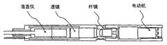
典型地,胶囊摄像机适用于在其被摄入之后在患者体内进行体内成像,拍摄例如胃肠道的图像场景。常规的胶囊摄像机(见图1)具有尺寸小于5cm×3cm的防水透明外部壳体30,从而使得胶囊不能被患者很容易地消化。透镜系统32位于胶囊壳体30的内部,并且位于例如电荷耦合器件(CCD)或互补金属-氧化物半导体(CMOS)图像传感器的成像传感器34的前方,以便提供成像场景到该传感器上。透镜32可以是单个元件透镜系统或多个元件透镜系统,并且可以包括聚焦和/或变焦功能。两个光源36、38(例如,激光或发光二极管(LED))与透镜32相邻,以将光线投射到周围的成像区域上。胶囊还典型地包括控制单元40,控制单元40包括图像存储器和/或将图像发送到外部拾取设备的图像发送装置,诸如微波发射机。通常也提供诸如电池或磁线圈组的动力源42。可利用磁信号在外部驱动动力源42以产生电力。此胶囊摄像机模块因此能够在紧凑、低功耗且轻型的模块中提供连续成像功能。Typically, a capsule camera is adapted for in vivo imaging inside a patient after it has been ingested, taking images of scenes such as the gastrointestinal tract. Conventional capsule cameras (see FIG. 1 ) have a waterproof transparent
在WO 2004/051323中,将可变透镜系统描述为放置在传感器的前方,从而允许紧凑、低功耗且轻型的模块中的可变聚焦/变焦和/或可变定向的成像功能。此公开只解决了成像功能,允许在一些情况下对胃肠道疾病进行诊断。并未解决利用光对这些疾病进行进一步检查或处置。为了允许利用光进行光学处置,则必须识别要被处置的区域,而且,一旦识别了要被处置的区域,就必须在胶囊向前移动时将光束聚焦到此区域上并且保持聚焦一段时间。In WO 2004/051323 a variable lens system is described as being placed in front of the sensor, allowing variable focus/zoom and/or variable orientation imaging functionality in a compact, low power and light weight module. This disclosure only addresses the imaging function, allowing in some cases the diagnosis of gastrointestinal diseases. The use of light for further examination or treatment of these diseases is not addressed. In order to allow optical treatment with light, the area to be treated must be identified and, once identified, the beam of light must be focused on this area and kept focused for a period of time as the capsule moves forward.
利用光学方法对某个感兴趣区域进行进一步检查,需要在胶囊向前移动时将聚焦的光束瞄准感兴趣区域,这时出现了另一问题。根据所提到的现有技术设备,由于光束为宽场照明束,则聚焦的检查是不可能的。Another problem arises when optical methods for further inspection of a region of interest require the focused beam of light to be aimed at the region of interest as the capsule moves forward. According to the mentioned prior art device, since the light beam is a wide-field illumination beam, inspection of the focus is not possible.
尽管已作出了努力,到目前为止,仍然需要这样的可摄入设备和/或可摄入系统,它们促进为处置或进一步检查而在期望的体内位置(例如所识别的病灶)上对光束进行有效聚焦。此外,仍然需要促进与胶囊摄像机结合的体内光聚焦和/或束导向的装置和/或系统。Despite efforts, there remains a need to date for ingestible devices and/or ingestible systems that facilitate directing a beam of light at a desired in vivo location (e.g., an identified lesion) for treatment or further examination. Effective focus. Furthermore, there remains a need for devices and/or systems that facilitate in vivo light focusing and/or beam steering in conjunction with capsule cameras.
通过本公开的设备、系统和方法满足了这些需要及其他需要。These needs and others are met by the devices, systems, and methods of the present disclosure.
根据本公开,公开了用于对动物或人类的胃肠道内的患病组织进行体内成像和/或处置的可摄入胶囊和方法。该胶囊包括:图像传感器;透镜系统,其用于将图像聚焦到图像传感器上;至少一个光源,并且优选为第一光源和第二光源,用于对感兴趣组织区域进行照明,第二光源任选地能够向患病区域提供光疗法处置;可变透镜系统,其位于第一光源的前方,其中,可变透镜系统包括用于将来自第一光源的光束定向并聚焦到患病组织区域上的束导向装置和聚焦装置;控制单元,其与图像传感器、第一光源和第二光源以及可变透镜系统相通信,控制单元包括图像存储装置、处理装置和图像发送装置;其中,控制单元使在胶囊通过所述胃肠道行进时从图像传感器接收到的存储图像相关联,并且对可变透镜系统的束导向装置和聚焦装置进行控制,以确保将来自第一光源的光束被定向并聚焦到患病组织区域上;动力源,其用于向图像传感器、第一光源和第二光源以及控制单元提供动力;以及非消化性、透明的外部保护性壳体,将其配置为通过胃肠道,并在其内部容纳图像传感器、透镜系统、第一和第二光源、可变透镜系统、控制单元以及动力源。According to the present disclosure, ingestible capsules and methods for in vivo imaging and/or treatment of diseased tissue within the gastrointestinal tract of an animal or human are disclosed. The capsule comprises: an image sensor; a lens system for focusing an image onto the image sensor; at least one light source, and preferably a first light source and a second light source, for illuminating a tissue region of interest, the second light source being either optionally capable of providing phototherapy treatment to the diseased area; a variable lens system positioned in front of the first light source, wherein the variable lens system includes a light beam for directing and focusing the beam from the first light source onto the diseased tissue area The beam guiding device and the focusing device; the control unit communicates with the image sensor, the first light source and the second light source and the variable lens system, and the control unit includes an image storage device, a processing device and an image sending device; wherein the control unit uses The stored images received from the image sensor as the capsule travels through the GI tract are correlated and the beam steering and focusing means of the variable lens system are controlled to ensure that the light beam from the first light source is directed and focused onto the diseased tissue area; a power source for powering the image sensor, the first and second light sources, and the control unit; and a non-digestible, transparent outer protective housing configured to pass through the gastrointestinal channel, and houses an image sensor, a lens system, first and second light sources, a variable lens system, a control unit, and a power source therein.
具体地,本发明的目的在于提供一种用于对动物或人类的胃肠道内的一个或多个感兴趣的患病组织区域进行体内成像和/或处置的可摄入胶囊,所述胶囊包括:In particular, it is an object of the present invention to provide an ingestible capsule for in vivo imaging and/or treatment of one or more diseased tissue regions of interest within the gastrointestinal tract of an animal or human, said capsule comprising :
图像传感器;Image Sensor;
透镜系统,其用于将感兴趣图像聚焦到所述图像传感器上;a lens system for focusing an image of interest onto said image sensor;
至少一个光源,其用于向所述胃肠道的所述组织提供照明;其中,所述至少一个光源任选地能够向所述患病组织区域提供光疗法处置;at least one light source for providing illumination to said tissue of said gastrointestinal tract; wherein said at least one light source is optionally capable of providing phototherapeutic treatment to said diseased tissue region;
可变透镜系统,其位于所述至少一个光源的前方,其中,所述可变透镜系统包括用于将来自所述至少一个光源的光束定向并聚焦到所述患病组织区域上的束导向装置和聚焦装置;a variable lens system positioned in front of the at least one light source, wherein the variable lens system includes a beam guide for directing and focusing a light beam from the at least one light source onto the diseased tissue region and focusing device;
控制单元,其与所述图像传感器、所述至少一个光源以及所述可变透镜系统相通信,所述控制单元包括图像存储装置、处理装置和图像发送装置;其中,所述控制单元使在所述胶囊通过所述胃肠道行进时从所述图像传感器接收到的存储图像相关联,并且对所述可变透镜系统的所述束导向装置和聚焦装置进行控制,以确保将来自所述至少一个光源的所述光束定向并聚焦到所述患病组织区域上;a control unit, which communicates with the image sensor, the at least one light source and the variable lens system, the control unit includes an image storage device, a processing device and an image sending device; wherein the control unit enables the correlating the stored images received from the image sensor as the capsule travels through the gastrointestinal tract, and controlling the beam steering and focusing of the variable lens system to ensure directing and focusing said light beam of a light source onto said diseased tissue region;
动力源,其用于向所述图像传感器、所述至少一个光源以及所述控制单元提供动力;以及a power source for powering the image sensor, the at least one light source and the control unit; and
非消化性、透明的外部保护性壳体,将其配置为通过所述胃肠道,并在其内部容纳所述图像传感器、所述透镜系统、所述至少一个光源、所述可变透镜系统、所述控制单元以及所述动力源。a non-digestible, transparent outer protective housing configured to pass through the gastrointestinal tract and house the image sensor, the lens system, the at least one light source, the variable lens system therein , the control unit and the power source.
另一目的在于提供一种用于对动物或人类的胃肠道内的一个或多个感兴趣的患病组织区域进行体内成像和/或处置的可摄入胶囊,所述胶囊包括:Another object is to provide an ingestible capsule for in vivo imaging and/or treatment of one or more diseased tissue regions of interest within the gastrointestinal tract of an animal or human, said capsule comprising:
图像传感器;Image Sensor;
透镜系统,其用于将感兴趣图像聚焦到所述图像传感器上;a lens system for focusing an image of interest onto said image sensor;
第一光源和第二光源,其用于向所述胃肠道的所述组织提供照明;其中,所述第二光源任选地能够向所述患病组织区域提供光疗法处置;a first light source and a second light source for providing illumination to said tissue of said gastrointestinal tract; wherein said second light source is optionally capable of providing phototherapeutic treatment to said diseased tissue region;
可变透镜系统,其位于所述第一光源的前方,其中,所述可变透镜系统包括用于将来自所述第一光源的光束定向并聚焦到所述患病组织区域上的束导向装置和聚焦装置;a variable lens system positioned in front of the first light source, wherein the variable lens system includes a beam guide for directing and focusing a light beam from the first light source onto the diseased tissue region and focusing device;
控制单元,其与所述图像传感器、所述第一光源和第二光源以及所述可变透镜系统相通信,所述控制单元包括图像存储装置、处理装置和图像发送装置;其中,所述控制单元使在所述胶囊通过所述胃肠道行进时从所述图像传感器接收到的存储图像相关联,并且对所述可变透镜系统的所述束导向装置和聚焦装置进行控制,以确保将来自所述第一光源的所述光束定向并聚焦到所述患病组织区域上;A control unit, which communicates with the image sensor, the first light source and the second light source, and the variable lens system, the control unit includes an image storage device, a processing device and an image sending device; wherein the control unit correlates stored images received from the image sensor as the capsule travels through the GI tract and controls the beam steering and focusing of the variable lens system to ensure that directing and focusing the light beam from the first light source onto the diseased tissue region;
动力源,其用于向所述图像传感器、所述第一光源和第二光源以及所述控制单元提供动力;以及a power source for powering the image sensor, the first and second light sources, and the control unit; and
非消化性、透明的外部保护性壳体,将其配置为通过所述胃肠道,并在其内部容纳所述图像传感器、所述透镜系统、所述第一光源和第二光源、所述可变透镜系统、所述控制单元以及所述动力源。a non-digestible, transparent outer protective housing configured to pass through the gastrointestinal tract and house the image sensor, the lens system, the first and second light sources, the A variable lens system, the control unit, and the power source.
另一目的在于提供一种胶囊,其中,所述第一光源和第二光源每个都为激光器。Another object is to provide a capsule wherein each of the first light source and the second light source is a laser.
另一目的在于提供一种胶囊,其中,所述可变透镜系统包括液体透镜系统。Another object is to provide a capsule wherein said variable lens system comprises a liquid lens system.
另一目的在于提供一种用于对动物或人类的胃肠道内的一个或多个感兴趣组织区域进行体内成像的方法,其包括:Another object is to provide a method for in vivo imaging of one or more tissue regions of interest within the gastrointestinal tract of an animal or human, comprising:
摄入一胶囊,所述胶囊包括:Ingestion of one capsule containing:
图像传感器;Image Sensor;
透镜系统,其用于将感兴趣图像聚焦到所述图像传感器上;a lens system for focusing an image of interest onto said image sensor;
至少一个光源,其用于向所述胃肠道的所述组织提供照明;其中,所述至少一个光源任选地能够向患病组织区域提供光疗法处置;at least one light source for providing illumination to said tissue of said gastrointestinal tract; wherein said at least one light source is optionally capable of providing phototherapeutic treatment to a diseased tissue region;
可变透镜系统,其位于所述至少一个光源的前方,其中,所述可变a variable lens system positioned in front of the at least one light source, wherein the variable
透镜系统包括用于将来自所述至少一个光源的光束定向并聚焦到所述患病组织区域上的束导向装置和聚焦装置;a lens system comprising beam directing means and focusing means for directing and focusing a light beam from said at least one light source onto said diseased tissue region;
控制单元,其与所述图像传感器、所述至少一个光源以及所述可变透镜系统相通信,所述控制单元包括图像存储装置、处理装置和图像发送装置;其中,所述控制单元使在所述胶囊通过所述胃肠道行进时从所述图像传感器接收到的存储图像相关联,并且对所述可变透镜系统的所述束导向装置和聚焦装置进行控制,以确保将来自所述至少一个光源的所述光束定向并聚焦到所述患病组织区域上;a control unit, which communicates with the image sensor, the at least one light source and the variable lens system, the control unit includes an image storage device, a processing device and an image sending device; wherein the control unit enables the correlating the stored images received from the image sensor as the capsule travels through the gastrointestinal tract, and controlling the beam steering and focusing of the variable lens system to ensure directing and focusing said light beam of a light source onto said diseased tissue region;
动力源,其用于向所述图像传感器、所述至少一个光源以及所述控制单元提供动力;以及a power source for powering the image sensor, the at least one light source and the control unit; and
非消化性、透明的外部保护性壳体,将其配置为通过所述胃肠道,并在其内部容纳所述图像传感器、所述透镜系统、所述至少一个光源、所述可变透镜系统、所述控制单元以及所述动力源;a non-digestible, transparent outer protective housing configured to pass through the gastrointestinal tract and house the image sensor, the lens system, the at least one light source, the variable lens system therein , the control unit and the power source;
将来自所述控制单元的所述图像数据发送至外部图像接收和查看单元,并且查看所述图像数据。The image data from the control unit is sent to an external image receiving and viewing unit, and the image data is viewed.
另一目的在于提供一种用于对动物或人类的胃肠道内的一个或多个感兴趣组织区域进行体内成像的方法,其包括:Another object is to provide a method for in vivo imaging of one or more tissue regions of interest within the gastrointestinal tract of an animal or human, comprising:
摄入一胶囊,所述胶囊包括:Ingestion of one capsule containing:
图像传感器;Image Sensor;
透镜系统,其用于将感兴趣图像聚焦到所述图像传感器上;a lens system for focusing an image of interest onto said image sensor;
第一光源和第二光源,其用于向所述胃肠道的所述组织提供照明;其中,所述第二光源任选地能够向患病组织区域提供光疗法处置;a first light source and a second light source for providing illumination to said tissue of said gastrointestinal tract; wherein said second light source is optionally capable of providing phototherapeutic treatment to a diseased tissue region;
可变透镜系统,其位于所述第一光源的前方,其中,所述可变透镜系统包括用于将来自所述第一光源的光束定向并聚焦到所述患病组织区域上的束导向装置和聚焦装置;a variable lens system positioned in front of the first light source, wherein the variable lens system includes a beam guide for directing and focusing a light beam from the first light source onto the diseased tissue region and focusing device;
控制单元,其与所述图像传感器、所述第一光源和第二光源以及所述可变透镜系统相通信,所述控制单元包括图像存储装置、处理装置和图像发送装置;其中,所述控制单元使在所述胶囊通过所述胃肠道行进时从所述图像传感器接收到的存储图像相关联,并且对所述可变透镜系统的所述束导向装置和聚焦装置进行控制,以确保将来自所述第一光源的所述光束定向并聚焦到所述患病组织区域上;A control unit, which communicates with the image sensor, the first light source and the second light source, and the variable lens system, the control unit includes an image storage device, a processing device and an image sending device; wherein the control unit correlates stored images received from the image sensor as the capsule travels through the GI tract and controls the beam steering and focusing of the variable lens system to ensure that directing and focusing the light beam from the first light source onto the diseased tissue region;
动力源,其用于向所述图像传感器、所述第一光源和第二光源以及所述控制单元提供动力;以及a power source for powering the image sensor, the first and second light sources, and the control unit; and
非消化性、透明的外部保护性壳体,将其配置为通过所述胃肠道,并在其内部容纳所述图像传感器、所述透镜系统、所述第一光源和第二光源、所述可变透镜系统、所述控制单元以及所述动力源;a non-digestible, transparent outer protective housing configured to pass through the gastrointestinal tract and house the image sensor, the lens system, the first and second light sources, the a variable lens system, said control unit and said power source;
将来自所述控制单元的所述图像数据发送至外部图像接收和查看单元,并且查看所述图像数据。The image data from the control unit is sent to an external image receiving and viewing unit, and the image data is viewed.
另一目的在于提供一种用于对动物或人类的胃肠道内的一个或多个感兴趣的患病组织区域进行体内处置的方法,其包括:Another object is to provide a method for in vivo treatment of one or more diseased tissue regions of interest within the gastrointestinal tract of an animal or human, comprising:
将荧光标记剂摄入所述胃肠道,该荧光标记剂将仅化学结合至所述患病组织区域,并且使得所述患病组织区域在受到光源刺激时发射荧光光束;ingesting a fluorescent marker into said gastrointestinal tract, which will chemically bind only to said diseased tissue region and cause said diseased tissue region to emit a fluorescent beam when stimulated by a light source;
此后摄入一胶囊,所述胶囊包括:Thereafter ingest one capsule containing:
图像传感器;Image Sensor;
透镜系统,其用于将感兴趣图像聚焦到所述图像传感器上;a lens system for focusing an image of interest onto said image sensor;
至少一个光源,其用于向所述胃肠道的所述组织提供照明;其中,所述至少一个光源为能够向所述患病组织区域提供光疗法处置的激光器;at least one light source for providing illumination to said tissue of said gastrointestinal tract; wherein said at least one light source is a laser capable of providing phototherapeutic treatment to said diseased tissue region;
可变透镜系统,其位于所述至少一个光源的前方,其中,所述可变透镜系统包括用于将来自所述至少一个光源的光束定向并聚焦到所述患病组织区域上的束导向装置和聚焦装置;a variable lens system positioned in front of the at least one light source, wherein the variable lens system includes a beam guide for directing and focusing a light beam from the at least one light source onto the diseased tissue region and focusing device;
控制单元,其与所述图像传感器、所述至少一个光源以及所述可变透镜系统相通信,所述控制单元包括图像存储装置、处理装置和图像发送装置;其中,所述控制单元使在所述胶囊通过所述胃肠道行进时从所述图像传感器接收到的存储图像相关联,并且对所述可变透镜系统的所述束导向装置和聚焦装置进行控制,以确保将来自所述至少一个光源的所述光束定向并聚焦到所述患病组织区域上;a control unit, which communicates with the image sensor, the at least one light source and the variable lens system, the control unit includes an image storage device, a processing device and an image sending device; wherein the control unit enables the correlating the stored images received from the image sensor as the capsule travels through the gastrointestinal tract, and controlling the beam steering and focusing of the variable lens system to ensure directing and focusing said light beam of a light source onto said diseased tissue region;
动力源,其用于向所述图像传感器、所述至少一个光源以及所述控制单元提供动力;以及a power source for powering the image sensor, the at least one light source and the control unit; and
非消化性、透明的外部保护性壳体,将其配置为通过所述胃肠道,并在其内部容纳所述图像传感器、所述透镜系统、所述至少一个光源、所述可变透镜系统、所述控制单元以及所述动力源;a non-digestible, transparent outer protective housing configured to pass through the gastrointestinal tract and house the image sensor, the lens system, the at least one light source, the variable lens system therein , the control unit and the power source;
使得所述至少一个光源照明并激发已结合了荧光剂的所述患病组织区域向所述透镜系统、所述图像传感器和所述控制单元发送荧光图像;其中,所述控制单元对所述可变透镜聚焦装置和束导向装置进行控制,以确保所述至少一个光源对所述患病区域的照明;以及causing the at least one light source to illuminate and excite the region of diseased tissue that has incorporated a fluorescent agent to send a fluorescence image to the lens system, the image sensor, and the control unit; The variable lens focusing device and the beam guiding device are controlled to ensure that the at least one light source illuminates the diseased area; and
使得所述至少一个光源对所述患病区域进行光处置。The at least one light source is caused to light treat the diseased area.
另一目的在于提供一种用于对动物或人类的胃肠道内的一个或多个感兴趣的患病组织区域进行体内处置的方法,其包括:Another object is to provide a method for in vivo treatment of one or more diseased tissue regions of interest within the gastrointestinal tract of an animal or human, comprising:
将荧光标记剂摄入所述胃肠道,该荧光标记剂将仅化学结合至所述患病组织区域,并且使得所述患病组织区域在受到光源刺激时发射荧光光束;ingesting a fluorescent marker into said gastrointestinal tract, which will chemically bind only to said diseased tissue region and cause said diseased tissue region to emit a fluorescent beam when stimulated by a light source;
此后摄入一胶囊,所述胶囊包括:Thereafter ingest one capsule containing:
图像传感器;Image Sensor;
透镜系统,其用于将感兴趣图像聚焦到所述图像传感器上;a lens system for focusing an image of interest onto said image sensor;
第一光源和第二光源,其用于向所述胃肠道的所述组织提供照明;其中,所述第二光源为能够向所述患病组织区域提供光疗法处置的激光器;a first light source and a second light source for providing illumination to the tissue of the gastrointestinal tract; wherein the second light source is a laser capable of providing phototherapeutic treatment to the diseased tissue region;
可变透镜系统,其位于所述第一光源的前方,其中,所述可变透镜系统包括用于将来自所述第一光源的光束定向并聚焦到所述患病组织区域上的束导向装置和聚焦装置;a variable lens system positioned in front of the first light source, wherein the variable lens system includes a beam guide for directing and focusing a light beam from the first light source onto the diseased tissue region and focusing device;
控制单元,其与所述图像传感器、所述第一光源和第二光源以及所述可变透镜系统相通信,所述控制单元包括图像存储装置、处理装置和图像发送装置;其中,所述控制单元使在所述胶囊通过所述胃肠道行进时从所述图像传感器接收到的存储图像相关联,并且对所述可变透镜系统的所述束导向装置和聚焦装置进行控制,以确保将来自所述第一光源的所述光束定向并聚焦到所述患病组织区域上;A control unit, which communicates with the image sensor, the first light source and the second light source, and the variable lens system, the control unit includes an image storage device, a processing device and an image sending device; wherein the control unit correlates stored images received from the image sensor as the capsule travels through the GI tract and controls the beam steering and focusing of the variable lens system to ensure that directing and focusing the light beam from the first light source onto the diseased tissue region;
动力源,其用于向所述图像传感器、所述第一光源和第二光源以及所述控制单元提供动力;以及a power source for powering the image sensor, the first and second light sources, and the control unit; and
非消化性、透明的外部保护性壳体,将其配置为通过所述胃肠道,并在其内部容纳所述图像传感器、所述透镜系统、所述第一光源和第二光源、所述可变透镜系统、所述控制单元以及所述动力源;a non-digestible, transparent outer protective housing configured to pass through the gastrointestinal tract and house the image sensor, the lens system, the first and second light sources, the a variable lens system, said control unit and said power source;
使得所述第一光源照明并激发已结合了荧光剂的所述患病组织区域向所述透镜系统、所述图像传感器和所述控制单元发送荧光图像;其中,所述控制单元对所述可变透镜聚焦装置和束导向装置进行控制,以确保所述第一光源对所述患病区域的照明;以及causing the first light source to illuminate and excite the diseased tissue region combined with a fluorescent agent to send a fluorescence image to the lens system, the image sensor and the control unit; The variable lens focusing device and the beam guiding device are controlled to ensure that the first light source illuminates the diseased area; and
使得所述第二光源对所述患病区域进行光处置。The second light source is caused to light treat the diseased area.
另一目的在于提供一种方法,其中,所述第一光源和第二光源每个都为激光器。Another object is to provide a method wherein the first light source and the second light source are each lasers.
另一目的在于提供一种方法,其中,所述可变透镜系统包括液体透镜系统。Another object is to provide a method wherein said variable lens system comprises a liquid lens system.
另一目的在于提供一种方法,其中,所述处置包括:对病灶或肿瘤的热消融;或者对出血血管或出血肿瘤的凝血。Another object is to provide a method, wherein the treatment comprises: thermal ablation of a lesion or a tumor; or coagulation of a bleeding vessel or a bleeding tumor.
通过参照下列实施例并参照附图对本发明的这些方面及其他方面进行更详细的解释。These and other aspects of the invention are explained in more detail by referring to the following examples and referring to the accompanying drawings.
图1为常规的胶囊摄像机的概念表示;Figure 1 is a conceptual representation of a conventional capsule camera;
图2为根据本发明的胶囊摄像机的概念表示;Figure 2 is a conceptual representation of a capsule camera according to the invention;
图3为根据本发明的具有共聚焦显微镜成像的胶囊摄像机的概念表示;Figure 3 is a conceptual representation of a capsule camera with confocal microscopy imaging according to the present invention;
图4为根据本发明的具有扩散光学成像功能的胶囊摄像机的概念表示;4 is a conceptual representation of a capsule camera with diffuse optical imaging according to the present invention;
图5为基于基本光纤的光学相干断层摄影(OCT)原理图的概念表示;Figure 5 is a conceptual representation of the basic fiber-based optical coherence tomography (OCT) schematic;
图6为基于结合具有简单聚焦机构(自聚焦透镜型)的光学传输系统(光纤)的光纤光学型设备的小型化导管型探头(直径约2mm)(根据OpticsLetters 29,2261(2004))的原理图。Fig. 6 is the principle of a miniaturized catheter-type probe (about 2mm in diameter) (according to OpticsLetters29 , 2261(2004)) based on a fiber optic type device combined with an optical transmission system (optical fiber) with a simple focusing mechanism (self-focusing lens type) picture.
为了克服与在先公开的医疗设备和可摄入胶囊相关的问题,在此公开的可摄入胶囊和方法利用放置于光源前方的可变透镜,例如液体透镜。该液体透镜已在WO 2003/069381中详细讨论过。为了允许束导向,同样必须将附加的电极用于WO 2004/051323(以全文引用的方式将其合并于此)中所描述的液体透镜的外部上。虽然利用能够实现光束的聚焦和束导向的液体,但是如何将焦斑保持在要被处置或进一步检查的组织区域上的问题仍然没有得到解决。为了解决此问题,我们利用与照明设备38相结合的摄像机系统,其包括32和34。当接通38时,摄像机拍摄图像,图像处理单元40(见图2)可从该图像识别感兴趣区域。在拍摄完此图像后不久,切断第一光源38,并且以低强度的光水平开启用于处置或进一步诊断的第二光源36。尽管在此将光源38和36描述为单独的光源,它们也可以是具有多重功能(例如,用于照明、用于处置等)的一个单一光源。同样,它们可以是多于两个光源,例如三个、四个或者更多。可以使利用源36的图像传感器上的此光斑的图像与利用源38的来自第一图像的感兴趣区域的图像相关联。一个附加的应用为,虽然胶囊已发生移动,摄像机所拍摄的两个图像也可以应用于对深度信息的立体摄影测量计算。这已被公开并且应用于内窥镜,但并没有应用于胶囊摄像机。如果所述区域并不重叠,则接着对液体透镜进行调节,并且重复该程序。如果达到了相关性,则将束36的强度切换至允许处置或进一步检查的水平。In order to overcome the problems associated with previously disclosed medical devices and ingestible capsules, the ingestible capsules and methods disclosed herein utilize a variable lens, such as a liquid lens, placed in front of a light source. This liquid lens has been discussed in detail in WO 2003/069381. In order to allow beam steering, additional electrodes must also be used on the exterior of the liquid lens described in WO 2004/051323 (which is hereby incorporated by reference in its entirety). While utilizing liquids that enable focusing and beam steering of the beam, the problem of how to maintain the focal spot on the area of tissue to be treated or further examined remains unresolved. In order to solve this problem, we utilize a camera system combined with a
在处置的情况下,利用摄像机诊断正确的组织区域的需求是很重要的。为了改进此步骤,可以使用荧光标记物。在这种情况下,光源38也应用于照明病灶,以诱导所靶向的标记物发出荧光。除了荧光之外,这可以推广为还包括自体荧光/生物发光。通过使处置光斑和荧光光斑以上述方式相关联,可以将此模态应用于瞄准处置光斑。荧光还会给出在何处必须执行消融或其他处置的指示。没有荧光的话,则需要对摄像机图像进行分析以找到病理。也可能使用多重荧光标签,其以不同的波长发射,并因而能够提供关于可能需要不同处置的病灶中的不同蛋白的信息。In the case of a procedure, the need to utilize a camera to diagnose the correct tissue region is important. To improve this step, fluorescent markers can be used. In this case, a
根据本发明的示范性应用:Exemplary application according to the invention:
处置的示例example of disposition
当处置光源为激光时,病灶或肿瘤上的热效应可以应用于对病灶或肿瘤的热消融。激光的热效应还可以应用于对出血血管或肿瘤的出血表面的凝血。When the treatment light source is a laser, the thermal effect on the lesion or tumor can be applied to thermal ablation of the lesion or tumor. The thermal effect of the laser can also be applied to the coagulation of bleeding vessels or bleeding surfaces of tumors.
另一示例为将光源用作用于光动力疗法(PDT)(参见例如DEJCDolmans、D Fukumura和RK Jain,“Photodynamic therapy for cancer”,NatureReviews Cancer 2003第3卷第380-387页)的非热光源。这需要使用光敏剂。也有可能在胶囊探测到可疑病灶时,由胶囊将光敏剂局部地释放(或在连续的基础上,这需要更大的贮存器)。Another example is the use of light sources as non-thermal light sources for photodynamic therapy (PDT) (see e.g. DEJ CDolmans, D Fukumura and RK Jain, "Photodynamic therapy for cancer", Nature Reviews Cancer 2003 Vol. 3 pp. 380-387). This requires the use of photosensitizers. It is also possible to release the photosensitizer locally from the capsule (or on a continuous basis, which would require a larger reservoir) when a suspicious lesion is detected by the capsule.
进一步检查的示例Example of further inspection
共聚焦显微镜confocal microscope
根据本发明的应用的一个示例为使用共聚焦显微镜(参见图2和图3)。在这种情况下,光源为聚焦到组织内的某处的点源。如此设置共聚焦显微镜的焦平面,从而使得在聚焦到摄像机34和32的图像传感器34上的情况下对此平面成像。An example of an application according to the invention is the use of confocal microscopy (see Figures 2 and 3). In this case, the light source is a point source focused somewhere within the tissue. The focal plane of the confocal microscope is arranged such that it is imaged with focus on the
在共聚焦扫描显微镜的情况下,由激光器38产生的光束通过分束器(partial beam splitter)60,并且由液体透镜44将其瞄准并聚焦到感兴趣病灶上。沿着同一返回路径的反射信号被液体透镜44聚焦,其经由在分束器60上的反射,经由位于探测器的前方的针孔或孔径,到达探测器62上。通过通过改变可变透镜44的设置来扫描光束,可以拍摄病灶的共聚焦显微图像。为了进行合适的图像重建,必须对胶囊的运动进行校正。这可以通过使用根据本发明的胶囊摄像机来实现。In the case of a confocal scanning microscope, the beam generated by the
荧光成像fluorescence imaging
在荧光成像过程的情况下,将激光束聚焦于病灶或肿瘤。探测器模块探测由光吸收诱导的荧光。In the case of fluorescence imaging procedures, a laser beam is focused on a lesion or tumor. The detector module detects fluorescence induced by light absorption.
我们可以使用例如共聚焦显微镜的如上所述的同样设置。由于我们正寻找由光吸收产生的荧光,我们使探测器适应于记录光谱分辨的荧光。We can use the same setup as above for eg a confocal microscope. Since we were looking for fluorescence arising from light absorption, we adapted the detector to record spectrally resolved fluorescence.
扩散光学断层摄影(DOT)Diffuse Optical Tomography (DOT)
在这种情况下,光源包括一个或多个光发射器,光发射器使得如对胶囊周围的相邻组织的照明的随后的点至少包含对光透明的位置(参见图4)。例如在光源38照明部位101时,光扩散地散射通过组织,并到达例如与探测器70连接的探测器光纤。收集所有探测光纤的信号,并将信号存储于存储器中。针对每个照明部位101’、101”等重复此测量。现将所有测量应用于执行图像重建(参见例如AP Gibson、JC Hebden和SR Arridge,“Recentadvances in diffuse optical imaging”,Phys.Med.Biol.2005年第50卷R1-R43)。为了补偿样本的移动,可以使用摄像机34、32上的图像。In this case, the light source comprises one or more light emitters such that subsequent points such as illumination of adjacent tissue around the capsule comprise at least the light-transparent locations (see FIG. 4 ). For example, when the
光学相干断层摄影(OCT)Optical Coherence Tomography (OCT)
OCT为以超高分辨率(几微米)达到几毫米(典型地1.5mm-2mm)的穿透深度、实时生成3D组织图像的成像技术。OCT提供3D结构图像(组织层、密度变化),在当前显示出提供光谱信息以及实现功能和分子成像的巨大潜力。OCT是基于干涉法的技术,能够测量小至-90dB的信号。图5示出了一个基于标准光纤的OCT装置。OCT is an imaging technique that generates 3D tissue images in real time with ultra-high resolution (several micrometers) to a penetration depth of several millimeters (typically 1.5mm-2mm). OCT provides 3D structural images (tissue layers, density changes) and currently shows great potential for providing spectral information and enabling functional and molecular imaging. OCT is an interferometry-based technique capable of measuring signals as small as -90dB. Figure 5 shows a standard optical fiber based OCT setup.
来自光源的光由耦合器进行分光。一个分支用作干涉仪的样本(参考)分支,而另一分支则将光传输至样本(样本分支)。扫描光学器件提供横向扫描能力,从而使得OCT装置获得针对每个横向位置的一个A-扫描(轴位扫描)。所有的A-扫描结合起来形成3D结构图像。当获得每个A-扫描时,参考镜的位移提供深度信息。已开发出一些更先进的技术,以比这里所示出的技术在更短的时间内获得深度信息。光谱OCT是这些技术中最先进的技术,其在不利用运动部件的情况下提供深度扫描数据。目前最快的OCT系统可以以约30fps产生图像,每帧具有多于1000次A-扫描。横向分辨率受到扫描光学器件和光聚焦系统的限制。轴向分辨率具有光源依赖性。商业上的SLD(超发光二极管,带宽约为70nm至930nm)当前可达到的典型轴向分辨率为约5μm。通过使用掺钛蓝宝石飞秒激光器实现一个约1.5μm的最佳显示分辨率。The light from the light source is split by the coupler. One branch serves as the sample (reference) branch of the interferometer, while the other transmits light to the sample (sample branch). Scanning optics provide lateral scanning capability such that the OCT device acquires one A-scan (axial scan) for each lateral position. All A-scans are combined to form a 3D structural image. As each A-scan is acquired, the displacement of the reference mirror provides depth information. Some more advanced techniques have been developed to obtain depth information in less time than shown here. Spectral OCT is the most advanced of these techniques, providing deep scan data without utilizing moving parts. The fastest current OCT systems can produce images at about 30 fps with more than 1000 A-scans per frame. Lateral resolution is limited by the scanning optics and light focusing system. Axial resolution is light source dependent. Typical axial resolutions currently achievable by commercial SLDs (Super Luminescent Diodes, bandwidth about 70 nm to 930 nm) are about 5 μm. An optimal display resolution of about 1.5 μm is achieved by using Ti:sapphire femtosecond lasers.
为使其适用于根据本发明的胶囊,系统必须小型化。从引擎侧起,OCT需要:To make it suitable for use in capsules according to the invention, the system must be miniaturized. From the engine side, OCT needs to:
i)光源。其决定轴向分辨率,轴向分辨率与源带宽成比例。商业上可得到的SLD(超发光二极管)获得约5μm的轴向分辨率。如果使用掺钛蓝宝石飞秒激光器或钨灯(很低的功率),则可以得到更好的分辨率(分辨率接近1μm)。傅立叶域OCT需要可调谐激光器。i) Light source. It determines the axial resolution, which is proportional to the source bandwidth. Commercially available SLDs (Super Luminescent Diodes) achieve an axial resolution of about 5 μm. Better resolution (resolution close to 1 μm) can be obtained if Ti:Sapphire femtosecond laser or tungsten lamp (very low power) is used. Fourier domain OCT requires tunable lasers.
ii)光纤光学组件。光纤提供光的传输,光纤耦合器和/或环行器实现迈克尔逊干涉仪。ii) Fiber optic components. Fiber optics provide the transmission of light, and fiber couplers and/or circulators implement Michelson interferometers.
iii)探测组件。其取决于OCT类型。典型地为光电二极管,但在频谱OCT的情况下,则需要频谱分辨的探测(频谱分析仪与线性CCD阵列相结合)。iii) Probing components. It depends on the type of OCT. Typically a photodiode, but in the case of spectral OCT, spectrally resolved detection (spectrum analyzer combined with a linear CCD array) is required.
iv)如果是偏振或相位OCT,则引擎和探头组件必须能够保持光的偏振特性。iv) In case of polarization or phase OCT, the engine and probe assembly must be able to preserve the polarization properties of the light.
尽管此时并不存在这种小型化的设备,设想在不远的将来,小型化允许这种设备的构造。例如,在Optics Letters 29,2261(2004)中已描述了小型化的探针设计(参见图6)。Although such miniaturized devices do not exist at this time, it is envisaged that in the near future, miniaturization will allow the construction of such devices. Miniaturized probe designs have been described, for example, in Optics Letters29 , 2261 (2004) (see Figure 6).
虽然已就本发明的具体实施例描述了本发明,本领域普通技术人员将认识到,可以在不偏离本发明的精神和范围的情况下实现许多修改、增强和/或改变。因此,本发明显然意在只受权利要求书及其等价物的范围的限制。Although the invention has been described in terms of specific embodiments thereof, those of ordinary skill in the art will recognize that many modifications, enhancements and/or changes can be made without departing from the spirit and scope of the invention. Accordingly, it is manifestly intended that this invention be limited only by the scope of the claims and the equivalents thereof.
Claims (6)
Translated fromChineseApplications Claiming Priority (3)
| Application Number | Priority Date | Filing Date | Title |
|---|---|---|---|
| US82012406P | 2006-07-24 | 2006-07-24 | |
| US60/820,124 | 2006-07-24 | ||
| PCT/IB2007/051323WO2008012701A1 (en) | 2006-07-24 | 2007-04-12 | Capsule camera with variable illumination of the surrounding tissue |
Publications (2)
| Publication Number | Publication Date |
|---|---|
| CN101495022A CN101495022A (en) | 2009-07-29 |
| CN101495022Btrue CN101495022B (en) | 2011-09-07 |
Family
ID=38481394
Family Applications (1)
| Application Number | Title | Priority Date | Filing Date |
|---|---|---|---|
| CN2007800282627AExpired - Fee RelatedCN101495022B (en) | 2006-07-24 | 2007-04-12 | Capsule camera with variable illumination of surrounding tissue |
Country Status (6)
| Country | Link |
|---|---|
| US (2) | US8771176B2 (en) |
| EP (1) | EP2046188B1 (en) |
| JP (1) | JP2009544392A (en) |
| CN (1) | CN101495022B (en) |
| RU (1) | RU2009106033A (en) |
| WO (1) | WO2008012701A1 (en) |
Families Citing this family (36)
| Publication number | Priority date | Publication date | Assignee | Title |
|---|---|---|---|---|
| KR20080060079A (en)* | 2006-12-26 | 2008-07-01 | 전자부품연구원 | Memory Endoscope Capsule Endoscope |
| US20090030279A1 (en)* | 2007-07-27 | 2009-01-29 | Zander Dennis R | Method and system for managing power consumption in a compact diagnostic capsule |
| US8636653B2 (en) | 2008-06-09 | 2014-01-28 | Capso Vision, Inc. | In vivo camera with multiple sources to illuminate tissue at different distances |
| US9138593B2 (en) | 2009-11-06 | 2015-09-22 | Universita' Degli Studi Di Firenze | Ingestible capsule for treating gastric infections, in particular for treating H. pylori infections |
| EP2571573A4 (en)* | 2010-03-17 | 2013-12-04 | Photopill Medical Ltd | Capsule phototherapy |
| US20120022338A1 (en)* | 2010-05-28 | 2012-01-26 | The General Hospital Corporation | Apparatus, systems, methods and computer-accessible medium for analyzing information regarding cardiovascular diseases and functions |
| US9277855B2 (en)* | 2010-08-10 | 2016-03-08 | Boston Scientific Scimed, Inc. | Endoscopic system for enhanced visualization |
| ES2384795B2 (en) | 2010-12-13 | 2013-01-29 | Medlumics, S.L. | GASTROINTESTINAL ELECTRONIC PILL. |
| CN103687646B (en) | 2011-03-17 | 2016-09-21 | 基文影像公司 | Capsule phototherapy |
| DE102011079958A1 (en) | 2011-07-28 | 2013-01-31 | Karl Storz Gmbh & Co. Kg | Endoscope with adjustable viewing direction |
| DE102011086325A1 (en) | 2011-11-15 | 2013-05-16 | Karl Storz Gmbh & Co. Kg | Illumination optics, observation device and method for generating an illumination light beam |
| CN102697438A (en)* | 2012-01-18 | 2012-10-03 | 广州宝胆医疗器械科技有限公司 | Capsule enteroscopy system with function of CCD (couple charged device) and function of optical coherence tomography |
| WO2013153860A1 (en)* | 2012-04-12 | 2013-10-17 | ソニー株式会社 | Capsule-shaped medical device and system |
| US11179028B2 (en)* | 2013-02-01 | 2021-11-23 | The General Hospital Corporation | Objective lens arrangement for confocal endomicroscopy |
| US10068334B2 (en)* | 2013-05-29 | 2018-09-04 | Capsovision Inc | Reconstruction of images from an in vivo multi-camera capsule |
| CN105848557B (en)* | 2013-12-27 | 2018-09-28 | 王康怀 | Capsule camera device with multispectral light source |
| US11571112B2 (en) | 2014-01-07 | 2023-02-07 | The General Hospital Corporation | Method and apparatus for recording microscopic images from within a living person or organism using an implantable device |
| CN103920246B (en)* | 2014-04-03 | 2017-11-10 | 深圳普门科技有限公司 | A kind of capsule photon treatment device of belt switch device |
| CN103920245B (en)* | 2014-04-03 | 2017-11-24 | 深圳普门科技有限公司 | Capsule photon treatment device, system and targeted therapy method with guide function |
| JP6335007B2 (en)* | 2014-04-18 | 2018-05-30 | オリンパス株式会社 | Capsule endoscope system, capsule endoscope, receiving apparatus, capsule endoscope operating method, and program |
| US10052154B2 (en)* | 2014-10-01 | 2018-08-21 | Verily Life Sciences Llc | System and method for fluorescence-based laser ablation |
| TWI564050B (en)* | 2015-01-27 | 2017-01-01 | 友達光電股份有限公司 | Phototherapy device |
| CN105361843B (en)* | 2015-11-30 | 2019-03-19 | 青岛大学附属医院 | Wireless capsule OCT endoscopic system for gastrointestinal tract diagnosis and treatment |
| WO2017203369A2 (en) | 2016-05-25 | 2017-11-30 | Carson Benjamin | Method, system, software, and device for remote, miniaturized, and three-dimensional imaging and analysis of human lesions. research and clinical applications thereof |
| IT201600129679A1 (en)* | 2016-12-21 | 2018-06-21 | Probiomedica S R L | Ingestible capsule for the phototherapeutic treatment of infections |
| DE102017109554A1 (en)* | 2017-05-04 | 2018-11-08 | Leica Microsystems Cms Gmbh | Optical arrangement for a confocal microscope, confocal microscope and method of operating a confocal microscope |
| CN107595385A (en)* | 2017-10-30 | 2018-01-19 | 中国联合网络通信集团有限公司 | A kind of thrombolysis method and microrobot |
| WO2019213594A1 (en) | 2018-05-03 | 2019-11-07 | The General Hospital Corporation | Systems, methods, and media for capsule-based multimode endoscopy |
| CN109770825B (en)* | 2019-03-06 | 2021-09-24 | 杭州行开医学影像技术有限公司 | Endoscope with 3D imaging function |
| CN109998456B (en)* | 2019-04-12 | 2024-08-09 | 安翰科技(武汉)股份有限公司 | Capsule type endoscope and control method thereof |
| US11622754B2 (en)* | 2019-06-28 | 2023-04-11 | Endiatx, Inc. | Ingestible device with propulsion and imaging capabilities |
| US11152664B2 (en)* | 2019-12-24 | 2021-10-19 | Anexa Labs Llc | Compact electronics with optical sensors |
| CN112353434A (en)* | 2020-10-16 | 2021-02-12 | 南方医科大学南方医院 | Gastric cancer surgical instrument |
| US11327443B1 (en) | 2020-10-23 | 2022-05-10 | Anexa Labs Llc | Wearable device for monitoring health metrics |
| US11776118B2 (en)* | 2020-12-02 | 2023-10-03 | International Business Machines Corporation | Recognition of partially digested medications |
| CN116138717A (en)* | 2022-09-08 | 2023-05-23 | 卓外(上海)医疗电子科技有限公司 | Visual photothermal therapy endoscopic device with adjustable light output angle |
Citations (2)
| Publication number | Priority date | Publication date | Assignee | Title |
|---|---|---|---|---|
| US6240312B1 (en)* | 1997-10-23 | 2001-05-29 | Robert R. Alfano | Remote-controllable, micro-scale device for use in in vivo medical diagnosis and/or treatment |
| US7044908B1 (en)* | 2003-07-08 | 2006-05-16 | National Semiconductor Corporation | Method and system for dynamically adjusting field of view in a capsule endoscope |
Family Cites Families (26)
| Publication number | Priority date | Publication date | Assignee | Title |
|---|---|---|---|---|
| US4931053A (en)* | 1988-01-27 | 1990-06-05 | L'esperance Medical Technologies, Inc. | Method and apparatus for enhanced vascular or other growth |
| US5098426A (en)* | 1989-02-06 | 1992-03-24 | Phoenix Laser Systems, Inc. | Method and apparatus for precision laser surgery |
| IL108352A (en) | 1994-01-17 | 2000-02-29 | Given Imaging Ltd | In vivo video camera system |
| US7405320B2 (en) | 1998-06-22 | 2008-07-29 | Immunomedics, Inc. | Therapeutic and diagnostic conjugates for use with multispecific antibodies |
| IL126727A (en) | 1998-10-22 | 2006-12-31 | Given Imaging Ltd | Method for delivering a device to a target location |
| US8636648B2 (en)* | 1999-03-01 | 2014-01-28 | West View Research, Llc | Endoscopic smart probe |
| IL134017A (en)* | 2000-01-13 | 2008-04-13 | Capsule View Inc | Camera for viewing inside intestines |
| US7039453B2 (en)* | 2000-02-08 | 2006-05-02 | Tarun Mullick | Miniature ingestible capsule |
| DE20122488U1 (en) | 2000-03-08 | 2005-12-15 | Given Imaging Ltd. | In vivo imaging system for use in applications such as imaging digestive tract, uses camera, illumination source and transmitter enclosed in capsule suitable for insertion into and passing through body lumens or cavities |
| US6709387B1 (en)* | 2000-05-15 | 2004-03-23 | Given Imaging Ltd. | System and method for controlling in vivo camera capture and display rate |
| EP1164406B1 (en)* | 2000-06-17 | 2019-04-17 | Leica Microsystems CMS GmbH | Method and device for illuminating an object |
| US8055329B2 (en) | 2001-01-22 | 2011-11-08 | Spectrum Dynamics Llc | Ingestible device for radioimaging of the gastrointestinal tract |
| AU2002213335A1 (en) | 2000-10-30 | 2002-05-15 | Motorola, Inc. | Ingestible capsule video transmitting fluorescent images |
| US7616986B2 (en)* | 2001-05-07 | 2009-11-10 | University Of Washington | Optical fiber scanner for performing multimodal optical imaging |
| JP2003260025A (en) | 2002-03-08 | 2003-09-16 | Olympus Optical Co Ltd | Capsule endoscope |
| AU2002951841A0 (en)* | 2002-09-30 | 2002-10-24 | Swinburne University Of Technology | Apparatus |
| AU2003280124A1 (en) | 2002-12-03 | 2004-06-23 | Koninklijke Philips Electronics N.V. | Apparatus for forming variable fluid meniscus configurations |
| JP2004275542A (en)* | 2003-03-17 | 2004-10-07 | Olympus Corp | Capsule type endoscope |
| DE10327034A1 (en) | 2003-06-16 | 2005-01-20 | Siemens Ag | Device for examination in the body with light |
| DE10346276B4 (en)* | 2003-10-06 | 2007-01-18 | Siemens Ag | System for the automated localization of lesions in the gastrointestinal tract for therapy with laser light of an endorobot |
| US20050137468A1 (en) | 2003-12-18 | 2005-06-23 | Jerome Avron | Device, system, and method for in-vivo sensing of a substance |
| JP2007525261A (en)* | 2004-01-16 | 2007-09-06 | ザ シティ カレッジ オブ ザ シティ ユニバーシティ オブ ニューヨーク | A microscale compact device for in vivo medical diagnostics combining optical imaging and point fluorescence spectroscopy |
| US7364543B2 (en)* | 2004-03-23 | 2008-04-29 | California Institute Of Technology | Paired angled rotation scanning probes and methods of use |
| WO2006070356A2 (en)* | 2004-12-30 | 2006-07-06 | Given Imaging Ltd. | Device, system, and method for adaptive imaging |
| US7530948B2 (en)* | 2005-02-28 | 2009-05-12 | University Of Washington | Tethered capsule endoscope for Barrett's Esophagus screening |
| US9220917B2 (en)* | 2006-04-12 | 2015-12-29 | The Invention Science Fund I, Llc | Systems for autofluorescent imaging and target ablation |
- 2007
- 2007-04-12CNCN2007800282627Apatent/CN101495022B/ennot_activeExpired - Fee Related
- 2007-04-12USUS12/374,506patent/US8771176B2/ennot_activeExpired - Fee Related
- 2007-04-12JPJP2009521385Apatent/JP2009544392A/enactivePending
- 2007-04-12WOPCT/IB2007/051323patent/WO2008012701A1/enactiveApplication Filing
- 2007-04-12EPEP07735484.3Apatent/EP2046188B1/ennot_activeNot-in-force
- 2007-04-12RURU2009106033/14Apatent/RU2009106033A/enunknown
- 2014
- 2014-06-06USUS14/298,246patent/US9011321B2/ennot_activeExpired - Fee Related
Patent Citations (2)
| Publication number | Priority date | Publication date | Assignee | Title |
|---|---|---|---|---|
| US6240312B1 (en)* | 1997-10-23 | 2001-05-29 | Robert R. Alfano | Remote-controllable, micro-scale device for use in in vivo medical diagnosis and/or treatment |
| US7044908B1 (en)* | 2003-07-08 | 2006-05-16 | National Semiconductor Corporation | Method and system for dynamically adjusting field of view in a capsule endoscope |
Also Published As
| Publication number | Publication date |
|---|---|
| US20140288368A1 (en) | 2014-09-25 |
| US20090177033A1 (en) | 2009-07-09 |
| CN101495022A (en) | 2009-07-29 |
| EP2046188A1 (en) | 2009-04-15 |
| RU2009106033A (en) | 2010-08-27 |
| US8771176B2 (en) | 2014-07-08 |
| WO2008012701A1 (en) | 2008-01-31 |
| EP2046188B1 (en) | 2019-03-27 |
| JP2009544392A (en) | 2009-12-17 |
| US9011321B2 (en) | 2015-04-21 |
Similar Documents
| Publication | Publication Date | Title |
|---|---|---|
| CN101495022B (en) | Capsule camera with variable illumination of surrounding tissue | |
| US11439307B2 (en) | Method for detecting fluorescence and ablating cancer cells of a target surgical area | |
| US10314490B2 (en) | Method and device for multi-spectral photonic imaging | |
| US20080214940A1 (en) | Medical imaging lens system, and method with high-efficiency light collection and collinear illumination | |
| US9615748B2 (en) | Endoscopic biopsy apparatus, system and method | |
| US20080058629A1 (en) | Optical fiber scope with both non-resonant illumination and resonant collection/imaging for multiple modes of operation | |
| JP2005514144A (en) | Apparatus and method for spectroscopic examination of the colon | |
| WO2008024101A1 (en) | Optical fiber scope with both non-resonant illumination and resonant collection/imaging for multiple modes of operation | |
| JP2017176811A (en) | Imaging apparatus, imaging method, and medical observation apparatus | |
| US20100076304A1 (en) | Invisible light irradiation apparatus and method for controlling invisible light irradiation apparatus | |
| KR102125226B1 (en) | Optical fiber probe and endoscope apparatus having the same | |
| CN118680505B (en) | Esophageal endoscopy diagnosis and treatment system and storage medium | |
| JP2008043383A (en) | Fluorescence observation endoscope device | |
| Wagnières et al. | Clinical imaging fluorescence apparatus for the endoscopic photodetection of early cancers by use of Photofrin II | |
| US10448817B2 (en) | Endoscopic device incorporating diode laser for PDD, PDT, and AF applications | |
| CN217338517U (en) | Broad Spectrum Fluorescence Endoscopy Device | |
| JP2004243034A (en) | Clinical capsule for measuring optical tomographic image | |
| JP3571689B2 (en) | Optical tomographic imaging system | |
| CN117770736A (en) | Confocal microscopic endoscopic imaging device and method based on far-end scanning | |
| Sujatha | Endoscopic Diagnostics in Biomedicine: Instrumentation and Applications |
Legal Events
| Date | Code | Title | Description |
|---|---|---|---|
| C06 | Publication | ||
| PB01 | Publication | ||
| C10 | Entry into substantive examination | ||
| SE01 | Entry into force of request for substantive examination | ||
| C14 | Grant of patent or utility model | ||
| GR01 | Patent grant | ||
| CF01 | Termination of patent right due to non-payment of annual fee | Granted publication date:20110907 Termination date:20200412 | |
| CF01 | Termination of patent right due to non-payment of annual fee |