CN101438143B - Cytometer cell counting and size measurement method - Google Patents
Cytometer cell counting and size measurement methodDownload PDFInfo
- Publication number
- CN101438143B CN101438143BCN200680023450.6ACN200680023450ACN101438143BCN 101438143 BCN101438143 BCN 101438143BCN 200680023450 ACN200680023450 ACN 200680023450ACN 101438143 BCN101438143 BCN 101438143B
- Authority
- CN
- China
- Prior art keywords
- particle
- particles
- channel
- image
- slit
- Prior art date
- Legal status (The legal status is an assumption and is not a legal conclusion. Google has not performed a legal analysis and makes no representation as to the accuracy of the status listed.)
- Expired - Fee Related
Links
Images


Classifications
- G—PHYSICS
- G01—MEASURING; TESTING
- G01N—INVESTIGATING OR ANALYSING MATERIALS BY DETERMINING THEIR CHEMICAL OR PHYSICAL PROPERTIES
- G01N15/00—Investigating characteristics of particles; Investigating permeability, pore-volume or surface-area of porous materials
- G01N15/10—Investigating individual particles
- G01N15/14—Optical investigation techniques, e.g. flow cytometry
- G01N15/1456—Optical investigation techniques, e.g. flow cytometry without spatial resolution of the texture or inner structure of the particle, e.g. processing of pulse signals
- G01N15/1459—Optical investigation techniques, e.g. flow cytometry without spatial resolution of the texture or inner structure of the particle, e.g. processing of pulse signals the analysis being performed on a sample stream
- G—PHYSICS
- G01—MEASURING; TESTING
- G01N—INVESTIGATING OR ANALYSING MATERIALS BY DETERMINING THEIR CHEMICAL OR PHYSICAL PROPERTIES
- G01N15/00—Investigating characteristics of particles; Investigating permeability, pore-volume or surface-area of porous materials
- G01N15/10—Investigating individual particles
- G01N15/14—Optical investigation techniques, e.g. flow cytometry
- G01N15/1429—Signal processing
- G01N15/1433—Signal processing using image recognition
- G—PHYSICS
- G01—MEASURING; TESTING
- G01N—INVESTIGATING OR ANALYSING MATERIALS BY DETERMINING THEIR CHEMICAL OR PHYSICAL PROPERTIES
- G01N15/00—Investigating characteristics of particles; Investigating permeability, pore-volume or surface-area of porous materials
- G01N15/10—Investigating individual particles
- G01N15/14—Optical investigation techniques, e.g. flow cytometry
- G01N15/1456—Optical investigation techniques, e.g. flow cytometry without spatial resolution of the texture or inner structure of the particle, e.g. processing of pulse signals
- G—PHYSICS
- G01—MEASURING; TESTING
- G01N—INVESTIGATING OR ANALYSING MATERIALS BY DETERMINING THEIR CHEMICAL OR PHYSICAL PROPERTIES
- G01N33/00—Investigating or analysing materials by specific methods not covered by groups G01N1/00 - G01N31/00
- G01N33/48—Biological material, e.g. blood, urine; Haemocytometers
- G01N33/50—Chemical analysis of biological material, e.g. blood, urine; Testing involving biospecific ligand binding methods; Immunological testing
- G01N33/72—Chemical analysis of biological material, e.g. blood, urine; Testing involving biospecific ligand binding methods; Immunological testing involving blood pigments, e.g. haemoglobin, bilirubin or other porphyrins; involving occult blood
- G01N33/721—Haemoglobin
- G—PHYSICS
- G01—MEASURING; TESTING
- G01N—INVESTIGATING OR ANALYSING MATERIALS BY DETERMINING THEIR CHEMICAL OR PHYSICAL PROPERTIES
- G01N15/00—Investigating characteristics of particles; Investigating permeability, pore-volume or surface-area of porous materials
- G01N15/10—Investigating individual particles
- G01N15/14—Optical investigation techniques, e.g. flow cytometry
- G01N15/1434—Optical arrangements
- G—PHYSICS
- G01—MEASURING; TESTING
- G01N—INVESTIGATING OR ANALYSING MATERIALS BY DETERMINING THEIR CHEMICAL OR PHYSICAL PROPERTIES
- G01N15/00—Investigating characteristics of particles; Investigating permeability, pore-volume or surface-area of porous materials
- G01N15/10—Investigating individual particles
- G01N15/14—Optical investigation techniques, e.g. flow cytometry
- G01N15/1484—Optical investigation techniques, e.g. flow cytometry microstructural devices
- G—PHYSICS
- G01—MEASURING; TESTING
- G01N—INVESTIGATING OR ANALYSING MATERIALS BY DETERMINING THEIR CHEMICAL OR PHYSICAL PROPERTIES
- G01N15/00—Investigating characteristics of particles; Investigating permeability, pore-volume or surface-area of porous materials
- G01N15/01—Investigating characteristics of particles; Investigating permeability, pore-volume or surface-area of porous materials specially adapted for biological cells, e.g. blood cells
- G01N2015/012—Red blood cells
- G—PHYSICS
- G01—MEASURING; TESTING
- G01N—INVESTIGATING OR ANALYSING MATERIALS BY DETERMINING THEIR CHEMICAL OR PHYSICAL PROPERTIES
- G01N15/00—Investigating characteristics of particles; Investigating permeability, pore-volume or surface-area of porous materials
- G01N15/01—Investigating characteristics of particles; Investigating permeability, pore-volume or surface-area of porous materials specially adapted for biological cells, e.g. blood cells
- G01N2015/018—Platelets
- G—PHYSICS
- G01—MEASURING; TESTING
- G01N—INVESTIGATING OR ANALYSING MATERIALS BY DETERMINING THEIR CHEMICAL OR PHYSICAL PROPERTIES
- G01N15/00—Investigating characteristics of particles; Investigating permeability, pore-volume or surface-area of porous materials
- G01N15/10—Investigating individual particles
- G01N2015/1027—Determining speed or velocity of a particle
- G—PHYSICS
- G01—MEASURING; TESTING
- G01N—INVESTIGATING OR ANALYSING MATERIALS BY DETERMINING THEIR CHEMICAL OR PHYSICAL PROPERTIES
- G01N15/00—Investigating characteristics of particles; Investigating permeability, pore-volume or surface-area of porous materials
- G01N15/10—Investigating individual particles
- G01N15/14—Optical investigation techniques, e.g. flow cytometry
- G01N15/1404—Handling flow, e.g. hydrodynamic focusing
- G01N2015/1413—Hydrodynamic focussing
- G—PHYSICS
- G01—MEASURING; TESTING
- G01N—INVESTIGATING OR ANALYSING MATERIALS BY DETERMINING THEIR CHEMICAL OR PHYSICAL PROPERTIES
- G01N15/00—Investigating characteristics of particles; Investigating permeability, pore-volume or surface-area of porous materials
- G01N15/10—Investigating individual particles
- G01N15/14—Optical investigation techniques, e.g. flow cytometry
- G01N2015/1486—Counting the particles
- G—PHYSICS
- G01—MEASURING; TESTING
- G01N—INVESTIGATING OR ANALYSING MATERIALS BY DETERMINING THEIR CHEMICAL OR PHYSICAL PROPERTIES
- G01N15/00—Investigating characteristics of particles; Investigating permeability, pore-volume or surface-area of porous materials
- G01N15/10—Investigating individual particles
- G01N15/14—Optical investigation techniques, e.g. flow cytometry
- G01N2015/1493—Particle size
Landscapes
- Chemical & Material Sciences (AREA)
- Health & Medical Sciences (AREA)
- Life Sciences & Earth Sciences (AREA)
- Immunology (AREA)
- General Physics & Mathematics (AREA)
- Pathology (AREA)
- Physics & Mathematics (AREA)
- General Health & Medical Sciences (AREA)
- Biochemistry (AREA)
- Engineering & Computer Science (AREA)
- Analytical Chemistry (AREA)
- Dispersion Chemistry (AREA)
- Hematology (AREA)
- Urology & Nephrology (AREA)
- Molecular Biology (AREA)
- Biomedical Technology (AREA)
- Medicinal Chemistry (AREA)
- Food Science & Technology (AREA)
- Microbiology (AREA)
- Cell Biology (AREA)
- Biotechnology (AREA)
- Signal Processing (AREA)
- Investigating Or Analysing Biological Materials (AREA)
Abstract
Description
Translated fromChinese背景技术Background technique
本发明要求于2005年4月29日提交的美国临时专利申请No.60/676,403的权益。因此2005年4月29日提交的美国临时专利申请No.60/676,403在此作为参照并入本文。This application claims the benefit of US Provisional Patent Application No. 60/676,403, filed April 29, 2005. US Provisional Patent Application No. 60/676,403, filed April 29, 2005, is hereby incorporated herein by reference.
本发明涉及确定颗粒的特性。具体地,本发明涉及确定颗粒数量和尺寸,更具体地,其涉及确定细胞的所述特性。The present invention relates to determining the characteristics of particles. In particular, the present invention relates to determining the number and size of particles, more particularly it relates to determining said properties of cells.
与本发明有关的专利和申请可以包括:于2003年7月22日授权并且标题为“Portable Flow Cytometry”的美国专利No.6,597,438;于2005年11月29日授权并且标题为“Optical Alignment Detection System”的美国专利No.6,970,245;于1998年11月17日授权并且标题为“Electrostatically ActuatedMesopump Having a Plurality of Elementary Cells”的美国专利No.5,836,750;于2004年12月30日提交并且标题为“Optical Detection System with PolarizingBeamsplitter”的美国专利申请No.11/027,134;于2005年5月16日提交并且标题为“Cytometer Analysis Cartridge Optical Configuration”的美国专利申请No.10/908,543;以及于2005年4月25日提交并且标题为“A Flow Control Systemofa Cartridge”的美国专利申请No.10/908,014;其全部作为参照并入本文。Patents and applications related to this invention may include: U.S. Patent No. 6,597,438 issued July 22, 2003 and entitled "Portable Flow Cytometry"; issued November 29, 2005 and entitled "Optical Alignment Detection System U.S. Patent No. 6,970,245 issued on November 17, 1998 and entitled "Electrostatically Actuated Mesopump Having a Plurality of Elementary Cells"; U.S. Patent No. 5,836,750 issued on December 30, 2004 and entitled "Optical Detection System with Polarizing Beamsplitter," U.S. Patent Application No. 11/027,134; U.S. Patent Application No. 10/908,543, filed May 16, 2005, and titled "Cytometer Analysis Cartridge Optical Configuration"; and U.S. Patent Application No. 10/908,014 filed and titled "A Flow Control System of a Cartridge"; incorporated herein by reference in its entirety.
发明内容Contents of the invention
本发明可以为一种用于计数、区分和测量颗粒诸如血细胞的系统。The invention may be a system for counting, differentiating and measuring particles such as blood cells.
附图简述Brief description of the drawings
图1为颗粒计数和尺寸测量系统的方块图;Figure 1 is a block diagram of a particle counting and size measurement system;
图2显示了一种说明性的血液分析盒;Figure 2 shows an illustrative blood analysis cartridge;
图3a为双狭缝光阑(dual slit aperture)的图形,带有流动通道(flowChannel)和进行分析的颗粒的图像;Figure 3a is a diagram of a dual slit aperture with images of the flow channel and the particles being analyzed;
图3b显示了来自双狭缝检测器的颗粒信号波形,此时所述颗粒的图像穿过狭缝;Figure 3b shows the particle signal waveform from a double-slit detector when the image of the particle passes through the slit;
图4a显示了当单细胞穿过狭缝时的图像;Figure 4a shows the image when a single cell passes through the slit;
图4b为两种尺寸的细胞信号图,此时所述细胞的图像穿过狭缝光阑;Figure 4b is a graph of the cell signal at two sizes when the image of the cell passes through the slit stop;
图5为用于各种颗粒类型的参数表;Figure 5 is a table of parameters for various particle types;
图6a为计数和测量系统的光源部分的图形;Figure 6a is a diagram of the light source portion of the counting and measuring system;
图6b为用于调节光束的会聚光学模块的图形;Figure 6b is a diagram of a converging optical module for conditioning beams;
图7a和7b显示了来自双狭缝检测器的波形,显示出流动通道的聚焦和散焦的图像,所述流动通道中有颗粒穿过双狭缝光阑;Figures 7a and 7b show waveforms from a double-slit detector showing in-focus and defocused images of a flow channel with particles passing through the double-slit aperture;
图8a显示了静态测试结果和珠子模型,以归一化信号相对于扫描距离作图;Figure 8a shows the static test results and the bead model, plotted with normalized signal versus scan distance;
图8b显示了带颗粒的流动通道图像的投影;Figure 8b shows the projection of the flow channel image with particles;
图9a为具有大约相同尺寸的颗粒的复杂波形的检测器波形;Figure 9a is a detector waveform of a complex waveform with particles of about the same size;
图9b为具有不同尺寸的颗粒的复杂波形的检测器波形;Figure 9b is a detector waveform of a complex waveform with particles of different sizes;
图10a为流速的直方图;Figure 10a is a histogram of flow velocity;
图10b和10c为测定的脉冲幅值和脉冲宽度(细胞直径)的直方图;Figures 10b and 10c are histograms of measured pulse amplitude and pulse width (cell diameter);
图10d为脉冲幅值相对于细胞直径的散点图,对于直径为5.43微米的精密珠子而言;Figure 10d is a scatter plot of pulse amplitude versus cell diameter for precision beads with a diameter of 5.43 microns;
图11a、11b和11c分别为测定的流速、细胞直径和脉冲幅值的直方图;Figures 11a, 11b and 11c are histograms of measured flow velocity, cell diameter and pulse amplitude, respectively;
图11d为脉冲幅值相对于细胞直径的散点图,对于血小板和血红细胞的混合物而言;以及Figure 11d is a scatter plot of pulse amplitude versus cell diameter for a mixture of platelets and red blood cells; and
图12a和12b为脉冲幅值相对于细胞直径的散点图,显示了样品中几种类型的细胞。Figures 12a and 12b are scatterplots of pulse amplitude versus cell diameter showing several types of cells in the sample.
发明描述Description of the invention
本发明可用于在血细胞计数器的通道中采用光学感测手段来计数和区分细胞(例如,RBC、PLT等)以及测定细胞的尺寸(直径,体积)。激光(或其它)光源可以被聚焦到血细胞计数器或流动通道中,要么作为一种伸长的线光源,要么作为两个独立的点光源。可以使细胞在血细胞计数器的通道中流动通过被聚焦的光。高质量的集光元件可以被用于形成细胞的清晰图像,并将光聚焦到含有一个、两个或多个平行狭缝的掩模、平板或不透明屏幕(screen)上,其纵轴优选被设置成正交于血细胞计数器通道中的流动方向。狭缝之间的距离可以例如是大约在血细胞计数器通道中期望的平均细胞分离(mean cell separation)。含有狭缝的不透明屏幕可以被放置在一个或多个检测器前。当细胞的图像通过狭缝时,它可以部分遮掩(obscure)入射到狭缝上的光,导致检测器上的信号减小,产生其宽度和细胞直径成比例的脉冲波形。当提供两个隔开的狭缝时,这两个波形可以允许计算细胞流速和细胞尺寸。可以获得高信噪比,并且这会使得容易计数事件并鉴别多个细胞事件。脉冲宽度和幅值可以进一步使得可以区分细胞类型。The present invention can be used to count and differentiate cells (eg, RBC, PLT, etc.) and determine the size (diameter, volume) of cells using optical sensing means in the channel of a hemocytometer. A laser (or other) light source can be focused into the hemocytometer or flow channel, either as an elongated line source or as two separate point sources. The cells can be flowed through the focused light in the channel of the hemocytometer. High quality light collecting elements can be used to form a sharp image of the cells and focus the light onto a mask, flat plate or opaque screen containing one, two or more parallel slits, the longitudinal axis of which is preferably Set orthogonal to the direction of flow in the hemocytometer channel. The distance between the slits may eg be about the expected mean cell separation in a hemocytometer channel. An opaque screen containing slits can be placed in front of one or more detectors. As the image of the cell passes through the slit, it can partially obscure the light incident on the slit, causing the signal on the detector to decrease, producing a pulse waveform whose width is proportional to the diameter of the cell. When provided with two spaced apart slits, these two waveforms can allow calculation of cell flow rate and cell size. High signal-to-noise ratios can be obtained and this will allow easy counting of events and identification of multiple cellular events. Pulse width and amplitude can further allow differentiation of cell types.
在一些情况下,细胞和光源的图像可以被投影到放置在检测器前的双狭缝光阑上。双狭缝光阑可以提供几何形状限定良好的光阑和高信噪比以计数细胞。来自狭缝的信号可以允许精确测量细胞流速,这进而可以帮助计算细胞直径。In some cases, images of the cells and light source can be projected onto a double slit diaphragm placed in front of the detector. The double slit aperture provides a geometrically well-defined aperture and a high signal-to-noise ratio for counting cells. The signal from the slit can allow precise measurement of cell flow velocity, which in turn can help calculate cell diameter.
图1显示了一种用于获得用于血液学分析参数的系统10。采用光学方法对血液样品进行测量可以获得四个主要的或重要的参数,即血红细胞(RBC)数(细胞数/μL),血小板(PLT)数(细胞数/μL),平均细胞体积(MCV)和红细胞分布宽度(RDW)。MCV可有效地表征RBC的平均尺寸。RDW为RBC中的尺寸变化。RBC的尺寸变化越大,RDW越大。Figure 1 shows a
RBC数为每单位体积的被分析血液中RBC的实际数目。Hct为血细胞比容,其为RBC×MCV,并可以折合起来表征血液的载氧容量(即,被分析的单位体积中所有细胞的总容量)。Hct也可以被视为RBC在血液中占据的空间量,或者在全血中由血红细胞组成的比例。MCH为“平均细胞血红蛋白”,其有效地为在每一RBC中的血红蛋白数量。MCH可以被视为单个RBC中的血红蛋白的平均质量或近以平均质量,单位为皮克。MCH=Hb÷RBC。Hb为被分析的每单位体积样品中血红蛋白的数量。MCHC为“平均细胞血红蛋白浓度”,其可以被视为在每一RBC中每单位体积的血红蛋白浓度。MCHC=Hb÷Hct。The RBC count is the actual number of RBCs per unit volume of blood analyzed. Hct is the hematocrit, which is RBC×MCV, and can be converted to characterize the oxygen carrying capacity of blood (ie, the total capacity of all cells in a unit volume analyzed). Hct can also be viewed as the amount of space that RBCs occupy in the blood, or the proportion of red blood cells in whole blood. MCH is "mean cellular hemoglobin", which is effectively the amount of hemoglobin in each RBC. MCH can be considered as the average or near-average mass of hemoglobin in a single RBC in picograms. MCH=Hb÷RBC. Hb is the amount of hemoglobin per unit volume of sample analyzed. MCHC is "Mean Cellular Hemoglobin Concentration", which can be considered as the concentration of hemoglobin per unit volume in each RBC. MCHC = Hb÷Hct.
系统10可以经由控制电子器件23运用基本光学技术提供信息,包括一套被测参数,其包括细胞流速(FR)、测量时间(T)、稀释因子(DF)、被计数的RBC数目(NRBc)、被计数的血小板数目(NPLT)、血红蛋白的数量(Hb),以及每一细胞i的有效直径(微米)(drbci)。<drbci>为细胞的细胞直径测量值的平均值,用集合{drbci}表示。一些主要的计算参数可以包括:RBC=NRBC÷(DF×FR×T);PLT=NPLT÷(DF×FR×T);MCV=(π/6)×<drbci3>;并且RDW=SD{[(π/6)drbci3]}÷MCV,其中,SD表示被测量的标准偏差。计算的参数可以包括:Hct=RBC×MCV;MCHC=Hb÷Hct;并且MCH=MCHC×MCV。
可以获得用于检测的血液样品11。独立模块12可以被用于确定血液样品中的血红蛋白(Hb)数量或血红蛋白浓度。模块12可以利用血红蛋白吸光来确定Hb。血液中的血红蛋白数量可以表示为克/升。A blood sample 11 may be obtained for testing. The stand-
血液样品11可以进入样品制备模块13。血红细胞可以从非球形转为球形。血红细胞的原始形状趋于扁平的杯形。这种再成形可以被称为等容球形化(isovolumetric sphering)。球形化流体可以被用于将血红细胞再成形为类似球形的细胞,例如Ornstein和Kim在U.S.4,412,004中所描述的。球形化流体似乎仅影响样品中的血红细胞。A blood sample 11 may enter a
而且,在样品制备中,可以为对样品11进行稀释。稀释用于减少每单位体积的血红细胞数目。例如,血液具有大约四到五百万红细胞/微升。目的是减小每单位体积的细胞密度,并适量地稀释样品,例如,以300∶1的比例,其可以以该比率减少每单位体积的红细胞数目。此外,每单位体积的血小板和白血细胞数目可以类似地被减少。正常地,存在大约300,000血小板/微升和8,000白血细胞/微升。由于白血细胞的数目相当低,不需要分离他们,这是由于它们对血红细胞和血小板计数的影响非常小。流体可以被导入图2所示卡片15的样品流体池或通道14中。一次性盒或卡片15仅仅是实施本发明的一部分的说明性例子。本发明可以不采用盒或卡片而被实施。选择样品和球形化/稀释流体的流速以获取预期的稀释因子。例如,样品可以在端口16中以大约1微升/分钟的速度流动,并且所述球形化稀释流体可以在端口17中以大约300微升/分钟的速度被推进,以便在转到汇合点24以加入球形通道25中的血液的地方形成300∶1的稀释因子。这些流速可以被放置在流体供应设备处的流动传感器检测。成鞘性流体(sheathing fluid)可以以需要的适当流速被输入到端口19,以便利用流体动力学聚焦(hydrodynamic focusing)21调整样品11流动以形成芯。样品、球形化稀释流体以及成鞘性流体的流速可以被流量控制器22控制,其接收来自控制电子器件23的速度信号。流体动力学聚焦21的结果是细胞在通道31中流动期间在所述芯中形成单纵列(a single file)。Also, in sample preparation, sample 11 may be diluted. Dilution is used to reduce the number of red blood cells per unit volume. For example, blood has about four to five million red blood cells per microliter. The aim is to reduce the cell density per unit volume, and to dilute the sample by an appropriate amount, for example, at a ratio of 300:1, which reduces the number of red blood cells per unit volume at that ratio. In addition, the number of platelets and white blood cells per unit volume can be similarly reduced. Normally, there are about 300,000 platelets/microliter and 8,000 white blood cells/microliter. Since the number of white blood cells is quite low, it is not necessary to separate them due to their minimal effect on red blood cell and platelet counts. Fluid can be introduced into the sample fluid reservoir or
样品可以被推入通道26中,该通道随着在向其与样品通道14的连接处延伸时可以逐渐变细。所述变细可用于抑制起泡。球形化试剂经通道27被推入端口17中,该通道比用于抑制起泡的推进通道26窄或小。出于相同的原因,从端口19到聚焦腔29的鞘流通道28可以稍微较小。成鞘性流体可以围绕从通道18流出并流入腔29的样品流动,并且液动式聚焦样品颗粒为单纵列,用于流经血细胞计数器的光学通道31。在流经该光学通道后,样品和其它流体可以经通道32流入废物池33。The sample may be pushed into
RBC、血小板和WBC 34可以流经光学通道31。当在光学通道31中时,颗粒34可以穿过来自激光器35的光束37。定位机构36可以调节激光器35,使它大致指向光学通道34。定位机构可以连接到电子器件模块23并被其控制。一套光束调节光学元件20可以提供点状分布并将光束37聚焦到通道31上。利用聚焦机构30可方便地采用会聚光学模块38而聚焦光束37,所述聚焦机构30连接到控制电子器件模块23并接收来自控制电子器件模块23的控制信号。当光37通过通道31时,它可以立即被通过通道的颗粒34遮掩(obscure)。当光37通过光学通道31后,它可以通过用于图像质量控制的会聚光学模块38。光束37可以通过双狭缝的不透明屏幕、平板或掩模39并入射到双狭缝检测器41上。检测器41可以为双的或普通的单感光器的检测器。它可以是针对具有多于两个狭缝的掩模的多传感器检测器。来自例如双狭缝检测器41的输出信号可以转到控制电子器件23,以获取数据以及分析并产生报告。通用用户界面(GUI)键盘和打印机42可以连接到控制电子器件23。来自控制电子器件模块23的输出可以为来自双狭缝检测器41的被所述电子器件模块23处理的一套被测参数43。控制电子器件23可以结合精密时钟,用于计时目的,诸如流动通道31中的颗粒34的速度,脉冲的取样数据点和其它的参数。从参数43可以提供一套计算出来的参数44。算法模块45可以提供算法给控制电子器件模块23以进行数据分析、参数计算以及其它的处理活动。RBCs, platelets and
在这里,系统10的重点可以是数据或参数获取和作表格。物体39可以为掩模或光阑设备,其具有两个类似槽的开口或狭缝47和48,如图1和3所示。物体39可以具有任何数目的开口或狭缝。然而,为了进行说明,可以提到两个狭缝。在光阑设备上可以有通道31的图像46,所述通道的芯部具有单纵列颗粒34。图3a显示了放大的物体39。颗粒34可以用图像49(或其阴影)表示。Here, the focus of
为了进行说明,示出了一个颗粒34,其可以为血红细胞,如图3a中的图像46所示(可以称为颗粒49)显示。当颗粒49穿过狭缝47时,它可以遮掩狭缝47的光37,并且检测器41可以感测光37的减少并输出代表该变暗的信号到控制电子器件23。该信号可以由图3b中幅值相对于时间图形的波形51表示。颗粒49可以继续前进通过通道图像46。当穿行一段距离53后,颗粒49可以穿过狭缝48并遮掩狭缝48的光37,并且检测器可以感测到光37的减少并输出代表该变暗的信号到控制电子器件23。该信号可以表示为图3b所示的波形52。尺寸54可以代表波形51和52之间的时间量(穿行时间),其为颗粒49穿行距离53所花费的时间量。尺寸53可以被视为狭缝间距。尺寸55可以为狭缝47和48的宽度。尺寸56可以代表波形或脉冲51和波形或脉冲52的宽度,其以时间来表示。尺寸57可以表示脉冲51和脉冲52的幅度或幅值。脉冲51和52的宽度和幅值可导致确定细胞直径和类型。颗粒或细胞34的直径58(即,颗粒34的图像49)可以等于校准常数乘以脉冲宽度(以距离表示)减去宽度55。几个公式可以包括:细胞直径58=C(脉冲宽度(以距离表示))减去狭缝宽度55;脉冲宽度(以距离表示)=流速乘以脉冲宽度56(以时间表示);脉冲宽度(以距离表示)=((狭缝间距53)除以(穿行时间54))乘以脉冲宽度56(以时间表示)。For purposes of illustration, one
图4a和4b分别显示了细胞图像49和狭缝检测器信号图的简单模型。细胞图像49可以为一种圆形细胞,具有直径尺寸58。在图4a中也显示了矩形狭缝47或48,具有宽度尺寸55和长度尺寸59。对于来自于相对于细胞和照射而位于狭缝光阑另一侧的检测器的脉冲波形的简单模型,可以假定均匀照射实际的细胞34以得到其图像49,不发生衍射。脉冲宽度(以距离表示)可以等于校准常数乘以细胞直径58加上狭缝宽度55的量。校准因子可以假定为等于位于焦点且无衍射的细胞图像49。多种类型细胞的相对脉冲幅值57的近似估计可以等于Figures 4a and 4b show a simple model of the
Cext-Csca(数值光阑),Cext -Csca (numerical aperture),
其中,Cext为总的Mie消光横截面,并且Csca为进入光学元件的数值光阑(NA)的Mie散射横截面。图4b的曲线显示了检测器脉冲61和62,其分别代表直径为4.5微米和5.5微米的血红细胞。具有2微米直径的血小板脉冲将大大小于脉冲61。图5显示了用于多种细胞类型、血小板(PBT)、血红细胞(RBC)和珠子的散射参数表,细胞直径、细胞横截面面积和Cext-Csca(NA=0.28)为微米尺寸。Cext-Csca可以为用于估计相对脉冲幅值的基础。where Cext is the total Mie extinction cross section and Csca is the Mie scattering cross section entering the numerical aperture (NA) of the optical element. The graph of Figure 4b shows
人们可以采用算法计算脉冲宽度、幅值和校准因子(C)。一种可应用的公式可以包括“脉冲宽度(以距离表示)=C(直径细胞+狭缝宽度)”。One can use algorithms to calculate pulse width, amplitude and calibration factor (C). One applicable formula may include "pulse width (in distance) = C(diametercells + slit width)".
至于聚焦灵敏度,大约10微米的聚焦变化可引起细胞或颗粒直径大约0.5微米的误差,即大约10%的直径误差,或大约30%的体积误差。校准可以随卡的变化而明显地改变(vary significantly from cart to card);例如,人们可以采用5.43微米的珠子计算校准因子(C),其在血液分析开始时就引进来,并将其应用到样品中后来的血细胞。因此,采用例如精密珠子原位确定校准因子的方法可能是必要的。As for focus sensitivity, a focus change of about 10 microns can cause an error of about 0.5 microns in cell or particle diameter, ie about 10% diameter error, or about 30% volume error. Calibration can vary significantly from cart to card; for example, one can calculate a calibration factor (C) using 5.43 micron beads, which is introduced at the beginning of blood analysis, and apply it to Later blood cells in the sample. Therefore, methods such as precision beads to determine calibration factors in situ may be necessary.
图6a显示了测试系统的光源支路(source leg)69。可以有670nm的激光器64,带有被准直的光束63。可以有用于控制光的光圈67、光束调节光学元件、以及聚焦光学元件68,用于将光束37聚焦到血细胞计数器的通道31上。光37可以分布到细胞34中。图像46可以从血细胞计数器的通道31投射到双狭缝光阑39上,如图6b所示,其显示了会聚光学模块38的一个说明性例子。带有图像46的光37可以从通道31发射到准直光学元件71(例如,Mitutoyo M Plan Apo 10,NA=0.28)。光学元件71离通道31可以有一段距离72。在该例子中,距离72可以为大约33.5mm。被准直的光37可以从光学元件71传播到聚焦光学元件73(例如,Mitutoyo M Plan Apo 10,NA=0.28)。光学元件73可以将含有图像46的光37聚焦到双狭缝光阑39上。可以有光圈74,用于控制光通过集光元件38的量。Figure 6a shows a
可以有聚焦或自动聚焦模块30。自动聚焦可能涉及闭环控制,其根据来自检测器41的波形51、52信号确定将光学元件73相对于光学元件71和/或光阑39移动多少位置。在一些构造中,光学元件71也可以移动以便聚焦30。There may be a focus or
图7a和7b显示波形51和52的图像,其显示了被聚焦的或未被聚焦的图像46在光阑39上的特征。这些波形类似于实际的检测器41输出波形,有可能带有引入到检测器41的一些噪音。控制电子器件23可以依赖的用于确定图像46的焦距的标准为波形51、52在零幅值上方的最高幅值75。为了有利的聚焦,该向上发射的幅值或距离75应当被保持在最小值。图7a为波形51、52的一说明性例子,其中,图像46处于聚焦状态。图7b为波形53、53的说明性例子,其中,图像46为散焦的。控制电子器件23结合检测器41以及聚焦模块30可导致调节或移动透镜73和/或7的位置,以便波形51、52的幅值75为最小。需要将图像聚焦到狭缝光阑上是因为所述焦点可能会随着盒的不同而改变。可以采用精密珠子进行聚焦。之后,可以利用精密测试珠子的已知直径计算校准因子。Figures 7a and 7b show images of
图8a显示了静态测试类型的结果,在系统中采用了5微米的珠子数据,采用了676.7nm的激光器和f/50的聚焦光学元件,该结果在归一化的信号相对于扫描距离的图形中由实线76代表。信噪比可以为大约2224。虚线77是采用图4a中所述的模型珠子对所述测量曲线的拟合。图8b显示了图像46的投射,颗粒34被显示出来。Figure 8a shows the results of a static test type, using 5 micron bead data in a system with a 676.7 nm laser and f/50 focusing optics, in a graph of normalized signal versus scan distance is represented by
可以具有用于光学测定RBC和PLT的算法。样品11可以被放入系统10中,确定鞘流速度并且计算稀释因子。当两种脉冲中都超过阈值时可以检测到事件。事件可以被分为简单的和复杂的事件。图3b显示了简单事件的例子。图9a显示了复杂事件的例子。复杂事件可能涉及穿过第一狭缝47的第一颗粒或细胞34的图像46和在第一颗粒或细胞34的图像46穿过第二狭缝48前穿过第一狭缝47的另一颗粒或细胞34的至少另一图像46。穿过狭缝47和48(图3a)的第一细胞34分别导致生成波形51和52。在第一颗粒34穿过狭缝48前穿过狭缝47的其它颗粒34可以导致生成波形78,其出现在图9a中的波形52前。其它颗粒34的穿过可以导致波形79。波形51和52之间的距离54可以和波形78和79之间的距离54相同,这是由于对于第一和第二颗粒34来说流速是相同的。波形51、52和78、79的幅值57和脉冲宽度56看起来相同,这意味着第一和第二颗粒可能具有大约相同的直径和尺寸。很可能这些颗粒为相同类型的。Algorithms for optical determination of RBC and PLT may be available. A sample 11 may be placed in the
图9b也显示了复杂事件。第一颗粒34的图像48可以通过第一狭缝47,并且该图像还可以显示出在第一颗粒34被显示穿过第二狭缝48前第二颗粒34穿过第一狭缝47。该活动可以通过来自检测器41的波形被揭示。波形51代表第一颗粒34的图像46穿过第一狭缝47,并且波形81代表第二颗粒34穿过第一狭缝47。波形52可以显示第一颗粒34的图像穿过第二狭缝48,并且波形82可以显示第二颗粒34的图像46穿过第二狭缝48。Figure 9b also shows complex events. An
波形51和52之间的距离54和波形81和82之间的距离54看起来是相同的,这是由于对于第一和第二颗粒34来说流速可能是大约相同的。波形51和81的脉冲宽度56和幅值57看起来不同。波形51的脉冲幅值57和宽度56明显大于波形81的脉冲幅值和宽度。这可以表示颗粒34具有两种不同的尺寸。例如,如果波形51和52代表RBC,则波形81和82可能代表血小板,特别是如果颗粒的类型已知就是这样的情形。因此,颗粒34可以相互区分开来,并且可以从来自检测器41的信息中获取数据、测定和计算参数,并转到控制电子器件23。The
需要重申的是,相对于用于光学测定RBC和PLT的算法,样品11、鞘流速度可以被测定,并且可以计算稀释因子。当两种脉冲都超过阈值时,可以检测到相关事件。事件可以被分为简单和复杂事件。简单事件可以首先被处理。颗粒34的局部流速可以被测量,并且可以测量以时间表示的脉冲宽度。可以计算以距离表示的脉冲宽度,即时间除以速度。可以从来自检测器41的波形信号计算脉冲幅值。可以产生幅值相对于直径的数据,并以表和/或图的形式表示。随后,可以分成PLT、多个PLT、单个RBC和多个RBC事件。RBC参数可以被累积为1*单个RBC+2*双RBC+3*三RBC以及...事件。PLT参数可以被累积为1*单个PLT+2*双PCT+3*三PCT+...事件。To reiterate, sample 11, sheath flow velocity can be measured and dilution factors can be calculated relative to the algorithms used for optical determination of RBC and PLT. A correlated event can be detected when both pulses exceed the threshold. Events can be divided into simple and complex events. Simple events can be handled first. The local velocity of the
随后,可以处理复杂事件。每一复杂事件可以被分解为简单的事件。接下来,如本文提到的,简单事件过程可以应用到后面的事件。稀释因子校正可以被应用到RBC和PLT参数。接着可以报告RBC和PLT参数。Subsequently, complex events can be processed. Every complex event can be decomposed into simpler events. Next, simple event procedures can be applied to subsequent events as mentioned in this article. Dilution factor corrections can be applied to RBC and PLT parameters. RBC and PLT parameters can then be reported.
采用本文描述的算法可以进行光学确定MCV和RDW。可以检测到在两种脉冲中都超过阈值的事件。局部流速和以时间表示的脉冲宽度可以被测定。利用时间除以速度可以计算以距离表示的脉冲宽度。可以计算脉冲幅值。可以为所述事件产生幅值和直径的相对关系。可以检查所述事件并指出每一个被鉴别的单个RBC。如果一个或多个事件被鉴别为RBC,则脉冲可以被转化为细胞直径。使用的公式可以为“脉冲宽度=C(细胞直径+狭缝宽度)”。直径可以被累加并且细胞直径可以被转化为细胞体积。对于进行这种转化,使用的公式可以为“细胞体积=(π/6)乘以(细胞直径)3”。随后可以计算并报告MCV和RDW。Optical determination of MCV and RDW can be performed using the algorithm described herein. Events that exceed the threshold in both pulses can be detected. Local flow velocity and pulse width in time can be determined. The pulse width expressed in distance can be calculated by dividing the time by the speed. The pulse amplitude can be calculated. A relative relationship of magnitude and diameter can be generated for the event. The events can be inspected and indicated for each individual RBC identified. Pulses can be converted to cell diameters if one or more events are identified as RBCs. The formula used may be "pulse width = C(cell diameter + slit width)". Diameters can be summed and cell diameters can be converted to cell volumes. For making this conversion, the formula used may be "cell volume = (π/6) times (cell diameter)3". MCV and RDW can then be calculated and reported.
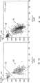
图10a、10b、10c和10d分别为被测的曲线80、83、90和98,显示了被建造以证实本发明的试验室测试床的典型珠子数据。图10a显示了事件数目相对于以米/秒(m/s)表示的流速的分布。图10b显示了事件数目相对于以微米表示的细胞直径的分布。来自这些数据的最终测定结果可以显示平均直径为5.430微米,其标准偏差为0.241微米,MCV为84.33,并且RDW为13.3。图10c显示了以毫伏表示的脉冲幅值相对于以微米表示的细胞直径的关系,对于136.1的计数率而言。图10d显示了事件数目相对于以毫伏表示的脉冲幅值的分布。这里的平均SN比看起来为61.224。Figures 10a, 10b, 10c and 10d are measured
图11a、11b、11c和11d为相似的结果,涉及检测并测量PLT和RBC的混合物。图11a显示了事件数目相对于流速(m/s)的直方图99。图11b显示了事件数目相对于以微米表示的细胞直径。人们可能注意到条块84围绕着1微米,和条块85围绕着4.5微米。这些块84和85看起来分别代表PLT和RBC。对于RBC,看起来平均直径为4.602微米,标准偏差为0.462微米,MCV为52.57并且RDW为29.7。Figures 11a, 11b, 11c and 11d show similar results involving the detection and measurement of mixtures of PLT and RBC. Figure 11a shows a
图11c显示了事件数目相对于以毫伏表示的脉冲幅值。这似乎是条块86的中心约为-45毫伏,以及条块87的中心约为-12毫伏。平均SN比看起来为大约38.911。绝对值最大的为块86,看起来代表RBC,并且另一块87看起来代表PLT。块84和86以及块85和87似乎是相互支持的。这种支持可以显示在图11d中,其中,根据分别来自图11b和11c的细胞直径绘制事件脉冲幅值。这种作图表面看来导致生成两个组88和89,其相应于PLT和RBC。这里的计数率可以为大约256.3。人们可能注意到块84和85之间隔开大约2.5微米,并且块86和87之间隔开大约22毫伏。人们可以利用这些数值作为图11d中的坐标,并通过该坐标以大约45度划线97,以显示PLT组88和RBC组89之间的近似分割线。Figure 11c shows the number of events versus pulse amplitude expressed in millivolts. It appears that the center of
图12a和12b为光学区分血液样品中的颗粒类型(即,RBC和PLT)提供了支持。试验以球形化前的血液作为样品类型。从这些图中,人们可以看到图12a中的两个组91和92和图12b中的组93和94分别代表PLT和RBC。线95和96可以以与图11d中线97相对于组88和89的方式相同的方式分别隔开两个组91和92以及组93和94。Figures 12a and 12b provide support for optically distinguishing particle types (ie, RBC and PLT) in blood samples. The test used blood before spherification as the sample type. From these figures, one can see that the two
在本申请的说明书中,尽管以另外的方式或时态进行了声明,一些主题可以是假定的或预言的。In the specification of the present application, some subject matter may be postulated or prophesied notwithstanding stated otherwise.
尽管已经相应于至少一种说明性的例子而描述了本发明,对本领域技术人员而言,通过阅读本申请的说明书,许多变形和修改将会是显而易见的。因此,其目的是附属的权利要求书应当根据现有技术尽可能广义地被解释,以包括所有的这些变形和修改。While the invention has been described with respect to at least one illustrative example, many variations and modifications will become apparent to those skilled in the art from the reading of the present specification. Therefore, it is intended that the appended claims be interpreted as broadly as possible in light of the prior art to encompass all such variations and modifications.
Claims (10)
Translated fromChineseApplications Claiming Priority (3)
| Application Number | Priority Date | Filing Date | Title |
|---|---|---|---|
| US67640305P | 2005-04-29 | 2005-04-29 | |
| US60/676,403 | 2005-04-29 | ||
| PCT/US2006/016440WO2006119106A1 (en) | 2005-04-29 | 2006-04-28 | Cytometer cell counting and size measurement method |
Publications (2)
| Publication Number | Publication Date |
|---|---|
| CN101438143A CN101438143A (en) | 2009-05-20 |
| CN101438143Btrue CN101438143B (en) | 2013-06-12 |
Family
ID=36829823
Family Applications (1)
| Application Number | Title | Priority Date | Filing Date |
|---|---|---|---|
| CN200680023450.6AExpired - Fee RelatedCN101438143B (en) | 2005-04-29 | 2006-04-28 | Cytometer cell counting and size measurement method |
Country Status (5)
| Country | Link |
|---|---|
| US (1) | US7688427B2 (en) |
| EP (1) | EP1875200A1 (en) |
| JP (1) | JP4965561B2 (en) |
| CN (1) | CN101438143B (en) |
| WO (1) | WO2006119106A1 (en) |
Families Citing this family (41)
| Publication number | Priority date | Publication date | Assignee | Title |
|---|---|---|---|---|
| US5966673A (en)* | 1997-01-10 | 1999-10-12 | Diamond Technologies, Inc. | System and method for computerized evaluation of gemstones |
| WO2010092727A1 (en)* | 2009-02-16 | 2010-08-19 | コニカミノルタオプト株式会社 | Blood test apparatus |
| ES2945686T3 (en) | 2009-05-13 | 2023-07-05 | Sartorius Bioanalytical Instr Inc | Flow measurement and control for enhanced particle quantification in flow cytometry |
| JP5304456B2 (en)* | 2009-06-10 | 2013-10-02 | ソニー株式会社 | Fine particle measuring device |
| FR2947626B1 (en) | 2009-07-03 | 2012-05-11 | Centre Nat Rech Scient | MEASURING DEVICE FOR THE CHARACTERIZATION OF DIPHASIC FLOWS. |
| US8573404B2 (en)* | 2009-12-17 | 2013-11-05 | Malvern Instruments, Ltd. | Continuous particle and macro-molecular zeta potential measurements using field flow fractionation combined micro-electrophoresis |
| US8101065B2 (en) | 2009-12-30 | 2012-01-24 | Lifescan, Inc. | Systems, devices, and methods for improving accuracy of biosensors using fill time |
| US8877034B2 (en)* | 2009-12-30 | 2014-11-04 | Lifescan, Inc. | Systems, devices, and methods for measuring whole blood hematocrit based on initial fill velocity |
| US9057676B2 (en) | 2010-02-05 | 2015-06-16 | Cytonome/St, Llc | Multiple flow channel particle analysis system |
| JP5443411B2 (en)* | 2011-03-02 | 2014-03-19 | 公益財団法人新産業創造研究機構 | Method and apparatus for detecting the size of particles in a liquid |
| WO2012158638A1 (en)* | 2011-05-13 | 2012-11-22 | Beckman Coulter, Inc. | Method and apparatus for diagnosing blood parasites |
| TWI654419B (en) | 2011-08-29 | 2019-03-21 | 美商安美基公司 | Method and apparatus for non-destructive detection - undissolved particles in a fluid |
| US10509976B2 (en) | 2012-06-22 | 2019-12-17 | Malvern Panalytical Limited | Heterogeneous fluid sample characterization |
| EP2864759A2 (en)* | 2012-06-22 | 2015-04-29 | Malvern Instruments Ltd | Particle characterization |
| US20150160246A1 (en) | 2013-12-11 | 2015-06-11 | Analiza, Inc. | Devices and methods for determining and/or isolating cells such as circulating cancer or fetal cells |
| US9709556B2 (en) | 2013-12-11 | 2017-07-18 | Analiza, Inc. | Devices and methods for determining and/or isolating circulating cancer cells |
| US9261452B2 (en)* | 2013-12-23 | 2016-02-16 | Palo Alto Research Center Incorporated | Flow cytometer |
| US10324020B2 (en) | 2013-12-23 | 2019-06-18 | Palo Alto Research Center Incorporated | Fluidic optical cartridge |
| US9952033B2 (en) | 2014-02-14 | 2018-04-24 | Palo Alto Research Center Incorporated | Spatial modulation of light to determine object length |
| US9207066B2 (en) | 2014-02-14 | 2015-12-08 | Palo Alto Research Center Incorporated | Spatial modulation of light to determine dimensional characteristics of objects in a flow path |
| US9528925B2 (en) | 2014-02-14 | 2016-12-27 | Palo Alto Research Center Incorporated | Spatial modulation of light to determine object position |
| US10451482B2 (en) | 2014-02-14 | 2019-10-22 | Palo Alto Research Center Incorporated | Determination of color characteristics of objects using spatially modulated light |
| JP6237417B2 (en)* | 2014-03-31 | 2017-11-29 | 株式会社Jvcケンウッド | Analysis apparatus and analysis method |
| US9400174B2 (en) | 2014-04-07 | 2016-07-26 | Palo Alto Research Center Incorporated | Monitor for particle injector |
| US9114606B1 (en) | 2014-04-07 | 2015-08-25 | Palo Alto Research Center Incorporated | Spatial light modulation method for determining droplet motion characteristics |
| RU2665344C2 (en)* | 2014-07-08 | 2018-08-29 | Ист Чайна Юниверсити Оф Сайенс Энд Текнолоджи | Method and device of synchronous high-speed photographic rotation of microparticle in hydrocyclone field |
| JP6942148B2 (en)* | 2016-05-11 | 2021-09-29 | エス.ディー.サイト ダイアグノスティクス リミテッド | Performing optical measurements on the sample |
| CN106018200A (en)* | 2016-05-21 | 2016-10-12 | 深圳市青核桃科技有限公司 | Method for improving measurement result accuracy of laser particle counter |
| US9704239B1 (en) | 2016-09-02 | 2017-07-11 | Amgen Inc. | Video trigger synchronization for improved particle detection in a vessel |
| CN106404637B (en)* | 2016-10-08 | 2018-11-13 | 重庆医科大学附属永川医院 | A kind of medicine detection cellanalyzer |
| CN106845622B (en)* | 2017-01-22 | 2019-04-23 | 江西特康科技有限公司 | Platelet count method and system |
| US10088660B2 (en) | 2017-02-10 | 2018-10-02 | Amgen Inc. | Imaging system for counting and sizing particles in fluid-filled vessels |
| CN107907471A (en)* | 2017-10-26 | 2018-04-13 | 刘峰 | A kind of cellular assay counting device |
| US10720755B2 (en)* | 2018-02-07 | 2020-07-21 | Elfi-Tech Ltd. | Ensemble-averaged measurement of stochastic motion by current-modulating of VCSEL wavelength |
| US12196652B2 (en) | 2018-08-01 | 2025-01-14 | Sartorius Bioanalytical Instruments, Inc. | Methods, kits and stain compositions for flow cytometry evaluation of unassociated virus-size particles using multiple fluorogenic dyes |
| US11137337B2 (en) | 2019-01-21 | 2021-10-05 | Essen Instruments, Inc. | Flow cytometry with data analysis for optimized dilution of fluid samples for flow cytometry investigation |
| US20220276250A1 (en)* | 2019-08-21 | 2022-09-01 | Waseda University | Cell analyzer system and cell analysis method |
| US11709116B2 (en) | 2020-02-04 | 2023-07-25 | Sartorius Bioanalytical Instruments, Inc. | Liquid flourescent dye concentrate for flow cytometry evaluation of virus-size particles and related products and methods |
| US11852577B2 (en)* | 2021-09-29 | 2023-12-26 | Orange Biomed Ltd., Co. | Apparatus for measuring properties of particles in a solution and related methods |
| CN118648020A (en)* | 2022-02-02 | 2024-09-13 | 贝克曼库尔特有限公司 | Measuring image quality of blood cell images |
| US20240175799A1 (en)* | 2022-11-30 | 2024-05-30 | Eaton Intelligent Power Limited | Systems and methods for optical particle detection |
Citations (4)
| Publication number | Priority date | Publication date | Assignee | Title |
|---|---|---|---|---|
| US5194909A (en)* | 1990-12-04 | 1993-03-16 | Tycko Daniel H | Apparatus and method for measuring volume and hemoglobin concentration of red blood cells |
| US6317511B1 (en)* | 1996-02-22 | 2001-11-13 | Hitachi, Ltd. | Method and apparatus for particle image analysis using flow cell |
| CN1502068A (en)* | 2000-08-02 | 2004-06-02 | ����Τ�����ʹ�˾ | Portable scattering and fluorescence cytometer |
| US20040223135A1 (en)* | 2000-08-25 | 2004-11-11 | Amnis Corporation | Methods of calibrating an imaging system using calibration beads |
Family Cites Families (109)
| Publication number | Priority date | Publication date | Assignee | Title |
|---|---|---|---|---|
| US3390605A (en)* | 1963-10-23 | 1968-07-02 | Yanagimoto Seisakusho Co Ltd | Device for measuring simultaneously both rotatory polarization and light absorption |
| US3657537A (en) | 1970-04-03 | 1972-04-18 | Bausch & Lomb | Computerized slit-scan cyto-fluorometer for automated cell recognition |
| US3822095A (en)* | 1972-08-14 | 1974-07-02 | Block Engineering | System for differentiating particles |
| US3928094A (en) | 1975-01-16 | 1975-12-23 | Fairchild Camera Instr Co | Method of aligning a wafer beneath a mask and system therefor and wafer having a unique alignment pattern |
| US3976862A (en) | 1975-03-18 | 1976-08-24 | Block Engineering, Inc. | Flow stream processor |
| US4577964A (en)* | 1978-09-06 | 1986-03-25 | Ortho Diagnostics, Inc. | Apparatus and method for detecting platelets in whole blood |
| US4293221A (en) | 1979-04-17 | 1981-10-06 | Research Corporation | Multidimensional slit-scan flow system |
| US4284412A (en) | 1979-07-13 | 1981-08-18 | Ortho Diagnostics, Inc. | Method and apparatus for automated identification and enumeration of specified blood cell subclasses |
| US4350892A (en) | 1980-07-31 | 1982-09-21 | Research Corporation | X'-, Y'-, Z'- axis multidimensional slit-scan flow system |
| US4412004A (en) | 1981-06-26 | 1983-10-25 | Technicon Instruments Corporation | Method for treating red blood cells to effect sphering and reagent therefor |
| JPS58143206A (en) | 1982-02-19 | 1983-08-25 | Canon Inc | Signal processing unit for detection of position |
| US4441816A (en)* | 1982-03-25 | 1984-04-10 | The United States Of America As Represented By The United States Department Of Energy | Optical double-slit particle measuring system |
| US4478077A (en) | 1982-09-30 | 1984-10-23 | Honeywell Inc. | Flow sensor |
| US4501144A (en) | 1982-09-30 | 1985-02-26 | Honeywell Inc. | Flow sensor |
| US4478076A (en) | 1982-09-30 | 1984-10-23 | Honeywell Inc. | Flow sensor |
| US4683159A (en) | 1982-09-30 | 1987-07-28 | Honeywell Inc. | Semiconductor device structure and processing |
| US4651564A (en) | 1982-09-30 | 1987-03-24 | Honeywell Inc. | Semiconductor device |
| JPS6166947A (en) | 1984-09-10 | 1986-04-05 | Canon Inc | Particle analysis device |
| US4695034A (en) | 1984-11-27 | 1987-09-22 | Stec Inc. | Fluid control device |
| US4745279A (en) | 1986-01-02 | 1988-05-17 | American Hospital Supply Corporation | Hematocrit measuring apparatus |
| US4704033A (en) | 1986-03-06 | 1987-11-03 | Micronix Corporation | Multiple wavelength linear zone plate alignment apparatus and method |
| NL8601000A (en) | 1986-04-21 | 1987-11-16 | Jan Greve T H Twente Afdeling | THE USE OF POLARIZED LIGHT IN FLOW CYTOMETRY. |
| JPS63100351A (en)* | 1986-10-17 | 1988-05-02 | Hitachi Ltd | Particle counting device |
| DE3640616A1 (en) | 1986-11-27 | 1988-06-09 | Standard Elektrik Lorenz Ag | ADJUSTMENT DEVICE |
| US4818263A (en) | 1987-06-11 | 1989-04-04 | Tektronix, Inc. | Method and apparatus for precisely positioning microlenses on optical fibers |
| US4874949A (en) | 1987-09-14 | 1989-10-17 | Vanderbilt University | Method of measuring lung vascular function and transcapillary transport by the use of nonradioactive markers |
| US4911616A (en) | 1988-01-19 | 1990-03-27 | Laumann Jr Carl W | Micro miniature implantable pump |
| JPH02118436A (en)* | 1988-10-28 | 1990-05-02 | Hitachi Ltd | Particle discrimination method, cell discrimination device, and cell fusion device |
| US5760900A (en) | 1989-03-18 | 1998-06-02 | Canon Kabushiki Kaisha | Method and apparatus for optically measuring specimen |
| US4932989A (en) | 1989-04-05 | 1990-06-12 | At&T Bell Laboratories | Method and apparatus for fabricating microlenses on optical fibers |
| CH679555A5 (en) | 1989-04-11 | 1992-03-13 | Westonbridge Int Ltd | |
| CA2016699C (en) | 1989-05-15 | 2003-11-18 | Paul N. Marshall | Lytic agents and uses thereof |
| CA2033181C (en) | 1989-06-14 | 2000-10-24 | Harald T. G. Van Lintel | Two valve micropump with improved outlet |
| DE3926066A1 (en) | 1989-08-07 | 1991-02-14 | Ibm Deutschland | MICROMECHANICAL COMPRESSOR CASCADE AND METHOD FOR INCREASING PRINTER AT EXTREMELY LOW WORKING PRESSURE |
| CH681168A5 (en) | 1989-11-10 | 1993-01-29 | Westonbridge Int Ltd | Micro-pump for medicinal dosing |
| KR910012538A (en) | 1989-12-27 | 1991-08-08 | 야마무라 가쯔미 | Micro pump and its manufacturing method |
| US5244537A (en) | 1989-12-27 | 1993-09-14 | Honeywell, Inc. | Fabrication of an electronic microvalve apparatus |
| US5082242A (en) | 1989-12-27 | 1992-01-21 | Ulrich Bonne | Electronic microvalve apparatus and fabrication |
| US5050429A (en) | 1990-02-22 | 1991-09-24 | Yamatake-Honeywell Co., Ltd. | Microbridge flow sensor |
| US5096388A (en) | 1990-03-22 | 1992-03-17 | The Charles Stark Draper Laboratory, Inc. | Microfabricated pump |
| US5159642A (en)* | 1990-07-13 | 1992-10-27 | Toa Medical Electronics Co., Ltd. | Particle image analyzing apparatus |
| EP0483469B1 (en) | 1990-10-30 | 1994-10-12 | Hewlett-Packard Company | Micropump |
| DE69129260T2 (en) | 1990-11-03 | 1998-11-19 | Horiba Ltd | Device for measuring the particle size distribution |
| US5108623A (en) | 1990-11-19 | 1992-04-28 | Gould Inc. | Moving web filter assembly |
| DE4119955C2 (en) | 1991-06-18 | 2000-05-31 | Danfoss As | Miniature actuator |
| US5176358A (en) | 1991-08-08 | 1993-01-05 | Honeywell Inc. | Microstructure gas valve control |
| WO1994001809A1 (en) | 1992-07-02 | 1994-01-20 | Indigo N.V. | Concentration detector for colored toner |
| JP3215175B2 (en) | 1992-08-10 | 2001-10-02 | シスメックス株式会社 | Particle analyzer |
| US5441597A (en) | 1992-12-01 | 1995-08-15 | Honeywell Inc. | Microstructure gas valve control forming method |
| WO1994027146A1 (en) | 1993-05-14 | 1994-11-24 | Coulter Corporation | Reticulocyte analyzing method and apparatus utilizing light scatter techniques |
| JPH07120375A (en)* | 1993-10-21 | 1995-05-12 | Hitachi Ltd | Flow type particle image analysis method and apparatus |
| US5540494A (en)* | 1994-06-03 | 1996-07-30 | Purvis, Jr.; Norman B. | Method and apparatus for determining absolute particle size, surface area and volume normalized fluorescence using forward angle light scatter intensity in flow cytometry |
| US5601080A (en) | 1994-12-28 | 1997-02-11 | Coretech Medical Technologies Corporation | Spectrophotometric blood analysis |
| US5793485A (en) | 1995-03-20 | 1998-08-11 | Sandia Corporation | Resonant-cavity apparatus for cytometry or particle analysis |
| US5528045A (en) | 1995-04-06 | 1996-06-18 | Becton Dickinson And Company | Particle analyzer with spatially split wavelength filter |
| AU6541596A (en) | 1995-06-16 | 1997-01-15 | University Of Washington | Microfabricated differential extraction device and method |
| EP0871539B1 (en) | 1995-06-16 | 2002-02-20 | University of Washington | Tangential flow planar microfabricated fluid filter |
| US5716852A (en) | 1996-03-29 | 1998-02-10 | University Of Washington | Microfabricated diffusion-based chemical sensor |
| US5717631A (en) | 1995-07-21 | 1998-02-10 | Carnegie Mellon University | Microelectromechanical structure and process of making same |
| US5633724A (en) | 1995-08-29 | 1997-05-27 | Hewlett-Packard Company | Evanescent scanning of biochemical array |
| US5726751A (en) | 1995-09-27 | 1998-03-10 | University Of Washington | Silicon microchannel optical flow cytometer |
| IT1280475B1 (en) | 1995-11-09 | 1998-01-20 | Fiat Ricerche | COLOR AND IMAGE SELECTIVE MICROFILTER DEVICES. |
| DE19546570C1 (en) | 1995-12-13 | 1997-03-27 | Inst Mikro Und Informationstec | Fluid micropump incorporated in silicon chip |
| JP3308441B2 (en) | 1995-12-19 | 2002-07-29 | シスメックス株式会社 | Urine particle analyzer |
| US5863502A (en) | 1996-01-24 | 1999-01-26 | Sarnoff Corporation | Parallel reaction cassette and associated devices |
| US5948684A (en) | 1997-03-31 | 1999-09-07 | University Of Washington | Simultaneous analyte determination and reference balancing in reference T-sensor devices |
| WO1997047390A1 (en) | 1996-06-14 | 1997-12-18 | University Of Washington | Absorption-enhanced differential extraction device |
| US5764674A (en) | 1996-06-28 | 1998-06-09 | Honeywell Inc. | Current confinement for a vertical cavity surface emitting laser |
| US5799030A (en) | 1996-07-26 | 1998-08-25 | Honeywell Inc. | Semiconductor device with a laser and a photodetector in a common container |
| WO1998014707A1 (en) | 1996-10-03 | 1998-04-09 | Westonbridge International Limited | Micro-machined device for fluids and method of manufacture |
| US6124663A (en) | 1996-12-16 | 2000-09-26 | The Boeing Company | Fiber optic connector having a microelectromechanical positioning apparatus and an associated fabrication method |
| US5683159A (en) | 1997-01-03 | 1997-11-04 | Johnson; Greg P. | Hardware mounting rail |
| US6097859A (en) | 1998-02-12 | 2000-08-01 | The Regents Of The University Of California | Multi-wavelength cross-connect optical switch |
| US5974867A (en) | 1997-06-13 | 1999-11-02 | University Of Washington | Method for determining concentration of a laminar sample stream |
| WO1998059233A1 (en) | 1997-06-23 | 1998-12-30 | Luminex Corporation | Interlaced lasers for multiple fluorescence measurement |
| US6082185A (en) | 1997-07-25 | 2000-07-04 | Research International, Inc. | Disposable fluidic circuit cards |
| US5880474A (en) | 1997-08-29 | 1999-03-09 | Becton Dickinson And Company | Multi-illumination-source flow particle analyzer with inter-location emissions crosstalk cancelation |
| US6007775A (en) | 1997-09-26 | 1999-12-28 | University Of Washington | Multiple analyte diffusion based chemical sensor |
| US5822170A (en) | 1997-10-09 | 1998-10-13 | Honeywell Inc. | Hydrophobic coating for reducing humidity effect in electrostatic actuators |
| US5901939A (en) | 1997-10-09 | 1999-05-11 | Honeywell Inc. | Buckled actuator with enhanced restoring force |
| US5836750A (en) | 1997-10-09 | 1998-11-17 | Honeywell Inc. | Electrostatically actuated mesopump having a plurality of elementary cells |
| US6106245A (en) | 1997-10-09 | 2000-08-22 | Honeywell | Low cost, high pumping rate electrostatically actuated mesopump |
| CA2221324A1 (en)* | 1997-11-17 | 1999-05-17 | Eagle Precision Technologies Inc. | Tub bending apparatus and method |
| US6116756A (en) | 1997-12-12 | 2000-09-12 | Xerox Corporation | Monolithic scanning light emitting devices |
| US6054335A (en) | 1997-12-12 | 2000-04-25 | Xerox Corporation | Fabrication of scanning III-V compound light emitters integrated with Si-based actuators |
| JP3522535B2 (en) | 1998-05-29 | 2004-04-26 | 忠弘 大見 | Gas supply equipment equipped with pressure type flow controller |
| US6091197A (en) | 1998-06-12 | 2000-07-18 | Xerox Corporation | Full color tunable resonant cavity organic light emitting diode |
| JP4001436B2 (en) | 1998-07-23 | 2007-10-31 | 三菱電機株式会社 | Optical switch and optical path switching device using optical switch |
| DE19835070B4 (en) | 1998-08-04 | 2006-03-16 | Carl Zeiss Jena Gmbh | Arrangement for adjustable wavelength-dependent detection in a fluorescence microscope |
| US6032689A (en) | 1998-10-30 | 2000-03-07 | Industrial Technology Research Institute | Integrated flow controller module |
| US6091537A (en) | 1998-12-11 | 2000-07-18 | Xerox Corporation | Electro-actuated microlens assemblies |
| US6184607B1 (en) | 1998-12-29 | 2001-02-06 | Honeywell International Inc. | Driving strategy for non-parallel arrays of electrostatic actuators sharing a common electrode |
| US6215221B1 (en) | 1998-12-29 | 2001-04-10 | Honeywell International Inc. | Electrostatic/pneumatic actuators for active surfaces |
| US6249341B1 (en) | 1999-01-25 | 2001-06-19 | Amnis Corporation | Imaging and analyzing parameters of small moving objects such as cells |
| US6097485A (en) | 1999-03-08 | 2000-08-01 | Integrated Waveguides, Inc. | Microchip optical transport technology for use in a personal flow cytometer |
| US6179586B1 (en) | 1999-09-15 | 2001-01-30 | Honeywell International Inc. | Dual diaphragm, single chamber mesopump |
| US6240944B1 (en) | 1999-09-23 | 2001-06-05 | Honeywell International Inc. | Addressable valve arrays for proportional pressure or flow control |
| US6813017B1 (en)* | 1999-10-20 | 2004-11-02 | Becton, Dickinson And Company | Apparatus and method employing incoherent light emitting semiconductor devices as particle detection light sources in a flow cytometer |
| US6281975B1 (en) | 2000-03-07 | 2001-08-28 | Eldex Laboratories, Inc. | Capillary flow cell with bulbous ends |
| US20010030743A1 (en) | 2000-03-10 | 2001-10-18 | Carlos Araujo | Laser alignment system with plural lasers for impingement on a single target |
| DE20008228U1 (en) | 2000-05-09 | 2002-01-03 | Prüftechnik Dieter Busch AG, 85737 Ismaning | Device for quantitative assessment of the aligned position of two machine parts, workpieces or the like. |
| US7978329B2 (en) | 2000-08-02 | 2011-07-12 | Honeywell International Inc. | Portable scattering and fluorescence cytometer |
| US7641856B2 (en) | 2004-05-14 | 2010-01-05 | Honeywell International Inc. | Portable sample analyzer with removable cartridge |
| US8071051B2 (en)* | 2004-05-14 | 2011-12-06 | Honeywell International Inc. | Portable sample analyzer cartridge |
| US6549275B1 (en) | 2000-08-02 | 2003-04-15 | Honeywell International Inc. | Optical detection system for flow cytometry |
| US6382228B1 (en) | 2000-08-02 | 2002-05-07 | Honeywell International Inc. | Fluid driving system for flow cytometry |
| JP4385049B2 (en)* | 2003-06-23 | 2009-12-16 | セウォン メディテック インコーポレイテッド | Blood cell deformability measuring device |
| US6907859B1 (en)* | 2004-05-11 | 2005-06-21 | Barnett Joel Robinson | Internal combustion engine with elevated expansion ratio |
| EP1966588B1 (en)* | 2005-12-29 | 2018-12-12 | Honeywell International Inc. | Assay implementation in a microfluidic format |
- 2006
- 2006-04-28CNCN200680023450.6Apatent/CN101438143B/ennot_activeExpired - Fee Related
- 2006-04-28USUS11/380,878patent/US7688427B2/enactiveActive
- 2006-04-28JPJP2008509209Apatent/JP4965561B2/enactiveActive
- 2006-04-28WOPCT/US2006/016440patent/WO2006119106A1/enactiveApplication Filing
- 2006-04-28EPEP06751902Apatent/EP1875200A1/ennot_activeCeased
Patent Citations (4)
| Publication number | Priority date | Publication date | Assignee | Title |
|---|---|---|---|---|
| US5194909A (en)* | 1990-12-04 | 1993-03-16 | Tycko Daniel H | Apparatus and method for measuring volume and hemoglobin concentration of red blood cells |
| US6317511B1 (en)* | 1996-02-22 | 2001-11-13 | Hitachi, Ltd. | Method and apparatus for particle image analysis using flow cell |
| CN1502068A (en)* | 2000-08-02 | 2004-06-02 | ����Τ�����ʹ�˾ | Portable scattering and fluorescence cytometer |
| US20040223135A1 (en)* | 2000-08-25 | 2004-11-11 | Amnis Corporation | Methods of calibrating an imaging system using calibration beads |
Also Published As
| Publication number | Publication date |
|---|---|
| EP1875200A1 (en) | 2008-01-09 |
| JP4965561B2 (en) | 2012-07-04 |
| WO2006119106A1 (en) | 2006-11-09 |
| US7688427B2 (en) | 2010-03-30 |
| JP2008539446A (en) | 2008-11-13 |
| US20060244964A1 (en) | 2006-11-02 |
| CN101438143A (en) | 2009-05-20 |
Similar Documents
| Publication | Publication Date | Title |
|---|---|---|
| CN101438143B (en) | Cytometer cell counting and size measurement method | |
| US10859484B2 (en) | Method for determining volume and hemoglobin content of individual red blood cells | |
| US5194909A (en) | Apparatus and method for measuring volume and hemoglobin concentration of red blood cells | |
| CA1230752A (en) | Method and apparatus for determining the volume and index of refraction of particles | |
| US7390662B2 (en) | Method and apparatus for performing platelet measurement | |
| EP2062056B1 (en) | Differentiation of flow cytometry pulses and applications | |
| US5798827A (en) | Apparatus and method for determination of individual red blood cell shape | |
| US6630990B2 (en) | Optical method and apparatus for red blood cell differentiation on a cell-by-cell basis, and simultaneous analysis of white blood cell differentiation | |
| JP5457560B2 (en) | How to flag a sample | |
| JP6076801B2 (en) | Blood cell analyzer and blood cell analysis method | |
| US7130046B2 (en) | Data frame selection for cytometer analysis | |
| CN104215562A (en) | Optical system of particle analyzer | |
| US9068915B2 (en) | Method and system for calibrating a flow cytometer | |
| Gray et al. | A new method for cell volume measurement based on volume exclusion of a fluorescent dye | |
| JP4301590B2 (en) | Particle measuring device | |
| JPH0792076A (en) | Particle analyzer | |
| JP2720069B2 (en) | Flow cell analyzer |
Legal Events
| Date | Code | Title | Description |
|---|---|---|---|
| C06 | Publication | ||
| PB01 | Publication | ||
| C10 | Entry into substantive examination | ||
| SE01 | Entry into force of request for substantive examination | ||
| C14 | Grant of patent or utility model | ||
| GR01 | Patent grant | ||
| CF01 | Termination of patent right due to non-payment of annual fee | ||
| CF01 | Termination of patent right due to non-payment of annual fee | Granted publication date:20130612 |