CN101426473A - Apparatus and formulation for suprachoroidal drug delivery - Google Patents
Apparatus and formulation for suprachoroidal drug deliveryDownload PDFInfo
- Publication number
- CN101426473A CN101426473ACNA2007800145013ACN200780014501ACN101426473ACN 101426473 ACN101426473 ACN 101426473ACN A2007800145013 ACNA2007800145013 ACN A2007800145013ACN 200780014501 ACN200780014501 ACN 200780014501ACN 101426473 ACN101426473 ACN 101426473A
- Authority
- CN
- China
- Prior art keywords
- suprachoroidal space
- pharmaceutical preparation
- tissue
- tip
- preparation
- Prior art date
- Legal status (The legal status is an assumption and is not a legal conclusion. Google has not performed a legal analysis and makes no representation as to the accuracy of the status listed.)
- Granted
Links
Images


Classifications
- A—HUMAN NECESSITIES
- A61—MEDICAL OR VETERINARY SCIENCE; HYGIENE
- A61K—PREPARATIONS FOR MEDICAL, DENTAL OR TOILETRY PURPOSES
- A61K9/00—Medicinal preparations characterised by special physical form
- A61K9/48—Preparations in capsules, e.g. of gelatin, of chocolate
- A61K9/50—Microcapsules having a gas, liquid or semi-solid filling; Solid microparticles or pellets surrounded by a distinct coating layer, e.g. coated microspheres, coated drug crystals
- A—HUMAN NECESSITIES
- A61—MEDICAL OR VETERINARY SCIENCE; HYGIENE
- A61K—PREPARATIONS FOR MEDICAL, DENTAL OR TOILETRY PURPOSES
- A61K9/00—Medicinal preparations characterised by special physical form
- A61K9/0012—Galenical forms characterised by the site of application
- A61K9/0048—Eye, e.g. artificial tears
- A—HUMAN NECESSITIES
- A61—MEDICAL OR VETERINARY SCIENCE; HYGIENE
- A61F—FILTERS IMPLANTABLE INTO BLOOD VESSELS; PROSTHESES; DEVICES PROVIDING PATENCY TO, OR PREVENTING COLLAPSING OF, TUBULAR STRUCTURES OF THE BODY, e.g. STENTS; ORTHOPAEDIC, NURSING OR CONTRACEPTIVE DEVICES; FOMENTATION; TREATMENT OR PROTECTION OF EYES OR EARS; BANDAGES, DRESSINGS OR ABSORBENT PADS; FIRST-AID KITS
- A61F9/00—Methods or devices for treatment of the eyes; Devices for putting in contact-lenses; Devices to correct squinting; Apparatus to guide the blind; Protective devices for the eyes, carried on the body or in the hand
- A61F9/0008—Introducing ophthalmic products into the ocular cavity or retaining products therein
- A61F9/0017—Introducing ophthalmic products into the ocular cavity or retaining products therein implantable in, or in contact with, the eye, e.g. ocular inserts
- A—HUMAN NECESSITIES
- A61—MEDICAL OR VETERINARY SCIENCE; HYGIENE
- A61F—FILTERS IMPLANTABLE INTO BLOOD VESSELS; PROSTHESES; DEVICES PROVIDING PATENCY TO, OR PREVENTING COLLAPSING OF, TUBULAR STRUCTURES OF THE BODY, e.g. STENTS; ORTHOPAEDIC, NURSING OR CONTRACEPTIVE DEVICES; FOMENTATION; TREATMENT OR PROTECTION OF EYES OR EARS; BANDAGES, DRESSINGS OR ABSORBENT PADS; FIRST-AID KITS
- A61F9/00—Methods or devices for treatment of the eyes; Devices for putting in contact-lenses; Devices to correct squinting; Apparatus to guide the blind; Protective devices for the eyes, carried on the body or in the hand
- A61F9/0008—Introducing ophthalmic products into the ocular cavity or retaining products therein
- A61F9/0026—Ophthalmic product dispenser attachments to facilitate positioning near the eye
- A—HUMAN NECESSITIES
- A61—MEDICAL OR VETERINARY SCIENCE; HYGIENE
- A61F—FILTERS IMPLANTABLE INTO BLOOD VESSELS; PROSTHESES; DEVICES PROVIDING PATENCY TO, OR PREVENTING COLLAPSING OF, TUBULAR STRUCTURES OF THE BODY, e.g. STENTS; ORTHOPAEDIC, NURSING OR CONTRACEPTIVE DEVICES; FOMENTATION; TREATMENT OR PROTECTION OF EYES OR EARS; BANDAGES, DRESSINGS OR ABSORBENT PADS; FIRST-AID KITS
- A61F9/00—Methods or devices for treatment of the eyes; Devices for putting in contact-lenses; Devices to correct squinting; Apparatus to guide the blind; Protective devices for the eyes, carried on the body or in the hand
- A61F9/007—Methods or devices for eye surgery
- A61F9/00736—Instruments for removal of intra-ocular material or intra-ocular injection, e.g. cataract instruments
- A—HUMAN NECESSITIES
- A61—MEDICAL OR VETERINARY SCIENCE; HYGIENE
- A61K—PREPARATIONS FOR MEDICAL, DENTAL OR TOILETRY PURPOSES
- A61K31/00—Medicinal preparations containing organic active ingredients
- A61K31/70—Carbohydrates; Sugars; Derivatives thereof
- A61K31/715—Polysaccharides, i.e. having more than five saccharide radicals attached to each other by glycosidic linkages; Derivatives thereof, e.g. ethers, esters
- A61K31/716—Glucans
- A61K31/717—Celluloses
- A—HUMAN NECESSITIES
- A61—MEDICAL OR VETERINARY SCIENCE; HYGIENE
- A61K—PREPARATIONS FOR MEDICAL, DENTAL OR TOILETRY PURPOSES
- A61K31/00—Medicinal preparations containing organic active ingredients
- A61K31/70—Carbohydrates; Sugars; Derivatives thereof
- A61K31/715—Polysaccharides, i.e. having more than five saccharide radicals attached to each other by glycosidic linkages; Derivatives thereof, e.g. ethers, esters
- A61K31/716—Glucans
- A61K31/718—Starch or degraded starch, e.g. amylose, amylopectin
- A—HUMAN NECESSITIES
- A61—MEDICAL OR VETERINARY SCIENCE; HYGIENE
- A61K—PREPARATIONS FOR MEDICAL, DENTAL OR TOILETRY PURPOSES
- A61K31/00—Medicinal preparations containing organic active ingredients
- A61K31/70—Carbohydrates; Sugars; Derivatives thereof
- A61K31/715—Polysaccharides, i.e. having more than five saccharide radicals attached to each other by glycosidic linkages; Derivatives thereof, e.g. ethers, esters
- A61K31/716—Glucans
- A61K31/722—Chitin, chitosan
- A—HUMAN NECESSITIES
- A61—MEDICAL OR VETERINARY SCIENCE; HYGIENE
- A61K—PREPARATIONS FOR MEDICAL, DENTAL OR TOILETRY PURPOSES
- A61K31/00—Medicinal preparations containing organic active ingredients
- A61K31/70—Carbohydrates; Sugars; Derivatives thereof
- A61K31/715—Polysaccharides, i.e. having more than five saccharide radicals attached to each other by glycosidic linkages; Derivatives thereof, e.g. ethers, esters
- A61K31/726—Glycosaminoglycans, i.e. mucopolysaccharides
- A61K31/728—Hyaluronic acid
- A—HUMAN NECESSITIES
- A61—MEDICAL OR VETERINARY SCIENCE; HYGIENE
- A61K—PREPARATIONS FOR MEDICAL, DENTAL OR TOILETRY PURPOSES
- A61K31/00—Medicinal preparations containing organic active ingredients
- A61K31/70—Carbohydrates; Sugars; Derivatives thereof
- A61K31/715—Polysaccharides, i.e. having more than five saccharide radicals attached to each other by glycosidic linkages; Derivatives thereof, e.g. ethers, esters
- A61K31/737—Sulfated polysaccharides, e.g. chondroitin sulfate, dermatan sulfate
- A—HUMAN NECESSITIES
- A61—MEDICAL OR VETERINARY SCIENCE; HYGIENE
- A61K—PREPARATIONS FOR MEDICAL, DENTAL OR TOILETRY PURPOSES
- A61K38/00—Medicinal preparations containing peptides
- A61K38/16—Peptides having more than 20 amino acids; Gastrins; Somatostatins; Melanotropins; Derivatives thereof
- A61K38/17—Peptides having more than 20 amino acids; Gastrins; Somatostatins; Melanotropins; Derivatives thereof from animals; from humans
- A61K38/39—Connective tissue peptides, e.g. collagen, elastin, laminin, fibronectin, vitronectin, cold insoluble globulin [CIG]
- A—HUMAN NECESSITIES
- A61—MEDICAL OR VETERINARY SCIENCE; HYGIENE
- A61K—PREPARATIONS FOR MEDICAL, DENTAL OR TOILETRY PURPOSES
- A61K47/00—Medicinal preparations characterised by the non-active ingredients used, e.g. carriers or inert additives; Targeting or modifying agents chemically bound to the active ingredient
- A61K47/30—Macromolecular organic or inorganic compounds, e.g. inorganic polyphosphates
- A61K47/36—Polysaccharides; Derivatives thereof, e.g. gums, starch, alginate, dextrin, hyaluronic acid, chitosan, inulin, agar or pectin
- A—HUMAN NECESSITIES
- A61—MEDICAL OR VETERINARY SCIENCE; HYGIENE
- A61K—PREPARATIONS FOR MEDICAL, DENTAL OR TOILETRY PURPOSES
- A61K9/00—Medicinal preparations characterised by special physical form
- A61K9/0012—Galenical forms characterised by the site of application
- A61K9/0048—Eye, e.g. artificial tears
- A61K9/0051—Ocular inserts, ocular implants
- A—HUMAN NECESSITIES
- A61—MEDICAL OR VETERINARY SCIENCE; HYGIENE
- A61K—PREPARATIONS FOR MEDICAL, DENTAL OR TOILETRY PURPOSES
- A61K9/00—Medicinal preparations characterised by special physical form
- A61K9/14—Particulate form, e.g. powders, Processes for size reducing of pure drugs or the resulting products, Pure drug nanoparticles
- A61K9/16—Agglomerates; Granulates; Microbeadlets ; Microspheres; Pellets; Solid products obtained by spray drying, spray freeze drying, spray congealing,(multiple) emulsion solvent evaporation or extraction
- A61K9/1605—Excipients; Inactive ingredients
- A61K9/1629—Organic macromolecular compounds
- A61K9/1635—Organic macromolecular compounds obtained by reactions only involving carbon-to-carbon unsaturated bonds, e.g. polyvinyl pyrrolidone, poly(meth)acrylates
- A—HUMAN NECESSITIES
- A61—MEDICAL OR VETERINARY SCIENCE; HYGIENE
- A61M—DEVICES FOR INTRODUCING MEDIA INTO, OR ONTO, THE BODY; DEVICES FOR TRANSDUCING BODY MEDIA OR FOR TAKING MEDIA FROM THE BODY; DEVICES FOR PRODUCING OR ENDING SLEEP OR STUPOR
- A61M5/00—Devices for bringing media into the body in a subcutaneous, intra-vascular or intramuscular way; Accessories therefor, e.g. filling or cleaning devices, arm-rests
- A61M5/48—Devices for bringing media into the body in a subcutaneous, intra-vascular or intramuscular way; Accessories therefor, e.g. filling or cleaning devices, arm-rests having means for varying, regulating, indicating or limiting injection pressure
- A61M5/486—Indicating injection pressure
- A—HUMAN NECESSITIES
- A61—MEDICAL OR VETERINARY SCIENCE; HYGIENE
- A61K—PREPARATIONS FOR MEDICAL, DENTAL OR TOILETRY PURPOSES
- A61K9/00—Medicinal preparations characterised by special physical form
- A61K9/14—Particulate form, e.g. powders, Processes for size reducing of pure drugs or the resulting products, Pure drug nanoparticles
- A—HUMAN NECESSITIES
- A61—MEDICAL OR VETERINARY SCIENCE; HYGIENE
- A61M—DEVICES FOR INTRODUCING MEDIA INTO, OR ONTO, THE BODY; DEVICES FOR TRANSDUCING BODY MEDIA OR FOR TAKING MEDIA FROM THE BODY; DEVICES FOR PRODUCING OR ENDING SLEEP OR STUPOR
- A61M5/00—Devices for bringing media into the body in a subcutaneous, intra-vascular or intramuscular way; Accessories therefor, e.g. filling or cleaning devices, arm-rests
- A61M5/178—Syringes
- A61M5/31—Details
- A61M2005/3103—Leak prevention means for distal end of syringes, i.e. syringe end for mounting a needle
- A—HUMAN NECESSITIES
- A61—MEDICAL OR VETERINARY SCIENCE; HYGIENE
- A61M—DEVICES FOR INTRODUCING MEDIA INTO, OR ONTO, THE BODY; DEVICES FOR TRANSDUCING BODY MEDIA OR FOR TAKING MEDIA FROM THE BODY; DEVICES FOR PRODUCING OR ENDING SLEEP OR STUPOR
- A61M2210/00—Anatomical parts of the body
- A61M2210/06—Head
- A61M2210/0612—Eyes
Landscapes
- Health & Medical Sciences (AREA)
- Life Sciences & Earth Sciences (AREA)
- Animal Behavior & Ethology (AREA)
- Veterinary Medicine (AREA)
- Public Health (AREA)
- General Health & Medical Sciences (AREA)
- Chemical & Material Sciences (AREA)
- Pharmacology & Pharmacy (AREA)
- Epidemiology (AREA)
- Medicinal Chemistry (AREA)
- Engineering & Computer Science (AREA)
- Molecular Biology (AREA)
- Ophthalmology & Optometry (AREA)
- Biomedical Technology (AREA)
- Bioinformatics & Cheminformatics (AREA)
- Heart & Thoracic Surgery (AREA)
- Vascular Medicine (AREA)
- Dermatology (AREA)
- Surgery (AREA)
- Nuclear Medicine, Radiotherapy & Molecular Imaging (AREA)
- Inorganic Chemistry (AREA)
- Anesthesiology (AREA)
- Hematology (AREA)
- Immunology (AREA)
- Gastroenterology & Hepatology (AREA)
- Zoology (AREA)
- Proteomics, Peptides & Aminoacids (AREA)
- Medicinal Preparation (AREA)
- Infusion, Injection, And Reservoir Apparatuses (AREA)
- Medicines That Contain Protein Lipid Enzymes And Other Medicines (AREA)
- Materials For Medical Uses (AREA)
- Pharmaceuticals Containing Other Organic And Inorganic Compounds (AREA)
Abstract
Description
Translated fromChinese相关申请的交叉参考Cross References to Related Applications
依照35 USC 119(e)请求2006年2月22日提出的美国临时申请序列号60/776,903的优先权。该临时申请通过参考整体引入此处。Priority is claimed pursuant to 35 USC 119(e) to US Provisional Application Serial No. 60/776,903, filed February 22, 2006. This provisional application is hereby incorporated by reference in its entirety.
发明领域field of invention
本发明涉及眼睛的药物递送领域。The present invention relates to the field of drug delivery to the eye.
发明背景Background of the invention
眼睛是具有多种特殊组织的复杂器官,提供视觉的光学和神经过程。组织的小尺寸和脆弱本质阻碍了进入眼睛进行医学治疗。眼睛的后部区域,包括视网膜、黄斑和视神经,特别难于进入,这是由于眼睛在眼眶内的凹陷位置。此外,局部滴眼剂很少渗透到后部区域内,进一步限制了治疗选择。The eye is a complex organ with many specialized tissues that provide the optical and neural processes of vision. The small size and fragile nature of the tissue hinder access to the eye for medical treatment. The posterior region of the eye, including the retina, macula, and optic nerve, is particularly difficult to access due to the sunken position of the eye within the orbit. In addition, topical eye drops rarely penetrate into the posterior region, further limiting treatment options.
脉络膜上方空间是眼睛中位于脉络膜(其是内血管膜)和眼睛外层的巩膜之间的潜在空间。该脉络膜上方空间从睫状体附近的眼睛前部延伸到视神经附近的眼睛后端。通常由于脉络膜由于眼睛的眼内压而与巩膜紧密并置,脉络膜上方空间并不明显。由于脉络膜与巩膜没有实质连接,当流体累积或其它状况发生时,组织分离形成脉络膜上方空间。该脉络膜上方空间提供了从眼睛前部区域进入的潜在途径用于治疗后部区域。The suprachoroidal space is the potential space in the eye between the choroid (which is the inner vascular membrane) and the sclera, the outer layer of the eye. This suprachoroidal space extends from the front of the eye near the ciliary body to the back of the eye near the optic nerve. Usually the suprachoroidal space is not evident due to the close apposition of the choroid to the sclera due to the intraocular pressure of the eye. Since the choroid does not have a substantial connection to the sclera, when fluid builds up or other conditions occur, tissue separates to form the suprachoroidal space. This suprachoroidal space provides potential access from the anterior region of the eye for treatment of the posterior region.
本发明涉及用于为脉络膜上方空间给药的药物制剂,和将药物和其它物质以最小侵入方式递送到该脉络膜上方空间的装置。The present invention relates to pharmaceutical formulations for administering drugs to the suprachoroidal space, and devices for the minimally invasive delivery of drugs and other substances into the suprachoroidal space.
发明内容Contents of the invention
提供了药物制剂,其特征在于零剪切粘度为至少300000mPas。一子类的药物制剂进一步特征在于在1000s-1的剪切速率下其粘度不超过约400mPas。A pharmaceutical formulation is provided characterized by a zero shear viscosity of at least 300000 mPas. A subclass of pharmaceutical formulations is further characterized by a viscosity of no more than about 400 mPas at a shear rate of 1000 s-1 .
为了注射到眼睛的脉络膜上方空间,包含生物活性物质和触变聚合物赋型剂,其用作胶状材料,用于在注射后铺展并将该药物均匀分布和定位在该脉络膜上方空间的区域内。在一种实施方案中,胶状材料在注射到该脉络膜上方空间之后发生交联。该生物活性物质可以包含微颗粒或微球。该聚合物赋型剂可以包括透明质酸、硫酸软骨素、明胶、聚甲基丙烯酸羟乙酯、硫酸皮肤素(dermatin sulfate)、聚环氧乙烷、聚乙二醇、聚环氧丙烷、聚丙二醇、藻酸盐、淀粉衍生物、水溶性甲壳质衍生物、水溶性纤维素衍生物或聚乙烯基吡咯烷酮。For injection into the suprachoroidal space of the eye, containing a biologically active substance and a thixotropic polymer excipient, which acts as a gel-like material for spreading and evenly distributing and localizing the drug in the area of the suprachoroidal space after injection Inside. In one embodiment, the gel-like material is cross-linked after injection into the suprachoroidal space. The biologically active substance may comprise microparticles or microspheres. The polymer excipient may include hyaluronic acid, chondroitin sulfate, gelatin, polyhydroxyethylmethacrylate, dermatin sulfate, polyethylene oxide, polyethylene glycol, polypropylene oxide, Polypropylene glycol, alginates, starch derivatives, water-soluble chitin derivatives, water-soluble cellulose derivatives or polyvinylpyrrolidone.
在另一实施方案中,提供了用于递送到眼睛的脉络膜上方空间的药物制剂,包含生物活性物质和具有在约1~33微米范围内的外径的微球。该微颗粒或微球另外可以包含受控释放涂层和/或组织亲和性表面。In another embodiment, there is provided a pharmaceutical formulation for delivery to the suprachoroidal space of the eye comprising a biologically active substance and microspheres having an outer diameter in the range of about 1 to 33 microns. The microparticles or microspheres may additionally comprise a controlled release coating and/or a tissue affinity surface.
该生物活性物质优选包括抗生素、甾族化合物、非甾族抗炎剂、神经保护剂、抗VEGF剂或新血管形成抑制剂。The biologically active substance preferably comprises antibiotics, steroids, non-steroidal anti-inflammatory agents, neuroprotective agents, anti-VEGF agents or inhibitors of neovascularization.
还提供了用于将药物制剂以最小侵入方式递送到眼睛的脉络膜上方空间的装置,包括具有其形状允许通过巩膜组织而不损坏下面的脉络膜组织的引导尖端的针,和用于引导所述尖端的设置以将所述制剂递送到该脉络膜上方空间附近或内部的传感器。Also provided is a device for minimally invasive delivery of a pharmaceutical formulation to the suprachoroidal space of the eye comprising a needle having a guiding tip shaped to allow passage of scleral tissue without damaging underlying choroidal tissue, and a needle for guiding said tip The sensor is configured to deliver the formulation to the sensor near or within the suprachoroidal space.
该传感器可以提供巩膜组织的图像。该传感器优选对超声、光、或压差响应。The sensor can provide images of scleral tissue. The sensor is preferably responsive to ultrasound, light, or differential pressure.
在另一实施方案中,提供了用于将药物制剂以最小侵入方式递送到眼睛的脉络膜上方空间的装置,包括具有其形状允许通过巩膜组织的引导尖端的针,和一旦接触脉络膜就为该脉络膜提供向内扩张行为以防止对其产生损伤的内部尖端。In another embodiment, there is provided a device for minimally invasive delivery of a drug formulation to the suprachoroidal space of the eye comprising a needle having an introducing tip shaped to allow passage through scleral tissue, and once in contact with the choroid An inner tip that provides an inward expansion behavior to prevent damage to it.
提供了用于为眼睛给药的方法,包括将包含生物活性物质和聚合物赋型剂的制剂放置在脉络膜上方空间中,使得所述赋型剂在递送之后凝胶以使所述生物活性物质定位。该制剂可以放置在脉络膜上方空间的后部或前部区域。A method for administering to the eye is provided comprising placing a formulation comprising a biologically active substance and a polymeric excipient in the suprachoroidal space such that the excipient gels after delivery so that the biologically active substance position. The preparation can be placed in the posterior or anterior region of the suprachoroidal space.
在另一实施方案中,提供了用于给药到眼睛的后部区域的方法,包括将包含含具有在约1~33微米范围内的外径的微球或微颗粒的生物活性物质的制剂放置在脉络膜上方空间的前部区域中,使得所述微球或微颗粒随后迁移到所述后部区域。该制剂优选包含用于将该微颗粒或微球均匀分散到该脉络膜上方空间中的聚合物赋型剂。In another embodiment, there is provided a method for administering to the posterior region of the eye comprising administering a formulation comprising a biologically active substance comprising microspheres or microparticles having an outer diameter in the range of about 1 to 33 microns. Placement in the anterior region of the suprachoroidal space allows the microspheres or microparticles to subsequently migrate to the posterior region. The formulation preferably comprises a polymeric excipient for uniform dispersion of the microparticles or microspheres into the suprachoroidal space.
在另一实施方案中,提供了用于为眼睛的脉络膜上方空间给药的方法,包括以下步骤:将针以至少该巩膜厚度一半的深度放置在朝向该脉络膜上方空间的巩膜组织中,和将药物制剂通过所述针注射到该巩膜中,使得所述制剂将与所述脉络膜上方空间相邻的该巩膜组织剖开并进入所述脉络膜上方空间。In another embodiment, there is provided a method for administering a drug to the suprachoroidal space of an eye comprising the steps of: placing a needle in scleral tissue facing the suprachoroidal space at a depth of at least half the thickness of the sclera, and placing A drug formulation is injected through the needle into the sclera such that the formulation dissects the scleral tissue adjacent to the suprachoroidal space and enters the suprachoroidal space.
在此处公开的方法中,该制剂优选包含触变聚合物。In the methods disclosed herein, the formulation preferably comprises a thixotropic polymer.
附图简述Brief description of the drawings
图1是依照实施例9在用针将透明质酸外科用粘弹性材料注射到巩膜中之后眼睛的一部分的超声图像。FIG. 1 is an ultrasound image of a portion of an eye after needle injection of a hyaluronic acid surgical viscoelastic material into the sclera according to Example 9. FIG.
图2是依照实施例9在用针将粘弹性材料和聚苯乙烯微球的1%溶液的1:1体积混合物注射到巩膜中之后眼睛的一部分的超声图像。2 is an ultrasound image of a portion of the eye following needle injection of a 1:1 volumetric mixture of viscoelastic material and a 1% solution of polystyrene microspheres into the sclera according to Example 9. FIG.
图3a和3b是具有扩张和切割或烧蚀尖端的依照本发明的递送装置的实施方案的图解。Figures 3a and 3b are diagrams of an embodiment of a delivery device according to the present invention having a dilating and cutting or ablating tip.
图4是显示依照本发明的递送装置相对于目标巩膜、脉络膜上方空间和脉络膜的定位的图解。Figure 4 is a diagram showing the positioning of a delivery device in accordance with the present invention relative to the target sclera, suprachoroidal space and choroid.
图5是具有用于设定该针穿透到眼睛中的深度和角度的挡板的依照本发明的递送装置的实施方案的图解。Figure 5 is a diagram of an embodiment of a delivery device according to the present invention with a barrier for setting the depth and angle of penetration of the needle into the eye.
图6是在手术过程中容纳微内窥镜和照相机以监控套管尖端的位置的递送装置的实施方案的图解。Figure 6 is an illustration of an embodiment of a delivery device housing a microendoscope and a camera to monitor the position of the cannula tip during a procedure.
图7是具有用于将药物通过导管递送到眼睛中的管腔和与光源相连以照亮套管尖端的光导纤维的递送装置的实施方案的图解。Figure 7 is a diagram of an embodiment of a delivery device having a lumen for delivering a drug through a catheter into the eye and a fiber optic connected to a light source to illuminate the tip of the cannula.
图8是依照本发明的装置与用于监控套管尖端位置的高分辨率成像装置相结合使用的实施方案的图解。Figure 8 is an illustration of an embodiment of a device according to the present invention used in conjunction with a high resolution imaging device for monitoring the position of the cannula tip.
优选实施方式preferred embodiment
本发明包括用于进入眼睛的脉络膜上方空间用于递送药物以治疗眼睛的药物制剂、装置和相关方法。特别地,本发明涉及用于通过对递送药物的定位而对脉络膜上方空间给药以治疗眼睛(包括眼睛的特定区域)的药物制剂。本发明还涉及用于直接将药物制剂和含药材料通过小针注射到脉络膜上方空间中的最小侵入性装置的设计和使用方法。The present invention includes pharmaceutical formulations, devices and related methods for delivering drugs into the suprachoroidal space of the eye for treatment of the eye. In particular, the present invention relates to pharmaceutical formulations for the administration of the suprachoroidal space to treat the eye, including specific regions of the eye, by localizing the delivery of the drug. The present invention also relates to the design and method of use of a minimally invasive device for direct injection of pharmaceutical formulations and drug-containing materials through small needles into the suprachoroidal space.
生物活性物质或材料是影响活体或生物学过程的药物或其它物质,包括在疾病的诊断、治疗、缓和、处理或预防中的应用以影响身体的结构或任何功能。药物制剂包含生物活性物质。A biologically active substance or material is a drug or other substance that affects a living organism or a biological process, including use in the diagnosis, therapy, mitigation, treatment or prevention of disease to affect the structure or any function of the body. Pharmaceutical preparations contain biologically active substances.
此处所用的眼睛的前部区域是通常容易从眼窝中的眼睛的暴露正面进入的眼睛区域。眼睛的后部区域通常是眼睛的如下剩余区域:所述区域主要通过外科手术通过眼睛的未暴露的、并因此通常需要眼睛临时缩回以到达的表面而到达的区域。As used herein, the anterior region of the eye is the region of the eye that is generally easily accessible from the exposed front of the eye in the eye socket. The posterior region of the eye is generally the remaining region of the eye that is primarily accessed surgically through the unexposed surface of the eye, and thus often requires the eye to be temporarily retracted to reach it.
制剂:preparation:
本发明的药物制剂提供了与脉络膜上方空间环境的相容性,可以经配制以控制通过该制剂的迁移分布该生物活性物质以及提供随时间的持续释放。该药物制剂包含与生理相容性赋型剂一起配制的一种或多种生物活性物质,其通常通过注射给药到眼睛的脉络膜上方空间中。适合的生物活性物质包括用于治疗感染的抗生素、用于治疗炎症和浮肿的甾族化合物和非甾族抗炎化合物、用于治疗视神经的神经保护剂(例如钙通道阻滞剂)和用于治疗黄斑变性的视网膜剂(例如抗VEGF化合物或新血管形成抑制剂)。The pharmaceutical formulations of the present invention provide compatibility with the environment of the suprachoroidal space, can be formulated to control migration distribution of the biologically active substance through the formulation and to provide sustained release over time. The pharmaceutical formulation comprises one or more biologically active substances formulated together with physiologically compatible excipients, which are usually administered by injection into the suprachoroidal space of the eye. Suitable bioactive substances include antibiotics for the treatment of infections, steroidal and non-steroidal anti-inflammatory compounds for the treatment of inflammation and edema, neuroprotective agents (such as calcium channel blockers) for the treatment of the optic nerve, and Retinal agents to treat macular degeneration (such as anti-VEGF compounds or inhibitors of neovascularization).
用于局部治疗的制剂:Preparations for topical treatment:
为了治疗眼睛的局部区域,例如,用于治疗黄斑病变(macularlesion)、后部视网膜或视神经,可以将药物制备在制剂中以限制在递送后的迁移并递送到病变区域。尽管不意于被特定理论限制,但我们观察到药物微颗粒通常在生理条件下朝向脉络膜上方空间的后部区域移动,可能是由于在该空间内的眼色素膜-巩膜流体流动。这种药物微颗粒可以制备具有足够的尺寸,任选具有组织表面亲和性,以限制药物迁移。可以通过在该微颗粒上添加聚合或脂质表面涂层,或者通过在该颗粒表面添加化学或生物部分来改性组织表面亲和性。由此由表面电荷、疏水性、或者例如可以引入微颗粒表面上的抗体或整联蛋白的生物目标试剂得到组织亲和性,用于提供与该组织的结合性质以限制药物移动。可替代地或与其相结合,可以将该药物与一种或多种聚合物赋型剂一起配制以限制药物迁移。可以选择和配制聚合物赋型剂以原位作为粘性胶状材料,并由此铺展到脉络膜上方空间的区域中并均匀分布和保持药物。可以选择和配制聚合物赋型剂以提供适合的粘度、流动性和溶解性质。例如,羧甲基纤维素是弱触变水溶性聚合物,其可以配制到在零剪切速率具有适合的粘度,以在脉络膜上方空间中形成胶状材料。聚合物的触变效应可以通过如下方式增强:对该聚合物进行适当化学改性来提高缔合性质比如加入疏水性部分,选择较高分子量聚合物或通过与适当的表面活性剂一起配制。优选使用具有强触变性质的高缔合性聚合物赋型剂,例如透明质酸,以使药物制剂的定位和药物保持性质最大化,同时允许该制剂注射通过小规格针。可以通过将聚合物赋型剂的水溶性、分子量和浓度在适合触变性质的范围内进行定制来调节该药物制剂的溶解性质,以可以通过小规格针进行递送并定位在脉络膜上方空间中。聚合物赋型剂可以经配制以提高粘度或在递送后交联以进一步限制该材料和结合的药物的迁移或溶解。例如,高触变性药物制剂将在通过小规格针注射过程中具有低粘度,但在零剪切条件下一旦在脉络膜上方空间中其有效粘度显著提高。透明质酸是强触变性天然聚合物,在以1~2重量百分比的浓度配制时显示出在零剪切下约为300000~7000000mPas的粘度,和通过小规格针注射所典型的在1000s-1剪切速率下150~400mPas的粘度,其精确粘度取决于分子量。也可以使用提高聚合物赋型剂分子量或交联度的化学方法来提高药物制剂,例如透明质酸和双环氧化物或二乙烯基砜交联剂的制剂,的原位定位。也可以使用脉络膜上方空间中的环境来诱发聚合物赋型剂的粘度或交联性的提高,例如由生理温度、pH值或与脉络膜上方空间有关的离子。也可以将该胶状材料配制具有表面电荷、疏水性或特别的组织亲和性以限制在脉络膜上方空间中的迁移。For the treatment of localized areas of the eye, eg, for the treatment of macular lesion, the posterior retina, or the optic nerve, the drug can be formulated to limit migration after delivery and delivered to the affected area. While not intending to be bound by a particular theory, we observed that drug microparticles generally migrated towards the posterior region of the suprachoroidal space under physiological conditions, possibly due to uveal-scleral fluid flow within this space. Such drug microparticles can be prepared to be of sufficient size, optionally with tissue surface affinity, to limit drug migration. Tissue surface affinity can be modified by adding polymeric or lipid surface coatings to the microparticles, or by adding chemical or biological moieties to the particle surface. Tissue affinity is thus derived from surface charge, hydrophobicity, or biological targeting agents such as antibodies or integrins that can be introduced on the surface of the microparticles to provide binding properties to the tissue to limit drug movement. Alternatively or in combination, the drug may be formulated with one or more polymeric excipients to limit drug migration. The polymeric excipient can be selected and formulated to act in situ as a viscous gel-like material and thereby spread into the region of the suprachoroidal space and evenly distribute and retain the drug. Polymeric excipients can be selected and formulated to provide suitable viscosity, flow and solubility properties. For example, carboxymethylcellulose is a weakly thixotropic water-soluble polymer that can be formulated to have a suitable viscosity at zero shear rate to form a gel-like material in the suprachoroidal space. The thixotropic effect of polymers can be enhanced by appropriate chemical modification of the polymer to enhance the association properties such as adding hydrophobic moieties, selecting higher molecular weight polymers or by formulating with appropriate surfactants. The use of highly associative polymeric excipients with strong thixotropic properties, such as hyaluronic acid, is preferred to maximize the localization and drug retention properties of the drug formulation while allowing injection of the formulation through small gauge needles. The dissolution properties of the drug formulation can be adjusted by tailoring the water solubility, molecular weight and concentration of the polymeric excipient within a range suitable for thixotropic properties, so that it can be delivered through small gauge needles and localized in the suprachoroidal space. Polymeric excipients can be formulated to increase viscosity or to crosslink after delivery to further limit migration or dissolution of the material and associated drug. For example, a highly thixotropic drug formulation will have a low viscosity during injection through a small gauge needle, but its effective viscosity will increase significantly once in the suprachoroidal space under zero shear conditions. Hyaluronic acid is a strongly thixotropic natural polymer that exhibits a viscosity of about 300,000 to 7,000,000 mPas at zero shear when formulated at a concentration of 1 to 2 weight percent, and a typical viscosity of 1,000 s-1 injected through a small gauge needle. The viscosity at the shear rate is 150-400mPas, the exact viscosity depends on the molecular weight. Chemical approaches that increase the molecular weight or degree of crosslinking of polymeric excipients can also be used to enhance in situ localization of drug formulations, such as formulations of hyaluronic acid and diepoxide or divinylsulfone crosslinkers. The environment in the suprachoroidal space can also be used to induce an increase in the viscosity or cross-linking of the polymeric excipient, for example by physiological temperature, pH, or ions associated with the suprachoroidal space. The gel-like material may also be formulated to have a surface charge, hydrophobicity, or specific tissue affinity to limit migration in the suprachoroidal space.
适用作依照本发明的聚合物赋型剂的生理相容的水溶性聚合物包括合成聚合物,例如聚乙烯醇、聚乙烯吡咯烷酮、聚乙二醇、聚环氧乙烷、聚甲基丙烯酸羟乙酯、聚丙二醇和环氧丙烷,和生物聚合物,例如纤维素衍生物、甲壳质衍生物、藻酸盐、明胶、淀粉衍生物、透明质酸、硫酸软骨素、硫酸皮肤素和其他糖基氨基聚糖(glycosoaminoglycans),和这些聚合物的混合物或共聚物。该聚合物赋型剂经选择以可以随时间溶解,其速度由聚合物的浓度、分子量、水溶性、交联度、酶不稳定性和组织粘合性质控制。特别有利的是如下聚合物赋型剂:其赋予该制剂强触变性质,以使该药物制剂在通过小规格针递送典型的高剪切速率下显示出低粘度以便于给药,但在零剪切下表现出高粘度以使该药物原位定位。Physiologically compatible water-soluble polymers suitable as polymer excipients according to the invention include synthetic polymers such as polyvinyl alcohol, polyvinylpyrrolidone, polyethylene glycol, polyethylene oxide, polymethacrylate hydroxy Ethyl esters, polypropylene glycol, and propylene oxide, and biopolymers such as cellulose derivatives, chitin derivatives, alginates, gelatin, starch derivatives, hyaluronic acid, chondroitin sulfate, dermatan sulfate, and other sugars Glycosoaminoglycans, and mixtures or copolymers of these polymers. The polymeric excipient is selected to dissolve over time, the rate of which is controlled by the concentration, molecular weight, water solubility, degree of crosslinking, enzymatic instability and tissue adhesive properties of the polymer. Particularly advantageous are polymeric excipients that impart strong thixotropic properties to the formulation such that the pharmaceutical formulation exhibits low viscosity for ease of administration at the high shear rates typical of delivery through small gauge needles, but at zero Exhibits high viscosity under shear to localize the drug in situ.
为了治疗眼睛的前部区域,可以将用于限制药物移动的聚合物赋型剂与药物组合,并注射到脉络膜上方空间的所需前部区域。To treat the anterior region of the eye, a polymeric excipient that restricts the movement of the drug can be combined with the drug and injected into the desired anterior region of the suprachoroidal space.
用于治疗眼睛的后部区域的一种方法包括将具有定位性质的药物制剂直接给药到脉络膜上方空间的后部区域。可以通过使用放置在脉络膜上方空间的前部区域中的柔性微套管并随后在递送药物和定位赋型剂之前使远端尖端前进到后部区域将药物制剂递送到脉络膜上方空间的后部区域。同样地,在递送具有用于定位所给药的药物的性质的药物制剂之前,柔性微套管可以前进到所需治疗区域(例如黄斑病变)的中心位置。One method for treating the posterior region of the eye involves administering a drug formulation having localized properties directly into the posterior region of the suprachoroidal space. Drug formulations can be delivered to the posterior region of the suprachoroidal space by using a flexible microcannula placed in the anterior region of the suprachoroidal space and then advancing the distal tip to the posterior region prior to delivering the drug and positioning the excipient . Likewise, the flexible microcannula can be advanced to the center of the desired treatment area (eg, macular degeneration) prior to delivery of a drug formulation having properties for localizing the administered drug.
通过使用本发明的药物制剂与用于使用相同发明人的美国专利申请60/566,776中所述的柔性装置将该制剂局部递送到脉络膜上方空间的各区域的给药装置相结合,便于治疗眼睛的局部区域,特别是后部区域,所述美国专利申请60/566,776通过参考将其整体引入此处。By using the pharmaceutical formulation of the present invention in combination with a drug delivery device for local delivery of the formulation to various regions of the suprachoroidal space using the flexible device described in U.S. Patent Application 60/566,776 by the same inventor, the treatment of ocular Local regions, particularly the rear region, said US patent application 60/566,776, which is hereby incorporated by reference in its entirety.
用于迁移到后部区域的制剂:Preparations for migration to the posterior area:
为了治疗眼睛的后部区域,例如治疗整个黄斑、脉络膜或视神经,该药物可以以允许在递送后迁移的形式制备并递送到脉络膜上方空间的前部区域。该药物可以以可溶性形式配制,具有快速溶解性的聚合物赋型剂或小的微颗粒或微球,以使在给药之后可以发生药物迁移。如果使用聚合物赋型剂,可以选择低粘度的快速吸收的制剂以将该药物均匀分布在给药区域中以使过高药物浓度的区域最小化,然后赋型剂溶解以使药物可以迁移到脉络膜上方空间的后部区域。特别适用的是这种聚合物赋型剂与药物微颗粒或微球结合使用。药物迁移的这种应用是有利的,因为可以将药物注射到医生容易访问的眼睛的前部区域,且可以用于治疗远离注射位置的后部区域,例如后部的脉络膜和黄斑。优选的微颗粒或微球是外径在约1~33微米范围内的那些。To treat the posterior region of the eye, eg, the entire macula, choroid, or optic nerve, the drug may be prepared and delivered to the anterior region of the suprachoroidal space in a form that allows migration after delivery. The drug can be formulated in soluble form with rapidly dissolving polymeric excipients or small microparticles or microspheres to allow drug migration after administration. If polymeric excipients are used, low-viscosity, rapidly-absorbing formulations can be chosen to distribute the drug evenly in the dosing area to minimize areas of excessive drug concentration, and then the excipient dissolves to allow the drug to migrate to the The posterior region of the suprachoroidal space. Particularly suitable is the use of such polymeric excipients in combination with pharmaceutical microparticles or microspheres. This application of drug transfer is advantageous because the drug can be injected into the anterior region of the eye, which is easily accessible to the physician, and can be used to treat posterior regions away from the injection site, such as the posterior choroid and macula. Preferred microparticles or microspheres are those having an outer diameter in the range of about 1 to 33 microns.
持续释放:Sustained release:
也可以使用药物微颗粒、一种或多种聚合物赋型剂或两者的组合来为该药物制剂提供持续释放性质。可以通过调节药物溶解度或施加受控释放涂层来定制从微颗粒中的药物递送速率。聚合物赋型剂也可以提供结合的药物的持续释放。聚合物赋型剂可以例如经选择以限制药物扩散或提供药物亲和性以减缓药物释放。聚合物赋型剂的溶解速率也可以经调节以控制其对持续释放性质的影响的动力学。Drug microparticles, one or more polymeric excipients, or a combination of both may also be used to provide sustained release properties to the pharmaceutical formulation. The rate of drug delivery from microparticles can be tailored by adjusting drug solubility or applying controlled release coatings. Polymeric excipients can also provide sustained release of the bound drug. Polymeric excipients may, for example, be selected to limit drug diffusion or provide drug affinity to slow drug release. The dissolution rate of the polymeric excipient can also be adjusted to control the kinetics of its effect on the sustained release profile.
递送装置:Delivery device:
用于将药物以最小侵入方式递送到脉络膜上方空间的装置可以包括用于将药物或包含药物的材料直接注射到脉络膜上方空间的针。该装置也可以包括用于使针通过结膜和巩膜组织前进到脉络膜上方空间中或其附近而不会造成内脉络膜层的穿孔或损伤的元件。可以通过非侵入成像(例如超声或光学相干性X线断层摄影术)、在该装置的组织接触部分的外部深度标记物或限深器(depth marker or stop)、在该装置中结合的深度或位置传感器或这种传感器的组合,来确定该递送装置的引导尖端的位置。例如,该递送装置可在该引导尖端加入传感器,例如光管或超声传感器以测定脉络膜的深度和位置,或压力传感器用于测定由于进入脉络膜上方空间而导致的局部流体压力的改变。The means for minimally invasive delivery of a drug into the suprachoroidal space may include a needle for injecting the drug or drug-containing material directly into the suprachoroidal space. The device may also include elements for advancing the needle through the conjunctival and scleral tissue into or near the suprachoroidal space without causing perforation or damage to the inner choroidal layer. Can be detected by non-invasive imaging (such as ultrasound or optical coherence tomography), external depth markers or depth markers (depth markers or stops) in the tissue contacting part of the device, depth or depth incorporated in the device. A position sensor, or a combination of such sensors, is used to determine the position of the introducer tip of the delivery device. For example, the delivery device may incorporate sensors at the introducer tip, such as light pipes or ultrasound sensors to measure the depth and position of the choroid, or pressure sensors to measure changes in local fluid pressure due to entry into the suprachoroidal space.
该递送装置的引导尖端优选经成形以便于通过切割、钝剖开或切割和钝剖开相结合穿透巩膜。该装置的特征元件可以包括在远端尖端处的能量递送元件以帮助组织穿透,例如超声、高流体压力或组织消融能量。该装置的组织接触部分的外径优选约20~25规格(gauge)针的尺寸(标称0.0358~0.0203英寸外径),以允许最小侵入方式应用,而不需用于组织剖开或伤口闭合的其他特征元件。适用于递送装置的材料包括高模量材料,例如金属包括不锈钢、钨和镍钛合金,和结构聚合物例如尼龙、聚乙烯、聚丙烯、聚酰亚胺和聚醚醚酮,和陶瓷。该装置的组织接触部分也可以包括表面处理,例如润滑涂层以帮助组织穿透,或能量反射或吸收涂层,以有助于在医疗成像过程中的定位和引导。The introducing tip of the delivery device is preferably shaped to facilitate penetration of the sclera by cutting, blunting, or a combination of cutting and blunting. Features of the device may include energy delivery elements at the distal tip to aid in tissue penetration, such as ultrasound, high fluid pressure, or tissue ablative energy. The outer diameter of the tissue-contacting portion of the device is preferably about the size of a 20-25 gauge (nominally 0.0358-0.0203 inch outer diameter) to allow minimally invasive application without the need for tissue dissection or wound closure other characteristic elements. Materials suitable for use in the delivery device include high modulus materials such as metals including stainless steel, tungsten, and nitinol, and structural polymers such as nylon, polyethylene, polypropylene, polyimide, and polyether ether ketone, and ceramics. The tissue-contacting portion of the device may also include surface treatments, such as lubricious coatings to aid tissue penetration, or energy-reflecting or absorbing coatings to aid in positioning and guidance during medical imaging.
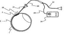
针可以以和板或固定机构成小角度(shallow angle)安装或可滑动地设置,以对插入的角度和深度进行定位和控制。该板,例如如图4中所示,可以包含注射端口,以允许该针前进通过预设置在眼球的表面(眼睛表面)上的所述板。该板可以进一步包括真空辅助密封件12,以提供该板对眼睛表面上的目标位置的稳定性。外部真空源,例如注射器或真空泵,通过管线13连接到该板上,以提供抽吸。该平板应当优选具有与眼球的曲度相合适的弯曲的底部或底缘。针11前进通过巩膜1直至进入脉络膜上方空间2,但不进入脉络膜3。The needle can be mounted at a shallow angle to the plate or fixture or slidably positioned to position and control the angle and depth of insertion. The plate, eg as shown in Figure 4, may contain injection ports to allow the needle to be advanced through the plate pre-disposed on the surface of the eyeball (surface of the eye). The plate may further include a vacuum assisted
也可以加入用于在注射过程中密封针管道的元件(例如沿管道的柔性法兰或真空密封)用于帮助递送。参照图4,示出了通过用与抽吸管线13连接的真空界面密封12定位的递送装置11相对于目标巩膜1、脉络膜上方空间2和脉络膜3的位置。Elements for sealing the needle conduit during injection (such as a flexible flange or vacuum seal along the conduit) may also be added to aid in delivery. Referring to FIG. 4 , the position of the
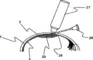
该装置也可以包括用于机械开启该脉络膜上方空间的元件,以使比可以用小内膛针递送的更大的微颗粒药物或药物递送植入物的注射。在一种实施方案中,这种递送装置可以包括提供来将该巩膜组织穿透到特定深度的第一元件,和可以前进并使脉络膜不损伤地向内扩张的第二元件,从而维持通向脉络膜上方空间的通道。该第二元件可以设置在第一元件的内部或放置在其附近。图3a和3b示出了具有所述元件的装置的实施方案。The device may also include elements for mechanically opening the suprachoroidal space to allow injection of larger microparticulate drugs or drug delivery implants than can be delivered with small bore needles. In one embodiment, such a delivery device may include a first element provided to penetrate the scleral tissue to a specific depth, and a second element that may advance and atraumatically expand the choroid inward, thereby maintaining access to the sclera. Channels in the suprachoroidal space. The second element may be disposed inside or placed adjacent to the first element. Figures 3a and 3b show an embodiment of a device having said elements.
参照图3a,示出了具有扩张尖端的递送装置。该递送装置包括在该装置末端的切割或消融尖端4和脉络膜扩张尖端8、和用于引导该装置通过该组织的超声传感器6。在该装置的近端(远离该切割尖端)提供luer连接器7。该按钮5与用于激活扩张尖端8的机构连接。该装置面对巩膜1设置,以访问邻近脉络膜3的脉络膜上方空间2。然后使用深度传感器进行引导,使该装置在巩膜组织中前进。当深度传感器显示该尖端4到达脉络膜上方空间2或到其附近时,激活该扩张尖端8以防止对脉络膜造成损伤。参照图3b,该按钮5已经激活以使该扩张尖端8前进到其激活位置9,导致脉络膜10扩张。由此获得通向脉络膜上方空间2的通道,而没有由于消融尖端4对脉络膜造成损伤。Referring to Figure 3a, there is shown a delivery device with an expanded tip. The delivery device includes a cutting or
在另一实施方案中,递送装置包括制备在引导尖端具有短的高角斜面的薄壁针,以使该斜面前进进入或通过巩膜组织。保持该斜面部分同时开口向内指向,会防止药物被从脉络膜上方空间挤出。通过将该针的尖端精确放置进入或通过巩膜组织可以实现各种类型的进入和递送。如果该针前进通过巩膜并进入脉络膜上方空间中,那么该针随后可以用于直接注射到该空间中,或者用作用于放置其他装置(例如微套管)的引入器。如果该针的放置距离巩膜的内部边界非常近,那么药物制剂通过该针的注射将允许流体剖开(fluid dissection)或流动通过任何剩下的、介于之间的巩膜组织并递送到脉络膜上方空间。图8中示出了适用于这种方式的装置的实施方案。In another embodiment, the delivery device comprises a thin walled needle prepared with a short high angle bevel at the introducing tip to advance the bevel into or through the scleral tissue. Keeping this beveled portion with the opening pointing inward prevents the drug from being squeezed out of the suprachoroidal space. Various types of access and delivery can be achieved by precise placement of the tip of the needle into or through scleral tissue. If the needle is advanced through the sclera and into the suprachoroidal space, the needle can then be used for direct injection into this space, or as an introducer for placing other devices such as a microcannula. If the needle is placed very close to the inner border of the sclera, injection of the drug formulation through the needle will allow fluid dissection or flow through any remaining, intervening sclera tissue and delivery to the suprachoroid space. An embodiment of a device suitable for use in this manner is shown in FIG. 8 .
在图8中,用于将物质注射到脉络膜上方空间2中的系统包括访问套管26和高分辨率成像装置27。该访问套管可以容纳皮下型针(未示出)或具有套管针(未示出)的导入护套。此外,该访问设备可以包括图4或图5中所示的板。该访问套管结合有斜面的、尖锐的远端尖端,其形状适于穿透组织。该成像装置可以包括实时特征,例如超声、光学相干性X线断层摄影术(OCT)或微计算机化X线断层摄影术(MicroCT)。使用该成像装置监控访问针或导入器通过巩膜的前进。该访问套管26前进直至该引导尖端接近该巩膜28的内边界,在该位置进行药物注射。药物制剂通过该针的注射将允许流体剖开或流动通过任何剩余的、介于之间的巩膜组织,并递送到脉络膜上方空间29。In FIG. 8 , the system for injecting a substance into the
在一种实施方案中,该递送装置可以特定角度进入组织,以提供将保持在巩膜内的管道或在不接触脉络膜的情况下穿透到脉络膜上方空间的组织通道。参照图5,示出了该装置的实施方案,其在近端具有luer连接器7,在远端具有斜面针尖端14。该针固定到倾斜的挡板15上,以设定针尖端14的穿透深度和角度。该组件前进直至该挡板接触到眼球的表面,将该针尖端放置在目标深度。该安装板还可以包含用于指示或引导针尖端位置的传感器。In one embodiment, the delivery device can enter the tissue at a specific angle to provide a tract that will remain within the sclera or penetrate the tissue passage into the suprachoroidal space without contacting the choroid. Referring to Figure 5, an embodiment of the device is shown having a luer connector 7 at the proximal end and a
在一种实施方案中,用于获得到脉络膜上方空间的最小侵入方式进入的系统包括访问套管和用于测定该访问套管远端尖端在组织管道中的位置的光学装置,以一旦进入脉络膜上方空间就提供直接反馈。巩膜(白色)和脉络膜(褐色)之间的色差可以用于提供位置信息,或者可以使用OCT方法确定脉络膜界面与巩膜之间的距离。该光学装置可以加入到微套管之中,或者可以是独立的装置,例如微内窥镜或纤维光学传感器和能够检测组织性质的转换器。该光学信号可以送到照相机和检测器中用于直接显现(如在内窥镜的情况下),或者送到光信号处理系统中,该光信号处理系统通过在光纤尖端发出组织性质改变的信号来显示深度。该访问微套管可以是由金属或塑料制成的针或导入器型装置。该访问套管的远端适于刺穿眼组织。如果是独立装置,在插入套管之后可以将该光学装置从该访问微套管中去除,以允许其它装置或注射物(injectate)访问所述空间以给药治疗。图6中示出了这种系统的实施方案。该光学装置包括柔性微内窥镜18,其与CCD摄像机16相连,在监控器19中观察图像。该内窥镜的尺寸使其可滑动地适配在外径优选小于1mm的访问套管17中。该访问套管17包括用于组织访问的斜面的、尖锐的远端尖端。该内窥镜的远端尖端位于该套管斜面的近端处以提供套管尖端的图像。该套管以小角度压靠睫状环区域处的眼睛表面前进,刺穿巩膜1,并前进直至该内窥镜图像显示进入脉络膜上方空间2。In one embodiment, a system for obtaining minimally invasive access to the suprachoroidal space includes an access cannula and optical means for determining the position of the distal tip of the access cannula within the tissue tract to determine once the access cannula has entered the choroid The space above provides immediate feedback. The color difference between the sclera (white) and the choroid (brown) can be used to provide positional information, or the distance between the choroidal interface and the sclera can be determined using OCT methods. The optical device can be incorporated into the microcannula, or it can be a separate device such as a microendoscope or a fiber optic sensor and transducer capable of detecting tissue properties. This optical signal can be fed to a camera and detector for direct visualization (as in the case of an endoscope), or to an optical signal processing system that signals tissue property changes at the tip of an optical fiber to show the depth. The access microcannula can be a needle or introducer type device made of metal or plastic. The distal end of the access cannula is adapted to penetrate ocular tissue. If a stand-alone device, the optical device can be removed from the access microcannula after insertion of the cannula to allow other devices or injectates to access the space to administer therapy. An embodiment of such a system is shown in FIG. 6 . The optical device includes a
在另一实施方案中,该系统的光学装置包括在远端尖端的聚焦照明光源。光散射量和光强度将根据组织类型和横穿组织的小光点的深度而变化。可以通过医生从表面观察或用传感器测定这种变化。可以结合聚焦点作为作为在微套管上的被照亮的信标尖端。参照图7,该访问装置包括柔性微套管或微导管20,其尺寸适于无创伤地访问脉络膜上方空间2。该微导管包括用于将材料递送到空间2中的管腔22和用于提供被照亮的远端尖端的光导纤维23。该光导纤维与照明光源24相连接,所述光源例如激光二极管、超亮LED、白炽灯或类似光源。该微导管可滑动地设置于访问套管21之中。随着访问套管前进通过组织,透照所述组织的光25改变。巩膜组织高度散射来自巩膜组织内部的光,然而,一旦进入脉络膜上方空间,在表面处看到的光强度和背散射就显著降低,表明该照亮的尖端已经经过巩膜1,现在处于脉络膜上方空间处的目标位置。In another embodiment, the optics of the system include a focused illumination source at the distal tip. The amount of light scattering and light intensity will vary depending on the tissue type and the depth of the small spot of light traversing the tissue. This change can be measured by a doctor visually or with sensors. The focal spot can be incorporated as an illuminated beacon tip on the microcannula. Referring to FIG. 7 , the access device includes a flexible microcannula or
递送装置的特别应用是如前所述的药物制剂,其与该递送装置相兼容。微颗粒形式的药物优选比管腔直径小的多,以防止在递送过程中管腔发生堵塞。优选微颗粒的平均外部尺寸约为该装置中最大管腔的10~20%。适合的制剂包括外径在约1~33微米范围内的微球或微颗粒。而且优选在药物制剂中使用聚合物赋型剂,以使该制剂能够注射到与脉络膜上方空间相邻的巩膜组织中,然后通过包含赋型剂的流体剖开远端尖端和脉络膜上方空间之间的组织,以形成药物进入脉络膜上方空间中的流动通道。对于通过小针管腔以及对于巩膜组织的流体剖开而言,具有触变性质的制剂是有利的。A particular application of the delivery device is a pharmaceutical formulation as previously described, which is compatible with the delivery device. Drugs in microparticle form are preferably much smaller than the diameter of the lumen to prevent clogging of the lumen during delivery. Preferably the average outer dimension of the microparticles is about 10-20% of the largest lumen in the device. Suitable formulations include microspheres or microparticles having an outer diameter in the range of about 1 to 33 microns. It is also preferred to use a polymeric excipient in the drug formulation to allow the formulation to be injected into the scleral tissue adjacent to the suprachoroidal space, followed by dissection between the distal tip and the suprachoroidal space by fluid containing the excipient tissue to form a flow channel for the drug into the suprachoroidal space. Formulations with thixotropic properties are advantageous for fluid dissection through small needle lumens and for scleral tissue.
以下实施例仅提供用于示例性的目的,决不意于限制本发明。The following examples are provided for illustrative purposes only and are by no means intended to limit the invention.
实施例1:Example 1:
使用悬浮在磷酸盐缓冲盐水中的荧光染色聚苯乙烯微球(FirefliTM,Duke Scientific,Inc.,Palo Alto,CA)做为模型药物,用于评价其中颗粒在脉络膜上方空间中从前部区域到后部区域迁移的尺寸范围。Fluorescently-stained polystyrene microspheres (Firefli™ , Duke Scientific, Inc., Palo Alto, CA) suspended in phosphate-buffered saline were used as a model drug for evaluating where particles travel from the anterior region to the suprachoroidal space. Size range of migration in the posterior region.
将摘除的人体尸体眼睛快速切开到睫状环区域中的脉络膜处,其位于眼睛的前部部分中。使用用钝的27规格针封端的注射器,将0.15ml的1体积%微球悬浮体(平均直径为6微米)递送到脉络膜上方空间的前部区域。回撤针,用氰基丙烯酸盐粘结剂密封切口。然后通过将通过灌注管连接到储存器的30规格针引入前腔,在10mm Hg压力下用磷酸盐缓冲盐水将眼睛灌注24小时。该储存器放置在实验室起重器上,升高以提供恒定的灌注压力。在测试前几个小时,将眼睛放置到甘油烧杯中用于通过脱水净化巩膜组织,从而使脉络膜上方空间可以直接看到。Enucleated human cadaver eyes were quickly incised to the choroid in the region of the ciliary ring, which is located in the anterior portion of the eye. Using a syringe capped with a blunt 27 gauge needle, 0.15 ml of a 1 vol% suspension of microspheres (mean
使用具有针对微球荧光选择的荧光滤光器的立体荧光显微镜(Model MZ-16,Leica,Inc.)观察微球。在低放大倍数(7~35X)下,可以清楚地看到该微球处于流状模式,从灌注位置相后朝向视神经区域运动,主要收集在脉络膜上方空间的后部区域。使用1、6、10、15、24和33微米直径的微球悬浮体重复试验,所得到的迁移和分布到眼睛的后部区域的模式相同。Microspheres were observed using a stereofluorescence microscope (Model MZ-16, Leica, Inc.) with fluorescence filters selected for microsphere fluorescence. At low magnification (7-35X), the microspheres can clearly be seen in a stream-like pattern moving posteriorly from the site of infusion towards the optic nerve region, collecting mainly in the posterior region of the suprachoroidal space. The experiment was repeated using microsphere suspensions of 1, 6, 10, 15, 24 and 33 micron diameter, and the resulting migration and distribution patterns to the posterior region of the eye were identical.
实施例2:Example 2:
重复实施例1的实验,除了将作为模型药物的6和33微米直径荧光微球混合物悬浮在聚合物赋型剂中,所述赋型剂包含外科手术用粘弹性体(Healon 5,Advanced Medical Optics,Inc.),即2.3%浓度的4000000道尔顿分子量透明质酸钠,具有7000000mPas零剪切粘度和在1000s-1剪切速率下400mPas粘度的触变性质。将该混合物以实施例1的方式引入该脉络膜上方空间中。在灌注24小时之后,微球仅滞留在脉络膜上方空间的前部灌注位置,没有显示出迁移的迹象,显示了触变聚合物赋型剂的定位作用。The experiment of Example 1 was repeated except that a mixture of 6 and 33 micron diameter fluorescent microspheres as a model drug was suspended in a polymeric vehicle comprising viscoelastic for surgical use (
实施例3:Example 3:
为了证明聚合物赋型剂粘性对药物定位的影响,重复实施例1的实验,除了将bevacizumab(AvastinTM,Genentech),即抗VEG抗体,吸附在5微米直径羧化荧光微球上,并与三种透明质酸基外科手术用粘弹性体(Healon,Healon GV,Healon 5,Advanced Medical Optics,Inc.)其中之一等体积混合,该三种透明质酸基外科手术用粘弹性体各自具有不同的粘性和触变性质。(Healon,在零剪切速率下粘度为300000mPas,在1000s-1剪切速率下粘度为150mPas;Healon GV,在零剪切速率下粘度为3000000mPas,在1000s-1剪切速率下粘度为200mPas;Healon 5,在零剪切速率下粘度为7000000mPas,在1000s-1剪切速率下粘度为400mPas)。将每种混合物以实施例1的方式引入眼睛前部区域中的睫状环处的脉络膜上方空间的前部区域中。在灌注24小时之后,发现在Healon和Healon GV中的微球处于到脉络膜上方空间的后部区域的迁移过程中,在睫状环灌注位置和后极都发现了该制剂。Healon 5中的微球保持分散在定位于脉络膜上方空间的睫状环区域中的原始注射位置处的粘弹性体中。In order to demonstrate the effect of polymer excipient viscosity on drug localization, the experiment of Example 1 was repeated, except that bevacizumab (Avastin™, Genentech), i.e. anti-VEG antibody, was adsorbed on 5 micron diameter carboxylated fluorescent microspheres, and mixed with three One of the hyaluronic acid-based surgical viscoelastics (Healon, Healon GV,
实施例4:Example 4:
重复实施例1的实验,除了使用1-乙基-3-(3-二甲基氨基-丙基)碳二亚胺(EDAC,Sigma-Aldrich)将bevacizumab(AvastinTM,Genentech)共价交联在5微米直径的羧化荧光微球上,并与三种外科手术用粘弹性体(Healon,Healon GV,Healon 5,Advanced Medical Optics,Inc.)其中之一等体积混合,该三种外科手术用粘弹性体各自具有如实施例3中所示不同的粘性和触变性质。将该混合物以实施例1的方式引入睫状环处的脉络膜上方空间中。在灌注24小时之后,对于所有粘弹性载体,微球都完全(exclusively)保持在脉络膜上方空间的睫状环区域中。The experiment of Example 1 was repeated except that bevacizumab (AvastinTM, Genentech) was covalently crosslinked on Carboxylated fluorescent microspheres with a diameter of 5 microns, and mixed with one of three surgical viscoelastic materials (Healon, Healon GV,
实施例5:Example 5:
为了证明交联聚合物赋型剂对药物定位的影响,重复实施例1的实验,除了将10微米直径的荧光微球混合到4%藻酸盐溶液中,并将其引入睫状环区域处的脉络膜上方空间中。在密封该切口位置之前,将等体积的1M CaCl2溶液在微球/藻酸盐悬浮液的位置灌输,以引发藻酸盐赋型剂的交联。在如实施例1中灌注之前,使该混合物凝胶5分钟。该微球完全(exclusively)保持在灌输位置,分散在交联聚合物赋型剂之中。To demonstrate the effect of cross-linked polymer excipients on drug localization, the experiment of Example 1 was repeated except that 10 μm diameter fluorescent microspheres were mixed into 4% alginate solution and introduced at the ciliary annulus region in the suprachoroidal space. Before sealing the incision site, an equal volume of 1MCaCl2 solution was instilled at the site of the microsphere/alginate suspension to initiate cross-linking of the alginate excipient. The mixture was allowed to gel for 5 minutes before being perfused as in Example 1. The microspheres remain exclusively at the infused site, dispersed in the cross-linked polymer excipient.
实施例6:Embodiment 6:
通过将1.5mg微颗粒形式的氟羟脱氢皮质醇丙酮化物(Triamcinolone acetonide)悬浮在15微升在零剪切速率下粘度为300000mPas和在1000s-1剪切速率下粘度为150mPas的Healon粘弹性体(Advanced Medical Optics,Irvine CA)中制备含药物注射物。将40个猪受试者麻醉,准备右眼并以消毒方式盖布。在上缘附近进行结膜环切,暴露并提供通向巩膜区域的手术通道。在巩膜中进行小径向切口,暴露裸脉络膜。将具有360微米直径尖端和325微米直径主体的柔性微导管(iTrack微导管,iScience Interventional Corp.)插入巩膜切口中并以后部方向前进到黄斑之后的目标区域。将药物悬浮液注射到脉络膜上方空间的后部区域中,观察到在目标区域的脉络膜和巩膜之间形成了层。回撤微导管,用7-0Vicryl缝线封闭巩膜和结膜切口。观察受试者,在12小时、24小时、48小时、4天、7天、14天、30天和90天恢复的眼睛组织。对受试者的血管造影、组织和照相研究显示没有后部部分病理信号。脉络膜的恢复样品显示在所有恢复时间周期药物的显著富集,在至少1mg/g组织的范围内。By suspending 1.5 mg triamcinolone acetonide in the form of microparticles in 15 microliters of Healon viscoelastic with a viscosity of 300,000 mPas at zero shear rate and 150 mPas at a shear rate of1000 s Drug-containing injections were prepared in vivo (Advanced Medical Optics, Irvine CA). Forty porcine subjects were anesthetized and the right eye was prepared and draped sterilely. A conjunctival circumcision is made near the superior border, exposing and providing surgical access to the scleral region. A small radial incision is made in the sclera exposing the bare choroid. A flexible microcatheter (iTrack microcatheter, iScience Interventional Corp.) with a 360 micron diameter tip and a 325 micron diameter body was inserted into the scleral incision and advanced posteriorly to the target area behind the macula. The drug suspension was injected into the posterior region of the suprachoroidal space and a layer was observed to form between the choroid and sclera in the targeted region. The microcatheter was withdrawn, and the scleral and conjunctival incisions were closed with 7-0 Vicryl sutures. Subjects were observed, and eye tissue recovered at 12 hours, 24 hours, 48 hours, 4 days, 7 days, 14 days, 30 days, and 90 days. Angiographic, histological, and photographic studies of the subject showed no signs of pathology in the posterior portion. Recovery samples of the choroid showed significant enrichment of drug over all recovery time periods, in the range of at least 1 mg/g tissue.
实施例7:Embodiment 7:
准备包含20mL Healon 5和50mL(1.5mg)bevacizumab(AvastinTM,Genentech)的含药物制剂。将18个猪受试者麻醉,准备右眼并以消毒方式盖布。在上缘附近进行结膜环切,暴露并提供通向巩膜区域的手术通道。在巩膜中进行小径向切口,暴露裸脉络膜。将具有360微米直径尖端和325微米直径主体的柔性微导管(iTrack微导管,iScienceInterventional Corp.)插入巩膜切口中并以后部方向前进到黄斑之后的目标区域。将药物制剂注射到脉络膜上方空间的后部区域中,观察到在目标区域处的脉络膜和巩膜之间形成了层。回缩微导管,用7-0Vicryl缝线封闭巩膜和结膜切口。将另外18个猪受试者麻醉,通过注射到玻璃体(Vitreous)中使其各自接收bevacizumab的50mL药丸。在注射后0.5、7、30、60、90和120天使评估和杀掉两组测试受试者。取出血清样品,使用酶基免疫测定法测试bevacizumab。在经玻璃体内注射受试者中发现了比脉络膜上层递送组更高的bevacizumab血浆水平和更长的滞留时间。摘除右眼球并进行解剖,以使用酶基免疫测定法定量特定组织和区域中的bevacizumab。酶免疫测定法证实通过玻璃体注射递送的bevacizumab遍布整个眼睛,而脉络膜上层递送时大部分保持在视网膜和脉络膜中,在玻璃体和前腔中几乎没有发现。A drug-containing formulation containing 20 mL of
实施例8:Embodiment 8:
重复实施例1的实验,除了制备0.2mL Healon 5、0.6mL Avastin和24mg氟羟脱氢皮质醇丙酮化物的药物制剂以提供抗炎和抗VEGF性质的治疗。在横切巩膜的睫状环区域中纵向形成约5mm长的切口,将已经通过在甘油中浸渍约30分钟进行清洗和用盐水在12mm Hg压力下灌注过的尸体眼球的脉络膜暴露。用该药物制剂灌注实施例6中的柔性微套管,将微套管尖端通过巩膜切口插入脉络膜上方空间中。借助于微套管尖端处的光导纤维信标,将该微套管的远端控制朝向眼球的后极,停在聚视神经约5mm的位置。使用Viscoelastic Injector(iScienceInterventional),将70微升的药物制剂注射到脉络膜上方空间的后部区域中。通过睫状环切口回缩,去除该微套管。通过净化的巩膜能够观察到该混合物,在视神经附近形成沉积物,该混合物也遵循着导管轨迹。用氰基丙烯酸酯(Locktite 4011)密封切口,再次用盐水以12mm Hg对眼球灌注3小时。通过浸渍在甘油中再次清洗巩膜,以检测给药的药物制剂。通过显微镜观察该药物制剂在脉络膜上方空间的后部区域中形成了在聚合物赋型剂中分散的药物层。The experiment of Example 1 was repeated except that a pharmaceutical formulation of 0.2 mL of
实施例9:Embodiment 9:
进行一系列实验以评估物质到脉络膜上方空间的最小侵入方式递送。实验目的是使用非侵入成像和流体剖开作为将物质通过巩膜组织递送到脉络膜上方空间而不直接穿透到脉络膜上方空间的方法。A series of experiments were performed to evaluate the minimally invasive delivery of substances into the suprachoroidal space. The aim of the experiment was to use non-invasive imaging and fluid dissection as a method of delivering substances through the scleral tissue into the suprachoroidal space without penetrating directly into the suprachoroidal space.
从眼库中获得人尸体眼睛,通过用磷酸盐缓冲盐水(PBS)使眼睛膨胀到约20mm Hg压力预备。使用不锈钢皮下管制备递送针,所述皮下管为255mm ID×355mm OD。将针远端尖端磨成双面短斜面点,长度为400um,角度为50°。然后将制备好的针银焊(silver-solder)成标准25规格×1英寸皮下注射针以完成该组件。Human cadaver eyes were obtained from eye banks and prepared by inflating the eyes to approximately 20 mm Hg pressure with phosphate buffered saline (PBS). Delivery needles were prepared using stainless steel hypodermic tubing, which was 255 mm ID x 355 mm OD. Grind the distal tip of the needle into a double-sided short bevel point with a length of 400 μm and an angle of 50°. The prepared needle was then silver-soldered to a standard 25
将针以相对于眼睛表面的锐角(<10°)轻柔前进到巩膜组织中。针的进入开始在距边缘约4mm的睫状环区域中,该针向后前进到巩膜组织中产生5~6mm长的管道,而不穿透通过巩膜进入脉络膜上方空间。使用高分辨率超声系统(iUltrasound,iScience Surgical Corp.)引导并证实针尖端放置在巩膜组织中以及证明注射。The needle is gently advanced into the scleral tissue at an acute angle (<10°) relative to the eye surface. Needle entry begins in the region of the ciliary ring about 4 mm from the edge, and the needle is advanced posteriorly into the scleral tissue creating a 5-6 mm long tract without penetrating through the sclera into the suprachoroidal space. A high-resolution ultrasound system (iUltrasound, iScience Surgical Corp.) was used to guide and confirm needle tip placement in scleral tissue and to demonstrate injection.
在第一组实验中,注射仅包含透明质酸外科手术用粘弹性体(Healon 5,Advanced Medical Optics,Inc.)的聚合物赋型剂。在第二组实验中,将该粘弹性体与10微米直径聚苯乙烯微球(Duke Scientific,Inc.)的1%水溶液以1:1比例混合以代表模型微颗粒药物。使用螺杆驱动注射器(ViscoInjector,iScience Surgical Corp.)通过该针递送该粘弹性体和该混合物,以控制递送体积和注射压力。进行注射,使针的斜面向内转向眼球中心。使用三个尸体眼睛的多个位置进行实验。In the first set of experiments, only a polymer vehicle containing hyaluronic acid surgical viscoelastomer (
在第一组实验中,针管道约3~4mm长,观察到注射物向后流出管道。将针的尖端放置在更长的管道中时,获得更高的注射压力,使注射物剖开通过巩膜的剩余的介于之间的层,并递送到脉络膜上方空间中。通过实验发现放置在巩膜外层中的针尖端(<1/2巩膜厚度)导致粘弹性体递送到巩膜内囊中或有时通过到眼球的外表面。随着针尖端接近巩膜底部,注射物剖开通过剩余的介于之间的巩膜组织,进入脉络膜上方空间,铺展以填充注射区域中的脉络膜上方空间。图1显示了清楚可见的针管道30(在去除针之后)和充满注射物的脉络膜上方空间的区域31。示出了巩膜1和脉络膜3。图2显示了充满包含微球和透明质酸赋型剂的注射物的脉络膜上方空间的区域33和在巩膜中的针尖端4和针的阴影32。In the first set of experiments, the needle conduit was approximately 3-4 mm long and the injectate was observed to flow out of the conduit backwards. When the needle tip is placed in the longer cannula, higher injection pressures are achieved, causing the injection to cut through the remaining intervening layers of the sclera and be delivered into the suprachoroidal space. It was found experimentally that a needle tip placed in the episclera (<1/2 the thickness of the sclera) resulted in delivery of the viscoelastic into the inner scleral capsule or sometimes through to the outer surface of the eyeball. As the needle tip approaches the base of the sclera, the injection dissects through the remaining intervening sclera tissue, into the suprachoroidal space, spreading to fill the suprachoroidal space in the injected area. Figure 1 shows the clearly visible needle tract 30 (after removal of the needle) and the region 31 of the suprachoroidal space filled with injectate. The
实施例10:Example 10:
进行实验使用微内窥镜成像以允许最小侵入方式进入人尸体眼睛的脉络膜上方空间。将定制的具有350微米外径且包含具有1200像素的成像束的柔性微内窥镜(Endoscopy Support Services,Brewster NY)安装在微米调节台上。该调节台安装在垂直台上,允许该内窥镜进行受控上下运动。将该微内窥镜与1/2"芯片CCD摄像机连接,然后连接到视频监视器上。将20规格皮下注射针放置在内窥镜上,以提供刺穿组织使进入的工具。Experiments were performed using microendoscopic imaging to allow minimally invasive access to the suprachoroidal space of human cadaver eyes. A custom-made flexible microendoscope (Endoscopy Support Services, Brewster NY) with an outer diameter of 350 micrometers and containing an imaging beam with 1200 pixels was mounted on a micrometer adjustment stage. The adjustment table is mounted on a vertical table, allowing controlled up and down movement of the endoscope. The microendoscope was connected to a 1/2" chip CCD camera and then to a video monitor. A 20 gauge hypodermic needle was placed on the endoscope to provide a tool for tissue penetration.
打开摄像机,使用具有光导管的外部光源(Model MI-150,DolanJenner,Boxborough,MA)提供穿巩膜成像照明。使针前进直至远端与边缘后面约4mm的人尸体整眼球的巩膜表面相接触为止。然后降低微内窥镜直至可以通过针的末端看到白色巩膜表面。然后通过轻微前后旋转使针缓慢前进到巩膜组织中。随着针以这种方式前进,降低内窥镜以跟随针产生的通道。在巩膜处或内部时,看到内窥镜图像是白色或灰白色的。随着针刺穿巩膜组织,图像颜色转变为表示存在深色脉络膜组织的深棕色,这显示手术进入了脉络膜上方空间。The camera was turned on and an external light source (Model MI-150, Dolan Jenner, Boxborough, MA) with a light guide was used to provide illumination for transscleral imaging. The needle was advanced until the distal end made contact with the scleral surface of the human cadaver eyeball approximately 4 mm posterior to the rim. The microendoscope is then lowered until the white scleral surface can be seen through the tip of the needle. The needle is then slowly advanced into the scleral tissue with a slight back and forth rotation. With the needle advanced in this manner, the endoscope is lowered to follow the channel created by the needle. See endoscopic image as white or off-white when at or within the sclera. As the needle penetrates the scleral tissue, the color of the image shifts to dark brown indicating the presence of dark choroidal tissue, which indicates that the surgery has entered the suprachoroidal space.
实施例11:Example 11:
进行实验使用光导纤维照明引导以允许最小侵入方式进入人尸体眼睛的脉络膜上方空间。将具有照明远端尖端的柔性微套管(iTrack-250A,iScience Interventional,Menlo Park,CA)放置在25规格皮下注射针中。该微套管包含塑料光导纤维,其可以对远端尖端照明。该微导管纤维连接器与635nm(红色)激光二极管光导纤维照明器(iLumin,iScience Interventional)相连,开启照明器以提供稳定的红光发射用于微套管尖端。将该微套管通过25规格针递送直至针的末梢斜面但不超过。Experiments were performed using fiber optic illumination guided to allow minimally invasive access to the suprachoroidal space of human cadaver eyes. A flexible microcannula (iTrack-250A, iScience Interventional, Menlo Park, CA) with an illuminated distal tip was placed in a 25 gauge hypodermic needle. The microcannula contains a plastic optical fiber that illuminates the distal tip. The microcatheter fiber connector was connected to a 635nm (red) laser diode fiber optic illuminator (iLumin, iScience Interventional), which was turned on to provide steady red emission for the microcannula tip. The microcannula was delivered through a 25 gauge needle up to but not beyond the distal bevel of the needle.
将针在人尸体整眼球的睫状环区域内缓慢前进直至针的尖端充分嵌入巩膜组织中以使该微套管可以轻微前进。由于巩膜组织对光发生明显散射,因此清楚看到来自微套管尖端的照亮。随着针缓慢前进,同时将微套管向前推动。当该皮下注射针的尖端刺穿足够的巩膜组织到达脉络膜上方空间时,该微套管尖端的红光立即变暗,因为照亮的尖端通出散射性巩膜组织进入其下的空间。使微套管前进,同时保持针静止,由此将微套管尖端放入脉络膜上方空间中。微套管以向后方向在脉络膜上方空间中的进一步前进可以透过巩膜看到为聚焦的红点,因为没有了在尖端位于巩膜组织内时看到的宽的光散射。使用高频超声系统(iUltraSound,iScience Interventional),确认了微套管在脉络膜上方空间中的位置。The needle was slowly advanced in the ciliary annulus region of the human cadaver eye until the tip of the needle was sufficiently embedded in the scleral tissue to allow slight advancement of the microcannula. Illumination from the tip of the microcannula is clearly seen due to the apparent scattering of light by scleral tissue. Push the microcannula forward at the same time as the needle is slowly advanced. When the tip of the hypodermic needle pierces enough scleral tissue to reach the suprachoroidal space, the red light at the tip of the microcannula dims immediately as the illuminated tip passes out the diffuse scleral tissue into the space below it. The microcannula tip is placed in the suprachoroidal space by advancing the microcannula while keeping the needle stationary. Further advancement of the microcannula in the suprachoroidal space in a posterior direction can be seen through the sclera as a focused red spot because of the absence of the broad light scatter seen when the tip is within the scleral tissue. Using a high-frequency ultrasound system (iUltraSound, iScience Interventional), the position of the microcannula in the suprachoroidal space was confirmed.
Claims (25)
Applications Claiming Priority (5)
| Application Number | Priority Date | Filing Date | Title |
|---|---|---|---|
| US77690306P | 2006-02-22 | 2006-02-22 | |
| US60/776,903 | 2006-02-22 | ||
| US11/709,941US20070202186A1 (en) | 2006-02-22 | 2007-02-21 | Apparatus and formulations for suprachoroidal drug delivery |
| US11/709,941 | 2007-02-21 | ||
| PCT/US2007/004874WO2007100745A2 (en) | 2006-02-22 | 2007-02-22 | Apparatus and formulations for suprachoroidal drug delivery |
Related Child Applications (1)
| Application Number | Title | Priority Date | Filing Date |
|---|---|---|---|
| CN2011100936446ADivisionCN102247239B (en) | 2006-02-22 | 2007-02-22 | Apparatus and formulations for suprachoroidal drug delivery |
Publications (2)
| Publication Number | Publication Date |
|---|---|
| CN101426473Atrue CN101426473A (en) | 2009-05-06 |
| CN101426473B CN101426473B (en) | 2011-06-08 |
Family
ID=38444302
Family Applications (2)
| Application Number | Title | Priority Date | Filing Date |
|---|---|---|---|
| CN2011100936446AActiveCN102247239B (en) | 2006-02-22 | 2007-02-22 | Apparatus and formulations for suprachoroidal drug delivery |
| CN2007800145013AActiveCN101426473B (en) | 2006-02-22 | 2007-02-22 | Apparatus and formulations for suprachoroidal drug delivery |
Family Applications Before (1)
| Application Number | Title | Priority Date | Filing Date |
|---|---|---|---|
| CN2011100936446AActiveCN102247239B (en) | 2006-02-22 | 2007-02-22 | Apparatus and formulations for suprachoroidal drug delivery |
Country Status (9)
| Country | Link |
|---|---|
| US (9) | US20070202186A1 (en) |
| EP (2) | EP3446679A1 (en) |
| JP (1) | JP2009531298A (en) |
| KR (1) | KR20080099285A (en) |
| CN (2) | CN102247239B (en) |
| AU (1) | AU2007221105A1 (en) |
| BR (1) | BRPI0708133A2 (en) |
| CA (1) | CA2643019A1 (en) |
| WO (1) | WO2007100745A2 (en) |
Cited By (10)
| Publication number | Priority date | Publication date | Assignee | Title |
|---|---|---|---|---|
| CN102458509A (en)* | 2009-05-15 | 2012-05-16 | I科学干预公司 | Methods and apparatus for sub-retinal catheterization |
| CN107249522A (en)* | 2014-09-19 | 2017-10-13 | 奥叙拉尔有限公司 | Ophthalmology delivery apparatus |
| CN107405306A (en)* | 2014-09-19 | 2017-11-28 | 奥叙拉尔有限公司 | Ophthalmic Pharmaceutical Composition |
| CN109009657A (en)* | 2018-09-10 | 2018-12-18 | 天津医科大学总医院 | A kind of choroid restorer |
| CN110893188A (en)* | 2012-11-08 | 2020-03-20 | 克莱尔塞德生物医学股份有限公司 | Methods and devices for treating ocular diseases in human subjects |
| CN111712228A (en)* | 2017-09-15 | 2020-09-25 | 奥叙拉尔有限公司 | Ophthalmic pharmaceutical composition |
| CN111936091A (en)* | 2017-10-18 | 2020-11-13 | J·莫雷诺 | Ophthalmic Microsurgery Instruments |
| CN113616417A (en)* | 2016-03-09 | 2021-11-09 | 陀螺仪治疗学有限公司 | Device for subretinal administration of therapeutic agents via curved needles |
| CN114366712A (en)* | 2022-01-25 | 2022-04-19 | 上海交通大学医学院附属第九人民医院 | Pharmaceutical gel mixture for treating choroidal neovascularization |
| WO2022236778A1 (en)* | 2021-05-13 | 2022-11-17 | Beijing Sightnovo Medical Technology Co., Ltd | Medical penetration device and system |
Families Citing this family (121)
| Publication number | Priority date | Publication date | Assignee | Title |
|---|---|---|---|---|
| US20130274837A1 (en)* | 1998-10-23 | 2013-10-17 | Babak Nemati | Systems and Methods to Enhance Optical Transparency of Biological Tissues for Photobiomodulation |
| KR20020035476A (en) | 1999-04-26 | 2002-05-11 | 지엠피 비젼 솔루션즈 인코포레이티드 | Shunt device and method for treating glaucoma |
| US7708711B2 (en) | 2000-04-14 | 2010-05-04 | Glaukos Corporation | Ocular implant with therapeutic agents and methods thereof |
| US6638239B1 (en)* | 2000-04-14 | 2003-10-28 | Glaukos Corporation | Apparatus and method for treating glaucoma |
| AU2002258754B2 (en) | 2001-04-07 | 2006-08-17 | Glaukos Corporation | Glaucoma stent and methods thereof for glaucoma treatment |
| US7431710B2 (en) | 2002-04-08 | 2008-10-07 | Glaukos Corporation | Ocular implants with anchors and methods thereof |
| US7488303B1 (en)* | 2002-09-21 | 2009-02-10 | Glaukos Corporation | Ocular implant with anchor and multiple openings |
| US7678065B2 (en) | 2001-05-02 | 2010-03-16 | Glaukos Corporation | Implant with intraocular pressure sensor for glaucoma treatment |
| US7951155B2 (en) | 2002-03-15 | 2011-05-31 | Glaukos Corporation | Combined treatment for cataract and glaucoma treatment |
| US9301875B2 (en) | 2002-04-08 | 2016-04-05 | Glaukos Corporation | Ocular disorder treatment implants with multiple opening |
| WO2007058966A1 (en)* | 2005-11-11 | 2007-05-24 | Massachusetts Institute Of Technology | Controlled needle-free eye injector |
| US20070202186A1 (en)* | 2006-02-22 | 2007-08-30 | Iscience Interventional Corporation | Apparatus and formulations for suprachoroidal drug delivery |
| US8197435B2 (en)* | 2006-05-02 | 2012-06-12 | Emory University | Methods and devices for drug delivery to ocular tissue using microneedle |
| US7909789B2 (en) | 2006-06-26 | 2011-03-22 | Sight Sciences, Inc. | Intraocular implants and methods and kits therefor |
| US20120123316A1 (en) | 2010-11-15 | 2012-05-17 | Aquesys, Inc. | Intraocular shunts for placement in the intra-tenon's space |
| US10085884B2 (en) | 2006-06-30 | 2018-10-02 | Aquesys, Inc. | Intraocular devices |
| US8721702B2 (en) | 2010-11-15 | 2014-05-13 | Aquesys, Inc. | Intraocular shunt deployment devices |
| US8852256B2 (en) | 2010-11-15 | 2014-10-07 | Aquesys, Inc. | Methods for intraocular shunt placement |
| EP2088976B1 (en) | 2006-11-10 | 2019-07-03 | Glaukos Corporation | Uveoscleral shunt |
| US8642067B2 (en)* | 2007-04-02 | 2014-02-04 | Allergen, Inc. | Methods and compositions for intraocular administration to treat ocular conditions |
| US11078262B2 (en) | 2007-04-30 | 2021-08-03 | Allergan, Inc. | High viscosity macromolecular compositions for treating ocular conditions |
| US8425449B2 (en) | 2009-07-09 | 2013-04-23 | Ivantis, Inc. | Ocular implants and methods for delivering ocular implants into the eye |
| US8734377B2 (en) | 2007-09-24 | 2014-05-27 | Ivantis, Inc. | Ocular implants with asymmetric flexibility |
| US20170360609A9 (en) | 2007-09-24 | 2017-12-21 | Ivantis, Inc. | Methods and devices for increasing aqueous humor outflow |
| US7740604B2 (en) | 2007-09-24 | 2010-06-22 | Ivantis, Inc. | Ocular implants for placement in schlemm's canal |
| US20090082862A1 (en) | 2007-09-24 | 2009-03-26 | Schieber Andrew T | Ocular Implant Architectures |
| US8512404B2 (en) | 2007-11-20 | 2013-08-20 | Ivantis, Inc. | Ocular implant delivery system and method |
| US8808222B2 (en) | 2007-11-20 | 2014-08-19 | Ivantis, Inc. | Methods and apparatus for delivering ocular implants into the eye |
| JP2011513002A (en) | 2008-03-05 | 2011-04-28 | イバンティス インコーポレイテッド | Method and apparatus for treating glaucoma |
| CN102238926B (en) | 2008-12-05 | 2015-09-16 | 伊万提斯公司 | Methods and devices for delivering ocular implants into the eye |
| JP5890182B2 (en) | 2009-02-12 | 2016-03-22 | インセプト エルエルシー | Drug delivery with hydrogel plugs |
| WO2010135369A1 (en) | 2009-05-18 | 2010-11-25 | Dose Medical Corporation | Drug eluting ocular implant |
| US10206813B2 (en) | 2009-05-18 | 2019-02-19 | Dose Medical Corporation | Implants with controlled drug delivery features and methods of using same |
| WO2012071476A2 (en) | 2010-11-24 | 2012-05-31 | David Haffner | Drug eluting ocular implant |
| AU2010271274B2 (en) | 2009-07-09 | 2015-05-21 | Alcon Inc. | Single operator device for delivering an ocular implant |
| CN102647960A (en) | 2009-10-23 | 2012-08-22 | 伊万提斯公司 | Ocular implant system and method |
| US8529492B2 (en) | 2009-12-23 | 2013-09-10 | Trascend Medical, Inc. | Drug delivery devices and methods |
| US8529622B2 (en) | 2010-02-05 | 2013-09-10 | Sight Sciences, Inc. | Intraocular implants and related kits and methods |
| US9510973B2 (en) | 2010-06-23 | 2016-12-06 | Ivantis, Inc. | Ocular implants deployed in schlemm's canal of the eye |
| JP5996544B2 (en)* | 2010-10-15 | 2016-09-21 | クリアサイド・バイオメディカル・インコーポレーテッドClearside Biomedical Incorporated | Eye access device |
| US20160256320A1 (en)* | 2010-11-15 | 2016-09-08 | Aquesys, Inc. | Intraocular shunt placement in the suprachoroidal space |
| US10285852B2 (en) | 2010-12-02 | 2019-05-14 | Tel Hashomer Medical Research Infrastructure And Services Ltd., The Chaim Sheba Medical Center | Subretinal delivery of therapeutic compositions |
| US10245178B1 (en) | 2011-06-07 | 2019-04-02 | Glaukos Corporation | Anterior chamber drug-eluting ocular implant |
| US8657776B2 (en) | 2011-06-14 | 2014-02-25 | Ivantis, Inc. | Ocular implants for delivery into the eye |
| EP2744563B1 (en) | 2011-08-16 | 2016-01-13 | Institut National de la Santé et de la Recherche Medicale | Device for the treatment of an ocular disease |
| JP2014524302A (en) | 2011-08-16 | 2014-09-22 | アンスティチュ ナショナル ドゥ ラ サンテ エ ドゥ ラ ルシェルシュ メディカル | Device for the treatment of eye diseases |
| EP2559443A1 (en) | 2011-08-16 | 2013-02-20 | INSERM (Institut National de la Santé et de la Recherche Médicale) | Methods and pharmaceutical compositions for the treatment of an ocular disease in a subject |
| EP2755549A1 (en) | 2011-09-13 | 2014-07-23 | Dose Medical Corporation | Intraocular physiological sensor |
| US10226417B2 (en) | 2011-09-16 | 2019-03-12 | Peter Jarrett | Drug delivery systems and applications |
| US9610195B2 (en) | 2013-02-27 | 2017-04-04 | Aquesys, Inc. | Intraocular shunt implantation methods and devices |
| US9808373B2 (en) | 2013-06-28 | 2017-11-07 | Aquesys, Inc. | Intraocular shunt implantation |
| US10080682B2 (en)* | 2011-12-08 | 2018-09-25 | Aquesys, Inc. | Intrascleral shunt placement |
| US8663150B2 (en) | 2011-12-19 | 2014-03-04 | Ivantis, Inc. | Delivering ocular implants into the eye |
| US20130189369A1 (en)* | 2012-01-23 | 2013-07-25 | Allergan, Inc | Time released biodegradable or bioerodible microspheres or microparticles suspended in a solidifying depot-forming injectable drug formulation |
| ES2842454T3 (en) | 2012-03-20 | 2021-07-14 | Sight Sciences Inc | Eye delivery systems |
| US9358156B2 (en) | 2012-04-18 | 2016-06-07 | Invantis, Inc. | Ocular implants for delivery into an anterior chamber of the eye |
| CN103767822A (en)* | 2012-10-24 | 2014-05-07 | 鸿富锦精密工业(深圳)有限公司 | Auxiliary device and method for feeding eye drops through auxiliary device |
| US10617558B2 (en) | 2012-11-28 | 2020-04-14 | Ivantis, Inc. | Apparatus for delivering ocular implants into an anterior chamber of the eye |
| US9022968B2 (en)* | 2013-01-08 | 2015-05-05 | University Of South Florida | Auto-regulation system for intraocular pressure |
| US9730638B2 (en) | 2013-03-13 | 2017-08-15 | Glaukos Corporation | Intraocular physiological sensor |
| WO2014159889A1 (en)* | 2013-03-14 | 2014-10-02 | Lumoptik, Inc. | Insertion tool guidance system for ocular access |
| CA2911290C (en) | 2013-05-03 | 2021-07-27 | Clearside Biomedical, Inc. | Apparatus and methods for ocular injection |
| EP3003454B1 (en) | 2013-06-03 | 2020-01-08 | Clearside Biomedical, Inc. | Apparatus for drug delivery using multiple reservoirs |
| CN105592828B (en)* | 2013-08-02 | 2019-01-22 | 堤乐哈修门医学研究基础建设及服务有限公司 | For delivering the composition to the device of eyes |
| JP2016539764A (en)* | 2013-11-01 | 2016-12-22 | アトリウム メディカル コーポレーションAtrium Medical Corporation | Positioning substance and method of use thereof |
| EP3068354B1 (en) | 2013-11-14 | 2023-06-28 | Aquesys, Inc. | Intraocular shunt inserter |
| WO2015085234A1 (en)* | 2013-12-06 | 2015-06-11 | Forsight Vision4, Inc. | Implantable therapeutic devices |
| US10010447B2 (en) | 2013-12-18 | 2018-07-03 | Novartis Ag | Systems and methods for subretinal delivery of therapeutic agents |
| CA2933900A1 (en)* | 2013-12-20 | 2015-06-25 | Georgia Tech Research Corporation | Formulations and methods for targeted ocular delivery of therapeutic agents |
| US9205181B2 (en) | 2014-01-09 | 2015-12-08 | Rainbow Medical, Ltd. | Injectable hydrogel implant for treating glaucoma |
| EP4555981A3 (en) | 2014-02-12 | 2025-08-06 | Genentech, Inc. | Method and apparatus for suprachoroidal administration of therapeutic agent |
| EP3677229A1 (en) | 2014-05-29 | 2020-07-08 | Glaukos Corporation | Implants with controlled drug delivery features |
| US9949874B2 (en) | 2014-06-06 | 2018-04-24 | Janssen Biotech, Inc. | Therapeutic agent delivery device with convergent lumen |
| US9925088B2 (en) | 2014-06-06 | 2018-03-27 | Janssen Biotech, Inc. | Sub-retinal tangential needle catheter guide and introducer |
| AU2015277133A1 (en)* | 2014-06-17 | 2017-02-02 | Clearside Biomedical, Inc. | Methods and devices for treating posterior ocular disorders |
| RU2710491C2 (en) | 2014-06-20 | 2019-12-26 | Клиасайд Байомедикал, Инк. | Device for drug injection into ocular tissue and method for drug injection into ocular tissue |
| WO2016011056A1 (en) | 2014-07-14 | 2016-01-21 | Ivantis, Inc. | Ocular implant delivery system and method |
| US10219936B2 (en)* | 2014-09-11 | 2019-03-05 | Orbit Biomedical Limited | Therapeutic agent delivery device with advanceable cannula and needle |
| US10258502B2 (en) | 2014-09-18 | 2019-04-16 | Orbit Biomedical Limited | Therapeutic agent delivery device |
| USD750223S1 (en) | 2014-10-14 | 2016-02-23 | Clearside Biomedical, Inc. | Medical injector for ocular injection |
| US10550187B2 (en) | 2014-10-24 | 2020-02-04 | Incept, Llc | Extra luminal scaffold |
| US10299958B2 (en) | 2015-03-31 | 2019-05-28 | Sight Sciences, Inc. | Ocular delivery systems and methods |
| MA42406A (en) | 2015-06-03 | 2018-05-16 | Aquesys Inc | IMPLEMENTATION OF INTRAOCULAR AB EXTERNO SHUNT |
| AU2016307951B2 (en) | 2015-08-14 | 2021-04-01 | Alcon Inc. | Ocular implant with pressure sensor and delivery system |
| US11925578B2 (en) | 2015-09-02 | 2024-03-12 | Glaukos Corporation | Drug delivery implants with bi-directional delivery capacity |
| KR101699229B1 (en)* | 2015-09-03 | 2017-01-25 | 부산대학교 산학협력단 | Light guide injection device |
| EP3349826B1 (en) | 2015-09-17 | 2023-04-12 | Oxular Limited | Ophthalmic injection device |
| US11564833B2 (en) | 2015-09-25 | 2023-01-31 | Glaukos Corporation | Punctal implants with controlled drug delivery features and methods of using same |
| WO2017106517A1 (en) | 2015-12-15 | 2017-06-22 | Ivantis, Inc. | Ocular implant and delivery system |
| EP3413851B1 (en) | 2016-02-10 | 2023-09-27 | Clearside Biomedical, Inc. | Packaging |
| KR102381775B1 (en) | 2016-03-16 | 2022-04-04 | 옥슬러 리미티드 | Ophthalmic delivery device and ophthalmic drug composition |
| CN109937025B (en) | 2016-04-20 | 2022-07-29 | 多斯医学公司 | Delivery device for bioabsorbable ocular drugs |
| KR102179241B1 (en)* | 2016-04-22 | 2020-11-16 | 국립암센터 | An injectable composition for labelling lesion |
| WO2017192565A1 (en) | 2016-05-02 | 2017-11-09 | Clearside Biomedical, Inc. | Systems and methods for ocular drug delivery |
| CN106691681B (en)* | 2016-05-25 | 2019-03-22 | 天津优视眼科技术有限公司 | It is a kind of for treating the surgery device of open-angle glaucoma |
| MX2018014763A (en) | 2016-06-02 | 2019-04-29 | Aquesys Inc | Intraocular drug delivery. |
| US10806629B2 (en) | 2016-06-17 | 2020-10-20 | Gyroscope Therapeutics Limited | Injection device for subretinal delivery of therapeutic agent |
| US10646374B2 (en) | 2016-06-17 | 2020-05-12 | Orbit Biomedical Limited | Apparatus and method to form entry bleb for subretinal delivery of therapeutic agent |
| US11000410B2 (en) | 2016-06-17 | 2021-05-11 | Gyroscope Therapeutics Limited | Guide apparatus for tangential entry into suprachoroidal space |
| EP3487586A4 (en)* | 2016-07-20 | 2020-03-04 | Emory University | FORMULAS FOR THE SUPRACHOROIDAL SPACE OF AN EYE AND METHOD |
| US10973681B2 (en) | 2016-08-12 | 2021-04-13 | Clearside Biomedical, Inc. | Devices and methods for adjusting the insertion depth of a needle for medicament delivery |
| WO2018112305A1 (en) | 2016-12-16 | 2018-06-21 | The Brigham And Women's Hospital, Inc. | System and method for resistance-dependent, self-regulated medical penetration |
| US11273072B2 (en) | 2017-01-13 | 2022-03-15 | Gyroscope Therapeutics Limited | Suprachoroidal injection device |
| US11076984B2 (en) | 2017-03-13 | 2021-08-03 | Gyroscope Therapeutics Limited | Method of performing subretinal drainage and agent delivery |
| SG11201908547VA (en) | 2017-03-22 | 2019-10-30 | Genentech Inc | Hydrogel cross-linked hyaluronic acid prodrug compositions and methods |
| US12090294B2 (en) | 2017-05-02 | 2024-09-17 | Georgia Tech Research Corporation | Targeted drug delivery methods using a microneedle |
| EP3687464A4 (en)* | 2017-09-27 | 2021-09-29 | REGENXBIO Inc. | TREATMENT OF EYE DISEASES WITH FULLY HUMAN, POST-TRANSLATIONAL MODIFIED ANTI-VEGF FAB |
| US10987247B2 (en)* | 2017-10-18 | 2021-04-27 | Jesus Moreno | Opthalmic microsurgical instrument |
| US11246753B2 (en) | 2017-11-08 | 2022-02-15 | Aquesys, Inc. | Manually adjustable intraocular flow regulation |
| CA3207829A1 (en) | 2018-02-22 | 2019-08-29 | Alcon Inc. | Ocular implant and delivery system |
| US10952898B2 (en) | 2018-03-09 | 2021-03-23 | Aquesys, Inc. | Intraocular shunt inserter |
| US11135089B2 (en) | 2018-03-09 | 2021-10-05 | Aquesys, Inc. | Intraocular shunt inserter |
| CN108653818B (en)* | 2018-05-24 | 2020-12-29 | 上海其胜生物制剂有限公司 | Reversible collagen stimulating filler and preparation method thereof |
| CN109692072B (en)* | 2019-01-09 | 2021-07-09 | 中南大学湘雅二医院 | A suprachoroidal pressurized hydrogel balloon device for rhegmatogenous retinal detachment |
| BR112021019825A2 (en)* | 2019-04-03 | 2021-12-21 | Regenxbio Inc | Subretinal administration method, suprachoroidal administration method, external scleral space administration method |
| US11504270B1 (en) | 2019-09-27 | 2022-11-22 | Sight Sciences, Inc. | Ocular delivery systems and methods |
| CN111228034A (en)* | 2020-01-16 | 2020-06-05 | 贵州省人民医院 | A drug-loaded controlled-release lacrimal duct embolus and preparation method thereof |
| WO2022076938A1 (en) | 2020-10-09 | 2022-04-14 | Clearside Biomedical, Inc. | Apparatus and methods of retinal injection |
| US11540940B2 (en) | 2021-01-11 | 2023-01-03 | Alcon Inc. | Systems and methods for viscoelastic delivery |
| WO2022172086A1 (en)* | 2021-02-09 | 2022-08-18 | Gyroscope Therapeutics Limited | Suprachoroidal injection device |
| WO2024226533A1 (en)* | 2023-04-24 | 2024-10-31 | Clearside Biomedical, Inc. | Methods for treating ocular disorders with integrin antagonist composition |
Family Cites Families (584)
| Publication number | Priority date | Publication date | Assignee | Title |
|---|---|---|---|---|
| US1527291A (en) | 1923-07-09 | 1925-02-24 | Zorraquin Guillermo | Safety-pressure-indicating needle |
| US2187259A (en) | 1936-07-11 | 1940-01-16 | George E Barnhart | Hypodermic needle |
| US2623521A (en) | 1951-03-12 | 1952-12-30 | Rose Shaw | Indicating stylet needle |
| US2841145A (en) | 1956-05-07 | 1958-07-01 | John A Epps | Syringe |
| US2939459A (en) | 1957-01-11 | 1960-06-07 | Jorge A Lazarte | Tandem syringe |
| US3477432A (en) | 1964-07-21 | 1969-11-11 | Joseph Denman Shaw | Combination mixing and injecting medical syringe |
| US3376999A (en) | 1967-05-31 | 1968-04-09 | Gen Dynamics Corp | Packaging, mixing and dispensing apparatus |
| US3892311A (en) | 1969-01-31 | 1975-07-01 | Vincent R Sneider | Disposable syringe kit |
| AT318132B (en) | 1969-08-01 | 1974-09-25 | Dentaire Ivoclar Ets | Mixing container for holding substances that react with one another for the production of ready-to-use paste-like dental preparations |
| US3762540A (en) | 1970-05-19 | 1973-10-02 | Dentaire Ivoclar Ets | Receptacle having at least three chambers |
| US3964482A (en) | 1971-05-17 | 1976-06-22 | Alza Corporation | Drug delivery device |
| US3838690A (en) | 1971-07-14 | 1974-10-01 | B Friedman | Replaceable cartridge hypodermic syringe having sterile maintenance of needle |
| US3788320A (en) | 1972-02-25 | 1974-01-29 | Kendall & Co | Spinal needle |
| US3962430A (en) | 1974-08-07 | 1976-06-08 | Merck & Co., Inc. | Sterilization of solid non-electrolyte medicinal agents employing sodium chloride |
| US4303071A (en) | 1978-08-07 | 1981-12-01 | Baxa Corporation | Syringe-type liquid container dispenser adapter |
| US4230112A (en) | 1978-08-07 | 1980-10-28 | Smith Philip E | Syringe-type liquid container dispenser adapter |
| US4226328A (en) | 1979-04-09 | 1980-10-07 | Baxter Travenol Laboratories, Inc. | Catheterization package |
| US4383530A (en) | 1981-06-05 | 1983-05-17 | John Bruno | Hypodermic needle and method of making needles |
| US4377897A (en)* | 1981-08-04 | 1983-03-29 | Ocular Associates | Ophthalmic needle and method for manufacturing the same |
| US4432964A (en) | 1981-08-24 | 1984-02-21 | Alza Corporation | Topical composition containing steroid in two forms released independently from polymeric carrier |
| JPS5828535B2 (en) | 1981-09-22 | 1983-06-16 | 茨城県 | Temperature sensor |
| US4525346A (en) | 1981-09-28 | 1985-06-25 | Alcon Laboratories, Inc. | Aqueous antimicrobial ophthalmic solutions |
| JPS5875563A (en) | 1981-10-30 | 1983-05-07 | テルモ株式会社 | Connector for stationary cathetel |
| US4564016A (en) | 1982-05-24 | 1986-01-14 | The Board Of Trustees Of The Leland Stanford Junior University | Apparatus for introducing ionized drugs into the posterior segment of the eye and method |
| JPS60501696A (en) | 1983-06-28 | 1985-10-11 | シユテリメ−ト ゲゼルシヤフト フユ−ル メデイツイ−ニツシエンベダルフ ミツト ベシユレンクテル ハフツング | Medical devices with introduction aids |
| US4573993A (en) | 1983-09-29 | 1986-03-04 | Instafil, Inc. | Fluid transfer apparatus |
| US4501363A (en) | 1983-10-25 | 1985-02-26 | Isbey Jr Edward K | Surgical kit |
| NZ212899A (en) | 1984-07-31 | 1987-10-30 | Phillips Pty Ltd N J | Piston operated adjustable volume dose injector for animals |
| US4708147A (en) | 1985-02-25 | 1987-11-24 | Haaga John R | Universal biopsy needle |
| US4826871A (en) | 1985-03-13 | 1989-05-02 | Gressel Philip D | Topical ophthalmic compositions containing one or more retinoids |
| US4689040A (en) | 1985-04-29 | 1987-08-25 | Thompson Robert J | Tip for a phacoemulsification needle |
| US4689042A (en) | 1985-05-20 | 1987-08-25 | Survival Technology, Inc. | Automatic medicament ingredient mixing and injecting apparatus |
| US4662870A (en) | 1985-07-15 | 1987-05-05 | Augustine Scott D | Needle penetration indicator and guide |
| US4826490A (en) | 1985-07-29 | 1989-05-02 | National Research Development Corporation | Safety device for hypodermic needle or the like |
| US4601708A (en) | 1985-09-09 | 1986-07-22 | Pavel Jordan | Automatic injection for syringe needle, and assembly |
| US4662878A (en) | 1985-11-13 | 1987-05-05 | Patents Unlimited Ltd. | Medicine vial adaptor for needleless injector |
| US5023087A (en) | 1986-02-10 | 1991-06-11 | Liposome Technology, Inc. | Efficient method for preparation of prolonged release liposome-based drug delivery system |
| US4627841A (en) | 1986-02-18 | 1986-12-09 | Dorr Robert T | Infusion needle |
| US4736850A (en) | 1986-10-17 | 1988-04-12 | W. L. Gore & Associates, Inc. | Endothelial cell harvesting kit |
| US4966773A (en) | 1986-11-25 | 1990-10-30 | Alcon Laboratories, Inc. | Topical ophthalmic compositions containing microfine retinoid particles |
| US4795432A (en) | 1987-02-19 | 1989-01-03 | Karczmer Claude M | Shield assembly for hypodermic injection devices |
| US4804371A (en) | 1987-05-06 | 1989-02-14 | Vaillancourt Vincent L | Post-injection needle sheath |
| ES2028192T3 (en) | 1987-07-10 | 1992-07-01 | B. Braun Melsungen Ag | CANNULA. |
| DE3802158A1 (en) | 1987-08-11 | 1989-02-23 | Hoechst Ag | DEVICE FOR APPLICATION OF IMPLANTS |
| US5547467A (en) | 1988-01-21 | 1996-08-20 | Massachusettes Institute Of Technology | Method for rapid temporal control of molecular transport across tissue |
| US5211638A (en) | 1988-01-25 | 1993-05-18 | Baxter International Inc. | Pre-slit injection site |
| US5100394A (en) | 1988-01-25 | 1992-03-31 | Baxter International Inc. | Pre-slit injection site |
| US5066276A (en) | 1988-06-21 | 1991-11-19 | Alcon Laboratories, Inc. | Method and apparatus for injecting viscous fluid into the eye to lift pre-retinal and post-retinal membrane with linear pressure control |
| KR0159754B1 (en) | 1988-09-23 | 1998-12-15 | 헌터 프랭크 | Oral hygiene measuring device |
| CA2001444C (en) | 1988-10-28 | 2000-07-25 | Darrel F. Untereker | Iontophoresis electrode |
| US5057072A (en) | 1988-10-28 | 1991-10-15 | Medtronic, Inc. | Iontophoresis electrode |
| US5817075A (en) | 1989-08-14 | 1998-10-06 | Photogenesis, Inc. | Method for preparation and transplantation of planar implants and surgical instrument therefor |
| US5164188A (en)* | 1989-11-22 | 1992-11-17 | Visionex, Inc. | Biodegradable ocular implants |
| US5025811A (en) | 1990-02-16 | 1991-06-25 | Dobrogowski Michael J | Method for focal destruction of eye tissue by electroablation |
| US5024662A (en) | 1990-03-13 | 1991-06-18 | Menes Cesar M | Resistance syringe for epidural anesthesia |
| US5015240A (en) | 1990-05-01 | 1991-05-14 | Ian Campbell Cree | Hypodermic needle shield |
| US5098389A (en) | 1990-06-28 | 1992-03-24 | Becton, Dickinson And Company | Hypodermic needle assembly |
| ES2023597A6 (en) | 1990-11-05 | 1992-01-16 | Rodiera Olive Jose Javier | Epidural cannula. |
| US5273530A (en) | 1990-11-14 | 1993-12-28 | The University Of Rochester | Intraretinal delivery and withdrawal instruments |
| US5284474A (en) | 1990-12-11 | 1994-02-08 | Adair Edwin Lloyd | Trochar system for laparoscopy |
| US5586991A (en) | 1990-12-18 | 1996-12-24 | Yoon; Inbae | Safety penetrating instrument |
| US5292310A (en) | 1990-12-27 | 1994-03-08 | Inbae Yoon | Safety needle |
| US5295974A (en) | 1991-01-07 | 1994-03-22 | Laughlin D Michael O | Shielded hypodermic needle with I.V. cannula |
| EP0501034A1 (en)* | 1991-01-30 | 1992-09-02 | CeramOptec GmbH | Illuminated leading probe device |
| GB9103291D0 (en) | 1991-02-15 | 1991-04-03 | Waverley Pharma Ltd | Transfer adaptor |
| US5137509A (en) | 1991-04-17 | 1992-08-11 | Dexide, Inc. | Surgical insufflation instrument |
| US5206267A (en) | 1991-05-09 | 1993-04-27 | Morton Shulman | Pain controlling composition and method of preparation |
| US5181909A (en) | 1991-05-15 | 1993-01-26 | Mcfarlane Richard H | Ampule-container medical syringe and methods |
| GB9111049D0 (en) | 1991-05-22 | 1991-07-17 | Parkin Adrian | Hypodermic needle |
| DE4120173A1 (en) | 1991-06-19 | 1992-12-24 | Hoechst Ag | POSITIVELY WORKING RADIATION-SENSITIVE MIXTURE AND PRODUCTION OF RADIATION-SENSITIVE RECORDING MATERIAL THEREFOR |
| US5104381A (en) | 1991-08-30 | 1992-04-14 | Origin Medsystems, Inc. | Pneumoneedle with removable stylet assembly |
| US5312361A (en)* | 1991-09-13 | 1994-05-17 | Zadini Filiberto P | Automatic cannulation device |
| US5172807A (en) | 1991-09-30 | 1992-12-22 | Centrix, Inc. | Cement mixing capsule |
| WO1993020784A1 (en) | 1992-04-10 | 1993-10-28 | State Of Oregon, Acting By And Through The Oregon State Board Of Higher Education On Behalf Of The Oregon Health Sciences University | A microneedle for injection of ocular blood vessels |
| US5767079A (en) | 1992-07-08 | 1998-06-16 | Celtrix Pharmaceuticals, Inc. | Method of treating ophthalmic disorders using TGF -β |
| CA2138644A1 (en) | 1992-07-08 | 1994-01-20 | Bert M. Glaser | Method of treating ophthalmic disorders using tgf-.beta. |
| US5295972A (en) | 1992-08-04 | 1994-03-22 | Metatech Corporation | Hypodermic syringe with protective cap |
| US5279564A (en) | 1992-09-11 | 1994-01-18 | Edward Weck Incorporated | Cannula retention device |
| US5300084A (en) | 1992-11-23 | 1994-04-05 | United States Surgical Corporation | Pneumoperitoneum needle |
| AU672862B2 (en) | 1992-12-02 | 1996-10-17 | Insite Vision Incorporated | Cyclodextrin and polymer based drug delivery system |
| US5320609A (en) | 1992-12-07 | 1994-06-14 | Habley Medical Technology Corporation | Automatic pharmaceutical dispensing syringe |
| US5681825A (en)* | 1993-03-15 | 1997-10-28 | Lindqvist; Bengt | Surgical method |
| US5399159A (en) | 1993-03-30 | 1995-03-21 | Origin Medsystems, Inc. | Apparatus and method for hand-held insufflation |
| US5358489A (en) | 1993-05-27 | 1994-10-25 | Washington Biotech Corporation | Reloadable automatic or manual emergency injection system |
| US5407070A (en) | 1993-08-18 | 1995-04-18 | Bascos; Christine M. | One-time resealable package for needled medical devices |
| US5415629A (en) | 1993-09-15 | 1995-05-16 | Henley; Julian L. | Programmable apparatus for the transdermal delivery of drugs and method |
| US5443505A (en)* | 1993-11-15 | 1995-08-22 | Oculex Pharmaceuticals, Inc. | Biocompatible ocular implants |
| US5354286A (en) | 1993-12-07 | 1994-10-11 | Survival Technology, Inc. | Injection device having polyparaxylylene coated container |
| US5397313A (en) | 1994-01-27 | 1995-03-14 | The Kendall Company | Low friction syringe |
| US5527306A (en) | 1994-04-18 | 1996-06-18 | Haining; Michael L. | Vial adapter |
| US5658256A (en) | 1994-08-22 | 1997-08-19 | Shields; Jack W. | Universal sharps shield |
| SE9402816D0 (en)* | 1994-08-24 | 1994-08-24 | Pharmacia Ab | Method and meams for drug administration |
| AU3584895A (en) | 1994-09-28 | 1996-04-19 | Celtrix Pharmaceuticals, Inc. | Method of treating macular degeneration using tgf-beta |
| US5792099A (en)* | 1995-02-14 | 1998-08-11 | Decamp; Dennis | Syringe and cannula for insertion of viscoelastic material into an eye and method of using same |
| IL114960A0 (en) | 1995-03-20 | 1995-12-08 | Medimop Medical Projects Ltd | Flow control device |
| US5779668A (en) | 1995-03-29 | 1998-07-14 | Abbott Laboratories | Syringe barrel for lyophilization, reconstitution and administration |
| US5968022A (en) | 1995-04-28 | 1999-10-19 | Saito; Yoshikuni | Medical hollow needle and method of production |
| US5575780A (en) | 1995-04-28 | 1996-11-19 | Saito; Yoshikuni | Medical hollow needle and a method of producing thereof |
| US5839715A (en) | 1995-05-16 | 1998-11-24 | Alaris Medical Systems, Inc. | Medical adapter having needleless valve and sharpened cannula |
| USD383049S (en) | 1995-06-30 | 1997-09-02 | Fiskars Inc. | Tool handle grip |
| WO1997015267A2 (en) | 1995-10-20 | 1997-05-01 | Schering Aktiengesellschaft | Adapter for extracting a liquid from a container closed with a stopper |
| US6280470B1 (en) | 1995-10-20 | 2001-08-28 | Gholam A. Peyman | Intrastromal corneal modification |
| DK0928182T3 (en) | 1996-01-11 | 2002-08-26 | Duoject Inc | Delivery system for medicines packed in pharmaceutical bottles |
| US5893397A (en) | 1996-01-12 | 1999-04-13 | Bioject Inc. | Medication vial/syringe liquid-transfer apparatus |
| US5766198A (en) | 1996-06-10 | 1998-06-16 | Li; Bing | Surgical knife employing a vacuum to ensure precise depth of incisions |
| US5752942A (en) | 1996-06-20 | 1998-05-19 | Becton Dickinson And Company | Five beveled point geometry for a hypodermic needle |
| US5788679A (en) | 1996-06-26 | 1998-08-04 | Gravlee, Jr.; Joseph F. | Phacoemulsification needle |
| US5976573A (en) | 1996-07-03 | 1999-11-02 | Rorer Pharmaceutical Products Inc. | Aqueous-based pharmaceutical composition |
| US5911223A (en) | 1996-08-09 | 1999-06-15 | Massachusetts Institute Of Technology | Introduction of modifying agents into skin by electroporation |
| US5951520A (en) | 1996-12-19 | 1999-09-14 | Bio-Plexus, Inc. | Self-blunting needle medical devices and methods of manufacture thereof |
| GB9701413D0 (en) | 1997-01-24 | 1997-03-12 | Smithkline Beecham Biolog | Novel device |
| US5919158A (en) | 1997-02-13 | 1999-07-06 | Emory University | Method and apparatus for preventing and clearing condensation on the lens during a fluid-gas exchange portion of a pars plana vitrectomy |
| CZ299180B6 (en)* | 1997-05-16 | 2008-05-14 | Amgen Inc. | Gel-forming pharmaceutical composition with protracted release and delayed gel formation |
| AUPO940697A0 (en) | 1997-09-23 | 1997-10-16 | Kaal, Joseph Hermes | Retractable syringe |
| FR2773320B1 (en) | 1998-01-05 | 2000-03-03 | Optisinvest | DEVICE FOR INTRAOCULAR TRANSFER OF ACTIVE PRODUCTS BY IONTOPHORESIS |
| WO1999034858A1 (en) | 1998-01-12 | 1999-07-15 | Georgia Tech Research Corporation | Assessment and control of acoustic tissue effects |
| US6003566A (en) | 1998-02-26 | 1999-12-21 | Becton Dickinson And Company | Vial transferset and method |
| US6059111A (en) | 1998-03-04 | 2000-05-09 | Medtronic, Inc. | Medical device packaging system |
| US6309347B1 (en) | 1998-03-17 | 2001-10-30 | Fuji Photo Optical Co., Ltd. | Air and water supply system for endoscopes |
| JP2951938B1 (en) | 1998-03-19 | 1999-09-20 | 日本ケミカルリサーチ株式会社 | Syringe with built-in drug dissolution mechanism |
| US6391005B1 (en)* | 1998-03-30 | 2002-05-21 | Agilent Technologies, Inc. | Apparatus and method for penetration with shaft having a sensor for sensing penetration depth |
| US6209738B1 (en) | 1998-04-20 | 2001-04-03 | Becton, Dickinson And Company | Transfer set for vials and medical containers |
| US6957745B2 (en) | 1998-04-20 | 2005-10-25 | Becton, Dickinson And Company | Transfer set |
| US6378714B1 (en) | 1998-04-20 | 2002-04-30 | Becton Dickinson And Company | Transferset for vials and other medical containers |
| US6540725B1 (en) | 1998-06-04 | 2003-04-01 | Biosense Webster, Inc. | Injection catheter with controllably extendable injection needle |
| US6503231B1 (en) | 1998-06-10 | 2003-01-07 | Georgia Tech Research Corporation | Microneedle device for transport of molecules across tissue |
| US6936053B1 (en)* | 1998-07-02 | 2005-08-30 | Jeffrey N. Weiss | Ocular implant needle |
| US6309374B1 (en) | 1998-08-03 | 2001-10-30 | Insite Vision Incorporated | Injection apparatus and method of using same |
| US6378526B1 (en) | 1998-08-03 | 2002-04-30 | Insite Vision, Incorporated | Methods of ophthalmic administration |
| FR2784034B1 (en) | 1998-10-01 | 2000-12-15 | Marc Brunel | SINGLE USE INJECTION DEVICE FOR PRE-FILLED |
| US6219575B1 (en) | 1998-10-23 | 2001-04-17 | Babak Nemati | Method and apparatus to enhance optical transparency of biological tissues |
| US20040141925A1 (en) | 1998-11-12 | 2004-07-22 | Elan Pharma International Ltd. | Novel triamcinolone compositions |
| US6670321B1 (en) | 1998-12-30 | 2003-12-30 | The Children's Medical Center Corporation | Prevention and treatment for retinal ischemia and edema |
| DE69932084T2 (en) | 1999-01-05 | 2007-06-28 | The Flinders University Of South Australia, Bedford Park | Antibody fragments for the local treatment of eye diseases |
| JP3492543B2 (en) | 1999-03-15 | 2004-02-03 | 金子工業有限会社 | Injection needle and its manufacturing method |
| DE19916979A1 (en) | 1999-04-15 | 2000-11-02 | Sican Gmbh | Level measurement method and level sensor |
| US6568439B1 (en) | 1999-04-20 | 2003-05-27 | Jms Co., Ltd. | Container cap and liquid communication adapter |
| US6699210B2 (en) | 1999-04-27 | 2004-03-02 | The Arizona Board Of Regents | Glaucoma shunt and a method of making and surgically implanting the same |
| US6159218A (en) | 1999-05-19 | 2000-12-12 | Aramant; Robert B. | Retinal tissue implantation tool |
| US6319240B1 (en) | 1999-05-25 | 2001-11-20 | Iomed, Inc. | Methods and apparatus for ocular iontophoresis |
| US6743211B1 (en) | 1999-11-23 | 2004-06-01 | Georgia Tech Research Corporation | Devices and methods for enhanced microneedle penetration of biological barriers |
| US6611707B1 (en) | 1999-06-04 | 2003-08-26 | Georgia Tech Research Corporation | Microneedle drug delivery device |
| US6256533B1 (en) | 1999-06-09 | 2001-07-03 | The Procter & Gamble Company | Apparatus and method for using an intracutaneous microneedle array |
| FR2794650B1 (en) | 1999-06-10 | 2001-09-14 | Marc Brunel | SINGLE USE INJECTION DEVICE |
| US6738526B1 (en) | 1999-07-30 | 2004-05-18 | Microsoft Corporation | Method and apparatus for filtering and caching data representing images |
| EP1207924A1 (en) | 1999-09-03 | 2002-05-29 | Pharmaspec Corporation | Multi-cannula catheter for administering therapy to disparate points in a vessel in which fluid flow past the catheter is precluded |
| US6494865B1 (en) | 1999-10-14 | 2002-12-17 | Becton Dickinson And Company | Intradermal delivery device including a needle assembly |
| US6569123B2 (en) | 1999-10-14 | 2003-05-27 | Becton, Dickinson And Company | Prefillable intradermal injector |
| HK1049438B (en) | 1999-10-21 | 2005-08-26 | Alcon, Inc. | Sub-tenon drug delivery |
| FR2801795B1 (en) | 1999-12-07 | 2002-07-05 | Plastef Investissements | SAFETY SUPPORT DEVICE FOR A SYRINGE AND ASSEMBLY OF SUCH A DEVICE AND A SYRINGE |
| DK1239774T3 (en) | 1999-12-10 | 2006-01-16 | Iscience Corp | Apparatus for the treatment of eye diseases |
| US20010051798A1 (en) | 1999-12-28 | 2001-12-13 | Hochman Mark N. | Method of performing an injection using a bi-directional rotational insertion technique |
| US6726676B2 (en) | 2000-01-05 | 2004-04-27 | Grieshaber & Co. Ag Schaffhausen | Method of and device for improving the flow of aqueous humor within the eye |
| US6503240B1 (en) | 2000-01-24 | 2003-01-07 | Brocco Diagnostics, Inc. | Vial access adapter |
| US6832994B2 (en) | 2000-01-24 | 2004-12-21 | Bracco Diagnostics Inc. | Table top drug dispensing vial access adapter |
| US6139534A (en) | 2000-01-24 | 2000-10-31 | Bracco Diagnostics, Inc. | Vial access adapter |
| US7799009B2 (en) | 2000-01-24 | 2010-09-21 | Bracco Diagnostics Inc. | Tabletop drug dispensing vial access adapter |
| US6544246B1 (en) | 2000-01-24 | 2003-04-08 | Bracco Diagnostics, Inc. | Vial access adapter and vial combination |
| RU14351U1 (en) | 2000-02-15 | 2000-07-20 | Государственное учреждение Межотраслевой научно-технический комплекс "Микрохирургия глаза" | GUIDEL NEEDLE FOR RETROBULBAR SPACE |
| US6558361B1 (en) | 2000-03-09 | 2003-05-06 | Nanopass Ltd. | Systems and methods for the transport of fluids through a biological barrier and production techniques for such systems |
| IL134997A0 (en) | 2000-03-09 | 2001-05-20 | Yehoshua Yeshurun | Health care system based on micro device |
| US6491670B1 (en)* | 2000-04-04 | 2002-12-10 | Duke University | Miniaturized surgical instruments especially useful for the opthalmologic surgical procedures and methods of making the same |
| JP4372310B2 (en) | 2000-04-10 | 2009-11-25 | ニプロ株式会社 | Adapter for mixed injection |
| US6622864B1 (en) | 2000-06-01 | 2003-09-23 | Osteotech, Inc. | Moisture resistant package for storing sterile items |
| US6589202B1 (en) | 2000-06-29 | 2003-07-08 | Becton Dickinson And Company | Method and apparatus for transdermally sampling or administering a substance to a patient |
| GB0017999D0 (en) | 2000-07-21 | 2000-09-13 | Smithkline Beecham Biolog | Novel device |
| US6530904B1 (en) | 2000-08-15 | 2003-03-11 | Evan T. Edwards | Medical injector |
| JP4187922B2 (en) | 2000-09-14 | 2008-11-26 | テルモ株式会社 | Liquid injection needle and liquid injection device |
| US6546283B1 (en) | 2000-10-18 | 2003-04-08 | Iomed, Inc. | High current density iontophoretic device and method of use thereof |
| EP1339348A2 (en) | 2000-10-31 | 2003-09-03 | Smith & Nephew, Inc. | Packaging and delivery system for bone graft particles |
| US20030060447A1 (en)* | 2002-04-24 | 2003-03-27 | Mutlu Karakelle | Non-aspirating transitional viscoelastics for use in surgery |
| JP2002177392A (en) | 2000-11-08 | 2002-06-25 | West Pharmaceutical Services Inc | Safety device of syringe |
| US6915901B2 (en) | 2000-12-11 | 2005-07-12 | Marc Feinberg | Packaging assembly for surgical use |
| US9302903B2 (en) | 2000-12-14 | 2016-04-05 | Georgia Tech Research Corporation | Microneedle devices and production thereof |
| US6387078B1 (en) | 2000-12-21 | 2002-05-14 | Gillespie, Iii Richard D. | Automatic mixing and injecting apparatus |
| US20030171722A1 (en)* | 2001-01-04 | 2003-09-11 | Michel Paques | Microsurgical injection and/or distending instruments and surgical method and apparatus utilizing same |
| EP2335660B1 (en) | 2001-01-18 | 2018-03-28 | The Regents of The University of California | Minimally invasive glaucoma surgical instrument |
| JP4083425B2 (en) | 2001-01-25 | 2008-04-30 | テルモ株式会社 | Liquid injection needle and liquid injection device |
| US6656433B2 (en) | 2001-03-07 | 2003-12-02 | Churchill Medical Systems, Inc. | Vial access device for use with various size drug vials |
| US7052829B2 (en) | 2001-03-30 | 2006-05-30 | The Arizona Board Of Regents On Behalf Of The University Of Arizona | Prevascularized constructs for implantation to provide blood perfusion |
| US20090088721A1 (en) | 2001-04-17 | 2009-04-02 | Eyegate Pharma S.A.S. | Enhanced retinal delivery of a nucleic acid through iontophoresis |
| US8337419B2 (en) | 2002-04-19 | 2012-12-25 | Sanofi-Aventis Deutschland Gmbh | Tissue penetration device |
| US7041068B2 (en) | 2001-06-12 | 2006-05-09 | Pelikan Technologies, Inc. | Sampling module device and method |
| US20030073609A1 (en) | 2001-06-29 | 2003-04-17 | Pinkerton Thomas C. | Enhanced pharmacokinetic profile of intradermally delivered substances |
| US6749792B2 (en) | 2001-07-09 | 2004-06-15 | Lifescan, Inc. | Micro-needles and methods of manufacture and use thereof |
| MXPA04001505A (en) | 2001-07-24 | 2004-05-31 | Artes Medical Usa Inc | Elongated syringe. |
| JP2003052819A (en) | 2001-08-10 | 2003-02-25 | Seikagaku Kogyo Co Ltd | Drug filling syringe package and sterilization or bactericidal method for it |
| CN100349629C (en) | 2001-09-12 | 2007-11-21 | 贝克顿迪肯森公司 | Microneedle-based pen device for drug delivery and method of using same |
| US8361037B2 (en) | 2001-09-19 | 2013-01-29 | Valeritas, Inc. | Microneedles, microneedle arrays, and systems and methods relating to same |
| CA2499838C (en) | 2001-09-21 | 2012-12-18 | Biovalve Technologies, Inc. | Gas pressure actuated microneedle arrays, and systems and methods relating to same |
| CA2461370A1 (en) | 2001-09-26 | 2003-05-15 | F. Hoffmann-La Roche Ag | Method and apparatus for sampling bodily fluid |
| US6715520B2 (en) | 2001-10-11 | 2004-04-06 | Carmel Pharma Ab | Method and assembly for fluid transfer |
| US7429258B2 (en) | 2001-10-26 | 2008-09-30 | Massachusetts Institute Of Technology | Microneedle transport device |
| US7569035B1 (en) | 2001-11-02 | 2009-08-04 | Meridian Medical Technologies, Inc. | Automatic injector with anti-coring needle |
| US7349733B2 (en) | 2001-11-02 | 2008-03-25 | Ceramatel, Inc. | Iontophoretic drug delivery systems |
| KR100900158B1 (en) | 2001-11-09 | 2009-06-02 | 알자 코포레이션 | Pneumatic powered autoinjector |
| WO2003043549A1 (en) | 2001-11-22 | 2003-05-30 | Eduard Anton Haefliger | Device and method for performing ophthalmological operations |
| GB0127983D0 (en) | 2001-11-22 | 2002-01-16 | Neutec Pharma Plc | Treatment of micro-organism infection |
| US7204823B2 (en) | 2001-12-19 | 2007-04-17 | Medtronic Minimed, Inc. | Medication delivery system and monitor |
| US6821286B1 (en) | 2002-01-23 | 2004-11-23 | Cardica, Inc. | System for preparing a graft vessel for anastomosis |
| US6875205B2 (en) | 2002-02-08 | 2005-04-05 | Alaris Medical Systems, Inc. | Vial adapter having a needle-free valve for use with vial closures of different sizes |
| KR20040096561A (en) | 2002-02-15 | 2004-11-16 | 앤태어스 파머, 인코퍼레이티드 | Injector with bypass channel |
| US20050033230A1 (en) | 2002-02-15 | 2005-02-10 | Alchas Paul G. | Prefillable intradermal delivery device with hidden needle and passive shielding |
| CA2476935C (en)* | 2002-02-22 | 2013-05-28 | Santen Pharmaceutical Co., Ltd. | Drug delivery system for subconjunctival administration of fine particles |
| CA2475764C (en) | 2002-03-13 | 2011-05-31 | Janssen Pharmaceutica N.V. | New inhibitors of histone deacetylase |
| KR20050000398A (en) | 2002-04-12 | 2005-01-03 | 셀진 코포레이션 | Methods for identification of modulators of angiogenesis, compounds discovered thereby, and methods of treatment using the compounds |
| US7708701B2 (en) | 2002-04-19 | 2010-05-04 | Pelikan Technologies, Inc. | Method and apparatus for a multi-use body fluid sampling device |
| JP4331675B2 (en) | 2002-05-16 | 2009-09-16 | スコット・ラボラトリーズ・インコーポレイテッド | Drug container insertion mechanism and method |
| US6979316B1 (en) | 2002-05-23 | 2005-12-27 | Seedlings Life Science Ventures Llc | Apparatus and method for rapid auto-injection of medication |
| US6945952B2 (en) | 2002-06-25 | 2005-09-20 | Theraject, Inc. | Solid solution perforator for drug delivery and other applications |
| EP1534376B1 (en) | 2002-06-25 | 2016-08-31 | Theraject, Inc. | Rapidly dissolving micro-perforator for drug delivery and other applications |
| EP1558306B1 (en) | 2002-07-11 | 2006-12-27 | Tecpharma Licensing AG | Device for intradermal and subcutaneous injection |
| US7316676B2 (en)* | 2002-08-20 | 2008-01-08 | Gholam A. Peyman | Treatment of retinal detachment |
| DE10242984B4 (en) | 2002-09-17 | 2010-09-23 | Sanatis Gmbh | Device for producing mixtures of two components |
| PL223153B1 (en) | 2002-09-18 | 2016-10-31 | Allergan Inc | Methods and apparatus for delivery of ocular implants |
| WO2004033021A1 (en) | 2002-10-07 | 2004-04-22 | Biovalve Technologies, Inc. | Microneedle array patch |
| IL152271A (en) | 2002-10-13 | 2006-04-10 | Meir Hefetz | Microneedles structures and production methods |
| US6883222B2 (en) | 2002-10-16 | 2005-04-26 | Bioject Inc. | Drug cartridge assembly and method of manufacture |
| JP2004196787A (en) | 2002-12-04 | 2004-07-15 | Santen Pharmaceut Co Ltd | Drug delivery system with subconjunctival depot |
| CN101336887A (en)* | 2002-12-04 | 2009-01-07 | 参天制药株式会社 | Drug delivery system using subconjunctival depot |
| EP2526996B1 (en) | 2002-12-20 | 2019-09-11 | Xeris Pharmaceuticals, Inc. | Formulation for intracutaneous injection |
| KR100563330B1 (en) | 2003-01-16 | 2006-03-22 | 포스트마이크로 주식회사 | Method for manufacturing microneedle array of polymer material using LIA process |
| US8328772B2 (en) | 2003-01-21 | 2012-12-11 | Carmel Pharma Ab | Needle for penetrating a membrane |
| GB0301934D0 (en) | 2003-01-28 | 2003-02-26 | Sundar Satish | Delivery apparatus and location method |
| US7578954B2 (en) | 2003-02-24 | 2009-08-25 | Corium International, Inc. | Method for manufacturing microstructures having multiple microelements with through-holes |
| CN1764484B (en) | 2003-02-24 | 2011-04-06 | 贝克顿迪金森法国公司 | Stand for injector main block packaging |
| DK1454609T3 (en) | 2003-03-05 | 2013-02-11 | Csl Behring Gmbh | Transmission facility |
| US20040186084A1 (en) | 2003-03-21 | 2004-09-23 | Akorn, Inc. | Triamcinolone formulations and methods for their preparation and use |
| US20050070819A1 (en) | 2003-03-31 | 2005-03-31 | Rosedale Medical, Inc. | Body fluid sampling constructions and techniques |
| US20040199130A1 (en)* | 2003-04-03 | 2004-10-07 | Chornenky Victor I. | Apparatus and method for treatment of macular degeneration |
| US7533514B2 (en) | 2003-04-25 | 2009-05-19 | Boston Scientific Scimed, Inc. | Method and apparatus for automated handling of medical devices during manufacture |
| US8403941B2 (en) | 2003-06-02 | 2013-03-26 | Abbott Medical Optics Inc. | Intraocular lens and cartridge packaging with lens-loading function |
| US7435237B2 (en) | 2003-06-02 | 2008-10-14 | Boston Scientific Scimed, Inc. | Mixing syringes with breakable septums |
| FR2855761B1 (en) | 2003-06-03 | 2006-02-24 | Optis France Sa | OCULAR DEVICE FOR VARIABLE DELIVERY OF ACTIVE PRINCIPLES BY IONTOPHORESIS |
| EP1633250A2 (en)* | 2003-06-04 | 2006-03-15 | Georgia Tech Research Corporation | Drilling microneedle device |
| US7025389B2 (en) | 2003-06-06 | 2006-04-11 | Baxter International Inc. | Method and device for transferring fluid |
| US7914803B2 (en) | 2003-06-13 | 2011-03-29 | Alcon, Inc. | Ophthalmic compositions containing a synergistic combination of three polymers |
| DE10327119A1 (en) | 2003-06-13 | 2004-12-30 | Aventis Pharma Deutschland Gmbh | Injection cap |
| ES2523454T3 (en) | 2003-06-16 | 2014-11-26 | Solx, Inc. | Referral for the treatment of glaucoma |
| ES2290738T3 (en) | 2003-06-30 | 2008-02-16 | Alza Corporation | PROCEDURE FOR COVERING SKIN PERFORATION MICROPROJECTIONS. |
| USD499153S1 (en) | 2003-07-08 | 2004-11-30 | Test Rite International Company, Ltd | Handle for jump rope |
| US20050009910A1 (en) | 2003-07-10 | 2005-01-13 | Allergan, Inc. | Delivery of an active drug to the posterior part of the eye via subconjunctival or periocular delivery of a prodrug |
| KR20060095974A (en) | 2003-09-23 | 2006-09-05 | 알콘, 인코퍼레이티드 | Triamcinolone Acetonide and Anecortave Acetate Injection Formulations |
| US7481818B2 (en) | 2003-10-20 | 2009-01-27 | Lifescan | Lancing device with a floating probe for control of penetration depth |
| US7497980B2 (en) | 2003-11-10 | 2009-03-03 | Agency For Science, Technology And Research | Microneedles and microneedle fabrication |
| US20060141049A1 (en) | 2003-11-12 | 2006-06-29 | Allergan, Inc. | Triamcinolone compositions for intravitreal administration to treat ocular conditions |
| US20090148527A1 (en)* | 2007-12-07 | 2009-06-11 | Robinson Michael R | Intraocular formulation |
| US20070224278A1 (en) | 2003-11-12 | 2007-09-27 | Lyons Robert T | Low immunogenicity corticosteroid compositions |
| US20050101882A1 (en) | 2003-11-12 | 2005-05-12 | Leira Enrique C. | Safety pressure device for body fluid extraction |
| US20050101582A1 (en)* | 2003-11-12 | 2005-05-12 | Allergan, Inc. | Compositions and methods for treating a posterior segment of an eye |
| WO2005069831A2 (en)* | 2004-01-12 | 2005-08-04 | Iscience Surgical Corporation | Injector for viscous materials |
| ES2318453T3 (en) | 2004-01-20 | 2009-05-01 | Allergan, Inc. | COMPOSITIONS FOR LOCALIZED EYE THERAPY, WHICH INCLUDE, PREFERENCE, ACETONID TRIAMCINOLONE AND HIALURONIC ACID. |
| DK1715827T3 (en)* | 2004-01-23 | 2011-04-18 | Iscience Interventional Corp | Compound ophthalmic micro cannula |
| JP2007520496A (en)* | 2004-02-04 | 2007-07-26 | レットメッド ピーティーワイ リミテッド | Sustained release steroid composition |
| US7678077B2 (en) | 2004-02-20 | 2010-03-16 | Boston Scientific Scimed, Inc. | Variable depth injection device and method |
| US20050256499A1 (en) | 2004-03-03 | 2005-11-17 | Pettis Ronald J | Methods and devices for improving delivery of a substance to skin |
| US7819822B2 (en) | 2004-03-06 | 2010-10-26 | Roche Diagnostics Operations, Inc. | Body fluid sampling device |
| US7108679B2 (en) | 2004-03-11 | 2006-09-19 | Becton, Dickinson And Company | Intradermal syringe and needle assembly |
| US20050203575A1 (en) | 2004-03-15 | 2005-09-15 | Unilever Home & Personal Care Usa, Division Of Conopco, Inc. | Skin microactivation system and method |
| US7361168B2 (en) | 2004-04-21 | 2008-04-22 | Acclarent, Inc. | Implantable device and methods for delivering drugs and other substances to treat sinusitis and other disorders |
| US20100173866A1 (en) | 2004-04-29 | 2010-07-08 | Iscience Interventional Corporation | Apparatus and method for ocular treatment |
| US20080058704A1 (en)* | 2004-04-29 | 2008-03-06 | Michael Hee | Apparatus and Method for Ocular Treatment |
| EP2193821A1 (en) | 2004-04-29 | 2010-06-09 | iScience Interventional Corporation | Apparatus for ocular treatment |
| BRPI0509334A (en) | 2004-04-30 | 2007-10-16 | Eyegate Pharma S A | ocular iontophoresis device, which reduces irritation |
| US20050244469A1 (en) | 2004-04-30 | 2005-11-03 | Allergan, Inc. | Extended therapeutic effect ocular implant treatments |
| US20050244463A1 (en) | 2004-04-30 | 2005-11-03 | Allergan, Inc. | Sustained release intraocular implants and methods for treating ocular vasculopathies |
| US8685435B2 (en) | 2004-04-30 | 2014-04-01 | Allergan, Inc. | Extended release biodegradable ocular implants |
| US20050244462A1 (en)* | 2004-04-30 | 2005-11-03 | Allergan, Inc. | Devices and methods for treating a mammalian eye |
| US8690865B2 (en) | 2004-05-28 | 2014-04-08 | Georgia Tech Research Corporation | Methods and devices for thermal treatment |
| US8323227B2 (en) | 2004-07-02 | 2012-12-04 | C. R. Bard, Inc. | Tip configurations for a multi-lumen catheter |
| JP2008505978A (en) | 2004-07-12 | 2008-02-28 | アラーガン、インコーポレイテッド | Ophthalmic composition and eye disease treatment method |
| US7615041B2 (en) | 2004-07-29 | 2009-11-10 | Boston Scientific Scimed, Inc. | Vial adaptor |
| US7503920B2 (en) | 2004-08-11 | 2009-03-17 | Tzony Siegal | Spinal surgery system and method |
| US7722669B2 (en) | 2004-08-13 | 2010-05-25 | Richard Foulkes | Method and insert for modifying eye color |
| JP4771043B2 (en) | 2004-09-06 | 2011-09-14 | 日本電気株式会社 | Thin film semiconductor device, driving circuit thereof, and apparatus using them |
| US8114110B2 (en) | 2004-09-22 | 2012-02-14 | St. Jude Medical, Atrial Fibrillation Division, Inc. | Transseptal puncture needle and needle assemblies |
| WO2006042252A2 (en) | 2004-10-08 | 2006-04-20 | Potentia Pharmeceuticals, Inc. | Viral complement control proteins for eye disorders |
| US7627938B2 (en) | 2004-10-15 | 2009-12-08 | Board Of Regents, The Univeristy Of Texas System | Tapered hollow metallic microneedle array assembly and method of making and using the same |
| US7604647B2 (en) | 2004-10-22 | 2009-10-20 | Medical Instrument Development Laboratories, Inc. | Ophthalmic cannula insertion tool and method |
| US7097776B2 (en) | 2004-10-22 | 2006-08-29 | Hewlett-Packard Development Company, L.P. | Method of fabricating microneedles |
| EP1827378A2 (en) | 2004-11-17 | 2007-09-05 | Government of The United States of America, as represented by Secretary, Department of Health and Human Services | Steroid formulation and methods of treatment using same |
| US7648482B2 (en) | 2004-11-22 | 2010-01-19 | Intelliject, Inc. | Devices, systems, and methods for medicament delivery |
| US8262715B2 (en) | 2004-11-23 | 2012-09-11 | Eye Delivery System, Llc | Medical device and method for temperature control and treatment of the eye and surrounding tissues via magnetic drug therapy |
| WO2006058189A2 (en) | 2004-11-23 | 2006-06-01 | Wong Jr Edward K | Medical device for temperature control and treatment of the eye and surrounding tissues |
| US20060173418A1 (en) | 2004-12-13 | 2006-08-03 | Arrow International, Inc. | Loss of resistance syringe |
| US8034105B2 (en) | 2004-12-16 | 2011-10-11 | Iscience Interventional Corporation | Ophthalmic implant for treatment of glaucoma |
| US20060169664A1 (en) | 2004-12-28 | 2006-08-03 | Holopack International Corp. | Vial for filling a syringe |
| US7803142B2 (en) | 2005-02-02 | 2010-09-28 | Summit Access Llc | Microtaper needle and method of use |
| TW200640443A (en) | 2005-02-23 | 2006-12-01 | Alcon Inc | Methods for treating ocular angiogenesis, retinal edema, retinal ischemia, and diabetic retinopathy using selective RTK inhibitors |
| US20060233858A1 (en)* | 2005-03-08 | 2006-10-19 | Allergan, Inc. | Systems and methods providing targeted intraocular drug delivery |
| CA2598234A1 (en) | 2005-03-11 | 2006-09-21 | Alcon, Inc. | Rnai-mediated inhibition of frizzled related protein-1 for treatment of glaucoma |
| US20070060904A1 (en) | 2005-03-14 | 2007-03-15 | Becton, Dickinson And Company | Filling system and method for syringes with short needles |
| WO2006110487A1 (en)* | 2005-04-08 | 2006-10-19 | Surmodics, Inc. | Sustained release implants for subretinal delivery |
| US7645264B2 (en) | 2005-04-11 | 2010-01-12 | Becton, Dickinson And Company | Injection device with secondary reservoir |
| US7722581B2 (en) | 2005-04-11 | 2010-05-25 | Gholam A. Peyman | Crystalline lens drug delivery |
| CA2967156C (en) | 2005-04-18 | 2021-06-01 | Francine Behar-Cohen | Improved methods and devices for delivering a therapeutic product to the ocular sphere of a subject. |
| JP2008539036A (en) | 2005-04-26 | 2008-11-13 | バイオレーズ テクノロジー インコーポレイテッド | Device for treating ocular symptoms |
| US20060259008A1 (en) | 2005-04-27 | 2006-11-16 | Allergan, Inc. | Apparatus and methods useful for intravitreal injection of drugs |
| CN100463668C (en) | 2005-05-09 | 2009-02-25 | 凌沛学 | Reversible thermal gelation aqueous pharmaceutical composition |
| US7648491B2 (en) | 2005-05-13 | 2010-01-19 | Bob Rogers | Medical substance transfer system |
| WO2006128034A1 (en) | 2005-05-25 | 2006-11-30 | Georgia Tech Research Corporation | Microneedles and methods for microinfusion |
| WO2006133411A1 (en) | 2005-06-08 | 2006-12-14 | Targegen, Inc. | Methods and compositions for the treatment of ocular disorders |
| US20080213461A1 (en) | 2005-06-17 | 2008-09-04 | Georgia Tech Research Corporation | Coated Microstructures and Methods of Manufacture Thereof |
| CN100430069C (en) | 2005-07-28 | 2008-11-05 | 凌沛学 | Reversible heat gelling aquatic pharmaceutical composition of a Chinese medicine and compound prescription thereof |
| US8070739B2 (en) | 2005-08-11 | 2011-12-06 | Medimop Medical Projects Ltd. | Liquid drug transfer devices for failsafe correct snap fitting onto medicinal vials |
| WO2007038687A2 (en) | 2005-09-27 | 2007-04-05 | Aciont, Inc. | Ocular administration of immunosuppressive agents |
| US20090082321A1 (en) | 2007-09-21 | 2009-03-26 | Allergan, Inc. | Steroid containing drug delivery systems |
| US8634907B2 (en) | 2005-10-24 | 2014-01-21 | Aciont, Inc. | Intraocular iontophoretic device and associated methods |
| US20070093877A1 (en) | 2005-10-26 | 2007-04-26 | Beecham Michael C | System for maintaining normal health of retinal cells and promoting regeneration of retinal cells |
| WO2007058966A1 (en) | 2005-11-11 | 2007-05-24 | Massachusetts Institute Of Technology | Controlled needle-free eye injector |
| CA2627392A1 (en) | 2005-11-14 | 2007-05-24 | Pactiv Corporation | Container having internal reservoir |
| FR2893852B1 (en) | 2005-11-29 | 2010-09-03 | Eyegate Pharma Sa | OCULAR IONTOPHORESIS DEVICE |
| US8099162B2 (en) | 2005-11-29 | 2012-01-17 | Eyegate Pharma, S.A.S. | Ocular iontophoresis device |
| US20070123947A1 (en) | 2005-11-30 | 2007-05-31 | Wenger William K | Medical device packaging system |
| JP2007159658A (en) | 2005-12-09 | 2007-06-28 | Fujifilm Corp | Blood collection needle |
| JP4358821B2 (en) | 2005-12-15 | 2009-11-04 | オリンパス株式会社 | Intra-subject introduction device |
| CN101351239A (en) | 2005-12-23 | 2009-01-21 | 导管治疗有限公司 | Irrigation catheter |
| US7981101B2 (en) | 2005-12-30 | 2011-07-19 | Carefusion 303, Inc. | Medical vial adapter with reduced diameter cannula and enlarged vent lumen |
| WO2007084582A2 (en) | 2006-01-17 | 2007-07-26 | Forsight Labs, Llc | Drug delivery treatment device |
| WO2007087061A2 (en) | 2006-01-17 | 2007-08-02 | Transcend Medical, Inc. | Glaucoma treatment device |
| US20070185495A1 (en) | 2006-01-30 | 2007-08-09 | Howmedica International S. De R. L. | Plug-in syringe stand |
| DK1820485T3 (en) | 2006-02-16 | 2010-03-08 | Hoffmann La Roche | Plant and apparatus for taking pills |
| US20070202186A1 (en) | 2006-02-22 | 2007-08-30 | Iscience Interventional Corporation | Apparatus and formulations for suprachoroidal drug delivery |
| US20080015539A1 (en) | 2006-02-28 | 2008-01-17 | Robert Pieroni | Bottle with adapter for receiving needleless syringe |
| FR2898812B1 (en) | 2006-03-24 | 2008-06-13 | Technoflex Sa | LUER CONNECTOR, MEDICAL CONNECTOR AND TRANSFER SET COMPRISING SUCH A CONNECTOR |
| US7547300B2 (en) | 2006-04-12 | 2009-06-16 | Icu Medical, Inc. | Vial adaptor for regulating pressure |
| ES2355937T3 (en) | 2006-04-27 | 2011-04-01 | Ecolab Inc. | ASSEMBLY OF DISTRIBUTION OF SOLID PRODUCTS. |
| US20210393436A1 (en) | 2006-05-02 | 2021-12-23 | Emory University | Methods and devices for drug delivery to ocular tissue using microneedle |
| US7918814B2 (en) | 2006-05-02 | 2011-04-05 | Georgia Tech Research Corporation | Method for drug delivery to ocular tissue using microneedle |
| US8197435B2 (en) | 2006-05-02 | 2012-06-12 | Emory University | Methods and devices for drug delivery to ocular tissue using microneedle |
| US20070270768A1 (en) | 2006-05-17 | 2007-11-22 | Bruno Dacquay | Mechanical Linkage Mechanism For Ophthalmic Injection Device |
| US20070270745A1 (en) | 2006-05-18 | 2007-11-22 | Camran Nezhat | Vacuum actuated tissue lifting device |
| USD598543S1 (en) | 2006-06-13 | 2009-08-18 | Angiomed Gmbh & Co. Medizintechnik Kg | Handle for a medical delivery device |
| US8668676B2 (en) | 2006-06-19 | 2014-03-11 | Allergan, Inc. | Apparatus and methods for implanting particulate ocular implants |
| US20070299386A1 (en) | 2006-06-23 | 2007-12-27 | Minu, L.L.C. | Delivery of an ocular agent using iontophoresis |
| US8802128B2 (en) | 2006-06-23 | 2014-08-12 | Allergan, Inc. | Steroid-containing sustained release intraocular implants and related methods |
| US8663303B2 (en) | 2010-11-15 | 2014-03-04 | Aquesys, Inc. | Methods for deploying an intraocular shunt from a deployment device and into an eye |
| US8974511B2 (en) | 2010-11-15 | 2015-03-10 | Aquesys, Inc. | Methods for treating closed angle glaucoma |
| US8852137B2 (en) | 2010-11-15 | 2014-10-07 | Aquesys, Inc. | Methods for implanting a soft gel shunt in the suprachoroidal space |
| US9205075B2 (en) | 2006-07-12 | 2015-12-08 | Mobius Therapeutics, Llc | Apparatus and method for reconstituting a pharmaceutical and preparing the reconstituted pharmaceutical for transient application |
| WO2008011125A2 (en) | 2006-07-20 | 2008-01-24 | Neurosystec Corporation | Devices, systems and methods for ophthalmic drug delivery |
| US20080027371A1 (en) | 2006-07-26 | 2008-01-31 | Higuchi John W | Method and device for minimally invasive site specific ocular drug delivery |
| US20080097335A1 (en) | 2006-08-04 | 2008-04-24 | Allergan, Inc. | Ocular implant delivery assemblies |
| WO2008020633A1 (en) | 2006-08-18 | 2008-02-21 | Toppan Printing Co., Ltd. | Microneedle and microneedle patch |
| US20080058717A1 (en) | 2006-09-04 | 2008-03-06 | Spector David M | Oral cavity liquid delivery system including pre-angled needle guidance assembly and method for using the same |
| US20080065002A1 (en) | 2006-09-07 | 2008-03-13 | Neurosystec Corporation | Catheter for Localized Drug Delivery and/or Electrical Stimulation |
| US8944069B2 (en) | 2006-09-12 | 2015-02-03 | Vidacare Corporation | Assemblies for coupling intraosseous (IO) devices to powered drivers |
| WO2008033426A1 (en) | 2006-09-12 | 2008-03-20 | Psivida Inc. | Injector apparatus and method of use |
| US20080097346A1 (en) | 2006-09-19 | 2008-04-24 | Alcon, Inc. | Trocar cannula |
| CA2665816C (en) | 2006-09-21 | 2016-07-12 | Vaxil Biotherapeutics Ltd. | Antigen specific multi epitope vaccines |
| US20080097390A1 (en) | 2006-09-27 | 2008-04-24 | Alcon Manufacturing, Ltd. | Spring actuated delivery system |
| US7849334B2 (en) | 2006-09-29 | 2010-12-07 | Intel Coporation | Transitioning a computing platform to a low power system state |
| US8167863B2 (en) | 2006-10-16 | 2012-05-01 | Carefusion 303, Inc. | Vented vial adapter with filter for aerosol retention |
| US20080234625A1 (en) | 2006-10-16 | 2008-09-25 | Bruno Dacquay | Fuse Assembly For Single Use Medical Device |
| EP2088976B1 (en) | 2006-11-10 | 2019-07-03 | Glaukos Corporation | Uveoscleral shunt |
| US8969415B2 (en) | 2006-12-01 | 2015-03-03 | Allergan, Inc. | Intraocular drug delivery systems |
| WO2008070479A2 (en) | 2006-12-01 | 2008-06-12 | Allergan, Inc. | Method for determining optimum intraocular locations for drug delivery systems |
| RU2344767C2 (en) | 2006-12-18 | 2009-01-27 | Наталья Александровна Уракова | Method of intra-arterial injection offered by a l urakov |
| TWI457336B (en) | 2006-12-28 | 2014-10-21 | Kinex Pharmaceuticals Llc | Composition and methods for modulating a kinase cascade |
| CA2674891C (en) | 2007-01-09 | 2016-03-15 | Fovea Pharmaceuticals | Apparatus for intra-ocular injection |
| US20080177239A1 (en) | 2007-01-18 | 2008-07-24 | Yong Li | Trocar cannula system |
| US9144516B2 (en) | 2007-02-08 | 2015-09-29 | Sharpsight B.V. | Device for intraocular administration of a substance, for example a medication, into a human or animal eye by means of a hypodermic needle |
| US7883499B2 (en) | 2007-03-09 | 2011-02-08 | Icu Medical, Inc. | Vial adaptors and vials for regulating pressure |
| GB0705782D0 (en) | 2007-03-26 | 2007-05-02 | Novartis Ag | Vial assemblies |
| DE102007018696A1 (en) | 2007-04-18 | 2008-10-23 | Sanofi-Aventis Deutschland Gmbh | Injection device for dispensing a medicament |
| US7975733B2 (en) | 2007-05-08 | 2011-07-12 | Carmel Pharma Ab | Fluid transfer device |
| USD590690S1 (en) | 2007-05-24 | 2009-04-21 | Domino S.P.A. | Handlebar hand grip |
| US8216312B2 (en) | 2007-05-31 | 2012-07-10 | Zimmer Spine, Inc. | Spinal interbody system and method |
| WO2009012406A1 (en) | 2007-07-17 | 2009-01-22 | Transcend Medical, Inc. | Ocular implant with hydrogel expansion capabilities reference to priority document |
| US20090030381A1 (en) | 2007-07-23 | 2009-01-29 | Lind Casey J | Arced Hypodermic Needle |
| CA2695996A1 (en) | 2007-08-16 | 2009-02-19 | East Carolina University | Smart injection syringe systems providing real-time user feedback of correct needle position |
| WO2009026443A2 (en) | 2007-08-21 | 2009-02-26 | Gilero, Llc | Vial access and injection system |
| CA2639322C (en) | 2007-09-07 | 2016-11-08 | Becton, Dickinson And Company | Pen needle hub having increased contact area |
| WO2009035567A2 (en) | 2007-09-07 | 2009-03-19 | Qlt Plug Delivery, Inc | Insertion and extraction tools for lacrimal implants |
| US20090076463A1 (en) | 2007-09-17 | 2009-03-19 | Jurg Attinger | Trocar Cannula |
| CA2700309A1 (en) | 2007-09-20 | 2009-03-26 | Transport Pharmaceuticals, Inc. | Method of enhancing iontophoretic delivery of a peptide |
| US20090081277A1 (en) | 2007-09-21 | 2009-03-26 | Allergan, Inc. | Pharmaceutical formulations and methods for treating ocular conditions |
| IL186290A0 (en) | 2007-09-25 | 2008-01-20 | Medimop Medical Projects Ltd | Liquid drug delivery devices for use with syringe having widened distal tip |
| US8083759B2 (en) | 2007-11-02 | 2011-12-27 | Refocus Ocular, Inc. | Apparatuses and methods for forming incisions in ocular tissue |
| KR101515062B1 (en) | 2007-12-17 | 2015-04-24 | 뉴 월드 파마슈티컬스, 엘엘시 | Integrated intra-dermal delivery, diagnostic and communication system |
| US8448786B2 (en) | 2009-06-30 | 2013-05-28 | Medline Industries, Inc. | Catheter tray, packaging system, instruction insert, and associated methods |
| US8631935B2 (en) | 2009-06-03 | 2014-01-21 | Medline Industries, Inc. | Catheter tray, packaging system, and associated methods |
| US8602959B1 (en) | 2010-05-21 | 2013-12-10 | Robert Park | Methods and devices for delivery of radiation to the posterior portion of the eye |
| AU2009203557B2 (en) | 2008-01-09 | 2014-02-20 | Novartis Ag | Unitary withdrawal spike unit suitable for factory fitting |
| JP2009183441A (en) | 2008-02-06 | 2009-08-20 | Ken Kondo | Intravenous needle assembly |
| CA2715894C (en) | 2008-02-18 | 2014-10-14 | Icu Medical, Inc. | Vial adaptor |
| CN101965183A (en) | 2008-02-21 | 2011-02-02 | 伊斯塔药品公司 | Eye NSAID as adjuvant |
| WO2009105690A2 (en) | 2008-02-21 | 2009-08-27 | Targeted Genetics Corporation | Devices and methods for delivering polynucleotides into retinal cells of the macula and fovea |
| US20100152646A1 (en) | 2008-02-29 | 2010-06-17 | Reshma Girijavallabhan | Intravitreal injection device and method |
| DK2262506T3 (en) | 2008-03-11 | 2014-06-10 | Alcon Res Ltd | HIGHLY FLOCCULATED TRIAMCINOLONACETONIDE SUSPENSIONS WITH LOW VISCOSITY FOR INTRAVITREAL INJECTION |
| FR2928536B1 (en) | 2008-03-14 | 2012-04-27 | Inst Nat Sante Rech Med | INJECTION DEVICE IN THE EYE |
| US20090259180A1 (en) | 2008-04-11 | 2009-10-15 | Jong Soo Choi | Injection method using injector with length-adjustable needle and injection apparatus using the same |
| CN201192452Y (en) | 2008-04-15 | 2009-02-11 | 黄蛟龙 | Air-operated massaging device |
| CN101559249A (en) | 2008-04-18 | 2009-10-21 | 崔钟洙 | Injection method and injection device using injector with length-adjustable needle head |
| US20090287161A1 (en) | 2008-05-15 | 2009-11-19 | Allergan, Inc | Metered, multiple dose/aliquot syringe |
| US20090312782A1 (en) | 2008-06-13 | 2009-12-17 | Maxwell Choongwon Park | Method and apparatus for repairing tendons |
| US9022940B2 (en) | 2008-07-18 | 2015-05-05 | Joseph H. Meier | Handheld imaging devices and related methods |
| US8821870B2 (en) | 2008-07-18 | 2014-09-02 | Allergan, Inc. | Method for treating atrophic age related macular degeneration |
| CN102164622B (en) | 2008-07-21 | 2013-10-23 | 史密斯医疗Asd公司 | tray assembly |
| WO2010022870A1 (en) | 2008-08-30 | 2010-03-04 | Sanofi-Aventis Deutschland Gmbh | Cartridge and needle system therefor |
| TWI580441B (en) | 2008-09-19 | 2017-05-01 | 愛爾康研究有限公司 | Stabilized pharmaceutical sub-micron suspensions and methods of forming same |
| US7678078B1 (en) | 2008-10-21 | 2010-03-16 | KMG Pharma LLC | Intravitreal injection device, system and method |
| US8221353B2 (en) | 2008-10-21 | 2012-07-17 | KMG Pharma, Inc | Intravitreal injection device and system |
| US20100098772A1 (en) | 2008-10-21 | 2010-04-22 | Allergan, Inc. | Drug delivery systems and methods for treating neovascularization |
| WO2010054660A1 (en) | 2008-11-11 | 2010-05-20 | Herlev Hospital | Double cannula system for anaesthetic needle |
| EP2355770A1 (en) | 2008-11-12 | 2011-08-17 | British Columbia Cancer Agency Branch | Vial handling and injection safety systems and connectors |
| CN102238926B (en) | 2008-12-05 | 2015-09-16 | 伊万提斯公司 | Methods and devices for delivering ocular implants into the eye |
| US20110282298A1 (en) | 2008-12-09 | 2011-11-17 | Nadav Agian | Device for injecting fluid isolated from microneedle hub with dead-space-reducing insert |
| US8876783B2 (en) | 2008-12-12 | 2014-11-04 | Shl Group Ab | Medicament delivery device |
| US20100152676A1 (en) | 2008-12-16 | 2010-06-17 | Alcon Research, Ltd. | Drug Loading Through Plunger |
| US20100160889A1 (en) | 2008-12-22 | 2010-06-24 | Baxter International Inc. | Vial access spike assembly |
| US8512309B2 (en) | 2009-01-15 | 2013-08-20 | Teva Medical Ltd. | Vial adapter element |
| US8545554B2 (en) | 2009-01-16 | 2013-10-01 | Allergan, Inc. | Intraocular injector |
| US20100191177A1 (en) | 2009-01-23 | 2010-07-29 | Iscience Interventional Corporation | Device for aspirating fluids |
| US8425473B2 (en) | 2009-01-23 | 2013-04-23 | Iscience Interventional Corporation | Subretinal access device |
| ES2378012B1 (en) | 2009-01-29 | 2013-02-12 | Innova Salud Desarrollos Sanitarios S.L. | DEVICE FOR THE ADMINISTRATION OF INJECTABLE PRODUCTS WITH CONTROLLED FLOW. |
| US8123736B2 (en) | 2009-02-10 | 2012-02-28 | Kraushaar Timothy Y | Cap adapters for medicament vial and associated methods |
| AU2010213840B2 (en) | 2009-02-10 | 2014-03-20 | Eyepoint Pharmaceuticals Us, Inc. | Ocular trocar assembly |
| US8162914B2 (en) | 2009-02-10 | 2012-04-24 | Kraushaar Timothy Y | Cap adapters for medicament vial and associated methods |
| US8057483B2 (en) | 2009-02-14 | 2011-11-15 | Ocular Transplantation Llc | Subretinal implantation instrument |
| CN201356711Y (en) | 2009-02-23 | 2009-12-09 | 徐学农 | Vitreous chamber flushing syringe needle |
| AU2010220388B2 (en) | 2009-03-02 | 2015-04-02 | Sanofi-Aventis Deutschland Gmbh | Medication delivery device with foldable finger pad |
| JP5570533B2 (en) | 2009-03-05 | 2014-08-13 | サノフィ−アベンティス・ドイチュラント・ゲゼルシャフト・ミット・ベシュレンクテル・ハフツング | Needle unit |
| CA2763555A1 (en) | 2009-03-05 | 2010-09-10 | Sanofi-Aventis Deutschland Gmbh | Needle assembly |
| EP2403569B1 (en) | 2009-03-05 | 2014-04-23 | Sanofi-Aventis Deutschland GmbH | Drug delivery device with retractable needle |
| CA2753919A1 (en) | 2009-03-05 | 2010-09-10 | Sanofi-Aventis Deutschland Gmbh | Drug delivery device |
| CA2753994A1 (en) | 2009-03-06 | 2010-09-10 | Sanofi-Aventis Deutschland Gmbh | Drug delivery device with retractable needle |
| US8287494B2 (en) | 2009-03-23 | 2012-10-16 | Colin Ma | Intravitreal injection devices and methods of injecting a substance into the vitreous chamber of the eye |
| SE0900371A1 (en) | 2009-03-24 | 2010-09-25 | Istvan Bartha | Device for distribution of liquid drugs |
| JP4505561B1 (en) | 2009-03-30 | 2010-07-21 | 篤志 今津 | Pressure puncture syringe |
| JP5654568B2 (en) | 2009-04-15 | 2015-01-14 | コーニンクレッカ フィリップス エヌ ヴェ | Needle with fiber incorporated into bevel cutting facet |
| EP2424610A2 (en) | 2009-04-29 | 2012-03-07 | Janisys Limited | A micro-needle device and apparatus and a method for applying a micro-needle element to a site on the skin of a subject |
| SG176067A1 (en) | 2009-05-15 | 2011-12-29 | Iscience Interventional Corp | Methods and apparatus for sub-retinal catheterization |
| JP5752683B2 (en) | 2009-06-25 | 2015-07-22 | サノフィ−アベンティス・ドイチュラント・ゲゼルシャフト・ミット・ベシュレンクテル・ハフツング | Cannula assembly for co-delivery of drugs |
| RU2428956C2 (en) | 2009-07-13 | 2011-09-20 | Александр Дмитриевич Ромащенко | Method of treating optic nerve by transplantation of autologic stem cells |
| EP2456491B1 (en) | 2009-07-23 | 2017-04-19 | Sid Technologies LLC | Intradermal injection adapter |
| US8535333B2 (en) | 2009-07-29 | 2013-09-17 | Transcend Medical, Inc. | Ocular implant applier and methods of use |
| WO2011018408A1 (en) | 2009-08-12 | 2011-02-17 | Sanofi-Aventis Deutschland Gmbh | Cap for a portable medical delivery device and such a medical delivery device |
| US20120259288A1 (en) | 2009-08-28 | 2012-10-11 | Sanofi-Aventis Deutschland Gmbh | Assembly kit for preparing of a pre-filled syringe |
| CA2811959A1 (en) | 2009-09-21 | 2011-03-24 | Harvard Bioscience, Inc. | Methods and apparatus for introducing cells at a tissue site |
| US9662271B2 (en) | 2009-10-23 | 2017-05-30 | Amgen Inc. | Vial adapter and system |
| WO2011057065A1 (en) | 2009-11-06 | 2011-05-12 | The Uab Research Foundation | Apparatuses and methods for delivering substances to the inner eye |
| WO2011057334A1 (en) | 2009-11-11 | 2011-05-19 | Unitract Syringe Pty Ltd | Clinical syringe |
| IL202069A0 (en) | 2009-11-12 | 2010-06-16 | Medimop Medical Projects Ltd | Fluid transfer device with sealing arrangement |
| US8480646B2 (en) | 2009-11-20 | 2013-07-09 | Carmel Pharma Ab | Medical device connector |
| DK2918302T3 (en) | 2009-12-22 | 2019-03-11 | Unl Holdings Llc | WITHDRAWAL SPRAY WITH IMPROVED ADMINISTRATION EFFICIENCY AND LOCKING SYSTEM |
| US8343106B2 (en) | 2009-12-23 | 2013-01-01 | Alcon Research, Ltd. | Ophthalmic valved trocar vent |
| US8529492B2 (en) | 2009-12-23 | 2013-09-10 | Trascend Medical, Inc. | Drug delivery devices and methods |
| CA2786366A1 (en) | 2010-01-05 | 2011-07-14 | Allergan, Inc. | Syringe comprising two plungers with locking means |
| CN201591741U (en) | 2010-01-22 | 2010-09-29 | 苏开志 | Sleeve acupuncture pin |
| US20110213317A1 (en) | 2010-03-01 | 2011-09-01 | Chen David E-Bin | Cannula for intraocular surgery |
| EP2364740A1 (en) | 2010-03-09 | 2011-09-14 | Sanofi-Aventis Deutschland GmbH | Arrangement for transferring a translation of a drive means to a plunger |
| GB201004102D0 (en) | 2010-03-12 | 2010-04-28 | Liversidge Barry P | Syringe barrels and handling systems |
| US9327105B2 (en) | 2010-03-26 | 2016-05-03 | Itrace Biomedical Inc. | Active transdermal drug delivery system and the method thereof |
| WO2011123722A1 (en) | 2010-03-31 | 2011-10-06 | Ocuject, Llc | Device and method for intraocular drug delivery |
| US9320647B2 (en) | 2010-03-31 | 2016-04-26 | Ocuject, Llc | Device and method for intraocular drug delivery |
| JP5931847B2 (en) | 2010-03-31 | 2016-06-08 | オキュジェクト, エルエルシー | Devices and methods for intraocular drug delivery |
| US9408746B2 (en) | 2010-03-31 | 2016-08-09 | Ocuject, Llc | Device and method for intraocular drug delivery |
| US8574217B2 (en) | 2010-04-08 | 2013-11-05 | Kmg Pharma, Llc | Sub-mucosal agent delivery method for the eye |
| US8430862B2 (en) | 2010-04-08 | 2013-04-30 | KMG Pharma LLC | Subconjunctival agent delivery apparatus, system and method |
| US8652118B2 (en) | 2010-04-08 | 2014-02-18 | Kmg Pharma, Llc | Sub-mucosal agent delivery, apparatus, system and method |
| US8632589B2 (en) | 2010-05-04 | 2014-01-21 | Abbott Medical Optics Inc. | IOL insertion system with semi-automatic trailing haptic configuration management |
| EP2571552A4 (en) | 2010-05-04 | 2014-11-26 | Unitract Syringe Pty Ltd | Syringe barrel adapter and needle assembly |
| US8932258B2 (en) | 2010-05-14 | 2015-01-13 | C. R. Bard, Inc. | Catheter placement device and method |
| US8758306B2 (en) | 2010-05-17 | 2014-06-24 | Icu Medical, Inc. | Medical connectors and methods of use |
| US20110295152A1 (en) | 2010-05-25 | 2011-12-01 | Nextier Corporation | Puncture method, incision needle, puncture needle, needle cap and instrument for blood purification treatment |
| US8545430B2 (en) | 2010-06-09 | 2013-10-01 | Transcend Medical, Inc. | Expandable ocular devices |
| WO2011155988A1 (en) | 2010-06-10 | 2011-12-15 | Colantonio Anthony J | Apparatus and method for safely inserting an introducer needle into epidural space |
| BR112012033388A2 (en) | 2010-07-02 | 2016-11-22 | Alcon Res Ltd | compounds for the treatment of posterior segment disorders and diseases |
| US8475009B2 (en) | 2010-07-08 | 2013-07-02 | Excelitas Technologies Corp. | Tailored side-emitter perimeter beacon |
| KR101180032B1 (en) | 2010-07-12 | 2012-09-05 | 인싸이토(주) | Method for manufacturing Hollow Microneedle with Controlled External Appearance Characteristics |
| TW201216948A (en) | 2010-07-19 | 2012-05-01 | Sanofi Aventis Deutschland | Medicament cartridges with non-standard dimensions |
| LT2600930T (en) | 2010-08-05 | 2021-04-12 | Forsight Vision4, Inc. | Injector apparatus for drug delivery |
| CN103209731A (en) | 2010-09-30 | 2013-07-17 | 赢创有限公司 | Drug delivery blade and methods for delivering a drug depot to a target site |
| JP5843870B2 (en) | 2010-10-01 | 2016-01-13 | サノフィ−アベンティス・ドイチュラント・ゲゼルシャフト・ミット・ベシュレンクテル・ハフツング | Needle assembly with release mechanism |
| EP2438939A1 (en) | 2010-10-08 | 2012-04-11 | Sanofi-Aventis Deutschland GmbH | Arrangement for coupling a plunger to either a syringe or a stopper |
| JP5996544B2 (en) | 2010-10-15 | 2016-09-21 | クリアサイド・バイオメディカル・インコーポレーテッドClearside Biomedical Incorporated | Eye access device |
| US20120101475A1 (en) | 2010-10-21 | 2012-04-26 | Meridian Medical Technologies, Inc. | High Efficiency Auto-Injector |
| EP2632516A1 (en) | 2010-10-25 | 2013-09-04 | Sanofi-Aventis Deutschland GmbH | Device for controlling a penetration depth of injection needle |
| EP2446866A1 (en) | 2010-11-02 | 2012-05-02 | Fovea Pharmaceuticals | Apparatus for injection into an eye |
| BR112013010342A8 (en) | 2010-11-03 | 2018-04-03 | Sanofi Aventis Deutschland | module containing medicine and needle cannula |
| US9241701B2 (en) | 2010-11-11 | 2016-01-26 | Depuy Mitek, Inc. | Cannula system and method for partial thickness rotator cuff repair |
| JP5704389B2 (en) | 2010-11-29 | 2015-04-22 | ニプロ株式会社 | Medical hollow needle and method for producing medical hollow needle |
| US10285852B2 (en) | 2010-12-02 | 2019-05-14 | Tel Hashomer Medical Research Infrastructure And Services Ltd., The Chaim Sheba Medical Center | Subretinal delivery of therapeutic compositions |
| US8608710B2 (en) | 2010-12-09 | 2013-12-17 | Becton Dickinson & Company | Pen needle assembly with different gauge needle cannulas |
| US9968337B2 (en) | 2010-12-20 | 2018-05-15 | Cook Medical Technologies Llc | Coring tissue biopsy needle and method of use |
| CA2828831C (en) | 2011-03-02 | 2019-05-07 | Aquilus Pharmaceuticals, Inc. | Compounds and methods for the treatment of pain and other disorders |
| RU2013145890A (en) | 2011-03-15 | 2015-04-20 | Юниверсити Оф Юта Рисерч Фаундейшн | METHODS FOR DIAGNOSIS AND TREATMENT OF VASCULAR ASSOCIATED MACULOPATHY AND ITS SYMPTOMS |
| USD667111S1 (en) | 2011-03-17 | 2012-09-11 | Surgical Innovations Limited | Handle for surgical instrument |
| EP2500002A1 (en) | 2011-03-17 | 2012-09-19 | Sanofi-Aventis Deutschland GmbH | Apparatus for intraocular injection |
| US20140088552A1 (en) | 2011-03-25 | 2014-03-27 | The Curators Of The University Of Missouri | Intravitreal Injection Device |
| WO2012131583A2 (en) | 2011-03-27 | 2012-10-04 | Microsert Ltd. | Miniature implanted drug delivery devices and inserter systems for introducing such devices |
| EP2510911A1 (en) | 2011-04-13 | 2012-10-17 | Sanofi-Aventis Deutschland GmbH | Apparatus for intraocular injection |
| EP2510902A1 (en) | 2011-04-15 | 2012-10-17 | Sanofi-Aventis Deutschland GmbH | Intraocular injection device |
| IL212420A0 (en) | 2011-04-17 | 2011-06-30 | Medimop Medical Projects Ltd | Liquid drug transfer assembly |
| EP2514461A1 (en) | 2011-04-20 | 2012-10-24 | Sanofi-Aventis Deutschland GmbH | Needle assembly for a medical device |
| EP2514684A1 (en) | 2011-04-20 | 2012-10-24 | Becton Dickinson France | Packaging for medical containers |
| EP2522318A1 (en) | 2011-05-12 | 2012-11-14 | Sanofi-Aventis Deutschland GmbH | Guide device for intraocular injection |
| WO2012162459A1 (en) | 2011-05-26 | 2012-11-29 | Pepose Jay | Device and method for administering eye medications |
| EP2540261A1 (en) | 2011-06-30 | 2013-01-02 | Sanofi-Aventis Deutschland GmbH | Intraocular medicament delivery device |
| JP2014521460A (en) | 2011-08-05 | 2014-08-28 | ユニトラクト シリンジ プロプライエタリイ リミテッド | Cannula with limited insertion depth |
| WO2013025696A1 (en) | 2011-08-15 | 2013-02-21 | Teva Pharmaceutical Industries Ltd. | Ophthalmic formulations and processes for their preparation |
| US20150110717A1 (en) | 2011-09-07 | 2015-04-23 | Screencell | Methods of increasing the number of target cells recovered from a fluid sample |
| WO2013037744A1 (en) | 2011-09-13 | 2013-03-21 | Sanofi-Aventis Deutschland Gmbh | Injection device |
| EP2574355A1 (en) | 2011-09-27 | 2013-04-03 | Sanofi-Aventis Deutschland GmbH | Package for a medicament delivery device |
| EP2578189A1 (en) | 2011-10-07 | 2013-04-10 | Sanofi-Aventis Deutschland GmbH | Apparatus for intraocular injection |
| EP2578190A1 (en) | 2011-10-07 | 2013-04-10 | Sanofi-Aventis Deutschland GmbH | Intraocular injection device |
| EP2578191A1 (en) | 2011-10-07 | 2013-04-10 | Sanofi-Aventis Deutschland GmbH | Apparatus for intraocular injection |
| EP2578260A1 (en) | 2011-10-07 | 2013-04-10 | Sanofi-Aventis Deutschland GmbH | Apparatus for intraocular injection |
| EP2586407A1 (en) | 2011-10-31 | 2013-05-01 | Sanofi-Aventis Deutschland GmbH | Aid device for intraocular injection |
| EP2596826A1 (en) | 2011-11-24 | 2013-05-29 | Sanofi-Aventis Deutschland GmbH | Safety syringe |
| EP2601988A1 (en) | 2011-12-08 | 2013-06-12 | Sanofi-Aventis Deutschland GmbH | Syringe carrier |
| EP2601990A1 (en) | 2011-12-08 | 2013-06-12 | Sanofi-Aventis Deutschland GmbH | Syringe carrier |
| PT2788054T (en) | 2011-12-08 | 2018-07-30 | Unl Holdings Llc | Accurate dose control mechanisms and drug delivery syringes |
| EP2601992A1 (en) | 2011-12-08 | 2013-06-12 | Sanofi-Aventis Deutschland GmbH | Syringe carrier |
| CN104105519A (en) | 2011-12-15 | 2014-10-15 | 赛诺菲-安万特德国有限公司 | Reservoir or medicated module for a drug delivery system and method of and assembly for filling the same |
| ES2533576T3 (en) | 2011-12-30 | 2015-04-13 | Q-Med Ab | Cannula without bruising |
| CN103889487B (en) | 2012-01-09 | 2017-04-12 | 费森尤斯卡比德国有限公司 | Port cannula for puncturing port catheters |
| AU344164S (en) | 2012-02-02 | 2012-09-04 | Terumo Corp | Safety device for indwelling needle |
| EP2626096A1 (en) | 2012-02-10 | 2013-08-14 | Sanofi-Aventis Deutschland GmbH | Medicament delivery device with needle assembly removal mechanism |
| EP2818190B1 (en) | 2012-02-20 | 2016-11-02 | Terumo Kabushiki Kaisha | Medical device packaging body |
| EP2816984B1 (en) | 2012-02-22 | 2019-04-03 | Schachar, Ira, H. | Device for treatment of retinal detachment and other maladies of the eye |
| JP6109203B2 (en) | 2012-02-23 | 2017-04-12 | ユニトラクト シリンジ プロプライエタリイ リミテッドUnitract Syringe Pty Ltd | Instrument for targeted delivery of therapeutic implants |
| CN104145048B (en) | 2012-02-23 | 2015-07-22 | 新明和工业株式会社 | How to make a puncture needle |
| USD672506S1 (en) | 2012-02-28 | 2012-12-11 | Idea Village Products, Inc. | Exfoliating device |
| WO2013127813A1 (en) | 2012-02-29 | 2013-09-06 | Sanofi-Aventis Deutschland Gmbh | Extraction device for a single extraction of a medicament from a container |
| KR20140135736A (en) | 2012-03-12 | 2014-11-26 | 유니트랙트 시린지 피티와이 엘티디 | Fill-finish cartridges for sterile fluid pathway assemblies and drug delivery devices incorporating fill-finish cartridges |
| EP2641629A1 (en) | 2012-03-22 | 2013-09-25 | Sanofi-Aventis Deutschland GmbH | Medical injection device with injection site pain reduction device |
| US9421129B2 (en) | 2012-04-02 | 2016-08-23 | Ocuject, Llc | Intraocular delivery devices and methods therefor |
| US9833523B2 (en) | 2012-04-24 | 2017-12-05 | The Ohio State University | Pharmacokinetic determination of intravitreal agents |
| US9889248B2 (en) | 2012-05-21 | 2018-02-13 | Baxalta GmbH | Syringe storage tray |
| CA2876116A1 (en) | 2012-06-14 | 2013-12-19 | Asahi Kasei Medical Co., Ltd. | Puncture needle |
| US9956341B2 (en) | 2012-07-03 | 2018-05-01 | Milestone Scientific, Inc. | Drug infusion with pressure sensing and non-continuous flow for identification of and injection into fluid-filled anatomic spaces |
| US8727117B2 (en) | 2012-07-16 | 2014-05-20 | Becton, Dickinson And Company | Package for syringe |
| WO2014028285A1 (en) | 2012-08-13 | 2014-02-20 | The Brigham And Women's Hospital, Inc. | Methods and devices for inserting a needle |
| EP2887982B1 (en) | 2012-08-27 | 2022-11-23 | Clearside Biomedical, Inc. | Apparatus for drug delivery using microneedles |
| USD715125S1 (en) | 2012-10-08 | 2014-10-14 | The Ames Companies, Inc. | Grip |
| KR20210133321A (en) | 2012-11-08 | 2021-11-05 | 클리어사이드 바이오메디컬, 인코포레이드 | Methods and devices for the treatment of ocular disease in human subjects |
| US20150297609A1 (en) | 2012-12-27 | 2015-10-22 | Sentiss Pharma Pvt. Ltd. | Sterile ophthalmic pharmaceutical suspensions |
| US9022968B2 (en) | 2013-01-08 | 2015-05-05 | University Of South Florida | Auto-regulation system for intraocular pressure |
| WO2014152959A1 (en) | 2013-03-14 | 2014-09-25 | Forsight Vision4, Inc. | Systems for sustained intraocular delivery of low solubility compounds from a port delivery system implant |
| US9211231B2 (en) | 2013-03-14 | 2015-12-15 | Carefusion 303, Inc. | Vial adapter for side engagement of vial cap |
| US9597260B2 (en) | 2013-03-15 | 2017-03-21 | Becton Dickinson and Company Ltd. | System for closed transfer of fluids |
| USD740098S1 (en) | 2013-03-19 | 2015-10-06 | Tong Yah Electronic Technology Co., Ltd. | Vehicular handle |
| US9987163B2 (en) | 2013-04-16 | 2018-06-05 | Novartis Ag | Device for dispensing intraocular substances |
| CA2911290C (en) | 2013-05-03 | 2021-07-27 | Clearside Biomedical, Inc. | Apparatus and methods for ocular injection |
| EP3003454B1 (en) | 2013-06-03 | 2020-01-08 | Clearside Biomedical, Inc. | Apparatus for drug delivery using multiple reservoirs |
| WO2015009523A1 (en) | 2013-07-16 | 2015-01-22 | 3M Innovative Properties Company | Hollow microneedle with bevel opening |
| CN105592828B (en) | 2013-08-02 | 2019-01-22 | 堤乐哈修门医学研究基础建设及服务有限公司 | For delivering the composition to the device of eyes |
| RU2016103794A (en) | 2013-08-07 | 2017-09-08 | Юниктракт Сириндж Пти Лтд | LUER CONNECTOR ADAPTER FOR SYRINGES |
| US9931480B2 (en) | 2013-08-07 | 2018-04-03 | Unitract Syringe Pty Ltd | Luer connection adapters for retractable needle syringes |
| USD718602S1 (en) | 2013-09-09 | 2014-12-02 | Midwest Tool And Cutlery Company | Hand grip for hand tools |
| JP2016530050A (en) | 2013-09-11 | 2016-09-29 | デバイオテック・ソシエテ・アノニム | Needle insertion device |
| USD713958S1 (en) | 2013-12-05 | 2014-09-23 | Becton, Dickinson And Company | Pen needle outer cover |
| CA2933900A1 (en) | 2013-12-20 | 2015-06-25 | Georgia Tech Research Corporation | Formulations and methods for targeted ocular delivery of therapeutic agents |
| ES2672327T3 (en) | 2014-01-27 | 2018-06-13 | Opia Technologies | Eye iontophoresis device |
| USD726908S1 (en) | 2014-02-06 | 2015-04-14 | St. Jude Medical, Cardiology Division, Inc. | Catheter handle |
| EP4555981A3 (en) | 2014-02-12 | 2025-08-06 | Genentech, Inc. | Method and apparatus for suprachoroidal administration of therapeutic agent |
| AU2015277133A1 (en) | 2014-06-17 | 2017-02-02 | Clearside Biomedical, Inc. | Methods and devices for treating posterior ocular disorders |
| RU2710491C2 (en) | 2014-06-20 | 2019-12-26 | Клиасайд Байомедикал, Инк. | Device for drug injection into ocular tissue and method for drug injection into ocular tissue |
| US9517307B2 (en) | 2014-07-18 | 2016-12-13 | Kaleo, Inc. | Devices and methods for delivering opioid antagonists including formulations for naloxone |
| KR20170060074A (en) | 2014-09-19 | 2017-05-31 | 옥슬러 리미티드 | Ophthalmic delivery device |
| AU2015316710B2 (en) | 2014-09-19 | 2020-07-23 | Oxular Limited | Ophthalmic drug compositions |
| JP5828535B1 (en) | 2014-10-10 | 2015-12-09 | 石根 三井 | Contact lenses for straightening corneal cross-linking |
| USD750223S1 (en) | 2014-10-14 | 2016-02-23 | Clearside Biomedical, Inc. | Medical injector for ocular injection |
| WO2016060925A1 (en) | 2014-10-16 | 2016-04-21 | Incept, Llc | Ocular gels or hydrogels and microinjectors |
| CN204364577U (en) | 2014-12-26 | 2015-06-03 | 林才桥 | Intramuscular injection device |
| JP2018515529A (en) | 2015-05-12 | 2018-06-14 | インセプト・リミテッド・ライアビリティ・カンパニーIncept,Llc | Drug delivery from hydrogels |
| MA42406A (en) | 2015-06-03 | 2018-05-16 | Aquesys Inc | IMPLEMENTATION OF INTRAOCULAR AB EXTERNO SHUNT |
| JP6807918B2 (en) | 2015-09-24 | 2021-01-06 | ベクトン・ディキンソン・アンド・カンパニーBecton, Dickinson And Company | 5 oblique needles of blood collection device |
| KR20180101488A (en) | 2016-01-08 | 2018-09-12 | 클리어사이드 바이오메디컬, 인코포레이드 | METHODS AND APPARATUS AND APPARATUS FOR THE TREATMENT OF POSTOPERGIC DISORDERS WITH APPLIABLEcept and other biological agents |
| WO2017120600A1 (en) | 2016-01-08 | 2017-07-13 | Clearside Biomedical, Inc. | Compositions and methods of treating wet age-related macular degeneration |
| EP3413851B1 (en) | 2016-02-10 | 2023-09-27 | Clearside Biomedical, Inc. | Packaging |
| KR102381775B1 (en) | 2016-03-16 | 2022-04-04 | 옥슬러 리미티드 | Ophthalmic delivery device and ophthalmic drug composition |
| WO2017190142A1 (en) | 2016-04-29 | 2017-11-02 | Clearside Biomedical, Inc. | Formulations and methods for reduction of intraocular pressure |
| WO2017192565A1 (en) | 2016-05-02 | 2017-11-09 | Clearside Biomedical, Inc. | Systems and methods for ocular drug delivery |
| US10973681B2 (en) | 2016-08-12 | 2021-04-13 | Clearside Biomedical, Inc. | Devices and methods for adjusting the insertion depth of a needle for medicament delivery |
| WO2018111862A1 (en) | 2016-12-15 | 2018-06-21 | Clearside Biomedical, Inc. | Systems and methods for delivering drugs to retinal tissue |
| US12090294B2 (en) | 2017-05-02 | 2024-09-17 | Georgia Tech Research Corporation | Targeted drug delivery methods using a microneedle |
| US20190269702A1 (en) | 2018-03-05 | 2019-09-05 | Clearside Biomedical, Inc. | Compositions and methods for treating noninfectious uveitis |
| US12142157B2 (en) | 2020-05-22 | 2024-11-12 | Clearside Biomedical, Inc. | Apparatus and methods for fabricating and using a simulated anatomical tissue structure |
| US20230363941A1 (en) | 2020-10-09 | 2023-11-16 | Clearside Biomedical, Inc. | Apparatus and methods of retinal injection |
| WO2022076938A1 (en) | 2020-10-09 | 2022-04-14 | Clearside Biomedical, Inc. | Apparatus and methods of retinal injection |
| US20220280386A1 (en) | 2021-03-02 | 2022-09-08 | Clearside Biomedical, Inc. | Methods and apparatus for extracting fluid from container |
- 2007
- 2007-02-21USUS11/709,941patent/US20070202186A1/ennot_activeAbandoned
- 2007-02-22EPEP18176149.5Apatent/EP3446679A1/ennot_activeWithdrawn
- 2007-02-22BRBRPI0708133-2Apatent/BRPI0708133A2/ennot_activeApplication Discontinuation
- 2007-02-22AUAU2007221105Apatent/AU2007221105A1/ennot_activeAbandoned
- 2007-02-22KRKR1020087020946Apatent/KR20080099285A/ennot_activeWithdrawn
- 2007-02-22EPEP07751620.1Apatent/EP1986605B1/enactiveActive
- 2007-02-22JPJP2008556462Apatent/JP2009531298A/enactivePending
- 2007-02-22CNCN2011100936446Apatent/CN102247239B/enactiveActive
- 2007-02-22CACA002643019Apatent/CA2643019A1/ennot_activeAbandoned
- 2007-02-22CNCN2007800145013Apatent/CN101426473B/enactiveActive
- 2007-02-22WOPCT/US2007/004874patent/WO2007100745A2/enactiveApplication Filing
- 2013
- 2013-03-15USUS13/842,218patent/US20130216623A1/ennot_activeAbandoned
- 2013-03-15USUS13/842,288patent/US20130245600A1/ennot_activeAbandoned
- 2016
- 2016-11-22USUS15/358,908patent/US20170290702A1/ennot_activeAbandoned
- 2017
- 2017-01-04USUS15/398,538patent/US20170340560A1/ennot_activeAbandoned
- 2020
- 2020-01-13USUS16/741,473patent/US20200390692A1/ennot_activeAbandoned
- 2021
- 2021-03-30USUS17/217,455patent/US20210212940A1/ennot_activeAbandoned
- 2022
- 2022-04-01USUS17/711,495patent/US11752101B2/enactiveActive
- 2023
- 2023-08-03USUS18/365,024patent/US11944703B2/enactiveActive
Cited By (16)
| Publication number | Priority date | Publication date | Assignee | Title |
|---|---|---|---|---|
| CN102458509B (en)* | 2009-05-15 | 2015-04-22 | I科学干预公司 | Devices for Subretinal Catheterization |
| CN102458509A (en)* | 2009-05-15 | 2012-05-16 | I科学干预公司 | Methods and apparatus for sub-retinal catheterization |
| CN110893188A (en)* | 2012-11-08 | 2020-03-20 | 克莱尔塞德生物医学股份有限公司 | Methods and devices for treating ocular diseases in human subjects |
| CN107249522A (en)* | 2014-09-19 | 2017-10-13 | 奥叙拉尔有限公司 | Ophthalmology delivery apparatus |
| CN107405306A (en)* | 2014-09-19 | 2017-11-28 | 奥叙拉尔有限公司 | Ophthalmic Pharmaceutical Composition |
| CN113616417A (en)* | 2016-03-09 | 2021-11-09 | 陀螺仪治疗学有限公司 | Device for subretinal administration of therapeutic agents via curved needles |
| CN111712228B (en)* | 2017-09-15 | 2024-05-07 | 奥叙拉尔有限公司 | Ophthalmic pharmaceutical composition |
| CN111712228A (en)* | 2017-09-15 | 2020-09-25 | 奥叙拉尔有限公司 | Ophthalmic pharmaceutical composition |
| CN116459073A (en)* | 2017-10-18 | 2023-07-21 | 新眸股份有限公司 | Ophthalmic microsurgical instrument |
| CN111936091B (en)* | 2017-10-18 | 2023-06-06 | 新眸股份有限公司 | Ophthalmic Microsurgery Instruments |
| CN111936091A (en)* | 2017-10-18 | 2020-11-13 | J·莫雷诺 | Ophthalmic Microsurgery Instruments |
| CN116585096A (en)* | 2017-10-18 | 2023-08-15 | 新眸股份有限公司 | Ophthalmic microsurgical instrument |
| CN109009657A (en)* | 2018-09-10 | 2018-12-18 | 天津医科大学总医院 | A kind of choroid restorer |
| WO2022236778A1 (en)* | 2021-05-13 | 2022-11-17 | Beijing Sightnovo Medical Technology Co., Ltd | Medical penetration device and system |
| CN114366712A (en)* | 2022-01-25 | 2022-04-19 | 上海交通大学医学院附属第九人民医院 | Pharmaceutical gel mixture for treating choroidal neovascularization |
| CN114366712B (en)* | 2022-01-25 | 2023-10-24 | 上海交通大学医学院附属第九人民医院 | Pharmaceutical gel mixture for treating choroidal neovascularization |
Also Published As
| Publication number | Publication date |
|---|---|
| US20210212940A1 (en) | 2021-07-15 |
| WO2007100745A3 (en) | 2008-07-24 |
| US20170290702A1 (en) | 2017-10-12 |
| EP1986605A2 (en) | 2008-11-05 |
| JP2009531298A (en) | 2009-09-03 |
| US11752101B2 (en) | 2023-09-12 |
| US20220347014A1 (en) | 2022-11-03 |
| EP3446679A1 (en) | 2019-02-27 |
| US20170340560A1 (en) | 2017-11-30 |
| US20130245600A1 (en) | 2013-09-19 |
| US20130216623A1 (en) | 2013-08-22 |
| US20070202186A1 (en) | 2007-08-30 |
| CN101426473B (en) | 2011-06-08 |
| EP1986605B1 (en) | 2018-06-06 |
| US20230372235A1 (en) | 2023-11-23 |
| AU2007221105A1 (en) | 2007-09-07 |
| US11944703B2 (en) | 2024-04-02 |
| CN102247239A (en) | 2011-11-23 |
| BRPI0708133A2 (en) | 2011-05-17 |
| WO2007100745A2 (en) | 2007-09-07 |
| EP1986605A4 (en) | 2013-02-13 |
| US20200390692A1 (en) | 2020-12-17 |
| CA2643019A1 (en) | 2007-09-07 |
| CN102247239B (en) | 2013-10-30 |
| KR20080099285A (en) | 2008-11-12 |
Similar Documents
| Publication | Publication Date | Title |
|---|---|---|
| US11944703B2 (en) | Ocular injector and methods for accessing suprachoroidal space of the eye | |
| US12390363B2 (en) | Ophthalmic drug compositions | |
| KR20110139772A (en) | Apparatus and Methods for Eye Care | |
| CN1846700A (en) | Slow-released 5-fluorouracil system in vitreous chamber |
Legal Events
| Date | Code | Title | Description |
|---|---|---|---|
| C06 | Publication | ||
| PB01 | Publication | ||
| C10 | Entry into substantive examination | ||
| SE01 | Entry into force of request for substantive examination | ||
| C14 | Grant of patent or utility model | ||
| GR01 | Patent grant | ||
| ASS | Succession or assignment of patent right | Owner name:CLEARSIDE BIOMEDICAL, INC. Free format text:FORMER OWNER: ISCIENCE SURGICAL CORP. Effective date:20150909 | |
| C41 | Transfer of patent application or patent right or utility model | ||
| TR01 | Transfer of patent right | Effective date of registration:20150909 Address after:Georgia, USA Patentee after:CLEARSIDE BIOMEDICAL INC Address before:American California Patentee before:Iscience Interventional Corporation |