CN101406710B - Suture thread containing bioactive components and preparation method thereof - Google Patents
Suture thread containing bioactive components and preparation method thereofDownload PDFInfo
- Publication number
- CN101406710B CN101406710BCN 200810203328CN200810203328ACN101406710BCN 101406710 BCN101406710 BCN 101406710BCN 200810203328CN200810203328CN 200810203328CN 200810203328 ACN200810203328 ACN 200810203328ACN 101406710 BCN101406710 BCN 101406710B
- Authority
- CN
- China
- Prior art keywords
- bioactive ingredients
- shell
- core
- suture
- fibre
- Prior art date
- Legal status (The legal status is an assumption and is not a legal conclusion. Google has not performed a legal analysis and makes no representation as to the accuracy of the status listed.)
- Expired - Fee Related
Links
Images

Landscapes
- Materials For Medical Uses (AREA)
Abstract
Description
Translated fromChinese技术领域technical field
本发明涉及医疗用的缝合线及其制备方法,更具体地涉及一种含有同轴共纺技术构建的具有芯—壳复合超细纤维结构、含可释放生物活性成分的缝合线及其制备方法。The present invention relates to a medical suture and a preparation method thereof, more specifically to a suture with a core-shell composite microfiber structure constructed by coaxial co-spinning technology, containing releasable bioactive components and a preparation method thereof .
背景技术Background technique
医用缝合线是一种用于伤口缝合、组织结扎和固定的无菌线,在创口愈合过程中起到减张及对合组织的作用。经手术缝合进入体内后,缝合线会被免疫系统视为“异物”而引起人体免疫排斥反应。作为一种从外界植入体内的物质,经常会引起体内不同程度的炎性反应,这样容易使伤口发生感染。缝合线的植入使得细菌有可乘之机,另外,缝线的结点也为细菌的入侵提供了便利,这些最终都会导致伤口的感染。还有少数一些人由于伤口感染加重不得不住院采取其他医疗措施。因此,理想缝合线除了要具备可调控的吸收性、可忽略的毒性、可持久的力学强度、良好的柔韧性、耐消毒、易灭菌外,还应具有抗菌和治疗功能。将抗菌消炎药、止痛药或其他具有治疗效果的药物如抗癌药或生物活性物质(如生长因子)引入到缝合线内部或表面,以消除伤口感染、促进伤口愈合、缓解患者痛苦,是医用缝合线的一个重要发展方向。常用的方法是将药物直接涂敷在缝合线表面,虽对控制术后感染有一定的作用,但很难实现药物的受控释放。Medical suture is a kind of sterile thread used for wound suturing, tissue ligation and fixation, and plays a role in reducing tension and abutting tissues during wound healing. After surgical sutures enter the body, the sutures will be regarded as "foreign objects" by the immune system and cause immune rejection by the human body. As a substance implanted into the body from the outside, it often causes different degrees of inflammatory reactions in the body, which can easily cause wound infection. The implantation of sutures makes it possible for bacteria to take advantage of them. In addition, the knots of the sutures also provide convenience for the invasion of bacteria, which will eventually lead to wound infection. A few others had to be hospitalized for other medical measures due to worsening wound infections. Therefore, an ideal suture should not only have adjustable absorbability, negligible toxicity, durable mechanical strength, good flexibility, resistance to disinfection, and easy sterilization, but also have antibacterial and therapeutic functions. Introduce antibacterial and anti-inflammatory drugs, analgesics or other therapeutic drugs such as anticancer drugs or biologically active substances (such as growth factors) into the interior or surface of sutures to eliminate wound infection, promote wound healing, and relieve patient pain. An important development direction of suture. The commonly used method is to directly coat the drug on the surface of the suture. Although it has a certain effect on controlling postoperative infection, it is difficult to achieve controlled release of the drug.
现有的缝合线一般由天然或合成材料经湿纺、干纺、熔融纺丝或凝胶纺丝等方法加工的微米级纤维制成。与这些传统的纺丝技术相比,静电纺丝能够制备亚微米或纳米级纤维,统称为超细纤维,具有直径小、比表面积大、力学强度高、柔韧性好等优点。静电纺丝的原理是将聚合物溶液或熔融体置于干净的通常是有封头的容器内,容器的下端连接一个直径很小的喷嘴,上端通常有一个移动活塞,便于控制从喷嘴喷出的液体速度与流量,当带电的导体与液体接通后,在电场力作用下,液体通过喷嘴后迅速雾化,转化成直径可以达到纳米级的超细纤维。Existing sutures are generally made of micron-scale fibers processed from natural or synthetic materials through methods such as wet spinning, dry spinning, melt spinning or gel spinning. Compared with these traditional spinning techniques, electrospinning can produce submicron or nanoscale fibers, collectively referred to as ultrafine fibers, which have the advantages of small diameter, large specific surface area, high mechanical strength, and good flexibility. The principle of electrospinning is to place the polymer solution or melt in a clean container usually with a head. The lower end of the container is connected to a nozzle with a small diameter, and the upper end usually has a moving piston to facilitate the control of spraying from the nozzle. When the charged conductor is connected to the liquid, under the action of the electric field force, the liquid is rapidly atomized after passing through the nozzle, and transformed into ultra-fine fibers whose diameter can reach nanometer scale.
本发明的申请人在中国专利号ZL200310108130.9,发明名称《静电纺丝基础上发明的同轴共纺技术》中公开了一种可制备出类似于铅笔的芯-壳两种不同成分的复合超细纤维的方法。该技术的另一特点是可以将其本身难以单独进行静电纺丝的材料或物质,借助于壳层的携带作用,纺制成复合超细纤维的芯层纤维。The applicant of the present invention discloses a composite of two different components of the core-shell similar to a pencil in the Chinese patent number ZL200310108130.9, the invention name "Coaxial Co-spinning Technology Invented on the Basis of Electrospinning" Microfiber method. Another feature of this technology is that materials or substances that are difficult to be electrospun alone can be spun into core fibers of composite ultrafine fibers by means of the carrying effect of the shell layer.
发明内容Contents of the invention
本发明所要解决的技术问题在于提供一种含有生物活性成分的缝合线,以生物可降解性材料为主体,降解后释放加载的生物活性成分,具有柔性、强度、释药及降解等性能的可控性。The technical problem to be solved by the present invention is to provide a suture containing bioactive components, which is made of biodegradable materials as the main body, releases the loaded bioactive components after degradation, and has the properties of flexibility, strength, drug release and degradation. controlling.
本发明所要解决的另一技术问题在于提供上述含有生物活性成分的缝合线的制备方法。Another technical problem to be solved by the present invention is to provide a method for preparing the above-mentioned suture thread containing bioactive components.
本发明解决上述技术问题所采取的技术方案是:一种含生物活性成分的缝合线,由多数根生物相容性的超细纤维通过同轴共纺技术及后处理制备而成,所述的多数根超细纤维为生物可降解性材料,至少有一根超细纤维为芯-壳结构的复合超细纤维,该芯材包含生物活性成分,壳为生物可降解性材料。The technical scheme adopted by the present invention to solve the above-mentioned technical problems is: a suture thread containing bioactive components, which is prepared from a plurality of biocompatible ultrafine fibers through coaxial co-spinning technology and post-treatment. Most ultrafine fibers are biodegradable materials, at least one ultrafine fiber is a composite ultrafine fiber with a core-shell structure, the core material contains bioactive components, and the shell is biodegradable material.
以药物或者生长因子等生物活性成分用作为芯层,以生物可降解材料作为壳层,通过同轴共纺可以实现将生物活性成分包覆到复合超细纤维芯层的目的,根据纤维材质不同,所含药物可实现快释、常释、缓释或受控释放,由于药物包裹在超细纤维的芯层,壳层聚合物起屏障作用,形成一种储库型、主要由壳层材料的降解来实现药物释放的系统,有利于药物的受控释放。Using bioactive ingredients such as drugs or growth factors as the core layer, and using biodegradable materials as the shell layer, the purpose of coating the bioactive ingredients on the composite ultrafine fiber core layer can be achieved through coaxial co-spinning, depending on the fiber material , the drug contained can realize fast release, regular release, sustained release or controlled release. Since the drug is wrapped in the core layer of the microfiber, the shell polymer acts as a barrier, forming a reservoir type, mainly composed of the shell material The degradation of the system to achieve drug release is conducive to the controlled release of drugs.
本发明的原理是:将同轴共纺得到的加载有生物活性成分的复合超细纤维,通过定向收集装置收集成沿着或者趋向于同一个方向排列的纤维束,再将这些纤维束组合成缝合线。The principle of the present invention is: the composite ultrafine fibers loaded with bioactive components obtained by coaxial co-spinning are collected into fiber bundles arranged along or tending to the same direction through a directional collection device, and then these fiber bundles are combined into sutures.
当这种缝合线置于生物体内后,随着单根超细纤维的逐步降解,其内所含的生物活性成分就不断释放出来。由于单根纤维直径非常细小,达到亚微米级或纳米级,因而,单根纤维可以在生物体内很容易实现降解,哪怕在缝合线置入的初期也能保证有生物活性成分释放出来;另一方面,缝合线是由成千上万根这种超细纤维组成的,初期的少量纤维降解并不会对缝合线的强度构成威胁,就如同钢丝绳中某些根细小钢丝的断裂并不会对钢丝绳的强度构成威胁一样。When the suture is placed in the living body, as the single ultrafine fiber gradually degrades, the bioactive components contained in it will be continuously released. Because the diameter of a single fiber is very small, reaching the submicron or nanometer level, a single fiber can be easily degraded in the body, and even in the initial stage of suture placement, it can ensure that there are bioactive components released; another On the one hand, the suture is composed of tens of thousands of such ultra-fine fibers, and the initial degradation of a small amount of fibers does not pose a threat to the strength of the suture, just as the breakage of some small steel wires in a steel wire rope does not pose a threat to the strength of the suture. The strength of the wire rope poses the same threat.
芯材中生物活性物资的含量和成分可以按要求进行调整:可以是纯的生物活性物质,也可以是生物活性物质与生物可降解性材料的混合物,其中,生物可降解性材料可以是与壳层相同的生物可降解性材料,也可以是不同于壳层材料的其他生物可降解性材料。The content and composition of bioactive substances in the core material can be adjusted as required: it can be pure bioactive substances, or a mixture of bioactive substances and biodegradable materials, wherein the biodegradable materials can be combined with the shell The same biodegradable material as the shell layer, or other biodegradable materials different from the shell layer material.
在上述方案的基础上,所述的生物活性成分为抗菌消炎药物、抗癌药物、止痛药物,其它通过伤口缝合处对人体产生作用的药物生长因子、抗凝血剂、促凝活性物质中的一种或其组合物。On the basis of the above scheme, the biologically active ingredients are antibacterial and anti-inflammatory drugs, anticancer drugs, analgesic drugs, and other drug growth factors, anticoagulants, and procoagulant active substances that act on the human body through wound sutures. one or a combination thereof.
在上述方案的基础上,所述的生物可降解性材料为聚乳酸、聚己内酯、聚乙醇酸、聚对二氧环己酮、聚羟基乙酸、乳酸/羟基乙酸共聚物(即丙交酯/乙交酯共聚物)中的一种。On the basis of the above scheme, the biodegradable material is polylactic acid, polycaprolactone, polyglycolic acid, polydioxanone, polyglycolic acid, lactic acid/glycolic acid copolymer (i.e. lactate ester/glycolide copolymer).
在上述方案的基础上,所述的超细纤维为亚微米级至纳米级,外径为0.01~10μm,优选外径为0.1~2μm。On the basis of the above solution, the ultra-fine fibers are submicron to nanometer, with an outer diameter of 0.01-10 μm, preferably 0.1-2 μm.
针对上述的含生物活性成分的缝合线的制备方法,包括下述步骤:The preparation method for the above-mentioned suture containing bioactive components may further comprise the steps:
第一步:将生物可降解性材料溶于溶剂中制成壳层纺丝溶液,将生物活性成分溶于溶剂中制成芯材纺丝溶液或将生物活性成分及生物可降解性材料的混合物溶于溶剂中制成混合芯材纺丝溶液,其中,生物可降解性材料为人工合成或天然或两者的混合材料,生物活性成分为脂溶性或水溶性或两者的混合材料;The first step: dissolving the biodegradable material in a solvent to make a shell spinning solution, dissolving the bioactive component in a solvent to make a core spinning solution or a mixture of the bioactive component and the biodegradable material Dissolving in a solvent to make a mixed core material spinning solution, wherein the biodegradable material is artificially synthesized or natural or a mixture of the two, and the bioactive component is fat-soluble or water-soluble or a mixture of the two;
第二步:用同轴共纺技术制成复合超细纤维;The second step: use coaxial co-spinning technology to make composite ultrafine fibers;
第三步:进行后处理,通过定向收集装置沿着或者趋向同一方向收集超细纤维成纤维束,收集装置由生物相容性材料(如不锈钢)制备,由调速电机驱动,将纤维束在40~300℃预热,并在该温度下拉伸,然后进行加捻处理,并保持加捻张力及温度定型0.5~72小时,根据不同的壳层和芯质材料采用不同的热处理温度、拉伸倍率、加捻系数、热定型温度和时间,制成直径均一的多股捻制的纤维束;The third step: carry out post-processing, and collect ultrafine fiber into fiber bundles along or tend to the same direction through a directional collection device. The collection device is made of biocompatible materials (such as stainless steel), driven by a speed-regulating motor, and the fiber bundles are Preheat at 40-300°C, stretch at this temperature, and then perform twisting treatment, and keep twisting tension and temperature setting for 0.5-72 hours, and adopt different heat treatment temperatures and drawing according to different shell and core materials. Elongation ratio, twist coefficient, heat setting temperature and time to make multi-twisted fiber bundles with uniform diameter;
第四步:采用微型编织机对多数股纤维束进行编织,制成结构紧凑、直径均一的含有生物活性成分的编织线;Step 4: Use a micro-braiding machine to weave most strands of fiber bundles to make a braided wire with a compact structure and uniform diameter containing bioactive components;
第五步:采用生物相容性材料对编织线进行浸胶、涂覆或浇注,形成一层光滑膜,制得缝合线,赋予其不同的功能,如降低表面摩擦、促凝作用、抗菌消炎、镇痛等。Step 5: Use biocompatible materials to dip, coat or pour the braided thread to form a smooth film to obtain sutures and endow them with different functions, such as reducing surface friction, promoting coagulation, antibacterial and anti-inflammatory , Analgesia, etc.
极限拉伸强度和打结强度是缝合线应用中的两个重要性能指标。由同轴共纺得到的复合超细纤维原丝往往力学性能比较低下,另外,单根复合超细纤维的直径也太小,因此,必须对纺出的纤维进行后处理。Ultimate tensile strength and knot strength are two important performance indicators in suture applications. The composite ultrafine fiber precursors obtained by coaxial co-spinning often have relatively low mechanical properties. In addition, the diameter of a single composite ultrafine fiber is too small. Therefore, the spun fibers must be post-treated.
首先,将复合超细纤维收集成定向排列的纤维束。只有定向排列的纤维束,其拉伸强度才最高。但静电纺丝或者同轴共纺过程中的纤维喷丝是杂乱无序的,通过定向收集装置实现纤维的定向排列。这种定向收集装置可以是一个带刀口状的金属边缘圆盘,随着圆盘的旋转,由于电场作用将带电的纤维丝聚集在边缘圆周上。上述定向收集装置已在现有技术中公开,故不作展开描述。First, the composite microfibers are collected into aligned fiber bundles. Only oriented fiber bundles have the highest tensile strength. However, the fiber spinning in the process of electrospinning or coaxial co-spinning is disorderly and disorderly, and the directional arrangement of the fibers is realized by the directional collection device. This directional collection device can be a metal edge disk with a knife-edge shape. As the disk rotates, the charged fiber filaments will be gathered on the edge circumference due to the action of the electric field. The above-mentioned directional collection device has been disclosed in the prior art, so it will not be described further.
其次,将收集到一定直径的纤维束通过热处理,即对纤维束加热(温度范围依所用材料不同而变,可以是40~300℃),在加热状态下对纤维束进行拉伸,加捻后置于合适的温度下保温一定时间(比如0.5~72小时),并且保持所施加的拉伸、加捻力,目的是使得捻制、拉伸后的纤维束形状固定,实现热定型。Secondly, the fiber bundles with a certain diameter are collected through heat treatment, that is, the fiber bundles are heated (the temperature range varies according to the materials used, and can be 40-300°C), and the fiber bundles are stretched under the heated state. Place it at a suitable temperature and keep it warm for a certain period of time (such as 0.5 to 72 hours), and keep the stretching and twisting force applied, so as to make the twisted and stretched fiber bundles fixed in shape and realize heat setting.
也可以在常温下拉伸、加捻之后热定型(升温到合适温度后保温足够长时间使纤维束形状固定),随后采用编织机将热定型后的多股纤维束进行编织,得到结构紧凑的编织线;还可以采用预拉伸、一次拉伸、二次拉伸等多道拉伸工序进一步提高其拉伸强度,必要时,可对编织线再次实施拉伸和热定型处理。It can also be stretched and twisted at room temperature and then heat-set (heated to a suitable temperature and kept warm for a long enough time to fix the shape of the fiber bundle), and then a braiding machine is used to weave the heat-set multi-strand fiber bundles to obtain a compact structure. Braided wire; multi-stretching processes such as pre-stretching, primary stretching, and secondary stretching can also be used to further increase its tensile strength. If necessary, the braided wire can be stretched and heat-set again.
最后,采用具有生物相容性的材料对编织线进行浸胶或涂覆,在编织线的表面形成一层光滑均匀的薄膜,一方面降低缝线在生物体内缝合时的摩擦阻力,另一方面可根据需要添加具有不同功效的生物活性成分到涂覆材料中,如添加抗菌消炎药、止痛药、促凝血物质等。Finally, the braided thread is dipped or coated with a biocompatible material to form a smooth and uniform film on the surface of the braided thread, which reduces the frictional resistance of the suture when it is sutured in the body, and Bioactive components with different effects can be added to the coating material as needed, such as adding antibacterial and anti-inflammatory drugs, analgesics, procoagulant substances, etc.
为了更好发挥缝线壳层材料各自的优点和弥补相互间的不足,还可以将多种生物相容材料,通过不同的匹配,包括在编织的过程中采用不同生物相容材料纤维束(其中某些甚至是不可降解的)和芯层包有不同生物活性物质的纤维束,以便实现可吸收或者半吸收缝合线的柔性、强度、释药及降解等性能的可控性。从而使所构建的缝合线满足各种不同组织缝合需求,既能缝合伤口又能减少感染、起到治疗作用。In order to give full play to the respective advantages of the suture shell materials and make up for the mutual deficiencies, multiple biocompatible materials can also be matched through different methods, including the use of different biocompatible material fiber bundles in the weaving process (wherein Some are even non-degradable) and the core layer is wrapped with fiber bundles of different bioactive substances in order to achieve the controllability of the flexibility, strength, drug release and degradation of absorbable or semi-absorbable sutures. Therefore, the constructed suture meets the suturing needs of various tissues, and can not only suture wounds, but also reduce infection and play a therapeutic role.
在上述方案的基础上,第一步中的同轴共纺为:将壳层纺丝溶液和芯材纺丝溶液分别置于壳层和芯材注射泵中,壳层注射泵的流速为0.1~100ml/h,输出电压为1~100KV,电压与芯材纺丝溶液接通,芯材注射泵的流速为0.01~10ml/h,接收距离为1~50cm,接收时间为0.5~30分钟。On the basis of the above scheme, the coaxial co-spinning in the first step is as follows: the shell spinning solution and the core spinning solution are placed in the shell and core injection pumps respectively, and the flow rate of the shell injection pump is 0.1 ~100ml/h, the output voltage is 1~100KV, the voltage is connected to the core material spinning solution, the flow rate of the core material injection pump is 0.01~10ml/h, the receiving distance is 1~50cm, and the receiving time is 0.5~30 minutes.
具体的,壳层注射泵的流速可以为0.1,1,5,10,20,50,80或100ml/h;输出电压可以为1,5,10,15,20,40,60,80或100KV;芯材注射泵的流速可以为0.01,0.1,0.5,1,2,5,8或10ml/h;接收距离可以为1,3,5,10,20,30或50cm;接收时间为0.5,1,5,10,20或30分钟。Specifically, the flow rate of the shell syringe pump can be 0.1, 1, 5, 10, 20, 50, 80 or 100ml/h; the output voltage can be 1, 5, 10, 15, 20, 40, 60, 80 or 100KV ; The flow rate of the core material injection pump can be 0.01, 0.1, 0.5, 1, 2, 5, 8 or 10ml/h; the receiving distance can be 1, 3, 5, 10, 20, 30 or 50cm; the receiving time is 0.5, 1, 5, 10, 20 or 30 minutes.
其中,优选的同轴共纺条件为:输出电压为12~30KV,壳层注射泵的流速为2.8~3.2ml/h,芯材注射泵的流速为0.15~0.25ml/h,接收距离为10~15cm,接收时间为3~5分钟。Among them, the preferred coaxial co-spinning conditions are: the output voltage is 12-30KV, the flow rate of the shell injection pump is 2.8-3.2ml/h, the flow rate of the core material injection pump is 0.15-0.25ml/h, and the receiving distance is 10 ~15cm, receiving time is 3~5 minutes.
在上述方案的基础上,第二步中的后处理为:纤维束在40~300℃下预热0.5~60分钟,并在该温度下拉伸,然后进行加捻处理,并保持加捻张力及40~300℃定型0.5~72小时,制成多数股纤维束。On the basis of the above scheme, the post-treatment in the second step is: the fiber bundle is preheated at 40-300°C for 0.5-60 minutes, stretched at this temperature, and then twisted, and the twisting tension is maintained And 40 ~ 300 ° C for 0.5 ~ 72 hours to make a plurality of strands of fiber bundles.
具体的,预热温度可以为40,60,80,100,150,200,250或300℃;预热时间可以为0.5,0.6,0.8,1,5,10,20,30,50或60分钟;定型温度可以为40,60,80,100,150,200,250或300℃,定型时间可以为0.5,1,2,6,12,24,36,48,60或72小时。Specifically, the preheating temperature can be 40, 60, 80, 100, 150, 200, 250 or 300°C; the preheating time can be 0.5, 0.6, 0.8, 1, 5, 10, 20, 30, 50 or 60 minutes The setting temperature can be 40, 60, 80, 100, 150, 200, 250 or 300°C, and the setting time can be 0.5, 1, 2, 6, 12, 24, 36, 48, 60 or 72 hours.
其中,优选的后处理条件为:40~120℃下预热0.5~1分钟,在60~150℃定型2~3小时,制成多数股纤维束。Among them, the preferred post-processing conditions are: preheating at 40-120° C. for 0.5-1 minute, setting at 60-150° C. for 2-3 hours, and making multi-strand fiber bundles.
在上述方案的基础上,第五步中,所述的生物相容性材料为壳聚糖、乳酸/羟基乙酸共聚物(即乙交酯/丙交酯共聚物,如Polyglactin370、Polyglactin 910、Polyglactin 460)、硬/软脂酸钙、钙磷盐、胶原、丝素蛋白等材料中的一种。On the basis of the above scheme, in the fifth step, the biocompatible material is chitosan, lactic acid/glycolic acid copolymer (i.e. glycolide/lactide copolymer, such as Polyglactin370, Polyglactin 910, Polyglactin 460), calcium hard/palmitate, calcium phosphate, collagen, silk fibroin and other materials.
在上述方案的基础上,所述的浸胶为将编织线浸泡在生物相容性材料溶液中1~300分钟,用氢氧化钠溶液洗涤,再用去离子水洗涤,晾干。具体的,浸泡时间可以为1,5,10,50,100,150,200,250或300分钟,优选浸泡时间为30~60分钟。On the basis of the above scheme, the dipping is to soak the braided wire in the biocompatible material solution for 1-300 minutes, wash it with sodium hydroxide solution, then wash it with deionized water, and dry it in the air. Specifically, the soaking time may be 1, 5, 10, 50, 100, 150, 200, 250 or 300 minutes, preferably 30-60 minutes.
在上述方案的基础上,所述的涂覆为将生物相容性材料溶液至于喷雾器中,缓慢喷涂到将编织线上,在20~100℃干燥,至溶剂完全挥发。具体的,干燥温度可以为20,40,60,80或100℃,优选干燥温度为20~30℃。On the basis of the above scheme, the coating is to put the biocompatible material solution in a sprayer, slowly spray it on the braided wire, and dry it at 20-100° C. until the solvent is completely volatilized. Specifically, the drying temperature may be 20, 40, 60, 80 or 100°C, preferably the drying temperature is 20-30°C.
在上述方案的基础上,所述的浇注为将生物相容性材料浇注于缝线外壁上,于-80~-30℃真空冻干处理,重复浇注冻干处理一次,再在80~150℃下进行热交联。具体的,冻干温度可以为-80,-70,-60,-50,-40或-30℃,处理时间可以为12,18,24,30,36,42或48小时;热交联温度可以为80,100,120或150℃,处理时间可以为12,18,24,30,36,42或48小时。优选真空冷冻温度为-60~-50℃;热交联温度为100~110℃。On the basis of the above scheme, the pouring is to pour the biocompatible material on the outer wall of the suture, vacuum freeze-drying at -80 to -30°C, repeat the pouring and freeze-drying once, and then freeze and dry at 80-150°C under thermal crosslinking. Specifically, the freeze-drying temperature can be -80, -70, -60, -50, -40 or -30°C, and the processing time can be 12, 18, 24, 30, 36, 42 or 48 hours; the thermal crosslinking temperature It can be 80, 100, 120 or 150°C, and the treatment time can be 12, 18, 24, 30, 36, 42 or 48 hours. Preferably, the vacuum freezing temperature is -60 to -50°C; the thermal crosslinking temperature is 100 to 110°C.
本发明的有益效果是:The beneficial effects of the present invention are:
本发明的含生物活性成分的缝合线要实现伤口的缝合,并避免二次开刀引起的伤口感染,以生物可降解性材料为基质材料作为同轴共纺复合超细纤维的壳层材料,将不同生物活性成分包裹到芯层,通过调节加工参数控制缝线的直径、结构、力学性能和药物的释放性能等,使所构建的缝合线满足各种不同组织缝合需求,既能缝合伤口又能减少感染,起到治疗作用。The suture thread containing biologically active components of the present invention should realize the suturing of the wound and avoid the wound infection caused by the second operation. The biodegradable material is used as the matrix material as the shell material of the coaxial co-spun composite ultrafine fiber. Different bioactive components are wrapped into the core layer, and the diameter, structure, mechanical properties and drug release performance of the suture are controlled by adjusting the processing parameters, so that the suture constructed can meet the suturing needs of various tissues, and can not only suture wounds but also Reduce infection and play a therapeutic role.
附图说明Description of drawings
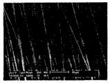
图1为本发明定向收集的复合超细纤维束放大5000倍的电子显微镜照片。Figure 1 is a 5000 times magnified electron micrograph of the composite ultrafine fiber bundles collected directional in the present invention.
图2为载药与非载药超细纤维缝合线力-位移曲线图。Figure 2 is the force-displacement curves of drug-loaded and non-drug-loaded microfiber sutures.
图3为实施例2缝合线放大150倍的形貌图。Fig. 3 is a 150-fold magnified topographical view of the suture in Example 2.
具体实施方式Detailed ways
实施例1:可释放抗生素具有促凝作用的缝合线制备Example 1: Preparation of sutures with releasable antibiotics that promote coagulation
(1)原材料溶液的配制(1) Preparation of raw material solution
壳层:生物可降解性材料为左旋聚乳酸(PLLA),溶剂为三氟乙醇,称取一定质量的左旋聚乳酸,溶于溶剂中配成浓度为8wt%的壳层纺丝溶液,在超声波中震荡4~5h,得到透明均匀的壳层纺丝溶液;Shell layer: The biodegradable material is L-polylactic acid (PLLA), and the solvent is trifluoroethanol. A certain mass of L-lactic acid is weighed, dissolved in a solvent to form a shell spinning solution with a concentration of 8wt%, and ultrasonically Medium shaking for 4-5 hours to obtain a transparent and uniform shell spinning solution;
芯层:生物活性成分为头孢噻肟钠,溶剂为去离子水,称取不同质量的头孢噻肟钠,分别溶于溶剂中配成浓度为10wt%、20wt%、30wt%的芯材纺丝溶液;Core layer: the bioactive ingredient is cefotaxime sodium, and the solvent is deionized water. Different qualities of cefotaxime sodium are weighed and dissolved in solvents respectively to make core materials with concentrations of 10wt%, 20wt%, and 30wt% for spinning solution;
涂层:生物可降解性材料壳聚糖(脱乙酰度80%,分子量10万左右),溶剂为1%醋酸溶液,壳聚糖溶于溶剂中配成浓度为1wt%的壳聚糖醋酸溶液。Coating: biodegradable material chitosan (deacetylation degree 80%, molecular weight about 100,000), solvent is 1% acetic acid solution, chitosan is dissolved in the solvent to form a concentration of 1wt% chitosan acetic acid solution .
(2)载药超细纤维线的制备(2) Preparation of drug-loaded ultrafine fiber lines
采用同轴共纺装置,将上述壳层纺丝溶液和芯材纺丝溶液分别置于壳层注射泵和芯材注射泵中,壳层注射泵的推动速度为3ml/h;芯材注射泵的推动速度为0.2ml/h,同轴共纺时控制输出电压为20~30KV,电压与芯层溶液接通,接收距离为10~13cm,接收时间为3~4min,制得的复合超细纤维的外径为0.1~0.68μm。Using a coaxial co-spinning device, the above-mentioned shell spinning solution and the core spinning solution are respectively placed in the shell injection pump and the core injection pump, and the driving speed of the shell injection pump is 3ml/h; The driving speed is 0.2ml/h, the control output voltage is 20-30KV during coaxial co-spinning, the voltage is connected to the core layer solution, the receiving distance is 10-13cm, and the receiving time is 3-4min. The outer diameter of the fiber is 0.1 to 0.68 μm.
纤维线的收集装置采用不锈钢材质的带刀口状的金属边缘圆盘,直径为28cm,控制转速为1207rpm,所收集的定向纤维线电子显微镜照片如图1所示。The collection device of the fiber line adopts a stainless steel knife-edge metal edge disc with a diameter of 28cm and a controlled speed of 1207rpm. The electron microscope photo of the collected oriented fiber line is shown in Figure 1.
(3)纤维束的制备(3) Preparation of fiber bundles
将制得的多数股纤维束置于80℃下预热30秒,并保持在此温度进行拉伸,控制拉伸伸长量50±5%,随后对其进行加捻处理,并保持在加捻张力以及90℃温度热定型2小时。Preheat the prepared multi-strand fiber bundles at 80°C for 30 seconds, and keep at this temperature for stretching, control the tensile elongation of 50±5%, then twist it, and keep it at the added temperature. Twist tension and heat setting at 90°C for 2 hours.
(4)载药编织线的制备(4) Preparation of drug-loaded braided wire
按上述实施步骤制得的经过预热、拉伸、加捻及热定型后处理的纤维束,采用8锭微型编织机进行编织,得到结构紧密、直径均一的载药超细纤维构成的编织线。The preheated, stretched, twisted and heat-set fiber bundles prepared according to the above-mentioned implementation steps are woven with an 8-spindle micro-braiding machine to obtain a braided wire composed of drug-loaded ultrafine fibers with a compact structure and uniform diameter. .
(5)缝合线的制备及表征(5) Preparation and characterization of suture
将上述实施步骤制得的载药超细纤维编织线完全浸泡在(1)中配制的壳聚糖醋酸溶液中30分钟,再用1wt%的氢氧化钠洗至中性,用去离子水冲洗3遍,在空气中放置晾干,使得壳聚糖在载药纳米纤维编织线上形成一层致密的薄膜涂敷层。The drug-loaded microfiber braided thread prepared by the above-mentioned implementation steps was completely soaked in the chitosan acetic acid solution prepared in (1) for 30 minutes, then washed to neutrality with 1wt% sodium hydroxide, and rinsed with deionized water 3 times, placed in the air to dry, so that the chitosan forms a dense film coating layer on the drug-loaded nanofiber braided wire.
对涂敷后的编织线进行拉伸试验,所得到的拉伸曲线见图2为载药(含生物活性成分)与非载药(不含生物活性成分)超细纤维缝合线力-位移曲线图。结果表明,这种缝合线基本满足美国联邦食品与药品检验局关于缝合线的使用标准。尤其值得指出的是,将载药缝合线与采用相同材料和步骤制备的非载药缝合线的拉伸性能进行对比,两者拉伸性能几无差异。Perform a tensile test on the coated braided thread, and the obtained tensile curve is shown in Figure 2, which is the force-displacement curve of drug-loaded (containing bioactive ingredients) and non-medicated (excluding bioactive ingredients) superfine fiber sutures picture. The results show that this suture basically meets the US Federal Food and Drug Inspection Service's use standards for sutures. In particular, it is worth noting that when comparing the tensile properties of drug-loaded sutures with non-drug-loaded sutures prepared using the same materials and procedures, there was little difference in tensile properties between the two.
为了进一步检验上述载药超细纤维缝合线的适用性,我们将该缝合线应用于CD大鼠上肢肌肉缝合,缝合过程中缝线具有一定的张力,没有出现断裂,且28天后的缝合、打结情况仍然良好,表明这种缝合线可以应用于生物组织的缝合。In order to further test the applicability of the above-mentioned drug-loaded microfiber suture, we applied the suture to the upper limb muscle suture of CD rats. During the suture process, the suture had a certain tension and did not break. The knot condition is still good, indicating that this suture can be applied to the suturing of biological tissues.
实施例2:具有释放抗癌药效缝线的制备Example 2: Preparation of sutures with the effect of releasing anticancer drugs
(1)原材料溶液的配制(1) Preparation of raw material solution
壳层:生物可降解性材料为聚已内酯(PCL),溶剂为体积分数为2:1的三氯甲烷:丙酮,将聚己内酯溶于溶剂中,溶液置于水浴恒温槽中,在60℃下水浴2小时即可溶解,保持PCL浓度为7wt%,制得壳层纺丝溶液;Shell layer: The biodegradable material is polycaprolactone (PCL), the solvent is chloroform: acetone with a volume fraction of 2:1, the polycaprolactone is dissolved in the solvent, and the solution is placed in a water bath constant temperature tank, It can be dissolved in a water bath at 60°C for 2 hours, and the PCL concentration is kept at 7wt% to obtain a shell spinning solution;
芯材:生物活性成分为白藜芦醇(RT),溶剂为无水乙醇,将白藜芦醇(RT)溶于溶剂中,在磁力搅拌下溶解,控制浓度为10wt%,制得芯材纺丝溶液;Core material: the bioactive component is resveratrol (RT), and the solvent is absolute ethanol. Resveratrol (RT) is dissolved in the solvent, dissolved under magnetic stirring, and the control concentration is 10wt%, and the core material is obtained spinning solution;
涂层:涂层溶质为乳酸/羟基乙酸共聚物(PGLA910),溶剂为丙酮,将溶质溶于溶剂中,配成浓度为1wt%的溶液。Coating: The solute of the coating is lactic acid/glycolic acid copolymer (PGLA910), and the solvent is acetone. The solute is dissolved in the solvent to prepare a solution with a concentration of 1 wt%.
(2)载药超细纤维线的制备(2) Preparation of drug-loaded ultrafine fiber lines
采用同轴共纺装置,将上述壳层纺丝溶液和芯材纺丝溶液分别置于壳层注射泵和芯材注射泵中,壳层注射泵的推动速度为3ml/h;芯材注射泵的推动速度为0.2ml/h,同轴共纺时控制输出电压为12~15KV,电压与芯层溶液接通,接收距离为13~15cm,接收时间为3~4min,制得的复合超细纤维的外径为0.2~1.58μm。Using a coaxial co-spinning device, the above-mentioned shell spinning solution and the core spinning solution are respectively placed in the shell injection pump and the core injection pump, and the driving speed of the shell injection pump is 3ml/h; The driving speed is 0.2ml/h, the control output voltage is 12-15KV during coaxial co-spinning, the voltage is connected to the core layer solution, the receiving distance is 13-15cm, and the receiving time is 3-4min. The outer diameter of the fiber is 0.2 to 1.58 μm.
纤维线的收集装置采用不锈钢材质的带刀口状的金属边缘圆盘,直径为28cm,控制转速为1207rpm。The collection device of the fiber line is a stainless steel knife-edge metal edge disc with a diameter of 28cm and a control speed of 1207rpm.
(3)纤维束的制备(3) Preparation of fiber bundles
将制得的多数股纤维束置于40℃下预热1分钟,并保持在此温度进行拉伸,控制拉伸伸长量60±8%,随后对其进行加捻处理,并保持在加捻张力以及100℃温度热定型2h。Preheat the prepared multi-strand fiber bundles at 40°C for 1 minute, and keep at this temperature for stretching, control the stretching elongation of 60±8%, then twist it, and keep it at the added temperature. Twist tension and heat setting at 100°C for 2 hours.
(4)RT-PCL载药编织线的制备(4) Preparation of RT-PCL drug-loaded braided wire
按上述实施步骤制得的经过预热、拉伸、加捻及热定型后处理的纤维束,采用8锭微型编织机进行编织,得到结构紧密、直径均一的载药超细纤维构成的编织线,如图3所示。The preheated, stretched, twisted and heat-set fiber bundles prepared according to the above-mentioned implementation steps are woven with an 8-spindle micro-braiding machine to obtain a braided wire composed of drug-loaded ultrafine fibers with a compact structure and uniform diameter. ,As shown in Figure 3.
(5)缝合线的制备(5) Preparation of sutures
将配制好的溶液置于喷雾器中,缓慢均匀的喷到上述实施步骤制得的载药超细纤维编织线上,随后置于20℃的干燥箱中干燥,直至溶剂完全挥发,即可制得表面光滑、具有缓释抗癌药功能的缝合线。Put the prepared solution in a sprayer, slowly and evenly spray it onto the drug-loaded microfiber braided wire prepared in the above steps, and then dry it in a drying oven at 20°C until the solvent is completely volatilized. A suture with a smooth surface and the function of slow-release anticancer drugs.
实施例3:可释放生长因子的医用缝合线制备Example 3: Preparation of medical sutures that can release growth factors
纤维细胞的培养、传代与鉴定:Fibroblast culture, subculture and identification:
将手术切下的新鲜皮肤洗净血迹,去除表皮及皮下组织,在无菌条件下剪成0.15~1mm3的组织块,贴附于培养瓶底面,于37℃、5%CO2细胞培养箱内静置培养4h,加入含20%FBS、100mg/L青霉素,100mg/L链霉素的DMEM培养液,继续培养,每4~6天用含20%FBS的DMEM换液1次,培养3~4周后进行传代培养。以细胞形态鉴定成纤维细胞。Clean the blood from the fresh skin excised by surgery, remove the epidermis and subcutaneous tissue, cut it into0.15-1mm3 tissue pieces under aseptic conditions, attach it to the bottom of the culture bottle, and store it in a cell culture incubator at 37°C and 5% CO2 After static culture for 4 hours, add DMEM culture solution containing 20% FBS, 100mg/L penicillin, and 100mg/L streptomycin, continue to cultivate, and change the medium once every 4 to 6 days with DMEM containing 20% FBS, and cultivate for 3 Subculture was carried out ~4 weeks later. Fibroblasts were identified by cell morphology.
(1)原材料溶液的配制(1) Preparation of raw material solution
壳层:生物可降解性材料为乳酸-聚羟基乙酸的共聚物(PLGA910),溶剂为3:1的四氢呋喃与N,N-二甲基甲酰胺,配制浓度为0.2g/ml的壳层纺丝溶液,在超声波中震荡4~5h,得到透明均匀的壳层纺丝溶液;Shell layer: The biodegradable material is lactic acid-polyglycolic acid copolymer (PLGA910), the solvent is 3:1 tetrahydrofuran and N,N-dimethylformamide, and the shell layer spinning concentration is 0.2g/ml. Shake the silk solution for 4 to 5 hours in an ultrasonic wave to obtain a transparent and uniform shell spinning solution;
芯层:生物活性成分为血管内皮生长因子(VEGF),溶剂为1:1的THF与DMF混合溶剂,将血管内皮生长因子及生物可降解性材料聚羟基乙酸(PGA)或乳酸-聚羟基乙酸共聚物(PLGA)的混合物溶于溶剂中,配成0.1~0.5μg/mL的芯层纺丝溶液;Core layer: The bioactive ingredient is vascular endothelial growth factor (VEGF), the solvent is a 1:1 mixture of THF and DMF, and the vascular endothelial growth factor and biodegradable materials polyglycolic acid (PGA) or lactic acid-polyglycolic acid The mixture of copolymers (PLGA) is dissolved in a solvent to prepare a core spinning solution of 0.1-0.5 μg/mL;
涂层:涂层为生物相容性材料I型胶原。Coating: Coating is biocompatible material type I collagen.
(2)载有生长因子超细纤维线的制备(2) Preparation of superfine fiber lines loaded with growth factors
采用同轴共纺装置,将上述壳层纺丝溶液和芯材纺丝溶液分别置于壳层注射泵和芯材注射泵中,壳层注射泵的推动速度为3ml/h;芯材注射泵的推动速度为0.3ml/h,同轴共纺时控制输出电压为15~30KV,电压与芯层溶液接通,接收距离为12~20cm,接收时间为3~4min,制得的复合超细纤维的外径为0.15~0.76μm。Using a coaxial co-spinning device, the above-mentioned shell spinning solution and the core spinning solution are respectively placed in the shell injection pump and the core injection pump, and the driving speed of the shell injection pump is 3ml/h; The driving speed is 0.3ml/h, the control output voltage is 15-30KV during coaxial co-spinning, the voltage is connected to the core layer solution, the receiving distance is 12-20cm, and the receiving time is 3-4min. The outer diameter of the fiber is 0.15 to 0.76 μm.
纤维线的收集装置采用不锈钢材质的带刀口状的金属边缘圆盘,直径为28cm,控制转速为1207rpm。The collection device of the fiber line is a stainless steel knife-edge metal edge disc with a diameter of 28cm and a control speed of 1207rpm.
(3)纤维束的制备(3) Preparation of fiber bundles
将制得的多数股纤维束置于50℃下预热20秒,并保持在此温度进行拉伸,控制拉伸伸长量50±5%,随后对其进行加捻处理,并保持在加捻张力以及80℃温度热定型1小时。Preheat the prepared multi-strand fiber bundles at 50°C for 20 seconds, and keep at this temperature for stretching, control the tensile elongation of 50±5%, then twist it, and keep it at the added temperature. Twist tension and heat setting at 80°C for 1 hour.
(4)载药编织线的制备(4) Preparation of drug-loaded braided wire
按上述实施步骤制得的经过预热、拉伸、加捻及热定型后处理的纤维束,采用8锭微型编织机进行编织,得到结构紧密、直径均一的含有生长因子的超细纤维编织线。The preheated, stretched, twisted and heat-set fiber bundles prepared according to the above steps are braided with an 8-spindle micro-braiding machine to obtain a superfine fiber braided line with a compact structure and uniform diameter containing growth factors .
(5)缝合线的制备(5) Preparation of sutures
将I型胶原浇注于缝线外壁上,预冻后于-50℃真空冻干处理24h,再重复浇注冻干1次。在真空干燥箱内真空度0.1bar,105℃热交联24h以使胶原分子间产生联结。经上述步骤制备成胶原海绵涂层附着于缝线外壁上,于缝线上形成了一层致密的薄膜涂敷层。Type I collagen was poured on the outer wall of the suture, pre-frozen and then vacuum freeze-dried at -50°C for 24 hours, and then repeated pouring and freeze-drying once. In a vacuum drying oven with a vacuum degree of 0.1 bar, heat crosslinking at 105° C. for 24 hours to generate linkages between collagen molecules. Prepared through the above steps, the collagen sponge coating is attached to the outer wall of the suture, forming a dense film coating layer on the suture.
Claims (7)
Priority Applications (1)
| Application Number | Priority Date | Filing Date | Title |
|---|---|---|---|
| CN 200810203328CN101406710B (en) | 2008-11-25 | 2008-11-25 | Suture thread containing bioactive components and preparation method thereof |
Applications Claiming Priority (1)
| Application Number | Priority Date | Filing Date | Title |
|---|---|---|---|
| CN 200810203328CN101406710B (en) | 2008-11-25 | 2008-11-25 | Suture thread containing bioactive components and preparation method thereof |
Publications (2)
| Publication Number | Publication Date |
|---|---|
| CN101406710A CN101406710A (en) | 2009-04-15 |
| CN101406710Btrue CN101406710B (en) | 2013-02-13 |
Family
ID=40570052
Family Applications (1)
| Application Number | Title | Priority Date | Filing Date |
|---|---|---|---|
| CN 200810203328Expired - Fee RelatedCN101406710B (en) | 2008-11-25 | 2008-11-25 | Suture thread containing bioactive components and preparation method thereof |
Country Status (1)
| Country | Link |
|---|---|
| CN (1) | CN101406710B (en) |
Families Citing this family (39)
| Publication number | Priority date | Publication date | Assignee | Title |
|---|---|---|---|---|
| GB2474851A (en)* | 2009-10-27 | 2011-05-04 | Univ Bolton | Wound dressing comprising anti-microbial honey encapsulated within biocompatible and biodegradable fibre, and the fibre's production |
| CN102071491B (en)* | 2011-01-18 | 2012-12-05 | 东华大学 | Medicinal gullet scaffold fiber and preparation method thereof |
| CN102389587A (en)* | 2011-09-19 | 2012-03-28 | 大连创达技术交易市场有限公司 | Manufacturing method of novel medical carbon fiber suture |
| CN102698315B (en)* | 2012-06-26 | 2014-01-15 | 单县润康缝合材料有限公司 | Coating method of antibacterial surgical suture made of high molecular absorbable material |
| CN104491920B (en)* | 2014-12-19 | 2017-09-19 | 山东博达医疗用品有限公司 | A kind of preparation method of absorbable suture |
| CN104524626B (en)* | 2014-12-19 | 2017-09-19 | 山东博达医疗用品有限公司 | A kind of absorbable collagen snare line and preparation method thereof |
| CN104940985A (en)* | 2015-05-29 | 2015-09-30 | 苏州乔纳森新材料科技有限公司 | Medical suture and production method thereof |
| CN104940987B (en)* | 2015-06-08 | 2017-12-08 | 浙江新城钮扣饰品有限公司 | A kind of high-intensity absorbable suture and preparation method thereof |
| CN104940989A (en)* | 2015-06-24 | 2015-09-30 | 苏州乔纳森新材料科技有限公司 | Absorbable suture line and method for preparing the same |
| CN104940988B (en)* | 2015-06-24 | 2017-11-14 | 吴丽敏 | A kind of antibacterial operation suture and preparation method thereof |
| CN105056295B (en)* | 2015-08-11 | 2018-01-05 | 安徽省康宁医疗用品有限公司 | A kind of anti-inflammatory, antibacterial absorbable medical suture and preparation method thereof |
| CN105079866B (en)* | 2015-08-11 | 2017-09-15 | 安徽省康宁医疗用品有限公司 | A kind of suture of high mechanical strength |
| CN105063799B (en)* | 2015-08-11 | 2017-12-15 | 安徽省康宁医疗用品有限公司 | A kind of preparation method of the good absorbable medical suture of tensile strength |
| CN105079869B (en)* | 2015-08-11 | 2017-08-15 | 安徽省康宁医疗用品有限公司 | A kind of absorbable medical suture of good mechanical property |
| CN105251042A (en)* | 2015-10-28 | 2016-01-20 | 钊桂英 | Medical absorbable polyglycolic acid (PGA) operation suture line and preparing method |
| CN105420917A (en)* | 2015-12-02 | 2016-03-23 | 江苏金松生物科技有限公司 | Polyglycolic acid suture manufacturing system |
| CN105544086A (en)* | 2015-12-02 | 2016-05-04 | 江苏金松生物科技有限公司 | Making method of polyglycolic acid suture |
| CN106075544A (en)* | 2016-01-26 | 2016-11-09 | 西北工业大学 | A kind of core shell composite construction medicine carrying stitching thread and preparation method thereof |
| CN105770978A (en)* | 2016-03-21 | 2016-07-20 | 江苏广达医材集团有限公司 | Biodegradable medical abdominal surgical suture material |
| CN105963764A (en)* | 2016-06-21 | 2016-09-28 | 林春梅 | Anti-infection and high-strength medical absorbable suture and preparation method thereof |
| CN106436314B (en)* | 2016-10-13 | 2019-06-18 | 上海太亨科贸有限公司 | A kind of composite fibre with self-expanding effect and preparation method thereof and drying unit |
| CN107213507A (en)* | 2017-07-03 | 2017-09-29 | 武汉医佳宝生物材料有限公司 | A kind of absorbable suture and preparation method thereof |
| CN108042844A (en)* | 2017-12-11 | 2018-05-18 | 青岛大学 | A kind of preparation method of suture |
| CN108543107A (en)* | 2018-04-19 | 2018-09-18 | 宁波诺丁汉新材料研究院有限公司 | A kind of human body medical absorbable suture and preparation method thereof |
| CN108607115A (en)* | 2018-06-13 | 2018-10-02 | 李华刚 | A kind of M lips shaping suture |
| CN108815559A (en)* | 2018-07-20 | 2018-11-16 | 苏州洛特兰新材料科技有限公司 | A kind of preparation method of medical sutures new material |
| CN108904869B (en)* | 2018-07-20 | 2020-09-01 | 南通纺织丝绸产业技术研究院 | Medical suture with natural antibacterial drug slow-release function and preparation method thereof |
| CN109385710B (en)* | 2018-09-12 | 2021-01-29 | 浙江星月生物科技股份有限公司 | Absorbable suture and preparation method thereof |
| CN109663144B (en)* | 2018-09-30 | 2020-12-29 | 温州医科大学 | A bioactive degradable surgical suture and preparation method thereof |
| CN109663142B (en)* | 2018-09-30 | 2021-06-22 | 温州医科大学 | Drug-loaded degradable surgical sewing thread and preparation method thereof |
| CN109876177B (en)* | 2019-01-26 | 2020-08-21 | 乐清市风杰电子科技有限公司 | Medical suture material and preparation method thereof |
| CN110302417A (en)* | 2019-06-18 | 2019-10-08 | 珠海稻田医疗科技有限公司 | A kind of antibacterial, biodegradable suture |
| CN111134746A (en)* | 2019-12-30 | 2020-05-12 | 山东省肿瘤防治研究院(山东省肿瘤医院) | Surgical suture for gastrointestinal surgery and manufacturing method thereof |
| CN113046884B (en)* | 2021-03-25 | 2022-06-10 | 台州市中心医院(台州学院附属医院) | Antibacterial degradable medical suture |
| CN113304304B (en)* | 2021-06-21 | 2022-07-01 | 青岛理工大学 | A kind of drug-loaded degradable absorbable medical suture thread and preparation method thereof |
| CN113718348B (en)* | 2021-09-13 | 2022-07-05 | 西北工业大学 | A repair factor-loaded suture production device |
| CN115874297A (en)* | 2021-09-27 | 2023-03-31 | 中国科学院理化技术研究所 | Preparation method and preparation device of medical absorbable antibacterial suture line |
| CN115094552B (en)* | 2022-06-17 | 2024-08-23 | 北京诺康达医药科技股份有限公司 | Pulling wire and preparation method thereof |
| CN120078926B (en)* | 2025-05-06 | 2025-09-02 | 上海傍成医疗科技有限公司 | A radio-stimulated surgical suture for promoting peripheral nerve healing, and its preparation method and application |
Citations (2)
| Publication number | Priority date | Publication date | Assignee | Title |
|---|---|---|---|---|
| CN1104917A (en)* | 1994-01-03 | 1995-07-12 | 北京医科大学 | Gene and medicament sewing thread |
| CN1537981A (en)* | 2003-10-23 | 2004-10-20 | 黄争鸣 | Coaxial composite continuous nano/micron fiber and its preparation method |
- 2008
- 2008-11-25CNCN 200810203328patent/CN101406710B/ennot_activeExpired - Fee Related
Patent Citations (2)
| Publication number | Priority date | Publication date | Assignee | Title |
|---|---|---|---|---|
| CN1104917A (en)* | 1994-01-03 | 1995-07-12 | 北京医科大学 | Gene and medicament sewing thread |
| CN1537981A (en)* | 2003-10-23 | 2004-10-20 | 黄争鸣 | Coaxial composite continuous nano/micron fiber and its preparation method |
Also Published As
| Publication number | Publication date |
|---|---|
| CN101406710A (en) | 2009-04-15 |
Similar Documents
| Publication | Publication Date | Title |
|---|---|---|
| CN101406710B (en) | Suture thread containing bioactive components and preparation method thereof | |
| US11828006B2 (en) | Methods of orienting multifilament yarn and monofilaments of poly-4-hydroxybutyrate and copolymers thereof | |
| JP6916950B2 (en) | Medical suture with sustained release function of natural antibacterial drug and its manufacturing method | |
| US20220202988A1 (en) | Yarns and fibers of poly(butylene succinate) and copolymers thereof, and methods of use thereof | |
| Dennis et al. | Suture materials—Current and emerging trends | |
| US9457127B2 (en) | Micro-fiber webs of poly-4-hydroxybutyrate and copolymers thereof produced by centrifugal spinning | |
| Hu et al. | Development of braided drug-loaded nanofiber sutures | |
| Arora et al. | Drug eluting sutures: A recent update | |
| US10874771B2 (en) | Oriented P4HB implants containing antimicrobial agents | |
| Soufdoost et al. | Surgical suture assembled with tadalafil/polycaprolactone drug-delivery for vascular stimulation around wound: validated in a preclinical model | |
| CN114059350A (en) | A kind of natural long-acting antibacterial and anti-inflammatory silk suture and preparation method thereof | |
| US12077885B2 (en) | Processing method and apparatus for micro-structured rope-like material | |
| CN104667340A (en) | A biodegradable suture with bioactivity | |
| Nicolae et al. | Polymer fibers in biomedical engineering | |
| CN111603609B (en) | Bionic tissue engineering scaffold and preparation method thereof | |
| Xu et al. | Electrospun Medical Sutures for Wound Healing: A Review. Polymers 2022, 14, 1637 | |
| Ghosh et al. | A Critique: Advancement and Applications of Surgical Sutures in Medical Implants | |
| Ghosh et al. | 11 A Critique | |
| Madheswaran | Fabrication and Characterization of Sutures Made from Composite Nanofibrous Yarns | |
| EP4284300A1 (en) | Minimally invasive breast suspension system |
Legal Events
| Date | Code | Title | Description |
|---|---|---|---|
| C06 | Publication | ||
| PB01 | Publication | ||
| C10 | Entry into substantive examination | ||
| SE01 | Entry into force of request for substantive examination | ||
| C14 | Grant of patent or utility model | ||
| GR01 | Patent grant | ||
| CF01 | Termination of patent right due to non-payment of annual fee | ||
| CF01 | Termination of patent right due to non-payment of annual fee | Granted publication date:20130213 Termination date:20151125 |