CN101283930A - Image diagnosis support system and image diagnosis support method - Google Patents
Image diagnosis support system and image diagnosis support methodDownload PDFInfo
- Publication number
- CN101283930A CN101283930ACNA2008100916440ACN200810091644ACN101283930ACN 101283930 ACN101283930 ACN 101283930ACN A2008100916440 ACNA2008100916440 ACN A2008100916440ACN 200810091644 ACN200810091644 ACN 200810091644ACN 101283930 ACN101283930 ACN 101283930A
- Authority
- CN
- China
- Prior art keywords
- information
- combination
- image
- diagnosis support
- user
- Prior art date
- Legal status (The legal status is an assumption and is not a legal conclusion. Google has not performed a legal analysis and makes no representation as to the accuracy of the status listed.)
- Granted
Links
Images


Classifications
- A—HUMAN NECESSITIES
- A61—MEDICAL OR VETERINARY SCIENCE; HYGIENE
- A61B—DIAGNOSIS; SURGERY; IDENTIFICATION
- A61B6/00—Apparatus or devices for radiation diagnosis; Apparatus or devices for radiation diagnosis combined with radiation therapy equipment
- A61B6/44—Constructional features of apparatus for radiation diagnosis
- A61B6/4494—Means for identifying the diagnostic device
- G—PHYSICS
- G16—INFORMATION AND COMMUNICATION TECHNOLOGY [ICT] SPECIALLY ADAPTED FOR SPECIFIC APPLICATION FIELDS
- G16H—HEALTHCARE INFORMATICS, i.e. INFORMATION AND COMMUNICATION TECHNOLOGY [ICT] SPECIALLY ADAPTED FOR THE HANDLING OR PROCESSING OF MEDICAL OR HEALTHCARE DATA
- G16H30/00—ICT specially adapted for the handling or processing of medical images
- G16H30/20—ICT specially adapted for the handling or processing of medical images for handling medical images, e.g. DICOM, HL7 or PACS
- G—PHYSICS
- G16—INFORMATION AND COMMUNICATION TECHNOLOGY [ICT] SPECIALLY ADAPTED FOR SPECIFIC APPLICATION FIELDS
- G16H—HEALTHCARE INFORMATICS, i.e. INFORMATION AND COMMUNICATION TECHNOLOGY [ICT] SPECIALLY ADAPTED FOR THE HANDLING OR PROCESSING OF MEDICAL OR HEALTHCARE DATA
- G16H50/00—ICT specially adapted for medical diagnosis, medical simulation or medical data mining; ICT specially adapted for detecting, monitoring or modelling epidemics or pandemics
- G16H50/20—ICT specially adapted for medical diagnosis, medical simulation or medical data mining; ICT specially adapted for detecting, monitoring or modelling epidemics or pandemics for computer-aided diagnosis, e.g. based on medical expert systems
- G—PHYSICS
- G16—INFORMATION AND COMMUNICATION TECHNOLOGY [ICT] SPECIALLY ADAPTED FOR SPECIFIC APPLICATION FIELDS
- G16H—HEALTHCARE INFORMATICS, i.e. INFORMATION AND COMMUNICATION TECHNOLOGY [ICT] SPECIALLY ADAPTED FOR THE HANDLING OR PROCESSING OF MEDICAL OR HEALTHCARE DATA
- G16H15/00—ICT specially adapted for medical reports, e.g. generation or transmission thereof
Landscapes
- Health & Medical Sciences (AREA)
- Engineering & Computer Science (AREA)
- Medical Informatics (AREA)
- Life Sciences & Earth Sciences (AREA)
- Public Health (AREA)
- General Health & Medical Sciences (AREA)
- Nuclear Medicine, Radiotherapy & Molecular Imaging (AREA)
- Radiology & Medical Imaging (AREA)
- Biomedical Technology (AREA)
- Primary Health Care (AREA)
- Epidemiology (AREA)
- Pathology (AREA)
- Heart & Thoracic Surgery (AREA)
- High Energy & Nuclear Physics (AREA)
- Surgery (AREA)
- Animal Behavior & Ethology (AREA)
- Physics & Mathematics (AREA)
- Optics & Photonics (AREA)
- Veterinary Medicine (AREA)
- Molecular Biology (AREA)
- Biophysics (AREA)
- Data Mining & Analysis (AREA)
- Databases & Information Systems (AREA)
- Medical Treatment And Welfare Office Work (AREA)
- Measuring And Recording Apparatus For Diagnosis (AREA)
- Apparatus For Radiation Diagnosis (AREA)
Abstract
Translated fromChineseDescription
Translated fromChinese技术领域technical field
本发明,涉及一种在判定医疗信息是否发送,或发送医疗信息的情况下,可以恰当地对其内容进行取舍选择等,使得对于需要医疗信息的用户,不会降低网络的通信效率、各装置的动作效率、读图时的作业效率中的至少一个的图像诊断支持系统及图像诊断支持方法。The present invention relates to a method for judging whether to send medical information, or when sending medical information, it is possible to appropriately select and select its content, so that the communication efficiency of the network is not reduced for users who need medical information, and each device An image diagnosis support system and an image diagnosis support method that provide at least one of operation efficiency and work efficiency during image reading.
背景技术Background technique
近年,医疗行为的专业领域被细化。例如,图像诊断被分为:患者的诊断图像的取得、取得了的诊断图像的读图及报告制作、基于报告结果的诊断结果和治疗方针的说明等各个作业。由各专业人员(负责的医师或负责技师)负责各个作业,通过这些全部作业来完成对患者的诊断等医疗行为。各专业人员基于在前级作业中由其他专业人员制作的信息,并通过适当参照过去的诊断信息等,执行各作业。In recent years, the professional field of medical practice has been refined. For example, image diagnosis is divided into various tasks such as acquisition of a patient's diagnostic image, image reading of the acquired diagnostic image, report creation, and explanation of a diagnosis result and a treatment policy based on the report result. Professionals (physicians in charge or technicians in charge) are in charge of each operation, and medical actions such as diagnosing patients are completed through all these operations. Each specialist executes each task by appropriately referring to past diagnostic information and the like based on information created by other specialists in previous tasks.
在医师终端、取得诊断图像的X射线CT装置、MRI装置等医用图像诊断装置、存储诊断图像的PACS服务器(医用图像保存装置)、用于对诊断图像进行读图的图像参照装置、图像诊断报告制作支持装置等的各自中,利用网络进行上述各作业。例如,主治医在自身的终端上,从医院信息系统(HIS:Hospital Information System)取得信息,一边参照读图报告等一边输入必要事项,利用放射科信息系统(RIS:Radiology Information System)进行定单(检查委托)的发布。操作医用图像诊断装置的技师等通过网络,接受发布的定单,基于该定单的内容决定摄影范围、摄影条件等,执行需要的图像采集。自动地或与规定地操作应答地,通过网络将取得的图像保存到医用图像保存装置。图像参照装置或图像诊断报告制作支持装置例如以读图医师的经过观察为目的,取得保存在医用图像保存装置中的图像、过去的报告等。在图像诊断报告制作支持装置中,执行作为诊断根据的图像(关键图像)的选择、报告制作。制作的报告、选择出的关键图像等通过网络发送并保存在医用图像保存装置中。Medical image diagnostic devices such as physician terminals, X-ray CT devices and MRI devices that acquire diagnostic images, PACS servers (medical image storage devices) that store diagnostic images, image reference devices that read diagnostic images, and image diagnostic reports In each of the production support devices and the like, each of the above-mentioned operations is performed using a network. For example, the attending doctor obtains information from the Hospital Information System (HIS: Hospital Information System) on his own terminal, enters necessary items while referring to a chart reading report, etc., and makes an order (examination) using the Radiology Information System (RIS: Radiology Information System). Commissioned) release. A technician or the like who operates a medical image diagnostic apparatus receives an issued order through a network, determines an imaging range, imaging conditions, and the like based on the content of the order, and executes necessary image acquisition. The obtained image is stored in the medical image storage device through the network automatically or in response to a predetermined operation. The image referencing device or the image diagnosis report preparation support device acquires images stored in the medical image storage device, past reports, etc., for the purpose of observation by a graphic reader, for example. In the image diagnosis report creation support device, selection of an image (key image) as a basis for diagnosis and creation of a report are executed. The prepared report, the selected key image, etc. are sent through the network and stored in the medical image storage device.
进而近年来,在这样的作业被细化了的图像诊断中,例如在特愿2006-319356中,提出了能够高效地利用过去的检查信息的系统。这是将对象(object)作为信息而共有化的技术,其中作为其内容,该对象包含与在过去的摄影中用过的条件或关键图像有关的信息、与在摄影时参照过的过去的检查有关的信息。用户可以通过在任意的装置中以任意的定时参照共有对象,得知过去的诊断所利用的关键图像和摄影条件,例如,可以摄影高精度地再现过去的检查且适于比较读图的图像。此外,由于参照过去而摄影的检查,将其参考历史也保存于共有对象中,所以在读图时,能根据该信息自动地确定并显示需要参考的比较对手,能大大降低读图医生在读图前的准备行为。Furthermore, in recent years, in image diagnosis in which such operations have been refined, for example, in Japanese Patent Application No. 2006-319356, a system capable of efficiently utilizing past examination information has been proposed. This is a technique for sharing an object as information, and as its content, the object includes information on conditions or key images used in past photography, and past inspections that were referred to in photography. relevant information. The user can refer to shared objects at any timing on any device to learn key images and imaging conditions used in past diagnoses. For example, images that reproduce past examinations with high precision and are suitable for comparative image reading can be captured. In addition, because the reference history of the inspections taken with reference to the past is also stored in the shared object, so when reading the picture, it can automatically determine and display the comparison partner that needs to be referred to according to the information, which can greatly reduce the need for the doctor to read the picture. preparatory behavior.
但是,在导入了利用共有对象的系统的情况下,例如未对应共有对象的显示装置(也包含显示客户端等)并不将共有对象与通常的图像信息区别地进行处理。因此,例如可能发生以下的问题。However, when a system using a shared object is introduced, for example, a display device (including a display client, etc.) that does not support the shared object does not process the shared object differently from normal image information. Therefore, for example, the following problems may occur.
即,由于服务器装置侧不将共有对象与通常的图像信息区别,因此有客户端装置(例如,查看器等)侧发送不需要的共有对象本身的情况。在该情况下,会在网络上进行无用的数据的发送、取得,会降低网络的通信效率、各装置的动作效率。That is, since the server device side does not distinguish the shared object from normal image information, the client device (for example, a viewer, etc.) side may transmit an unnecessary shared object itself. In this case, useless data is transmitted and acquired on the network, and the communication efficiency of the network and the operation efficiency of each device are lowered.
此外,例如在客户端装置中显示了不需要的共有对象的情况下,读图所需要的图像(例如关键图像等)的显示区域变小。因此,会产生共有对象的一部分或全部的删除动作等无用的作业,降低读图时的作业效率。Also, for example, when an unnecessary shared object is displayed on the client device, the display area of an image (for example, a key image, etc.) required for image reading becomes small. Therefore, useless work such as deletion of part or all of shared objects occurs, reducing work efficiency in image reading.
进而,可以说:不限于共有对象,在服务器装置、客户端装置之间,通过网络收发医用图像报告、图像数据等各种医疗信息的情况下,这样的网络的通信效率、各装置的动作效率、读图时的作业效率的降低也是一个很大的问题。Furthermore, it can be said that the communication efficiency of such a network and the operation efficiency of each device are not limited to the shared objects, and when various medical information such as medical image reports and image data are transmitted and received between the server device and the client device through the network. 1. The reduction of work efficiency when reading pictures is also a big problem.
发明内容Contents of the invention
本发明是鉴于以上的问题而提出的,其目的在于:提供一种对于需要以共有对象或报告为代表的医疗信息的客户端装置,可以恰当地发送其内容的全部或一部分的图像诊断支持系统、以及图像诊断支持方法,使得不发生网络的通信效率、各装置的动作效率、读图时的作业效率中的至少一个的降低。The present invention has been made in view of the above problems, and an object of the present invention is to provide an image diagnosis support system that can appropriately transmit all or part of the content to a client device that requires medical information represented by shared objects or reports , and the image diagnosis support method so that at least one of network communication efficiency, operation efficiency of each device, and work efficiency at the time of image reading does not decrease.
根据本发明的一个方式,提供了一种图像诊断支持系统,其特征在于包括:存储单元,存储包含摄影条件、摄影范围、作为诊断根据的关键图像位置中的至少一个和图像的多个对象;接受单元,从用户接受成为参照的检查的确定信息以及用户识别信息和关于利用场景的组合;判定单元,根据用于对用户识别信息和与利用场景有关的信息的每个组合而判定是否向用户提供上述对象的第一判定信息,判定是否在上述接受的组合中提供对象;收集单元,在上述判定单元判定为提供对象的情况下,从上述存储单元收集根据上述检查的确定信息确定的对象;显示单元,显示根据上述检查的确定信息确定的对象。According to one aspect of the present invention, there is provided an image diagnosis support system, which is characterized by comprising: a storage unit for storing a plurality of objects including at least one of imaging conditions, imaging ranges, key image positions used as diagnosis basis, and images; The accepting unit accepts from the user the confirmation information of the check to be referred to and the combination of the user identification information and the usage scene; providing the first judgment information of the object, judging whether the object is provided in the above-mentioned accepted combination; the collection unit, when the judging unit judges that it is the object to be provided, collecting the object determined according to the confirmation information of the above-mentioned inspection from the above-mentioned storage unit; A display unit for displaying the object determined based on the determination information of the above inspection.
根据本发明的另一个方式,提供了一种图像诊断支持服务器,其特征在于包括:接受单元,从用户接受成为参照的检查的确定信息以及用户识别信息和与利用场景有关的信息的组合;判定单元,根据用于对用户识别信息和与利用场景有关的信息的每个组合而判定是否向用户提供多个医疗信息中的预定的医疗信息的判定信息,判定是否在上述接受的组合中提供上述医疗信息;收集单元,在上述判定单元判定为提供上述医疗信息的情况下,根据上述检查的确定信息,从存储多个医疗信息的存储单元收集应该提供的上述医疗信息。According to another aspect of the present invention, there is provided an image diagnosis support server, which is characterized by including: a receiving unit that receives from a user a combination of identification information of an examination to be referred to, user identification information, and information related to a usage scene; A unit for judging whether to provide predetermined medical information among a plurality of medical information to the user for each combination of the user identification information and the information related to the usage scene, and judging whether to provide the above-mentioned medical information in the above-mentioned accepted combination. medical information; a collection unit that collects the medical information to be provided from a storage unit that stores a plurality of medical information based on confirmation information of the examination when the judging unit determines that the medical information is to be provided.
根据本发明的另一个方式,提供了一种图像诊断支持服务器,其特征在于包括:存储单元,存储包含摄影条件、摄影范围、作为诊断根据的关键图像位置中的至少一个和图像的多个对象;接受单元,从用户接受成为参照的检查的确定信息以及用户识别信息和与利用场景有关的信息的组合;判定单元,根据用于对用户识别信息和与利用场景有关的信息的每个组合而判定是否向用户提供上述对象的第一判定信息,判定是否在上述接受的组合中提供对象;收集单元,在上述判定单元判定为提供对象的情况下,从上述存储单元收集根据上述检查的确定信息而确定的对象。According to another aspect of the present invention, there is provided an image diagnosis support server, which is characterized in that it includes: a storage unit that stores a plurality of objects including at least one of imaging conditions, imaging ranges, key image positions used as diagnosis basis, and images. The accepting unit accepts from the user a combination of confirmation information and user identification information and information related to the use scene from the user; judging whether to provide the first judgment information of the above-mentioned object to the user, and judging whether to provide the object in the above-mentioned accepted combination; the collection unit, in the case where the above-mentioned judging unit judges that the object is to be provided, collects the determination information according to the above-mentioned inspection from the above-mentioned storage unit and determined objects.
根据本发明的另一个方式,提供了一种图像诊断支持方法,其特征在于包括:从用户接受成为参照的检查的确定信息以及用户识别信息和与利用场景有关的信息的组合;根据用于对用户识别信息和与利用场景有关的信息的每个组合而判定是否向用户提供多个医疗信息中的规定的医疗信息的判定信息,判定是否在上述接受的组合中提供上述医疗信息;在判定为提供上述医疗信息的情况下,根据上述检查的确定信息,从存储多个医疗信息的存储单元收集应该提供的上述医疗信息。According to another aspect of the present invention, there is provided an image diagnosis support method characterized by comprising: receiving from a user a combination of determination information of an examination to be referred to, user identification information, and information related to a usage scene; Judgment information for judging whether to provide prescribed medical information among a plurality of medical information to the user for each combination of user identification information and information related to the utilization scene, and judging whether to provide the above-mentioned medical information in the above-mentioned accepted combination; When providing the above-mentioned medical information, the above-mentioned medical information to be provided is collected from a storage unit storing a plurality of medical information based on the determination information of the above-mentioned examination.
根据本发明的另一个方式,提供了一种图像诊断支持方法,其特征在于包括:从用户接受成为参照的检查的确定信息以及用户识别信息和与利用场景有关的信息的组合的步骤;根据用于对用户识别信息和与利用场景有关的信息的每个组合而判定是否向用户提供包含摄影条件、摄影范围、作为诊断根据的关键图像位置中的至少一个和图像的对象的第一判定信息,判定是否在上述接受的组合中提供对象的步骤;在上述判定中判定为提供对象的情况下,从存储包含摄影条件、摄影范围、作为诊断根据的关键图像位置中的至少一个和图像的多个对象的存储单元中,收集根据上述检查的确定信息确定的对象的步骤。According to another aspect of the present invention, there is provided an image diagnosis support method, characterized by including the steps of: receiving from a user a combination of confirmation information of an examination to be referred to, user identification information, and information related to a usage scene; determining for each combination of the user identification information and the information on the usage scene whether to provide the user with first determination information including at least one of the shooting condition, the shooting range, the key image position as a basis for diagnosis, and the object of the image, A step of judging whether to provide an object in the above-mentioned accepted combination; if it is determined in the above-mentioned judgment that the object is to be provided, a plurality of images including at least one of the imaging condition, the imaging range, and the key image position as the diagnosis basis and the image are stored. In the object storage unit, a step of collecting the objects identified based on the identification information of the above inspection.
附图说明Description of drawings
图1是表示实施例1的实现图像诊断支持系统的医院内网络系统1的结构的图。FIG. 1 is a diagram showing the configuration of an in-
图2表示实施例1的利用管理表的一个例子。FIG. 2 shows an example of the utilization management table of the first embodiment.
图3表示提供信息管理表的一个例子。Fig. 3 shows an example of the provision information management table.
图4是用于说明共有对象的结构的图。FIG. 4 is a diagram for explaining the structure of shared objects.
图5是表示共有对象的生成、管理的流程的一个例子的流程图。FIG. 5 is a flowchart showing an example of the flow of creation and management of shared objects.
图6是表示实施例2的图像诊断支持处理的流程的流程图。6 is a flowchart showing the flow of image diagnosis support processing in the second embodiment.
图7表示实施例2的利用管理表的一个例子。FIG. 7 shows an example of the use management table of the second embodiment.
图8是表示实施例2的图像诊断支持处理的流程的流程图。8 is a flowchart showing the flow of image diagnosis support processing in the second embodiment.
图9是表示实施例3的医用图像诊断支持系统1的结构的图。FIG. 9 is a diagram showing the configuration of a medical image
图10是表示实施例3的图像诊断支持处理的流程的流程图。10 is a flowchart showing the flow of image diagnosis support processing in the third embodiment.
图11是表示实施例4的利用管理表的一个例子的图。FIG. 11 is a diagram showing an example of a usage management table according to the fourth embodiment.
具体实施方式Detailed ways
以下,根据附图说明本发明的实施例。另外,在以下的说明中,对于具有基本一样的功能及结构的结构要素,附加同一标记,仅在必要时进行重复说明。Hereinafter, embodiments of the present invention will be described with reference to the drawings. In addition, in the following description, the same code|symbol is attached|subjected to the structural element which has substantially the same function and structure, and it repeats description only when necessary.
另外,在以下的实施例中,为了具体地进行说明,而说明服务器装置恰当地向需要作为医疗信息的共有对象的客户端装置发送其内容的全部或一部分等的图像诊断支持系统以及图像诊断支持方法等。但是,处理的医疗信息并不限于共有对象,例如对于医用报告、医用图像数据,也可以应用本发明的技术思想。In addition, in the following embodiments, for concrete description, an image diagnosis support system and an image diagnosis support system in which a server device appropriately transmits all or part of the content to a client device that needs to share medical information will be described. method etc. However, the medical information to be processed is not limited to shared objects, for example, the technical idea of the present invention can also be applied to medical reports and medical image data.
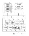
图1是表示本实施例的实现图像诊断支持系统的医院内网络系统1的结构的图。如该图所示,本医院内网络系统1具备医用图像诊断装置2、客户端装置3和作为服务器装置的医用图像保存装置5。FIG. 1 is a diagram showing the configuration of an in-
[医用图像诊断装置][Medical Image Diagnosis Device]
医用图像诊断装置2是X射线计算机断层摄影装置(X射线CT装置)、磁共振成像装置、超声波诊断装置、核医学诊断装置、X射线诊断装置等图像诊断装置。在本实施例中,为了具体地进行说明,设定医用图像诊断装置2是X射线CT装置的情况。The medical image
此外,医用图像诊断装置2除了取得与患者有关的诊断图像的摄影系统之外,还具备控制部件21、共有对象生成部件23、收发部件25、显示部件、数据存储部件、操作部件(没有参考标记的未图示)等。In addition, the medical image
控制部件21统一地控制本医用图像诊断装置2的静态或动态的动作。The
共有对象生成部件23为了有效地利用过去的医疗行为实施时使用过的信息(例如,定位图像、摄影位置、摄影范围、摄影条件、图像生成条件等),生成由图像信息和附带(文字或数值)信息构成的共有对象。将在后文中详细说明该共有对象的结构。Shared
收发部件25经由网络N从其它设备接收或向其它设备发送图像、共有对象等的医疗信息。The
[客户端装置][client device]
客户端装置3是对服务器装置(本实施例中的医用图像保存装置5)请求共有对象的装置,具有控制部件31、收发部件33、显示部件、数据存储部件、操作部件(没有参考标记的未图示)等。The
控制部件31在向医用图像保存装置5发送在过去的检查中取得的图像信息等的取得请求时,自动地发送现在的用户ID(例如,登录时或上述取得请求指示时输入的用户ID)、用于识别该客户端装置3的装置ID、在该客户端装置3中正在使用的应用程序名。此外,控制部件3采用从医用图像保存装置5接受的数据,进行用于在显示部件中以预定的方式显示各种图像、共有对象等的控制。The
收发部件33对预定的操作进行应答地,向医用图像保存装置5发送在过去的检查中取得的图像信息等的取得请求指示、现在的用户ID、装置ID、应用程序名等。此外,收发部件33从医用图像保存装置5接收加工后的共有对象数据等。In response to a predetermined operation, the transmitting and receiving
[医用图像保存装置][Medical image storage device]
医用图像保存装置5根据患者ID、序列UID等管理并保存在医用图像诊断装置2中生成的图像等。此外,医用图像保存装置5分析在医用图像诊断装置2中生成的共有对象,将各种数据保存到预定的位置。进而,医用图像保存装置5作为实现后述的图像诊断支持功能的图像诊断支持系统而动作。The medical
本医用图像保存装置5具备数据库50、数据保存部件51、操作部件52、显示部件53、收发部件54、判定部件55、共有对象加工部件56、信息收集部件57以及控制部件58。The medical
数据库50是管理用于确定各种图像的实体数据和共有对象的实体数据的保存位置(所在)的信息(所在确定信息)的数据库。此外,数据库50存储在图像诊断支持功能中被利用的利用管理表、提供信息管理表。The
在此,利用管理表是用于定义是否对用户和利用场景的每个组合提供共有对象、以及在提供共有对象的情况下决定其内容的信息区分的表。提供信息管理表是用于按每个信息区分定义包含在共有对象中的内容(提供信息)的表。Here, the use management table is a table for defining whether to provide a shared object for each combination of a user and a use scene, and when providing a shared object, to determine the information classification of its content. The provided information management table is a table for classifying and defining the content (provided information) included in the common object for each information.
图2表示利用管理表的一个例子。如该图所示,在利用管理表中,对利用场景(在该图例子中,是应用程序名和使用装置)与用户的每个组合,定义是否提供共有对象,以及提供共有对象时的信息区分。Fig. 2 shows an example of the utilization management table. As shown in the figure, in the use management table, for each combination of the use scene (in this example, the application name and the device used) and the user, it is defined whether to provide shared objects, and information classification when providing shared objects .
图3表示提供信息管理表的一个例子。如该图所示,在提供信息管理表中,对每个信息区分,定义定位图像、摄影条件、图像条件等应该包含在共有对象中的内容。Fig. 3 shows an example of the provision information management table. As shown in the figure, in the provided information management table, for each piece of information, contents to be included in shared objects, such as positioning images, shooting conditions, and image conditions, are defined.
数据保存部件51通过收发部件54接受图像数据、共有对象等各种数据,并写入、保存到适当的位置。此外,数据保存部件51在决定了保存位置时、删除后时、或者变更后的情况下,与数据库50通信,修正数据库50管理的共有对象管理信息和所在确定信息。另外,该数据保存部件51不必内置于医用图像保存装置5内,也可以位于网络上的其他位置。The
此外,在本实施例中,在数据库50中保存各种图像的实体数据和共有对象的所在确定信息,在数据保存部件51中接受并保存接受的图像数据、共有对象等各种数据。当然这些只是示例,本发明的技术思想并不限于各种数据的保存位置。例如,也可以将数据库50和数据保存部件51作为一个数据保存部件等,可以根据设计事项的范畴任意变更各种数据的保存方式。In addition, in the present embodiment, the
操作部件52是具备键盘、各种开关、鼠标等,可输入来自操作者的指示的装置。The
显示部件53是用于显示操作画面和规定的图像的监视器。The
收发部件54通过网络N从其它机器接收或向其它机器发送图像、共有对象等医疗信息。The transmitting and receiving
判定部件55基于通过收发部件54接受的用户和利用场景的组合和利用管理表,判定在该用户和利用场景下是否提供共有对象、以及提供共有对象时的信息区分。此外,判定部件55基于判定出的信息区分和提供信息管理表,判定与该信息区分对应的提供信息。The judging
共有对象加工部件56根据在判定部件55中判定出的提供信息,加工已有的共有对象使得仅含有该提供信息。The shared
信息收集部件57从数据保存部件51或网络上的其它装置,收集被客户端装置3请求的各种信息、在判定部件55中判定出的信息区分所对应的提供信息。The
控制部件58统一地控制本医用图像保存装置5的静态或动态的动作。此外,通过在未图示的存储器中展开存储在数据保存部件51中的专用程序,来实现后述的图像诊断支持功能。The
(共有对象)(shared objects)
下面,说明共有对象。所谓共有对象,例如如图4所示,是为了有效地利用医疗行为实施时使用过的信息(例如:定位图像、摄影位置、摄影范围、摄影条件、图像生成条件、与关键图像有关的信息、与报告有关的信息等),由图像信息和附带(文字或数值)信息构成,例如作为在每次检查或每个序列中与DICOM规格一致的图像而被生成、管理的。另外,序列是指将时间(是何时生成的)、空间(是何地生成的)、信息的临床特性(有怎样的临床含义)作为符号而对信息进行分类的结果。Next, shared objects will be described. The so-called common objects, such as shown in Figure 4, are used in order to effectively utilize the information (for example: positioning image, shooting position, shooting range, shooting conditions, image generation conditions, information related to key images, Report-related information, etc.) is composed of image information and incidental (character or numerical) information, and is created and managed as an image conforming to the DICOM standard for each examination or each sequence, for example. In addition, the sequence refers to the result of classifying information by using time (when it was generated), space (where it was generated), and clinical characteristics of information (what kind of clinical meaning does it have) as symbols.
[图像信息][image information]
共有对象具备的图像信息是指用于参考位置或范围的一个或者多个定位图像(例如,在X射线CT装置中所用的扫描图像、MRI装置中的导航扫描的顶向(coronal)像等。也被称为“侦察图像”、“定位器”)。在此,范围是指实际上医用图像诊断装置通过X射线和高频波等而供给能量,检测器基于供给的能量进行信号检测或图像生成的对象的物理范围。例如,在X射线CT装置1的情况下,是基于由检测器检测到的投影数据而重构的体轴方向的范围(重构范围),在MRI装置的情况下,是扫描范围。一般在扫描前取得的定位图像上用虚线等明确表示范围,也有与表示体轴方向的图像生成间距的线一起表示的情况。此外,根据需要,也有该图像信息含有在定位图像上表示关键图像位置的标志,进而含有关键图像本身(关键图像的实体数据)的情况。The image information possessed by the shared object refers to one or more positioning images used for reference positions or ranges (for example, scan images used in X-ray CT apparatuses, coronal images of navigation scans in MRI apparatuses, etc.). Also known as "scout imagery", "locator"). Here, the range refers to the physical range of an object where the medical imaging diagnostic apparatus actually supplies energy with X-rays, high-frequency waves, etc., and the detector performs signal detection or image generation based on the supplied energy. For example, in the case of the
另外,如果在后述的图像诊断支持功能中允许编辑处理,则依照从医用报告制作支持系统3接收到的编辑指示,对作为该图像信息而被管理着的关键图像的实体数据、定位图像上的关键图像位置等进行编辑。In addition, if the editing process is permitted in the image diagnosis support function described later, according to the editing instruction received from the medical report
[附带信息][accompanying information]
共有对象具备的附带信息大致分为5个种类:与该共有对象对应的检查、序列的确定信息;在该检查中参照过的检查、序列的确定信息;与该共有对象所对应的检查有关的报告的确定信息;与该共有对象所对应的检查的关键图像有关的信息;与该共有对象对应的检查、序列的再现信息。The incidental information possessed by the shared object can be roughly divided into five categories: the inspection and sequence identification information corresponding to the shared object; the inspection and sequence identification information referred to in the inspection; and the inspection related to the shared object. Confirmation information of the report; information related to the key image of the examination corresponding to the common object; reproduction information of the examination and sequence corresponding to the common object.
[附带信息1:与该共有对象对应的检查、序列的确定信息][Supplementary information 1: Confirmation information of the inspection and sequence corresponding to the shared object]
该附带信息是用于区别该共有对象和其它共有对象的信息,包含该(共有)对象的标识符(对象UID)、管理对象序列标识符(管理对象序列UID)、管理对象检查标识符(管理对象检查UID)。The additional information is information used to distinguish the shared object from other shared objects, including the identifier (object UID) of the (shared) object, the sequence identifier of the managed object (sequence UID of the managed object), the check identifier of the managed object (managed object check UID).
对象UID是用于区别(用于确定)该对象和其他对象的信息,由各装置的对象生成部件在生成共有对象时以不重复的体系进行编号。管理对象序列UID是用于确定将该共有对象作为其管理对象的序列的信息;管理对象检查UID是用于确定将该共有对象作为管理对象的检查的信息。The object UID is information for distinguishing (for identifying) the object from other objects, and is numbered in a non-overlapping system when the object generating means of each device generates common objects. The management object sequence UID is information for specifying the sequence of the shared object as its management object; the management object check UID is information for specifying the inspection of the shared object as the management object.
[附带信息2:在该检查中参照过的检查、序列的确定信息][Supplementary information 2: Confirmation information of the examination and sequence referred to in this examination]
该附带信息是用于表示该共有对象和其他共有对象的关联性的信息,包含该父(共有)对象标识符(父对象UID)、关系序列标识符(关系序列UID)、该序列UID、关系检查标识符。The additional information is information used to indicate the association between the shared object and other shared objects, including the parent (shared) object identifier (parent object UID), relationship sequence identifier (relation sequence UID), the sequence UID, relationship Check the identifier.
该父对象UID是用于确定在生成该对象时参照了的对象(父对象)的信息。关系序列UID是用于确定采用了与该共有对象一样的条件(例如摄影条件、定位图像等)的序列的信息。对于该关系序列UID,在其性质上,有在对象固有信息内存在多个的情况。这时,理想的是同时将该序列的附带信息(序列日时、序列号码、序列描述、造影种类)等也与序列UID关联地进行附带。该序列UID是用于确定通过该共有对象来表示摄影条件等的序列的标识符。The parent object UID is information for specifying the object (parent object) referred to when generating the object. The relational sequence UID is information for specifying a sequence using the same conditions (for example, imaging conditions, positioning images, etc.) as the shared object. The nature of this relational sequence UID may include multiple objects in the object specific information. In this case, it is desirable to attach the sequence UID in association with the sequence UID at the same time, such as the incidental information of the sequence (sequence date, sequence number, sequence description, type of contrast). The sequence UID is an identifier for specifying a sequence representing imaging conditions and the like by the common object.
另外,对于根据各UID确定的数据,通过指向链接,从而基于各UID访问链接目标的数据,可以迅速地追溯该图像组的派生的检查经过。此外,共有对象的制作日、制作时间也可以包含在对象固有信息中。In addition, for the data determined according to each UID, by pointing to the link, the data of the link target is accessed based on each UID, and the derived inspection process of the image group can be quickly traced. In addition, the creation date and creation time of the shared object may be included in the object specific information.
[附带信息3:与该共有对象所对应的检查有关的报告的确定信息][Supplementary information 3: Confirmation information of the report related to the inspection corresponding to the shared object]
该附带信息是用于确定在该检查中生成了的报告的标识符(报告标识符)。另外,对于预定的检查,有在日后修正临时制作的报告、或另行重新生成报告的情况。在对这些报告编号不同的标识符时,含有所有的报告标识符、或根据预定的条件选择出的报告标识符。This incidental information is an identifier (report identifier) for specifying a report generated in the inspection. In addition, for a scheduled inspection, there may be a case where the temporarily prepared report is corrected or the report is newly generated separately in the future. When different identifiers are numbered for these reports, all report identifiers or report identifiers selected based on predetermined conditions are included.
[附带信息4:与该共有对象所对应的检查的关键图像有关的信息][Supplementary information 4: information about the key image of the examination corresponding to the common object]
该附加信息是用于确定在医用图像保存装置5侧的部件中在读图或图像诊断时利用的关键图像的信息(例如,DICOM规格的SOPInstanceUID等),是用于确定关键图像的实体数据、关键图像的位置及方向等(例如,z轴坐标位置、观察时的方向、放大率、WW/WL这样的信息等)的信息。此外,在关键图像是MPR像时,该附带信息也可以与图像生成条件一样,包含与作为关键图像的MPR图像有关的位置、方向、生成条件等。This additional information is information for specifying a key image used in image reading or image diagnosis (for example, SOPInstanceUID of the DICOM standard, etc.) among components on the side of the medical
在后述的图像诊断支持处理中,与报告制作者即用户和在该报告制作时所用的装置的组合对应地,判定编辑处理的可否及编辑处理的种类,根据该判定结果管理该附带信息。In image diagnosis support processing to be described later, the possibility of editing and the type of editing are judged according to the combination of the user who is the report creator and the device used for the report creation, and the incidental information is managed based on the judgment result.
[附带信息5:与该共有对象对应的检查或序列的再现信息][Supplementary information 5: reproduction information of the examination or sequence corresponding to the shared object]
该附带信息是用于再现过去的检查或在序列中执行了的处理的信息,包含摄影条件、图像生成条件等。This incidental information is information for reproducing past examinations or processes performed in a sequence, and includes imaging conditions, image generation conditions, and the like.
摄影条件是指为了通过摄影动作从患者收集作为图像生成的来源的物理数据所需要的物理条件。该条件的内容依赖于设备的种类。例如,X射线CT装置的摄影条件是扫描的开始位置和范围(卧台移动量)、X射线管球的KV/mA、与可得到的图像切片的总宽对应的一次旋转的卧台移动量(射束间距)这样的物理量。但是,摄影条件的内容不限于该例子。例如,可以含有检查时的被检体插入方向(是从脚还是从头进入装置的信息)、造影剂投入的有无、投入量、药剂的种类、患者的体位(在诊断上为躺卧方向、姿势)等。进而,最近有为了降低辐射以规定的画质自动控制KV/mA的功能,但在这样的情况下,也可以在摄影条件中包含作为控制量的图像噪声(SD值)。The imaging conditions refer to physical conditions required to collect physical data from a patient as a source of image generation through imaging operations. The content of this condition depends on the type of device. For example, the imaging conditions of the X-ray CT apparatus are the start position and range of the scan (table movement amount), the KV/mA of the X-ray tube, and the bed movement amount of one rotation corresponding to the total width of the available image slices. (beam spacing) such a physical quantity. However, the contents of the shooting conditions are not limited to this example. For example, it may include the insertion direction of the subject during the examination (information about whether to enter the device from the feet or the head), the presence or absence of injection of contrast medium, the amount of injection, the type of drug, and the patient's body position (in the diagnosis, lying direction, posture), etc. Furthermore, recently, there is a function of automatically controlling KV/mA with a predetermined image quality in order to reduce radiation, but in such a case, image noise (SD value) as a control amount may be included in the imaging conditions.
此外,例如在MRI装置的情况下,摄影条件可以包含摄影范围、患者的插入方向和体位、磁场强度、脉冲序列、检测线圈种类、检测线圈的设置位置、心电同步、呼吸同步的有无、卧台送风的有无、摄影中心的身体部位、安装位置这样的参数。In addition, for example, in the case of an MRI apparatus, the imaging conditions may include the imaging range, the insertion direction and body position of the patient, the magnetic field strength, the pulse sequence, the type of detection coil, the installation position of the detection coil, the presence or absence of electrocardiographic synchronization, respiratory synchronization, Parameters such as the presence or absence of air supply on the bed, the body parts of the photography center, and the installation location.
图像生成条件是指用于根据摄影得到的物理数据重构图像的参数,例如是重构范围、时间相位、图像的位置、方向、厚度、FOV(放大率)、重构函数等过滤处理参数等。此外,在该图像生成条件中,也包含在各种医用图像诊断装置和图像参照装置中执行的体数据绘制和MPR处理等的图像处理中使用的条件。例如,在MPR处理的情况下,相当于基准坐标、法线向量、切片厚、范围等。Image generation conditions refer to parameters for reconstructing images from physical data obtained by photography, such as reconstruction range, time phase, image position, direction, thickness, FOV (magnification), reconstruction function and other filter processing parameters, etc. . In addition, the image generation conditions include conditions used in image processing such as volume data rendering and MPR processing executed in various medical image diagnostic devices and image referencing devices. For example, in the case of MPR processing, it corresponds to reference coordinates, normal vector, slice thickness, range, and the like.
另外,对于重构条件的范围,可以通过附带表示重构范围的定位图像来定义。在该情况下,在一个共有对象中,存储表示多个重构范围的多个定位图像。In addition, the range of the reconstruction condition can be defined by attaching a positioning image indicating the reconstruction range. In this case, a plurality of positioning images representing a plurality of reconstruction ranges are stored in one shared object.
此外,对于根据各UID确定的数据,通过指向链接,从而基于各UID访问链接目标的数据,可以迅速地追溯到该图像组的派生的检查经过。此外,共有对象的制作日、制作时间也可以包含在对象固有信息中。In addition, for the data determined according to each UID, by pointing to the link, the data of the link target is accessed based on each UID, and the derived inspection process of the image group can be quickly traced. In addition, the creation date and creation time of the shared object may be included in the object specific information.
通过保存以上的附带信息,可以在检查读图开始时不遗漏掉可与前次图像进行比较的图像而恰当地摄影。另外,共有对象不需要具有上述所示的全部信息,如果是能够有效地利用医疗行为实施时使用了的信息的方案,根据利用的装置和目的,其内容可以进行各种变化。例如,医用图像诊断装置(设备)所用的共有对象可以包括由患者ID、与扫描范围(重构范围)有关的位置信息、界标构成的附带信息和作为图像信息的参考像。此外,PACS所用的共有对象可以包括由患者ID、关键图像的位置信息、界标构成的附带信息和作为图像信息的参考像。进而,在不需要参考像,而希望仅利用过去的摄影条件等的做法的情况下,可以在仅由含有摄影条件等的附带信息构成的结构下,生成共有对象。By storing the above incidental information, it is possible to properly photograph without missing an image that can be compared with the previous image at the start of inspection image reading. In addition, the shared object does not need to have all the above-mentioned information, and as long as it can effectively use the information used in the implementation of medical actions, its content can be changed in various ways according to the device and purpose of use. For example, a common object used by a medical image diagnosis apparatus (equipment) may include incidental information consisting of a patient ID, position information related to a scan range (reconstruction range), landmarks, and a reference image as image information. In addition, the shared objects used by PACS may include incidental information consisting of patient ID, position information of key images, landmarks, and reference images as image information. Furthermore, when it is desired to use only past imaging conditions and the like without requiring a reference image, shared objects can be generated with a configuration consisting only of incidental information including imaging conditions and the like.
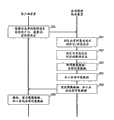
图5是表示共有对象的生成、管理的流程的一个例子的流程图。如该图所示,首先,在医用图像诊断装置2中,例如一边参照过去的共有对象一边执行扫描计划,并执行摄影(图像取得)(步骤S1、S2)。FIG. 5 is a flowchart showing an example of the flow of creation and management of shared objects. As shown in the figure, first, in the medical image
共有图像生成部件23根据患者信息、参考利用了的共有对象、该摄影所用的定位图像、摄影条件等,生成共有对象。这时,为了与通常的图像数据区别,或者为了容易进行事后的检索,例如可以在附带信息中嵌入表示是共有对象的信息。The shared
收发部件25通过网络向医用图像保存装置5发送通过摄影取得的图像数据、生成了的共有对象的数据(步骤S3)。The transmitting and receiving
接着,在分析部件57中分析由收发部件54接收到的共有对象(步骤S4),生成与该共有对象有关的所在确定信息、共有对象管理信息(步骤S5)。此外,将该共有对象的实体数据保存于数据保存部件51的预定的位置(步骤S6)。Next, the
例如在日后的摄影中作为参考数据而利用被保存的共有对象。即,如果从收发部件25向医用图像保存装置5发送共有对象数据的请求(步骤S7),则控制部件58例如基于患者信息等和共有对象管理信息,确定参照所用的共有对象。此外,控制部件58用所在信息表检索数据保存部件51,抽出该共有对象应该含有的各种信息(步骤S8)。将被抽出的各种信息作为参照用的共有对象发送给医用图像诊断装置2(步骤S9)。医用图像诊断装置2接收共有对象,一边参照该共有对象一边执行扫描计划,执行摄影(图像取得)(步骤S10、S11)。For example, the stored shared objects are used as reference data in future photography. That is, when a request for shared object data is sent from the transmitting and receiving
(图像诊断支持处理)(Image diagnosis support processing)
下面,说明本实施例的在医用图像保存装置5中执行的采用图像诊断支持功能的处理(图像诊断支持处理)。Next, the processing using the image diagnosis support function (image diagnosis support processing) executed in the medical
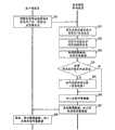
图6是表示实施例1的图像诊断支持处理的流程的流程图。如该图所示,首先,客户端装置2的控制部件21为了在现在的检查中参照,经由收发部件25向医用图像保存装置5发送在过去的检查中取得的图像信息等的取得请求、以及患者ID、用于确定应该参照的检查的ID、序列ID等。此外,客户端装置2的控制部件21经由收发部件25,与该取得请求等一起,向医用图像保存装置5发送操作者的用户ID、客户端装置2的装置ID(例如,站ID、AE(Application Entity,应用程序入口)等)、在客户端装置2中使用的应用程序名(步骤S20)。6 is a flowchart showing the flow of image diagnosis support processing in the first embodiment. As shown in the figure, first, the
然后,医用图像保存装置5的判定部件55通过参照利用管理表,基于从客户端装置2接收到的用户ID、装置ID、应用程序名的组合,判定共有对象的显示的有无、信息区分(步骤S21)。例如,在从客户端装置2接收到“Dr.TARO”(用户ID)、“读图终端1”(装置ID)、“共有对象viewer”(应用程序名)作为用户和使用场景的组合的情况下,判定部件55参照利用管理表,判定为“显示共有对象”,并且判定为信息区分是“全部”。另外,如果在从客户端装置2接收到“Dr.TARO”(用户ID)、“读图终端1”(装置ID)、“Dicom viewer”(应用程序名)作为用户和使用场景的组合的情况下,判定部件55参照利用管理表,判定为“不显示共有对象”,并且判定为信息区分是“无”。Then, the judging
接着,判定部件55基于在步骤S21中判定出的信息区分和提供信息管理表,判定与该信息区分对应的提供信息(共有对象应该含有的信息)(步骤S22)。例如,在步骤S21中判定为信息区分是“全部”的情况下,判定部件55根据图3所示的提供信息管理表,判定为“定位图像”、“摄影范围”、“摄影条件”、“图像生成条件”、“检查历史”、“与关键图像有关的信息”等全部信息是提供信息。此外,例如在步骤S21中判定为信息区分是“摄影条件1”的情况下,判定部件55根据提供信息管理表,判定为“定位图像”、“摄影范围”、“摄影条件”、“检查历史”的各信息是提供信息。Next, the judging
接着,信息收集部件57通过参照数据库50,确定与在步骤S21中接受的患者ID、检查ID、序列ID等相关联的图像数据、共有对象数据的所在,从数据保存部件51或者根据需要经由网络从其它的装置收集图像数据、共有对象数据的实体数据(步骤S23)。Next, the
接着,共有对象加工部件57加工共有对象数据使得仅含有根据信息区分判定出的提供信息(步骤S24)。例如,在步骤S22中,将“定位图像”、“摄影范围”、“摄影条件”、“检查历史”判定为共有对象应该含有的提供信息的情况下,共有对象加工部件57通过将与这4个项目对应的信息以外的信息删除或处理为不可读取的形式,从而加工共有对象使得仅含有与信息区分对应的提供信息。另外,在将包含在共有对象中的信息中的与信息区分对应的提供信息以外的信息设为不可读取的形式的情况下,从网络的通信效率的观点出发,理想的是进行处理使得数据尺寸尽量地小。此外,在本实施例中,作为与DICOM规格一致的附带信息而保存“定位图像”、“摄影范围”、“摄影条件”、“检查历史”等各信息。在该DICOM规格中,可以利用规定的标识符区别各信息并取得。Next, the sharing object processing means 57 processes the sharing object data so as to include only the provided information determined by information classification (step S24). For example, in step S22, when "locator image", "shooting range", "shooting condition", and "examination history" are determined as the provided information that should be included in the sharing object, the sharing
接着,收发部件54经由网络向客户端装置3发送收集到的图像数据、以及加工后的共有对象数据(步骤S25)。在客户端装置3侧,基于接受的图像数据以预定的方式显示需要的图像,并且基于加工后的共有对象数据,以预定的方式显示依据信息区分的提供信息(步骤S26)。Next, the transmitting and receiving
另外,在步骤S21中判定为“无信息区分”的情况下,在各个步骤S22~S26中,分别省略与共有对象有关的处理。In addition, when it is determined in step S21 that "there is no information classification", in each of steps S22 to S26, the processing related to the shared object is omitted.
(效果)(Effect)
根据以上所述的结构,可以得到以下的效果。According to the configuration described above, the following effects can be obtained.
根据本图像诊断支持系统1,根据用户和利用场景的组合,判定共有对象的提供的有无、以及提供共有对象时的信息区分,基于判定出的信息区分判定共有对象应该含有的提供信息。进而,加工共有对象使得仅含有判定出的该提供信息,例如经由网络将其提供给客户端装置。因此,在服务器装置侧,在客户端装置侧不需要共有对象的情况下,不发送共有对象,并且在客户端装置需要共有对象的情况下,提供仅含有必要最小限度信息的共有对象。因此,用户不需要进行没用信息的删除等无用的作业,可以排除降低读图时的作业效率的主要原因。此外,由于不进行无用的数据的收发和重放处理,可以排除网络的通信效率、各装置的动作效率降低的主要原因。According to the image
(实施例2)(Example 2)
接着,说明本发明的实施例2。Next,
例如在想要在现在的检查中利用而取得了共有对象时,有以下的情况:不存储与摄影条件或参考图像等有关的实体数据(或用于确定实体数据的信息),而替代地存储用于确定应该参考的其它共有对象(关联共有对象)的信息(例如,其UID、表示所在的信息、链接信息等。以下称为“关联共有对象确定信息”)。For example, when a shared object is acquired in order to be used in the current inspection, there may be cases where entity data (or information for specifying entity data) related to imaging conditions or reference images are not stored, but stored instead. Information for specifying other shared objects (associated shared objects) that should be referred to (for example, its UID, information indicating its location, link information, etc., hereinafter referred to as "associated shared object specifying information").
因此,在本实施例中,说明在该情况下,进而访问关联共有对象,而取得与信息区分对应的提供信息的医用图像诊断支持系统1。Therefore, in this embodiment, in this case, the medical image
图7表示实施例2的利用管理表的一个例子。在与图2所示的利用管理表进行比较时,在对用户ID和利用场景的每个组合定义了“世代”这一点上不同。在此,“世代”是指在取得的共有对象内存储有关联共有对象确定信息的情况下,将该取得的共有对象作为第一世代,定义从此开始追溯访问几个关联共有对象而进行需要的信息的检索的指标。FIG. 7 shows an example of the use management table of the second embodiment. When compared with the use management table shown in FIG. 2 , it differs in that a "generation" is defined for each combination of a user ID and a use scene. Here, "generation" refers to the case where related shared object identification information is stored in the obtained shared object, and the obtained shared object is defined as the first generation, and the number of related shared objects that need to be retrospectively accessed from then on is defined. Index of information retrieval.
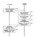
图8是表示实施例2的图像诊断支持处理的流程的流程图。如该图所示,首先,客户端装置2的控制部件21通过收发部件25,向医用图像保存装置5发送在过去的检查中取得的图像信息等的取得请求、患者ID、检查ID、序列ID、操作者的用户ID、装置ID、应用程序名(步骤S30)。8 is a flowchart showing the flow of image diagnosis support processing in the second embodiment. As shown in the figure, first, the
医用图像保存装置5的判定部件55通过参照利用管理表,判定与从客户端装置2接收到的用户ID、装置ID、应用程序的组合有关的共有对象的显示的有无、信息区分及世代(步骤S31)。例如,在作为用户与使用场景的组合,从客户端装置2接收到“Dr.TARO”(用户ID)、“读图终端1”(装置ID)、“共有对象viewer”(应用程序名)的情况下,判定部件55参照利用管理表,判定为“显示共有对象”,并且判定为信息区分是“全部”。并且,判定部件55参照利用管理表,判定与该组合有关的世代是“第二世代”。The judging
接着,判定部件55基于判定出的信息区分和提供信息管理表,判定与该信息区分对应的提供信息(步骤S32)。信息收集部件57通过参照数据库50,确定与在步骤S21中接受的患者ID、检查ID、序列ID等相关联的图像数据、共有对象数据的所在,从数据保存部件51或者根据需要经由网络从其它的装置,收集图像数据、共有对象数据的实体数据(步骤S33)。Next, the
接着,判定部件55判定收集到的共有对象中是否含有关联共有对象确定信息(步骤S34)。在其结果是判断为不包含关联共有对象确定信息时,转移到步骤S36。Next, the judging
另一方面,当判断为含有关联共有对象确定信息时,信息收集部件57访问由关联共有对象确定信息确定的关联共有对象,取得其含有的信息(步骤S35)。On the other hand, when it is judged that the associated shared object identification information is included, the
另外,在步骤S35中访问的关联共有对象中进而含有用于确定应该参照的共有对象的信息的情况下,在步骤S31中判定为第三世代时,进而访问关联共有对象。In addition, when the related shared objects accessed in step S35 further include information for specifying the shared objects to be referred to, when it is determined in step S31 that it is the third generation, further related shared objects are accessed.
接着,由共有对象加工部件57加工共有对象数据使得仅含有根据信息区分判定出的提供信息(步骤S36)。收发部件54通过网络向客户端装置3发送收集到的图像数据、以及加工后的共有对象数据(步骤S37)。在客户端装置3侧,基于接受的图像数据以预定的方式显示需要的图像,并且基于加工后的共有对象数据,以预定的方式表示依据信息区分的提供信息(步骤S38)。Next, the shared object data is processed by the shared object processing means 57 so as to contain only the provided information determined by information classification (step S36). The transmitting and receiving
另外,在步骤S31中判定为“无信息区分”的情况下,在各个步骤S32~S38中,分别省略与共有对象有关的处理。In addition, when it is determined in step S31 that "there is no information classification", in each of steps S32 to S38, the processing related to the shared object is omitted.
根据以上描述的结构,在所利用的共有对象中不存储与摄影条件和参考图像等有关的实体数据,而替代地存储关联共有对象确定信息时,也可以通过追溯到可取得需要的信息的其它共有对象,来取得需要的信息,可以实现与实施例1同样的效果。According to the structure described above, when entity data related to imaging conditions, reference images, etc. are not stored in the shared objects used, but related shared object specifying information is stored instead, it is also possible to obtain the required information by going back to other The same effect as that of
(实施例3)(Example 3)
接着,说明实施例3。Next, Example 3 will be described.
在实施例1和2中,在服务器装置侧加工共有对象使得仅含有与信息区分对应的提供信息。与此相对,在本实施例中,对客户端装置加以显示限制,使得仅显示共有对象所含有的信息中的与信息区分对应的提供信息。In
图9是表示实施例3的医用图像诊断支持系统1的结构的图。与图2的结构比较时,在客户端装置2进而具备判定部件35这一点上不同。FIG. 9 is a diagram showing the configuration of a medical image
判定部件35判定从医用图像保存装置5接受的共有对象所含有的信息中的与从医用图像保存装置5接受的信息区分对应的提供信息。The judging unit 35 judges the provision information corresponding to the information received from the medical
控制部件31在显示共有对象的情况下,控制显示部件使得仅显示判断部件35中的与信息区分对应的提供信息。When displaying shared objects, the
图10是表示实施例3的图像诊断支持处理的流程的流程图。如该图所示,首先,客户端装置2的控制部件21通过收发部件25,向医用图像保存装置5发送在过去的检查中取得的图像信息等的取得请求、患者ID、检查ID、序列ID、操作者的用户ID、装置ID、应用程序名(步骤S40)。10 is a flowchart showing the flow of image diagnosis support processing in the third embodiment. As shown in the figure, first, the
医用图像保存装置5的判定部件55通过参照利用管理表,判定与从客户端装置2接收到的用户ID、装置ID、应用程序名的组合有关的共有对象的显示的有无、信息区分(步骤S41)。The judging
此外,判定部件55在判定为显示共有对象的情况下,基于与该组合有关的信息区分和提供信息管理表,判定与该信息区分对应的提供信息,生成表示该提供信息是什么样的信息的提供信息列表(步骤S42)。例如,在信息区分是“摄影条件1”时,判定部件55判定为“定位图像”、“摄影范围”、“摄影条件”、“检查历史”是提供信息,生成表示这4者是提供信息的提供信息列表。Furthermore, when it is determined that the shared object is displayed, the
接着,信息收集部件57通过参照数据库50,确定与步骤S21接受的患者ID、检查ID、序列ID等相关联的图像数据、共有对象数据的所在,从数据保存部件51或者根据需要经由网络从其它的装置,收集图像数据、共有对象数据的实体数据(步骤S43)。Next, the
接着,通过网络向客户端装置3发送收集到的图像数据、共有对象数据等、以及提供信息列表(步骤S44)。Next, the collected image data, shared object data, etc., and the provision information list are transmitted to the
接着,客户端装置3的判定部件35基于接受的提供信息列表,判定共有对象数据所含有的信息中的与提供信息对应的信息(步骤S45)。此外,客户端装置3的控制部件31加以显示限制地在显示部件中显示共有对象,使得仅显示共有对象所含有的信息中的与信息区分对应的提供信息。Next, the judging unit 35 of the
另外,在步骤S41中判定为“无信息区分”的情况下,在各个步骤S32~S46中,分别省略与共有对象有关的处理。In addition, when it is determined in step S41 that "there is no information classification", in each of steps S32 to S46, the processing related to the shared object is omitted.
根据以上描述地结构,可以实现与实施例1或2同样的效果。According to the structure described above, the same effect as that of
(实施例4)(Example 4)
接着,说明实施例4。本实施例将所处理的医疗信息作为共有对象及医用报告,进行实施例1~3的任意一个的图像诊断支持处理。Next, Example 4 will be described. In this embodiment, the image diagnosis support process in any one of
图11是表示实施例4的利用管理表的一个例子的图。与图2所示的例子比较时,在对用户ID和利用场景的每个组合定义了医用报告的提供有无及仅关键图像的提供有无这一点上不同。FIG. 11 is a diagram showing an example of a usage management table according to the fourth embodiment. Compared with the example shown in FIG. 2 , it is different in that the availability of medical reports and the availability of only key images are defined for each combination of user ID and usage scenario.
医用图像保存装置5的判定部件55例如在步骤S21、步骤S31、步骤S41的任意一个中,通过参照图11所示的利用管理表,在已述的判定的基础上,判定与现在的用户和使用场景的组合有关的医用报告的提供有无。例如,在作为用户和使用场景的组合,从客户端装置2接收到“Dr.TARO”(用户ID)、“读图终端1”(装置ID)、“Dicomviewer”(应用程序名)时,判定部件55判定为在该读图终端1中显示用带注释图像和DICOM规格中的Key Image Note确定的关键图像,并且判定为合并医用报告而显示。The judging
此外,虽未图示,但根据需要,通过另行对每个各信息区分,预先生成定义包含在医用报告中的内容(提供信息)的提供信息管理表并存储于数据库50,从而利用与共有对象时同样的手法,对于医用报告也可以判定根据信息区分提供的医用报告应该含有的信息。其结果是在向客户端装置侧提供的医用信息例如包含医用报告的情况下,也可以实现与实施例1~3同样的效果。In addition, although not shown, if necessary, by separately distinguishing each piece of information, a provided information management table defining the contents (provided information) included in the medical report is generated in advance and stored in the
另外,本发明并不限于上述实施例的原样,在实施阶段可以在不脱离其宗旨的范围内对结构要素进行变形、具体化实现。作为具体的变形例,例如有如下的形式。In addition, the present invention is not limited to the above-mentioned embodiments as they are, and the structural elements can be modified and realized in practice within the range not departing from the gist thereof. As a specific modified example, there are, for example, the following forms.
(1)通过将执行相应处理的程序安装在工作站等计算机上,并将其展开到存储器上,也可以实现本实施例的各功能。此时,可以将能够使计算机实现该方法的程序存储在磁盘(软盘(注册商标)、硬盘等)、光盘(CD-ROM、DVD等)、半导体存储器等记录介质中来分发。(1) The functions of this embodiment can also be realized by installing a program for executing the corresponding processing on a computer such as a workstation, and expanding it on a memory. In this case, a program that enables a computer to realize the method can be stored in a recording medium such as a magnetic disk (floppy disk (registered trademark), hard disk, etc.), optical disk (CD-ROM, DVD, etc.), semiconductor memory, and the like, and distributed.
(2)在上述各实施例中,使客户端装置2和医用图像保存装置5(服务器装置)分体。但是,并不限于此,也可以是在一个装置中实现各装置的功能的结构。此外,例如可以将医用图像保存装置5的一部分功能设置在客户端装置2侧,或者将客户端装置2的一部分功能设置在医用图像保存装置5侧,作为实现图像诊断支持功能的服务器而构成。(2) In each of the above-described embodiments, the
另外,通过上述实施例所公开的多个结构要素的适当的组合,可以形成各种发明。例如,可以从实施例所示的全部结构要素中删除几个结构要素。进而,也可以将不同实施例中的结构要素适当地组合。In addition, various inventions can be formed by appropriately combining a plurality of constituent elements disclosed in the above embodiments. For example, some structural elements may be deleted from all the structural elements shown in the embodiments. Furthermore, constituent elements in different embodiments may be appropriately combined.
Claims (22)
Translated fromChineseApplications Claiming Priority (2)
| Application Number | Priority Date | Filing Date | Title |
|---|---|---|---|
| JP2007-105330 | 2007-04-12 | ||
| JP2007105330AJP5053690B2 (en) | 2007-04-12 | 2007-04-12 | Image diagnosis support system and image diagnosis support program |
Publications (2)
| Publication Number | Publication Date |
|---|---|
| CN101283930Atrue CN101283930A (en) | 2008-10-15 |
| CN101283930B CN101283930B (en) | 2011-01-26 |
Family
ID=39853753
Family Applications (1)
| Application Number | Title | Priority Date | Filing Date |
|---|---|---|---|
| CN2008100916440AExpired - Fee RelatedCN101283930B (en) | 2007-04-12 | 2008-04-11 | Image diagnosis support system and image diagnosis support method |
Country Status (3)
| Country | Link |
|---|---|
| US (1) | US8238628B2 (en) |
| JP (1) | JP5053690B2 (en) |
| CN (1) | CN101283930B (en) |
Cited By (2)
| Publication number | Priority date | Publication date | Assignee | Title |
|---|---|---|---|---|
| CN103366081A (en)* | 2012-03-26 | 2013-10-23 | 株式会社东芝 | Medical information management device |
| CN104114083A (en)* | 2012-02-14 | 2014-10-22 | 佳能株式会社 | Diagnosis support device and control method thereof |
Families Citing this family (16)
| Publication number | Priority date | Publication date | Assignee | Title |
|---|---|---|---|---|
| JP4905967B2 (en)* | 2007-03-02 | 2012-03-28 | 富士フイルム株式会社 | Similar case retrieval apparatus, method, and program |
| JP5329911B2 (en)* | 2007-11-02 | 2013-10-30 | 株式会社東芝 | Medical image management apparatus and medical image system |
| JP5200631B2 (en)* | 2008-04-02 | 2013-06-05 | コニカミノルタエムジー株式会社 | Medical image system, imaging apparatus, and program |
| JP5226370B2 (en)* | 2008-04-18 | 2013-07-03 | 株式会社東芝 | Interpretation report creation support system |
| JP5330009B2 (en)* | 2009-02-06 | 2013-10-30 | 株式会社東芝 | Medical communication management apparatus, medical image diagnostic apparatus, and medical communication management system |
| JP5232710B2 (en)* | 2009-04-17 | 2013-07-10 | 株式会社日立ソリューションズ | Document protection system log display method |
| JP5633358B2 (en)* | 2010-12-20 | 2014-12-03 | コニカミノルタ株式会社 | Image inspection apparatus and image detection system |
| JP6099865B2 (en)* | 2011-01-11 | 2017-03-22 | 東芝メディカルシステムズ株式会社 | Diagnostic imaging equipment |
| US9323250B2 (en) | 2011-01-28 | 2016-04-26 | Intouch Technologies, Inc. | Time-dependent navigation of telepresence robots |
| US9098611B2 (en)* | 2012-11-26 | 2015-08-04 | Intouch Technologies, Inc. | Enhanced video interaction for a user interface of a telepresence network |
| US9361021B2 (en) | 2012-05-22 | 2016-06-07 | Irobot Corporation | Graphical user interfaces including touchpad driving interfaces for telemedicine devices |
| WO2013176760A1 (en) | 2012-05-22 | 2013-11-28 | Intouch Technologies, Inc. | Graphical user interfaces including touchpad driving interfaces for telemedicine devices |
| JP5841107B2 (en)* | 2013-09-10 | 2016-01-13 | 日立アロカメディカル株式会社 | Ultrasonic diagnostic equipment |
| DE102013221603A1 (en)* | 2013-10-24 | 2015-04-30 | Siemens Aktiengesellschaft | Computed Unit CT System and Method for Reconstructing and Diagnosing CT Imaging |
| JP2015192788A (en)* | 2014-03-31 | 2015-11-05 | 富士フイルム株式会社 | Inspection report preparation support system, medical image diagnostic device, inspection report preparation support method, and program |
| CN110517767B (en)* | 2019-08-27 | 2022-12-13 | 北京百度网讯科技有限公司 | Auxiliary diagnosis method, auxiliary diagnosis device, electronic equipment and storage medium |
Family Cites Families (19)
| Publication number | Priority date | Publication date | Assignee | Title |
|---|---|---|---|---|
| US6021404A (en)* | 1997-08-18 | 2000-02-01 | Moukheibir; Nabil W. | Universal computer assisted diagnosis |
| US6520912B1 (en)* | 2000-11-09 | 2003-02-18 | Acuson Corporation | Method and system for displaying medical data such as a diagnostic medical ultrasound image at an automatically-selected display resolution |
| US20080146943A1 (en)* | 2006-12-14 | 2008-06-19 | Ep Medsystems, Inc. | Integrated Beam Former And Isolation For An Ultrasound Probe |
| JP2003337861A (en)* | 2002-05-21 | 2003-11-28 | Srl Inc | Medical information provision system |
| KR20020061572A (en)* | 2002-06-25 | 2002-07-24 | 주식회사 베베콤 | Method for providing of integrated medical treatment service and apparatus thereof |
| US20040086163A1 (en)* | 2002-10-31 | 2004-05-06 | Konica Minolta Holdings, Inc. | Medical image radiographing system, medical image management method and portable terminal |
| JP2004254952A (en)* | 2003-02-26 | 2004-09-16 | Toshiba Corp | Information collection and provision system and information collection and provision method |
| JP4474846B2 (en) | 2003-05-21 | 2010-06-09 | コニカミノルタエムジー株式会社 | Medical information management system and medical information management method |
| JP2005063080A (en)* | 2003-08-11 | 2005-03-10 | Hitachi Medical Corp | Medical image processor |
| US7483557B2 (en)* | 2003-09-30 | 2009-01-27 | Kabushiki Kaisha Toshiba | Medical imaging communication system, method and software |
| US7189000B2 (en)* | 2003-12-22 | 2007-03-13 | Kabushiki Kaisha Toshiba | Image-quality control system |
| JP2006018817A (en)* | 2004-06-01 | 2006-01-19 | Toshiba Corp | Medical image storage device, medical image device, medical image system, and medical image information storage method |
| DE102004059182A1 (en)* | 2004-12-08 | 2006-06-14 | Siemens Ag | Operating method for a computer and corresponding devices |
| JP5283839B2 (en) | 2005-11-25 | 2013-09-04 | 東芝メディカルシステムズ株式会社 | Medical diagnostic imaging system |
| JP5525675B2 (en)* | 2006-03-07 | 2014-06-18 | 東芝メディカルシステムズ株式会社 | Medical image diagnosis support apparatus, medical image diagnosis apparatus, or medical image diagnosis support system |
| JP2006319356A (en) | 2006-07-07 | 2006-11-24 | Fujitsu Ltd | Printed wiring board |
| US7698246B2 (en)* | 2006-09-07 | 2010-04-13 | International Business Machines Corporation | System and method for optimal and adaptive process unification of decision support functions associated with managing a chaotic event |
| JP4218772B2 (en)* | 2007-02-16 | 2009-02-04 | 国立大学法人名古屋大学 | Diagnostic imaging support device |
| JP4257441B2 (en)* | 2007-02-16 | 2009-04-22 | 国立大学法人名古屋大学 | Medical information storage device and medical image diagnostic device |
- 2007
- 2007-04-12JPJP2007105330Apatent/JP5053690B2/ennot_activeExpired - Fee Related
- 2008
- 2008-04-10USUS12/100,780patent/US8238628B2/ennot_activeExpired - Fee Related
- 2008-04-11CNCN2008100916440Apatent/CN101283930B/ennot_activeExpired - Fee Related
Cited By (5)
| Publication number | Priority date | Publication date | Assignee | Title |
|---|---|---|---|---|
| CN104114083A (en)* | 2012-02-14 | 2014-10-22 | 佳能株式会社 | Diagnosis support device and control method thereof |
| US9734300B2 (en) | 2012-02-14 | 2017-08-15 | Canon Kabushiki Kaisha | Diagnosis support apparatus and method of controlling the same |
| CN108053884A (en)* | 2012-02-14 | 2018-05-18 | 佳能株式会社 | Diagnosis support device |
| CN103366081A (en)* | 2012-03-26 | 2013-10-23 | 株式会社东芝 | Medical information management device |
| CN103366081B (en)* | 2012-03-26 | 2016-09-07 | 东芝医疗系统株式会社 | Medical information management device |
Also Published As
| Publication number | Publication date |
|---|---|
| JP2008259707A (en) | 2008-10-30 |
| US20080253629A1 (en) | 2008-10-16 |
| JP5053690B2 (en) | 2012-10-17 |
| CN101283930B (en) | 2011-01-26 |
| US8238628B2 (en) | 2012-08-07 |
Similar Documents
| Publication | Publication Date | Title |
|---|---|---|
| CN101283930A (en) | Image diagnosis support system and image diagnosis support method | |
| CN103279637B (en) | Medical imaging diagnosis supporting apparatus and image diagnosis supporting method | |
| US8401259B2 (en) | Image diagnosis support system | |
| CN101123911B (en) | Medical Imaging Diagnostic Devices | |
| US20090182577A1 (en) | Automated information management process | |
| US7418120B2 (en) | Method and system for structuring dynamic data | |
| JP2009230304A (en) | Medical report creation support system, program, and method | |
| JP4257441B2 (en) | Medical information storage device and medical image diagnostic device | |
| US10037405B2 (en) | Medical image generation apparatus, medical image storage apparatus, medical image display apparatus, and medical image display system | |
| JP6021468B2 (en) | Medical image display device | |
| JP5631914B2 (en) | Database search apparatus, method, and program | |
| JP2021100515A (en) | Medical device and program | |
| JP5363962B2 (en) | Diagnosis support system, diagnosis support program, and diagnosis support method | |
| US8594406B2 (en) | Single scan multi-procedure imaging | |
| JP5305700B2 (en) | Image diagnosis support system and image diagnosis support method | |
| JP2009069977A (en) | MEDICAL REPORT CREATION SYSTEM, MEDICAL REPORT CREATION DEVICE, AND MEDICAL REPORT CREATION METHOD | |
| JP2020154630A (en) | Medical information collection device | |
| JP2007072649A (en) | Interpretation report creation device | |
| JP2010152623A (en) | Medical image management system | |
| JP2010152624A (en) | Medical image management system | |
| JP2007007190A (en) | Diagnostic system, management server, and image data management method | |
| US12076176B2 (en) | Dynamic image processing apparatus and storage medium | |
| KR20130088730A (en) | Apparatus for sharing and managing information in picture archiving communication system and method thereof | |
| JP2006309550A (en) | Double reading support system | |
| JP2006048665A (en) | Image management apparatus and image management method |
Legal Events
| Date | Code | Title | Description |
|---|---|---|---|
| C06 | Publication | ||
| PB01 | Publication | ||
| C10 | Entry into substantive examination | ||
| SE01 | Entry into force of request for substantive examination | ||
| C14 | Grant of patent or utility model | ||
| GR01 | Patent grant | ||
| C41 | Transfer of patent application or patent right or utility model | ||
| TR01 | Transfer of patent right | Effective date of registration:20160726 Address after:Japan Tochigi Patentee after:TOSHIBA MEDICAL SYSTEMS Corp. Address before:Tokyo, Japan Patentee before:Toshiba Corp. Patentee before:TOSHIBA MEDICAL SYSTEMS Corp. | |
| CF01 | Termination of patent right due to non-payment of annual fee | Granted publication date:20110126 | |
| CF01 | Termination of patent right due to non-payment of annual fee |