CN101278863A - Ultrasonic diagnostic device, breast imaging system, and breast imaging method - Google Patents
Ultrasonic diagnostic device, breast imaging system, and breast imaging methodDownload PDFInfo
- Publication number
- CN101278863A CN101278863ACNA2008100918111ACN200810091811ACN101278863ACN 101278863 ACN101278863 ACN 101278863ACN A2008100918111 ACNA2008100918111 ACN A2008100918111ACN 200810091811 ACN200810091811 ACN 200810091811ACN 101278863 ACN101278863 ACN 101278863A
- Authority
- CN
- China
- Prior art keywords
- mentioned
- breast
- unit
- oppressed
- image
- Prior art date
- Legal status (The legal status is an assumption and is not a legal conclusion. Google has not performed a legal analysis and makes no representation as to the accuracy of the status listed.)
- Granted
Links
Images


Classifications
- A—HUMAN NECESSITIES
- A61—MEDICAL OR VETERINARY SCIENCE; HYGIENE
- A61B—DIAGNOSIS; SURGERY; IDENTIFICATION
- A61B8/00—Diagnosis using ultrasonic, sonic or infrasonic waves
- A61B8/08—Clinical applications
- A61B8/0825—Clinical applications for diagnosis of the breast, e.g. mammography
- A—HUMAN NECESSITIES
- A61—MEDICAL OR VETERINARY SCIENCE; HYGIENE
- A61B—DIAGNOSIS; SURGERY; IDENTIFICATION
- A61B6/00—Apparatus or devices for radiation diagnosis; Apparatus or devices for radiation diagnosis combined with radiation therapy equipment
- A61B6/04—Positioning of patients; Tiltable beds or the like
- A61B6/0407—Supports, e.g. tables or beds, for the body or parts of the body
- A61B6/0414—Supports, e.g. tables or beds, for the body or parts of the body with compression means
- A—HUMAN NECESSITIES
- A61—MEDICAL OR VETERINARY SCIENCE; HYGIENE
- A61B—DIAGNOSIS; SURGERY; IDENTIFICATION
- A61B6/00—Apparatus or devices for radiation diagnosis; Apparatus or devices for radiation diagnosis combined with radiation therapy equipment
- A61B6/44—Constructional features of apparatus for radiation diagnosis
- A61B6/4417—Constructional features of apparatus for radiation diagnosis related to combined acquisition of different diagnostic modalities
- A—HUMAN NECESSITIES
- A61—MEDICAL OR VETERINARY SCIENCE; HYGIENE
- A61B—DIAGNOSIS; SURGERY; IDENTIFICATION
- A61B6/00—Apparatus or devices for radiation diagnosis; Apparatus or devices for radiation diagnosis combined with radiation therapy equipment
- A61B6/50—Apparatus or devices for radiation diagnosis; Apparatus or devices for radiation diagnosis combined with radiation therapy equipment specially adapted for specific body parts; specially adapted for specific clinical applications
- A61B6/502—Apparatus or devices for radiation diagnosis; Apparatus or devices for radiation diagnosis combined with radiation therapy equipment specially adapted for specific body parts; specially adapted for specific clinical applications for diagnosis of breast, i.e. mammography
- A—HUMAN NECESSITIES
- A61—MEDICAL OR VETERINARY SCIENCE; HYGIENE
- A61B—DIAGNOSIS; SURGERY; IDENTIFICATION
- A61B6/00—Apparatus or devices for radiation diagnosis; Apparatus or devices for radiation diagnosis combined with radiation therapy equipment
- A61B6/52—Devices using data or image processing specially adapted for radiation diagnosis
- A61B6/5211—Devices using data or image processing specially adapted for radiation diagnosis involving processing of medical diagnostic data
- A61B6/5229—Devices using data or image processing specially adapted for radiation diagnosis involving processing of medical diagnostic data combining image data of a patient, e.g. combining a functional image with an anatomical image
- A61B6/5247—Devices using data or image processing specially adapted for radiation diagnosis involving processing of medical diagnostic data combining image data of a patient, e.g. combining a functional image with an anatomical image combining images from an ionising-radiation diagnostic technique and a non-ionising radiation diagnostic technique, e.g. X-ray and ultrasound
- A—HUMAN NECESSITIES
- A61—MEDICAL OR VETERINARY SCIENCE; HYGIENE
- A61B—DIAGNOSIS; SURGERY; IDENTIFICATION
- A61B8/00—Diagnosis using ultrasonic, sonic or infrasonic waves
- A61B8/40—Positioning of patients, e.g. means for holding or immobilising parts of the patient's body
- A61B8/403—Positioning of patients, e.g. means for holding or immobilising parts of the patient's body using compression means
- A—HUMAN NECESSITIES
- A61—MEDICAL OR VETERINARY SCIENCE; HYGIENE
- A61B—DIAGNOSIS; SURGERY; IDENTIFICATION
- A61B8/00—Diagnosis using ultrasonic, sonic or infrasonic waves
- A61B8/44—Constructional features of the ultrasonic, sonic or infrasonic diagnostic device
- A61B8/4405—Device being mounted on a trolley
- A—HUMAN NECESSITIES
- A61—MEDICAL OR VETERINARY SCIENCE; HYGIENE
- A61B—DIAGNOSIS; SURGERY; IDENTIFICATION
- A61B8/00—Diagnosis using ultrasonic, sonic or infrasonic waves
- A61B8/44—Constructional features of the ultrasonic, sonic or infrasonic diagnostic device
- A61B8/4416—Constructional features of the ultrasonic, sonic or infrasonic diagnostic device related to combined acquisition of different diagnostic modalities, e.g. combination of ultrasound and X-ray acquisitions
- A—HUMAN NECESSITIES
- A61—MEDICAL OR VETERINARY SCIENCE; HYGIENE
- A61B—DIAGNOSIS; SURGERY; IDENTIFICATION
- A61B8/00—Diagnosis using ultrasonic, sonic or infrasonic waves
- A61B8/48—Diagnostic techniques
- A61B8/483—Diagnostic techniques involving the acquisition of a 3D volume of data
- A—HUMAN NECESSITIES
- A61—MEDICAL OR VETERINARY SCIENCE; HYGIENE
- A61B—DIAGNOSIS; SURGERY; IDENTIFICATION
- A61B8/00—Diagnosis using ultrasonic, sonic or infrasonic waves
- A61B8/52—Devices using data or image processing specially adapted for diagnosis using ultrasonic, sonic or infrasonic waves
- A61B8/5207—Devices using data or image processing specially adapted for diagnosis using ultrasonic, sonic or infrasonic waves involving processing of raw data to produce diagnostic data, e.g. for generating an image
Landscapes
- Health & Medical Sciences (AREA)
- Life Sciences & Earth Sciences (AREA)
- Engineering & Computer Science (AREA)
- Medical Informatics (AREA)
- Radiology & Medical Imaging (AREA)
- Biomedical Technology (AREA)
- Biophysics (AREA)
- Veterinary Medicine (AREA)
- Public Health (AREA)
- Nuclear Medicine, Radiotherapy & Molecular Imaging (AREA)
- General Health & Medical Sciences (AREA)
- Pathology (AREA)
- Animal Behavior & Ethology (AREA)
- Physics & Mathematics (AREA)
- Heart & Thoracic Surgery (AREA)
- Molecular Biology (AREA)
- Surgery (AREA)
- Optics & Photonics (AREA)
- High Energy & Nuclear Physics (AREA)
- Computer Vision & Pattern Recognition (AREA)
- Dentistry (AREA)
- Oral & Maxillofacial Surgery (AREA)
- Ultra Sonic Daignosis Equipment (AREA)
- Apparatus For Radiation Diagnosis (AREA)
Abstract
Translated fromChineseDescription
Translated fromChinese技术领域technical field
本发明涉及在X射线成像和超声波成像的融合型的乳房成像中所使用的系统以及在该融合型成像中使用的超声波诊断装置、乳房成像系统以及乳房成像方法。The present invention relates to a system used in fusion-type breast imaging of X-ray imaging and ultrasound imaging, an ultrasound diagnostic apparatus, a breast imaging system, and a breast imaging method used in the fusion imaging.
背景技术Background technique
超声波诊断用只让超声波探头从体表接触的简单的操作就能够以实时显示得到心脏的脉动和胎儿的活动的样子,并且因为安全性高所以能够反复进行检查。此外,系统的规模小于X射线、CT、MRI等其他的诊断设备,可以说是向床边一侧移动的检查也容易进行等简便的诊断方法。在该超声波诊断中使用的超声波诊断装置因其具备的功能的种类不同而各自不同,还开发了小型的可单手提起的装置,超声波诊断不会象X射线等那样受到辐射的影响,还能够在妇产科和家庭医疗等中使用。Ultrasonic diagnosis can obtain the pulse of the heart and the movement of the fetus in real-time display by a simple operation that only touches the ultrasonic probe from the body surface, and it is safe to perform repeated inspections. In addition, the scale of the system is smaller than that of other diagnostic equipment such as X-ray, CT, and MRI, and it can be said that it is a simple diagnostic method such as examinations that can be moved to the side of the bed can be easily performed. Ultrasonic diagnostic devices used in this ultrasonic diagnosis are different because of the types of functions they have. Small devices that can be picked up with one hand have also been developed. Ultrasonic diagnostics will not be affected by radiation like X-rays, and can also Used in obstetrics and gynecology and family medicine, etc.
本超声波诊断装置和使用乳房拍摄用X射线诊断装置的摄影法(乳房X线照相术:MMG)一样还多用于乳腺癌的检查。一般来说,使用了MMG的检查(MMG检查)在微细石灰化的检查能力方面优异,另一方面,在生物体的组织构造和肿瘤的描画中超声波检查一方优异。MMG检查和超声波检查分别具有长处与短处,对于乳腺癌检查起到互补的作用。因而,如果能够正确地取得用MMG得到的图像(X摄像乳房图像)和超声波图像的位置关系的对应,则能够期待诊断精度的提高。This ultrasonic diagnostic apparatus is also frequently used for breast cancer examinations, as is the imaging method (mammography: MMG) using an X-ray diagnostic apparatus for breast imaging. In general, inspection using MMG (MMG inspection) is excellent in the ability to inspect fine calcifications, and on the other hand, ultrasonic inspection is excellent in drawing the tissue structure of a living body and tumor. MMG examination and ultrasonography have advantages and disadvantages respectively, and they play a complementary role in the examination of breast cancer. Therefore, if the correspondence between the positional relationship between the image obtained by MMG (X-radiography breast image) and the ultrasonic image can be accurately acquired, it is expected that the diagnostic accuracy will be improved.
但是,在以往的X射线成像和超声波成像的融合型的乳房成像中存在以下那样的问题。However, conventional X-ray imaging and ultrasonic imaging fusion type breast imaging has the following problems.
即,一般的MMG检查需要压迫乳房,超声波检查在非压迫状态下对乳房进行超声波扫描。因此,在图像取得时的乳房的状态不同,X射线乳房图像和超声波图像的正确的位置关系的对应困难。That is, the general MMG examination needs to compress the breast, and the ultrasonography scans the breast in a non-compressed state. Therefore, the state of the breast at the time of image acquisition is different, and it is difficult to associate an accurate positional relationship between an X-ray breast image and an ultrasonic image.
此外,为了实现X射线乳房图像和超声波图像的位置关系的对应关系,提出了从乳房拍摄用X射线诊断装置的压迫板的上进行超声波扫描的方法,和从压迫板之间进行超声波检查的方法等。但是,在该方法中,为了取得与乳房全体有关的超声波图像,用形成有用于插入超声波探头的多个孔的压迫板压迫乳房,必须一边挪动超声波探头一边在各孔的位置上取得超声波图像。因此,除了在超声波图像的取得中需要许多时间外,经常因为用许多孔空着的压迫板压迫乳房而伴随痛苦,对被检查者来说增加了很大负担。进而,因为用形成有许多孔的压迫板压迫乳房,所以还存在压迫强度下降的情况。In addition, in order to realize the corresponding relationship between the positional relationship between X-ray breast images and ultrasonic images, a method of performing ultrasonic scanning from above the compression plate of the X-ray diagnostic apparatus for breast imaging, and a method of performing ultrasonic examination from between the compression plates have been proposed. wait. However, in this method, in order to obtain an ultrasonic image of the entire breast, the breast is compressed by a compression plate having a plurality of holes for inserting the ultrasonic probe, and it is necessary to obtain an ultrasonic image at the position of each hole while moving the ultrasonic probe. Therefore, in addition to taking a lot of time to obtain an ultrasonic image, the breast is often compressed by the compression plate with many holes, which is often accompanied by pain, which greatly increases the burden on the examinee. Furthermore, since the breast is compressed by the compression plate in which many holes are formed, the compression strength may decrease.
发明内容Contents of the invention
本发明就是鉴于上述情况而提出的,其目的在于提供一种与以往相比在缩短用于取得超声波图像的时间并且减少由压迫带来的痛苦的同时,能够进行X射线乳房图像和超声波图像的正确的位置对应的超声波诊断装置、乳房成像系统以及乳房成像方法。The present invention has been made in view of the above circumstances, and its object is to provide a method that can perform X-ray breast images and ultrasonic images while shortening the time for obtaining ultrasonic images and reducing the pain caused by compression compared with the past. The correct position corresponds to an ultrasonic diagnostic device, a breast imaging system, and a breast imaging method.
根据本发明的一种涉及乳房用于取得X射线图像和超声波图像的乳房成像系统,其特征在于包括:照射X射线的X射线照射单元;X射线检测单元,检测入射到检测面上的X射线;压迫单元,具有用于设置超声波探头的开口部,用于通过使用第1块板和第2块板进行夹着来压迫乳房;X射线图像生成单元,基于用上述X射线检测单元检测到的X射线,生成X射线图像;超声波探头,设置在上述开口部上,基于所提供的驱动信号,将超声波发送到上述受到压迫的乳房,接收来自该乳房的反射波,发生回波信号;发送单元,向上述超声波探头提供上述驱动信号;以及图像生成单元,基于上述回波信号和包含了由上述压迫单元产生的超声波的反射的超声波的传播路径所涉及的信息,生成涉及上述受到压迫的乳房的超声波图像。A breast imaging system for obtaining X-ray images and ultrasonic images related to breasts according to the present invention is characterized in that it includes: an X-ray irradiation unit for irradiating X-rays; an X-ray detection unit for detecting X-rays incident on the detection surface The compression unit has an opening for setting the ultrasonic probe, and is used to compress the breast by sandwiching the first plate and the second plate; the X-ray image generation unit is based on the X-ray detection unit detected X-ray to generate an X-ray image; the ultrasonic probe is installed on the above-mentioned opening, and based on the provided drive signal, sends the ultrasonic wave to the above-mentioned compressed breast, receives the reflected wave from the breast, and generates an echo signal; the sending unit , providing the drive signal to the ultrasound probe; and an image generation unit, based on the echo signal and information related to the propagation path of the ultrasonic wave including the reflection of the ultrasonic wave generated by the compression unit, generating images related to the compressed breast Ultrasound image.
根据本发明的另一方面的超声波诊断装置,包括:超声波探头,基于所提供的驱动信号,对用压迫单元压迫的乳房发送超声波,接收来自该乳房的反射波,发生回波信号;发送单元,向上述超声波探头提供上述驱动信号;以及图像生成单元,基于上述回波信号和包含了由上述压迫单元产生的超声波的反射的超声波的传播路径所涉及的信息,生成涉及上述受到压迫的乳房的超声波图像。An ultrasonic diagnostic apparatus according to another aspect of the present invention includes: an ultrasonic probe for transmitting ultrasonic waves to a breast compressed by a compression unit based on a supplied drive signal, receiving a reflected wave from the breast to generate an echo signal; a transmission unit, supplying the drive signal to the ultrasound probe; and an image generating unit that generates ultrasound related to the compressed breast based on the echo signal and information related to the propagation path of the ultrasound including the reflection of the ultrasound generated by the compression unit image.
根据本发明的再一方面的涉及乳房用于取得X射线图像和超声波图像的乳房成像方法,其特征在于包括:对于用压迫单元压迫的乳房照射X射线,所述压迫单元具有用于设置超声波探头的开口部、第1块板和第2块板,检测透过上述乳房的X射线,基于上述检测到的X射线,生成X射线图像,设置在上述开口部上,基于所提供的驱动信号将超声波发送到上述受到压迫的乳房,接收来自上述乳房的反射波,发生回波信号,基于上述回波信号和包含了由上述压迫单元产生的超声波的反射的超声波的传播路径所涉及的信息,生成涉及上述受到压迫的乳房的超声波图像。According to another aspect of the present invention, the breast imaging method related to the breast for obtaining X-ray images and ultrasonic images is characterized in that it includes: irradiating X-rays to the breast compressed by a compression unit, and the compression unit has a device for setting an ultrasound probe The opening of the opening, the first plate and the second plate detect the X-rays transmitted through the breast, generate an X-ray image based on the detected X-rays, and set them on the opening. Ultrasonic waves are transmitted to the compressed breasts, reflected waves from the breasts are received, echo signals are generated, and based on the echo signals and information related to the propagation path of ultrasonic waves including the reflection of ultrasonic waves generated by the compression unit, generate An ultrasound image involving the above-mentioned compressed breast.
根据本发明的再一方面的超声波诊断方法,包括:基于所提供的驱动信号,对用压迫板压迫的乳房发送超声波,接收来自上述自乳房的反射波,发生回波信号,基于上述回波信号和包含了由上述压迫单元产生的超声波的反射的超声波的传播路径所涉及的信息,生成涉及上述受到压迫的乳房的超声波图像。An ultrasonic diagnostic method according to still another aspect of the present invention includes: transmitting ultrasonic waves to the breast compressed by the compression plate based on the provided drive signal, receiving the reflected wave from the breast to generate an echo signal, and generating an echo signal based on the echo signal An ultrasonic image related to the compressed breast is generated with information related to a propagation path of ultrasonic waves including reflections of ultrasonic waves generated by the compressing means.
附图说明Description of drawings
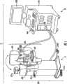
图1表示实施方式所涉及的乳房成像系统1的外观图。FIG. 1 shows an external view of a
图2表示乳房拍摄用X射线诊断装置2的方框结构图。FIG. 2 shows a block configuration diagram of an X-ray
图3是表示压迫单元35的结构的一例的图。FIG. 3 is a diagram showing an example of the configuration of the
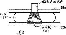
图4是表示用压迫单元35压迫乳房配置的状态的图。FIG. 4 is a diagram showing a state in which the breast is compressed by the
图5是表示压迫单元35的结构的另一个例子的图。FIG. 5 is a diagram showing another example of the configuration of the
图6表示本超声波诊断装置6的方框结构图。FIG. 6 shows a block configuration diagram of the ultrasonic diagnostic apparatus 6 .
图7(a)、7(b)是用于说明本压迫乳房图像重构功能的图。7( a ) and 7 ( b ) are diagrams for explaining the compression breast image reconstruction function of the present invention.
图8是用于说明压迫乳房图像和X射线乳房图像的位置对应的图。FIG. 8 is a diagram for explaining the positional correspondence between compressed breast images and X-ray breast images.
图9是表示在使用了乳房成像装置1的图像取得中执行的各处理的流程的流程图。FIG. 9 is a flowchart showing the flow of each process executed in image acquisition using the
图10是表示第2种实施方式所涉及的压迫单元35的结构的图。FIG. 10 is a diagram showing the configuration of a
图11是用于说明第3种实施方式的压迫乳房图像重构功能的图。Fig. 11 is a diagram for explaining the compression breast image reconstruction function of the third embodiment.
具体实施方式Detailed ways
以下,根据附图说明本发明的第1种实施方式以及第2种实施方式。而且,在以下的说明中,对于具有大致相同的功能以及构成的构成要素,标注相同符号并只在需要时进行重复说明。Hereinafter, a first embodiment and a second embodiment of the present invention will be described with reference to the drawings. In addition, in the following description, the same code|symbol is attached|subjected to the component which has substantially the same function and a structure, and it repeats description only when necessary.
(第1种实施方式)(first embodiment)
图1表示涉及本实施方式的乳房成像系统1的外观图。如同一图所示,本乳房成像系统1含有乳房拍摄用X射线诊断装置2、超声波诊断装置6。以下,说明各装置的构成。FIG. 1 shows an external view of a
(乳房拍摄用X射线诊断装置)(X-ray diagnostic equipment for breast photography)
如图1所示,本乳房拍摄用X射线诊断装置2含有:支架部20、支柱部25。在支架部20上设置相互对置的X射线源装置33以及平面检测器40、由上侧板35a以及下侧板35b组成的压迫单元35等。此外,在支柱部25上设置显示板27a、脚踏开关21a、21b、触摸板21c等。支架部20能够相对支柱部25上下移动以及以轴26为中心转动。通过将该支架部20设置成规定的姿态,用上侧板35a和设置在平面检测器40的检测面上的下侧板35b压迫固定乳房,能够从头尾方向、内外方向、内外斜方向等的任意的方向取得图像。As shown in FIG. 1 , the present X-ray
图2表示本乳房拍摄用X射线诊断装置2的方框结构图。如同一图所示,本乳房拍摄用X射线诊断装置2具备X射线控制部31、X射线源装置33、压迫单元35、压迫板驱动部36、压迫板控制部37、平面检测器40、存储器41、图像处理装置43、D/A变换器45、显示部47、中央控制部50、操作部51、存储部53。FIG. 2 shows a block configuration diagram of the present X-ray
X射线控制部31根据来自中央控制部50的指示控制X射线源装置33,使其以规定速率照射规定强度的X射线。The
X射线源装置33具有X射线管330、X射线照射区域限定器331。X射线管330是发生X射线的真空管,用来自高压发生装置的高电压加速电子,通过让电子撞击标靶发生X射线。X射线照射区域限定器331将从X射线管330照射的X射线形成为规定的形状。The
压迫单元35具备上侧板35a和下侧板35b,设置在X射线管330和平面检测器40之间。成为检查对象的乳房通过用上侧板35a和下侧板35b夹持压迫,在拍摄时平且薄地固定。上侧板35a以及下侧板35b将其材质设为具有X射线透过性的同时超声波反射性高的材料(例如,充分研磨过的丙烯板)作为其材料。通过在该上侧板和下侧板之间压迫固定乳房(定位),减少来自被拍摄体的散射X射线,能够减少乳房组织的重叠,能够改善图像对比度、防止身体活动产生的噪声发生等,能够实现照射线量的降低等。The
另外,压迫单元35的下侧板35b和平面检测器40也可以不一体化。此外,在压迫单元35的上侧板35a上如图3所示,例如设置一维超声波探头形成用于滑动的开口部350。如图4所示那样在将乳房压迫固定在上侧板35a和下侧板35b之间的状态下在开口部350上设置超声波探头通过一边滑动一边进行超声波扫描,能够在和乳房X线照相术拍摄时同样的状态下取得超声波图像。In addition, the
而且,图3所示的开口部35的形状是一个例子。即,开口部35的形状如果是可以在压迫固定乳房的状态下滑动一维超声波探头进行体积扫描的形状,则可以是任何形状,因而,例如如图5所示那样即使形成为和图3垂直的方向上也能够实现同样的作用效果。此外,开口部350为了防止体积扫描时的超声波探头的上下移动,理想的是设置在音响上透明且不使乳房较大露出的程度上有压迫力的薄膜。In addition, the shape of the
此外,在本实施方式中,假设设置上侧板35a和下侧板35b的结构。但是不限于此,例如如果平面检测器40的检测面具有超声波反射性高的特性和压迫耐久性等能够作为下侧板35b利用,则不需要特别设置下侧板35b。In addition, in this embodiment, the structure which provided the
压迫板驱动部36在压迫板控制部37的控制下,驱动压迫单元35。The compression
压迫板控制部37在中央控制部50的指示下,为了进行压迫单元35的定位,控制压迫板驱动部36。此外,压迫板控制部37测量压迫固定的乳房的压迫厚度(即,上侧板35a和下侧板35b之间的距离),送到中央控制部30。The compression
平面检测器40具有闪烁器和发光二极管阵列,通过将透过了被拍摄体的X射线照在光电膜上生成电子空穴,将它在半导体开关中蓄积,通过作为电信号读出将X摄像变换为电信号检测。而且,变换方式可以是从X射线变换为电信号的直接变换,也可以是从X射线经由光变换为电信号的间接变换。The
存储器41暂时存储从平面检测器40提供的数字信号。The
图像处理装置43从存储器41对每帧读出数字信号,根据需要进行减法处理等的规定的图像处理。The
D/A变换器45将从图像处理装置43输入的图像数据的数字信号串变换为模拟信号串。The D/
显示部47除了具有根据图像处理装置43接收到的信号显示X射线诊断图像等的CRT、等离子显示器、液晶显示器等外,具有表示装置的动作状态的显示板27a。The
中央控制部50是涉及进行与图像数据的收集有关的控制、以及收集到的图像数据的图像处理、涉及图像再生处理等的控制的中央处理装置。此外,中央控制部50在以后说明的压迫乳房图像的取得时,根据需要将压迫固定的乳房的压迫厚度、设置在上侧板35a上的超声波探头35的平面检测器40的检测面上的位置等发送到超声波诊断装置6。而且,设置在上侧板35a上的超声波探头35的平面检测器40的检测面上的位置例如能够通过设置在上侧板35a的开口部350上检测超声波探头12的位置的传感器等取得。The
操作部51是具备键盘和各种开关、鼠标、脚踏开关21a、21b、触摸板21c等的输入装置,在输入患者信息(患者ID、检测部位、检测目的等)的输入、压迫单元35的上下移动、拍摄指示、脉冲速率选择、图像选择等时使用。The
存储部53存储在图像处理装置43中的图像处理前或者处理后的图像数据等。此外,存储部53存储用于实现以后说明的数字自动曝光控制功能的专用程序。The
(超声波诊断装置)(ultrasonic diagnostic device)
如图1所示,本超声波诊断装置6具备装置主体61、超声波探头62、监视器63、输入装置64。As shown in FIG. 1 , the ultrasonic diagnostic apparatus 6 includes an apparatus
超声波探头62具有根据来自装置主体61的驱动信号发生超声波,将来自被检测体的反射波变换为电信号的多个压电振子;设置在该压电振子上的整合层;防止从该压电振子向后方的超声波的传播的填充材料等。如果从该超声波探头12向被检测体P发送超声波,则该发送超声波在体内组织的音响阻抗的不连续面上接连不断地反射,作为回波信号被超声波探头12接收。该回波信号的振幅依赖于成为反射的不连续面上的音响阻抗的差。此外,发送出的超声波脉冲在移动的血流和心脏壁等的表面上反射时的回波根据多普勒效应依赖于移动体的超声波发送方向的速度成分,受到频率偏移。The
输入装置64具有与装置主体61连接,用于将来自操作者的各种指示、条件、关心区域(ROI)的设定指示、各种画质条件设定等取入到装置主体61的各种开关、键、跟踪球、鼠标、键盘等。例如,如果操作者对输入装置64的结束键、FRWWZE键进行操作,则超声波的发送接收结束,该超声波诊断装置暂时变成停止状态。The
监视器63根据来自装置主体61的视频信号,将生物体的形态学信息、血流信息作为图像显示。The
图6表示本超声波诊断装置6的方框构成图。如同一图所示,在本超声波诊断装置6的装置主体61上设置超声波发送单元71、超声波接收单元72、B模式处理单元73、多普勒处理单元74、图像生成单元75、图像存储器76、图像合成单元77、控制处理器(CPU)78、内部存储部79、接口部80。以下,说明各个构成要素的功能。FIG. 6 shows a block configuration diagram of the ultrasonic diagnostic apparatus 6 . As shown in the same figure, an
超声波发送单元71具有未图示的触发发生电路、延迟电路以及脉冲发生器电路等。在脉冲发生器电路中,以规定的速率频率fr Hz(周期:1/fr秒),反复发生用于形成发送超声波的速率脉冲。此外,在延迟电路中,将对每个通道把超声波会聚成束状并且决定发送指向性所需要的延迟时间给予各速率脉冲。触发发生电路以基于该速率脉冲的时刻,向探头62施加驱动脉冲。The
而且,超声波发送单元21为了按照控制处理器78的指示执行规定的扫描顺序,具有可以瞬间改变发送频率、发送驱动电压等的功能。特别是对于发送驱动电压的改变,用可以瞬间切换其值的线性放大器型的发送电路,或者对多个电源单元进行电切换的机构实现。Furthermore, the ultrasonic transmission unit 21 has a function of instantaneously changing the transmission frequency, the transmission drive voltage, etc. in order to execute a predetermined scanning sequence according to the instruction of the
超声波接收单元72具有未图示的放大器电路、A/D变换器、加法器等。在放大器电路中,对每个通道放大经由探头62取入的回波信号。在A/D变换器中,对经过放大的回波信号给予为了决定接收指向性而需要的延迟时间,在其后在加法器中进行加法处理。通过该加算,强调来自与回波信号的接收指向性相应的方向的反射成分,用接收指向性和发送指向性形成超声波发送接收的综合的束。The
B模式处理单元73从发送接收单元71接收回波信号,实施对数放大、包络线检波处理等,生成用亮度的明暗表现信号强度的数据。The B-
多普勒处理单元74根据由发送接收单元71接收到的回波信号对速度信息进行频率解析,抽出多普勒效应产生的血流、组织、造影剂回波成分,对多点求平均速度、离散、能量等的血流信息。将得到的血流信息送到图像生成单元75,作为平均速度图像、离散图像、能量图像、它们的组合图像在监视器63上进行彩色显示。The
图像生成单元75将来自B模式处理单元73的信号列(超声波扫描的扫描线信号列)变换为以电视等为代表的一般的视频格式的扫描线信号列,生成作为显示图像的超声波诊断图像。此时,还实施边缘强调和时间平滑化等各种图像滤波,能够提供适应于用户喜好的画质。而且,在输入到该图像生成单元75之前的数据常被称为“原始数据”。The
图像存储器76例如是保存与刚停止之前的多个帧对应的超声波图像的存储器。通过连续显示(电影式显示)存储在该图像存储器76中的图像,也可以显示超声波运动图像。但是,在此,需要留意成为包含虚像的图像。The
图像合成单元77将从图像生成单元75收到的图像与各种参数的文字信息和刻度等合成,作为视频信号输出到监视器63。此外,图像合成监视单元77根据控制处理器78的控制,使用作为用在压迫单元35上反射的超声波得到的虚像的超声波图像,执行重构与实际的乳房有关的超声波图像(即,涉及用压迫单元35压迫被固定的乳房的超声波图像(压迫乳房图像))的压迫乳房图像重构功能(以后说明)。进而,图像合成单元77根据控制处理器78的控制,进行压迫乳房图像和X射线乳房图像的位置对应。The
控制处理器78具有作为信息处理装置(计算机)的功能,控制本超声波诊断装置主体的动作。控制处理器78从内部存储部79中读出用于执行图像生成/显示等的控制程序并展开在自身所具有的存储器上,执行与各种处理有关的计算/控制等。此外,控制处理器78通过将专用程序展开在存储器上,实现让扫描线扩散地分布的超声波扫描、压迫乳房图像重构功能。The
内部存储部79保管用于执行发送接收条件、图像生成、显示处理的控制程序、诊断信息(或者ID,医生的意见等)、诊断协议、探头的扫描线角度信息、探头的位置信息、身体标记生成程序、用于实现压迫乳房图像重构功能的专用程序及其他的数据组。此外,根据需要,还在图像存储器76中的图像的保管等中使用。内部存储部79的数据还可以经由接口电路80向乳房拍摄用X射线诊断装置2等的其他的装置转送。The
接口部80是涉及输入装置64、网络、新的外部存储装置(未图示)的接口。用该装置得到的超声波图像等的数据和解析结果等可以用接口部80经由网络转送到其他的装置。The
(压迫乳房图像重构功能)(Compressed breast image reconstruction function)
以下,说明乳房图像成像装置1具有的压迫乳房重构功能。该功能通过根据与包含了由压迫单元产生的超声波的反射的超声波的传播路径有关的信息,将用超声波扫描器得到的回波信号与受到压迫的二维或者三维乳房形状对应并将其影像化,生成压迫乳房图像。而且,在本实施方式中,为了具体地说明,将在根据回波信号生成一般的超声波图像后,使用该一般的超声波图像重构压迫乳房图像的情况为例子。但是,一般的超声波图像的生成不是必须的,通过根据与包含由压迫单元产生的超声波的反射的超声波的传播路径有关的信息,直接将回波信号或者由其产生的值与受到压迫的乳房形状对应起来影像化,能够生成压迫乳房图像。The compressed breast reconstruction function of the
图7(a)、7(b)是用于说明本压迫乳房图像重构功能的图。即,图7(a)是将压迫单元35压迫固定的乳房分割为实际区域R1~R5的图。此外,图7(b)是将把在开口部350上设置超声波探头62执行超声波扫描得到的回波信号以扇形形状进行影像化得到的一般的超声波图像P分割为图像小区域P1~P9的图。而且,图像小区域P1(R1)P2、P5、图像小区域P3和P6、图像小区域P4和P7分别相互呈镜像。此外,图像小区域P1和实际区域R1对应,但此外的图像小区域不与实际区域对应。在该意义下,图像小区域P2~P9是虚像区域,包含它们的超声波图像P可以说是虚像。7(a) and 7(b) are diagrams for explaining the compression breast image reconstruction function of the present invention. That is, FIG. 7( a ) is a diagram in which the breast compressed and fixed by the
图7(b)中的图像小区域P1是只使用来自被压迫单元35压迫固定的乳房内组织的反射波生成的图像,与将实际区域R1直接影像化的图像对应。此外,例如在图7(b)中的图像小区域P3内的任意点X1’中的值(即回波信号强度)是因通过实际从超声波探头62照射的超声波在涉及下侧板35b-点X1’的下侧板35b的(实际区域R2内的)对称点X1-下侧板35b这一路径上反射得到的反射波而产生的。因而,实际区域R2能够用图像小区域P3影像化。同样,实际区域R3能够用图像小区域P4影像化。The small image region P1 in FIG. 7( b ) is an image generated using only reflected waves from the intramammary tissue compressed and fixed by the
进而,图7(b)中的图像小区域P9内的任意的点X2’中的值(即回波信号强度)是因通过实际从超声波探头62照射的超声波在涉及下侧板35b-上侧板35a-点X2’的下侧板35b和虚像上侧板35a’的(实际区域R5内的)对称点X2-上侧板35a-下侧板35b这一路径反射而得到的反射波产生的。因而,实际区域R5能够用图像小区域P9影像化。同样,实际区域R4用图像小区域P8,实际区域R2用图像小区域R6,实际区域R3用图像小区域R7分别能够影像化。Furthermore, the value (i.e. echo signal strength) at any point X2' in the image small area P9 in FIG.
由此,如实际区域R1用图像小区域P1、P2、P5,将实际区域R2用图像小区域P3或者P6中,将实际小区域R3用图像小区域P4或者P7,将实际区域R4用图像小区域P8,将实际区域R5用图像小区域P9分别置换那样重构图像,由此能够在用被压迫单元35压迫固定的形态下进行乳房的影像化。Thus, for example, the actual region R1 uses the image small regions P1, P2, and P5, the actual region R2 uses the image small region P3 or P6, the actual small region R3 uses the image small region P4 or P7, and the actual region R4 uses the image small region P4 or P7. In the area P8 , the image is reconstructed by replacing the actual area R5 with the small image area P9 , thereby making it possible to visualize the breast while being compressed and fixed by the
在该重构中,对于用对应的哪个图像小区域置换各实际区域没有特别限定。例如,当将实际区域R1称为图像小区域P5、将实际区域R2称为图像小区域P6、将实际区域R3称为图像小区域P7、将实际小区域R4称为图像小区域P8、将实际区域R5称为图像小区域P9的状况中,当将超声波图像P在上侧板35a和下侧板35b之间的距离L上在深度方向上分割为多个层的情况下,使用分类为同样层的小区域(即,包含在压迫单元35上的反射次数是2次或者3次的回波信号的图像小区域),也可以分别重构。通过根据这样在压迫单元35上的反射次数选择图像小区域并重构,能够进行使用了衰减是相同程度的反射波的重构,能够提高画质。此外,例如对于从实际区域R4或者R5向外侧的区域,通过使用比虚像下侧板35b’下侧的图像小区域可以重构图像。In this reconstruction, there is no particular limitation as to which corresponding small image area is used to replace each actual area. For example, when the actual area R1 is called the small image area P5, the actual area R2 is called the small image area P6, the actual area R3 is called the small image area P7, the actual small area R4 is called the small image area P8, and the actual In the case where the region R5 is referred to as the small image region P9, when the ultrasonic image P is divided into a plurality of layers in the depth direction at the distance L between the
而且,在根据以上所述的压迫乳房图像重构功能的处理(压迫乳房图像重构处理)中,和对被压迫单元35压迫固定的状态的乳房进行超声波扫描得到的超声波图像P一同,需要涉及上侧板35a和下侧板35b间的距离L的信息。该信息能够通过通信从乳房拍摄用X射线诊断装置2取得。但是,并不拘泥于此,例如也可以根据被超声波图像P影像化的上侧板35a和下侧板35b之间的距离来求得。Furthermore, in the processing (compressed breast image reconstruction processing) according to the above-mentioned compressed breast image reconstruction function, together with the ultrasonic image P obtained by ultrasonic scanning the breast in a state compressed and fixed by the
此外,这样的压迫乳房图像重构处理对用体积扫描得到的各超声波图像执行,生成与各自对应的多个压迫乳房图像。从得到的多个压迫乳房图像中,还可以制成涉及压迫乳房的体积数据。用户通过使用涉及该压迫乳房的体积数据,能够选择与受到压迫的乳房有关的任意剖面图像(B模式图像,C模式图像)进行观察。In addition, such compressed breast image reconstruction processing is performed on each ultrasonic image obtained by volume scanning, and a plurality of compressed breast images corresponding to each are generated. From the obtained multiple images of the compressed breast, volume data relating to the compressed breast can also be generated. By using the volume data related to the compressed breast, the user can select any cross-sectional image (B-mode image, C-mode image) related to the compressed breast for observation.
压迫乳房图像重构处理的执行后,中央控制部50或者图像合成单元77在重构的压迫乳房图像和X射线乳房图像之间进行位置对应,该位置对应能够通过在压迫单元35的开口部350或者超声波探头62自身上设置位置传感器,例如特定在平面检测器40上的超声波探头62的位置来实现。此时,例如如图8所示,在X射线乳房图像上表示超声波图像的位置(或者在超声波图像上表示X射线乳房图像的位置),根据需要通过点击该位置,自动地与该位置对应的超声波图像(或者X射线乳房图像)自动地同时或者有选择地显示X射线乳房图像(或者超声波图像)。After the compressed breast image reconstruction process is executed, the
(超声波扫描)(ultrasound scan)
当执行上述压迫乳房图像重构功能的情况下,如图4所示,执行扫描线扩散地分布那样的超声波扫描。由此,容易引起在压迫单元35中的超声波反射,沿着压迫单元35的面方向可以进行宽区域的超声波成像。In the case of executing the compressed breast image reconstruction function described above, as shown in FIG. 4 , an ultrasonic scan is performed such that scan lines are diffusely distributed. This makes it easy to cause ultrasonic reflection in the compressing
而且,这样的超声波扫描依照扫描线的位置计算用于相位控制的延迟时间,通过根据它控制各压电振子的驱动时刻来实现。In addition, such ultrasonic scanning is realized by calculating the delay time for phase control according to the position of the scanning line, and controlling the driving timing of each piezoelectric vibrator based on this.
(动作)(action)
以下,说明包含压迫乳房图像重构处理的本乳房成像装置1的动作。Hereinafter, the operation of the present
图9是表示在使用了乳房成像装置1的图像取得中执行的各处理的流程的流程图。如该图所示,首先,用上侧板35a和平面检测器40的检测面(即,下侧板35b)压迫/固定患者的乳房,进行被拍摄体的定位(步骤S1)。以下,经由操作部51、输入装置64将患者信息、拍摄条件等分别输入到乳房拍摄用X射线诊断装置2、超声波诊断装置6(步骤S2)。而且,所谓拍摄条件在是乳房拍摄用X射线诊断装置2的情况下,表示X射线条件(管电压,mAs值、焦点-拍摄面距离、用于实现扩散的扫描线分布的每个通道的延迟时间等),当是超声波诊断装置6的情况下表示发送条件(发送电压,焦点位置等)等。如乳房拍摄用X射线诊断装置2的中央控制部50根据输入的拍摄条件照射X射线,根据透过乳房的X射线取得X射线乳房图像(步骤S3)。FIG. 9 is a flowchart showing the flow of each process executed in image acquisition using the
以下,通过在上侧板35a的开口部350上设置超声波探头62(步骤S4),一边让超声波62滑动一边进行超声波扫描,取得涉及乳房的体积数据(步骤S5)。图像合成单元77通过对构成已取得的体积数据的各超声波图像进行已说明的重构处理,生成压迫乳房图像(步骤S6)。Thereafter, by installing the
此外,乳房拍摄用X射线诊断装置2的中央控制部50或者超声波诊断装置6的控制处理器78例如根据在平面检测器40上的超声波探头62的位置进行X射线乳房图像和压迫乳房图像的位置对应(步骤S7)。相互对应的X射线乳房图像以及压迫乳房图像在显示部47或者监视器63上,或者用另外的显示装置以规定的形态显示(步骤S8)。In addition, the
(效果)(Effect)
如果采用以上叙述的构成,则能够得到以下的效果。According to the configuration described above, the following effects can be obtained.
如果采用本乳房成像系统,则超声波探头插入开口部的数与以往相比能够以少的压迫板在充分压迫固定的状态下对乳房进行超声波扫描。因而,能够在和X射线乳房图像取得时相同的被拍摄体配置取得超声波图像,能够使用该超声波图像重构乳房压迫图像。其结果,在能够取得容易对应且容易比较的X射线乳房图像和压迫乳房图像的同时,还能够正确并且容易地进行图像间的位置对应。According to this breast imaging system, the number of ultrasonic probes inserted into the openings can be ultrasonically scanned on the breast in a sufficiently compressed and fixed state with fewer compression plates than in the past. Therefore, it is possible to acquire an ultrasonic image in the same subject arrangement as when acquiring an X-ray breast image, and to reconstruct a breast compression image using the ultrasonic image. As a result, it is possible to obtain X-ray breast images and compressed breast images that are easy to correspond and compare, and to accurately and easily perform positional correspondence between the images.
此外,如果采用本乳房成像系统,则将超声波探头嵌入开口部,使用通过一边在和超声波扫描面垂直方向滑动一边进行超声波扫描而得到的体积数据,能够取得与受到压迫固定的乳房有关的体积数据,因而,与以往相比,能够缩短用于超声波图像取得的时间,能够减轻给患者的负担。In addition, according to this breast imaging system, the ultrasound probe is inserted into the opening, and the volume data related to the compressed and fixed breast can be acquired using volume data obtained by performing ultrasound scanning while sliding in a direction perpendicular to the ultrasound scanning surface. Therefore, compared with conventional methods, it is possible to shorten the time for obtaining an ultrasonic image and reduce the burden on the patient.
此外,在本乳房成像系统中,压迫板的开口部只设置成嵌入超声波探头让其滑动的大小即可,因而,不需要用开着多个孔的压迫板压迫乳房,在能够减少在乳房的压迫固定中的痛苦的同时,能够防止压迫强度的下降。In addition, in this breast imaging system, the opening of the compression plate can only be set to a size that allows the ultrasound probe to be embedded to allow it to slide. Therefore, it is not necessary to use a compression plate with a plurality of holes to compress the breast. While compressing the pain in immobilization, it is possible to prevent a decrease in compression strength.
(第2种实施方式)(the second embodiment)
以下,说明本发明的第2种实施方式。本实施方式说明实现使用了二维超声波探头的压迫乳房图像重构功能的乳房成像装置1。Next, a second embodiment of the present invention will be described. In this embodiment, a
即,当使用超声波振子排列成二维的二维阵列探头的情况下,即使不沿着开口部350移动也能够用三维扫描取得体积数据。因而,当超声波探头62是二维阵列探头的情况下,压迫单元35例如如图10所示构成为具有带有与该二维阵列探头对应的形状的开口部351。That is, when using a two-dimensional array probe in which ultrasonic vibrators are arranged two-dimensionally, volume data can be obtained by three-dimensional scanning without moving along the
而且,图像合成单元77通过对构成所收集到的体积数据的任意的各剖面进行压迫乳房图像重构处理,能够生成任意的压迫乳房图像。此外,通过对构成所收集到的体积数据的各剖面进行压迫乳房图像重构处理,能够取得与受到压迫固定的乳房有关的体积数据。Furthermore, the
此外,通过根据与包含由压迫单元产生的超声波的反射的超声波的传播路径有关的信息,将用超声波扫描得到的回波信号与受到压迫的三维乳房形状对应地直接影像化,能够取得与受到压迫固定的乳房有关的体积数据。In addition, by directly visualizing the echo signal obtained by ultrasonic scanning in correspondence with the three-dimensional shape of the compressed breast based on the information about the propagation path of the ultrasonic wave including the reflection of the ultrasonic wave generated by the compression unit, it is possible to obtain a corresponding image of the compressed breast. Fixed breast-related volume data.
如果采用以上那样的构成,则与第1种实施方式的情况下相比,在能够进一步减少在乳房的压迫固定中的痛苦的同时,能够防止压迫强度的下降。According to the above configuration, compared with the case of the first embodiment, it is possible to further reduce the pain in compression and fixation of the breast, and prevent a decrease in the compression strength.
(第3种实施方式:梯形)(The third embodiment: trapezoid)
以下,说明本发明的第3种实施方式。本实施方式说明实现使用了一维阵列超声波探头的压迫乳房图像重构功能的乳房成像装置1。Next, a third embodiment of the present invention will be described. This embodiment describes the
即,当使用将超声波振子排列成一维的一维阵列探头的情况下,例如在图10所示的开口部351上设置超声波探头62,如超声波扫描面变成扩展形状(例如,梯形)那样,进行超声波发送接收。That is, when using a one-dimensional array probe in which ultrasonic vibrators are arranged one-dimensionally, for example, the
如果采用这样的构成,则即使不让探头62沿着开口部350移动,也能够取得与乳房的所希望的剖面有关的超声波图像。因而,能够进一步减轻乳房的图像诊断时的痛苦。According to such a configuration, without moving the
(第4种实施方式:倾斜)(Fourth embodiment: tilt)
以下,说明本发明的第4种实施方式。本实施方式说明一边使用一维超声波探头,一边不让探头62沿着开口部350移动而取得体积数据的例子。Hereinafter, a fourth embodiment of the present invention will be described. This embodiment describes an example in which volume data is acquired without moving the
图11是用于说明涉及本实施方式的压迫乳房图像重构功能的图。送到受到压迫的乳房内的扫描面Q0的超声波在所示的压迫板表面上反射,例如按照路径Q1、Q2、Q3的顺序在乳房内衰减传播。因而,例如在来自传播路径Q1的发射波和传播路径Q2的反射波中,接收时刻不同。根据该接收时刻的不同接收到的反射波区别是从乳房内的超声波传播路径上的哪个位置来的反射波。如图11表示的那样提供一边展开一边变换,能够生成将传播路径Q1、Q2、Q3作为扫描面的二维超声波图像。FIG. 11 is a diagram for explaining the compressed breast image reconstruction function according to the present embodiment. Ultrasonic waves transmitted to the scanning plane Q0 in the compressed breast are reflected on the surface of the compression plate shown, and propagate through the breast attenuated, for example, in the order of paths Q1, Q2, and Q3. Therefore, for example, the reception timing differs between the transmitted wave from the propagation path Q1 and the reflected wave from the propagation path Q2. Depending on the receiving timing, the received reflected wave is distinguished from which position on the ultrasonic propagation path in the breast the reflected wave came from. As shown in FIG. 11 , by providing transformation while unfolding, it is possible to generate a two-dimensional ultrasonic image with propagation paths Q1 , Q2 , and Q3 as scanning planes.
即,将在压迫单元35中一次反射与在传播路径Q1上传播的超声波对应的反射波(例如来自任意的点X1的反射波)根据其接收时刻,在传播路径Q1和处于镜像关系的虚像区域r1的对应的位置(点X1’)上影像化。同样,将用压迫单元35进行了二次反射与在路径Q2上传播的超声波对应的反射波影像化在与虚像区域r2对应的位置上,将用压迫单元35进行了三次反射与在传播路径Q3上传播的超声波对应的反射波影像化在与虚像区域r3对应的位置上。That is, the reflection wave corresponding to the ultrasonic wave propagating on the propagation path Q1 once reflected in the compressing unit 35 (for example, a reflection wave from an arbitrary point X1) is reflected on the propagation path Q1 and the virtual image region in a mirror image relationship according to the receiving time. The corresponding position (point X1') of r1 is visualized. Similarly, the reflection wave corresponding to the ultrasonic wave propagating on the path Q2 that has been reflected twice by the compressing
此外,对于用未图示的摇摆单元让扫描面Q0沿着方向D扇动得到的乳房的体积数据,通过对各扫描面的每个实施同样的影像化处理,能够重构与乳房的虚像有关的体积数据。In addition, for the volume data of the breast obtained by fanning the scanning plane Q0 along the direction D with a swing unit not shown, by performing the same imaging process on each scanning plane, it is possible to reconstruct the virtual image of the breast. volume data.
如果采用以上所述的构成,则即使不让探头62沿着开口部350移动,也能够取得与乳房的所希望的三维区域有关的超声波图像。因而,能够进一步减轻在乳房的图像诊断时的痛苦。With the configuration described above, it is possible to acquire an ultrasonic image of a desired three-dimensional region of the breast without moving the
而且,本发明并不限于上述实施方式自身,在实施阶段在不脱离其主要内容的范围中改变构成要素能够具体化。作为具体化的变形例子有以下的例子。In addition, the present invention is not limited to the above-mentioned embodiment itself, and can be realized by changing constituent elements within a range not departing from the main contents at the stage of implementation. Examples of specific modifications include the following.
(1)本实施方式的各功能也能够通过将执行该处理的程序安装在工作站等的计算机中,将它们展开在存储器上实现。此时,能够让计算机执行该方法的程序也可以存储在磁盘(软盘(注册商标)、硬盘等)、光盘(CD-ROM,DVD等)、半导体存储器等的记录媒体中发布。(1) Each function of the present embodiment can also be realized by installing a program for executing the processing in a computer such as a workstation, and expanding them on a memory. In this case, the program that enables a computer to execute the method may be stored in a recording medium such as a magnetic disk (floppy disk (registered trademark), hard disk, etc.), optical disk (CD-ROM, DVD, etc.), semiconductor memory, and the like for distribution.
(2)在本乳房成像系统中,可以任意改变超声波扫描线的角度。在该决定中虽然没有特别限定,但例如根据患者的乳房的大小、压迫板的宽度、深度、上侧板35a和下侧板35b之间的距离L等,可以人为地选择任意的值或者预先分类的规定的值。(2) In this breast imaging system, the angle of the ultrasonic scanning line can be changed arbitrarily. Although there are no particular limitations on this determination, for example, according to the size of the patient's breast, the width and depth of the compression plate, the distance L between the
(3)在上述各实施方式中,说明了将超声波探头设定在X射线路径区域上的构成。但是,并不拘泥于此,也可以在X射线路径区域外(例如上侧板35a和下侧板35b之间)配置超声波探头,用和各实施方式一样的方法执行乳房成像。(3) In each of the above-mentioned embodiments, the configuration in which the ultrasonic probe is set on the X-ray path region has been described. However, it is not limited to this, and the ultrasound probe may be arranged outside the X-ray path area (for example, between the
此外,通过在上述实施方式中公开的多个构成要素的适宜的组合,能够形成各种发明。例如,也可以从在实施方式中表示的全构成要素中删除几个构成要素。进而,也可以适宜地组合涉及不同的实施方式的构成要素。In addition, various inventions can be formed by appropriate combinations of a plurality of constituent elements disclosed in the above-mentioned embodiments. For example, some constituent elements may be deleted from all the constituent elements shown in the embodiments. Furthermore, components related to different embodiments may be combined as appropriate.
Claims (23)
Applications Claiming Priority (2)
| Application Number | Priority Date | Filing Date | Title |
|---|---|---|---|
| JP2007-099691 | 2007-04-05 | ||
| JP2007099691 | 2007-04-05 |
Publications (2)
| Publication Number | Publication Date |
|---|---|
| CN101278863Atrue CN101278863A (en) | 2008-10-08 |
| CN101278863B CN101278863B (en) | 2012-05-02 |
Family
ID=39643759
Family Applications (1)
| Application Number | Title | Priority Date | Filing Date |
|---|---|---|---|
| CN2008100918111AExpired - Fee RelatedCN101278863B (en) | 2007-04-05 | 2008-04-03 | Ultrasonic diagnostic device, breast imaging system, and breast imaging method |
Country Status (4)
| Country | Link |
|---|---|
| US (1) | US20080249415A1 (en) |
| EP (1) | EP1977692A3 (en) |
| JP (1) | JP5481038B2 (en) |
| CN (1) | CN101278863B (en) |
Cited By (8)
| Publication number | Priority date | Publication date | Assignee | Title |
|---|---|---|---|---|
| CN102697526A (en)* | 2012-06-15 | 2012-10-03 | 华东医院 | Ultrasonic scanning tomography device for volumes of superficial tissues and organs |
| CN103189000A (en)* | 2010-06-15 | 2013-07-03 | 影像麦宁公司 | Fiducial systems for mammography |
| CN103860183A (en)* | 2012-12-17 | 2014-06-18 | 财团法人工业技术研究院 | Photoacoustic detector, photoacoustic plate, and detector using the photoacoustic plate |
| CN105193447A (en)* | 2015-10-09 | 2015-12-30 | 汕头市超声仪器研究所有限公司 | X-ray and ultrasound combined breast examination device and fusion imaging method thereof |
| CN109907769A (en)* | 2017-12-12 | 2019-06-21 | 西门子医疗保健有限责任公司 | The method of breast X-ray machine and the image data set of offer chest |
| CN110996801A (en)* | 2017-08-11 | 2020-04-10 | 豪洛捷公司 | Breast compression plate with proximal corners |
| CN112603342A (en)* | 2020-12-23 | 2021-04-06 | 达影医疗(中山)有限公司 | Compression device, mammary gland X-ray imaging machine and mammary gland detection imaging method |
| CN117982167A (en)* | 2024-04-03 | 2024-05-07 | 南京大学 | Second harmonic focuser based on micron contrast agent |
Families Citing this family (18)
| Publication number | Priority date | Publication date | Assignee | Title |
|---|---|---|---|---|
| NL2002092C (en)* | 2008-10-13 | 2010-04-14 | Academisch Medisch Ct Van De Universiteit Van Amsterdam | Mammography-apparatus and method for screening the occurrence of malignant cells. |
| CA2740350A1 (en)* | 2008-10-13 | 2010-04-22 | Gerhard Johan Den Heeten | Mammography-apparatus and method for screening the occurrence of malignant cells |
| US8942342B2 (en) | 2008-12-29 | 2015-01-27 | Analogic Corporation | Multi-modality image acquisition |
| US7831015B2 (en)* | 2009-03-31 | 2010-11-09 | General Electric Company | Combining X-ray and ultrasound imaging for enhanced mammography |
| NL2005509C2 (en) | 2010-02-19 | 2011-08-23 | Academisch Medisch Ct Bij De Universiteit Van Amsterdam | Mammography-apparatus. |
| US9730659B2 (en) | 2010-11-16 | 2017-08-15 | Analogic Corporation | Multi-modality image acquisition |
| US8886293B2 (en)* | 2010-11-24 | 2014-11-11 | Mayo Foundation For Medical Education And Research | System and method for tumor analysis and real-time biopsy guidance |
| JP2013146539A (en)* | 2011-12-21 | 2013-08-01 | Nippon Koden Corp | Cuff and method for observing tissue under pressure by using the same |
| CN103006272B (en)* | 2013-01-09 | 2014-12-03 | 东南大学 | Velocity distribution measuring method based on ultrasonic interleave programming |
| CN105286796B (en)* | 2015-10-30 | 2021-11-23 | 上海联影医疗科技股份有限公司 | Mammography system and photography method thereof |
| JP6730919B2 (en)* | 2016-12-12 | 2020-07-29 | 株式会社日立製作所 | Ultrasonic CT device |
| NL2019124B1 (en) | 2017-06-27 | 2019-01-07 | Sigmascreening B V | Mammography apparatus |
| NL2019834B1 (en) | 2017-10-31 | 2019-05-08 | Sigmascreening B V | Mammography apparatus and method of pressurizing a breast |
| AU2019290182B2 (en)* | 2018-06-22 | 2025-01-30 | Hologic, Inc. | Multi-position ultrasound system |
| JP7169430B2 (en)* | 2019-03-27 | 2022-11-10 | 富士フイルム株式会社 | Imaging control device, method and program |
| CN111281344A (en)* | 2020-02-26 | 2020-06-16 | 浙江杜比医疗科技有限公司 | Mammary gland imaging system and mammary gland imaging method |
| US20220192632A1 (en)* | 2020-12-20 | 2022-06-23 | Shabbir Bakir Bambot | Method and apparatus for breast imaging |
| WO2025070512A1 (en)* | 2023-09-29 | 2025-04-03 | 富士フイルム株式会社 | Image processing device, medical image capture system, image processing method, and image processing program |
Family Cites Families (14)
| Publication number | Priority date | Publication date | Assignee | Title |
|---|---|---|---|---|
| WO1995011627A1 (en)* | 1993-10-29 | 1995-05-04 | Neovision Corporation | Methods and apparatus for performing sonomammography and enhanced x-ray imaging |
| US5776062A (en)* | 1996-10-15 | 1998-07-07 | Fischer Imaging Corporation | Enhanced breast imaging/biopsy system employing targeted ultrasound |
| JP3668822B2 (en)* | 1996-11-11 | 2005-07-06 | 株式会社島津製作所 | Medical image processing device |
| US6731966B1 (en)* | 1997-03-04 | 2004-05-04 | Zachary S. Spigelman | Systems and methods for targeting a lesion |
| US6122542A (en)* | 1998-11-25 | 2000-09-19 | Rubicor Medical, Inc. | Breast stabilization devices and imaging and interventional methods using the same |
| US6574499B1 (en)* | 1998-11-25 | 2003-06-03 | Xdata Corporation | Mammography method and apparatus |
| DE19901730A1 (en)* | 1999-01-18 | 2000-07-20 | Siemens Ag | X-ray device for simultaneous breast diagnosis using ultrasound |
| US6607489B2 (en)* | 2001-04-05 | 2003-08-19 | General Electric Company | Focus correction for ultrasound imaging through mammography compression plate |
| US20030149364A1 (en)* | 2002-02-01 | 2003-08-07 | Ajay Kapur | Methods, system and apparatus for digital imaging |
| US7285092B2 (en)* | 2002-12-18 | 2007-10-23 | Barbara Ann Karmanos Cancer Institute | Computerized ultrasound risk evaluation system |
| JP2004141447A (en)* | 2002-10-25 | 2004-05-20 | Japan Science & Technology Agency | Reflection / transmission type ultrasonic inverse scattering CT system |
| US6846289B2 (en)* | 2003-06-06 | 2005-01-25 | Fischer Imaging Corporation | Integrated x-ray and ultrasound medical imaging system |
| CN101031244A (en)* | 2004-09-29 | 2007-09-05 | 皇家飞利浦电子股份有限公司 | Methods and apparatus for performing enhanced ultrasound diagnostic breast imaging |
| JP4575738B2 (en)* | 2004-09-29 | 2010-11-04 | 富士フイルム株式会社 | Ultrasonic image boundary extraction method, ultrasonic image boundary extraction device, and ultrasonic imaging device |
- 2008
- 2008-03-28JPJP2008088624Apatent/JP5481038B2/ennot_activeExpired - Fee Related
- 2008-03-31EPEP08006511Apatent/EP1977692A3/ennot_activeWithdrawn
- 2008-04-01USUS12/060,679patent/US20080249415A1/ennot_activeAbandoned
- 2008-04-03CNCN2008100918111Apatent/CN101278863B/ennot_activeExpired - Fee Related
Cited By (12)
| Publication number | Priority date | Publication date | Assignee | Title |
|---|---|---|---|---|
| CN103189000A (en)* | 2010-06-15 | 2013-07-03 | 影像麦宁公司 | Fiducial systems for mammography |
| CN103189000B (en)* | 2010-06-15 | 2015-08-26 | 影像麦宁公司 | Mammogram benchmark system |
| CN102697526A (en)* | 2012-06-15 | 2012-10-03 | 华东医院 | Ultrasonic scanning tomography device for volumes of superficial tissues and organs |
| CN103860183A (en)* | 2012-12-17 | 2014-06-18 | 财团法人工业技术研究院 | Photoacoustic detector, photoacoustic plate, and detector using the photoacoustic plate |
| CN105193447A (en)* | 2015-10-09 | 2015-12-30 | 汕头市超声仪器研究所有限公司 | X-ray and ultrasound combined breast examination device and fusion imaging method thereof |
| CN110996801A (en)* | 2017-08-11 | 2020-04-10 | 豪洛捷公司 | Breast compression plate with proximal corners |
| CN110996801B (en)* | 2017-08-11 | 2023-10-03 | 豪洛捷公司 | Breast compression plate with near corners |
| CN109907769A (en)* | 2017-12-12 | 2019-06-21 | 西门子医疗保健有限责任公司 | The method of breast X-ray machine and the image data set of offer chest |
| CN109907769B (en)* | 2017-12-12 | 2020-10-16 | 西门子医疗保健有限责任公司 | Breast X-ray machine and method for providing an image data record of a breast |
| US10874366B2 (en) | 2017-12-12 | 2020-12-29 | Siemens Healthcare Gmbh | Mammography imaging |
| CN112603342A (en)* | 2020-12-23 | 2021-04-06 | 达影医疗(中山)有限公司 | Compression device, mammary gland X-ray imaging machine and mammary gland detection imaging method |
| CN117982167A (en)* | 2024-04-03 | 2024-05-07 | 南京大学 | Second harmonic focuser based on micron contrast agent |
Also Published As
| Publication number | Publication date |
|---|---|
| JP5481038B2 (en) | 2014-04-23 |
| EP1977692A2 (en) | 2008-10-08 |
| EP1977692A3 (en) | 2008-12-10 |
| US20080249415A1 (en) | 2008-10-09 |
| CN101278863B (en) | 2012-05-02 |
| JP2008272459A (en) | 2008-11-13 |
Similar Documents
| Publication | Publication Date | Title |
|---|---|---|
| JP5481038B2 (en) | Ultrasound diagnostic apparatus, breast imaging system, and breast imaging program | |
| JP5143333B2 (en) | System and method for performing image processing for observing abnormal parts in different types of images | |
| US8275447B2 (en) | Medical image diagnostic system, medical imaging apparatus, medical image storage apparatus, and medical image display apparatus | |
| CN104640506B (en) | Diagnostic ultrasound equipment, medical image-processing apparatus and medical image processing method | |
| JP4934263B2 (en) | Digital imaging method, system and apparatus | |
| JP6608232B2 (en) | Medical image diagnostic apparatus, medical image processing apparatus, and medical information display control method | |
| US11266380B2 (en) | Medical ultrasound image processing device | |
| JP6058295B2 (en) | Ultrasonic diagnostic apparatus, medical image processing apparatus, medical image processing method, and medical image processing program | |
| JP2007216003A (en) | Ultrasonic inspection equipment | |
| JP2011224346A (en) | Ultrasound diagnosis apparatus, image processing apparatus, and image processing method | |
| JP5462076B2 (en) | Ultrasonic diagnostic apparatus and image information management apparatus | |
| JP2011224354A (en) | Ultrasonic diagnostic apparatus, ultrasonic image processor, and medical image diagnostic apparatus | |
| JP2009072410A (en) | Composite image diagnostic equipment | |
| CN1606965A (en) | Device capable of displaying medical trend map and relevant information | |
| CN101601593A (en) | Ultrasonic diagnostic device | |
| JP2014028029A (en) | Ultrasonic diagnostic apparatus and ultrasonic diagnostic apparatus control program | |
| JP2009028381A (en) | Medical imaging system | |
| JP2018015556A (en) | Ultrasonic diagnostic apparatus and measurement program | |
| CN102626328B (en) | Diagnostic ultrasound equipment, Ultrasonographic device and adquisitiones | |
| JP7195218B2 (en) | Radiation imaging system, medical imaging system, control method, and control program | |
| JP2012030053A (en) | Ultrasound diagnosis apparatus, image processing apparatus and image processing method | |
| JP5134897B2 (en) | Breast examination system | |
| JP2012075794A (en) | Ultrasonic diagnostic apparatus, medical image processor, and medical image processing program | |
| KR20140137037A (en) | ultrasonic image processing apparatus and method | |
| JP5366372B2 (en) | Ultrasonic diagnostic apparatus and ultrasonic image data generation program |
Legal Events
| Date | Code | Title | Description |
|---|---|---|---|
| C06 | Publication | ||
| PB01 | Publication | ||
| C10 | Entry into substantive examination | ||
| SE01 | Entry into force of request for substantive examination | ||
| C14 | Grant of patent or utility model | ||
| GR01 | Patent grant | ||
| CF01 | Termination of patent right due to non-payment of annual fee | Granted publication date:20120502 Termination date:20160403 | |
| CF01 | Termination of patent right due to non-payment of annual fee |