CN101242862B - Sheet-like composition - Google Patents
Sheet-like compositionDownload PDFInfo
- Publication number
- CN101242862B CN101242862BCN2006800305840ACN200680030584ACN101242862BCN 101242862 BCN101242862 BCN 101242862BCN 2006800305840 ACN2006800305840 ACN 2006800305840ACN 200680030584 ACN200680030584 ACN 200680030584ACN 101242862 BCN101242862 BCN 101242862B
- Authority
- CN
- China
- Prior art keywords
- amnion
- cells
- amniotic membrane
- epithelium
- sheet
- Prior art date
- Legal status (The legal status is an assumption and is not a legal conclusion. Google has not performed a legal analysis and makes no representation as to the accuracy of the status listed.)
- Active
Links
- 239000000203mixtureSubstances0.000titleclaimsabstractdescription120
- 210000001691amnionAnatomy0.000claimsabstractdescription600
- HDTRYLNUVZCQOY-UHFFFAOYSA-Nα-D-glucopyranosyl-α-D-glucopyranosideNatural productsOC1C(O)C(O)C(CO)OC1OC1C(O)C(O)C(O)C(CO)O1HDTRYLNUVZCQOY-UHFFFAOYSA-N0.000claimsabstractdescription104
- HDTRYLNUVZCQOY-WSWWMNSNSA-NTrehaloseNatural productsO[C@@H]1[C@@H](O)[C@@H](O)[C@@H](CO)O[C@@H]1O[C@@H]1[C@H](O)[C@@H](O)[C@@H](O)[C@@H](CO)O1HDTRYLNUVZCQOY-WSWWMNSNSA-N0.000claimsabstractdescription104
- HDTRYLNUVZCQOY-LIZSDCNHSA-Nalpha,alpha-trehaloseChemical compoundO[C@@H]1[C@@H](O)[C@H](O)[C@@H](CO)O[C@@H]1O[C@@H]1[C@H](O)[C@@H](O)[C@H](O)[C@@H](CO)O1HDTRYLNUVZCQOY-LIZSDCNHSA-N0.000claimsabstractdescription104
- 210000002469basement membraneAnatomy0.000claimsabstractdescription49
- 210000004027cellAnatomy0.000claimsdescription364
- 210000000981epitheliumAnatomy0.000claimsdescription162
- 238000011282treatmentMethods0.000claimsdescription120
- 238000000034methodMethods0.000claimsdescription111
- 239000000463materialSubstances0.000claimsdescription101
- 108090000631TrypsinProteins0.000claimsdescription96
- 102000004142TrypsinHuman genes0.000claimsdescription96
- 239000012588trypsinSubstances0.000claimsdescription96
- 210000002919epithelial cellAnatomy0.000claimsdescription90
- 108010049003FibrinogenProteins0.000claimsdescription61
- 102000008946FibrinogenHuman genes0.000claimsdescription61
- 229940012952fibrinogenDrugs0.000claimsdescription61
- 108090000190ThrombinProteins0.000claimsdescription49
- 229960004072thrombinDrugs0.000claimsdescription49
- 102000008186CollagenHuman genes0.000claimsdescription47
- 108010035532CollagenProteins0.000claimsdescription47
- 229920001436collagenPolymers0.000claimsdescription47
- 239000000853adhesiveSubstances0.000claimsdescription46
- 230000001070adhesive effectEffects0.000claimsdescription46
- 210000003560epithelium cornealAnatomy0.000claimsdescription44
- 108010039627AprotininProteins0.000claimsdescription36
- 229960004405aprotininDrugs0.000claimsdescription36
- ZPNFWUPYTFPOJU-LPYSRVMUSA-NiniprolChemical compoundC([C@H]1C(=O)NCC(=O)NCC(=O)N[C@H]2CSSC[C@H]3C(=O)N[C@@H](CCCCN)C(=O)N[C@@H](C)C(=O)N[C@@H](CCCNC(N)=N)C(=O)N[C@H](C(N[C@H](C(=O)N[C@@H](CCCNC(N)=N)C(=O)N[C@@H](CC=4C=CC(O)=CC=4)C(=O)N[C@@H](CC=4C=CC=CC=4)C(=O)N[C@@H](CC=4C=CC(O)=CC=4)C(=O)N[C@@H](CC(N)=O)C(=O)N[C@@H](C)C(=O)N[C@@H](CCCCN)C(=O)N[C@@H](C)C(=O)NCC(=O)N[C@@H](CC(C)C)C(=O)N[C@@H](CSSC[C@H](NC(=O)[C@H](CC(O)=O)NC(=O)[C@H](CCC(O)=O)NC(=O)[C@H](C)NC(=O)[C@H](CO)NC(=O)[C@H](CCCCN)NC(=O)[C@H](CC=4C=CC=CC=4)NC(=O)[C@H](CC(N)=O)NC(=O)[C@H](CC(N)=O)NC(=O)[C@H](CCCNC(N)=N)NC(=O)[C@H](CCCCN)NC(=O)[C@H](C)NC(=O)[C@H](CCCNC(N)=N)NC2=O)C(=O)N[C@@H](CCSC)C(=O)N[C@@H](CCCNC(N)=N)C(=O)N[C@@H]([C@@H](C)O)C(=O)N[C@@H](CSSC[C@H](NC(=O)[C@H](CC=2C=CC=CC=2)NC(=O)[C@H](CC(O)=O)NC(=O)[C@H]2N(CCC2)C(=O)[C@@H](N)CCCNC(N)=N)C(=O)N[C@@H](CC(C)C)C(=O)N[C@@H](CCC(O)=O)C(=O)N2[C@@H](CCC2)C(=O)N2[C@@H](CCC2)C(=O)N[C@@H](CC=2C=CC(O)=CC=2)C(=O)N[C@@H]([C@@H](C)O)C(=O)NCC(=O)N2[C@@H](CCC2)C(=O)N3)C(=O)NCC(=O)NCC(=O)N[C@@H](C)C(O)=O)C(=O)N[C@@H](CCC(N)=O)C(=O)N[C@H](C(=O)N[C@@H](CC=2C=CC=CC=2)C(=O)N[C@H](C(=O)N1)C(C)C)[C@@H](C)O)[C@@H](C)CC)=O)[C@@H](C)CC)C1=CC=C(O)C=C1ZPNFWUPYTFPOJU-LPYSRVMUSA-N0.000claimsdescription36
- 238000004519manufacturing processMethods0.000claimsdescription24
- 210000000056organAnatomy0.000claimsdescription21
- 210000003491skinAnatomy0.000claimsdescription19
- 238000007710freezingMethods0.000claimsdescription17
- 230000008014freezingEffects0.000claimsdescription17
- 238000002360preparation methodMethods0.000claimsdescription17
- 238000001035dryingMethods0.000claimsdescription16
- 238000005406washingMethods0.000claimsdescription15
- 108010028309kalininProteins0.000claimsdescription10
- 230000000968intestinal effectEffects0.000claimsdescription9
- KCXVZYZYPLLWCC-UHFFFAOYSA-NEDTAChemical compoundOC(=O)CN(CC(O)=O)CCN(CC(O)=O)CC(O)=OKCXVZYZYPLLWCC-UHFFFAOYSA-N0.000claimsdescription7
- 230000001954sterilising effectEffects0.000claimsdescription7
- 238000004659sterilization and disinfectionMethods0.000claimsdescription7
- 238000010257thawingMethods0.000claimsdescription7
- 239000002738chelating agentSubstances0.000claimsdescription6
- 238000002844meltingMethods0.000claimsdescription6
- 230000008018meltingEffects0.000claimsdescription6
- 230000000451tissue damageEffects0.000claimsdescription5
- 231100000827tissue damageToxicity0.000claimsdescription5
- 210000003780hair follicleAnatomy0.000claimsdescription4
- URDCARMUOSMFFI-UHFFFAOYSA-N2-[2-[bis(carboxymethyl)amino]ethyl-(2-hydroxyethyl)amino]acetic acidChemical compoundOCCN(CC(O)=O)CCN(CC(O)=O)CC(O)=OURDCARMUOSMFFI-UHFFFAOYSA-N0.000claimsdescription3
- QPCDCPDFJACHGM-UHFFFAOYSA-NN,N-bis{2-[bis(carboxymethyl)amino]ethyl}glycineChemical compoundOC(=O)CN(CC(O)=O)CCN(CC(=O)O)CCN(CC(O)=O)CC(O)=OQPCDCPDFJACHGM-UHFFFAOYSA-N0.000claimsdescription3
- 210000001533respiratory mucosaAnatomy0.000claimsdescription3
- 239000007916tablet compositionSubstances0.000claimsdescription3
- UZVUJVFQFNHRSY-OUTKXMMCSA-Jtetrasodium;(2s)-2-[bis(carboxylatomethyl)amino]pentanedioateChemical compound[Na+].[Na+].[Na+].[Na+].[O-]C(=O)CC[C@@H](C([O-])=O)N(CC([O-])=O)CC([O-])=OUZVUJVFQFNHRSY-OUTKXMMCSA-J0.000claimsdescription3
- 239000000790retinal pigmentSubstances0.000claimsdescription2
- 210000003583retinal pigment epitheliumAnatomy0.000claimsdescription2
- 230000009545invasionEffects0.000claims1
- 238000004108freeze dryingMethods0.000abstractdescription25
- 238000003860storageMethods0.000abstractdescription14
- 230000006378damageEffects0.000abstractdescription9
- 239000010410layerSubstances0.000description157
- 210000001519tissueAnatomy0.000description122
- 239000000306componentSubstances0.000description86
- 239000000243solutionSubstances0.000description78
- 238000010186stainingMethods0.000description38
- 238000002054transplantationMethods0.000description33
- LFQSCWFLJHTTHZ-UHFFFAOYSA-NEthanolChemical compoundCCOLFQSCWFLJHTTHZ-UHFFFAOYSA-N0.000description32
- 238000012744immunostainingMethods0.000description26
- 239000002609mediumSubstances0.000description24
- 210000005081epithelial layerAnatomy0.000description22
- 239000006144Dulbecco’s modified Eagle's mediumSubstances0.000description17
- 238000012258culturingMethods0.000description17
- 230000006870functionEffects0.000description17
- 238000012360testing methodMethods0.000description16
- 230000000694effectsEffects0.000description15
- 238000007490hematoxylin and eosin (H&E) stainingMethods0.000description15
- 238000011156evaluationMethods0.000description14
- 238000010899nucleationMethods0.000description14
- XLYOFNOQVPJJNP-UHFFFAOYSA-NwaterSubstancesOXLYOFNOQVPJJNP-UHFFFAOYSA-N0.000description14
- 210000004087corneaAnatomy0.000description13
- 239000001963growth mediumSubstances0.000description13
- 239000011159matrix materialSubstances0.000description13
- CSCPPACGZOOCGX-UHFFFAOYSA-NAcetoneChemical compoundCC(C)=OCSCPPACGZOOCGX-UHFFFAOYSA-N0.000description12
- 210000001136chorionAnatomy0.000description12
- GNBHRKFJIUUOQI-UHFFFAOYSA-NfluoresceinChemical compoundO1C(=O)C2=CC=CC=C2C21C1=CC=C(O)C=C1OC1=CC(O)=CC=C21GNBHRKFJIUUOQI-UHFFFAOYSA-N0.000description12
- 238000001727in vivoMethods0.000description12
- 239000000758substrateSubstances0.000description12
- 238000001356surgical procedureMethods0.000description12
- 230000007547defectEffects0.000description11
- 210000002826placentaAnatomy0.000description11
- IJGRMHOSHXDMSA-UHFFFAOYSA-NAtomic nitrogenChemical compoundN#NIJGRMHOSHXDMSA-UHFFFAOYSA-N0.000description10
- 102000012422Collagen Type IHuman genes0.000description10
- 108010022452Collagen Type IProteins0.000description10
- 102000009123FibrinHuman genes0.000description10
- 108010073385FibrinProteins0.000description10
- BWGVNKXGVNDBDI-UHFFFAOYSA-NFibrin monomerChemical compoundCNC(=O)CNC(=O)CNBWGVNKXGVNDBDI-UHFFFAOYSA-N0.000description10
- PEDCQBHIVMGVHV-UHFFFAOYSA-NGlycerineChemical compoundOCC(O)COPEDCQBHIVMGVHV-UHFFFAOYSA-N0.000description10
- 238000004113cell cultureMethods0.000description10
- 229950003499fibrinDrugs0.000description10
- 210000002950fibroblastAnatomy0.000description10
- 230000014509gene expressionEffects0.000description10
- 210000004379membraneAnatomy0.000description10
- 239000012528membraneSubstances0.000description10
- 210000000130stem cellAnatomy0.000description10
- 210000001835visceraAnatomy0.000description10
- 108091003079Bovine Serum AlbuminProteins0.000description9
- 102100025759Keratin, type II cytoskeletal 3Human genes0.000description9
- 108010070918Keratin-3Proteins0.000description9
- 239000006285cell suspensionSubstances0.000description9
- 239000012091fetal bovine serumSubstances0.000description9
- 210000004303peritoneumAnatomy0.000description9
- 241000283690Bos taurusSpecies0.000description8
- 102000004190EnzymesHuman genes0.000description8
- 108090000790EnzymesProteins0.000description8
- 102000016359FibronectinsHuman genes0.000description8
- 108010067306FibronectinsProteins0.000description8
- 102000011782KeratinsHuman genes0.000description8
- 108010076876KeratinsProteins0.000description8
- 210000000683abdominal cavityAnatomy0.000description8
- QVGXLLKOCUKJST-UHFFFAOYSA-Natomic oxygenChemical compound[O]QVGXLLKOCUKJST-UHFFFAOYSA-N0.000description8
- 230000015572biosynthetic processEffects0.000description8
- 210000000795conjunctivaAnatomy0.000description8
- 238000005138cryopreservationMethods0.000description8
- 229940088598enzymeDrugs0.000description8
- 210000002615epidermisAnatomy0.000description8
- 238000002474experimental methodMethods0.000description8
- 210000001508eyeAnatomy0.000description8
- 239000007788liquidSubstances0.000description8
- 239000001301oxygenSubstances0.000description8
- 229910052760oxygenInorganic materials0.000description8
- 210000003903pelvic floorAnatomy0.000description8
- 230000000704physical effectEffects0.000description8
- 230000008569processEffects0.000description8
- 238000012545processingMethods0.000description8
- 230000035755proliferationEffects0.000description8
- 210000000717sertoli cellAnatomy0.000description8
- 206010052428WoundDiseases0.000description7
- 208000027418Wounds and injuryDiseases0.000description7
- 230000004888barrier functionEffects0.000description7
- 239000000512collagen gelSubstances0.000description7
- 230000007423decreaseEffects0.000description7
- 108010007093dispaseProteins0.000description7
- 238000002156mixingMethods0.000description7
- 239000002904solventSubstances0.000description7
- OKKJLVBELUTLKV-UHFFFAOYSA-NMethanolChemical compoundOCOKKJLVBELUTLKV-UHFFFAOYSA-N0.000description6
- 229910019142PO4Inorganic materials0.000description6
- FAPWRFPIFSIZLT-UHFFFAOYSA-MSodium chlorideChemical compound[Na+].[Cl-]FAPWRFPIFSIZLT-UHFFFAOYSA-M0.000description6
- 210000003815abdominal wallAnatomy0.000description6
- 230000033115angiogenesisEffects0.000description6
- 239000008366buffered solutionSubstances0.000description6
- 210000005252bulbus oculiAnatomy0.000description6
- 210000002808connective tissueAnatomy0.000description6
- -1ethanol (for exampleChemical class0.000description6
- 210000001035gastrointestinal tractAnatomy0.000description6
- 230000005847immunogenicityEffects0.000description6
- 239000011259mixed solutionSubstances0.000description6
- 210000003101oviductAnatomy0.000description6
- NBIIXXVUZAFLBC-UHFFFAOYSA-KphosphateChemical compound[O-]P([O-])([O-])=ONBIIXXVUZAFLBC-UHFFFAOYSA-K0.000description6
- 239000010452phosphateSubstances0.000description6
- 238000004321preservationMethods0.000description6
- 230000002829reductive effectEffects0.000description6
- 230000000241respiratory effectEffects0.000description6
- 210000002966serumAnatomy0.000description6
- 210000004291uterusAnatomy0.000description6
- 102100040487Keratin, type I cytoskeletal 13Human genes0.000description5
- 108010065070Keratin-13Proteins0.000description5
- 210000004369bloodAnatomy0.000description5
- 239000008280bloodSubstances0.000description5
- 230000004663cell proliferationEffects0.000description5
- 238000006243chemical reactionMethods0.000description5
- 230000035606childbirthEffects0.000description5
- 239000011248coating agentSubstances0.000description5
- 238000000576coating methodMethods0.000description5
- 230000004069differentiationEffects0.000description5
- 210000002219extraembryonic membraneAnatomy0.000description5
- 235000011187glycerolNutrition0.000description5
- 239000003102growth factorSubstances0.000description5
- 229910052757nitrogenInorganic materials0.000description5
- 210000004197pelvisAnatomy0.000description5
- 239000004033plasticSubstances0.000description5
- 229920003023plasticPolymers0.000description5
- 230000002980postoperative effectEffects0.000description5
- 239000002243precursorSubstances0.000description5
- 108090000623proteins and genesProteins0.000description5
- 210000003786scleraAnatomy0.000description5
- 210000000813small intestineAnatomy0.000description5
- 208000010412GlaucomaDiseases0.000description4
- WZUVPPKBWHMQCE-UHFFFAOYSA-NHaematoxylinChemical compoundC12=CC(O)=C(O)C=C2CC2(O)C1C1=CC=C(O)C(O)=C1OC2WZUVPPKBWHMQCE-UHFFFAOYSA-N0.000description4
- 206010061218InflammationDiseases0.000description4
- KFZMGEQAYNKOFK-UHFFFAOYSA-NIsopropanolChemical compoundCC(C)OKFZMGEQAYNKOFK-UHFFFAOYSA-N0.000description4
- 102100023970Keratin, type I cytoskeletal 10Human genes0.000description4
- 108010065038Keratin-10Proteins0.000description4
- 206010027476MetastasesDiseases0.000description4
- 241001465754MetazoaSpecies0.000description4
- NWIBSHFKIJFRCO-WUDYKRTCSA-NMytomycinChemical compoundC1N2C(C(C(C)=C(N)C3=O)=O)=C3[C@@H](COC(N)=O)[C@@]2(OC)[C@@H]2[C@H]1N2NWIBSHFKIJFRCO-WUDYKRTCSA-N0.000description4
- 206010028980NeoplasmDiseases0.000description4
- 241000283973Oryctolagus cuniculusSpecies0.000description4
- 201000002154PterygiumDiseases0.000description4
- 241000282887SuidaeSpecies0.000description4
- 208000025865UlcerDiseases0.000description4
- 230000009471actionEffects0.000description4
- 230000001464adherent effectEffects0.000description4
- 210000003425amniotic epithelial cellAnatomy0.000description4
- 230000008901benefitEffects0.000description4
- 230000000903blocking effectEffects0.000description4
- 210000000601blood cellAnatomy0.000description4
- 210000001185bone marrowAnatomy0.000description4
- 201000011510cancerDiseases0.000description4
- 239000003795chemical substances by applicationSubstances0.000description4
- 238000004140cleaningMethods0.000description4
- 238000007796conventional methodMethods0.000description4
- 210000003298dental enamelAnatomy0.000description4
- 210000004207dermisAnatomy0.000description4
- 230000006866deteriorationEffects0.000description4
- 201000010099diseaseDiseases0.000description4
- 208000037265diseases, disorders, signs and symptomsDiseases0.000description4
- 210000000744eyelidAnatomy0.000description4
- 239000011521glassSubstances0.000description4
- 230000012010growthEffects0.000description4
- 239000012535impuritySubstances0.000description4
- 230000004054inflammatory processEffects0.000description4
- NOESYZHRGYRDHS-UHFFFAOYSA-NinsulinChemical compoundN1C(=O)C(NC(=O)C(CCC(N)=O)NC(=O)C(CCC(O)=O)NC(=O)C(C(C)C)NC(=O)C(NC(=O)CN)C(C)CC)CSSCC(C(NC(CO)C(=O)NC(CC(C)C)C(=O)NC(CC=2C=CC(O)=CC=2)C(=O)NC(CCC(N)=O)C(=O)NC(CC(C)C)C(=O)NC(CCC(O)=O)C(=O)NC(CC(N)=O)C(=O)NC(CC=2C=CC(O)=CC=2)C(=O)NC(CSSCC(NC(=O)C(C(C)C)NC(=O)C(CC(C)C)NC(=O)C(CC=2C=CC(O)=CC=2)NC(=O)C(CC(C)C)NC(=O)C(C)NC(=O)C(CCC(O)=O)NC(=O)C(C(C)C)NC(=O)C(CC(C)C)NC(=O)C(CC=2NC=NC=2)NC(=O)C(CO)NC(=O)CNC2=O)C(=O)NCC(=O)NC(CCC(O)=O)C(=O)NC(CCCNC(N)=N)C(=O)NCC(=O)NC(CC=3C=CC=CC=3)C(=O)NC(CC=3C=CC=CC=3)C(=O)NC(CC=3C=CC(O)=CC=3)C(=O)NC(C(C)O)C(=O)N3C(CCC3)C(=O)NC(CCCCN)C(=O)NC(C)C(O)=O)C(=O)NC(CC(N)=O)C(O)=O)=O)NC(=O)C(C(C)CC)NC(=O)C(CO)NC(=O)C(C(C)O)NC(=O)C1CSSCC2NC(=O)C(CC(C)C)NC(=O)C(NC(=O)C(CCC(N)=O)NC(=O)C(CC(N)=O)NC(=O)C(NC(=O)C(N)CC=1C=CC=CC=1)C(C)C)CC1=CN=CN1NOESYZHRGYRDHS-UHFFFAOYSA-N0.000description4
- 238000002955isolationMethods0.000description4
- 230000009401metastasisEffects0.000description4
- 210000000214mouthAnatomy0.000description4
- 239000002504physiological saline solutionSubstances0.000description4
- 210000005059placental tissueAnatomy0.000description4
- 239000011148porous materialSubstances0.000description4
- 230000002062proliferating effectEffects0.000description4
- 230000008929regenerationEffects0.000description4
- 238000011069regeneration methodMethods0.000description4
- 238000011160researchMethods0.000description4
- 238000007790scrapingMethods0.000description4
- 238000007789sealingMethods0.000description4
- 239000012679serum free mediumSubstances0.000description4
- 229960005322streptomycinDrugs0.000description4
- 239000000126substanceSubstances0.000description4
- 231100000397ulcerToxicity0.000description4
- 241000282693CercopithecidaeSpecies0.000description3
- 241000283086EquidaeSpecies0.000description3
- 108060003393GranulinProteins0.000description3
- 241000282412HomoSpecies0.000description3
- 241000124008MammaliaSpecies0.000description3
- 241000699666Mus <mouse, genus>Species0.000description3
- 239000004677NylonSubstances0.000description3
- 241000282579PanSpecies0.000description3
- 241001494479PecoraSpecies0.000description3
- 210000001015abdomenAnatomy0.000description3
- 210000004102animal cellAnatomy0.000description3
- 239000003242anti bacterial agentSubstances0.000description3
- 229940088710antibiotic agentDrugs0.000description3
- 239000012503blood componentSubstances0.000description3
- GEHJBWKLJVFKPS-UHFFFAOYSA-Nbromochloroacetic acidChemical compoundOC(=O)C(Cl)BrGEHJBWKLJVFKPS-UHFFFAOYSA-N0.000description3
- 238000012136culture methodMethods0.000description3
- LOKCTEFSRHRXRJ-UHFFFAOYSA-Idipotassium trisodium dihydrogen phosphate hydrogen phosphate dichlorideChemical compoundP(=O)(O)(O)[O-].[K+].P(=O)(O)([O-])[O-].[Na+].[Na+].[Cl-].[K+].[Cl-].[Na+]LOKCTEFSRHRXRJ-UHFFFAOYSA-I0.000description3
- 238000004090dissolutionMethods0.000description3
- 210000001339epidermal cellAnatomy0.000description3
- 238000007654immersionMethods0.000description3
- 208000015181infectious diseaseDiseases0.000description3
- 230000008595infiltrationEffects0.000description3
- 238000001764infiltrationMethods0.000description3
- 230000002401inhibitory effectEffects0.000description3
- 208000014674injuryDiseases0.000description3
- 210000000936intestineAnatomy0.000description3
- 238000002350laparotomyMethods0.000description3
- 210000004877mucosaAnatomy0.000description3
- 210000003928nasal cavityAnatomy0.000description3
- 229920001778nylonPolymers0.000description3
- 230000002093peripheral effectEffects0.000description3
- 239000002953phosphate buffered salineSubstances0.000description3
- 229920000747poly(lactic acid)Polymers0.000description3
- 239000004417polycarbonateSubstances0.000description3
- 229920000515polycarbonatePolymers0.000description3
- 239000004626polylactic acidSubstances0.000description3
- 239000003761preservation solutionSubstances0.000description3
- 239000011780sodium chlorideSubstances0.000description3
- 238000005507sprayingMethods0.000description3
- 239000007858starting materialSubstances0.000description3
- 230000001225therapeutic effectEffects0.000description3
- 210000000115thoracic cavityAnatomy0.000description3
- 238000002834transmittanceMethods0.000description3
- 230000008733traumaEffects0.000description3
- CPKVUHPKYQGHMW-UHFFFAOYSA-N1-ethenylpyrrolidin-2-one;molecular iodineChemical compoundII.C=CN1CCCC1=OCPKVUHPKYQGHMW-UHFFFAOYSA-N0.000description2
- GSDSWSVVBLHKDQ-UHFFFAOYSA-N9-fluoro-3-methyl-10-(4-methylpiperazin-1-yl)-7-oxo-2,3-dihydro-7H-[1,4]oxazino[2,3,4-ij]quinoline-6-carboxylic acidChemical compoundFC1=CC(C(C(C(O)=O)=C2)=O)=C3N2C(C)COC3=C1N1CCN(C)CC1GSDSWSVVBLHKDQ-UHFFFAOYSA-N0.000description2
- 102000009016Cholera ToxinHuman genes0.000description2
- 108010049048Cholera ToxinProteins0.000description2
- 102000004266Collagen Type IVHuman genes0.000description2
- 108010042086Collagen Type IVProteins0.000description2
- 208000028006Corneal injuryDiseases0.000description2
- 208000005422Foreign-Body reactionDiseases0.000description2
- WSFSSNUMVMOOMR-UHFFFAOYSA-NFormaldehydeChemical compoundO=CWSFSSNUMVMOOMR-UHFFFAOYSA-N0.000description2
- 108010010803GelatinProteins0.000description2
- AEMRFAOFKBGASW-UHFFFAOYSA-NGlycolic acidChemical compoundOCC(O)=OAEMRFAOFKBGASW-UHFFFAOYSA-N0.000description2
- 102000004877InsulinHuman genes0.000description2
- 108090001061InsulinProteins0.000description2
- 102100025758Keratin, type II cytoskeletal 4Human genes0.000description2
- 108010070921Keratin-4Proteins0.000description2
- TWRXJAOTZQYOKJ-UHFFFAOYSA-LMagnesium chlorideChemical compound[Mg+2].[Cl-].[Cl-]TWRXJAOTZQYOKJ-UHFFFAOYSA-L0.000description2
- CTQNGGLPUBDAKN-UHFFFAOYSA-NO-XyleneChemical compoundCC1=CC=CC=C1CCTQNGGLPUBDAKN-UHFFFAOYSA-N0.000description2
- 239000004952PolyamideSubstances0.000description2
- 239000004698PolyethyleneSubstances0.000description2
- WCUXLLCKKVVCTQ-UHFFFAOYSA-MPotassium chlorideChemical compound[Cl-].[K+]WCUXLLCKKVVCTQ-UHFFFAOYSA-M0.000description2
- 230000002776aggregationEffects0.000description2
- 238000004220aggregationMethods0.000description2
- 150000001298alcoholsChemical class0.000description2
- 150000001413amino acidsChemical class0.000description2
- 230000003115biocidal effectEffects0.000description2
- 238000001574biopsyMethods0.000description2
- 230000008859changeEffects0.000description2
- 210000000038chestAnatomy0.000description2
- 210000004978chinese hamster ovary cellAnatomy0.000description2
- 230000003749cleanlinessEffects0.000description2
- 210000004748cultured cellAnatomy0.000description2
- 238000005520cutting processMethods0.000description2
- 238000000354decomposition reactionMethods0.000description2
- 238000011161developmentMethods0.000description2
- 230000018109developmental processEffects0.000description2
- 238000010586diagramMethods0.000description2
- 239000012153distilled waterSubstances0.000description2
- 210000001198duodenumAnatomy0.000description2
- 238000005538encapsulationMethods0.000description2
- 238000005516engineering processMethods0.000description2
- YQGOJNYOYNNSMM-UHFFFAOYSA-NeosinChemical compound[Na+].OC(=O)C1=CC=CC=C1C1=C2C=C(Br)C(=O)C(Br)=C2OC2=C(Br)C(O)=C(Br)C=C21YQGOJNYOYNNSMM-UHFFFAOYSA-N0.000description2
- 210000005175epidermal keratinocyteAnatomy0.000description2
- 210000004920epithelial cell of skinAnatomy0.000description2
- 230000008472epithelial growthEffects0.000description2
- 210000003237epithelioid cellAnatomy0.000description2
- 238000000605extractionMethods0.000description2
- 230000005251gamma rayEffects0.000description2
- 238000004388gamma ray sterilizationMethods0.000description2
- 238000011902gastrointestinal surgeryMethods0.000description2
- 239000000499gelSubstances0.000description2
- 229920000159gelatinPolymers0.000description2
- 239000008273gelatinSubstances0.000description2
- 235000019322gelatineNutrition0.000description2
- 235000011852gelatine dessertsNutrition0.000description2
- 230000035876healingEffects0.000description2
- 238000009396hybridizationMethods0.000description2
- JYGXADMDTFJGBT-VWUMJDOOSA-NhydrocortisoneChemical compoundO=C1CC[C@]2(C)[C@H]3[C@@H](O)C[C@](C)([C@@](CC4)(O)C(=O)CO)[C@@H]4[C@@H]3CCC2=C1JYGXADMDTFJGBT-VWUMJDOOSA-N0.000description2
- 206010020718hyperplasiaDiseases0.000description2
- 229940125396insulinDrugs0.000description2
- 230000005732intercellular adhesionEffects0.000description2
- 210000002429large intestineAnatomy0.000description2
- 239000006166lysateSubstances0.000description2
- 229960004857mitomycinDrugs0.000description2
- 229960001699ofloxacinDrugs0.000description2
- 230000003287optical effectEffects0.000description2
- 230000008520organizationEffects0.000description2
- 210000004798organs belonging to the digestive systemAnatomy0.000description2
- 210000000496pancreasAnatomy0.000description2
- 230000002688persistenceEffects0.000description2
- 239000008055phosphate buffer solutionSubstances0.000description2
- 239000000049pigmentSubstances0.000description2
- 229920002647polyamidePolymers0.000description2
- 229920000573polyethylenePolymers0.000description2
- 238000003672processing methodMethods0.000description2
- 230000001737promoting effectEffects0.000description2
- 102000004169proteins and genesHuman genes0.000description2
- 230000005855radiationEffects0.000description2
- 239000002994raw materialSubstances0.000description2
- 238000011084recoveryMethods0.000description2
- 210000000664rectumAnatomy0.000description2
- 238000007670refiningMethods0.000description2
- 230000002787reinforcementEffects0.000description2
- 230000008439repair processEffects0.000description2
- 210000002345respiratory systemAnatomy0.000description2
- 230000004044responseEffects0.000description2
- 210000004872soft tissueAnatomy0.000description2
- 210000002784stomachAnatomy0.000description2
- 230000001629suppressionEffects0.000description2
- 239000000725suspensionSubstances0.000description2
- 230000009885systemic effectEffects0.000description2
- 230000017423tissue regenerationEffects0.000description2
- 210000003954umbilical cordAnatomy0.000description2
- 230000035899viabilityEffects0.000description2
- 239000008096xyleneSubstances0.000description2
- LCSKNASZPVZHEG-UHFFFAOYSA-N3,6-dimethyl-1,4-dioxane-2,5-dione;1,4-dioxane-2,5-dioneChemical groupO=C1COC(=O)CO1.CC1OC(=O)C(C)OC1=OLCSKNASZPVZHEG-UHFFFAOYSA-N0.000description1
- 208000021970Abdominal wall defectDiseases0.000description1
- 229930024421AdenineNatural products0.000description1
- GFFGJBXGBJISGV-UHFFFAOYSA-NAdenineChemical compoundNC1=NC=NC2=C1N=CN2GFFGJBXGBJISGV-UHFFFAOYSA-N0.000description1
- 206010003445AscitesDiseases0.000description1
- 208000009137Behcet syndromeDiseases0.000description1
- 201000004569BlindnessDiseases0.000description1
- UXVMQQNJUSDDNG-UHFFFAOYSA-LCalcium chlorideChemical compound[Cl-].[Cl-].[Ca+2]UXVMQQNJUSDDNG-UHFFFAOYSA-L0.000description1
- 206010009900Colitis ulcerativeDiseases0.000description1
- 102000001187Collagen Type IIIHuman genes0.000description1
- 108010069502Collagen Type IIIProteins0.000description1
- 206010009944Colon cancerDiseases0.000description1
- 208000001333Colorectal NeoplasmsDiseases0.000description1
- 208000035473Communicable diseaseDiseases0.000description1
- 241001573498CompactaSpecies0.000description1
- 206010051559Corneal defectDiseases0.000description1
- 208000011231Crohn diseaseDiseases0.000description1
- 206010056340Diabetic ulcerDiseases0.000description1
- 102100021238Dynamin-2Human genes0.000description1
- IAYPIBMASNFSPL-UHFFFAOYSA-NEthylene oxideChemical compoundC1CO1IAYPIBMASNFSPL-UHFFFAOYSA-N0.000description1
- 206010019909HerniaDiseases0.000description1
- 101000817607Homo sapiens Dynamin-2Proteins0.000description1
- 206010020751HypersensitivityDiseases0.000description1
- 206010021333Ileus paralyticDiseases0.000description1
- 201000005081Intestinal Pseudo-ObstructionDiseases0.000description1
- 206010022678Intestinal infectionsDiseases0.000description1
- 102100023967Keratin, type I cytoskeletal 12Human genes0.000description1
- 108010065086Keratin-12Proteins0.000description1
- 241000699670Mus sp.Species0.000description1
- 206010029113NeovascularisationDiseases0.000description1
- 206010067776Ocular pemphigoidDiseases0.000description1
- 206010030113OedemaDiseases0.000description1
- 241000283977OryctolagusSpecies0.000description1
- 206010033128Ovarian cancerDiseases0.000description1
- 206010061535Ovarian neoplasmDiseases0.000description1
- 241000282320Panthera leoSpecies0.000description1
- 102000057297Pepsin AHuman genes0.000description1
- 108090000284Pepsin AProteins0.000description1
- 102000035195PeptidasesHuman genes0.000description1
- 108091005804PeptidasesProteins0.000description1
- 208000012896Peritoneal diseaseDiseases0.000description1
- 208000002151Pleural effusionDiseases0.000description1
- 206010060932Postoperative adhesionDiseases0.000description1
- 229920000153Povidone-iodinePolymers0.000description1
- 241000700159RattusSpecies0.000description1
- 206010038848Retinal detachmentDiseases0.000description1
- 208000007014Retinitis pigmentosaDiseases0.000description1
- 101710151387Serine protease 1Proteins0.000description1
- 102100032491Serine protease 1Human genes0.000description1
- 208000005718Stomach NeoplasmsDiseases0.000description1
- 208000007107Stomach UlcerDiseases0.000description1
- 101710172711Structural proteinProteins0.000description1
- AUYYCJSJGJYCDS-LBPRGKRZSA-NThyrolarChemical compoundIC1=CC(C[C@H](N)C(O)=O)=CC(I)=C1OC1=CC=C(O)C(I)=C1AUYYCJSJGJYCDS-LBPRGKRZSA-N0.000description1
- 102000004338TransferrinHuman genes0.000description1
- 108090000901TransferrinProteins0.000description1
- 101710119665Trypsin-1Proteins0.000description1
- 201000006704Ulcerative ColitisDiseases0.000description1
- 208000036142Viral infectionDiseases0.000description1
- 208000016807X-linked intellectual disability-macrocephaly-macroorchidism syndromeDiseases0.000description1
- 230000003187abdominal effectEffects0.000description1
- 230000002159abnormal effectEffects0.000description1
- 239000002253acidSubstances0.000description1
- 229960000643adenineDrugs0.000description1
- 210000000577adipose tissueAnatomy0.000description1
- 230000002411adverseEffects0.000description1
- 206010064930age-related macular degenerationDiseases0.000description1
- 238000007605air dryingMethods0.000description1
- 239000003513alkaliSubstances0.000description1
- 208000026935allergic diseaseDiseases0.000description1
- 230000007815allergyEffects0.000description1
- 230000003698anagen phaseEffects0.000description1
- 230000003110anti-inflammatory effectEffects0.000description1
- 230000005875antibody responseEffects0.000description1
- 229940121375antifungal agentDrugs0.000description1
- 239000003429antifungal agentSubstances0.000description1
- 210000001742aqueous humorAnatomy0.000description1
- 208000006673asthmaDiseases0.000description1
- 201000009310astigmatismDiseases0.000description1
- 230000001580bacterial effectEffects0.000description1
- 238000005452bendingMethods0.000description1
- 230000009286beneficial effectEffects0.000description1
- 108050002883beta-defensinProteins0.000description1
- 102000012265beta-defensinHuman genes0.000description1
- 239000000227bioadhesiveSubstances0.000description1
- 239000002981blocking agentSubstances0.000description1
- 210000000845cartilageAnatomy0.000description1
- 210000004534cecumAnatomy0.000description1
- 230000024245cell differentiationEffects0.000description1
- 230000010261cell growthEffects0.000description1
- 238000005119centrifugationMethods0.000description1
- 210000003467cheekAnatomy0.000description1
- 201000010002cicatricial pemphigoidDiseases0.000description1
- 238000003501co-cultureMethods0.000description1
- 206010009887colitisDiseases0.000description1
- 238000002052colonoscopyMethods0.000description1
- 230000000052comparative effectEffects0.000description1
- 150000001875compoundsChemical class0.000description1
- 239000000470constituentSubstances0.000description1
- 238000010276constructionMethods0.000description1
- 238000011109contaminationMethods0.000description1
- 230000007812deficiencyEffects0.000description1
- 239000000645desinfectantSubstances0.000description1
- 230000000249desinfective effectEffects0.000description1
- 238000007865dilutingMethods0.000description1
- 238000007598dipping methodMethods0.000description1
- 150000002016disaccharidesChemical class0.000description1
- BFMYDTVEBKDAKJ-UHFFFAOYSA-Ldisodium;(2',7'-dibromo-3',6'-dioxido-3-oxospiro[2-benzofuran-1,9'-xanthene]-4'-yl)mercury;hydrateChemical compoundO.[Na+].[Na+].O1C(=O)C2=CC=CC=C2C21C1=CC(Br)=C([O-])C([Hg])=C1OC1=C2C=C(Br)C([O-])=C1BFMYDTVEBKDAKJ-UHFFFAOYSA-L0.000description1
- 239000006196dropSubstances0.000description1
- 239000003814drugSubstances0.000description1
- 230000002500effect on skinEffects0.000description1
- 210000002889endothelial cellAnatomy0.000description1
- 210000000871endothelium cornealAnatomy0.000description1
- 230000009786epithelial differentiationEffects0.000description1
- 210000003238esophagusAnatomy0.000description1
- 230000003203everyday effectEffects0.000description1
- 238000004299exfoliationMethods0.000description1
- 239000003889eye dropSubstances0.000description1
- 229940012356eye dropsDrugs0.000description1
- 239000003885eye ointmentSubstances0.000description1
- 210000000887faceAnatomy0.000description1
- 230000002349favourable effectEffects0.000description1
- 238000011049fillingMethods0.000description1
- 239000003517fumeSubstances0.000description1
- 239000007789gasSubstances0.000description1
- 206010017758gastric cancerDiseases0.000description1
- 230000002496gastric effectEffects0.000description1
- ZDXPYRJPNDTMRX-UHFFFAOYSA-NglutamineNatural productsOC(=O)C(N)CCC(N)=OZDXPYRJPNDTMRX-UHFFFAOYSA-N0.000description1
- 210000002216heartAnatomy0.000description1
- 210000005003heart tissueAnatomy0.000description1
- 238000010438heat treatmentMethods0.000description1
- 210000005260human cellAnatomy0.000description1
- 210000004754hybrid cellAnatomy0.000description1
- 229960000890hydrocortisoneDrugs0.000description1
- 230000003301hydrolyzing effectEffects0.000description1
- 208000030843hydrosalpinxDiseases0.000description1
- 230000002390hyperplastic effectEffects0.000description1
- 238000009802hysterectomyMethods0.000description1
- 230000008105immune reactionEffects0.000description1
- 238000003018immunoassayMethods0.000description1
- 230000001506immunosuppresive effectEffects0.000description1
- 238000002513implantationMethods0.000description1
- 230000006872improvementEffects0.000description1
- 230000002779inactivationEffects0.000description1
- 230000036512infertilityEffects0.000description1
- 208000000509infertilityDiseases0.000description1
- 231100000535infertilityToxicity0.000description1
- 230000028709inflammatory responseEffects0.000description1
- 230000005764inhibitory processEffects0.000description1
- 229910010272inorganic materialInorganic materials0.000description1
- 239000011147inorganic materialSubstances0.000description1
- 208000028774intestinal diseaseDiseases0.000description1
- 210000004347intestinal mucosaAnatomy0.000description1
- 208000003243intestinal obstructionDiseases0.000description1
- 230000001678irradiating effectEffects0.000description1
- 230000003780keratinizationEffects0.000description1
- 238000000370laser capture micro-dissectionMethods0.000description1
- 210000000088lipAnatomy0.000description1
- 210000004185liverAnatomy0.000description1
- 230000007774longtermEffects0.000description1
- 210000004072lungAnatomy0.000description1
- 208000002780macular degenerationDiseases0.000description1
- 229910001629magnesium chlorideInorganic materials0.000description1
- 238000005259measurementMethods0.000description1
- 238000002406microsurgeryMethods0.000description1
- 239000008267milkSubstances0.000description1
- 210000004080milkAnatomy0.000description1
- 235000013336milkNutrition0.000description1
- 238000012986modificationMethods0.000description1
- 230000004048modificationEffects0.000description1
- 230000004899motilityEffects0.000description1
- 210000002200mouth mucosaAnatomy0.000description1
- 239000002324mouth washSubstances0.000description1
- 229940051866mouthwashDrugs0.000description1
- 210000004400mucous membraneAnatomy0.000description1
- 238000011328necessary treatmentMethods0.000description1
- 229920002113octoxynolPolymers0.000description1
- 210000002747omentumAnatomy0.000description1
- 210000001328optic nerveAnatomy0.000description1
- 210000001672ovaryAnatomy0.000description1
- 210000003254palateAnatomy0.000description1
- 201000007620paralytic ileusDiseases0.000description1
- 210000000505parietal peritoneumAnatomy0.000description1
- 229940111202pepsinDrugs0.000description1
- 239000008363phosphate bufferSubstances0.000description1
- 229940012957plasminDrugs0.000description1
- 229920000728polyesterPolymers0.000description1
- 239000001103potassium chlorideSubstances0.000description1
- 235000011164potassium chlorideNutrition0.000description1
- 229960001621povidone-iodineDrugs0.000description1
- 238000004393prognosisMethods0.000description1
- 238000000746purificationMethods0.000description1
- 239000008213purified waterSubstances0.000description1
- 230000000306recurrent effectEffects0.000description1
- 230000009467reductionEffects0.000description1
- 238000007634remodelingMethods0.000description1
- 210000001525retinaAnatomy0.000description1
- 230000004264retinal detachmentEffects0.000description1
- 230000002207retinal effectEffects0.000description1
- 230000036573scar formationEffects0.000description1
- 230000037390scarringEffects0.000description1
- 239000002356single layerSubstances0.000description1
- 238000007390skin biopsyMethods0.000description1
- 238000002791soakingMethods0.000description1
- 239000007787solidSubstances0.000description1
- 230000003068static effectEffects0.000description1
- 150000003431steroidsChemical class0.000description1
- 201000011549stomach cancerDiseases0.000description1
- 238000005728strengtheningMethods0.000description1
- 239000002344surface layerSubstances0.000description1
- 230000008961swellingEffects0.000description1
- 229920003002synthetic resinPolymers0.000description1
- 239000000057synthetic resinSubstances0.000description1
- 230000008685targetingEffects0.000description1
- 238000010998test methodMethods0.000description1
- 238000012546transferMethods0.000description1
- 239000012581transferrinSubstances0.000description1
- 229940035722triiodothyronineDrugs0.000description1
- 238000009966trimmingMethods0.000description1
- 238000001291vacuum dryingMethods0.000description1
- 238000010200validation analysisMethods0.000description1
- 230000009385viral infectionEffects0.000description1
- 230000009278visceral effectEffects0.000description1
- 230000004393visual impairmentEffects0.000description1
- 230000003313weakening effectEffects0.000description1
- 238000009736wettingMethods0.000description1
- 230000037303wrinklesEffects0.000description1
Images


Classifications
- A—HUMAN NECESSITIES
- A61—MEDICAL OR VETERINARY SCIENCE; HYGIENE
- A61L—METHODS OR APPARATUS FOR STERILISING MATERIALS OR OBJECTS IN GENERAL; DISINFECTION, STERILISATION OR DEODORISATION OF AIR; CHEMICAL ASPECTS OF BANDAGES, DRESSINGS, ABSORBENT PADS OR SURGICAL ARTICLES; MATERIALS FOR BANDAGES, DRESSINGS, ABSORBENT PADS OR SURGICAL ARTICLES
- A61L27/00—Materials for grafts or prostheses or for coating grafts or prostheses
- A61L27/36—Materials for grafts or prostheses or for coating grafts or prostheses containing ingredients of undetermined constitution or reaction products thereof, e.g. transplant tissue, natural bone, extracellular matrix
- A61L27/3604—Materials for grafts or prostheses or for coating grafts or prostheses containing ingredients of undetermined constitution or reaction products thereof, e.g. transplant tissue, natural bone, extracellular matrix characterised by the human or animal origin of the biological material, e.g. hair, fascia, fish scales, silk, shellac, pericardium, pleura, renal tissue, amniotic membrane, parenchymal tissue, fetal tissue, muscle tissue, fat tissue, enamel
- A—HUMAN NECESSITIES
- A01—AGRICULTURE; FORESTRY; ANIMAL HUSBANDRY; HUNTING; TRAPPING; FISHING
- A01N—PRESERVATION OF BODIES OF HUMANS OR ANIMALS OR PLANTS OR PARTS THEREOF; BIOCIDES, e.g. AS DISINFECTANTS, AS PESTICIDES OR AS HERBICIDES; PEST REPELLANTS OR ATTRACTANTS; PLANT GROWTH REGULATORS
- A01N1/00—Preservation of bodies of humans or animals, or parts thereof
- A01N1/10—Preservation of living parts
- A01N1/12—Chemical aspects of preservation
- A01N1/122—Preservation or perfusion media
- A01N1/126—Physiologically active agents, e.g. antioxidants or nutrients
- A—HUMAN NECESSITIES
- A61—MEDICAL OR VETERINARY SCIENCE; HYGIENE
- A61F—FILTERS IMPLANTABLE INTO BLOOD VESSELS; PROSTHESES; DEVICES PROVIDING PATENCY TO, OR PREVENTING COLLAPSING OF, TUBULAR STRUCTURES OF THE BODY, e.g. STENTS; ORTHOPAEDIC, NURSING OR CONTRACEPTIVE DEVICES; FOMENTATION; TREATMENT OR PROTECTION OF EYES OR EARS; BANDAGES, DRESSINGS OR ABSORBENT PADS; FIRST-AID KITS
- A61F2/00—Filters implantable into blood vessels; Prostheses, i.e. artificial substitutes or replacements for parts of the body; Appliances for connecting them with the body; Devices providing patency to, or preventing collapsing of, tubular structures of the body, e.g. stents
- A61F2/02—Prostheses implantable into the body
- A61F2/14—Eye parts, e.g. lenses or corneal implants; Artificial eyes
- A—HUMAN NECESSITIES
- A61—MEDICAL OR VETERINARY SCIENCE; HYGIENE
- A61L—METHODS OR APPARATUS FOR STERILISING MATERIALS OR OBJECTS IN GENERAL; DISINFECTION, STERILISATION OR DEODORISATION OF AIR; CHEMICAL ASPECTS OF BANDAGES, DRESSINGS, ABSORBENT PADS OR SURGICAL ARTICLES; MATERIALS FOR BANDAGES, DRESSINGS, ABSORBENT PADS OR SURGICAL ARTICLES
- A61L27/00—Materials for grafts or prostheses or for coating grafts or prostheses
- A61L27/36—Materials for grafts or prostheses or for coating grafts or prostheses containing ingredients of undetermined constitution or reaction products thereof, e.g. transplant tissue, natural bone, extracellular matrix
- A61L27/3683—Materials for grafts or prostheses or for coating grafts or prostheses containing ingredients of undetermined constitution or reaction products thereof, e.g. transplant tissue, natural bone, extracellular matrix subjected to a specific treatment prior to implantation, e.g. decellularising, demineralising, grinding, cellular disruption/non-collagenous protein removal, anti-calcification, crosslinking, supercritical fluid extraction, enzyme treatment
- A—HUMAN NECESSITIES
- A61—MEDICAL OR VETERINARY SCIENCE; HYGIENE
- A61L—METHODS OR APPARATUS FOR STERILISING MATERIALS OR OBJECTS IN GENERAL; DISINFECTION, STERILISATION OR DEODORISATION OF AIR; CHEMICAL ASPECTS OF BANDAGES, DRESSINGS, ABSORBENT PADS OR SURGICAL ARTICLES; MATERIALS FOR BANDAGES, DRESSINGS, ABSORBENT PADS OR SURGICAL ARTICLES
- A61L27/00—Materials for grafts or prostheses or for coating grafts or prostheses
- A61L27/36—Materials for grafts or prostheses or for coating grafts or prostheses containing ingredients of undetermined constitution or reaction products thereof, e.g. transplant tissue, natural bone, extracellular matrix
- A61L27/38—Materials for grafts or prostheses or for coating grafts or prostheses containing ingredients of undetermined constitution or reaction products thereof, e.g. transplant tissue, natural bone, extracellular matrix containing added animal cells
- A61L27/3804—Materials for grafts or prostheses or for coating grafts or prostheses containing ingredients of undetermined constitution or reaction products thereof, e.g. transplant tissue, natural bone, extracellular matrix containing added animal cells characterised by specific cells or progenitors thereof, e.g. fibroblasts, connective tissue cells, kidney cells
- A61L27/3813—Epithelial cells, e.g. keratinocytes, urothelial cells
- A—HUMAN NECESSITIES
- A61—MEDICAL OR VETERINARY SCIENCE; HYGIENE
- A61L—METHODS OR APPARATUS FOR STERILISING MATERIALS OR OBJECTS IN GENERAL; DISINFECTION, STERILISATION OR DEODORISATION OF AIR; CHEMICAL ASPECTS OF BANDAGES, DRESSINGS, ABSORBENT PADS OR SURGICAL ARTICLES; MATERIALS FOR BANDAGES, DRESSINGS, ABSORBENT PADS OR SURGICAL ARTICLES
- A61L27/00—Materials for grafts or prostheses or for coating grafts or prostheses
- A61L27/50—Materials characterised by their function or physical properties, e.g. injectable or lubricating compositions, shape-memory materials, surface modified materials
- A—HUMAN NECESSITIES
- A61—MEDICAL OR VETERINARY SCIENCE; HYGIENE
- A61P—SPECIFIC THERAPEUTIC ACTIVITY OF CHEMICAL COMPOUNDS OR MEDICINAL PREPARATIONS
- A61P41/00—Drugs used in surgical methods, e.g. surgery adjuvants for preventing adhesion or for vitreum substitution
Landscapes
- Health & Medical Sciences (AREA)
- Life Sciences & Earth Sciences (AREA)
- Engineering & Computer Science (AREA)
- Chemical & Material Sciences (AREA)
- Biomedical Technology (AREA)
- General Health & Medical Sciences (AREA)
- Animal Behavior & Ethology (AREA)
- Veterinary Medicine (AREA)
- Public Health (AREA)
- Medicinal Chemistry (AREA)
- Transplantation (AREA)
- Oral & Maxillofacial Surgery (AREA)
- Dermatology (AREA)
- Epidemiology (AREA)
- Chemical Kinetics & Catalysis (AREA)
- Botany (AREA)
- Zoology (AREA)
- Cell Biology (AREA)
- Molecular Biology (AREA)
- Urology & Nephrology (AREA)
- Wood Science & Technology (AREA)
- Dentistry (AREA)
- Environmental Sciences (AREA)
- Bioinformatics & Cheminformatics (AREA)
- General Chemical & Material Sciences (AREA)
- Surgery (AREA)
- Nuclear Medicine, Radiotherapy & Molecular Imaging (AREA)
- Pharmacology & Pharmacy (AREA)
- Organic Chemistry (AREA)
- Ophthalmology & Optometry (AREA)
- Cardiology (AREA)
- Heart & Thoracic Surgery (AREA)
- Vascular Medicine (AREA)
- Materials For Medical Uses (AREA)
- Micro-Organisms Or Cultivation Processes Thereof (AREA)
- Medicinal Preparation (AREA)
Abstract
Translated fromChinese
Description
Translated fromChinese技术领域technical field
本发明涉及使用羊膜的片状组合物及其制作方法。本发明的片状组合物可以用作例如用于制作人工组织(角膜上皮片等)的培养基质、眼表面或皮肤等再建用的移植材料、或者抗粘连剂。The present invention relates to a sheet composition using amnion and a method for making the same. The sheet-like composition of the present invention can be used, for example, as a culture substrate for producing artificial tissue (corneal epithelial sheet, etc.), a graft material for reconstruction of the ocular surface or skin, or an anti-adhesion agent.
背景技术Background technique
羊膜具有活体适合性高、富有柔软性等作为移植材料而优选的特性,正在谋求在以角膜上皮的再建为代表的各种组织再建中的利用(例如参照专利文献1~4)。羊膜的利用方法大致可划分为两种。即,谋求羊膜直接应用于患处的组织再建的方法和羊膜作为细胞培养用基质而使用的方法。对于这些中的任一种利用方法而言,羊膜的柔软性都是重要的特性。因为具有高柔软性,羊膜可无间隙地被覆患处,并且也可以与患处良好的粘附并成活,发挥出良好的治疗效果。另一方面,作为细胞培养用基质使用羊膜时,由于具有高柔软性因此可以在羊膜上进行良好的细胞增殖以及正常的组织化(分化)。Amniotic membrane has properties that are preferable as transplant materials, such as high biocompatibility and flexibility, and its use in reconstruction of various tissues represented by reconstruction of corneal epithelium is being sought (for example, refer to
专利文献1:特开平5-56987号公报Patent Document 1: JP-A-5-56987
专利文献2:国际公开第03/043542 A1号文本Patent Document 2: Text of International Publication No. 03/043542 A1
专利文献3:国际公开第03/092762 A1号文本Patent Document 3: Text of International Publication No. 03/092762 A1
专利文献4:国际公开第2004/078225 A1号文本Patent Document 4: Text of International Publication No. 2004/078225 A1
发明内容Contents of the invention
羊膜是哺乳动物中覆盖子宫和胎盘最表层的膜,由分娩时获取。因此,并非能够经常得到新鲜状态的羊膜,如果考虑到今后的临床应用,期待着提供可以长时间保存并且操作性也优异的羊膜。因此,将采取的羊膜制成干燥状态,使保存性以及操作性提高。通过实施干燥处理羊膜的保存性等虽然显著提高了,但是发生结构蛋白质的变性等,结果是当回到湿润状态时柔软性大大地降低。如果柔软性降低,还发生不能无间隙地被覆患处的情况,对患处的粘附性、成活性也降低。另一方面,经干燥处理的羊膜,在其上的细胞增殖率降低、多层化也受阻、不发生正常的组织化(分化)。认为这是由于伴随着柔软性的降低,羊膜表面的平坦性大幅降低而引起的。The amnion is the outermost membrane covering the uterus and placenta in mammals and is harvested during childbirth. Therefore, it is not always possible to obtain amnion in a fresh state, and in consideration of future clinical applications, it is expected to provide amnion that can be stored for a long time and is also excellent in operability. Therefore, the collected amnion is made into a dry state to improve storage and handling properties. The storage stability of the amniotic membrane is significantly improved by drying, but the structural protein is denatured, and as a result, the softness is greatly reduced when it returns to a wet state. If the flexibility is lowered, the affected area may not be covered without gaps, and the adhesiveness and activity to the affected area may also decrease. On the other hand, the amniotic membrane subjected to the drying process has a reduced cell proliferation rate, is also prevented from multilayering, and does not undergo normal organization (differentiation). This is considered to be caused by a large decrease in the flatness of the amniotic membrane surface accompanying the decrease in flexibility.
本发明是鉴于以上课题而完成的,其主要目的在于提供含有保存性以及操作性优异,并且使用时具有高柔软性的羊膜的片状组合物。The present invention has been made in view of the above problems, and its main object is to provide a sheet-like composition containing amniotic membrane which is excellent in storage and handling properties and has high flexibility when used.
为了实现上述目的,本发明人等试着通过对羊膜实施修饰,来提高柔软性。结果发现了用作为二糖之一的海藻糖处理后的羊膜,即使随后实施干燥处理,在回到湿润状态时柔软性也恢复、其柔软性是与未处理的羊膜(未加工羊膜)同等的水平。总之,明确了海藻糖处理对提高羊膜的柔软性是有效的。In order to achieve the above object, the inventors of the present invention attempted to improve the flexibility by modifying the amniotic membrane. As a result, it was found that the amniotic membrane treated with trehalose, which is one of the disaccharides, regains its softness when it returns to a wet state even if it is subsequently dried. level. In summary, it was clarified that trehalose treatment is effective in improving the flexibility of the amniotic membrane.
并且,看到了通过经由海藻糖处理,羊膜的透明度也提高了这个惊人的现象。由上述事实表明,在对透明性的要求尽可能高的用途(例如角膜再建)中使用时,将羊膜进行海藻糖处理是极其有效的。In addition, it was found that the transparency of the amniotic membrane was also improved by trehalose treatment. From the above facts, it is shown that trehalose treatment of amniotic membrane is extremely effective when used in applications requiring as high transparency as possible (eg, corneal reconstruction).
另一方面,通过海藻糖处理能够获得羊膜的拉伸强度也提高、比未加工羊膜还强韧的羊膜。由上述事实弄清了海藻糖处理对于羊膜操作性的提高、移植后的形态保持也是有效的。On the other hand, the tensile strength of the amnion is also increased by trehalose treatment, and the amnion which is stronger than the unprocessed amnion can be obtained. From the above facts, it was found that trehalose treatment is also effective in improving the operability of amniotic membrane and maintaining the shape after transplantation.
此外,在验证海藻糖处理后的羊膜的活体适合性时,看到与未加工羊膜同样的高活体适合性。由此,确认了海藻糖处理对于活体适合性没有影响。In addition, when the in vivo suitability of trehalose-treated amnion was verified, the same high in vivo suitability as that of unprocessed amnion was observed. From this, it was confirmed that trehalose treatment has no effect on biocompatibility.
得到以上见解后,检查海藻糖对羊膜在作为细胞培养基质时的功能所带来的影响。具体而言,在海藻糖处理后的羊膜上培养角膜上皮细胞,检查细胞增殖率、多层化的程度。其结果是看到了良好的细胞增殖,并且发生了5~7层的多层化。这比未实施海藻糖处理的羊膜上的多层化(1~2层)的程度大幅提高。如此,表明羊膜在作为细胞培养基质使用时,海藻糖处理也是有效的。Following these insights, the effect of trehalose on the function of amnion as a cell culture substrate was examined. Specifically, corneal epithelial cells were cultured on amniotic membranes treated with trehalose, and the cell proliferation rate and the degree of multilayering were examined. As a result, good cell proliferation was observed, and multilayering of 5 to 7 layers occurred. This was significantly higher than the degree of multilayering (1 to 2 layers) on the amniotic membrane that was not treated with trehalose. Thus, it was shown that trehalose treatment is also effective when amnion is used as a cell culture substrate.
接着,将在羊膜上形成有细胞层的片移植到动物眼表面,验证其再建效果。其结果是,显示出良好的粘附性和成活性,能够无缺损地再建眼表面,并维持高透明性。Next, the sheet with the cell layer formed on the amniotic membrane was transplanted to the surface of the animal eye to verify its reconstruction effect. As a result, it exhibited good adhesion and viability, and was able to reconstruct the ocular surface without defects while maintaining high transparency.
本发明主要基于以上见解,提供以下的片状组合物。The present invention mainly provides the following sheet-like compositions based on the above findings.
一种片状组合物,具备附加了海藻糖的羊膜而成。A sheet-like composition comprising amniotic membrane added with trehalose.
根据[1]所述的片状组合物,是冻结状态或者干燥状态。The sheet-like composition according to [1], which is in a frozen state or a dried state.
根据[2]所述的片状组合物,是冻干状态。The sheet-like composition according to [2], which is in a lyophilized state.
根据[1]~[3]中任一项所述的片状组合物,所述羊膜是去除了上皮细胞层的羊膜。The sheet-form composition according to any one of [1] to [3], wherein the amnion is amnion from which an epithelial cell layer has been removed.
根据[1]~[4]中任一项所述的片状组合物,检测出所述羊膜的作为基底膜成分的胶原IV、胶原VII、层粘连蛋白5与未处理的羊膜为同等强度。According to the sheet-like composition according to any one of [1] to [4], collagen IV, collagen VII, and
根据[1]~[5]中任一项所述的片状组合物,所述羊膜是人羊膜。The sheet-form composition according to any one of [1] to [5], wherein the amniotic membrane is human amniotic membrane.
根据[1]~[6]中任一项所述的片状组合物,在所述羊膜上形成含有活体来源的细胞的细胞层。The sheet-like composition according to any one of [1] to [6], wherein a cell layer containing living body-derived cells is formed on the amniotic membrane.
根据[7]所述的片状组合物,在所述细胞层中所述活体来源的细胞是多层化的。The sheet-like composition according to [7], wherein the living body-derived cells are multilayered in the cell layer.
根据[7]所述的片状组合物,所述活体来源的细胞是来源于角膜上皮、结膜上皮、皮肤表皮、毛囊上皮、口腔粘膜上皮、虹膜色素上皮、视网膜色素上皮、呼吸道粘膜上皮或者肠管粘膜上皮的细胞。According to the sheet composition described in [7], the cells derived from living organisms are derived from corneal epithelium, conjunctival epithelium, skin epithelium, hair follicle epithelium, oral mucosal epithelium, iris pigment epithelium, retinal pigment epithelium, respiratory tract mucosal epithelium or intestinal tract cells of the mucosal epithelium.
根据[7]所述的片状组合物,所述细胞层是由多层化为约5~7层的细胞构建的,并且具备与角膜上皮类似的性质。The sheet-like composition according to [7], wherein the cell layer is composed of cells multilayered into about 5 to 7 layers, and has properties similar to those of corneal epithelium.
根据[1]~[6]中任一项所述的片状组合物,作为抗粘连用材料、或者作为由于手术侵害导致的脏器或器官的表面组织损害的再建用材料而使用。The sheet-like composition according to any one of [1] to [6], which is used as an anti-adhesion material or as a reconstruction material for viscera or superficial tissue damage of an organ due to surgical trauma.
根据[1]~[11]中任一项所述的片状组合物,在所述羊膜的绒毛膜侧的表面附着粘附成分。The sheet-like composition according to any one of [1] to [11], wherein an adhesive component is attached to the surface of the amniotic membrane on the chorionic side.
根据[12]所述的片状组合物,所述粘附成分为纤维蛋白原及凝血酶。The sheet-like composition according to [12], wherein the adhesive component is fibrinogen and thrombin.
根据[12]所述的片状组合物,所述粘附成分为纤维蛋白原、凝血酶以及抑肽酶。The sheet-like composition according to [12], wherein the adhesive component is fibrinogen, thrombin, and aprotinin.
根据[1]~[14]中任一项所述的片状组合物,用活体吸收性材料被覆所述羊膜的绒毛膜侧的表面。The sheet-like composition according to any one of [1] to [14], wherein the chorionic surface of the amniotic membrane is covered with a bioabsorbable material.
作为其他方面本发明提供以下的移植方法。As another aspect, the present invention provides the following transplantation methods.
一种移植方法,使用[1]~[15]中任一项的片状组合物作为移植材料。A transplantation method using the sheet-like composition according to any one of [1] to [15] as a transplant material.
作为其他方面,本发明还提供以下的制作方法。As other aspects, the present invention also provides the following production methods.
一种片状组合物的制作方法,包括以下步骤:A preparation method of a sheet composition, comprising the following steps:
(a)制备羊膜的步骤;(a) the step of preparing amniotic membrane;
(b)向所述羊膜附加海藻糖的步骤。(b) A step of adding trehalose to the amniotic membrane.
根据[17]所述的制作方法,还包括以下步骤:According to the preparation method described in [17], the following steps are also included:
(c)在步骤b之后进行冻结处理或干燥处理的步骤。(c) A step of performing freezing treatment or drying treatment after step b.
根据[18]所述的制作方法,还包括以下步骤:According to the preparation method described in [18], it also includes the following steps:
(d)在步骤c之后进行灭菌处理的步骤。(d) A step of performing a sterilization treatment after step c.
根据[17]~[19]中任一项所述的制作方法,所述步骤a包括以下步骤:According to the production method described in any one of [17] to [19], the step a includes the following steps:
(a1)从羊膜去除上皮的步骤。(a1) Step of removing epithelium from amnion.
根据[20]所述的制作方法,所述步骤a1包括以下步骤:According to the production method described in [20], the step a1 includes the following steps:
(1)准备由活体分离的羊膜的步骤;(1) a step of preparing amnion isolated from a living body;
(2)对所述羊膜实施冻融处理的步骤;(2) the step of implementing freeze-thaw treatment to the amniotic membrane;
(3)对冻融处理后的羊膜实施胰蛋白酶处理的步骤;(3) the step of implementing trypsin treatment to the amnion after freezing and thawing;
(4)清洗胰蛋白酶处理后的羊膜的步骤。(4) A step of washing the amnion treated with trypsin.
根据[21]所述的制作方法,其特征在于,在所述冻融处理中,冻结温度为约-20℃~约-80℃,融解温度为约4℃~约50℃。The production method according to [21], wherein in the freeze-thaw treatment, the freezing temperature is about -20°C to about -80°C, and the melting temperature is about 4°C to about 50°C.
根据[21]或[22]所述的制作方法,其特征在于,所述冻融处理反复实施2次以上。The production method according to [21] or [22], wherein the freeze-thaw treatment is repeated two or more times.
根据[20]~[23]中任一项所述的制作方法,其特征在于,使用胰蛋白酶浓度为约0.01%(w/v)~约0.05%(w/v)的胰蛋白酶溶液实施所述胰蛋白酶处理。The production method according to any one of [20] to [23], wherein the trypsin solution with a trypsin concentration of about 0.01% (w/v) to about 0.05% (w/v) is used to carry out the preparation method. Trypsin treatment as described above.
根据[24]所述的制作方法,其特征在于,所述胰蛋白酶溶液含有约0.1mM~约0.6mM的从EDTA、NTA、DTPA、HEDTA、GLDA以及它们的任意组合中选择的螯合剂。The production method according to [24], wherein the trypsin solution contains about 0.1 mM to about 0.6 mM of a chelating agent selected from EDTA, NTA, DTPA, HEDTA, GLDA and any combination thereof.
根据[20]~[25]中任一项所述的制作方法,其特征在于,在胰蛋白酶溶液仅接触羊膜上皮侧的条件下,实施所述胰蛋白酶处理。The production method according to any one of [20] to [25], wherein the trypsin treatment is performed under the condition that the trypsin solution contacts only the amnion epithelium side.
根据[20]~[26]中任一项所述的制作方法,在步骤b之后实施以下的步骤:According to the production method described in any one of [20] to [26], the following steps are implemented after step b:
(A)在所述羊膜上形成含有活体来源的细胞的细胞层。(A) A cell layer containing living-derived cells is formed on the amnion.
附图说明Description of drawings
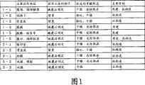
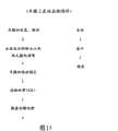
图1是汇总了作为组织再建用材料的片状组合物的用途(主要应用部位、应用方法的例子,优选的羊膜形态、主要目的)的表。Fig. 1 is a table summarizing the uses (main application sites, examples of application methods, preferred amniotic membrane form, main purpose) of sheet-like compositions as materials for tissue reconstruction.
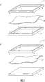
图2是说明羊膜的固定法的图。(a)是以一对框夹持羊膜;(b)是以框和平板状部件夹持羊膜。Fig. 2 is a diagram illustrating a method of fixing amnion. (a) the amniotic membrane is clamped by a pair of frames; (b) the amniotic membrane is clamped by a frame and a plate-shaped part.
图3是显示海藻糖处理·冻干羊膜的物性评价试验(厚度)结果的图。Fig. 3 is a graph showing the results of a physical property evaluation test (thickness) of trehalose-treated and freeze-dried amniotic membrane.
图4是显示海藻糖处理·冻干羊膜的物性评价试验(透明度)结果的图。Fig. 4 is a graph showing the results of a physical property evaluation test (transparency) of trehalose-treated and freeze-dried amnion.
图5是显示海藻糖处理·冻干羊膜的物性评价试验(拉伸强度)结果的图。Fig. 5 is a graph showing the results of a physical property evaluation test (tensile strength) of trehalose-treated and freeze-dried amniotic membrane.
图6是显示海藻糖处理·冻干羊膜的物性评价试验(柔软性)结果的图。Fig. 6 is a graph showing the results of a physical property evaluation test (softness) of trehalose-treated and freeze-dried amniotic membrane.
图7是显示海藻糖处理·冻干羊膜的活体适合性评价试验结果的图。将海藻糖处理·冻干羊膜移植到家兔的角膜实质层间,观察眼表面的状态。左边是刚移植完的眼表面状态,右边是移植后一个月的眼表面状态。Fig. 7 is a graph showing the results of an in vivo suitability evaluation test of trehalose-treated and freeze-dried amnion. The trehalose-treated and freeze-dried amniotic membrane was transplanted into the corneal parenchyma of rabbits, and the state of the ocular surface was observed. On the left is the state of the eye surface just after transplantation, and on the right is the state of the eye surface one month after transplantation.
图8是显示海藻糖处理·冻干羊膜的活体适合性评价试验结果的图。将海藻糖处理·冻干羊膜移植到家兔的角膜实质层间,在移植后1个月时将包含移植部的角膜的一部分摘出进行HE染色。Fig. 8 is a graph showing the results of an in vivo suitability evaluation test of trehalose-treated and freeze-dried amnion. The trehalose-treated and freeze-dried amniotic membrane was transplanted between the parenchymal layers of the rabbit cornea, and a part of the cornea including the transplanted part was removed one month after the transplantation and subjected to HE staining.
图9是显示在海藻糖处理·冻干羊膜上培养角膜上皮细胞时的器具等状态的剖面模式图。培养嵌套12静置于培养皿11内,在培养皿11底面形成3T3细胞层15。此外,显示了如下状态:在培养嵌套12的底面上静置羊膜13,在其上培养角膜上皮细胞14。符号16为培养基。Fig. 9 is a schematic cross-sectional view showing the state of equipment and the like when culturing corneal epithelial cells on trehalose-treated and freeze-dried amniotic membrane. The
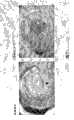
图10是显示在海藻糖处理·冻干羊膜上形成细胞层的图(HE染色图像)。将使用了未进行海藻糖处理而进行冻干所得到的羊膜(未处理冻干羊膜)时所形成的细胞层状态作为比较对照。Fig. 10 is a diagram showing the formation of a cell layer on trehalose-treated and freeze-dried amnion (HE staining image). The state of the cell layer formed when the amniotic membrane obtained by lyophilization without trehalose treatment (untreated lyophilized amniotic membrane) was used was used as a comparative control.
图11是在海藻糖处理·冻干羊膜上形成细胞层的片(培养角膜上皮片)的HE染色图像和免疫染色图像。在免疫染色图像中,各抗体的信号为绿色。细胞核以红色表示。角膜型角蛋白3表达(+)、皮肤角化型角蛋白10(-)、结膜型角蛋白13(-)。Fig. 11 shows HE staining images and immunostaining images of a sheet (cultured corneal epithelial sheet) in which a cell layer was formed on trehalose-treated and freeze-dried amnion. In the immunostaining images, the signal of each antibody is green. Nuclei are indicated in red.
图12是显示使用海藻糖处理·冻干羊膜制作的培养角膜上皮片的再建效果的图。在培养角膜上皮片移植后第二天以及第十四天的眼表面的状态(上面)以及荧光素染色图像(下面)。Fig. 12 is a graph showing the reconstruction effect of a cultured corneal epithelial sheet prepared using trehalose-treated and freeze-dried amniotic membrane. State of the ocular surface (top) and fluorescein staining images (bottom) on the second day and fourteenth day after cultured corneal epithelial sheet transplantation.
图13是移植后第二周的培养角膜上皮片的HE染色图像(左上边)和关于各种角蛋白的免疫染色图像(右上边、下边)。记号**表示海藻糖处理·冻干羊膜(TH-AM)。Fig. 13 shows HE staining images (upper left) and immunostaining images for various keratins (upper right, lower) of cultured corneal epithelial sheets at the second week after transplantation. The mark ** represents trehalose-treated and freeze-dried amniotic membrane (TH-AM).
图14是使用了对基底膜特异性成分的抗体以及对致密层特异性成分的抗体将海藻糖处理·冻干羊膜进行免疫染色后的结果。显示将海藻糖处理·冻干羊膜的免疫染色图像(C)与未加工羊膜(A)的免疫染色图像以及未经海藻糖处理但进行冻干的羊膜的免疫染色图像(B)进行比较。1:胶原I的染色图像;2:胶原III的染色图像;3:胶原IV的染色图像;4:胶原V的染色图像;5:胶原Ⅶ的染色图像;6:层粘连蛋白5的染色图像;7:纤连蛋白的染色图像。Fig. 14 shows the results of immunostaining of trehalose-treated and lyophilized amniotic membranes using antibodies to basement membrane-specific components and antibodies to compact layer-specific components. The immunostaining image (C) of trehalose-treated-lyophilized amniotic membrane is shown in comparison with that of unprocessed amniotic membrane (A) and immunostaining image of non-trehalose-treated but lyophilized amniotic membrane (B). 1: Collagen I staining image; 2: Collagen III staining image; 3: Collagen IV staining image; 4: Collagen V staining image; 5: Collagen VII staining image; 6:
图15是显示羊膜上皮的去除顺序的流程图。Fig. 15 is a flowchart showing the removal sequence of the amniotic epithelium.
图16是显示胰蛋白酶处理的方法的图。(a)是将框固定的羊膜的上皮侧朝下浸渍于胰蛋白酶溶液中。(b)是向框内加入胰蛋白酶溶液,从而使胰蛋白酶作用于羊膜的上皮侧。Fig. 16 is a graph showing the method of trypsin treatment. (a) is immersing the epithelial side of the frame-fixed amnion in a trypsin solution. (b) is adding a trypsin solution into the frame so that trypsin acts on the epithelial side of the amniotic membrane.
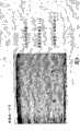
图17是经胰蛋白酶处理的羊膜、未加工羊膜(有上皮)、用手处理的羊膜的HE染色图像。Figure 17 is HE staining images of trypsinized amnion, unprocessed amnion (with epithelium), and hand-treated amnion.
图18是经胰蛋白酶处理的羊膜的免疫染色图像。Figure 18 is an immunostaining image of trypsinized amnion.
图19同样是经胰蛋白酶处理的羊膜的免疫染色图像。Figure 19 is also an immunostaining image of trypsinized amnion.
图20是未加工羊膜(有上皮)的免疫染色图像。Figure 20 is an immunostained image of naive amnion (with epithelium).
图21是用手处理的羊膜的免疫染色图像。Figure 21 is an immunostained image of amnion processed by hand.
图22是汇总HE染色实验以及免疫实验结果的表。Fig. 22 is a table summarizing the results of HE staining experiments and immunoassays.
标号说明Label description
1、2、3框1, 2, 3 boxes
4 板状部件4 plate parts
5 胰蛋白酶溶液5 trypsin solution
10 羊膜10 amnion
11 培养皿(culture dish)11 culture dish
12 培养嵌套(培养插入容器)12 culture nests (culture inserts)
13 羊膜13 amnion
14 角膜上皮细胞14 Corneal epithelial cells
15 3T3细胞层15 3T3 cell layer
16 培养基16 media
具体实施方式Detailed ways
本发明的第1方面涉及片状组合物。在本发明的片状组合物中使用羊膜作为其主要构成成分。由于羊膜所具有的高透明性及强韧性,所以可以构成透明性及强度优异的片状组合物。此外,由于羊膜的高活体亲和性及低免疫原性,所以可以构成活体亲和性更加优异且免疫原性低的片状组合物。通过使用羊膜,可以进一步期待抗炎症作用、抑制瘢痕形成、阻碍血管新生的作用。在本发明的片状组合物含有细胞层的情况下,在良好地形成该细胞层方面,也优选使用羊膜。即,如后所述,具备细胞层的片状组合物是通过以羊膜为基质(支持体)并在其上播种、培养规定的细胞而形成,结果由于羊膜具有使细胞在其上良好地粘附及增殖的特性,所以使用羊膜则细胞可以良好地粘附及增殖而形成细胞层。The first aspect of the present invention relates to a sheet-like composition. Amniotic membrane is used as the main constituent in the sheet-like composition of the present invention. Due to the high transparency and toughness of the amniotic membrane, it is possible to form a sheet-like composition with excellent transparency and strength. In addition, due to the high in vivo affinity and low immunogenicity of amniotic membrane, it is possible to constitute a sheet-like composition with further excellent in vivo affinity and low immunogenicity. By using amniotic membrane, further anti-inflammatory action, scar formation suppression, and angiogenesis-inhibiting action can be expected. When the sheet-like composition of the present invention contains a cell layer, it is also preferable to use amnion in order to form the cell layer well. That is, as described later, the sheet-like composition with the cell layer is formed by seeding and culturing predetermined cells on the amniotic membrane as a matrix (support). It has the property of proliferation, so when amnion is used, the cells can adhere and proliferate well to form a cell layer.
(羊膜的来源)(source of amniotic membrane)
“羊膜”是哺乳动物中覆盖子宫和胎盘最表层的膜,在胶原丰富的软组织上形成基底膜、上皮层而构成。可以使用人、猴子、黑猩猩、猪、马、牛等的羊膜。其中,优选使用人羊膜。这是因为在包括免疫原性、病毒感染的安全性方面有利。"Amniotic membrane" is the membrane that covers the outermost layer of the uterus and placenta in mammals, and is composed of a basement membrane and an epithelial layer on collagen-rich soft tissues. Amniotic membranes of humans, monkeys, chimpanzees, pigs, horses, cows, etc. can be used. Among them, human amnion is preferably used. This is because it is advantageous in aspects including immunogenicity and safety of viral infection.
(海藻糖的附加)(addition of trehalose)
本发明的片状组合物使用附加了海藻糖的羊膜。本发明人等的研究结果表明,通过附加海藻糖,可以提高羊膜的柔软性、特别是可大幅改善处于冻干状态时的柔软性。并且,如后述实施例所示,表明了附加海藻糖的羊膜作为细胞培养用基质发挥了良好的功能。基于这些见解,构成的本发明的片状组合物具有高度柔软性、另一方面在作为细胞培养用基质使用时可进行良好的细胞增殖和多层化。The sheet composition of the present invention uses trehalose-added amniotic membrane. As a result of research by the inventors of the present invention, it has been revealed that the addition of trehalose can improve the flexibility of the amniotic membrane, especially the flexibility in a freeze-dried state can be greatly improved. Furthermore, as shown in Examples described below, it was shown that the amniotic membrane to which trehalose was added functions well as a substrate for cell culture. Based on these findings, the sheet-like composition of the present invention constituted has high flexibility and, on the other hand, enables good cell growth and multilayering when used as a substrate for cell culture.
这里,如果羊膜内部的基质蛋白变弱,则羊膜的强度降低,并且容易受到损害。并且,不能将水分牢固地保持在内部,会变脆弱而失去弹性。海藻糖作用于基质蛋白内结合松散的部位从而强化了蛋白质间的结合,由此使羊膜内部的保持水分的功能正常化,预想维持了羊膜本来的湿润、聚集、弹性。认为通过预先附加海藻糖,可有效防止伴随着冻干处理的羊膜内部的基质蛋白变柔软,特别是在水中的膨润变弱的情况。Here, if the matrix protein inside the amnion becomes weak, the strength of the amnion decreases and it is easily damaged. Also, it cannot hold moisture firmly inside and becomes weak and loses its elasticity. Trehalose acts on the loosely bonded part of the matrix protein to strengthen the bond between proteins, thereby normalizing the function of retaining water inside the amnion, and it is expected to maintain the original moisture, aggregation, and elasticity of the amnion. It is considered that adding trehalose in advance can effectively prevent the softening of the matrix protein inside the amniotic membrane accompanying the freeze-drying process, especially the weakening of swelling in water.
海藻糖(物质名,通称)是以α-D-吡喃葡糖基(1,1)-α-D-吡喃葡萄糖苷来表示的化合物。Trehalose (substance name, general name) is a compound represented by α-D-glucopyranosyl(1,1)-α-D-glucopyranoside.
对羊膜附加海藻糖,可以通过例如用海藻糖溶液处理羊膜来进行。另外,后面详细描述了海藻糖的附加方法。Adding trehalose to the amnion can be performed, for example, by treating the amnion with a trehalose solution. In addition, the method of adding trehalose will be described in detail later.
另外,本发明的一种实施方式实质上是仅由附加了海藻糖的羊膜构成的。In addition, one embodiment of the present invention consists substantially only of amniotic membrane to which trehalose has been added.
(羊膜的状态)(state of amniotic membrane)
本发明的一种实施方式是使用去除了上皮细胞层的羊膜。去除了上皮细胞层的羊膜完全没有因上皮细胞所引起的免疫排斥等问题,安全性变得极高。并且,细胞在去除了上皮的羊膜上更加良好地粘附以及增殖,因此也可获得能够在更短时间构建高品质的细胞片这种制作方面的优点。One embodiment of the invention is the use of amnion from which the epithelial cell layer has been removed. The amniotic membrane from which the epithelial cell layer has been removed has no problems such as immune rejection caused by epithelial cells, and the safety becomes extremely high. In addition, since the cells adhere and proliferate more favorably on the amniotic membrane from which the epithelium has been removed, there is also an advantage of being able to construct a high-quality cell sheet in a shorter time.
通过检查本发明的片状组合物中不含羊膜上皮层的细胞,能够确认是去除了上皮层的羊膜。By examining cells that do not contain the amniotic epithelial layer in the sheet-like composition of the present invention, it can be confirmed that the amniotic membrane is the amniotic membrane from which the epithelial layer has been removed.
另一方面,也可以使用残存有上皮层的羊膜来构建本发明的片状组合物。通过预先使羊膜的上皮层残存,可在制作阶段进行γ射线处理等充分的灭菌处理,可实现安全性的提高。On the other hand, amniotic membrane with the remaining epithelial layer can also be used to construct the sheet-like composition of the present invention. By leaving the epithelial layer of the amniotic membrane in advance, sufficient sterilization treatment such as γ-ray treatment can be performed at the production stage, and safety can be improved.
(再构建后的羊膜的使用)(use of reconstituted amnion)
使用再构建后的羊膜也能构成本发明的片状组合物。具体而言,例如可以使用经匀浆器、超声波、酶处理而暂时分解,并再次构建成膜状的形状的羊膜。处理方法优选使用匀浆器。这是由于能期待基底膜的微小结构体较高度地保持。匀浆器处理的条件(转数)为例如3000rpm~50000rpm,优选为10000rpm~40000rpm,更优选为约30000rpm。The sheet-like composition of the present invention can also be formed using reconstituted amnion. Specifically, for example, amnion that is once decomposed by a homogenizer, ultrasonic wave, or enzyme treatment, and then reconstructed into a membrane-like shape can be used. The processing method preferably uses a homogenizer. This is because the microstructure of the basement membrane can be expected to be maintained at a relatively high level. The conditions (number of rotations) of the homogenizer treatment are, for example, 3000 rpm to 50000 rpm, preferably 10000 rpm to 40000 rpm, more preferably about 30000 rpm.
(厚度)(thickness)
通过使用羊膜,本发明的片状组合物可构成非常薄的片状。本发明的片状组合物可调制成例如10μm~500μm的厚度。如此的非常薄的片状会增加通用性。也可以使用去除了绒毛膜侧的致密层的一部分(例如10μm~30μm左右)的羊膜来构建本发明的片状组合物,还可以通过涂敷活体吸收性原材料而制成例如100μm~500μm左右的厚度。By using amnion, the sheet composition of the present invention can be formed into a very thin sheet. The sheet-like composition of the present invention can be adjusted to have a thickness of, for example, 10 μm to 500 μm. Such a very thin sheet adds versatility. The sheet-like composition of the present invention can also be constructed using amniotic membrane from which a part of the dense layer on the chorionic membrane side (for example, about 10 μm to 30 μm) has been removed, or it can be formed by coating a bioabsorbable material, for example, with a thickness of about 100 μm to 500 μm. thickness.
(粘附成分的使用)(Use of Adhesive Components)
本发明的一种实施方式是将纤维蛋白原以及凝血酶(以下,也将它们统称为“粘附成分”)附着于羊膜的表面。由此,在移植本发明的片状组合物时,首先,通过凝血酶特异性地水解纤维蛋白原而生成纤维蛋白,接着,纤维蛋白聚合而形成稳定的纤维蛋白块,从而发挥粘附作用。如果这样提高了粘附性,在将片状组合物应用于患处时,无需缝合也能获得足够的粘附力,由此可实现手术方式的简便化。另外,在本说明书中,将具有上皮且附着有粘附成分的羊膜还称作“粘附成分附着·有上皮羊膜”,将无上皮但附有粘附成分的羊膜还称作“粘附成分附着·无上皮羊膜”。One embodiment of the present invention is to attach fibrinogen and thrombin (hereinafter, these are also collectively referred to as "adhesion components") to the surface of the amniotic membrane. Thus, when the sheet-like composition of the present invention is transplanted, first, fibrin is generated by thrombin specifically hydrolyzing fibrinogen, and then the fibrin is polymerized to form a stable fibrin mass, thereby exerting an adhesion function. If the adhesiveness is improved in this way, when the sheet-like composition is applied to the affected area, sufficient adhesive force can be obtained without suturing, thereby enabling the simplification of the surgical procedure. In addition, in this specification, the amniotic membrane having epithelium and adherent components is also referred to as "adhesive component-adhered and epithelial amnion", and the amniotic membrane without epithelium but with adhesive components is also referred to as "adhesive component". Adhesive · Epithelial Amniotic Membrane".
纤维蛋白原及凝血酶根据本发明的片状组合物的用途,附着在羊膜的单面或两面。单面附着时,无论羊膜上有无上皮,羊膜的绒毛膜侧表面(即,与上皮侧相反侧的表面)成为附着面。因此,使用如此构成的片状组合物时,使存在羊膜上皮的侧朝上,移植到应用部位。另外,以抗粘连为目的的片状组合物时,制成粘附成分附着于羊膜的单面。以活体粘附剂为目的移植到活体内时,优选在羊膜的两面附着粘附成分。Fibrinogen and thrombin adhere to one or both sides of the amniotic membrane according to the use of the sheet-like composition of the present invention. In the case of single-sided attachment, the chorionic surface of the amnion (that is, the surface opposite to the epithelial side) becomes the attachment surface regardless of whether there is epithelium on the amnion. Therefore, when using the sheet-like composition constituted in this way, the side where the amniotic epithelium is present is turned upward, and transplanted to the application site. In addition, in the case of a sheet-like composition for the purpose of anti-adhesion, the adhesive component is made to adhere to one side of the amniotic membrane. When transplanting into a living body for the purpose of a bioadhesive, it is preferable to attach an adhesive component to both sides of the amniotic membrane.
如后所述,该方式的片状组合物考虑到使用目的等,制备成适当的状态(例如干燥状态或湿润状态),并经过在羊膜表面附着纤维蛋白原及凝血酶的工序来制备。因此,根据其制作过程中及/或其最终状态如何,可预想直到被使用为止的期间由一部分纤维蛋白原生成纤维蛋白。因此,本发明的片状组合物还包括附着有因这样的原因生成的纤维蛋白或纤维蛋白块的物质。As will be described later, the sheet-like composition of this embodiment is prepared in an appropriate state (for example, dry state or wet state) in consideration of the purpose of use, and is prepared through a process of attaching fibrinogen and thrombin to the surface of the amniotic membrane. Therefore, depending on the production process and/or its final state, it is expected that a part of fibrinogen will generate fibrin until it is used. Therefore, the sheet-like composition of the present invention also includes those to which fibrin or fibrin lumps generated for such reasons adhere.
纤维蛋白原及凝血酶的来源没有特别限定。例如,可以把人、猴子、黑猩猩、牛、马、羊、猪等的血液作为材料来制备纤维蛋白原及凝血酶。比外,作为纤维蛋白原及凝血酶,可以使用利用培养细胞(例如CHO细胞或COS细胞)而得到的重组体(recombinant)。优选使用人来源(特别是人来源的重组体)的纤维蛋白原及凝血酶。这是因为在包括免疫原性的安全性方面有利。此外,如果考虑可以使用稳定品质的物质及感染的问题,则特别优选使用重组体。The sources of fibrinogen and thrombin are not particularly limited. For example, fibrinogen and thrombin can be prepared from the blood of humans, monkeys, chimpanzees, cows, horses, sheep, pigs, and the like. In addition, as fibrinogen and thrombin, recombinants obtained from cultured cells (such as CHO cells or COS cells) can be used. Fibrinogen and thrombin of human origin (especially recombinant human origin) are preferably used. This is because it is advantageous in terms of safety including immunogenicity. In addition, it is particularly preferable to use a recombinant in consideration of the possibility of using a substance of stable quality and the problem of infection.
特别优选使用来源于接受本发明的片状组合物移植的患者(recipient)的血液的纤维蛋白原及凝血酶。这是因为不用担心会发生起因于这些粘附成分的免疫排斥反应。It is particularly preferable to use fibrinogen and thrombin derived from the blood of a recipient who has been transplanted with the sheet-like composition of the present invention. This is because there is no fear of immune rejection due to these adhesion components.
另外,纤维蛋白原及凝血酶的来源可以不必相同。如果列举一例,可以将人血液来源的纤维蛋白原和牛血液来源的凝血酶组合使用。In addition, the sources of fibrinogen and thrombin do not have to be the same. As an example, fibrinogen derived from human blood and thrombin derived from bovine blood can be used in combination.
纤维蛋白原及凝血酶的附着量没有特别限定。例如,纤维蛋白原的附着量可以设定在每1cm2羊膜0.1mg~50mg的范围。同样地,凝血酶的附着量可以设定在每1cm2羊膜0.5μmg~10mg的范围。The attached amounts of fibrinogen and thrombin are not particularly limited. For example, the attached amount of fibrinogen can be set in the range of 0.1 mg to 50 mg per 1 cm2 of amniotic membrane. Similarly, the amount of thrombin attached can be set in the range of 0.5 μmg to 10 mg per 1 cm2 of amniotic membrane.
设定纤维蛋白原及凝血酶的附着量时首要考虑粘附力。即,为了获得所希望的粘附力,必须设定这些成分的附着量。另一方面,纤维蛋白原或凝血酶的附着量过多时,虽然根据所使用的纤维蛋白原的来源来决定,但是容易发生免疫反应或血管新生这一点成为问题。Adhesion force is the primary consideration when setting the attachment amount of fibrinogen and thrombin. That is, in order to obtain a desired adhesive force, it is necessary to set the adhesion amount of these components. On the other hand, when the attached amount of fibrinogen or thrombin is too large, although it depends on the source of the fibrinogen used, it becomes a problem that an immune reaction or angiogenesis is likely to occur.
在此,例如在片状组合物被应用于眼表面的再建等的情况下,在移植后这些成分导致的血管新生成为问题时,为了尽可能抑制血管新生的发生,优选减少这些成分的附着量。通过将这些成分的附着量设定在可能范围内的低值,可抑制移植后的血管新生,期待高的治疗效果。本发明人等的研究结果表明:当将纤维蛋白原与羊膜组合使用的情况下,当纤维蛋白原的附着量为每1cm2羊膜约0.5mg以上时,可获得对眼表面的良好粘附力。即使凝血酶的附着量为每1cm2羊膜约1μg时,也可获得对眼表面的良好粘附力。基于这些见解,纤维蛋白原附着量优选的范围为每1cm2羊膜0.5~20mg,进一步优选的范围为每1cm2羊膜0.5mg~10mg,更进一步优选的范围为每1cm2羊膜0.5mg~6mg(具体地说,例如约0.5mg、约1mg、约2mg)。同样地,凝血酶附着量优选的范围为每1cm2羊膜1μg~1mg,进一步优选的范围为每1cm2羊膜5μg~200μg,更进一步优选的范围为每1cm2羊膜10μg~100μg(具体地说,例如约10μg、约20μg、约30μg)。Here, for example, when the sheet-like composition is applied to reconstruction of the ocular surface, etc., when angiogenesis caused by these components after transplantation becomes a problem, it is preferable to reduce the amount of adhesion of these components in order to suppress the occurrence of angiogenesis as much as possible. . By setting the adhesion amount of these components to a low value within the possible range, angiogenesis after transplantation can be suppressed, and a high therapeutic effect can be expected. As a result of research by the inventors of the present invention, when fibrinogen is used in combination with amniotic membrane, good adhesion to the ocular surface can be obtained when the attachment amount of fibrinogen is about 0.5 mg or more per 1 cm2 of amniotic membrane . Good adhesion to the ocular surface was obtained even when the amount of thrombin attached was about 1 μg per 1 cm ofamniotic membrane. Based on these findings, the preferred range of fibrinogen adhesion is 0.5-20 mg per 1 cm2 of amniotic membrane, a more preferred range is 0.5 mg-10 mg per 1 cm2 of amniotic membrane, and a further preferred range is 0.5 mg-6 mg per 1 cm2 of amniotic membrane ( Specifically, eg about 0.5 mg, about 1 mg, about 2 mg). Likewise, the thrombin adhesion amount preferably ranges from 1 μg to 1 mg per 1 cmof amniotic membrane, a more preferred range of 5 μg to 200 μg per1 cmof amniotic membrane, and an even more preferred range of 10 μg to 100 μg per 1 cm of amniotic membrane (specifically, eg about 10 μg, about 20 μg, about 30 μg).
在本发明的一种实施方式中,除了纤维蛋白原及凝血酶以外,还有抑肽酶附着在羊膜表面。抑肽酶阻碍如下过程:通过凝血酶的作用而形成的纤维蛋白块被纤维蛋白溶酶溶解。因此,通过并用抑肽酶可抑制纤维蛋白块的分解,可维持甚至强化粘附力。In one embodiment of the present invention, in addition to fibrinogen and thrombin, aprotinin is attached to the amniotic membrane surface. Aprotinin blocks the process by which fibrin clots formed by the action of thrombin are dissolved by plasmin. Therefore, by using aprotinin in combination, the decomposition of fibrin lumps can be suppressed, and the adhesive force can be maintained or even strengthened.
抑肽酶的来源没有特别限定。例如,可以使用牛、马、羊、猪、猴子、黑猩猩等的胰脏来源的抑肽酶。此外,还可以使用利用培养细胞(例如CHO细胞或COS细胞)而得到的重组体(recombinant)的抑肽酶。如果考虑可以使用稳定品质的物质及感染问题,则优选使用重组体。The source of aprotinin is not particularly limited. For example, aprotinin derived from the pancreas of bovines, horses, sheep, pigs, monkeys, chimpanzees, etc. can be used. In addition, recombinant aprotinin obtained from cultured cells (eg, CHO cells or COS cells) can also be used. Considering the possibility of using a substance of stable quality and the problem of infection, it is preferable to use a recombinant.
使用抑肽酶时,其附着量没有特别限定。例如,可以将抑肽酶的附着量设定在每1cm2羊膜0.1KIU~200KIU的范围。根据上述的纤维蛋白原附着量的研究,使抑肽酶的附着量变化来研究对于粘附力的影响,结果表明,在与羊膜组合使用时即使将抑肽酶量设定在1KIU~2KIU的范围,也可以得到对眼表面的充分粘附力。基于该见解,抑肽酶附着量优选的范围为每1cm2羊膜1KIU~100KIU,进一步优选的范围为每1cm2羊膜1KIU~20KIU,更进一步优选的范围为每1cm2羊膜1KIU~10KIU(具体地说,例如约1KIU、约2KIU、约3KIU)。抑肽酶的量过多时,自然会引起制造成本的上升,起因于抑肽酶自身的免疫原性等的副作用的可能性增大。另一方面,抑肽酶的量过少时,可能不能充分发挥抑制纤维蛋白块分解的抑肽酶的作用。When aprotinin is used, the attached amount is not particularly limited. For example, the amount of aprotinin attached can be set in the range of 0.1 KIU to 200 KIU per 1 cm2 of amniotic membrane. According to the study on the amount of aprotinin attached above, the effect on the adhesion was studied by changing the amount of aprotinin attached. range, sufficient adhesion to the ocular surface can also be obtained. Based on this finding, the preferred range of aprotinin adhesion is 1 KIU to 100 KIU per 1 cmof amniotic membrane, the more preferred range is 1 KIU to 20 KIU per1 cm of amniotic membrane, and the further preferred range is 1 KIU to 10 KIU per 1 cm of amnioticmembrane (specifically Say, for example, about 1 KIU, about 2 KIU, about 3 KIU). When the amount of aprotinin is too large, the production cost will naturally increase, and the possibility of side effects such as immunogenicity of aprotinin itself will increase. On the other hand, when the amount of aprotinin is too small, the action of aprotinin for inhibiting the decomposition of fibrin clumps may not be sufficiently exhibited.
另外,在各种用途中纤维蛋白块被用作粘附剂,在那样的用途中一般并用抑肽酶。本发明人等的研究结果表明,与羊膜进行组合时,即使不使用抑肽酶也可得到对于活体的充分粘附力。可以不使用抑肽酶,意味着不仅简化构成而在制作上及成本方面上有利,而且没有必要考虑起因于抑肽酶自身的免疫原性等的副作用。In addition, fibrin clots are used as adhesives in various applications, and aprotinin is generally used in combination in such applications. As a result of research by the inventors of the present invention, it has been shown that when combined with amnion, sufficient adhesion to a living body can be obtained without using aprotinin. The fact that aprotinin can not be used means that not only the structure is simplified, which is advantageous in terms of production and cost, but also there is no need to consider side effects such as immunogenicity of aprotinin itself.
(利用活体吸收性材料的强化)(reinforcement by bioabsorbent material)
以活体吸收性材料被覆羊膜的绒毛膜侧,由此能够提高本发明的片状组合物的强度。作为以该目的使用的活体吸收性材料,优选采用在较早期被羊膜分解·吸收的材料。例如可适当使用例如聚乳酸羟基乙酸(polyglactin)910、明胶、胶原、聚乳酸等作为此处的活体吸收性材料。用于强化的活体吸收性材料的形态没有特别限定。例如以成形为网眼状或者片状的活体吸收性材料被覆羊膜绒毛膜侧从而强化羊膜。羊膜的状态在进行强化的过程中可以是湿润状态、干燥状态任一种。但是,产品的最终形态优选羊膜为干燥状态。这是因为如果是干燥状态则操作性以及保存性优异。需要说明的是,本说明书中的实施了强化的羊膜也称作“杂化羊膜”。The strength of the sheet-like composition of the present invention can be increased by covering the chorionic side of the amniotic membrane with a bioabsorbable material. As the bioabsorbable material used for this purpose, it is preferable to use a material that is decomposed and absorbed by the amnion at an early stage. For example, polylactic acid glycolic acid (polyglactin) 910, gelatin, collagen, polylactic acid, etc. can be used suitably as a bioabsorbable material here. The form of the bioabsorbable material used for reinforcement is not particularly limited. For example, the amniotic membrane is reinforced by covering the amniotic chorion side with a bioabsorbable material formed into a mesh shape or a sheet shape. The state of the amniotic membrane can be either a wet state or a dry state during the strengthening process. However, the final form of the product is preferably the amnion in a dry state. This is because handling properties and storage properties are excellent in a dry state. In addition, the reinforced amniotic membrane in this specification is also called "hybrid amniotic membrane".
(细胞层)(cell layer)
本发明的片状组合物的一种实施方式为在羊膜上具有细胞层。在该实施方式中,并用粘附成分时,在未形成细胞层的侧的羊膜表面附着有粘附成分(纤维蛋白原等)。One embodiment of the sheet-like composition of the present invention has a cell layer on the amnion. In this embodiment, when an adhesion component is used in combination, the adhesion component (fibrinogen, etc.) adheres to the surface of the amniotic membrane on the side where the cell layer is not formed.
在该实施方式中,使用去除了上皮的羊膜。从而,在上皮存在的一侧形成细胞层。此处的细胞层由活体来源的细胞构成。构成细胞层的细胞的来源没有特别限定。例如,由角膜上皮、结膜上皮、皮肤表皮、毛囊上皮、口腔粘膜上皮、虹膜色素上皮、视网膜色素上皮、呼吸道粘膜上皮或者肠道粘膜上皮来源的细胞构成细胞层。可以由相互不同的二种以上的细胞构成细胞层。这样,通过含有来源不同的二种以上的细胞来构成,本说明书中也称之为“杂化”。经杂化的细胞层中的细胞的含有形态(杂化状态)没有特别限定,例如,各细胞可以分散,任意细胞(或者多种类的细胞)可以具有一定的聚集而存在。此外,各细胞的含有率在细胞层整体中可以不是均匀的。细胞层可以是单层,也可以是多层(多层化的状态)。In this embodiment, amnion from which the epithelium has been removed is used. Thus, a cell layer is formed on the side where the epithelium exists. The cell layer here consists of cells of living origin. The source of cells constituting the cell layer is not particularly limited. For example, the cell layer is composed of cells derived from corneal epithelium, conjunctival epithelium, skin epithelium, hair follicle epithelium, oral mucosal epithelium, iris pigmented epithelium, retinal pigmented epithelium, respiratory tract mucosal epithelium, or intestinal mucosal epithelium. The cell layer may be composed of two or more different types of cells. In this way, it is constituted by containing two or more types of cells from different origins, which is also referred to as "hybridization" in this specification. The form of cells contained in the hybridized cell layer (hybridization state) is not particularly limited, for example, individual cells may be dispersed, and arbitrary cells (or a plurality of types of cells) may exist in a certain aggregation. In addition, the content rate of each cell may not be uniform throughout the entire cell layer. The cell layer may be a single layer or a multilayer (multilayered state).
具有经杂化的细胞层时,构成细胞层的细胞种类,以构建用作角膜上皮的再建的本发明的片状组合物的情况为例,详述如下。When there is a hybridized cell layer, the cell types constituting the cell layer will be described in detail below, taking the case of constructing the sheet-like composition of the present invention used for reconstruction of the corneal epithelium as an example.
经杂化的细胞层含有二种以上的细胞。因此,为了方便说明,把细胞层所含有的细胞种类之一当作第1细胞,把与其种类不同的细胞种类当作第2细胞。首先,使用自体细胞作为第1细胞。本说明书中的“自体”是指应用本发明的片状组合物的对象,即接受移植的人(recipient)。另一方面,这样的“自体”以外的人称为“他人”。只要在与后述的第2细胞杂化时能够形成角膜上皮样的粘膜上皮层,则第1细胞的种类没有特别限定。作为第1细胞的例子,可列举来源于口腔粘膜上皮、结膜上皮、鼻腔粘膜上皮等粘膜上皮的细胞,或者来源于能够构建这样的粘膜上皮的未分化细胞(即粘膜上皮的干细胞)的细胞。在此“来源于或者来源的”是指为了特别指定起始材料而使用的。因此,例如,如果说来源于口腔粘膜上皮的(或者口腔粘膜上皮来源的)细胞,则是指以口腔粘膜上皮细胞为起始材料而获得的细胞。此外,本发明中的“能够构建粘膜上皮的未分化细胞”是指具有向构成该粘膜上皮的细胞分化的能力的细胞。例如,如果说能够构建口腔粘膜上皮的未分化细胞,则是指能够向口腔粘膜上皮细胞分化的细胞。作为未分化细胞的具体例,可举出口腔粘膜上皮或结膜上皮等、构成特定组织的细胞的前体或者干细胞、或者分化度低的上皮系干细胞等。A hybridized cell layer contains two or more types of cells. Therefore, for convenience of description, one of the cell types included in the cell layer is regarded as a first cell, and a cell type different from the cell type is regarded as a second cell. First, autologous cells are used as the first cells. "Autologous" in this specification refers to a subject to whom the sheet-like composition of the present invention is applied, that is, a recipient of a transplant. On the other hand, people other than such "self" are called "others". The type of the first cell is not particularly limited as long as it can form a corneal epithelial-like mucosal epithelial layer when hybridized with the second cell described later. Examples of the first cells include cells derived from mucosal epithelium such as oral mucosal epithelium, conjunctival epithelium, and nasal cavity mucosal epithelium, or cells derived from undifferentiated cells capable of constructing such mucosal epithelium (i.e., mucosal epithelial stem cells). "Derived from or derived from" herein means used in order to specifically designate a starting material. Thus, for example, if a cell derived from oral mucosal epithelium (or oral mucosal epithelium-derived) is said, it means a cell obtained from oral mucosal epithelial cells as a starting material. In addition, "undifferentiated cells capable of constructing mucosal epithelium" in the present invention refers to cells having the ability to differentiate into cells constituting the mucosal epithelium. For example, undifferentiated cells capable of constructing oral mucosal epithelium refer to cells capable of differentiating into oral mucosal epithelial cells. Specific examples of undifferentiated cells include oral mucosal epithelium, conjunctival epithelium, etc., precursors or stem cells of cells constituting specific tissues, or epithelial stem cells with a low degree of differentiation.
经杂化的细胞层可以含有不同的二种以上的第1细胞。例如,可以在含有来源于口腔粘膜上皮的细胞和来源于结膜上皮的细胞的状态下构建细胞层。The hybridized cell layer may contain two or more different first cells. For example, a cell layer can be constructed in a state containing cells derived from oral mucosal epithelium and cells derived from conjunctival epithelium.
本发明中的“口腔粘膜上皮”包括口腔内缘粘膜上皮部、口唇部、口上颚部、颊部等。其是来源于口腔粘膜上皮的细胞,可以以表达口腔粘膜上皮特异性的角蛋白4、或角蛋白13为指标来确认。或者,还可以以表达角蛋白3为指标。已知这种角蛋白3是角膜特异性的角蛋白之一,但在口腔粘膜上皮中也确认其有表达。另外,从表达这种作为角膜特异性角蛋白的角蛋白3的角度讲,可优选使用口腔粘膜上皮细胞作为制作角膜上皮移植用的组合物的材料。The "oral mucosal epithelium" in the present invention includes oral mucosal epithelium, lips, palate, cheeks, and the like. These are cells derived from the oral mucosal epithelium, and can be confirmed by expressing oral mucosal epithelium-specific keratin 4 or keratin 13 as an index. Alternatively, expression of
另一方面,通过研究口腔粘膜上皮细胞特异性的基因的表达,可以确认其为口腔粘膜上皮来源。On the other hand, by studying the expression of genes specific to oral mucosal epithelial cells, it can be confirmed that it is derived from oral mucosal epithelium.
另外,来源于口腔粘膜上皮以外的组织的细胞,也同样地可以通过检查该组织中特异性的标记或者基因的表达来确认其来源。In addition, cells derived from tissues other than the oral mucosal epithelium can similarly be identified by examining the expression of markers or genes specific to the tissue.
作为第2细胞的具体例,可列举来源于角膜上皮、结膜上皮、或者羊膜上皮的细胞。其中,优选第2细胞为来源于角膜上皮或者结膜上皮的细胞。这是因为,对于由来源于眼表面组织的细胞而构建的细胞层来说,可具有与角膜上皮更接近的特性。特别优选第2细胞为来源于角膜上皮的细胞。这是因为,可成为与角膜上皮更近似特性的细胞层。Specific examples of the second cells include cells derived from corneal epithelium, conjunctival epithelium, or amniotic epithelium. Among them, the second cells are preferably cells derived from corneal epithelium or conjunctival epithelium. This is because a cell layer constructed of cells derived from ocular surface tissue can have properties closer to those of the corneal epithelium. It is particularly preferable that the second cells are cells derived from corneal epithelium. This is because it can be a cell layer having characteristics more similar to corneal epithelium.
第2细胞可以是自体细胞或者他人细胞的任何一种。如果是由自体细胞构建而得的细胞层,则会形成没有或很少免疫排斥问题的细胞层。如果是由他人细胞构建而得的细胞层,由于作为原材料的细胞容易获得,所以形成在其制作方面有利的细胞层。本发明的细胞层可以含有不同的二种以上的第2细胞。例如,可以在含有来源于角膜上皮的细胞和来源于结膜上皮的细胞的状态下构建细胞层。The second cells may be either autologous cells or other cells. If the cell layer is constructed from autologous cells, it will form a cell layer with no or few immune rejection problems. In the case of a cell layer constructed from other people's cells, since cells as raw materials are easily available, the cell layer is advantageous in terms of its production. The cell layer of the present invention may contain two or more different second cells. For example, a cell layer can be constructed in a state containing cells derived from corneal epithelium and cells derived from conjunctival epithelium.
本发明的片状组合物中的细胞层含有来源于角膜上皮的细胞,可以以例如角膜上皮特异性的角蛋白3或者角蛋白12的表达为指标来确认。此外,还可以以表达角蛋白4为指标。The cell layer in the sheet-like composition of the present invention contains corneal epithelium-derived cells, and it can be confirmed by, for example, the expression of corneal epithelium-
另外,来源于角膜上皮以外的组织的细胞也同样地可以通过检查该组织中特异性的标记或者基因的表达来确认其来源。In addition, the origin of cells derived from tissues other than the corneal epithelium can also be confirmed by examining the expression of specific markers or genes in the tissue.
本发明的片状组合物由于使用羊膜作为支持体,所以可以构建成非常薄的状态。可以将本发明的片状组合物制备成如下厚度:在不含细胞层时制备成例如10μm~100μm的厚度,在含有细胞层时制备成例如20μm~200μm的厚度。这样,非常薄也是本发明的一大特征。由于具有该特征,可以在各种用途中应用。特别是,可以利用高透明性在眼表面的再建等中应用。Since the sheet-like composition of the present invention uses amniotic membrane as a support, it can be constructed in a very thin state. The sheet-like composition of the present invention can be prepared to have a thickness of, for example, 10 μm to 100 μm when not containing a cell layer, and to have a thickness of, for example, 20 μm to 200 μm when containing a cell layer. Like this, very thin also is a big characteristic of the present invention. Due to this feature, it can be used in various applications. In particular, high transparency can be used for reconstruction of the ocular surface and the like.
(片状组合物的状态)(state of sheet composition)
本发明的片状组合物的状态没有特别限定,可以以湿润状态(例如被浸于溶液中的状态)、冻结状态或者干燥状态(包括半干燥状态)提供。如果是冻结状态或者干燥状态,则操作容易、保存性也优异。如果是干燥状态,即使在常温(例如约10℃~约35℃)等也能够保存。即,在使用前无需在冷冻或者冷藏环境下进行管理,其操作(保存、运输等)变得容易。但是,即使是干燥状态,也可以根据需要进行冷冻或者冷藏保存。The state of the sheet-like composition of the present invention is not particularly limited, and may be provided in a wet state (for example, a state immersed in a solution), a frozen state, or a dry state (including a semi-dry state). If it is in a frozen state or a dry state, it is easy to handle and excellent in storage stability. If it is in a dry state, it can be stored even at normal temperature (for example, about 10°C to about 35°C). That is, there is no need to manage it in a frozen or refrigerated environment before use, and its handling (storage, transportation, etc.) becomes easy. However, even in a dry state, it can be frozen or refrigerated as needed.
特别地,如制成冻干状态则在操作方面优异,而且应用时良好地粘附于患处(在患处表面浸润而发挥粘附力),因此在应用后基本上不需要缝合(但是,为了更加牢固地粘附在患处,也可以进行缝合处理)。不需要缝合的话,就大大减轻了患者以及医生的负担。In particular, if it is made into a lyophilized state, it is excellent in handling, and it adheres well to the affected area during application (wetting on the surface of the affected area to exert adhesive force), so there is basically no need for sutures after application (however, for more firmly adheres to the affected area, suture treatment is also possible). If there is no need for sutures, the burden on patients and doctors will be greatly reduced.
优选使用具有上皮层的冻结状态、具有上皮层的干燥状态(冻干状态)、没有上皮层的湿润状态、没有上皮层的冻结状态或者没有上皮层的干燥状态(冻干状态)的羊膜来构建本发明的片状组合物。Amnions are preferably constructed using amnion in a frozen state with an epithelial layer, in a dry state with an epithelial layer (lyophilized state), in a wet state without an epithelial layer, in a frozen state without an epithelial layer, or in a dry state without an epithelial layer (lyophilized state) Tablet composition of the present invention.
另外,本说明书中,具有上皮层的冻结状态的羊膜也称作“冻结保存·有上皮羊膜”,具有上皮层的冻干状态的羊膜也称作“冻干·有上皮羊膜”,没有上皮层的冻结状态的羊膜称作“冻结保存·无上皮羊膜”,没有上皮层的冻干状态的羊膜也称作“冻干·无上皮羊膜”。In addition, in this specification, amnion in a frozen state with an epithelial layer is also referred to as "freeze-preserved amnion with epithelial layer", and amniotic membrane in a freeze-dried state with an epithelial layer is also referred to as "lyophilized amnion with epithelial layer". Amnion in a frozen state is called "freeze-preserved amnion without epithelium", and amnion in a freeze-dried state without an epithelial layer is also called "freeze-dried amnion without epithelium".
干燥状态的片状组合物容易操作,具体来讲,在常温(例如约10℃~约35℃)等也能保存。即,在使用前无需在冷冻或者冷藏环境下进行管理,其操作(保存、运输等)变得容易。A sheet-like composition in a dry state is easy to handle, and specifically, can be stored at room temperature (for example, about 10°C to about 35°C). That is, there is no need to manage it in a frozen or refrigerated environment before use, and its handling (storage, transportation, etc.) becomes easy.
另一方面,由于处在干燥状态,在不影响其使用的情况下可以进行有效的灭菌处理。并且,由于是干燥状态经时劣化极小,经长时间仍可维持其高品质状态。On the other hand, since it is in a dry state, it can be effectively sterilized without affecting its use. Moreover, since it is in a dry state, deterioration over time is extremely small, and its high-quality state can be maintained for a long time.
为了发挥本发明的片状组合物所期待的功能(例如作为用于形成细胞层的基质的功能、作为组织再建材料的功能等),认为保持基底膜的结构是重要的。此处本发明优选的一种实施方式,其特征在于,使用残存有基底膜成分(胶原IV(α1、α2和α5)、胶原VII、层粘连蛋白5)的羊膜。是否残存有基底膜成分,可以通过实施以该成分为检测对象的免疫染色来鉴定。本发明的一种实施方式,是将这些成分中的至少一种、优选多种、更优选全部进行检测。并且,优选检测的这些成分的强度与以未处理的羊膜(即,从活体分离后未进行冻结等处理的羊膜)为对象检测时的强度没有显著不同(优选几乎是相等的强度)。In order to exhibit the expected functions of the sheet-like composition of the present invention (for example, the function as a matrix for forming a cell layer, the function as a tissue reconstruction material, etc.), it is considered important to maintain the structure of the basement membrane. Here, a preferred embodiment of the present invention is characterized in that amniotic membrane in which basement membrane components (collagen IV (α1, α2, and α5), collagen VII, laminin 5) remain remains is used. Whether or not a basement membrane component remains can be identified by performing immunostaining targeting the component. One embodiment of the present invention is to detect at least one, preferably multiple, and more preferably all of these components. Furthermore, it is preferable that the intensity of these components to be detected is not significantly different (preferably nearly equal in intensity) from the intensity when detected with unprocessed amniotic membrane (ie, amniotic membrane isolated from a living body and not subjected to processing such as freezing).
另一方面,优选使用还残存有致密层成分(胶原I、III、V、纤连蛋白)的羊膜。致密层成分的残存状态与上述基底膜成分的情况同样地可通过免疫染色进行鉴定。On the other hand, it is preferable to use amnion in which the components of compact layer (collagen I, III, V, fibronectin) still remain. The remaining state of the compact layer components can be identified by immunostaining similarly to the case of the above-mentioned basement membrane components.
通过对羊膜附加海藻糖,即使是实施冻干处理也可以高度地维持基底膜以及致密层的结构。By adding trehalose to the amnion, the structures of the basement membrane and the compact layer can be maintained to a high degree even after freeze-drying.
(提供形态)(provide form)
例如,可以以收纳于玻璃或塑料等容器的状态、或者使用透明膜或者遮光片等进行包装后的状态来提供本发明的片状组合物。For example, the sheet-like composition of the present invention can be provided in a state housed in a container such as glass or plastic, or in a state packaged with a transparent film, a light-shielding sheet, or the like.
优选以基本上不与氧接触的状态包装本发明的片状组合物来提供。在该形态下,没有因氧而引起的品质劣化,经长时间仍能维持高品质。作为“基本上不与氧接触的状态”,可列举以容器内处于真空状态或者将氮填充到容器内(氮置换)的状态、或者用膜、片材等密封包装的状态。另外,本发明的片状组合物通常是事先进行灭菌处理。Preferably, the sheet-like composition of the present invention is packaged and provided in a state where it is not substantially in contact with oxygen. In this form, there is no quality deterioration due to oxygen, and high quality can be maintained over a long period of time. Examples of the "state substantially not in contact with oxygen" include a state in which the container is vacuumed, nitrogen is filled in the container (nitrogen replacement), or the state is sealed and packaged with a film, sheet, or the like. In addition, the sheet-like composition of the present invention is usually sterilized in advance.
(用途)(use)
本发明的片状组合物可以作为组织再建用移植材料、抗粘连剂等使用。本发明的片状组合物的应用领域可列举眼科领域、消化器外科领域、妇科领域、皮肤科领域。The sheet-like composition of the present invention can be used as a graft material for tissue reconstruction, an anti-adhesion agent, and the like. The fields of application of the sheet-like composition of the present invention include the fields of ophthalmology, gastrointestinal surgery, gynecology, and dermatology.
将本发明的片状组合物的应用例(应用部位、应用方法等)分为本发明的片状组合物不具备细胞层的情况(A)和具备细胞层的情况(B)进行记载。Application examples (application sites, application methods, etc.) of the sheet-like composition of the present invention are described in the case of the sheet-like composition of the present invention having no cell layer (A) and the case of having a cell layer (B).
A.不具备细胞层的片状组合物的应用例A. Application example of sheet-like composition without cell layer
不具备细胞层的片状组合物可应用于巩膜、角膜的再建(对于翼状胬肉或者角膜上皮缺损等的治疗)、皮肤(表皮)溃疡、烫伤时的被覆等。并且,也可用作组织再建用材料。此处的“组织再建用材料”是指活体组织的再建(再生)所使用的材料。不具备细胞层的片状组合物,优选用在以再建因手术侵害引起的脏器或者器官的表面组织损害为目的的治疗中。本发明的片状组合物特别适合作为用于再建在通常的治愈过程中产生粘连的表面组织的材料。此处的术语“组织再建”典型地意味着将表面组织损伤处恢复成正常状态,由于防止了该脏器或者器官等的表面组织的再粘连,也作为包括使该脏器等恢复成正常状态(例如防止输卵管的粘连剥离后的再粘连,使其恢复正常状态)的情况的术语使用。The sheet-like composition without a cell layer can be applied to the reconstruction of sclera and cornea (for the treatment of pterygium or corneal epithelial defect, etc.), skin (epidermis) ulcers, coverings for burns, and the like. Furthermore, it can also be used as a material for tissue reconstruction. The "material for tissue reconstruction" herein refers to a material used for reconstruction (regeneration) of living tissue. The sheet-like composition without a cell layer is preferably used in a treatment aimed at rebuilding an organ or surface tissue damage of an organ caused by surgical trauma. The sheet-like composition of the present invention is particularly suitable as a material for rebuilding superficial tissue that has adhered during the usual healing process. The term "tissue reconstruction" herein typically means to restore the surface tissue damage to a normal state, since the re-adhesion of the surface tissue of the viscera or organ etc. is prevented, and also includes restoring the viscera etc. to a normal state (For example, to prevent re-adhesion after the adhesion of the fallopian tube is dissected, so that it can return to a normal state).
作为本发明的再建对象的组织的适合例有腹部、胸部或者骨盆内的脏器或者器官(胃、大肠、小肠、盲肠、十二指肠、心脏、肺、输卵管、直肠、肝脏、卵巢、子宫等)的表面组织;或者腹腔、胸腔、骨盆腔、口腔、鼻腔、耳或者咽喉的表面组织;或者眼组织。因此,本发明的片状组合物可应用于消化器外科、妇产科、胸外科、口腔外科、耳鼻喉外科以及眼外科领域。本发明的片状组合物特别适合于作为用于再建腹部、胸部或者骨盆内的脏器或者器官的表面组织,或者腹腔、胸腔或者表面组织的材料。但是,即使是上述这些领域之外,本发明也可广泛应用于伴有外科手术的领域。以下,对本发明的片状组合物的应用部位、应用方法等进行了详细的记载。Suitable examples of tissues to be reconstructed in the present invention include viscera or organs (stomach, large intestine, small intestine, cecum, duodenum, heart, lung, fallopian tube, rectum, liver, ovary, uterus) in the abdomen, chest, or pelvis. etc.); or surface tissue of the abdominal cavity, chest cavity, pelvic cavity, oral cavity, nasal cavity, ear, or throat; or ocular tissue. Therefore, the sheet-like composition of the present invention can be applied to the fields of digestive organ surgery, obstetrics and gynecology, thoracic surgery, oral surgery, ENT surgery, and eye surgery. The sheet-like composition of the present invention is particularly suitable as a material for reconstructing viscera or superficial tissues of organs in the abdomen, chest or pelvis, or abdominal cavity, chest cavity or superficial tissues. However, the present invention can be widely applied to fields involving surgical operations even in fields other than the above-mentioned fields. Hereinafter, the application site, the application method, etc. of the sheet-like composition of this invention are described in detail.
在作为组织再建用材料使用时,本发明的片状组合物的用途以应用方法和应用目的为基准,可大致可划为以下三类。When used as a material for tissue reconstruction, the use of the sheet-like composition of the present invention can be roughly classified into the following three categories based on the application method and application purpose.
(1)被覆组织再建用材料(应用方法)·组织再建(应用目的)型(1) Covered tissue reconstruction material (application method) and tissue reconstruction (application purpose) type
将组织再建用材料贴于脏器表面、腹膜表面等,目的在于再建受损的组织表面的用法(使用方法)。该用途的具体例为下述的1-1、1-4,1-5,1-6。The usage (method of use) of sticking the material for tissue reconstruction on the surface of an organ, the surface of the peritoneum, etc., for the purpose of rebuilding the surface of a damaged tissue. Specific examples of this use are the following 1-1, 1-4, 1-5, and 1-6.
(2)被覆组织再建用材料(应用方法)·抗粘连(应用目的)型(2) Covered tissue reconstruction material (application method) and anti-adhesion (application purpose) type
将羊膜贴于脏器表面、腹膜表面等,目的在于抑制与周边组织形成粘连的用法(使用方法)。该用途的具体例为下述的2-1、3-1,3-2,3-3。The usage (method of use) of sticking the amniotic membrane on the surface of organs, the surface of the peritoneum, etc., for the purpose of inhibiting the formation of adhesion with surrounding tissues. Specific examples of this use are the following 2-1, 3-1, 3-2, and 3-3.
(3)留置组织再建用材料(应用方法)·抗粘连(应用目的)型(3) Materials for indwelling tissue reconstruction (application method) and anti-adhesion (application purpose) type
通过将羊膜留置于形成高度粘连的部位,抑制粘连形成的用法(使用方法)。该用途的具体例,为下述的1-2、1-3、2-2。以往的抗粘连剂大多采用该使用形态。例如作为抗粘连剂现在正使用的生物膜与下述的1-2是同样的使用形态。但是,在下述的1-3,2-2的使用形态下生物膜却不能使用。Use (method of use) for suppressing adhesion formation by leaving amniotic membrane in a site where high adhesion is formed. Specific examples of this use include the following 1-2, 1-3, and 2-2. Conventional anti-blocking agents are mostly used in this form. For example, the biofilm currently used as an anti-adhesion agent is in the same usage form as in 1-2 below. However, the biofilm cannot be used in the following 1-3, 2-2 usage forms.
以下显示了作为组织再建用材料的片状组合物的用途的具体例(参照图1)。Specific examples of the use of the sheet-like composition as a material for tissue reconstruction are shown below (see FIG. 1 ).
1.在消化器外科领域的应用1. Application in the field of digestive organ surgery
1-1受损脏器的再建、抗粘连用途的应用法1-1 Reconstruction of damaged organs and application of anti-adhesion
在进行各种外科手术时在操作过程中会对脏器产生微小损伤。损伤区域失去浆膜结构,如在术后早期不能再建该结构的话,就成为脏器间形成粘连的原因,也存在损伤基本功能的情况,这是不利的。有效利用羊膜的组织再建能力、抗粘连能力,可以解决上述问题。具体地讲,用组织再建用材料覆盖损伤脏器表面来实现组织再建以及抗粘连。对于这种目的,可优选使用以冻结保存·有上皮羊膜、冻结保存·无上皮羊膜、冻干·有上皮羊膜、粘附成分附着·无上皮羊膜等构建的组织再建用材料。为了简便操作,优选以干燥状态的羊膜(例如冻干·有上皮羊膜、冻干·无上皮羊膜)构建的组织再建用材料,在心脏等需要载体柔软性的领域中也可以选择以冻结保存状态的羊膜(例如冻结保存·有上皮羊膜、冻结保存·无上皮羊膜)构建的组织再建用材料。应用方法,是在外科手术结束的阶段,以羊膜基底膜面向腹腔侧的状态用组织再建用材料直接被覆脏器的损伤区域。之后,根据需要进行固定。固定可以使用医用缝合线(vicryl)等缝合线。在使用以干燥状态的羊膜构建的组织再建用材料时,可期待高粘接力,因此可以省略缝合等固定处理。在使用用粘附成分构建的组织再建用材料时,也同样可期待高粘接力。这样,优选无需另行进行缝合等固定处理,来实现组织再建用材料固定于应用部位。这是由于进行缝合等时,操作变得繁琐并且有可能会促进发生炎症而形成粘连。特别地,如果使用以冻结保存·有上皮羊膜构建的组织再建用材料,则可发挥对应用部位的高粘接力,并且不会发生由粘附成分引起的异物反应的问题,可以说特别优选。When various surgical operations are performed, tiny damages will occur to the organs during the operation. If the serosa structure is lost in the damaged area, if the structure cannot be rebuilt in the early postoperative period, it will cause adhesion between organs and damage basic functions, which is disadvantageous. The above-mentioned problems can be solved by effectively utilizing the tissue reconstruction ability and anti-adhesion ability of the amniotic membrane. Specifically, tissue reconstruction and anti-adhesion are achieved by covering the surface of damaged organs with tissue reconstruction materials. For this purpose, materials for tissue reconstruction constructed with cryopreserved amnion with epithelium, amnion without epithelium cryopreserved, freeze-dried amnion with epithelium, amnion with adhesive components attached, and amnion without epithelium can be preferably used. For ease of handling, tissue reconstruction materials constructed from amnion in a dry state (e.g., freeze-dried amnion with epithelium, amnion without epithelium) are preferred, and frozen preservation can also be selected in fields that require carrier flexibility, such as the heart Tissue reconstruction materials for the construction of amniotic membrane (such as cryopreservation · epithelial amnion, cryopreservation · epithelial amnion). The application method is to directly cover the injured area of the organ with the tissue reconstruction material in the state where the amniotic basement membrane faces the abdominal cavity at the end of the surgical operation. Afterwards, fix as needed. Sutures such as medical sutures (vicryl) can be used for fixation. When using a material for tissue reconstruction constructed from amniotic membrane in a dry state, high adhesive force can be expected, so it is possible to omit fixation such as suturing. When using materials for tissue reconstruction constructed with adhesive components, high adhesive force can also be expected. In this way, it is preferable that the tissue reconstruction material is fixed to the application site without additional fixing treatment such as suturing. This is because when suturing or the like is performed, operations become cumbersome and inflammation may be promoted to form adhesions. In particular, it can be said that it is particularly preferable to use a tissue reconstruction material that is cryopreserved and constructed with epithelial amniotic membrane, because it can exhibit high adhesive force to the application site and there will be no problem of foreign body reaction caused by the adhesive components. .
通过以上操作,可期待在术后早期浆膜结构的再建,并且可实现损伤脏器的再建·抗粘连。Through the above operations, the reconstruction of the serosal structure in the early postoperative period can be expected, and the reconstruction and anti-adhesion of damaged organs can be realized.
1-2.受损脏器-创面间的抗粘连用途的应用法1-2. Application method for anti-adhesion between damaged organs and wound surface
在进行各种外科手术后,有时会在腹腔内脏器和创面间形成粘连。如果腹腔内脏器与创面发生粘连,则脏器被物理性固定了成为脏器的运动性减弱,内腔梗阻、麻痹性肠梗阻的原因。有效利用羊膜的抗粘连能力,可以解决上述问题。对于这种目的,可使用以冻结保存·有上皮羊膜、冻结保存·无上皮羊膜、冻干·有上皮羊膜进行强化的杂化羊膜等构建的组织再建用材料。为了获得不褶皱的足够的强度,优选使用用干燥状态的羊膜构建的组织再建用材料,更优选使用以干燥状态的羊膜且进行强化的杂化羊膜构建的组织再建用材料。应用方法例如下面内容。在外科手术结束的阶段,将组织再建用材料插入创面下,直接留置。此时,以羊膜的基底膜侧成为腹腔侧、绒毛膜侧成为腹壁侧的方式应用组织再建用材料。应用后,可利用缝合等进行固定,优选不固定仅留置的方式。Adhesions sometimes form between abdominal viscera and wounds after various surgical procedures. If the viscera in the abdominal cavity adheres to the wound surface, the viscera will be physically fixed, which will lead to weakened motility of the viscera, lumen obstruction, and paralytic ileus. Effective use of the anti-adhesion ability of the amniotic membrane can solve the above problems. For this purpose, materials for tissue reconstruction constructed from cryopreserved amnion with epithelium, amnion without epithelium cryopreserved, freeze-dried amnion with epithelium reinforced, and the like can be used. In order to obtain sufficient strength without wrinkling, it is preferable to use a tissue reconstruction material constructed from amniotic membrane in a dry state, more preferably a tissue reconstruction material constructed from a hybrid amniotic membrane reinforced with dry amniotic membrane. The application method is as follows. At the end of the surgical operation, tissue reconstruction materials are inserted under the wound and left directly. At this time, the tissue reconstruction material was applied so that the basement membrane side of the amniotic membrane became the abdominal cavity side, and the chorion side became the abdominal wall side. After application, it can be fixed by suturing or the like, and it is preferable not to fix it but to leave it alone.
通过以上操作,可以实现术后脏器和创面间的抗粘连。Through the above operations, anti-adhesion between the postoperative organs and the wound can be achieved.
1-3.骨盆底抗粘连的应用1-3. Application of pelvic floor anti-adhesion
在骨盆内,存在直肠、子宫等脏器表面的一部分没有被腹膜覆盖的腹腔外脏器。在对这种腹腔外脏器进行手术时,产生在骨盆内没有腹膜存在的区域,小肠落入骨盆底,形成骨盆壁和小肠的粘连。一旦形成粘连的话,大多难以剥离。因此预防措施是重要的。使用羊膜的浆膜再建能力可以预防骨盆底的粘连。对于这种目的,可优选使用以冻结保存·有上皮羊膜、冻结保存·无上皮羊膜、冻干·有上皮羊膜进行强化的杂化羊膜等构建的组织再建用材料。组织再建用材料所要求的性质与1-2.相同。应用方法例如下面内容。在外科手术结束的阶段,将组织再建用材料插入骨盆底,轻轻地压于腹膜而被覆腹膜。此时,以羊膜的基底膜侧成为腹腔侧、绒毛膜侧成为腹膜侧的方式应用组织再建用材料。应用后,可利用缝合等进行固定,优选不固定仅被覆的方式。In the pelvis, there are extra-abdominal organs such as the rectum and uterus that are not covered by peritoneum. When operating on such extra-abdominal organs, an area in the pelvis where no peritoneum exists, the small intestine falls into the pelvic floor, forming adhesions between the pelvic wall and the small intestine. Once the adhesion is formed, it is mostly difficult to peel off. Therefore precautions are important. Adhesions of the pelvic floor can be prevented using the serosa-remodeling capabilities of the amnion. For this purpose, materials for tissue reconstruction constructed with cryopreserved amnion with epithelium, amnion without epithelium cryopreserved, or hybrid amnion reinforced with lyophilized amnion with epithelium can be preferably used. The required properties of the material for tissue reconstruction are the same as 1-2. The application method is as follows. At the end of the surgical procedure, a tissue reconstruction material is inserted into the pelvic floor and gently pressed against the peritoneum to coat the peritoneum. At this time, the material for tissue reconstruction was applied so that the basement membrane side of the amniotic membrane became the abdominal cavity side, and the chorion side became the peritoneal side. After application, it may be fixed by suturing or the like, but it is preferable to cover without fixing.
通过以上操作,可以实现术后骨盆底的抗粘连。Through the above operations, postoperative anti-adhesion of the pelvic floor can be achieved.
1-4.壁侧腹膜的再建用途的应用法1-4. Application method for reconstruction of the parietal peritoneum
由于进行数次手术的情况或腹壁瘢痕疝等腹膜疾病引起壁侧腹膜的缺损。可使用羊膜作为缺损腹壁的填补用载体。对于这种目的,可优选使用以冻结保存·有上皮羊膜、冻结保存·无上皮羊膜、冻干·有上皮羊膜、粘附成分附着·无上皮羊膜等构建的组织再建用材料。腹膜的缺损大、重度的情况下,从强度角度出发优选使用以冻结保存·有上皮羊膜构建的组织再建用材料。应用在外科手术结束阶段进行。首先将组织再建用材料置于腹壁缺损区域进行覆盖。以羊膜的基底膜侧面向腹腔侧的状态用组织再建用材料被覆腹壁缺损区域。之后,还可以使用缝合线等将组织再建用材料固定。使用以附有粘附成分的羊膜构建的组织再建用材料时,通过粘附成分完成固定即可。通过以上操作可以期待腹壁的再建。The defect of the peritoneum on the side of the wall caused by several surgeries or peritoneal diseases such as cicatricial hernia of the abdominal wall. Amniotic membrane can be used as a filling vehicle for the defected abdominal wall. For this purpose, materials for tissue reconstruction constructed with cryopreserved amnion with epithelium, amnion without epithelium cryopreserved, freeze-dried amnion with epithelium, amnion with adhesive components attached, and amnion without epithelium can be preferably used. When the peritoneal defect is large and severe, it is preferable to use a material for tissue reconstruction that is cryopreserved and constructed with epithelial amniotic membrane from the viewpoint of strength. Application is performed at the end of the surgical procedure. First, tissue reconstruction materials are placed in the defect area of the abdominal wall to cover it. The abdominal wall defect region was covered with the material for tissue reconstruction with the basement membrane side of the amniotic membrane facing the abdominal cavity. After that, the tissue reconstruction material can also be fixed using sutures or the like. When using a tissue reconstruction material constructed of amniotic membrane with an adhesive component, it is only necessary to complete the fixation with the adhesive component. The reconstruction of the abdominal wall can be expected through the above operations.
1-5.抑制腹膜播种性转移的应用1-5. Application of inhibition of peritoneal seeding metastasis
腹膜播种是指胃癌、大肠癌、卵巢癌发展、癌细胞从组织游离通过胸水、腹水转移至体腔而引起的病例。伴随着腹膜播种的癌症的预后极差,抑制转移的方法是所人们期望的,但目前还未知有效的方法。有效利用羊膜的性质,存在能够抑制转移的可能性。作为癌细胞以高频率发生转移的部位已知有大网膜、横膈膜、肠间膜等。将这些乳斑(milkyspots)预先以组织再建用材料被覆形成屏障,由此可实现转移的抑制。对于这种目的,可使用以冻结保存·有上皮羊膜、冻结保存·无上皮羊膜、冻干·有上皮羊膜、粘附成分附着·无上皮羊膜等构建的组织再建用材料,从操作简便出发,优选使用以冻干·有上皮羊膜构建的组织再建用材料。作为应用方法,例如以包覆组织的状态用组织再建用材料被覆应用部分。并且,还可以通过在一部分以补丁的形式进行缝合或者通过附着于羊膜的粘附成分将组织再建用材料固定在应用部位。组织再建用材料的应用时期可以是癌症进展发生转移后,也可以在未开始播种的阶段(预备性的使用)。Peritoneal seeding refers to cases caused by the development of gastric cancer, colorectal cancer, and ovarian cancer, and the transfer of cancer cells from tissues to body cavities through pleural effusion and ascites. The prognosis of cancer with peritoneal seeding is extremely poor, and methods to suppress metastasis are desired, but no effective method is currently known. There is a possibility that metastasis can be suppressed by effectively utilizing the properties of amniotic membrane. The omentum, diaphragm, mesenterium, and the like are known as sites where cancer cells metastasize at high frequency. These milk spots (milkyspots) are pre-coated with materials for tissue reconstruction to form a barrier, thereby achieving suppression of metastasis. For this purpose, tissue reconstruction materials constructed by cryopreservation, amnion with epithelium, amnion without epithelium, freeze-drying, amnion with epithelium, amnion with adhesive components, etc. can be used. A material for tissue reconstruction constructed from freeze-dried and epithelialized amniotic membrane is preferably used. As an application method, for example, the applied part is covered with the tissue reconstruction material in a state of covering the tissue. In addition, the tissue reconstruction material may be fixed to the application site by suturing a part in the form of a patch or by an adhesive component attached to the amniotic membrane. The application period of the material for tissue reconstruction can be after the cancer has progressed and metastasized, or at the stage before seeding (preparatory use).
1-6.防止复发粘连的应用1-6. Application to prevent recurrent adhesions
在进行剖腹手术后,发生肠间或者肠和腹壁的粘连折曲,引发通路障碍、陷入功能衰竭的地步,而引起肠梗阻。即使实施粘连解除术,由于在粘连组织特别是出现高度炎症而瘢痕化的组织中发生浆膜缺损而经常在术后导致再粘连。使用羊膜可实现防止术后粘连复发以及修复、强化浆膜缺损部。对于这种目的,可使用以冻结保存·有上皮羊膜、冻结保存·无上皮羊膜、冻干·有上皮羊膜、粘附成分附着·无上皮羊膜等构建的组织再建用材料。从操作简便出发,优选使用以冻干·有上皮羊膜构建的组织再建用材料。作为应用方法例如,剖腹后在将粘连进行物理性剥离的阶段,用组织再建用材料以筒状包裹肠。此时,适合以羊膜的基底膜侧面向腹腔侧的状态使用组织再建用材料。之后,通常进行固定,可以用医用缝合线等缝合线将脏器与羊膜进行缝合,还可以缝合羊膜之间(羊膜成为管状)或者不缝合而进行留置。使用以附有粘附成分的羊膜构建的组织再建用材料时通过该粘附成分完成固定即可。优选无需另行进行缝合等固定处理,来实现将组织再建用材料固定于应用部位。进行缝合等操作时操作变得繁琐,并且有可能会促进引发炎症而形成粘连。特别是如果使用以干燥状态的羊膜构建的组织再建用材料,可以对应用部位发挥高粘接力,并且没有因粘附成分所引发的异物反应问题,可以说特别优选。After laparotomy, adhesions and bending between intestines or between intestines and the abdominal wall occur, leading to obstruction of the passage, leading to functional failure, and intestinal obstruction. Even with adhesiolysis, re-adhesions often occur postoperatively due to serosal defects in the adherent tissue, especially in highly inflamed and scarred tissue. The use of amniotic membrane can prevent the recurrence of postoperative adhesions and repair and strengthen the serous defect. For this purpose, materials for tissue reconstruction constructed by cryopreservation, amnion with epithelium, amnion without epithelium, freeze-drying, amnion with epithelium, amnion with adhesive components, etc. can be used. From the viewpoint of ease of operation, it is preferable to use a material for tissue reconstruction constructed from freeze-dried epithelial amniotic membrane. As an application method, for example, at the stage of physically peeling off the adhesion after laparotomy, the intestine is wrapped in a cylindrical shape with a tissue reconstruction material. In this case, it is suitable to use the material for tissue reconstruction with the basement membrane side of the amniotic membrane facing the abdominal cavity side. After that, fixation is usually performed, and the organ and the amniotic membrane can be sutured with sutures such as medical sutures, or between the amniotic membranes (the amniotic membrane becomes tubular) or left without suturing. When using a material for tissue reconstruction constructed with amnion attached with an adhesive component, it is only necessary to complete the fixation by the adhesive component. Preferably, the tissue reconstruction material can be fixed to the application site without additional fixation such as suture. Operations such as suturing become cumbersome, and inflammation may be promoted to form adhesions. In particular, it can be said that it is particularly preferable to use a tissue reconstruction material constructed of amniotic membrane in a dry state, which can exhibit high adhesive force to the application site and has no problem of foreign body reaction caused by the adhesive components.
通过以上操作,可以预防粘连的复发,并可期待在术后早期浆膜结构的再建。Through the above operations, the recurrence of adhesions can be prevented, and the reconstruction of the serosa structure can be expected in the early postoperative period.
2.妇产科领域的应用2. Application in the field of obstetrics and gynecology
2-1.输卵管阻塞的应用2-1. Application of blocked fallopian tubes
如果输卵管与腹膜等发生粘连,则输卵管闭塞卵子不能通过,导致不孕。使用羊膜可以防止输卵管的粘连。对于这种目的,组织再建用材料的应用方法例如下面内容。将已粘连的部分剥离,进行通常的输卵管伞端成形术。在术后闭合腹部前以组织再建用材料被覆输卵管区域。对于这种目的,可使用以冻结保存·有上皮羊膜、冻结保存·无上皮羊膜、冻干·有上皮羊膜、粘附成分附着·无上皮羊膜等构建的组织再建用材料,从操作简便出发,优选使用以冻干·有上皮羊膜构建的组织再建用材料。作为应用方法,例如以包覆组织的状态用组织再建用材料被覆应用部分。并且,还可以通过在一部分以补丁的形式进行缝合或者通过附着于羊膜的粘附成分将组织再建用材料固定在应用部位。If the fallopian tube adheres to the peritoneum, the fallopian tube is blocked and the egg cannot pass through, resulting in infertility. Adhesion of the fallopian tubes can be prevented with the use of amnion. For this purpose, the application method of the material for tissue reconstruction is as follows. The adhered part is peeled off, and the usual tubal fimbriaplasty is performed. The fallopian tube area is covered with a tissue reconstruction material before the abdomen is closed postoperatively. For this purpose, tissue reconstruction materials constructed by cryopreservation, amnion with epithelium, amnion without epithelium, freeze-drying, amnion with epithelium, amnion with adhesive components, etc. can be used. A material for tissue reconstruction constructed from freeze-dried and epithelialized amniotic membrane is preferably used. As an application method, for example, the applied part is covered with the tissue reconstruction material in a state of covering the tissue. In addition, the tissue reconstruction material may be fixed to the application site by suturing a part in the form of a patch or by an adhesive component attached to the amniotic membrane.
2-2.防止骨盆底粘连的应用2-2. Application to prevent pelvic floor adhesions
骨盆内的子宫是腹腔外脏器,脏器表面的约50%没有被腹膜覆盖。为此,如果进行子宫摘除手术则产生腹膜缺损部位,结果导致骨盆底和小肠的粘连形成。使用羊膜可防止骨盆底的粘连。对于这种目的所使用的组织再建用材料的形态、应用方法与1-3.相同。The uterus in the pelvis is an extra-abdominal viscera, about 50% of the visceral surface is not covered by peritoneum. For this reason, if a hysterectomy is performed, a peritoneal defect site is created, resulting in the formation of adhesions between the pelvic floor and the small intestine. Adhesions to the pelvic floor are prevented with the use of amnion. The form and application method of the tissue reconstruction material used for this purpose are the same as those in 1-3.
3.眼科领域中的应用3. Application in the field of ophthalmology
3-1.青光眼手术的应用3-1. Application of glaucoma surgery
青光眼是视神经受损、视野变窄而视力下降的疾病。青光眼的治疗是通过切除小梁形成新的房水排出系统来进行手术的,术后巩膜和结膜发生粘连,有时治疗效果并不理想。针对这种问题,认为羊膜的使用是有效的。具体来讲,进行一般的小梁切除后,将组织再建用材料插入结膜下。对于这种目的,可使用以冻结保存·有上皮羊膜、冻结保存·无上皮羊膜、冻干·有上皮羊膜、粘附成分附着·无上皮羊膜等构建的羊膜,从操作简便出发,优选使用以冻干·有上皮羊膜构建的组织再建用材料。应用后,还可以将组织再建用材料经缝合等固定在应用部位。Glaucoma is a disease in which the optic nerve is damaged, narrowing the field of vision and reducing vision. The treatment of glaucoma is to remove the trabeculae to form a new aqueous humor drainage system. After the operation, the sclera and conjunctiva adhere, and sometimes the treatment effect is not satisfactory. In response to this problem, the use of amniotic membrane is considered to be effective. Specifically, after general trabeculectomy, tissue reconstruction material is inserted under the conjunctiva. For this purpose, amnion constructed by cryopreservation, amnion with epithelium, amnion without epithelium, freeze-drying, amnion with epithelium, amnion with adhesive components, amnion without epithelium, etc. can be used. Freeze-dried·Material for tissue reconstruction with epithelial amniotic membrane structure. After application, the material for tissue reconstruction may be fixed to the application site by suturing or the like.
3-2.睑球粘连的应用3-2. Application of symblephane
睑球粘连是指从眼睑结膜到眼球发生瘢痕化,眼睑与眼球发生粘连的疾病。发生睑球粘连时,眼表面大范围受损的情况居多,将粘连的组织剥离后多复发。认为利用羊膜可以抑制睑球粘连。作为具体的应用方法,例如将眼睑与眼球间的粘连剥离后,将瘢痕化的结膜组织剥离,使巩膜露出,以组织再建用材料被覆。对于这种目的,可使用以冻结保存·有上皮羊膜、冻结保存·无上皮羊膜、冻干·有上皮羊膜、粘附成分附着·无上皮羊膜等构建的组织再建用材料,从操作简便等理由出发,优选使用以粘附成分附着·无上皮羊膜构建的组织再建用材料。使用该组织再建用材料时,主要可利用粘附成分实现固定于应用部位。以组织再建用材料被覆的部位可以是睑侧也可以是巩膜。Symblephane is a disease in which the eyelids and the eyeballs are stuck together due to scarring from the conjunctiva of the eyelids to the eyeball. When symblephane occurs, the ocular surface is often damaged extensively, and often recurs after the adherent tissue is peeled off. It is believed that the use of amniotic membrane can inhibit symblephane. As a specific application method, for example, after the adhesion between the eyelid and the eyeball is peeled off, the scarred conjunctival tissue is peeled off to expose the sclera, which is covered with a tissue reconstruction material. For this purpose, tissue reconstruction materials constructed by cryopreservation, amnion with epithelium, amnion without epithelium, freeze-drying, amnion with epithelium, amnion with adhesive components, amnion without epithelium, etc. can be used for reasons such as ease of operation. From this point of view, it is preferable to use a material for tissue reconstruction constructed with an adhesive component attached and an epithelial-free amniotic membrane. When using this tissue reconstruction material, it can be fixed to the application site mainly by utilizing the adhesive component. The site covered with the material for tissue reconstruction may be the eyelid side or the sclera.
3-3.复发翼状胬肉的应用3-3. Application of recurrent pterygium
翼状胬肉是指结膜组织发生异常增生,增生组织粘连于角膜引起散光、视力下降的疾病。认为羊膜对于该疾病是有效的。作为具体的应用方法,例如将翼状胬肉组织剥离使巩膜露出后,以组织再建用材料被覆。对于这种目的,可使用以冻结保存·有上皮羊膜、冻结保存·无上皮羊膜、冻干·有上皮羊膜、粘附成分附着·无上皮羊膜等构建的组织再建用材料,从操作简便等理由出发,优选使用以粘附成分附着·无上皮羊膜构建的组织再建用材料。使用该组织再建用材料时,主要可利用粘附成分实现固定于应用部位。Pterygium refers to a disease in which conjunctival tissue hyperplasia occurs abnormally, and hyperplastic tissue adheres to the cornea, causing astigmatism and vision loss. Amnion is thought to be effective for this disease. As a specific application method, for example, the pterygium tissue is peeled off to expose the sclera, and then covered with a tissue reconstruction material. For this purpose, tissue reconstruction materials constructed by cryopreservation, amnion with epithelium, amnion without epithelium, freeze-drying, amnion with epithelium, amnion with adhesive components, amnion without epithelium, etc. can be used for reasons such as ease of operation. From this point of view, it is preferable to use a material for tissue reconstruction constructed with an adhesive component attached and an epithelial-free amniotic membrane. When using this tissue reconstruction material, it can be fixed to the application site mainly by utilizing the adhesive component.
B.具有细胞层的片状组合物的应用例B. Application example of sheet-like composition with cell layer
具有细胞层的片状组合物可以应用于角膜或视网膜的再建(对于史蒂芬强森综合症、热化学外伤、眼类天疱疮、视网膜脱落、老年性黄斑变性症、青光眼、视网膜色素变性症等的治疗)、表皮-糖尿病性溃疡(表皮)、表皮水疱症、或者烫伤的治疗等。The sheet-like composition with the cell layer can be applied to the reconstruction of the cornea or retina (for Steven Johnson's syndrome, thermochemical trauma, ocular pemphigoid, retinal detachment, age-related macular degeneration, glaucoma, retinitis pigmentosa, etc. treatment), epidermis-diabetic ulcers (epidermis), epidermal vesicles, or the treatment of burns, etc.
(片状组合物的制作方法)(Manufacturing method of sheet-like composition)
本发明的其他方面涉及片状组合物的制作方法。本发明的制作方法包括下面的步骤。即,(a)制备羊膜的步骤和(b)对上述羊膜附加海藻糖的步骤。Other aspects of the invention relate to methods of making the sheet-like composition. The manufacturing method of the present invention includes the following steps. That is, (a) a step of preparing amniotic membrane and (b) a step of adding trehalose to the amniotic membrane.
1.羊膜的制备:步骤(a)1. Preparation of Amniotic Membrane: Step (a)
“羊膜”是哺乳动物中覆盖子宫和胎盘的最表层的膜,在胶原丰富的软组织上形成基底膜、上皮层而构成。作为羊膜,优选使用人羊膜。人羊膜可以从例如分娩时产后得到的人胎膜、胎盘等中获取。具体地说,通过将分娩后立即得到的含有人胎膜、胎盘及脐带的一体物进行处理、精制,可以制备人羊膜。处理、精制方法可以采用特开平5-56987号中记载的方法等公知的方法。即,可以从分娩时得到的胎膜剥离羊膜,通过超声波清洗等物理处理及酶处理等将残存组织去除,经过适当的清洗工序来制备人羊膜。"Amniotic membrane" is the outermost membrane covering the uterus and placenta in mammals, and is composed of a basement membrane and an epithelial layer on collagen-rich soft tissues. As the amnion, human amnion is preferably used. Human amnion can be obtained, for example, from human fetal membranes obtained after childbirth, placenta, and the like. Specifically, human amniotic membrane can be prepared by processing and refining a body including human fetal membranes, placenta, and umbilical cord obtained immediately after childbirth. As the treatment and purification methods, known methods such as the method described in JP-A-5-56987 can be used. That is, human amnion can be prepared by peeling the amniotic membrane from the fetal membrane obtained at the time of delivery, removing residual tissue by physical treatment such as ultrasonic washing or enzyme treatment, and performing an appropriate washing process.
这样制备得到的人羊膜可以事先冻结保存到使用时为止。人羊膜的冻结可以在例如-80℃、将DMEM(Dulbecco′s modified Eagle′smedium)和甘油以体积比等量混合而得的溶液中进行。通过冻结保存可提高操作性是勿庸置疑的,还有望降低抗原性。The human amnion prepared in this way can be frozen and preserved until use. Human amnion can be frozen, for example, at -80°C in a solution obtained by mixing equal volumes of DMEM (Dulbecco's modified Eagle's medium) and glycerin. It is undoubted that operability can be improved by freezing preservation, and antigenicity is also expected to be reduced.
还可以直接利用羊膜,优选利用通过进行刮除处理等从羊膜上去除了上皮的羊膜。由于去除上皮,所以抗原性降低。例如,将如上那样冻结保存的人羊膜解冻之后,用EDTA或蛋白分解酶处理,缓解细胞间的粘附,接着用细胞刮器等刮除上皮,从而可以制备去除了上皮的人羊膜。The amnion can also be used as it is, preferably the amnion from which the epithelium has been removed by scraping treatment or the like. Antigenicity is reduced due to the removal of the epithelium. For example, human amnion from which the epithelium has been removed can be prepared by thawing human amniotic membrane frozen and preserved as above, treating with EDTA or a proteolytic enzyme to loosen intercellular adhesion, and then scraping off the epithelium with a cell scraper or the like.
优选以包括以下步骤的方法去除羊膜的上皮。Preferably, the epithelium of the amniotic membrane is removed in a method comprising the following steps.
(1)准备由活体分离的羊膜的步骤。(1) A step of preparing amnion isolated from a living body.
(2)对上述羊膜实施冻融处理的步骤。(2) A step of freeze-thawing the amniotic membrane.
(3)对冻融处理后的羊膜实施胰蛋白酶处理的步骤。(3) A step of performing trypsin treatment on the frozen-thawed amnion.
(4)清洗胰蛋白酶处理后的羊膜的步骤。(4) A step of washing the amnion treated with trypsin.
根据该上皮去除法,可以与以往的用手的上皮去除法同等程度地在抑制基底膜损伤的同时完全去除上皮。即,根据该上皮去除法,可获得上皮被完全去除,并且具有良好地保持了本来结构的基底膜的羊膜。该羊膜例如作为细胞培养用的基质(基材)发挥良好功能。另一方面,以下的上皮去除法与以往的用手方法相比,操作非常简便,操作时间也缩短了。并且,同时还可以容易地处理多数羊膜。进而,不特殊要求熟练的技术,因而也容易进行自动化。According to this epithelium removal method, the epithelium can be completely removed while suppressing basement membrane damage to the same extent as the conventional manual epithelium removal method. That is, according to this epithelial removal method, the epithelium is completely removed, and the amniotic membrane having the basement membrane with its original structure well maintained can be obtained. This amniotic membrane functions well, for example, as a matrix (substrate) for cell culture. On the other hand, the following epithelial removal method is very easy to operate and shortens the operation time compared with the conventional manual method. And, at the same time, most amniotic membranes can be easily processed. Furthermore, since skilled technology is not particularly required, it is also easy to automate.
以下,详细说明上皮去除法的各步骤。Hereinafter, each step of the epithelial removal method will be described in detail.
(羊膜的准备:步骤1)(Preparation of Amnion: Step 1)
在步骤(1)中准备羊膜。这里的羊膜优选人羊膜。人羊膜可以从例如分娩时产后得到的人胎膜、胎盘等中获取。具体地说,可以通过将分娩后立即得到的含有人胎膜、胎盘及脐带的一体物进行处理、精制,来制备人羊膜。这样的人羊膜制备方法可以采用特开平5-56987号中记载的方法等公知的方法。典型地按着以下顺序实施该步骤。Prepare the amnion in step (1). The amniotic membrane here is preferably human amniotic membrane. Human amnion can be obtained, for example, from human fetal membranes obtained after childbirth, placenta, and the like. Specifically, human amniotic membrane can be prepared by processing and refining a body including human fetal membranes, placenta, and umbilical cord obtained immediately after childbirth. A known method such as the method described in JP-A-5-56987 can be used for such a method for preparing human amniotic membrane. The steps are typically performed in the following order.
(1-1)羊膜的采取(1-1) Collection of amniotic membrane
分娩时采取胎盘组织的一部分,接着用手将羊膜组织从胎盘组织剥离。还可以在该阶段暂时冻结。A portion of the placental tissue is taken at the time of delivery, and the amniotic membrane tissue is then separated from the placental tissue by hand. It is also possible to freeze temporarily at this stage.
(1-2)血球成分等的去除(1-2) Removal of blood cell components, etc.
将羊膜上附着的血球成分用生理盐水等清洗·去除。并且,用手将绒毛膜剥离、去除。如此,优选在这个阶段制成去除了血球成分和绒毛膜的羊膜,在后述的冻融处理(步骤2)后,还可以进行血球成分的去除和/或绒毛膜的剥离。The blood cell components adhering to the amniotic membrane are washed and removed with physiological saline or the like. And, the chorion is peeled off and removed by hand. In this way, it is preferable to prepare the amniotic membrane from which the blood cell components and chorion are removed at this stage, and after the freeze-thaw treatment (step 2) described later, the removal of the blood cell component and/or the peeling of the chorion may also be performed.
这样制备得到的人羊膜可以冻结保存直到下面的处理为止。人羊膜的冻结可以在例如-80℃、将DMEM(Dulbecco′s modified Eagle′smedium)和甘油以体积比等量混合而得的溶液中进行。通过冻结保存可提高操作性是勿庸置疑的,还有望降低抗原性。The human amniotic membrane thus prepared can be stored frozen until the following processing. Human amnion can be frozen, for example, at -80°C in a solution obtained by mixing equal volumes of DMEM (Dulbecco's modified Eagle's medium) and glycerin. It is undoubted that operability can be improved by freezing preservation, and antigenicity is also expected to be reduced.
(框固定)(frame fixed)
将按着以上顺序制备的羊膜固定于框后,优选进行以下处理。通过以框固定羊膜,操作变得容易。After the amniotic membrane prepared in the above procedure is fixed in a frame, the following treatments are preferably performed. Operation is facilitated by fixing the amnion with a frame.
羊膜的固定方法的具体例如图2所示。可使用与图2a的例同样形状的两个框(1、2)。羊膜10被这两个框夹住其边缘部而被固定。另外,以羊膜展开的状态固定。图2b的例中,使用框3和板状部件4固定羊膜10。首先,将羊膜10以展开的状态置于板状部件4上。此时羊膜10的上皮侧朝上。接着,在羊膜10的上面放置框3,以板状部件4和框3夹持羊膜10的边部。其结果是仅有羊膜10的上皮侧露出。因此,在实施后述的胰蛋白酶处理时,可以使胰蛋白酶溶液仅与羊膜的上皮侧接触(例如,向框3的内侧添加胰蛋白酶溶液)。由此,可以在不影响上皮以外的部分(羊膜致密层以及基底膜)的情况下进行胰蛋白酶处理。即,使胰蛋白酶作用于羊膜上皮,同时保护羊膜致密层等使其免受胰蛋白酶的作用。A specific example of the method for fixing the amnion is shown in FIG. 2 . Two frames (1, 2) of the same shape as in the example of Fig. 2a can be used. The
(冻融处理:步骤2)(freeze-thaw treatment: step 2)
在该步骤中,使羊膜暂时冻结,之后使其融解。通过该冻融处理,在后面的胰蛋白酶处理时,羊膜上皮层容易剥离。考虑这是由于羊膜上皮层与基底膜之间的粘附状态(粘接状态)松散引起的。In this step, the amnion is temporarily frozen and then allowed to thaw. By this freeze-thaw treatment, the amniotic epithelial layer was easily peeled off during the subsequent trypsin treatment. This is considered to be caused by a loose adhesion state (adhesion state) between the amniotic epithelial layer and the basement membrane.
冻结温度可采用约-20℃~约-80℃。从可以获得足够的冻结状态、可以利用通用的冷冻装置等角度进行考虑,优选在约-80℃下使其冻结。另一方面,融解温度可以采用约4℃~约50℃。优选将融解温度设为约37℃。The freezing temperature can be from about -20°C to about -80°C. It is preferably frozen at about -80° C. from the viewpoints that a sufficient frozen state can be obtained and a general-purpose freezer can be used. On the other hand, the melting temperature may be from about 4°C to about 50°C. Preferably, the melting temperature is set at about 37°C.
优选反复实施冻融处理。通过反复实施,在之后的胰蛋白酶处理中,增强了容易剥离上皮的这种冻融处理的效果。但是,反复进行必要以上的处理时,预想到还会对上皮以外的部分产生坏影响。因此,优选例如反复实施2~4次冻融处理。本发明人等经研究,结果发现通过实施2次冻结温度-80℃、融解温度37℃的冻融处理,可获得足够必要的效果。基于上述见解,优选在冻结温度-80℃、融解温度37℃的条件下实施2次冻融处理。It is preferable to repeatedly perform freeze-thaw treatment. By repeated implementation, the effect of the freeze-thaw treatment for easy exfoliation of the epithelium was enhanced in the subsequent trypsin treatment. However, when more than necessary treatment is repeated, it is expected to have adverse effects on parts other than the epithelium. Therefore, it is preferable to repeatedly implement freeze-
反复实施冻融处理时的各次的条件(冻结温度、融解温度)可以全部相同,也可以一部分不同,或者也可以互不相同。但是从操作性的角度出发,优选各次的条件相同。The conditions (freezing temperature, thawing temperature) for each time when the freeze-thaw treatment is repeated may be all the same, partly different, or mutually different. However, from the standpoint of operability, it is preferable that the conditions are the same for each time.
(胰蛋白酶处理:步骤3)(Trypsin treatment: step 3)
在该步骤中,将冻融处理后的羊膜用胰蛋白酶处理。胰蛋白酶处理是通过使羊膜与胰蛋白酶溶液接触而实施的。作为胰蛋白酶溶液,可使用例如胰蛋白酶浓度为约0.01%(w/v)~约0.05%(w/v)的胰蛋白酶溶液。优选使用胰蛋白酶浓度为约0.02%(w/v)的胰蛋白酶溶液。如果胰蛋白酶溶液的胰蛋白酶浓度过低,则不能发挥充分的胰蛋白酶作用。另一方面,如果胰蛋白酶溶液的胰蛋白酶浓度过高,则能够使胰蛋白酶良好地作用于羊膜上皮相反地,胰蛋白酶也对羊膜致密层以及基底膜发生作用,该部分被损伤。In this step, the frozen-thawed amnion is treated with trypsin. Trypsinization was performed by contacting the amniotic membrane with a trypsin solution. As the trypsin solution, for example, a trypsin solution having a trypsin concentration of about 0.01% (w/v) to about 0.05% (w/v) can be used. Preferably a trypsin solution with a trypsin concentration of about 0.02% (w/v) is used. If the trypsin concentration of the trypsin solution is too low, sufficient trypsin action cannot be exerted. On the other hand, if the trypsin concentration of the trypsin solution is too high, trypsin can act well on the amniotic epithelium. Conversely, trypsin also acts on the amnion compact layer and basement membrane, and these parts are damaged.
胰蛋白酶大多是市场上出售的牛来源、猪来源、人来源等的胰蛋白酶。例如可以优选使用Trypsin-EDTA(Invitrogen公司)、trypsin1:250(Sigma公司)。Most trypsin is commercially available trypsin derived from bovine, porcine, or human origin. For example, trypsin-EDTA (Invitrogen) and trypsin 1:250 (Sigma) can be preferably used.
预先向胰蛋白酶溶液中添加普通的螯合剂,螯合剂不是必须的。作为螯合剂可使用EDTA、NTA、DTPA、HEDTA、GLDA等。可以将这些任意组合使用。添加螯合剂使其浓度为例如约0.1mM~约0.6mM。Add a common chelating agent to the trypsin solution in advance, and the chelating agent is not necessary. As the chelating agent, EDTA, NTA, DTPA, HEDTA, GLDA, etc. can be used. Any combination of these can be used. The chelating agent is added at a concentration of, for example, about 0.1 mM to about 0.6 mM.
优选在仅使羊膜上皮侧与胰蛋白酶溶液接触的条件下实施胰蛋白酶处理。这是为了保护羊膜上皮以外的部分使其免受胰蛋白酶的作用。例如,通过仅使羊膜上皮侧浸渍于胰蛋白酶溶液,在羊膜上皮侧添加涂布胰蛋白酶溶液,封闭羊膜绒毛膜侧以使其不与溶液接触而进行加工之后,全部浸渍于胰蛋白酶溶液等,由此可以仅使羊膜上皮侧与胰蛋白酶溶液接触。如上所述,如果使用预先固定于图2b所示的框的羊膜(框固定羊膜),则成为仅露出羊膜的上皮侧的状态,所以例如即使通过将框固定羊膜浸渍于胰蛋白酶溶液,也可以仅使羊膜上皮侧与胰蛋白酶溶液接触。在该方法中,还具有以框固定的羊膜进行浸渍这种简便操作就能够实施胰蛋白酶处理的这个优点。另外,在使用如此固定于框的羊膜时,不仅可以连同框一起浸渍于胰蛋白酶溶液的方法,还可以通过仅使羊膜的上皮侧部分浸渍于胰蛋白酶溶液(例如将羊膜的上皮侧朝下浸渍于胰蛋白酶溶液)、向框内添加胰蛋白酶溶液或者将胰蛋白酶溶液涂布于羊膜上皮侧,从而仅使羊膜的上皮侧与胰蛋白酶溶液接触。Preferably, the trypsin treatment is carried out under the condition that only the amnion epithelium side is brought into contact with the trypsin solution. This is to protect parts other than the amniotic epithelium from trypsin. For example, by immersing only the amnion epithelium side in a trypsin solution, adding and coating the amnion epithelium side with a trypsin solution, sealing the amnion chorion side so that it does not come into contact with the solution for processing, and then immersing the whole in a trypsin solution, etc., Thus, only the amniotic epithelium side can be brought into contact with the trypsin solution. As described above, if the amniotic membrane fixed in advance to the frame shown in FIG. Only the amniotic epithelium side was exposed to the trypsin solution. In this method, there is also an advantage that trypsin treatment can be performed by a simple operation such as immersing the frame-fixed amnion. In addition, when the amniotic membrane fixed to the frame is used in this way, not only the method of immersing the frame in the trypsin solution but also the method of immersing only the epithelial side of the amniotic membrane in the trypsin solution (for example, immersing the epithelial side of the amnion downward) trypsin solution), add the trypsin solution to the frame, or spread the trypsin solution on the epithelial side of the amnion so that only the epithelial side of the amnion is in contact with the trypsin solution.
胰蛋白酶处理时间(胰蛋白酶溶液的接触时间)例如为约5分钟~约60分钟。优选约10分钟~约20分钟,更优选约15分钟。如果处理时间过短,则不能使胰蛋白酶充分地作用,结果是羊膜上皮的除去不充分。另一方面,如果处理时间过长,则有时胰蛋白酶也对羊膜的基底膜、致密层产生作用,可能使该部分损伤。The trypsin treatment time (contact time of the trypsin solution) is, for example, about 5 minutes to about 60 minutes. It is preferably about 10 minutes to about 20 minutes, more preferably about 15 minutes. If the treatment time is too short, trypsin cannot sufficiently act, resulting in insufficient removal of the amniotic epithelium. On the other hand, if the treatment time is too long, trypsin may also act on the basement membrane and compact layer of the amnion, which may damage this part.
胰蛋白酶处理的温度条件为例如约25℃~约42℃,以使得胰蛋白酶良好地发挥作用。The temperature condition of the trypsin treatment is, for example, about 25° C. to about 42° C. so that trypsin can function well.
与胰蛋白酶溶液接触期间优选维持羊膜在静置的状态。因为考虑这样使胰蛋白酶溶液更难于浸透到基底膜、致密层。The amnion is preferably maintained at rest during contact with the trypsin solution. Because it is considered that it is more difficult for the trypsin solution to penetrate into the basement membrane and dense layer.
也可以分多次实施胰蛋白酶处理。The trypsin treatment may also be performed in multiple times.
(清洗:步骤4)(cleaning: step 4)
用以上的方法与胰蛋白酶溶液接触后,清洗羊膜。通过该清洗,去除附着的胰蛋白酶溶液,同时去除羊膜上皮(上皮细胞)。例如,通过置于具有适当流动性的液体中(例如流水中)、以浸于适当的液体中的状态使其振荡(例如上下振荡)、或者以浸于适当的液体中的状态施加超声波等,由此清洗胰蛋白酶处理后的羊膜。作为清洗中所使用的液体,可示例生理盐水、磷酸系缓冲液、精制水、DMEM。After contacting with the trypsin solution by the above method, wash the amniotic membrane. By this washing, the adhering trypsin solution is removed, and the amniotic epithelium (epithelial cells) is also removed. For example, by placing in a liquid with appropriate fluidity (such as running water), oscillating (such as shaking up and down) while being immersed in a suitable liquid, or applying ultrasonic waves while being immersed in a suitable liquid, In this way, the trypsinized amniotic membrane was washed. Examples of the liquid used for washing include physiological saline, phosphate buffer, purified water, and DMEM.
清洗后的羊膜也可以预先冷藏或者冷冻保存直到使用时。例如可以在浸渍于含有甘油的保存液(例如含有50%甘油的DMEM(Dulbecco’sModified Eagle’s Medium:GIBCOBRL公司))的状态进行保存。Washed amnion can also be pre-refrigerated or frozen until use. For example, it can be preserved in a state of being immersed in a preservation solution containing glycerin (for example, DMEM (Dulbecco's Modified Eagle's Medium: GIBCOBRL) containing 50% glycerin).
2.海藻糖的附加:步骤(b)2. Addition of trehalose: step (b)
对羊膜附加海藻糖,可通过例如将羊膜浸渍于海藻糖溶液来进行。例如将羊膜浸渍于将海藻糖溶解于蒸馏水、磷酸缓冲的生理盐水(PBS(-))使其浓度为5%(w/v)~20%(w/v)所得到的溶液中。浸渍时的温度在例如4℃~37℃的范围内。浸渍时间在例如1小时~1天的范围内。另外,海藻糖可利用例如株式会社林原商事提供的“Toreha”(注册商标)或H plus V Lifescience公司提供的“Torehainochi”等。Adding trehalose to the amnion can be performed, for example, by immersing the amnion in a trehalose solution. For example, the amniotic membrane is immersed in a solution obtained by dissolving trehalose in distilled water and phosphate-buffered saline (PBS(-)) to a concentration of 5% (w/v) to 20% (w/v). The temperature at the time of immersion exists in the range of 4 degreeC - 37 degreeC, for example. The immersion time is, for example, in the range of 1 hour to 1 day. In addition, as trehalose, for example, "Toreha" (registered trademark) provided by Hayashibara Shoji Co., Ltd., "Torehainochi" provided by H plus V Lifescience, etc. can be used.
作为向羊膜附加海藻糖的方法,可采用将海藻糖溶液涂布于羊膜表面的方法、将海藻糖溶液喷到羊膜表面的方法、将海藻糖直接添加在羊膜表面的方法等。As a method of adding trehalose to the amniotic membrane, a method of applying a trehalose solution to the surface of the amniotic membrane, a method of spraying a trehalose solution on the surface of the amniotic membrane, a method of directly adding trehalose to the surface of the amniotic membrane, and the like can be used.
另外,在从羊膜除去上皮的步骤(例如上述步骤2~4)之前,可实施附加海藻糖的步骤。In addition, the step of adding trehalose may be performed before the step of removing the epithelium from the amniotic membrane (for example, steps 2 to 4 above).
3.冻结处理或者干燥处理:步骤(c)3. Freezing or drying: step (c)
本发明的一种实施方式是将附加了海藻糖后的羊膜冻结或者干燥。通过该处理提高保存性、操作也变得容易。特别是通过干燥处理,可制成在保存性方面以及操作方面极其优异的片状组合物。并且,由于伴随着干燥的表面形态的变化,可期待羊膜对活体组织的亲和性(粘附性)提高。羊膜的干燥优选以冻干处理实施。这是由于可抑制羊膜的柔软性的降低。并且,冻干处理从保持羊膜的基底膜成分的结构的角度也优选。在冻干处理中,一般通过在沸点约-20℃(107Pa、0.8托)~约-50℃(4Pa、0.03托)这样的低气压环境(真空)下,从冻结固化状态的试样(例如在约-40℃下冻结后的试样)升华来除去水分。通过冻干处理,可以从内部均匀地脱水并且实现高干燥度,因此可以将本来的功能以及形态以高度地保持的状态使其干燥。并且冻干处理具有如下特征:1.处理中的劣化少;2.能够容易地无菌化;3.得到复元性优异的干燥体;4.得到保存性优异的干燥体等。One embodiment of the present invention is to freeze or dry the amniotic membrane to which trehalose has been added. Preservability is improved by this treatment, and handling becomes easy. In particular, drying treatment can provide a sheet-like composition that is extremely excellent in terms of storage stability and handling. In addition, it is expected that the affinity (adhesiveness) of the amniotic membrane to the living tissue will be improved due to the change of the surface morphology accompanying drying. Drying of the amnion is preferably carried out by freeze-drying. This is because the decrease in the flexibility of the amnion can be suppressed. Furthermore, freeze-drying is also preferable from the viewpoint of maintaining the structure of the basement membrane component of the amnion. In the freeze-drying process, it is generally carried out from a sample in a frozen solidified state (for example The sample after freezing at about -40°C) is sublimated to remove water. By freeze-drying, it can be dehydrated uniformly from the inside and achieve a high degree of dryness, so it can be dried while maintaining its original function and form to a high degree. In addition, the freeze-drying treatment has the following characteristics: 1. Less deterioration during treatment; 2. Can be easily sterilized; 3. Obtain a dried product with excellent recovery properties; 4. Obtain a dried product with excellent storage stability.
冻干处理可通过具有真空室、冷却-加热装置、排气装置(冷凝管以及真空泵)的冻干机进行。多数的冻干装置为市场上出售的,可以使用从其中进行任意选择的装置实施干燥处理。另外,处理条件可基于所使用的装置中附有的使用说明书进行设定。此时,可以考虑进行干燥处理的试样的大小、干燥度等。干燥度可以设定成例如水活性(AW)小于0.5。Freeze-drying can be performed by a freeze-dryer equipped with a vacuum chamber, a cooling-heating device, and an exhaust device (condenser and vacuum pump). Many freeze-drying apparatuses are commercially available, and drying can be performed using any apparatus selected from them. In addition, processing conditions can be set based on the instruction manual attached to the apparatus used. At this time, the size and dryness of the sample to be dried may be taken into consideration. Dryness can be set such that the water activity (AW) is less than 0.5.
通过切断冻干后的羊膜,或者剪断羊膜,可得到期望大小以及形状的干燥羊膜。也可以将得到的干燥羊膜固定于支持体或框。By cutting the lyophilized amnion, or cutting the amnion, a dried amnion of a desired size and shape can be obtained. The obtained dried amnion may also be fixed to a support or a frame.
本发明的一种实施方式,是将经干燥处理得到的干燥羊膜以基本上不与氧接触的状态收纳于适当的容器中。以基本上隔绝于氧的状态包装,由此制成保存性极其优异的不含上皮的干燥羊膜。In one embodiment of the present invention, the dried amniotic membrane obtained by the drying process is stored in a suitable container in a state where it does not substantially come into contact with oxygen. It is packaged in a state of being substantially isolated from oxygen, thereby producing dry amniotic membrane that does not contain epithelium and is extremely excellent in preservation.
例如,将干燥处理后的羊膜收纳于适当的容器内后,通过吸除容器内的空气形成真空状态或者对容器内进行氮置换等,羊膜可以以基本上隔绝于氧的状态进行包装。或者,也可以通过将脱氧剂一同封入容器内,来除去残存的氧。另外,还可以将上述方法任意组合使用。作为容器可以使用例如包含可塑性合成树脂的袋状或者管状的容器(将两张片材重叠将周边部密封后的容器)或者由玻璃等无机材料形成的瓶状容器等。For example, after storing the dried amnion in a suitable container, the amnion can be packaged in a state that is substantially isolated from oxygen by sucking out the air in the container to form a vacuum state or performing nitrogen replacement in the container. Alternatively, residual oxygen can also be removed by enclosing a deoxidizer in the container together. In addition, the above methods can also be used in any combination. As the container, for example, a bag-shaped or tube-shaped container made of plastic synthetic resin (a container obtained by overlapping two sheets and sealing the peripheral part) or a bottle-shaped container formed of an inorganic material such as glass can be used.
在实施冻结处理或者干燥处理之前或者之后,还可以实施以活体吸收性材料被覆羊膜的绒毛膜侧的步骤。通过该处理可提高羊膜的强度。作为活体吸收性材料可例示聚乳酸羟基乙酸910(polyglactin)、明胶、胶原、聚乳酸等。Before or after the freezing treatment or the drying treatment, a step of coating the chorionic membrane side of the amnion with a bioabsorbable material may also be performed. This treatment increases the strength of the amnion. Examples of bioabsorbable materials include polylactic-glycolic acid 910 (polyglactin), gelatin, collagen, polylactic acid, and the like.
4.灭菌处理步骤:步骤(d)4. Sterilization treatment step: step (d)
通过实施灭菌处理可以将混入菌的风险最小化。可以通过EOG(环氧乙烷气体)、UV(紫外线)、γ-射线处理等进行羊膜的灭菌。其中,优选采用γ射线灭菌。这是由于羊膜的物性降低少。在γ-射线灭菌中,辐射剂量为例如2kGy~50kGy,优选为10kGy~30kGy,更优选为15kGy~25kGy。The risk of bacterial contamination can be minimized by performing sterilization. Sterilization of the amnion can be performed by EOG (ethylene oxide gas), UV (ultraviolet light), γ-ray treatment, or the like. Among them, gamma-ray sterilization is preferred. This is because the physical properties of the amniotic membrane were less deteriorated. In gamma-ray sterilization, the radiation dose is, for example, 2 kGy to 50 kGy, preferably 10 kGy to 30 kGy, more preferably 15 kGy to 25 kGy.
优选在将实施了一系列处理后的羊膜收纳于容器中的状态或者以膜或者片等包装的状态下实施灭菌处理。因此,优选在灭菌处理前实施将羊膜在容器等中收纳等的步骤。另外,优选以基本上不与氧接触的状态将羊膜进行收纳或者包装。这是由于可以抑制品质的劣化、能够长期保存。It is preferable to sterilize the amniotic membrane subjected to a series of treatments in a state of being housed in a container or packaged in a film, sheet, or the like. Therefore, it is preferable to carry out a step of storing the amniotic membrane in a container or the like before the sterilization treatment. In addition, it is preferable to store or package the amniotic membrane in a state where it is not substantially in contact with oxygen. This is because deterioration of quality can be suppressed and long-term storage is possible.
5.粘附成分的附着:步骤(e)5. Attachment of Adhesive Components: Step (e)
本发明的一种实施方式是将纤维蛋白原以及凝血酶附着于羊膜的表面。这些成分的附着可以在例如上述干燥处理后实施。通过使用干燥状态的羊膜,可以使纤维蛋白原等粘附成分更好地附着。One embodiment of the present invention is to attach fibrinogen and thrombin to the surface of the amniotic membrane. Attachment of these components can be performed, for example, after the above-mentioned drying treatment. Adhesive components such as fibrinogen can be better attached by using amnion in a dry state.
纤维蛋白原及凝血酶向羊膜表面的附着可分别单独进行或者同时进行。附着方法没有特别限定。作为附着方法的例子,可以举出将附着成分的溶解液涂布、滴加、或者喷雾到羊膜表面的方法,或者将羊膜浸渍在附着成分的溶解液中的方法。此外,将纤维蛋白原自身(或者凝血酶自身)、或者将纤维蛋白原(或者凝血酶)溶解到适当的溶剂中之后而析出的成分添加(涂抹)到羊膜表面,从而可以使纤维蛋白原(或者凝血酶)附着在羊膜表面。Attachment of fibrinogen and thrombin to the amniotic membrane surface can be performed independently or simultaneously. The attachment method is not particularly limited. Examples of the attachment method include a method of applying, dripping, or spraying a solution of the attachment component on the surface of the amnion, or a method of immersing the amnion in a solution of the attachment component. In addition, fibrinogen itself (or thrombin itself) or a component precipitated after dissolving fibrinogen (or thrombin) in an appropriate solvent can be added (applied) to the surface of the amniotic membrane to make the fibrinogen ( or thrombin) attaches to the surface of the amniotic membrane.
优选制备这2种成分的混合液,使用该混合液来涂布、滴加等,由此使纤维蛋白原及凝血酶同时附着在羊膜表面。以下,说明这样的将2种成分同时附着的方法的具体例。It is preferable to prepare a mixed solution of these two components, and use the mixed solution to apply, drop, etc., whereby fibrinogen and thrombin are simultaneously attached to the surface of the amniotic membrane. A specific example of the method of simultaneously attaching such two components will be described below.
首先,制备纤维蛋白原溶解液。具体地说,将纤维蛋白原溶解在乙醇(例如94%乙醇)等的溶剂(溶媒)中,使之达到所希望的浓度。除了乙醇之外,还可以使用无水乙醇、异丙醇、甲醇等醇类或丙酮等作为溶剂。另一方面,以同样的顺序另外制备凝血酶溶解液。作为此时的溶剂,可以使用例如乙醇(例如99.5%乙醇)、无水乙醇、异丙醇、甲醇等醇类或丙酮等。First, a fibrinogen solution is prepared. Specifically, fibrinogen is dissolved in a solvent (vehicle) such as ethanol (for example, 94% ethanol) to a desired concentration. In addition to ethanol, alcohols such as absolute ethanol, isopropanol, and methanol, acetone, or the like can also be used as a solvent. On the other hand, a thrombin lysate was separately prepared in the same procedure. As a solvent in this case, alcohols such as ethanol (for example, 99.5% ethanol), absolute ethanol, isopropanol, and methanol, or acetone can be used.
接着,混合以以上的顺序准备好的纤维蛋白原溶解液及凝血酶溶解液。使用如此得到的混合液,如上所述,实施向羊膜上的涂布、滴加等。如上这样将纤维蛋白原溶解液和凝血酶溶解液混合,使用混合液实施附着操作,此时,优选注意不让混合液中的水分量增多。如果混合液中的水分量增多,则在附着操作前会发生纤维蛋白原、凝血酶间的反应,给附着操作带来障碍。此外,为了移植后获得良好的粘附力,优选在事前不发生作用的状态下将纤维蛋白原和凝血酶附着在羊膜上。如果考虑以上方面,则优选采用水溶性且水分量少的挥发性溶剂分别作为纤维蛋白原的溶剂及凝血酶的溶剂。Next, the fibrinogen lysate and thrombin lyse prepared in the above procedure were mixed. Using the mixed liquid thus obtained, coating, dripping, etc. on the amniotic membrane are carried out as described above. The fibrinogen solution and the thrombin solution are mixed as described above, and the mixed solution is used to carry out the attachment operation. At this time, it is preferable to pay attention not to increase the water content in the mixed solution. If the amount of water in the mixture increases, a reaction between fibrinogen and thrombin will occur before the attachment operation, which will hinder the attachment operation. In addition, in order to obtain good adhesion after transplantation, it is preferable to attach fibrinogen and thrombin to the amniotic membrane in a state where they do not act beforehand. In consideration of the above points, it is preferable to use water-soluble and low-moisture volatile solvents as the fibrinogen solvent and the thrombin solvent, respectively.
纤维蛋白原溶解液及凝血酶溶解液、或者纤维蛋白原和凝血酶的混合液的涂布、滴加等,典型地说,是对羊膜表面的全部区域均匀地实施,但例如也可以仅在一部分区域实施(例如,以点滴的方式间隔设置并在多个区域实施,或仅在周边部实施),或以不同的附着量来实施。The fibrinogen dissolving solution, thrombin dissolving solution, or fibrinogen and thrombin dissolving solution, or the fibrinogen and thrombin mixed solution coating, dripping, etc., is typically carried out uniformly on the entire surface of the amniotic membrane, but for example, it may be only in the It can be performed on a part of the area (for example, it can be applied to a plurality of areas at intervals by dripping, or it can be implemented only on the peripheral part), or it can be applied with different adhesion amounts.
在上述方法中,可以同时进行纤维蛋白原以及凝血酶的附着,也可以在不同的步骤中将各自的成分附着。即,还可以将纤维蛋白原的附着和凝血酶的附着作为2阶段的步骤实施。但是,从可简化操作方面、以及能够使纤维蛋白原以及凝血酶以更加均匀的分散状态附着的方面,优选使用纤维蛋白原和凝血酶的混合液以一步进行附着操作。In the above method, the attachment of fibrinogen and thrombin may be performed simultaneously, or the respective components may be attached in different steps. That is, the attachment of fibrinogen and the attachment of thrombin may be performed as a two-stage procedure. However, it is preferable to use a mixture of fibrinogen and thrombin to perform the attachment operation in one step in terms of simplifying the operation and allowing the attachment of fibrinogen and thrombin in a more uniformly dispersed state.
另外,纤维蛋白原及凝血酶可以依照常规方法由血液来制备。还可以使用重组体的纤维蛋白原等,此时可以由适当的培养细胞的培养液或者细胞破碎液通过常规方法来制备。此外,还可以使用市场上出售的纤维蛋白原等。例如,人来源的纤维蛋白原可以从Baxter公司购入。同样地,人来源的凝血酶可以从Baxter公司购入。In addition, fibrinogen and thrombin can be prepared from blood according to conventional methods. Recombinant fibrinogen and the like can also be used, and in this case, it can be prepared by a conventional method from an appropriate culture medium for culturing cells or a cell disruption solution. In addition, commercially available fibrinogen and the like can also be used. For example, fibrinogen of human origin is commercially available from Baxter Corporation. Likewise, thrombin of human origin is commercially available from Baxter Corporation.
除了纤维蛋白原及凝血酶以外,还可以使抑肽酶附着在羊膜表面。即,在该实施方式中,进一步实施使抑肽酶附着的步骤(步骤b-1)。抑肽酶的附着可以通过与纤维蛋白原同样的方法及顺序来实施。即,通过采用抑肽酶溶解液的涂布、滴加、喷雾、浸渍等,可以在羊膜表面附着抑肽酶。抑肽酶溶解液可以通过将抑肽酶溶解在氯化钠溶液(例如0.85%溶液)、氯化钾溶液、氯化钙溶液、氯化镁溶液等中来制备。In addition to fibrinogen and thrombin, aprotinin can also be attached to the amniotic membrane surface. That is, in this embodiment, a step of attaching aprotinin (step b-1) is further carried out. Attachment of aprotinin can be carried out by the same method and procedure as for fibrinogen. That is, aprotinin can be attached to the amniotic membrane surface by applying, dripping, spraying, immersing, etc. with a solution of aprotinin. Aprotinin solution can be prepared by dissolving aprotinin in sodium chloride solution (for example, 0.85% solution), potassium chloride solution, calcium chloride solution, magnesium chloride solution and the like.
应说明的是,抑肽酶可以依照常规方法由牛的胰脏来制备。还可以使用重组体的抑肽酶,此时可以由适当的培养细胞的培养液或者细胞破碎液通过常规方法来制备。此外,还可以使用市场上出售的抑肽酶。例如,牛来源的抑肽酶可以从Bayer药品公司购入。It should be noted that aprotinin can be prepared from bovine pancreas according to conventional methods. Recombinant aprotinin can also be used, and in this case, it can be prepared by a conventional method from an appropriate culture medium or cell disruption medium for culturing cells. In addition, commercially available aprotinin can also be used. For example, aprotinin of bovine origin is commercially available from Bayer Pharmaceuticals.
使抑肽酶附着的步骤还可以单独实施,优选与使纤维蛋白原及凝血酶附着的步骤同时实施。这是因为粘附成分的附着操作作为整体被简化了。此外,还因为能够将纤维蛋白原、凝血酶及抑肽酶以更均匀分散的状态附着在羊膜表面。例如,制备纤维蛋白原、凝血酶、及抑肽酶的混合液,将其涂布等,从而可以实施这3种成分向羊膜的同时附着。这3种成分的混合顺序没有特别限定。The step of attaching aprotinin may be performed alone, but it is preferably performed simultaneously with the step of attaching fibrinogen and thrombin. This is because the attachment operation of the adhesive components is simplified as a whole. In addition, it is also possible to attach fibrinogen, thrombin, and aprotinin to the amniotic membrane surface in a more evenly dispersed state. For example, by preparing a mixed solution of fibrinogen, thrombin, and aprotinin, and applying it, these three components can be simultaneously attached to the amniotic membrane. The mixing order of these three components is not particularly limited.
纤维蛋白原等的附着可以对羊膜的单面或者两面实施。在前者的情况下,原则上无论羊膜中有无上皮,在与上皮侧(上皮存在的一侧)呈相反侧的表面(即绒毛膜侧)进行纤维蛋白原等的附着。Attachment of fibrinogen and the like can be performed on one or both sides of the amniotic membrane. In the former case, in principle, regardless of the presence or absence of epithelium in the amniotic membrane, fibrinogen or the like adheres to the surface opposite to the epithelial side (the side where the epithelium exists) (that is, the chorion side).
附着纤维蛋白原及凝血酶(根据情况还有抑肽酶)之后,根据需要实施干燥处理。如此,形成保存稳定性优异的片状组合物。此外,成为在处理(输送、移植操作等)方面也优选的形态。After attaching fibrinogen and thrombin (or aprotinin in some cases), drying treatment is performed as necessary. In this way, a sheet-like composition excellent in storage stability is formed. In addition, it is also a preferred form in terms of handling (transportation, transplantation, etc.).
作为干燥处理,可以采用风干、真空干燥、减压干燥、冻干等一般的干燥处理方法。As the drying treatment, general drying treatment methods such as air drying, vacuum drying, reduced-pressure drying, and freeze-drying can be employed.
(含有细胞层的片状组合物的制备方法)(Method for producing sheet-like composition containing cell layer)
在本发明的一种实施方式中,在羊膜上形成使用活体来源的细胞的细胞层。形成细胞层的步骤可以按着下面的顺序实施。首先,准备适当的活体来源的细胞(制备活体来源的细胞的步骤)。作为活体来源的细胞,使用适合于最终得到的片状组合物的用途的细胞。例如,制作皮肤表皮组织的再生用片时,优选使用皮肤表皮细胞(包括其干细胞、前体细胞)或毛囊上皮细胞(包括其干细胞、前体细胞)等。同样地,以角膜上皮组织的再生为目的时,优选使用角膜上皮细胞(包括其干细胞、前体细胞);以粘膜上皮组织的再生为目的时,优选使用粘膜上皮细胞(包括其干细胞、前体细胞)。作为粘膜上皮细胞的例子,可以举出口腔粘膜上皮细胞、肠道粘膜上皮细胞、呼吸道粘膜上皮细胞等。In one embodiment of the present invention, a cell layer using living-derived cells is formed on the amnion. The steps of forming the cell layer can be carried out in the following order. First, an appropriate living body-derived cell is prepared (step of preparing a living body-derived cell). As the living body-derived cells, cells suitable for the use of the finally obtained sheet-like composition are used. For example, when producing a sheet for skin epidermal tissue regeneration, it is preferable to use skin epidermal cells (including stem cells and precursor cells thereof) or hair follicle epithelial cells (including stem cells and precursor cells thereof). Similarly, when the regeneration of corneal epithelial tissue is aimed at, corneal epithelial cells (including stem cells and precursor cells thereof) are preferably used; when regeneration of mucosal epithelial tissue is aimed at, mucosal epithelial cells (including stem cells and precursor cells thereof) are preferably used. cell). Examples of mucosal epithelial cells include oral mucosal epithelial cells, intestinal mucosal epithelial cells, respiratory tract mucosal epithelial cells, and the like.
对于活体来源的细胞的制备方法,以皮肤表皮细胞、角膜上皮细胞、口腔粘膜上皮细胞、肠道粘膜上皮细胞及呼吸道粘膜上皮细胞为例进行说明。For the preparation method of living-derived cells, skin epithelial cells, corneal epithelial cells, oral mucosal epithelial cells, intestinal mucosal epithelial cells, and respiratory mucosal epithelial cells are taken as examples to illustrate.
(皮肤表皮细胞)(skin epidermal cells)
首先,采取皮肤时,预先用聚乙烯吡咯酮碘等消毒药预防性地消毒采取部位,进行抗真菌剂的外用涂布之后,依照皮肤活检采取小皮肤片。培养表皮角化细胞时,用剪子从皮肤片中尽可能地剔除脂肪组织和真皮,用杜氏磷酸缓冲液(PBS)清洗数次。在70%乙醇中浸泡1分钟来进行灭菌。切成长方条状,浸于分散酶液中,在4℃下静置一晚。接着将表皮从真皮上剥离。把剥离得到的表皮清洗后,切碎表皮片,调配表皮角化细胞悬浮液。把细胞悬浮在无血清培养基中,播种于胶原被覆培养皿上,进行传代培养。First, when the skin is collected, the collection site is pre-sterilized preventively with a disinfectant such as povidone-iodine, and after external application of an antifungal agent, a small skin piece is collected according to a skin biopsy. When culturing epidermal keratinocytes, remove adipose tissue and dermis as much as possible from the skin piece with scissors, and wash several times with Duchenne's phosphate buffered solution (PBS). Sterilize by soaking in 70% ethanol for 1 minute. Cut into rectangular strips, soak in dispase solution, and let stand overnight at 4°C. The epidermis is then peeled away from the dermis. After the peeled epidermis is washed, the epidermis slices are chopped to prepare an epidermal keratinocyte suspension. Cells were suspended in serum-free medium, seeded on collagen-coated culture dishes, and subcultured.
(角膜上皮细胞)(corneal epithelium)
角膜上皮细胞可从角膜缘部组织中获得。例如,从角膜缘部组织剥离去除内皮细胞,切除结膜,制作单一细胞悬浮液。然后把它保存于氮罐中,接着迅速在37℃下融解来调配角膜上皮细胞悬浮液。根据需要,进行传代培养。对传代培养而言,例如,可使用无血清培养基EpilifeTM(CASCADE公司)、MCDB153培养基(日水制药株式会社)或改变这些培养基的氨基酸组成等而制成的培养基等。Corneal epithelial cells can be obtained from limbal tissue. For example, endothelial cells are removed by stripping the limbal tissue, the conjunctiva is excised, and a single cell suspension is made. It was then stored in a nitrogen tank, followed by rapid thawing at 37°C to prepare a corneal epithelial cell suspension. Subculture was carried out as needed. For subculture, for example, serum-free medium Epilife™ (CASCADE), MCDB153 medium (Nissui Pharmaceutical Co., Ltd.), or a medium prepared by changing the amino acid composition of these mediums, etc. can be used.
(口腔粘膜上皮细胞)(oral mucosal epithelium)
作为口腔粘膜上皮细胞,可以使用存在于齿根部的细胞(口腔内缘粘膜上皮细胞)、口唇部细胞、口上颚部细胞、颊部细胞等。其中,口腔内缘粘膜上皮细胞因其增殖能力高且抗原性低,而特别优选使用它。口腔粘膜上皮细胞可以用手术刀等切除目标细胞存在的部位,或通过进行刮除来采取。对于口腔内缘粘膜上皮细胞,可以通过将拔牙时附着的口腔粘膜上皮从牙釉质牙骨质移行部分离出来,由此进行采取。在此,为了去除结缔组织等杂质,优选进行用分散酶或胰蛋白酶等酶的处理或过滤器处理。As the oral mucosal epithelial cells, cells present in the root of the tooth (oral inner marginal mucosal epithelial cells), lip cells, oropharyngeal cells, buccal cells, and the like can be used. Among them, oral mucosal epithelial cells are particularly preferably used because of their high proliferative ability and low antigenicity. Oral mucosal epithelial cells can be collected by excising a site where target cells exist with a scalpel or by scraping. Oral inner marginal mucosal epithelium can be collected by separating the oral mucosal epithelium attached at the time of tooth extraction from the enamel-cementotransitional part. Here, in order to remove impurities such as connective tissue, treatment with an enzyme such as dispase or trypsin or filter treatment is preferably performed.
(肠道粘膜上皮细胞)(intestinal mucosal epithelial cells)
肠道粘膜上皮细胞是在大肠内窥镜下从肠道上皮活检组织中采取,或者用剖腹手术时常规的手法采取。还可以用激光捕捉显微解剖(lazercapture microdissection)方法从组织中切除上皮细胞。本发明技术也适用于食道、胃、十二指肠、小肠、大肠的人的所有消化道上皮细胞而制作的片状组合物。由于溃疡、炎症等,人的消化道上皮受到损伤时,骨髓来源细胞发挥应对紧急事态的急救作用,而修复上皮。消化道上皮细胞的一部分也可以由骨髓制作。从这个意义上本发明的意义可以与使用角膜上皮细胞视为同等。通常1000个中只有少数几个可以由骨髓生成的上皮细胞,在胃溃疡、大肠炎等引起的消化道内面的溃疡(伤口)治愈的过程中增加50倍到100倍,已确认大约10个中有1个消化道上皮细胞为骨髓来源。这里制作的消化道粘膜上皮细胞来源片状组合物,认为对于认定为难症的重症肠道感染症、溃疡性大肠炎、克隆病、贝切特病等肠道病的难治溃疡、炎症而言,在促进肠道上皮的再生方面也是颇有意义的。也期待着对肠道变态反应的可用性。Intestinal mucosal epithelial cells were collected from intestinal epithelial biopsy tissue under colonoscopy, or by routine methods during laparotomy. Epithelial cells can also be excised from tissue using laser capture microdissection. The technology of the present invention is also applicable to the sheet-like composition made from all human digestive tract epithelial cells of esophagus, stomach, duodenum, small intestine and large intestine. When the epithelium of the human digestive tract is damaged due to ulcers, inflammation, etc., bone marrow-derived cells play a first-aid role in emergency situations and repair the epithelium. Part of the epithelium of the digestive tract can also be made from bone marrow. In this sense, the meaning of the present invention can be regarded as equivalent to the use of corneal epithelial cells. Usually only a few out of 1,000 epithelial cells that can be produced by
(呼吸道粘膜上皮细胞)(respiratory mucosal epithelial cells)
呼吸道粘膜上皮细胞容易从呼吸道粘膜的活检组织中得到,与上述的组织相同,为了去除结缔组织等杂质,优选进行用分散酶或胰蛋白酶等酶处理或过滤器处理。呼吸道粘膜上皮细胞通过β防御素的生物合成、释放等,对各种感染症的病情发展起重要的作用。而且,呼吸道粘膜上皮对哮喘、变态反应疾病中所起的作用也高。对受到组织损伤的呼吸道粘膜提供本发明的由呼吸道粘膜上皮细胞制作的片状组合物,不仅是紧急时进行应对,还可用于人工呼吸道的提供。尤其羊膜上制作的片所具有的免疫抑制作用是有益的。Respiratory mucosal epithelial cells are easily obtained from biopsy tissues of the respiratory tract mucosa. Like the above-mentioned tissues, in order to remove impurities such as connective tissue, they are preferably treated with enzymes such as dispase or trypsin or treated with filters. Respiratory mucosal epithelial cells play an important role in the development of various infectious diseases through the biosynthesis and release of β-defensins. Furthermore, the respiratory mucosal epithelium plays a high role in asthma and allergic diseases. Providing the sheet-like composition made of respiratory mucosal epithelial cells of the present invention to the airway mucosa with tissue damage can be used not only for emergency response but also for provision of artificial airway. In particular, the immunosuppressive effect of tablets made on amniotic membrane is beneficial.
口腔粘膜上皮细胞、肠道粘膜上皮细胞等,特别是在采取组织之后,为了去除结缔组织等杂质,优选进行用分散酶或胰蛋白酶等酶的处理或过滤器处理。Oral mucosal epithelial cells, intestinal mucosal epithelial cells, and the like are preferably treated with enzymes such as dispase or trypsin or filter-treated to remove impurities such as connective tissue after tissue collection.
活体来源的细胞优选由接受片状组合物移植的受者(recipient)制备。即、优选活体来源的细胞的供体和活体移植片的受者是相同的人。通过使用这样的自体细胞,可解决免疫排斥的问题。Cells of living origin are preferably prepared from recipients transplanted with the sheet-like composition. That is, it is preferable that the donor of the living body-derived cells and the recipient of the living body graft be the same person. By using such autologous cells, the problem of immune rejection can be solved.
制备好的活体来源的细胞被播种到羊膜上(将活体来源的细胞播种到羊膜上的步骤),供给其后的培养(培养经播种的活体来源的细胞并使其增殖的步骤)。The prepared living body-derived cells are seeded on the amnion (step of seeding the living body-derived cells on the amnion), and used for subsequent culture (step of culturing and proliferating the seeded living body-derived cells).
在该实施方式中,特别优选使用去除了上皮的羊膜。通过去除上皮,可以期待抗原性的降低,此外,通过事先去除不需要的细胞,可以良好地形成目的细胞层。使用去除了上皮的羊膜时,优选在去除上皮而表露出的面侧(即基底膜侧)播种活体来源的细胞。这是因为在该面侧含有丰富的IV型胶原,认为播种的活体来源的细胞的增殖、多层化会良好地进行。In this embodiment, it is particularly preferred to use amnion from which the epithelium has been removed. Reduction of antigenicity can be expected by removing the epithelium, and by removing unnecessary cells in advance, the desired cell layer can be formed satisfactorily. When using the amnion from which the epithelium has been removed, it is preferable to inoculate cells derived from a living body on the side exposed by the removal of the epithelium (that is, the basement membrane side). This is because type IV collagen is abundantly contained on this surface side, and it is considered that the proliferation and multilayering of the seeded living body-derived cells proceed favorably.
在此,也可以通过使用二种以上的细胞,形成经杂化的细胞层。对于这种情况的细胞层的具体形成方法,以制作角膜上皮再建用的片状组合物为例,详述如下。Here, a hybrid cell layer may also be formed by using two or more types of cells. The specific method for forming the cell layer in this case will be described in detail below by taking the preparation of a sheet-like composition for corneal epithelial reconstruction as an example.
首先,作为细胞层形成中使用的细胞种之一(第1细胞),优选使用口腔粘膜上皮细胞、结膜上皮细胞、鼻腔粘膜上皮细胞、或其他粘膜上皮细胞、或者能够构建这些中的任意一种粘膜上皮的未分化细胞。另一方面,作为与第1细胞一起用于细胞层形成的细胞种(第2细胞),优选使用角膜上皮细胞、结膜上皮细胞、或羊膜上皮细胞。这些细胞从其存在的活体组织中采取。具体地说,例如,用手术刀等采取目的细胞存在的组织的一部分之后,经过结缔组织的去除、细胞的分离等处理,制备成细胞悬浮液(悬浊液)的形态。另外,作为第1细胞可以使用不同的二种以上的细胞。对于第2细胞也同样地可以使用不同的二种以上的细胞。First, as one of the cell types (first cells) used in the formation of the cell layer, oral mucosal epithelial cells, conjunctival epithelial cells, nasal cavity mucosal epithelial cells, or other mucosal epithelial cells are preferably used, or any of these can be constructed. Undifferentiated cells of the mucosal epithelium. On the other hand, it is preferable to use corneal epithelial cells, conjunctival epithelial cells, or amniotic membrane epithelial cells as the cell type (second cell) used for cell layer formation together with the first cells. These cells are taken from the living tissue in which they exist. Specifically, for example, a part of the tissue in which the target cells are present is collected with a scalpel or the like, and then treated to remove connective tissue, separate cells, etc., to prepare a cell suspension (suspension). In addition, two or more different types of cells may be used as the first cells. Similarly, two or more different types of cells can be used for the second cells.
在优选作为第1细胞采取源的口腔粘膜上皮中提示有干细胞的存在,可认为容易实施向形成上皮样细胞层的细胞分化诱导。另外,利用口腔粘膜上皮细胞具有如下优点:容易采取,可大量采取细胞,进而在处置双眼性患者的情况下也可以用自体的细胞制备移植材料等。尤其是,对于不能采取角膜上皮细胞的患者,可以应用自体细胞来源的移植材料,其优点是,可以大幅度地解除临床上极其重要的排斥反应问题。Stem cells are suggested in the oral mucosal epithelium, which is a preferable first cell collection source, and it is considered that it is easy to induce cell differentiation to form an epithelioid cell layer. In addition, the use of oral mucosal epithelial cells has the advantage of being easy to collect, a large number of cells can be collected, and in the case of treating binocular patients, autologous cells can also be used to prepare transplant materials. In particular, for patients who cannot obtain corneal epithelial cells, autologous cell-derived transplant materials can be used, and the advantage is that the rejection problem, which is extremely important clinically, can be largely resolved.
作为口腔粘膜上皮细胞可以采用存在于齿根部的细胞(口腔内缘粘膜上皮细胞)、口唇部细胞、口上颚部细胞、颊部细胞等。其中,口腔内缘粘膜上皮细胞由于其增殖性能高,而且抗原性低,故特别优选采用它。口腔粘膜上皮细胞可以从目标细胞所在部位用手术刀等切除,或进行刮除来采取。对于口腔内缘粘膜上皮细胞,可以通过将拔牙时附着的口腔粘膜上皮从牙釉质牙骨质移行部分离来采取。另外,为了去除结缔组织等杂质,优选实施用分散酶或胰蛋白酶等酶处理或过滤器处理。As oral mucosal epithelial cells, cells present at the root of the tooth (oral inner edge mucosal epithelial cells), lip cells, oropharyngeal cells, buccal cells, and the like can be used. Among them, oral mucosal epithelial cells are particularly preferably used because of their high proliferative properties and low antigenicity. Oral mucosal epithelial cells can be collected from the site where the target cells are located by excision with a scalpel or by scraping. Oral inner marginal mucosal epithelium can be collected by separating the oral mucosal epithelium attached at the time of tooth extraction from the enamel-cementotransitional part. In addition, in order to remove impurities such as connective tissue, it is preferable to perform enzyme treatment such as dispase or trypsin or filter treatment.
也可以采用从准备移植本发明制作的片状组合物的患者以外的口腔所采取的口腔粘膜上皮细胞,但若考虑免疫排斥反应,优选从患者自身的口腔采取口腔粘膜上皮细胞进行培养。Oral mucosal epithelial cells collected from the oral cavity other than the patient's oral cavity to be transplanted with the sheet composition produced by the present invention can also be used, but in consideration of immune rejection, oral mucosal epithelial cells are preferably collected from the patient's own oral cavity for culture.
由于口腔粘膜增殖能力高,通常,术后数日内服抗生素,通过漱口药水(Isodine)消毒等来治愈创伤,因而认为通过采取粘膜引起的患者自身的侵害是轻度的。Due to the high proliferative ability of the oral mucosa, the wound is usually healed by taking antibiotics orally for a few days after the operation and disinfecting with mouthwash (Isodine). Therefore, it is considered that the damage to the patient itself caused by the collection of the mucosa is mild.
另一方面,作为第2细胞,可以优选用他人(allo)的角膜上皮细胞。这样的角膜上皮细胞,例如可以从眼库(Northwest lions eye bank等)中获得没有感染症的供者的眼球。作为第2细胞可以使用的细胞不限于角膜上皮细胞,也可以使用结膜上皮细胞、羊膜上皮细胞等。但是,若采用作为在活体中构成角膜上皮的细胞的角膜上皮细胞、或在其附近存在的结膜上皮细胞,则认为可构建更好地再现角膜上皮特性的片状组合物。本发明人等的研究结果确认,使用角膜上皮细胞作为第2细胞时,可以构建类似于角膜上皮的细胞层。这一事实支持上述的预想,并且印证了特别优选角膜上皮细胞作为第2细胞。另一方面,使用羊膜上皮细胞作为第2细胞时,也被确认可形成良好地再现角膜所要求的特性的细胞层。这一事实表明,还可以优选使用羊膜上皮细胞作为第2细胞。On the other hand, corneal epithelial cells of other people (allo) can be preferably used as the second cells. Such corneal epithelial cells can be obtained from, for example, eyeballs of donors without infection from eye banks (Northwest lions eye bank, etc.). Cells that can be used as the second cells are not limited to corneal epithelial cells, and conjunctival epithelial cells, amniotic membrane epithelial cells, and the like can also be used. However, using corneal epithelial cells, which are cells constituting the corneal epithelium in vivo, or conjunctival epithelial cells in the vicinity thereof, it is considered possible to construct a sheet-like composition that better reproduces the characteristics of the corneal epithelium. As a result of research by the inventors of the present invention, it has been confirmed that when corneal epithelial cells are used as the second cells, a cell layer similar to corneal epithelium can be constructed. This fact supports the above prediction, and confirms that corneal epithelial cells are particularly preferred as the second cells. On the other hand, it was also confirmed that when amniotic epithelial cells are used as the second cells, a cell layer that satisfactorily reproduces the characteristics required for the cornea can be formed. This fact indicates that amniotic epithelial cells can also be preferably used as the second cells.
可以利用自体的细胞作为第2细胞,但是如果采用他人的细胞则可以更容易获得细胞。例如,即使为双眼性患者制作治疗用片状组合物时,也可以获得作为第2细胞的角膜上皮细胞。It is possible to use your own cells as the second cells, but it is easier to obtain cells if you use other people's cells. For example, corneal epithelial cells can be obtained as the second cells even when a sheet-like composition for treatment is produced for binocular patients.
将分别调制好的第1细胞和第2细胞(以下,把这些统称为“第1细胞等”)播种在羊膜上,进行培养。通常,将调制成细胞悬浮液形态的第1细胞和第2细胞分别往羊膜上滴加,实施培养。The prepared first cells and second cells (hereinafter, these are collectively referred to as "first cells and the like") were seeded on the amnion and cultured. Usually, the first cells and the second cells prepared in the form of a cell suspension are dropped onto the amniotic membrane, respectively, and cultured.
典型地说,第1细胞的播种和第2细胞的播种同时(这里的“同时”当然包括文字所表示的同时,也包括一方播种后没有实质性的时间间隔地播种另一方的情况)实施,例如,在播种第1细胞后经过数分~数小时的时间,播种第2细胞等,也可以在不同的时机播种两者。这样通过错开播种时机,可以构建例如富有来源于第1细胞的细胞的区域局部存在的细胞层等,可以构建非均质的细胞层。Typically, the seeding of the first cell and the seeding of the second cell are carried out at the same time (the "simultaneous" here certainly includes the simultaneous expression in the text, and also includes the case of seeding the other without a substantial time interval after one is seeded), For example, the second cells may be seeded after a few minutes to several hours after seeding the first cells, or both may be seeded at different timings. By shifting the seeding timing in this way, for example, a cell layer locally present in a region rich in cells derived from the first cell can be constructed, and a heterogeneous cell layer can be constructed.
播种的第1细胞等的比率没有特别限定,典型地说,播种大体等数的第1细胞和第2细胞。应说明的是,在采用口腔粘膜上皮细胞作为第1细胞,采用角膜上皮细胞作为第2细胞的实验中,比较第1细胞数与第2细胞数的比为3∶7的情况、5∶5的情况以及7∶3的情况,结果表明在细胞增殖及多层化方面,它们之间没有明显的差异(未出示数据)。The ratio of the first cells and the like to be seeded is not particularly limited. Typically, approximately equal numbers of first cells and second cells are seeded. It should be noted that in the experiment using oral mucosal epithelial cells as the first cells and corneal epithelial cells as the second cells, the ratio of the number of the first cells to the number of the second cells was 3:7 and 5:5. and 7:3, the results showed that there was no significant difference between them in terms of cell proliferation and multilayering (data not shown).
通过在羊膜上培养第1细胞及第2细胞,这些细胞增殖而形成细胞层(认为该过程中至少一部分细胞分化)。形成细胞层之后,实施使该细胞层的表层接触空气的步骤。另外,在本说明书中也将该步骤称为空气上举(Air-lifting)。该步骤是为了使形成细胞层的细胞分化以及诱导屏障功能而实施的。By culturing the first cell and the second cell on the amnion, these cells proliferate to form a cell layer (at least some of the cells are considered to differentiate during this process). After forming the cell layer, a step of exposing the surface layer of the cell layer to air is carried out. In addition, this step is also referred to as air-lifting (Air-lifting) in this specification. This step is performed to differentiate the cells forming the cell layer and induce barrier function.
该步骤可以如下进行:通过用滴液管、移液管等暂时去除一部分培养液而使培养液表面降低,由此使细胞层的最表层暂时露出于培养液外。或者,也可以通过将细胞层连同羊膜一起往上提升,将最表层暂时从培养液表面露出。进而,可以用管等把空气送入培养液中,使细胞层的最上层与空气接触。从操作容易性的观点出发,优选通过降低培养液表面而使细胞层的最表层露出的方法进行。This step can be carried out by temporarily removing a part of the culture solution with a dropper, pipette or the like to lower the surface of the culture solution, thereby temporarily exposing the outermost layer of the cell layer to the outside of the culture solution. Alternatively, the outermost layer can be temporarily exposed from the surface of the culture medium by lifting the cell layer together with the amnion upward. Furthermore, air can be sent into the culture solution through a tube or the like, so that the uppermost layer of the cell layer can be brought into contact with the air. From the viewpoint of ease of handling, it is preferable to perform the method by lowering the surface of the culture solution to expose the outermost layer of the cell layer.
进行该步骤的时间,即是多层化细胞层的最表层与空气接触的时间,根据细胞的状态、培养条件等而变化,例如3天~2周左右,优选为1周以内,更优选为3日以内。The time for performing this step, that is, the time for the outermost layer of the multilayered cell layer to be in contact with the air, varies depending on the state of the cells, culture conditions, etc., for example, about 3 days to 2 weeks, preferably within 1 week, and more preferably within 1 week. Within 3 days.
根据以上的本发明的方法,在羊膜上形成第1细胞等多层化的角膜上皮样的细胞层。这样得到的片状组合物,可以与作为第1细胞等的培养基质而采用的羊膜一起用作对于角膜损伤、缺损等的患者的移植材料(代替角膜上皮)。在这种情况下,以羊膜成为眼球侧的方式移植到角膜上皮缺损部位。According to the method of the present invention described above, a multilayered corneal epithelial-like cell layer such as first cells is formed on the amnion. The sheet-like composition thus obtained can be used as a transplant material (in place of the corneal epithelium) for patients with corneal damage, defect, etc., together with amniotic membrane used as a culture substrate for the first cells or the like. In this case, the amniotic membrane is transplanted to the corneal epithelial defect site so that it becomes the eyeball side.
在本发明的一种实施方式中,在支持细胞的存在下进行活体来源的细胞的培养。所谓支持细胞也称饲养(feeder)细胞,在培养液中供给成长因子等等。由于在与支持细胞的共存下进行培养,提高细胞的增殖效率。支持细胞可以使用例如3T3细胞(瑞士小鼠3T3细胞、小鼠NIH3T3细胞、3T3J2细胞等)等。其中,从增殖效率和操作容易等观点出发,优选使用小鼠NIH3T3细胞作为支持细胞。In one embodiment of the invention, the culturing of cells of living origin is carried out in the presence of supporting cells. The so-called supporting cells are also called feeder cells, which supply growth factors and the like in the culture medium. By culturing in the coexistence with supporting cells, the proliferation efficiency of cells is improved. As support cells, for example, 3T3 cells (Swiss mouse 3T3 cells, mouse NIH3T3 cells, 3T3J2 cells, etc.) etc. can be used. Among them, mouse NIH3T3 cells are preferably used as supporting cells from the viewpoint of growth efficiency and ease of handling.
优选预先用丝裂酶素C等对支持细胞实施非活性化。这是为了防止随着支持细胞自身增殖而阻碍活体来源的细胞的增殖,来提高活体来源的细胞的增殖效率。这种非活性化也可以通过放射线处理等来实施。Preferably, the Sertoli cells are inactivated in advance with mitomycin C or the like. This is to prevent growth of living-derived cells from being hindered by the proliferation of supporting cells themselves, and to increase the proliferation efficiency of living-derived cells. Such inactivation can also be performed by radiation treatment or the like.
支持细胞的细胞密度可以是,例如约1×10²个/cm2以上,优选约1×102个/cm2~约1×107个/cm2,更优选约1×103个/cm2~约1×105个/cm2。用与第1细胞等的对比来说,例如,可以在使用的支持细胞数相对于活体来源的细胞的总数为1/103倍~1×102倍、优选1/102~1倍的条件下进行培养。若支持细胞数少,则活体来源的细胞的增殖率降低,在过少的情况下,得不到活体来源的细胞良好的增殖及多层化。另一方面,在支持细胞数过多的情况下,活体来源的细胞的增殖率反而下降,因而不优选。The cell density of supporting cells may be, for example, about 1×10² cells/cm2 or more, preferably about 1×10² cells/cm2 to about 1×10⁷ cells/cm2 , more preferably about 1×10³ cells/
在支持细胞共存下进行活体来源的细胞的培养时,优选在支持细胞与羊膜之间设置支持细胞不能通过的孔径尺寸的隔离膜。通过利用该隔离膜,在培养时可防止支持细胞侵入到羊膜侧(即活体细胞侧)。其结果是不用担心最终获得的片状组合物内混有支持细胞。这就意味着可以制作没有因支持细胞所致的免疫排斥问题的片状组合物,在临床上有极其重要的意义。When culturing cells derived from a living body in the coexistence of Sertoli cells, it is preferable to provide an isolation membrane with a pore size that the Sertoli cells cannot pass between the Sertoli cells and the amnion. By using this isolation membrane, it is possible to prevent Sertoli cells from invading into the amnion side (that is, the living cell side) during culture. As a result, there is no need to worry about supporting cells being mixed in the finally obtained sheet-like composition. This means that a sheet-like composition free from the problem of immune rejection caused by Sertoli cells can be produced, which is clinically extremely important.
作为隔离膜,可以适当选择使用具有支持细胞不能通过的孔径尺寸的公知的隔离膜。例如,可以使用聚碳酸酯制的、孔径尺寸约0.4μm~3.0μm的膜。隔离膜的材质没有特别限定,除了聚碳酸酯以外,也可以是聚酯等。这样的隔离膜在市场上出售,能容易地得到。As the isolation membrane, a known isolation membrane having a pore size through which supporting cells cannot pass can be appropriately selected and used. For example, a membrane made of polycarbonate and having a pore size of about 0.4 μm to 3.0 μm can be used. The material of the separator is not particularly limited, and may be polyester or the like in addition to polycarbonate. Such a separator is commercially available and can be easily obtained.
作为使用隔离膜时的培养方法的例子可举出以下的方法。首先,通过在培养皿等容器(第1容器)中播种培养非活性化的支持细胞,从而在容器表面形成支持细胞层。其次,把用隔离膜形成底面的第2容器设置于第1容器内。这时,调整第2容器的位置,使得第2容器的底面处于培养液中。继而,在第2容器的底面、即隔离膜上载置或附着羊膜。然后,在羊膜上播种活体来源的细胞,进行培养。Examples of the culture method when using a separator include the following methods. First, inactivated supporter cells are seeded and cultured in a container (first container) such as a petri dish to form a supporter cell layer on the surface of the container. Next, the second container with the bottom surface formed by the separator is set in the first container. At this time, the position of the second container was adjusted so that the bottom surface of the second container was in the culture solution. Next, the amniotic membrane is placed or adhered on the bottom surface of the second container, that is, the separator. Then, live-derived cells are seeded on the amniotic membrane and cultured.
在第2容器的底面预先载置或附着羊膜(例如,在第2容器的底面载置去除了上皮的羊膜,在该状态下暂时进行干燥处理),将该第2容器设置于播种了支持细胞的第1容器内,然后可以在羊膜上播种活体来源的细胞,进行培养。Place or attach amnion on the bottom of the second container (for example, place the amnion from which the epithelium has been removed on the bottom of the second container and temporarily dry it in this state), and place the second container on the seeded surface of the Sertoli cells. In the first container, cells from living organisms can be seeded on the amniotic membrane and cultured.
培养活体来源的细胞时使用的培养液,只要能使该细胞增殖、多层化就没有特别限定。例如,可以使用如下的培养基:以规定比例混合在上皮细胞的成长中通常使用的DMEM(Dulbecco’s modified Eagle’smedium)和Ham’s F12培养基(Ham’s F12 medium),添加FBS、成长因子、抗生素等而得到的培养基。具体例可举出,添加有FBS(10%)、胰岛素(5mg/ml)、霍乱毒素(0.1nM)、上皮细胞生长因子(EGF)(10ng/ml)以及青霉素-链霉素(50IU/ml)的、DMEM和Ham’s F12培养基的混合培养基(混合体积比1∶1)。此外,还可以使用进一步添加有三碘甲状腺原氨酸(例如2nM)、谷氨酰胺(例如4mM)、转铁蛋白(例如5mg/ml)、腺嘌呤(例如0.18mM)和/或氢化可的松(例如0.4mg/ml)的DMEM/Ham’s F12混合培养基。The culture medium used for culturing cells derived from a living body is not particularly limited as long as the cells can proliferate and form multilayers. For example, a medium in which DMEM (Dulbecco's modified Eagle's medium) and Ham's F12 medium (Ham's F12 medium) commonly used in the growth of epithelial cells are mixed in a predetermined ratio, and FBS, growth factors, antibiotics, etc. are added to the medium can be used. obtained medium. Specific examples include, adding FBS (10%), insulin (5mg/ml), cholera toxin (0.1nM), epithelial growth factor (EGF) (10ng/ml) and penicillin-streptomycin (50IU/ml ), DMEM and Ham's F12 medium (mixing volume ratio 1:1). In addition, further supplemented with triiodothyronine (eg 2nM), glutamine (eg 4mM), transferrin (eg 5mg/ml), adenine (eg 0.18mM) and/or hydrocortisone can also be used (eg 0.4mg/ml) DMEM/Ham's F12 mixed medium.
还可以在异种动物细胞不存在的条件下进行活体来源的细胞的培养。本发明中的“异种动物细胞不存在的条件下”是指,作为培养活体来源的细胞时的条件,不使用对该活体来源的细胞而言为异种的动物细胞。具体地说,是指使用人细胞(例如人皮肤表皮细胞或人角膜上皮细胞)作为活体来源的细胞时,小鼠或大鼠等人以外的动物种的细胞在培养液中不存在(不共存)的条件。通过在这样的条件下实施培养,不用担心在最终得到的移植材料(即片状组合物)中混入异种来源的成分(包括异种细胞自身)。Cells derived from living organisms can also be cultured in the absence of xenogeneic animal cells. "Under the absence of xenogeneic animal cells" in the present invention means that the conditions for culturing the living body-derived cells do not use animal cells that are heterogeneous to the living body-derived cells. Specifically, when using human cells (for example, human skin epithelial cells or human corneal epithelial cells) as cells derived from a living body, cells of animal species other than humans such as mice or rats do not exist in the culture medium (do not coexist. )conditions of. By cultivating under such conditions, there is no fear of mixing xenogeneic components (including xenogeneic cells themselves) into the final graft material (ie, sheet-like composition).
培养活体来源的细胞时使用的培养基,只要能使该细胞增殖就没有特别限定。例如,可以使用如下培养基:MCDB153培养基(日水制药株式会社)、或EpiLifeTM(Cascade公司)、改变这些培养基的氨基酸组成等而制成的培养基、以规定比例混合上皮细胞的成长中通常使用的DMEM(Dulbecco′s modified Eagle′s medium)和Ham′s F12培养基(Ham′s F12 medium)而得的培养基等。特别优选在本发明中使用无血清且不含异种动物来源的蛋白质的培养基。另一方面,可以使用添加有成长因子或抗生素等的培养基。但是,优选使用不含血清的培养基。即,优选采用无血清培养法作为本发明中的培养方法。这是因为可以避免因混入血清来源的成分而导致的免疫排斥等问题。还可以在含有血清的培养基中培养,此时,优选使用同种来源的血清(培养人的活体来源的细胞时为人来源的血清)、或者使用自体血清。当然,只要可能,优选使用不会担心引起免疫排斥反应的自体血清。The medium used for culturing cells derived from living organisms is not particularly limited as long as the cells can proliferate. For example, the following medium can be used: MCDB153 medium (Nissui Pharmaceutical Co., Ltd.), or EpiLifeTM (Cascade Co., Ltd.), a medium prepared by changing the amino acid composition of these mediums, etc., and a growth medium in which epithelial cells are mixed at a predetermined ratio. A medium obtained from DMEM (Dulbecco's modified Eagle's medium) and Ham's F12 medium (Ham's F12 medium) commonly used in the medium and the like. It is particularly preferred to use a serum-free medium that is free of proteins of xenogeneic animal origin in the present invention. Alternatively, a medium to which growth factors, antibiotics, etc. are added can be used. However, it is preferred to use a serum-free medium. That is, a serum-free culture method is preferably employed as the culture method in the present invention. This is because problems such as immune rejection caused by mixing serum-derived components can be avoided. Culture can also be performed in a serum-containing medium. In this case, it is preferable to use serum of the same origin (serum of human origin in the case of culturing cells derived from a living human organism) or autologous serum. Of course, as long as possible, it is preferable to use autologous serum without fear of causing immune rejection.
为了良好地增殖活体来源的细胞,在培养步骤过程中也可以变更培养条件。In order to favorably proliferate the cells derived from a living body, the culture conditions may also be changed during the culture step.
培养步骤的结果是活体来源的细胞在羊膜上增殖。在有必要将这样得到的细胞层的表层角化时(例如使用皮肤表皮细胞制作皮肤表皮片时、使用角膜上皮细胞制作角膜上皮片时),可以实施上述的空气上举(Air-lifting)。As a result of the culturing step, cells of living origin proliferate on the amnion. When it is necessary to keratinize the surface of the cell layer obtained in this way (for example, when using skin epidermal cells to prepare a skin epithelial sheet or when using corneal epithelial cells to prepare a corneal epithelial sheet), the above-mentioned air-lifting (Air-lifting) can be performed.
将活体来源的细胞播种到羊膜上,使得例如细胞密度达到约1×10³个/cm2以上、优选约1×103个/cm2~约1×107个/cm2、进一步优选约1×104个/cm2~约1×106个/cm2。Seeding cells from a living body on the amniotic membrane such that, for example, the cell density reaches about 1×10³ cells/cm2 or more, preferably about 1×10³ cells/cm2 to about 1×10⁷ cells/cm2 , more preferably about 1×10 7 cells/
在优选的一种实施方式中,在预先制备好的含有人成纤维细胞的胶原基质上载置羊膜,其后,在羊膜上播种活体来源的细胞,进行培养。即,在该实施方式中,实施如下步骤:在胶原凝胶内培养人成纤维细胞的步骤(步骤B)、及在上述胶原凝胶上载置羊膜之后,将上述活体来源的细胞播种或载置在该羊膜上的步骤(步骤C)。按这样的顺序制成的片状组合物含有在载置于含人成纤维细胞的胶原凝胶上的羊膜上增殖的活体来源的细胞。该实施方式的片状组合物在去除胶原基质之后可以用作移植材料。此外,也可以与胶原基质一起用作移植材料。In a preferred embodiment, the amniotic membrane is placed on the pre-prepared collagen matrix containing human fibroblasts, and then cells from living organisms are seeded on the amniotic membrane and cultured. That is, in this embodiment, the step of culturing human fibroblasts in the collagen gel (step B), and placing the amnion on the collagen gel, and then seeding or placing the living body-derived cells Step on the amniotic membrane (step C). The sheet-like composition prepared in this way contains living-derived cells proliferated on amniotic membrane mounted on collagen gel containing human fibroblasts. The sheet-like composition of this embodiment can be used as a graft material after removing the collagen matrix. In addition, it can also be used as a graft material together with a collagen matrix.
“胶原凝胶”是作为人成纤维细胞的培养基质而发挥作用。作为胶原凝胶原材料的胶原种类没有特殊限定,可以使用I型胶原、III型胶原、IV型胶原等。也可以使用多种胶原混合存在的胶原。这些胶原可以通过酸溶解法、碱溶解法、酶溶解法等从猪、牛、羊等动物的皮肤、软骨等结缔组织中提取、精制。在此,为了降低抗原性,优选使用通过用胃蛋白酶或胰蛋白酶等分解酶处理来去除端肽的、所谓的组织修复胶原(atherocollagen)。作为胶原凝胶的材料,可以使用羊膜来源的、特别是人羊膜来源的胶原。在此,所谓的羊膜来源是指广义的以羊膜为初始原材料而得到的物质。"Collagen gel" functions as a culture substrate for human fibroblasts. The type of collagen used as the raw material of the collagen gel is not particularly limited, and type I collagen, type III collagen, type IV collagen, and the like can be used. Collagens in which multiple types of collagens are mixed may also be used. These collagens can be extracted and refined from connective tissues such as skin and cartilage of animals such as pigs, cattle, and sheep by acid dissolution, alkali dissolution, and enzyme dissolution. Here, in order to reduce antigenicity, it is preferable to use so-called tissue repair collagen (atherocollagen) from which telopeptides are removed by treatment with a decomposing enzyme such as pepsin or trypsin. As a material for the collagen gel, collagen derived from amnion, especially human amnion, can be used. Here, the so-called amniotic membrane source refers to a substance obtained from amniotic membrane as a starting material in a broad sense.
胶原凝胶所含有的人成纤维细胞的来源没有特别限定,只要产生胶原则哪一种组织来源都可以,例如可以使用由皮肤组织、口腔粘膜组织等制备得到的人成纤维细胞。The source of the human fibroblasts contained in the collagen gel is not particularly limited, and any tissue source may be used as long as the collagen is produced. For example, human fibroblasts prepared from skin tissue, oral mucosal tissue, etc. can be used.
显示胶原基质的制作方法的具体例子。首先,按以下的顺序制备人成纤维细胞。采取皮肤,接着从皮肤剥离真皮。把真皮切碎,使其附着于I型胶原被覆试皿。静置培养,将从真皮片游离的人成纤维细胞进行传代培养。使细胞从试皿底面剥离,调配细胞悬浮液,播种于细胞培养用试皿。适当地冻存(例如在液氮中保存)细胞。A specific example of the production method of the collagen matrix is shown. First, human fibroblasts were prepared in the following procedure. The skin is taken, and then the dermis is peeled off from the skin. The dermis was minced and attached to type I collagen-coated dishes. In static culture, human fibroblasts dissociated from dermal sheets were subcultured. The cells are peeled off from the bottom of the test dish, the cell suspension is prepared, and seeded on the test dish for cell culture. Cells are appropriately cryopreserved (eg, in liquid nitrogen).
另一方面,用I型胶原调制中和胶原液(参照后述的实施例)。把它添加到培养容器(例如培养嵌套)中,在室温中静置10分钟左右,使其凝胶化。接着,用上述方法将预先培养好的对数增殖期的人成纤维细胞混合于该凝胶中,再次使其凝胶化。然后,静置培养。通过以上顺序得到含有人成纤维细胞的胶原基质。通过该创意努力,形成了有必要的强度、且能够载置羊膜层、活体来源的细胞的胶原基质,成为本发明的基础。在胶原基质上载置(附着)另行准备的羊膜。其后,按上述顺序实施细胞的播种、培养。On the other hand, a neutralized collagen solution was prepared using type I collagen (see Examples described later). Add it to a culture container (such as a culture nest) and let it gel at room temperature for about 10 minutes. Next, human fibroblasts in the logarithmic growth phase cultured in advance were mixed with the gel by the method described above, and gelled again. Then, let stand and cultivate. A collagen matrix containing human fibroblasts was obtained through the above procedure. As a result of this creative effort, a collagen matrix capable of supporting the amnion layer and cells derived from a living body was formed with the required strength, which became the basis of the present invention. Separately prepared amnion is placed (attached) on the collagen matrix. Thereafter, seeding and culturing of cells were carried out in the order described above.
另外,伴随着使粘附成分(纤维蛋白原等)附着于羊膜表面的操作,在粘附成分的附着操作之前形成细胞层。即,在该实施方式中,在羊膜上形成细胞层之后,使纤维蛋白原等粘附成分附着于羊膜表面(未形成细胞层一侧的表面)。In addition, a cell layer is formed prior to the operation of attaching the adhesion components (such as fibrinogen) to the amniotic membrane surface. That is, in this embodiment, after the cell layer is formed on the amnion, an adhesion component such as fibrinogen is attached to the surface of the amnion (the surface on the side where the cell layer is not formed).
实施例1Example 1
1.海藻糖处理·冻干羊膜的制备1. Trehalose treatment and preparation of freeze-dried amnion
1-1.羊膜的采取1-1. Collection of amniotic membrane
对于没有全身合并症的准备剖腹产的孕妇事先会同妇产科医生进行认真的说明、取得同意后,在手术室剖腹产时采取羊膜。操作要注意清洁,按手术操作洗手后穿上手术袍。分娩前准备清洁的采取羊膜用搪瓷盘和清洗用生理盐水。分娩后将胎盘组织移到搪瓷盘上,用手把羊膜组织从胎盘剥离。羊膜与胎盘的粘连牢固的部分用剪刀切除。For pregnant women who do not have systemic complications and are about to undergo caesarean section, amnion membranes will be taken during caesarean section in the operating room after a careful explanation with the obstetrician and gynecologist in advance, and after obtaining consent. The operation should pay attention to cleanliness, wash hands according to the operation operation, and then put on the surgical gown. Prepare clean enamel dishes for taking amniotic membranes before delivery and wash with saline. After delivery the placental tissue is moved to an enamel tray and the amniotic tissue is separated from the placenta by hand. The part where the amniotic membrane adhered firmly to the placenta was excised with scissors.
1-2.羊膜的处理1-2. Treatment of amniotic membrane
羊膜处理的过程按(1)清洗、(2)修整、(3)保存的顺序实施。在全部过程中,优选在清洁的通风橱内操作,全部使用经灭菌的容器和器具,培养皿等使用经灭菌的一次性(可任意处理的)培养皿。用生理盐水边清洗边去除附着于所采取的羊膜上的血液成分,再用足够量的生理盐水(添加0.005%氧氟沙星)清洗。接着,用添加有青霉素-链霉素(50IU)的磷酸缓冲液(PBS)总计清洗3次。接着,把羊膜移到培养皿内,用剪刀进行约4×3cm左右大小的分割。分割后以形状、厚度等为基准,选择状态良好的羊膜。The process of amniotic membrane processing is carried out in the order of (1) cleaning, (2) trimming, and (3) preservation. During the whole process, it is preferable to operate in a clean fume hood, all use sterilized containers and utensils, and use sterilized disposable (disposable) petri dishes for petri dishes. The collected amniotic membrane was washed with physiological saline to remove blood components attached to it, and then washed with a sufficient amount of normal saline (adding 0.005% ofloxacin). Next, the cells were washed a total of 3 times with phosphate buffered solution (PBS) added with penicillin-streptomycin (50 IU). Next, move the amniotic membrane into a petri dish, and use scissors to divide it into about 4×3 cm. After division, the amniotic membrane in good condition is selected based on the shape and thickness.
1-3.羊膜的保存1-3. Preservation of amniotic membrane
在2cc的灭菌低温管中各注入1cc的保存液,把每一片经采取、清洗后选出的羊膜装入,加上标记后,保存于-80℃的低温冰箱中。保存液使用含有50%灭菌过的甘油的DMEM(Dulbecco’S Modofied EagleMedium:GIBCOBRL公司)。保存的羊膜使用期限是3个月,若过期就进行焚烧处理。另外,未进行这样的保存处理也可以实施以下的上皮处理。Inject 1 cc of preservation solution into 2 cc of sterilized cryogenic tubes, put each piece of amnion selected after collection and cleaning into each piece, mark it, and store it in a low-temperature refrigerator at -80°C. As a preservation solution, DMEM (Dulbecco's Modofied Eagle Medium: GIBCOBRL) containing 50% sterilized glycerin was used. The preserved amnion can be used for 3 months, and if it expires, it will be incinerated. In addition, the following epithelial treatment may be performed without such storage treatment.
1-4.羊膜上皮的处理1-4. Treatment of amniotic epithelium
将在-80℃下保存的羊膜在室温下解冻之后,用添加有青霉素-链霉素(50IU)的磷酸缓冲液(PBS)清洗2次。将清洗后的羊膜浸于0.02%EDTA溶液(Nacalai tesque公司)(100mm培养皿)中、在37℃的CO2培养箱内反应1小时。反应后用足量的PBS清洗羊膜2次,在实体显微镜下,用细胞刮器(Nunc公司USA)用手刮除(去除)上皮。另外,以光学以及电子显微镜操作(扫描型电子显微镜)确认用这种处理方法是否完全去除刮除了一层羊膜上皮。After thawing the amnion stored at -80°C at room temperature, it was washed twice with phosphate buffered solution (PBS) added with penicillin-streptomycin (50 IU). The washed amnion was soaked in 0.02% EDTA solution (Nacalai Tesque Co.) (100 mm Petri dish), and reacted in a CO2 incubator at 37° C. for 1 hour. After the reaction, the amniotic membrane was washed twice with a sufficient amount of PBS, and the epithelium was scraped (removed) by hand with a cell scraper (Nunc, USA) under a solid microscope. In addition, it was confirmed by optical and electron microscope operations (scanning electron microscope) whether a layer of amniotic epithelium was completely removed by this treatment method.
1-5.海藻糖处理·冻干羊膜的制备1-5. Trehalose treatment and preparation of freeze-dried amnion
将去除了上皮的羊膜在10%(w/v)海藻糖溶液中在37℃浸渍2小时。海藻糖溶液是将海藻糖(Torehainochi,H plus V Lifescience公司、林原制)以蒸馏水稀释而制备的。溶液的pH保持在7~10的范围。将羊膜用一组灭菌后的塑料框夹持,之后以夹具固定。和框一起移入-80℃的低温冰箱内,待确认羊膜冻结后,使用真空冻干机(Yamato、NEOCOOL)进行冻干处理(-110℃、约1小时)。根据使用说明书,以能获得充分干燥体来进行条件设定。从框中将冻干处理后的羊膜取下,移入外侧为聚酰胺尼龙、内侧为聚乙烯所制成的双层结构的袋内,使用家用真空包装器(FLAME NUOVA、MAGIC VAC)进行真空包装。对如此获得的真空袋状态的羊膜进行γ射线照射(约25kGy)来灭菌。将灭菌处理后的羊膜直接以真空袋状态常温保存至即将使用时。另外,从保存开始经12个月,也能维持刚冻干后的状态。下面的实验使用常温保存一个月后的干燥羊膜。The amniotic membrane from which the epithelium was removed was immersed in a 10% (w/v) trehalose solution at 37° C. for 2 hours. The trehalose solution was prepared by diluting trehalose (Torehainochi, H plus V Lifescience, Hayashibara) with distilled water. The pH of the solution was kept in the range of 7-10. The amnion is clamped with a set of sterilized plastic frames, and then fixed with clamps. Move it together with the frame into a low-temperature refrigerator at -80°C, and after confirming that the amnion is frozen, use a vacuum freeze dryer (Yamato, NEOCOOL) for freeze-drying (-110°C, about 1 hour). According to the instruction manual, set the conditions to obtain a sufficiently dry body. Remove the lyophilized amniotic membrane from the frame, put it into a double-layer bag made of polyamide nylon on the outside and polyethylene on the inside, and vacuum pack it with a household vacuum packer (FLAME NUOVA, MAGIC VAC) . The amniotic membrane thus obtained in a vacuum bag state was irradiated with gamma rays (approximately 25 kGy) to sterilize it. The sterilized amniotic membrane is directly stored in a vacuum bag at room temperature until it is about to be used. In addition, the state immediately after freeze-drying was maintained even after 12 months from storage. The following experiments used dried amniotic membranes stored at room temperature for one month.
2.海藻糖处理·冻干羊膜的物性评价2. Evaluation of physical properties of trehalose-treated and freeze-dried amnion
以上述顺序制备的海藻糖处理·冻干羊膜(也称作海藻糖处理FD-AM)在室温下浸渍于PBS至充分复元后,进行其物性评价。试验项目、试验方法如下。另外,将分别制备的五片海藻糖处理·冻干羊膜供给各试验,将得到的测定值的平均值用于评价。此外,准备未实施上皮处理、海藻糖处理以及冻干处理的羊膜(未加工羊膜)以及除了未实施海藻糖处理以外以同样顺序制备的冻干羊膜,作为物性评价的基准(比较对照)。The trehalose-treated and freeze-dried amniotic membrane (also referred to as trehalose-treated FD-AM) prepared in the above procedure was immersed in PBS at room temperature until fully recovered, and then its physical properties were evaluated. The test items and test methods are as follows. In addition, five trehalose-treated and freeze-dried amniotic membranes prepared separately were used for each test, and the average value of the obtained measured values was used for evaluation. In addition, amniotic membranes not subjected to epithelial treatment, trehalose treatment, and freeze-drying treatment (unprocessed amniotic membrane) and freeze-dried amniotic membranes prepared in the same order except that trehalose treatment was not performed were prepared as references for physical property evaluation (comparison control).
(1)厚度(1) Thickness
用Keyence公司制的双扫描高精度激光测定器(LT-9010M)进行厚度的测定。The thickness was measured with a double-scanning high-precision laser measuring device (LT-9010M) manufactured by Keyence Corporation.
(2)透明度(2) Transparency
使用日本电色社制造的浊度计(NDH2000)测定浊度,用于透明度的评价。浊度以下式计算。雾度(浊度)=漫透射率(DF)/全光线透射率(TT)The turbidity was measured using a turbidity meter (NDH2000) manufactured by Nippon Denshoku Co., Ltd., and used for evaluation of transparency. The turbidity is calculated by the following formula. Haze (turbidity) = diffuse transmittance (DF) / total light transmittance (TT)
(3)拉伸强度(3) Tensile strength
拉伸强度的测定使用A&D公司制造的拉伸强度计(TensilonRTC-1210A)。The measurement of tensile strength used the tensile strength meter (TensilonRTC-1210A) made by A&D company.
(4)柔软性(4) Softness
为了测定柔软性,使用Karlzeiss制、显微外科手术用显微镜以肉眼检测褶皱的存在。用3人分别测定在眼球整个面的范围内褶皱的个数,由其平均值评价柔软性。For the determination of softness, the presence of wrinkles was visually detected using a microsurgery microscope manufactured by Karlzeiss. The number of creases over the entire surface of the eyeball was measured by three persons, and the softness was evaluated from the average value.
试验结果如图3~图6所示。首先,如图3所示可知海藻糖处理·冻干羊膜比冻干羊膜厚。认为是由于经海藻糖处理后水分保持能力提高的缘故。另外,反映出去除上皮层的情况,即海藻糖处理·冻干羊膜比未加工羊膜(AM)还薄很多。The test results are shown in Figures 3 to 6. First, as shown in FIG. 3 , it was found that the trehalose-treated and freeze-dried amnion was thicker than the freeze-dried amnion. This is considered to be due to the improvement in water retention ability after trehalose treatment. In addition, reflecting the removal of the epithelial layer, ie, the trehalose-treated and freeze-dried amniotic membrane was much thinner than the unprocessed amniotic membrane (AM).
其次,如图4所示海藻糖处理·冻干羊膜比冻干羊膜的透明度高。如此,表明了海藻糖处理导致透明性提高这个令人惊讶的事实。与海藻糖处理·冻干羊膜相比,未加工羊膜的透明度差是由于上皮层的存在引起的。Secondly, as shown in Figure 4, trehalose-treated and freeze-dried amnion has higher transparency than freeze-dried amnion. Thus, the surprising fact that trehalose treatment leads to an increase in transparency is demonstrated. The poor transparency of unprocessed amnion compared with trehalose-treated·lyophilized amnion was due to the presence of an epithelial layer.
另一方面,如图5所示,海藻糖处理·冻干羊膜比冻干羊膜的拉伸强度高。令人惊讶的是,即使与具备上皮的羊膜(未加工羊膜)相比,也是海藻糖处理·冻干羊膜的强度高。如此,就表明了海藻糖处理对于提高羊膜的强度是非常有效的。On the other hand, as shown in Fig. 5, the tensile strength of the trehalose-treated and lyophilized amniotic membrane was higher than that of the lyophilized amniotic membrane. Surprisingly, trehalose-treated and freeze-dried amnion had higher strength than amnion with epithelium (unprocessed amnion). Thus, it was shown that trehalose treatment is very effective in improving the strength of the amniotic membrane.
并且,如图6所示,海藻糖处理·冻干羊膜的柔软性与冻干羊膜大为差异,和未加工羊膜是同等的。如此,海藻糖处理因提高羊膜的柔软性而非常有效。Furthermore, as shown in FIG. 6 , the flexibility of the trehalose-treated and freeze-dried amnion was significantly different from that of the freeze-dried amnion, and was equivalent to that of the unprocessed amnion. Thus, trehalose treatment is very effective for improving the flexibility of the amniotic membrane.
3.海藻糖处理·冻干羊膜的活体适合性评价3. In vivo suitability evaluation of trehalose-treated and freeze-dried amnion
对于移植于活体的材料的活体适合性要求高。因此,海藻糖处理·冻干羊膜的活体适合性按着以下顺序评价。The biocompatibility requirements for materials transplanted into living bodies are high. Therefore, the in vivo suitability of trehalose-treated and freeze-dried amnion was evaluated in the following procedure.
用手术刀由6周龄日本家兔的眼表面切入角膜实质层。继而,将修整为适当大小的海藻糖处理·冻干羊膜插入到实质层的切口内。在以上移植术后,经时观察眼表面的状态。刚移植完和移植后一个月眼表面的状态示于图7。在移植后一个月时,没有看到从周围新生血管,也没有炎症反应。并且,眼表面的透明性也比刚移植完显著提高了。从该结果判断,海藻糖处理·冻干羊膜的活体适合性与未处理的羊膜是同等的。The corneal parenchyma was incised from the ocular surface of 6-week-old Japanese rabbits with a scalpel. Next, trehalose-treated and freeze-dried amnion trimmed to an appropriate size was inserted into the incision in the parenchyma. After the above transplantation, the state of the ocular surface was observed over time. The states of the ocular surface immediately after transplantation and one month after transplantation are shown in Fig. 7 . At one month post-transplantation, no surrounding neovascularization was seen, nor was there an inflammatory response. In addition, the transparency of the ocular surface is also significantly improved compared with that just after transplantation. From these results, it was judged that the in vivo suitability of trehalose-treated and freeze-dried amnion was equivalent to that of untreated amnion.
为了对活体适合性作更详细调查,在移植后一个月时,将包含移植部的角膜的一部分摘出进行HE染色。HE染色图像示于图8。移植片(即,海藻糖处理·冻干羊膜)用箭头指示。由图8所示,上皮的分化异常、角膜内血管新生、上皮浮肿、以及细胞浸润都没有看到。由该结果确认了海藻糖处理·冻干羊膜的抗原性极低、活体适合性优异。In order to examine the in vivo suitability in more detail, a part of the cornea including the transplanted part was excised one month after the transplantation, and HE staining was performed. HE staining images are shown in Figure 8. Grafts (ie, trehalose-treated·lyophilized amnion) are indicated by arrows. As shown in FIG. 8 , abnormal epithelial differentiation, intracorneal angiogenesis, epithelial edema, and cell infiltration were not observed. From these results, it was confirmed that the trehalose-treated and freeze-dried amnion had extremely low antigenicity and excellent in vivo compatibility.
4.使用海藻糖处理·冻干羊膜制备培养角膜上皮片4. Preparation of cultured corneal epithelial slices using trehalose-treated and freeze-dried amniotic membrane
4-1.回收角膜上皮细胞4-1. Recovery of corneal epithelial cells
从6周龄的日本白色家兔采取的角膜浸渍于含有10%牛胎儿血清(FBS)的DMEM中,切除结膜、角膜内皮等不需要的组织。之后,将组织用磷酸缓冲液(PBS)清洗,在37℃下浸渍于含有1.2U/ml分散酶的(Nacalai tesque公司)的磷酸缓冲液(PBS)中1小时。取出处理后的组织,在室温下浸渍于0.02%EDTA中2分钟,接着在室温下浸渍于磷酸缓冲液中2分钟,使分散酶活性停止。在含有10%胎牛血清(FBS)的DMEM中刮除角膜上皮细胞,之后经离心处理来浓缩、回收角膜上皮细胞。Corneas collected from 6-week-old Japanese white rabbits were immersed in DMEM containing 10% fetal bovine serum (FBS), and unnecessary tissues such as conjunctiva and corneal endothelium were excised. Thereafter, the tissue was washed with phosphate buffered solution (PBS), and immersed in phosphate buffered solution (PBS) containing 1.2 U/ml dispase (Nacalai Tesque) at 37° C. for 1 hour. The treated tissue was taken out, immersed in 0.02% EDTA for 2 minutes at room temperature, and then immersed in phosphate buffer solution for 2 minutes at room temperature to stop dispase activity. Corneal epithelial cells were scraped in DMEM containing 10% fetal bovine serum (FBS), and then concentrated and recovered by centrifugation.
4-2.制备共培养细胞4-2. Preparation of co-cultured cells
作为共培养细胞(支持细胞),使用NIH-3T3细胞(以下简称“3T3细胞”)。将事先培养的长满75F烧瓶(BD Falcon公司)的3T3细胞在0.05%丝裂酶素C溶液中浸渍2小时,抑制3T3的增殖活性。接着,用磷酸缓冲液(PBS)清洗数次去除丝裂酶素C。用0.05%胰蛋白酶-EDTA溶液处理细胞后,移液操作制作细胞悬浊液(3T3细胞悬浊液)。As co-culture cells (support cells), NIH-3T3 cells (hereinafter referred to as "3T3 cells") were used. The pre-cultured 3T3 cells overgrown with 75F flasks (BD Falcon Company) were immersed in 0.05% mitogenin C solution for 2 hours to inhibit the proliferation activity of 3T3. Next, the cells were washed several times with phosphate buffered saline (PBS) to remove mitomycin C. After the cells were treated with a 0.05% trypsin-EDTA solution, a cell suspension (3T3 cell suspension) was prepared by pipetting.
4-3.细胞层的形成4-3. Formation of cell layer
将在1.中得到的海藻糖处理·冻干羊膜在室温下浸渍于PBS中直至充分复元。将如此制备的人羊膜作为基质,将角膜上皮细胞和3T3细胞按以下的顺序进行共培养。培养器具使用6孔的培养皿(culture dish)(Corning公司,NY)和培养嵌套(培养插入容器)(聚碳酸酯制、平均孔径尺寸3.0μm、Corning公司,NY)。The trehalose-treated and lyophilized amniotic membrane obtained in 1. was soaked in PBS at room temperature until fully reconstituted. Using the thus prepared human amniotic membrane as a matrix, corneal epithelial cells and 3T3 cells were co-cultured in the following procedure. A 6-well culture dish (Corning, NY) and a culture nest (culture insert) (made of polycarbonate, with an average pore size of 3.0 μm, Corning, NY) were used as the culture equipment.
首先,在培养皿上以约1×10⁴个/cm2的细胞密度播种3T3细胞悬浮液,在37℃、5%CO2的条件下培养。此外,在培养嵌套上将基底膜侧(存在上皮的一侧)朝上贴附羊膜,在室温下干燥10分钟。其后,在贴附羊膜的培养嵌套上以约1×105个/cm2的细胞密度播种角膜上皮细胞悬浮液。First, seed 3T3 cell suspension on a culture dish at a cell density of about 1×10⁴ cells/cm2 and culture it at 37°C and 5% CO2 . In addition, the amnion was attached on the culture nest with the basement membrane side (the side where the epithelium was present) facing upward, and dried at room temperature for 10 minutes. Thereafter, the corneal epithelial cell suspension was seeded at a cell density of about 1×10⁵ cells/cm2 on the amnion-attached culture nests.
以上操作后,如图9所示,在培养皿内设置培养嵌套,在同一个培养基中培养3T3细胞以及角膜上皮细胞。另外,图9是表示培养中的状态的剖面模式图。培养嵌套12静置于培养皿11内,在培养皿11底面形成3T3细胞层15。此外,还显示了如下状态:在培养嵌套12的底面上静置羊膜13,在其上培养有角膜上皮细胞14。符号16为培养基。After the above operations, as shown in FIG. 9 , a culture nest was set in a culture dish, and 3T3 cells and corneal epithelial cells were cultured in the same culture medium. In addition, FIG. 9 is a schematic cross-sectional view showing a state during cultivation. The
使用如下培养基:在DMEM/Ham’s F12混合培养基(混合体积比1∶1)中添加10%FBS、胰岛素(5mg/ml)、霍乱毒素(0.1nM)、青霉素-链霉素(50IU/ml)、人重组上皮细胞生长因子(EGF)(10ng/ml)。Use the following medium: add 10% FBS, insulin (5mg/ml), cholera toxin (0.1nM), penicillin-streptomycin (50IU/ml) to DMEM/Ham's F12 mixed medium (mixing volume ratio 1:1) ), human recombinant epithelial growth factor (EGF) (10ng/ml).
在上述的培养基中培养了3周(Submerge)。之后,为了分化诱导粘膜上皮,利用所谓的空气上举(Air-lifting)法培养一周。所谓空气上举法是将培养基的液面达到细胞层面为止,把该细胞层的表面暴露在空气中的方法,所述细胞层面是在羊膜上形成的角膜上皮细胞来源的细胞层面。Submerge中隔天更换培养基,空气上举后每天更换培养基进行培养。其结果是在羊膜上形成细胞层。Cultured in the above medium for 3 weeks (Submerge). Thereafter, in order to induce differentiation of the mucosal epithelium, they were cultured for one week by the so-called air-lifting method. The so-called air lifting method is a method of exposing the surface of the cell layer to the air until the liquid level of the culture medium reaches the cell layer derived from corneal epithelial cells formed on the amniotic membrane. In Submerge, the culture medium is replaced every other day, and the culture medium is replaced every day after air lifting. The result is a layer of cells on top of the amnion.
5.培养角膜上皮片的组织学特征的验证5. Validation of Histological Characteristics of Cultured Corneal Epithelial Slices
空气上举后在培养2天时,形成了近似于角膜上皮的细胞层(图10右栏)。可知细胞层与正常角膜上皮同样细胞多层化为5~7层。在该细胞层的羊膜侧存在类圆柱形的类似基底细胞的细胞群。并且,最表层的细胞呈扁平形但具有核,与皮肤不同其表面没有角化。如此确认羊膜上形成了类似角膜上皮的细胞层(角膜上皮样层)。另外,以未进行海藻糖处理而冻干的羊膜(冻干羊膜)作为基质,同样地进行角膜上皮细胞培养时,虽然形成了细胞层,但是只看到1~2层的多层化(图10左栏)。由上述事实可以说,实施了海藻糖处理的冻干羊膜发挥了促进角膜正常分化的功能。At 2 days of culture after air uplift, a cell layer similar to corneal epithelium was formed (Fig. 10 right column). It can be seen that the cell layer is multilayered into 5 to 7 layers like the normal corneal epithelium. Cylindrical, basal-like cell populations are present on the amniotic side of this cell layer. Also, the outermost cells have a flat shape but have nuclei, and unlike skin, the surface is not keratinized. Thus, it was confirmed that a corneal epithelium-like cell layer (corneal epithelial-like layer) was formed on the amniotic membrane. In addition, when corneal epithelial cells were cultured in the same way using freeze-dried amnion without trehalose treatment (lyophilized amnion) as a substrate, although cell layers were formed, only 1-2 layers of multilayering were observed (Fig. 10 left column). From the above facts, it can be said that the freeze-dried amniotic membrane treated with trehalose has the function of promoting the normal differentiation of the cornea.
其次,为了进一步研究细胞层的组织学特性进行免疫染色。首先,将得到的细胞层与羊膜一起切成适当的大小,冻结包埋于OCT化合物中。之后,用冷冻切片机进行薄切,制作载物切片。在进行免疫染色时,对作为代表性的细胞骨架蛋白的角蛋白相关内容进行研究。即,检查角膜特异性角蛋白3、表皮特异性角蛋白10以及结膜特异性角蛋白13的表达。方法如下:以磷酸缓冲液(PBS)清洗载物切片后,用1%胎牛血清(FBS)进行封闭,抑制非特异性抗体反应。然后,在室温下使对于各角蛋白的抗体(一次抗体)反应1小时。反应后,以含有triton-X的PBS在15分钟、3次的条件下进行洗涤,接着在室温下使荧光标记抗体(2次抗体)反应1小时。反应后,用磷酸缓冲液(PBS)在15分钟、3次的条件下进行洗涤,封入后用共聚焦显微镜观察组织。Second, immunostaining was performed in order to further investigate the histological properties of the cell layer. First, the resulting cell layer was cut to an appropriate size together with amnion, frozen and embedded in OCT compound. Afterwards, use a cryostat to perform thin sections to make load slices. When performing immunostaining, the content related to keratin, which is a representative cytoskeletal protein, was studied. That is, the expressions of cornea-
对细胞层的各种角蛋白的抗体反应如下。首先都未看到对表皮特异性角蛋白10以及结膜特异性角蛋白13的染色性(图11下边)。另一方面,在大范围内看到对角膜特异性角蛋白3的染色性(图11右上边)。对角蛋白3的染色性在细胞层的上部强。从以上结果可确认形成了近似于正常角膜上皮的上皮细胞层。Antibody responses to various keratins of the cell layer are as follows. First, no staining of epidermis-
6.使用培养角膜上皮片的移植实验6. Transplantation experiments using cultured corneal epithelial sheets
使用4.制作的、在羊膜上形成有角膜上皮样细胞层的片(培养角膜上皮片)进行以下的移植实验。Using the sheet (cultured corneal epithelial sheet) prepared in 4. in which the corneal epithelioid cell layer was formed on the amniotic membrane, the following transplantation experiments were performed.
首先,对家兔进行角膜上皮细胞的采取,使用月型刀从距离缘部4mm的外侧将角膜和结膜上皮以100μm的厚度全部去除。通过该操作,由于含有角膜上皮干细胞的上皮细胞消失,所以可认为再现了人为的眼表面干细胞缺乏症。First, corneal epithelial cells were collected from rabbits, and the corneal and conjunctival epithelium were completely removed with a thickness of 100 μm from the outer side 4 mm from the edge using a crescent knife. By this operation, since the epithelial cells including corneal epithelial stem cells disappear, it is considered that artificial ocular surface stem cell deficiency is reproduced.
接着,将培养角膜上皮片移植到缘部偏内侧的区域。移植时使用10-0尼龙线缝合周围组织。移植后,在移植片上缝上治疗用软性隐形眼镜。术后,涂上抗生素和类固醇眼用软膏2次/日。在移植后的时间点,显示出与移植前的培养角膜上皮片同样的透明度。Next, the cultured corneal epithelium was transplanted to the inner side of the border. Surrounding tissue was sutured with 10-0 nylon suture during transplantation. After transplantation, therapeutic soft contact lenses are sewn onto the graft. After operation, apply antibiotic and
在移植后2天以及14天观察实施移植术的眼表面。并且,实施荧光素染色试验。另外,荧光素染色试验如下进行:使荧光素试纸含抗生素的眼药等水分,并直接涂于眼表面,使之眨眼2、3次后,观察眼表面的荧光素染色性。如果角膜上皮残存,则由于其紧密的细胞间粘附结构而荧光色素不浸润,未见到由荧光素引起的染色。The transplanted ocular surface was observed at 2 days and 14 days after transplantation. Furthermore, a fluorescein staining test was performed. In addition, the fluorescein staining test was carried out as follows: fluorescein test paper containing water such as antibiotic eye drops was directly applied to the surface of the eye, blinked 2 or 3 times, and the fluorescein staining of the eye surface was observed. If the corneal epithelium remains, no staining due to fluorescein is seen due to its tight intercellular adhesion structure without fluorochrome infiltration.
在移植后2天,眼表面维持透明性(图12左上边)。另外,通过荧光素染色,确认了培养角膜片无缺损地残存在眼表面(图12左下边)。另一方面,移植的培养角膜上皮片未显示荧光素染色性,由此确认了培养角膜上皮片具备角膜上皮同样的屏障功能。另外,在移植的培养角膜上皮片周围的周边范围看到了荧光素染色性,确认了存在于移植部的组织没有污染周围残存的结膜上皮。At 2 days post-implantation, the ocular surface remained transparent (Fig. 12 upper left). In addition, by fluorescein staining, it was confirmed that the cultured corneal piece remained intact on the ocular surface (lower left in FIG. 12 ). On the other hand, the transplanted cultured corneal epithelium did not show fluorescein staining, thus confirming that the cultured corneal epithelium has the same barrier function as the corneal epithelium. In addition, fluorescein staining was observed in the peripheral area around the transplanted cultured corneal epithelium, and it was confirmed that the tissue present in the transplanted part did not contaminate the remaining conjunctival epithelium.
另外,对于一般的角膜上皮而言,细胞之间以紧密粘附结构结合,因此荧光色素不会浸润其表面。即,在荧光素染色试验中不显示染色性。另一方面,当细胞间的粘附松弛,细胞自身剥离屏障功能受损时,发生荧光色素浸润而将组织染色。因此,通过检查由荧光素染色引起的染色性,可以确认移植的培养角膜上皮片是否具有与角膜上皮同样的屏障功能。In addition, in general corneal epithelium, the cells are bound together in a tightly adherent structure, so the fluorescent pigment does not infiltrate the surface. That is, it did not show stainability in the fluorescein staining test. On the other hand, when the adhesion between cells is loosened and the cell self-stripping barrier function is damaged, the infiltration of fluorescent pigment occurs and the tissue is stained. Therefore, whether or not the transplanted cultured corneal epithelium has the same barrier function as the corneal epithelium can be confirmed by examining the staining property caused by fluorescein staining.
另一方面,即使是在移植后14天培养角膜上皮片仍残存于眼表面,并且与移植后2天的状态比较向周围扩展,被覆了眼表面整体(图12右上边)。并且,眼表面未显示荧光素染色性,确认了培养角膜上皮片维持了屏障功能(图12右下边)。就透明性而言,与移植后2天的时间点比较无变化,依然维持高透明性(图12右上边)。On the other hand, the cultured corneal epithelial sheet remained on the ocular surface even 14 days after transplantation, and expanded to cover the entire ocular surface compared with the
由以上结果证实了:将海藻糖处理·冻干羊膜作为基质得到的培养角膜上皮片具备对眼表面良好的成活性,其经长期仍能维持。并且,还确认了在移植后向周围扩展,同时在长时间发挥作为角膜上皮所必需的屏障功能,并且维持高透明性。即,确认了:用上述方法得到的培养角膜上皮片作为角膜上皮的替代发挥着良好功能,并且可适合用作在角膜损伤、缺损等情况下再建眼表面的移植材料。From the above results, it was confirmed that the cultured corneal epithelial sheet obtained by using trehalose-treated and freeze-dried amniotic membrane as a matrix has good viability on the ocular surface, and it can be maintained over a long period of time. In addition, it was confirmed that the corneal epithelium exhibits a barrier function necessary for the corneal epithelium while expanding to the periphery after transplantation, and maintains high transparency. That is, it was confirmed that the cultured corneal epithelium sheet obtained by the above method functions well as a substitute for corneal epithelium, and can be suitably used as a transplant material for rebuilding the ocular surface in the case of corneal injury or defect.
7.移植后的培养角膜上皮片的组织学特性评价7. Evaluation of Histological Properties of the Transplanted Cultured Corneal Epithelial Slices
然后,将移植后2周的培养角膜上皮片摘出,研究其组织学特性。在图13的左上边显示了培养角膜上皮片的HE染色图像。可以确认在海藻糖处理·冻干羊膜(TH-AM、记号**)上,与正常角膜上皮同样,细胞是规则整齐排列而构成的细胞层。该细胞层越往上层扁平形状的细胞越多,维持了极其近似于角膜上皮的结构。Then, the cultured
在图13的右上边以及下边,显示了对各种角蛋白染色试验的结果。大体上,显示与移植前的培养角膜上皮片同样的染色性。即,都没有见到对表皮特异性角蛋白10以及结膜特异性角蛋白13的染色性(图13下边);在细胞层整体看到了对角膜特异性角蛋白3的染色性(图13右上边)。如上述确认了即使在移植培养角膜上皮片后,也维持了角膜特异性的角蛋白。其结果是,从组织学的观点强有力地支持了培养角膜上皮片在长时间发挥着与角膜上皮同样的功能。In the upper right and lower sides of Fig. 13, the results of various keratin staining tests are shown. In general, it showed the same staining as that of the cultured corneal epithelium before transplantation. That is, neither the staining of epidermis-
8.对基底膜成分以及致密层成分的免疫染色8. Immunostaining for Basement Membrane Components as well as Lamina Compacta Components
为了使羊膜良好地作为细胞培养用的基质而发挥作用,认为优选基底膜和致密层保持其本来的结构。基底膜和致密层是否保持其本来的结构,可以通过分别对其有无特征成分(是否残存)进行检查而评价。因此,以下面所示的免疫染色法来检查海藻糖处理·冻干羊膜中是否残存基底膜成分和致密层成分。另外,以未进行上皮处理、海藻糖处理以及冻干处理的羊膜(未加工羊膜)以及除了未进行海藻糖处理以外与海藻糖处理·冻干羊膜同样的方法制备的羊膜(冻干羊膜)作为比较对象。In order for the amnion to function well as a matrix for cell culture, it is considered preferable that the basement membrane and the compact layer maintain their original structures. Whether the basement membrane and the dense layer maintain their original structures can be evaluated by checking whether there are characteristic components (whether they remain or not). Therefore, whether basement membrane components and compact layer components remained in the trehalose-treated and freeze-dried amnion was examined by the immunostaining method shown below. In addition, amniotic membranes (unprocessed amniotic membranes) that were not subjected to epithelial treatment, trehalose treatment, and freeze-drying treatment, and amniotic membranes prepared in the same way as trehalose-treated and freeze-dried amniotic membranes (freeze-dried amniotic membranes) were used as Compare objects.
首先,将各羊膜分别切成1.5×1.5cm的大小,用OCT化合物进行包埋,在-80℃冻结制成冻结标本。将该标本直接以冻结状态使用冷冻切片机(CM1900 Leica公司制)以厚度8μm在垂直方向对着羊膜面切下,固定于载玻片上制作冻结切片。使用该切片以下面的顺序进行免疫染色。First, each amniotic membrane was cut into a size of 1.5×1.5 cm, embedded with OCT compound, and frozen at -80°C to prepare a frozen specimen. This specimen was cut in a frozen state perpendicular to the amniotic membrane with a thickness of 8 μm using a cryostat (CM1900 manufactured by Leica Co., Ltd.), and fixed on a glass slide to prepare a frozen section. This section was used for immunostaining in the following order.
1.丙酮固定5分钟、2.PBS清洗30分钟、3.利用PBS/3%BSA进行封闭15分钟、4.一次抗体1小时、5.PBS清洗30分钟、6、利用PBS/3%BSA进行封闭15分钟、7.二次抗体1小时、8.PBS清洗30分钟、9.封入。1. Fix with acetone for 5 minutes, 2. Wash with PBS for 30 minutes, 3. Block with PBS/3% BSA for 15 minutes, 4. Primary antibody for 1 hour, 5. Wash with PBS for 30 minutes, 6. Use PBS/3%BSA for blocking Blocking for 15 minutes, 7. Secondary antibody for 1 hour, 8. Washing with PBS for 30 minutes, 9. Encapsulation.
在荧光显微镜(Leica DMIRB)下观察封入后的样品。The encapsulated samples were observed under a fluorescence microscope (Leica DMIRB).
另外,所使用的抗体如下,应用量遵从各制造商的操作指南。In addition, the antibodies used are as follows, and the application amounts were in accordance with the manuals of the respective manufacturers.
胶原I(collagen I):LSL LB-1190;胶原III(collagen III):LSLLB1300;胶原IV(collagen IV):LSL LB-1407;胶原V(collagen V):LSLLB1581;胶原VII(collagen VII):Chemicon MAB1345;层粘连蛋白5(Laminin-5):Chemicon MAB19562;纤连蛋白(fibronectin):LSLLB-1021。Collagen I: LSL LB-1190; Collagen III: LSL LB1300; Collagen IV: LSL LB-1407; Collagen V: LSLLB1581; Collagen VII: Chemicon MAB1345; Laminin-5: Chemicon MAB19562; Fibronectin: LSLLB-1021.
在羊膜基底膜层有胶原IV、VII、层粘连蛋白5表达,在致密层有胶原I、III、V、纤连蛋白表达。为此,通过进行各抗体的免疫染色能够观察羊膜基底膜以及致密层的残存。并且,在本实验中还进行PI染色,因此也可同时辨别羊膜上皮细胞的有无。Collagen IV, VII, and
免疫染色的结果如图14所示。A列为未加工羊膜的染色图像、B列为冻干羊膜的染色图像、C列为海藻糖处理·冻干羊膜的染色图像。各列中从上开始按顺序为(1)胶原I的染色图像;(2)胶原III的染色图像;(3)胶原IV的染色图像;(4)胶原V的染色图像;(5)胶原VII的染色图像;(6)层粘连蛋白5的染色图像;(7)纤连蛋白的染色图像。The results of immunostaining are shown in FIG. 14 . Column A is the stained image of unprocessed amniotic membrane, column B is the stained image of freeze-dried amniotic membrane, and column C is the stained image of trehalose-treated and freeze-dried amniotic membrane. In order from the top in each column are (1) staining image of collagen I; (2) staining image of collagen III; (3) staining image of collagen IV; (4) staining image of collagen V; (5) collagen VII (6) staining image of
由染色结果表明以下事项。首先,相比于冻干羊膜,海藻糖处理·冻干羊膜的基底膜成分(胶原IV、胶原VII、层粘连蛋白5)的信号强,即基底膜成分所涉及的染色性近似于未加工羊膜的染色性。这个事实暗示着海藻糖处理·冻干羊膜高度保持了基底膜本来的结构。The following matters were revealed from the staining results. First, compared with freeze-dried amnion, trehalose-treated and freeze-dried amnion has stronger signal of basement membrane components (collagen IV, collagen VII, laminin 5), that is, staining of basement membrane components is similar to that of unprocessed amnion dyeability. This fact implies that trehalose-treated and freeze-dried amnion highly maintains the original structure of the basement membrane.
在一部分的染色结果(胶原IV、纤连蛋白)中,冻干羊膜在整体确认了染色性,而海藻糖处理·冻干羊膜与未加工羊膜同样,染色的部位和未染色的部位的边界比较明确。这个事实暗示着海藻糖处理·冻干羊膜高度保持了基底膜和致密层各自的本来结构。In part of the staining results (collagen IV, fibronectin), staining was confirmed for the freeze-dried amniotic membrane as a whole, but the trehalose-treated and freeze-dried amniotic membrane was compared with the boundary between the stained part and the unstained part in the same way as the unprocessed amnion clear. This fact implies that the trehalose-treated and freeze-dried amnion highly maintained the respective original structures of the basement membrane and the compact layer.
由致密层所涉及的染色结果可知,海藻糖处理·冻干羊膜的致密层比冻干羊膜的致密层厚。由上述事实可知:通过实施海藻糖处理在回到至湿润状态时,致密层良好地膨润,恢复至接近于未加工羊膜的厚度。From the staining results concerning the compact layer, it was found that the compact layer of the trehalose-treated and freeze-dried amnion was thicker than that of the freeze-dried amnion. From the above facts, it can be seen that the compact layer swells well and returns to a thickness close to that of unprocessed amniotic membrane when returning to a wet state by trehalose treatment.
如上述表明了海藻糖处理·冻干羊膜的基底膜和致密层的结构近似于未加工羊膜的基底膜和致密层结构。即,确认了通过实施海藻糖处理,能防止在冻干时基底膜和致密层结构的损伤,并且可获得具备近似于未加工羊膜的结构的基底膜和致密层的羊膜。As mentioned above, it was shown that the structures of the basement membrane and the compact layer of the trehalose-treated and freeze-dried amnion were similar to those of the unprocessed amnion. That is, it was confirmed that by performing trehalose treatment, damage to the structure of the basement membrane and compact layer during freeze-drying can be prevented, and amnion having a structure similar to that of unprocessed amnion and a structure of the basement membrane and compact layer can be obtained.
9.上皮去除法的研究9. Study of Epithelial Removal Method
以图15所示的顺序处理羊膜,制备上皮去除后的羊膜(不含上皮羊膜)。以下,详细说明各步骤的操作方法(处理方法)、操作条件(处理条件)等。Amniotic membranes were processed in the order shown in FIG. 15 to prepare amniotic membranes after deepithelialization (amniotic membranes without epithelium). Hereinafter, the operation method (processing method), operation conditions (processing conditions), and the like of each step will be described in detail.
9-1.羊膜的采取9-1. Collection of amniotic membrane
对于没有全身合并症的准备剖腹产的孕妇事先会同妇产科医生进行认真的说明、取得同意后,在手术室剖腹产时采取羊膜。操作要注意清洁,按手术操作洗手后穿上手术袍。分娩前准备清洁的采取羊膜用搪瓷盘和清洗用生理盐水。分娩后将胎盘组织移到搪瓷盘上,用手把羊膜组织从胎盘剥离。羊膜与胎盘的粘连牢固的部分用剪刀切除。For pregnant women who do not have systemic complications and are about to undergo caesarean section, amnion membranes will be taken during caesarean section in the operating room after a careful explanation with the obstetrician and gynecologist in advance, and after obtaining consent. The operation should pay attention to cleanliness, wash hands according to the operation operation, and then put on the surgical gown. Prepare clean enamel dishes for taking amniotic membranes before delivery and wash with saline. After delivery the placental tissue is moved to an enamel tray and the amniotic tissue is separated from the placenta by hand. The part where the amniotic membrane adhered firmly to the placenta was excised with scissors.
9-2.血液成分的去除、绒毛膜的剥离9-2. Removal of blood components and stripping of chorion
首先,用生理盐水边清洗边去除附着于所采取的羊膜上的血液成分,再用足量的生理盐水(添加0.005%氧氟沙星)清洗。接着,用手去掉粘附于羊膜的绒毛膜。First, blood components adhering to the collected amniotic membrane were removed while washing with physiological saline, and then washed with a sufficient amount of normal saline (adding 0.005% ofloxacin). Next, the chorion adhered to the amnion is removed by hand.
9-3.羊膜的固定9-3. Fixation of amniotic membrane
以图2b所示的方法固定羊膜。首先,上皮侧朝上将羊膜放在已灭菌的氟树脂片上。之后,以不变得褶皱、松弛的状态拉伸羊膜,将已灭菌的氟树脂片框放在羊膜上。用夹具等固定氟树脂片和氟树脂片框。由此,羊膜就成为被氟树脂片和氟树脂片框夹持的状态(框固定)。将向外侧伸出的多余部分的羊膜切下来。在实体显微镜下观察,确认上皮侧是朝上的。The amnion was fixed as shown in Figure 2b. First, place the amnion epithelium-side up on a sterilized fluororesin sheet. After that, the amniotic membrane is stretched so that it does not become wrinkled or loose, and a sterilized fluororesin sheet frame is placed on the amniotic membrane. Fix the fluororesin sheet and fluororesin sheet frame with a jig or the like. As a result, the amniotic membrane is sandwiched between the fluororesin sheet and the fluororesin sheet frame (fixed by the frame). Cut off the excess amniotic membrane protruding outward. Check under a stereomicroscope to confirm that the epithelial side is facing up.
9-4.冻融处理9-4. Freeze-thaw treatment
将框固定的羊膜移入-80℃的低温冰箱内,放置约30分钟(冻结)。从低温冰箱中取出后,移入37℃的培养箱内,放置约30分钟(融解)。再进行一次以上操作。The amniotic membrane fixed in the frame was moved into a low-temperature refrigerator at -80°C, and left for about 30 minutes (freezing). After taking it out from the low-temperature refrigerator, move it into an incubator at 37°C and let it stand for about 30 minutes (thaw). Do the above operation one more time.
9-5.胰蛋白酶处理9-5. Trypsin treatment
冻融处理后,将羊膜的上皮侧浸渍于胰蛋白酶溶液(0.02%胰蛋白酶、含0.2mM EDTA的磷酸缓冲液),放置约15分钟(37℃)。浸渍方法采用图16a所示的方法。即,在保持被框固定的羊膜10在氟树脂片4朝下面的状态下,将胰蛋白酶溶液5加到氟树脂片框3内。由此,仅是羊膜10的上皮部分浸渍于胰蛋白酶溶液5中。另外,如图16b所示,将框固定的羊膜10,以其上皮侧朝下的状态放入容器6内,由此也可以实施向胰蛋白酶溶液5的浸渍。在该例中,设置于容器6内侧的突起7卡住氟树脂片框3,由此实现了胰蛋白酶溶液5的液面5a和羊膜10处于所期望的位置关系。After the freeze-thaw treatment, the epithelial side of the amniotic membrane was immersed in trypsin solution (0.02% trypsin, phosphate buffer solution containing 0.2mM EDTA), and left for about 15 minutes (37°C). The dipping method adopts the method shown in Fig. 16a. That is, the
9-6.清洗9-6. Cleaning
将胰蛋白酶处理后的羊膜从氟树脂片和氟树脂片框取下移入PBS中,使其振荡来进行清洗(140rpm 15分钟2次)。通过该操作,去除胰蛋白酶溶液以及羊膜上皮细胞。The trypsinized amniotic membrane was removed from the fluororesin sheet and the fluororesin sheet frame, transferred into PBS, and washed by shaking (140 rpm, 15 minutes twice). Through this operation, the trypsin solution and the amnion epithelial cells were removed.
9-7.冻干9-7. Freeze-drying
将清洗后的羊膜用一组灭菌后的塑料框夹持,之后以夹具固定。和框一起移入-80℃的低温冰箱内,待确认羊膜冻结后,使用真空冻干机(Yamato、NEOCOOL)进行冻干处理(-110℃、约1小时)。根据使用说明书,以能获得充分干燥体来进行条件设定。由塑料框将冻干处理后的羊膜取下,移入外侧为聚酰胺尼龙、内侧为聚乙烯所制成的双层结构的袋内,使用家用真空包装器(FLAEM NUOVA、MAGIC VAC)进行真空包装。对如此获得的真空袋状态的羊膜进行γ射线照射(约25kGy)进行灭菌。将灭菌处理后的羊膜直接以真空袋状态常温保存至即将使用时。The washed amnion is clamped with a set of sterilized plastic frames, and then fixed with clamps. Move it together with the frame into a low-temperature refrigerator at -80°C, and after confirming that the amnion is frozen, use a vacuum freeze dryer (Yamato, NEOCOOL) for freeze-drying (-110°C, about 1 hour). According to the instruction manual, set the conditions to obtain a sufficiently dry body. Remove the freeze-dried amniotic membrane from the plastic frame, put it into a double-layer bag made of polyamide nylon on the outside and polyethylene on the inside, and vacuum pack it with a household vacuum packer (FLAEM NUOVA, MAGIC VAC) . The thus obtained amniotic membrane in a vacuum bag state was sterilized by irradiating with gamma rays (about 25 kGy). The sterilized amniotic membrane is directly stored in a vacuum bag at room temperature until it is about to be used.
9-8.利用冻融处理以及胰蛋白酶处理的上皮去除法的评价9-8. Evaluation of epithelial removal method by freeze-thaw treatment and trypsin treatment
用以下手法评价上述的羊膜上皮去除法。评价实验中使用胰蛋白酶处理后,清洗得到的不含上皮的羊膜(胰蛋白酶处理羊膜)。The above-mentioned amnion epithelium removal method was evaluated in the following manner. In the evaluation experiment, the obtained amnion without epithelium was washed after the trypsin treatment (trypsin treatment amnion).
(1)HE染色(1) HE staining
按着下面的顺序将不含上皮的羊膜(胰蛋白酶处理羊膜)进行HE染色。另外,将未去除上皮的羊膜(未加工羊膜)和用手去除上皮的羊膜(用手处理的羊膜)作为比较对象。Amniotic membranes without epithelium (trypsinized amniotic membranes) were subjected to HE staining in the following procedure. In addition, the amnion from which the epithelium was not removed (unprocessed amnion) and the amnion from which the epithelium was removed by hand (hand-processed amnion) were compared.
首先,将不含上皮的羊膜(胰蛋白酶处理羊膜)、未加工羊膜、用手去除上皮的羊膜(用手处理的羊膜)分别切成1.5×1.5cm的大小,用OCT化合物进行包埋,在-80℃冻结制成冻结标本。将该标本直接以冻结状态使用冷冻切片机(CM1900 Leica公司制)在垂直方向对着羊膜面以厚度8μm切下,固定于载玻片上制作冻结切片。First, the amniotic membrane without epithelium (trypsin-treated amniotic membrane), unprocessed amniotic membrane, and amniotic membrane with the epithelium removed by hand (hand-treated amniotic membrane) were cut into 1.5×1.5 cm in size, embedded with OCT compound, and placed in Freeze at -80°C to make frozen specimens. This specimen was cut in a frozen state with a thickness of 8 μm perpendicular to the amnion surface using a cryostat (CM1900 manufactured by Leica Co., Ltd.), and fixed on a glass slide to prepare a frozen section.
HE染色的顺序以及条件如下。1.10%福尔马林5分钟、2.流水清洗15分钟、3.苏木精溶液10秒、4.流水清洗15分钟、5.曙红溶液10秒、6.流水清洗15分钟、7.70%乙醇10秒、8.90%乙醇10秒、9.95%乙醇10秒、10.100%乙醇10秒、11.100%二甲苯10秒、12.100%二甲苯30分钟、13.封入。The procedure and conditions of HE staining are as follows. 1. 10% formalin for 5 minutes, 2. Washing with running water for 15 minutes, 3. Hematoxylin solution for 10 seconds, 4. Washing with running water for 15 minutes, 5. Eosin solution for 10 seconds, 6. Washing with running water for 15 minutes, 7.70
在光学显微镜(奥林巴斯BX50)下观察封入后的样品。The sealed sample was observed under an optical microscope (Olympus BX50).
使用羊膜以苏木精进行染色时,羊膜上皮细胞和致密层有核细胞被染色。另一方面,以曙红进行染色时,致密层被染色。因此,根据HE染色可辨别上皮细胞有无剥离、致密层有无损伤。When the amnion is stained with hematoxylin, the amnion epithelium and the nucleated cells of the compact layer are stained. On the other hand, when stained with eosin, the compact layer was stained. Therefore, according to HE staining, it can be distinguished whether the epithelial cells have been peeled off and whether the compact layer has been damaged.
HE染色结果示于图17。在未加工羊膜中能确认细胞层(上皮)。在经胰蛋白酶处理羊膜中未观察到与用手处理的羊膜同样的细胞层(上皮)。由该结果可确认根据上述方法可以将上皮完全地、并且均匀地去除。并且,未看到有致密层的损伤。The results of HE staining are shown in FIG. 17 . Cell layers (epithelium) can be identified in raw amnion. The same cell layer (epithelium) as in the hand-treated amnion was not observed in the trypsinized amnion. From this result, it was confirmed that the epithelium can be completely and uniformly removed by the method described above. Also, no damage to the dense layer was observed.
(2)对基底膜成分和致密层成分的免疫染色(2) Immunostaining of basement membrane components and compact layer components
为了良好地发挥出作为细胞培养用基质的作用,认为除了完全去除上皮层以外,还优选使基底膜和致密层保持其本来的结构。可以通过分别对其特征性成分有无(是否残存)的检查来评价基底膜和致密层是否维持了结构。因此,对于胰蛋白酶处理羊膜而言,以下面显示的免疫染色法来检查是否残存基底膜成分和致密层成分。另外,和HE染色的情况相同地,将未去除上皮的羊膜(未加工羊膜)和用手去除上皮的羊膜(用手处理的羊膜)作为比较对象。In order to function well as a substrate for cell culture, it is considered preferable to maintain the original structure of the basement membrane and compact layer in addition to completely removing the epithelial layer. Whether the structure of the basement membrane and the dense layer is maintained can be evaluated by checking whether the characteristic components are present (whether they remain or not). Therefore, for trypsinized amniotic membranes, the immunostaining method shown below was used to check for residual basement membrane components and compact layer components. In addition, as in the case of HE staining, the amnion from which the epithelium was not removed (unprocessed amnion) and the amnion from which the epithelium was removed by hand (hand-processed amnion) were used as objects of comparison.
羊膜冻结切片的制作方法和HE染色的情况相同。使用该切片按着以下的顺序进行免疫染色。The method of making frozen sections of amnion is the same as that of HE staining. Using this section, immunostaining was performed in the following procedure.
1.丙酮固定5分钟、2.PBS清洗30分钟、3.利用PBS/3%BSA进行封闭15分钟、4.一次抗体1小时、5.PBS清洗30分钟、6、利用PBS/3%BSA进行封闭15分钟、7.二次抗体1小时、8.PBS清洗30分钟、9.封入。1. Fix with acetone for 5 minutes, 2. Wash with PBS for 30 minutes, 3. Block with PBS/3% BSA for 15 minutes, 4. Primary antibody for 1 hour, 5. Wash with PBS for 30 minutes, 6. Use PBS/3%BSA for blocking Blocking for 15 minutes, 7. Secondary antibody for 1 hour, 8. Washing with PBS for 30 minutes, 9. Encapsulation.
在荧光显微镜(Leica DMIRB)下观察封入后的样品。The encapsulated samples were observed under a fluorescence microscope (Leica DMIRB).
另外,所使用的抗体如下,应用量遵从各制造商的操作指南。In addition, the antibodies used are as follows, and the application amounts were in accordance with the manuals of the respective manufacturers.
胶原I(collagen I):LSL LB-1190;胶原III(collagen III):LSLLB1300;胶原IV(collagen IV):LSL LB-1407;胶原V(collagen V):LSLLB1581;胶原VII(collagen VII):Chemicon MAB1345;层粘连蛋白5(Laminin-5):Chemicon MAB19562;纤连蛋白(fibronectin):LSLLB-1021。Collagen I: LSL LB-1190; Collagen III: LSL LB1300; Collagen IV: LSL LB-1407; Collagen V: LSLLB1581; Collagen VII: Chemicon MAB1345; Laminin-5: Chemicon MAB19562; Fibronectin: LSLLB-1021.
在羊膜基底膜层有胶原IV、VII、层粘连蛋白5表达,在致密层有胶原I、III、V、纤连蛋白表达。为此,通过进行各抗体的免疫染色能够观察羊膜基底膜以及致密层的残存。并且,在本实验中还进行PI染色,因此也可同时辨别羊膜上皮细胞的有无。Collagen IV, VII, and
免疫染色的结果如图18~图21所示。图18和19为胰蛋白酶处理的羊膜的免疫染色图像、图20为未加工羊膜(有上皮)的免疫染色图像、图21为用手处理的羊膜的免疫染色图像。将免疫染色的结果汇总后的表示于图22中。从这些结果可知,在胰蛋白酶处理的羊膜中,还残存着与用手处理的羊膜同等程度的基底膜成分、致密层成分。即,依照上述处理方法,能够残存与以往用手处理方法同等的基底膜成分和致密层成分。The results of immunostaining are shown in FIGS. 18 to 21 . Figures 18 and 19 are immunostaining images of trypsin-treated amnion, Figure 20 is an immunostaining image of unprocessed amnion (with epithelium), and Figure 21 is an immunostaining image of hand-processed amnion. The results of immunostaining are summarized in Fig. 22 . From these results, it was found that in the amniotic membrane treated with trypsin, the basement membrane components and compact layer components remained to the same extent as those in the amniotic membrane treated by hand. That is, according to the above-mentioned treatment method, the same basement membrane components and dense layer components as in the conventional manual treatment method can remain.
9-9.总结9-9. Summary
由以上的实验结果可确认:用组合了冻融处理和胰蛋白酶处理的方法处理羊膜,可以使羊膜的基底膜和致密层基本上没有损伤(即以良好地保持本来结构的状态)地、完全地去除上皮。From the above experimental results, it can be confirmed that the amniotic membrane treated with the combination of freeze-thaw treatment and trypsin treatment can make the basement membrane and compact layer of the amniotic membrane substantially without damage (that is, in a state that maintains the original structure well) and completely to remove the epithelium.
产业上的可应用性Industrial Applicability
本发明的片状组合物作为组织再建用移植材料、抗粘连剂等用于广泛的领域。作为本发明的片状组合物的适用领域,可示例眼科领域、消化器外科领域、妇科领域、皮肤科领域。The sheet-like composition of the present invention is used in a wide range of fields as graft materials for tissue reconstruction, anti-adhesion agents, and the like. Fields of application of the sheet-like composition of the present invention include the fields of ophthalmology, gastrointestinal surgery, gynecology, and dermatology.
本发明不限于上述发明的实施方式及实施例的说明。在不超出所要求保护范围的记载、且是本领域技术人员能容易想到的范围内,各种变形方式都包括在发明中。The present invention is not limited to the description of the embodiments and examples of the invention described above. Various modifications are included in the invention within the scope that does not exceed the description of the claims and can be easily conceived by those skilled in the art.
本说明书中写明的论文、公开专利公报及专利公报等的内容,通过援引其全部内容而引用。The contents of papers, published patent publications, and patent publications described in this specification are incorporated by reference in their entirety.
Claims (29)
Translated fromChineseApplications Claiming Priority (3)
| Application Number | Priority Date | Filing Date | Title |
|---|---|---|---|
| JP2005214339 | 2005-07-25 | ||
| JP214339/2005 | 2005-07-25 | ||
| PCT/JP2006/314245WO2007013331A1 (en) | 2005-07-25 | 2006-07-19 | Sheet-like composition |
Publications (2)
| Publication Number | Publication Date |
|---|---|
| CN101242862A CN101242862A (en) | 2008-08-13 |
| CN101242862Btrue CN101242862B (en) | 2012-01-04 |
Family
ID=37683232
Family Applications (1)
| Application Number | Title | Priority Date | Filing Date |
|---|---|---|---|
| CN2006800305840AActiveCN101242862B (en) | 2005-07-25 | 2006-07-19 | Sheet-like composition |
Country Status (8)
| Country | Link |
|---|---|
| US (2) | US8231908B2 (en) |
| EP (1) | EP1944045B1 (en) |
| JP (1) | JP5109030B2 (en) |
| KR (1) | KR101313033B1 (en) |
| CN (1) | CN101242862B (en) |
| CA (1) | CA2616794A1 (en) |
| ES (1) | ES2437865T3 (en) |
| WO (1) | WO2007013331A1 (en) |
Families Citing this family (53)
| Publication number | Priority date | Publication date | Assignee | Title |
|---|---|---|---|---|
| US8372437B2 (en) | 2006-08-17 | 2013-02-12 | Mimedx Group, Inc. | Placental tissue grafts |
| WO2008042441A1 (en)* | 2006-10-03 | 2008-04-10 | Anthrogenesis Corporation | Use of umbilical cord biomaterial for ocular surgery |
| US8658851B2 (en)* | 2006-10-20 | 2014-02-25 | Keracure, Inc. | Devices with cells cultured on flexible supports |
| EP2197270B8 (en) | 2007-09-07 | 2020-03-04 | Surgical Biologics, LLC | Placental tissue grafts and improved methods of preparing and using the same |
| US9358320B2 (en) | 2008-04-25 | 2016-06-07 | Allosource | Multi-layer tissue patches |
| US9480549B2 (en) | 2008-04-25 | 2016-11-01 | Allosource | Multi-layer tissue patches |
| US8109997B2 (en)* | 2009-01-18 | 2012-02-07 | Eyeon Medical Ltd. | Hydrophobic pseudo-endothelial implants for treating corneal edema |
| GB0909136D0 (en)* | 2009-05-28 | 2009-07-01 | Profibrix Bv | Dry powder composition |
| WO2011074208A1 (en)* | 2009-12-18 | 2011-06-23 | 国立大学法人東北大学 | Method for preparation of corneal transplantation material by transparentization of skin dermis or amnion |
| US10130736B1 (en) | 2010-05-14 | 2018-11-20 | Musculoskeletal Transplant Foundation | Tissue-derived tissuegenic implants, and methods of fabricating and using same |
| US9352003B1 (en) | 2010-05-14 | 2016-05-31 | Musculoskeletal Transplant Foundation | Tissue-derived tissuegenic implants, and methods of fabricating and using same |
| US8883210B1 (en) | 2010-05-14 | 2014-11-11 | Musculoskeletal Transplant Foundation | Tissue-derived tissuegenic implants, and methods of fabricating and using same |
| CN201743979U (en)* | 2010-08-11 | 2011-02-16 | 柏登生医股份有限公司 | Biosheets for Eye Tissue Repair |
| US9433490B2 (en) | 2010-12-22 | 2016-09-06 | University Of Florida Research Foundation, Inc. | Multilayered implant materials derived from amniotic membrane, methods of making the multilayered implant materials, and method of using multilayered implant materials |
| EP3461509A1 (en) | 2011-07-13 | 2019-04-03 | Vivex Biomedical, Inc. | Spinal implants with stem cells |
| US10314594B2 (en) | 2012-12-14 | 2019-06-11 | Corquest Medical, Inc. | Assembly and method for left atrial appendage occlusion |
| US10813630B2 (en) | 2011-08-09 | 2020-10-27 | Corquest Medical, Inc. | Closure system for atrial wall |
| US10307167B2 (en) | 2012-12-14 | 2019-06-04 | Corquest Medical, Inc. | Assembly and method for left atrial appendage occlusion |
| US9162011B2 (en) | 2011-12-19 | 2015-10-20 | Allosource | Flowable matrix compositions and methods |
| JP6078838B2 (en)* | 2012-03-31 | 2017-02-15 | 学校法人早稲田大学 | Method for treating biological tissue and method for producing biological tissue for storage |
| EP2884944B1 (en)* | 2012-08-15 | 2020-10-07 | MiMedx Group, Inc. | Reinforced placental tissue grafts and methods of making and using the same |
| US20140142689A1 (en) | 2012-11-21 | 2014-05-22 | Didier De Canniere | Device and method of treating heart valve malfunction |
| AU2013370471A1 (en)* | 2012-12-27 | 2015-07-02 | Lotus Tissue Repair Inc. | Administration of recombinant collagen 7 for the treatment of age related disorders |
| EP2951289B1 (en)* | 2013-01-30 | 2019-10-02 | Nutech Medical, Inc. | Placental membrane preparation and methods of making and using same |
| AU2014248873B2 (en) | 2013-03-13 | 2017-03-23 | Allosource | Fascia fibrous compositions and methods for their use and manufacture |
| KR102312720B1 (en) | 2013-03-15 | 2021-10-13 | 알로소스 | Cell repopulated collagen matrix for soft tissue repair and regeneration |
| GB201309963D0 (en)* | 2013-06-04 | 2013-07-17 | Univ Nottingham | Amniotic membrane |
| US9566443B2 (en) | 2013-11-26 | 2017-02-14 | Corquest Medical, Inc. | System for treating heart valve malfunction including mitral regurgitation |
| KR101832203B1 (en) | 2013-12-06 | 2018-02-27 | 알로소스 | Method of drying sheets of tissue |
| KR101495281B1 (en)* | 2014-01-10 | 2015-02-24 | (주)안트로젠 | Composition for skin regeneration or wound healing comprising Mesenchymal Stem cells-Hydrogel-Biodegradable scaffold or Mesenchymal Stem cells-Hydrogel-Nondegradable scaffold |
| WO2016033385A1 (en)* | 2014-08-28 | 2016-03-03 | Mimedx Group, Inc. | Collagen reinforced tissue grafts |
| JP6599089B2 (en)* | 2014-09-03 | 2019-10-30 | テルモ株式会社 | Laminated body of sheet-shaped cell culture having reinforcing portion and fibrin gel |
| JP6495603B2 (en)* | 2014-09-03 | 2019-04-03 | テルモ株式会社 | Method for producing laminate of sheet-shaped cell culture and fibrin gel |
| US10842626B2 (en) | 2014-12-09 | 2020-11-24 | Didier De Canniere | Intracardiac device to correct mitral regurgitation |
| CA2980316A1 (en) | 2015-03-24 | 2016-09-29 | Osiris Therapeutics, Inc. | Compositions comprising meniscal tissues and uses thereof |
| CA2986702C (en) | 2015-05-21 | 2023-04-04 | David Wang | Modified demineralized cortical bone fibers |
| CN104998294B (en)* | 2015-08-03 | 2017-11-14 | 浙江奥奇医用敷料有限公司 | Preparation method of a bactericidal and hemostatic medical dressing |
| US11116871B2 (en) | 2015-09-17 | 2021-09-14 | Stimlabs Llc | Compositions derived from placenta and methods of producing the same |
| MA42829A (en) | 2015-09-17 | 2021-04-14 | Stimlabs Llc | COMPOSITIONS DERIVED FROM PLACENTA AND PROCESSES FOR PRODUCING THEM |
| EP3474665A4 (en)* | 2016-06-24 | 2020-01-01 | Osiris Therapeutics, Inc. | VIABLE LYOPHILIZED COMPOSITIONS OBTAINED FROM HUMAN TISSUES, AND METHODS OF MAKING SAME |
| EP3474869B1 (en) | 2016-06-24 | 2025-04-09 | Osiris Therapeutics, Inc. | Human tissue derived compositions and uses thereof |
| IL266381B2 (en)* | 2016-11-02 | 2024-04-01 | Axogen Corp | Amnion tissue grafts and methods of preparing and using same |
| EP4455265A3 (en) | 2016-12-07 | 2025-01-01 | Mayo Foundation for Medical Education and Research | Methods and materials for using fibrin supports for retinal pigment epithelium transplantation |
| US10772986B2 (en) | 2017-01-26 | 2020-09-15 | Allosource | Fascia fibrous compositions and methods for their use and manufacture |
| US20190365948A1 (en)* | 2017-01-27 | 2019-12-05 | Aziyo Biologics, Inc. | Lyophilized placental composite sheet and uses thereof |
| CN108570415A (en)* | 2017-03-10 | 2018-09-25 | 上海睿泰生物科技股份有限公司 | A kind of culture apparatus being used to prepare epithelial cell piece and its application |
| US11154641B2 (en) | 2017-12-22 | 2021-10-26 | Stimlabs Llc | Translucent, dehydrated placental tissue and methods of producing and using the same |
| KR102015903B1 (en)* | 2018-02-08 | 2019-08-28 | 주식회사 엘지생활건강 | Composition for optical clearing of skin comprising optical clearing agents |
| WO2020117165A1 (en)* | 2018-12-03 | 2020-06-11 | Bulut Nebahat | Use of animal originated cornea membrane products in treatment of eye and out of eye wounds |
| JP6846402B2 (en)* | 2018-12-21 | 2021-03-24 | テルモ株式会社 | Method for producing a laminate of sheet-shaped cell culture and fibrin gel |
| WO2021173650A1 (en) | 2020-02-28 | 2021-09-02 | Aziyo Biologics Inc. | Placental tissue compositions and methods |
| WO2025030604A1 (en)* | 2023-08-10 | 2025-02-13 | 赛克赛斯生物科技股份有限公司 | Cross-linked amniotic membrane material, and preparation method therefor and use thereof |
| KR102832342B1 (en)* | 2024-12-03 | 2025-07-11 | 주식회사 엠에스바이오 | Amnion dressing with improved transparency and method therefor |
Citations (2)
| Publication number | Priority date | Publication date | Assignee | Title |
|---|---|---|---|---|
| CN1088118A (en)* | 1992-12-15 | 1994-06-22 | 吕平 | Medical protective tectorial membrane for surfaces of wound |
| CN1522568A (en)* | 2003-09-03 | 2004-08-25 | 江西省科学院住友生物工程技术有限公 | Amnia preservation and handling method |
Family Cites Families (8)
| Publication number | Priority date | Publication date | Assignee | Title |
|---|---|---|---|---|
| JPH0556987A (en) | 1991-09-05 | 1993-03-09 | A M D:Kk | Production of membrane material for raw material of medical treating implement |
| JP4033510B2 (en)* | 1995-12-27 | 2008-01-16 | ロート製薬株式会社 | Ophthalmic pharmaceutical composition containing trehalose |
| AUPQ196799A0 (en)* | 1999-08-02 | 1999-08-26 | Pinfold, Ann | Method of preservation of amnion and uses thereof |
| NL1019308C2 (en)* | 2001-11-06 | 2003-05-07 | Stichting Tech Wetenschapp | Method for selecting genetically transformed cells. |
| AU2002343774A1 (en) | 2001-11-19 | 2003-06-10 | Amniotec. Inc. | Ectocornea-like sheet and method of constructing the same |
| US20050214259A1 (en) | 2002-04-30 | 2005-09-29 | Yoichiro Sano | Corneal endothelium-like sheet and method of constructing the same |
| CA2516316A1 (en) | 2003-02-26 | 2004-09-16 | Takahiro Nakamura | Amnion-origin medical material and method of preparing the same |
| CN100503814C (en)* | 2003-06-16 | 2009-06-24 | 华北制药集团新药研究开发有限责任公司 | Methods of preparing a transplantable product for treatment of skin defects |
- 2006
- 2006-07-19USUS11/989,296patent/US8231908B2/enactiveActive
- 2006-07-19CNCN2006800305840Apatent/CN101242862B/enactiveActive
- 2006-07-19ESES06781252.9Tpatent/ES2437865T3/enactiveActive
- 2006-07-19CACA002616794Apatent/CA2616794A1/ennot_activeAbandoned
- 2006-07-19EPEP06781252.9Apatent/EP1944045B1/enactiveActive
- 2006-07-19JPJP2007528424Apatent/JP5109030B2/enactiveActive
- 2006-07-19KRKR1020087004151Apatent/KR101313033B1/enactiveActive
- 2006-07-19WOPCT/JP2006/314245patent/WO2007013331A1/enactiveApplication Filing
- 2012
- 2012-07-09USUS13/544,518patent/US20120276080A1/ennot_activeAbandoned
Patent Citations (2)
| Publication number | Priority date | Publication date | Assignee | Title |
|---|---|---|---|---|
| CN1088118A (en)* | 1992-12-15 | 1994-06-22 | 吕平 | Medical protective tectorial membrane for surfaces of wound |
| CN1522568A (en)* | 2003-09-03 | 2004-08-25 | 江西省科学院住友生物工程技术有限公 | Amnia preservation and handling method |
Also Published As
| Publication number | Publication date |
|---|---|
| EP1944045A1 (en) | 2008-07-16 |
| US20120276080A1 (en) | 2012-11-01 |
| US8231908B2 (en) | 2012-07-31 |
| CN101242862A (en) | 2008-08-13 |
| EP1944045B1 (en) | 2013-09-18 |
| EP1944045A4 (en) | 2012-01-25 |
| WO2007013331A1 (en) | 2007-02-01 |
| JPWO2007013331A1 (en) | 2009-02-05 |
| KR20080032212A (en) | 2008-04-14 |
| KR101313033B1 (en) | 2013-10-01 |
| CA2616794A1 (en) | 2007-02-01 |
| ES2437865T3 (en) | 2014-01-14 |
| JP5109030B2 (en) | 2012-12-26 |
| US20090175954A1 (en) | 2009-07-09 |
Similar Documents
| Publication | Publication Date | Title |
|---|---|---|
| CN101242862B (en) | Sheet-like composition | |
| EP1847277A1 (en) | Sheet-shaped composition utilizing amnion and method of preparing the same | |
| JP5795166B2 (en) | Biotechnological tissue constructs and methods for production and use | |
| EP2211922B1 (en) | Anisotropic implant and its method of production | |
| JPWO2004078225A1 (en) | Amnion-derived medical material and production method thereof | |
| US20080039940A1 (en) | Biological Tissue Sheet, Method Of Forming The Same And Transplantation Method By Using The Sheet | |
| US20140220102A1 (en) | Ectocornea-like sheet and method of constructing the same | |
| NZ535799A (en) | Collagen biofabric from a placenta and methods of preparation and use therefor | |
| CN100368534C (en) | Biologically derived amniotic membrane, composite biologically derived amniotic membrane and preparation method thereof | |
| EP1782846A1 (en) | Corneal epithelial sheet and process for producing the same | |
| WO2006129673A1 (en) | Material for tissue reconstruction and utilization of the same | |
| WO2007029676A1 (en) | Biological tissue sheet and method for preparation thereof | |
| CA2559582A1 (en) | Corneal epithelial sheet, method of constructing the same and transplantation method using the sheet | |
| WO2006090696A1 (en) | Method for preparing epithelium-denuded amnion | |
| HK1192693B (en) | Collagen biofabric and use thereof | |
| HK1085384B (en) | Collagen biofabric and methods of preparation and use therefor |
Legal Events
| Date | Code | Title | Description |
|---|---|---|---|
| C06 | Publication | ||
| PB01 | Publication | ||
| C10 | Entry into substantive examination | ||
| SE01 | Entry into force of request for substantive examination | ||
| C14 | Grant of patent or utility model | ||
| GR01 | Patent grant | ||
| ASS | Succession or assignment of patent right | Owner name:PUBLIC FOUNDATION APEX MEDICAL PROMOTION FOUNDATIO Free format text:FORMER OWNER: ARBLAST CO., LTD. Effective date:20130125 | |
| C41 | Transfer of patent application or patent right or utility model | ||
| TR01 | Transfer of patent right | Effective date of registration:20130125 Address after:Hyogo Patentee after:FOUNDATION FOR BIOMEDICAL RESEARCH AND INNOVATION Patentee after:Mu Xiamao Address before:Hyogo Patentee before:Arblast Co., Ltd. Patentee before:Mu Xiamao | |
| ASS | Succession or assignment of patent right | Owner name:LEIJINSI CORPORATION Free format text:FORMER OWNER: PUBLIC FOUNDATION APEX MEDICAL PROMOTION FOUNDATION Effective date:20140910 | |
| C41 | Transfer of patent application or patent right or utility model | ||
| TR01 | Transfer of patent right | Effective date of registration:20140910 Address after:Tokyo, Japan, Japan Patentee after:Regis Corporation Patentee after:Mu Xiamao Address before:Hyogo Patentee before:FOUNDATION FOR BIOMEDICAL RESEARCH AND INNOVATION Patentee before:Mu Xiamao | |
| C41 | Transfer of patent application or patent right or utility model | ||
| TR01 | Transfer of patent right | Effective date of registration:20160701 Address after:Hyogo Patentee after:FOUNDATION FOR BIOMEDICAL RESEARCH AND INNOVATION Patentee after:Mu Xiamao Address before:Tokyo, Japan, Japan Patentee before:Regis Corporation Patentee before:Mu Xiamao | |
| CP01 | Change in the name or title of a patent holder | ||
| CP01 | Change in the name or title of a patent holder | Address after:Hyogo Co-patentee after:Mu Xiamao Patentee after:Public welfare corporates, Kobe medical industry city promotion organization Address before:Hyogo Co-patentee before:Mu Xiamao Patentee before:FOUNDATION FOR BIOMEDICAL RESEARCH AND INNOVATION |