CN101220097A - Monoclonal antibody to H5 subtype avian influenza virus hemagglutinin protein or its binding active fragment and use thereof - Google Patents
Monoclonal antibody to H5 subtype avian influenza virus hemagglutinin protein or its binding active fragment and use thereofDownload PDFInfo
- Publication number
- CN101220097A CN101220097ACNA2007100060981ACN200710006098ACN101220097ACN 101220097 ACN101220097 ACN 101220097ACN A2007100060981 ACNA2007100060981 ACN A2007100060981ACN 200710006098 ACN200710006098 ACN 200710006098ACN 101220097 ACN101220097 ACN 101220097A
- Authority
- CN
- China
- Prior art keywords
- seq
- monoclonal antibody
- antibody
- hybridoma cell
- cell line
- Prior art date
- Legal status (The legal status is an assumption and is not a legal conclusion. Google has not performed a legal analysis and makes no representation as to the accuracy of the status listed.)
- Pending
Links
- 241000712461unidentified influenza virusSpecies0.000titleclaimsabstractdescription163
- 230000027455bindingEffects0.000titleclaimsabstractdescription131
- 101710154606HemagglutininProteins0.000titleclaimsabstractdescription80
- 101710093908Outer capsid protein VP4Proteins0.000titleclaimsabstractdescription80
- 101710135467Outer capsid protein sigma-1Proteins0.000titleclaimsabstractdescription80
- 101710176177Protein A56Proteins0.000titleclaimsabstractdescription80
- 238000009739bindingMethods0.000titleclaimsdescription125
- 239000012634fragmentSubstances0.000titledescription97
- 108090000765processed proteins & peptidesProteins0.000claimsabstractdescription92
- 210000004408hybridomaAnatomy0.000claimsabstractdescription73
- 150000007523nucleic acidsChemical class0.000claimsabstractdescription54
- 239000000185hemagglutininSubstances0.000claimsabstractdescription53
- 108020004707nucleic acidsProteins0.000claimsabstractdescription40
- 102000039446nucleic acidsHuman genes0.000claimsabstractdescription40
- 238000001514detection methodMethods0.000claimsdescription157
- 238000000034methodMethods0.000claimsdescription134
- 150000001413amino acidsChemical class0.000claimsdescription111
- 238000012360testing methodMethods0.000claimsdescription94
- 239000003153chemical reaction reagentSubstances0.000claimsdescription88
- 241000700605VirusesSpecies0.000claimsdescription76
- 210000004027cellAnatomy0.000claimsdescription74
- 235000001014amino acidNutrition0.000claimsdescription63
- 238000006243chemical reactionMethods0.000claimsdescription59
- 239000000758substrateSubstances0.000claimsdescription46
- 239000002245particleSubstances0.000claimsdescription43
- 239000000203mixtureSubstances0.000claimsdescription38
- 239000012528membraneSubstances0.000claimsdescription35
- 239000007787solidSubstances0.000claimsdescription34
- 230000000694effectsEffects0.000claimsdescription29
- 229920001220nitrocellulosPolymers0.000claimsdescription23
- 239000007790solid phaseSubstances0.000claimsdescription23
- 239000000020NitrocelluloseSubstances0.000claimsdescription21
- 238000004321preservationMethods0.000claimsdescription19
- 239000013604expression vectorSubstances0.000claimsdescription16
- 239000002773nucleotideSubstances0.000claimsdescription14
- 125000003729nucleotide groupChemical group0.000claimsdescription14
- 239000008194pharmaceutical compositionSubstances0.000claimsdescription14
- 108091028043Nucleic acid sequenceProteins0.000claimsdescription12
- 230000004044responseEffects0.000claimsdescription8
- FWMNVWWHGCHHJJ-SKKKGAJSSA-N4-amino-1-[(2r)-6-amino-2-[[(2r)-2-[[(2r)-2-[[(2r)-2-amino-3-phenylpropanoyl]amino]-3-phenylpropanoyl]amino]-4-methylpentanoyl]amino]hexanoyl]piperidine-4-carboxylic acidChemical compoundC([C@H](C(=O)N[C@H](CC(C)C)C(=O)N[C@H](CCCCN)C(=O)N1CCC(N)(CC1)C(O)=O)NC(=O)[C@H](N)CC=1C=CC=CC=1)C1=CC=CC=C1FWMNVWWHGCHHJJ-SKKKGAJSSA-N0.000claimsdescription7
- 241001529936MurinaeSpecies0.000claimsdescription6
- 150000003839saltsChemical class0.000claimsdescription6
- 230000009385viral infectionEffects0.000claimsdescription5
- 239000004615ingredientSubstances0.000claimsdescription4
- 239000004816latexSubstances0.000claimsdescription4
- 229920000126latexPolymers0.000claimsdescription4
- 239000006249magnetic particleSubstances0.000claimsdescription4
- 241000894007speciesSpecies0.000claimsdescription4
- 230000000840anti-viral effectEffects0.000claimsdescription3
- 239000011230binding agentSubstances0.000claimsdescription3
- 201000010099diseaseDiseases0.000claimsdescription3
- 208000037265diseases, disorders, signs and symptomsDiseases0.000claimsdescription3
- 230000002255enzymatic effectEffects0.000claimsdescription3
- 238000004737colorimetric analysisMethods0.000claimsdescription2
- 125000003275alpha amino acid groupChemical group0.000claims27
- 108090000623proteins and genesProteins0.000abstractdescription78
- 102000004169proteins and genesHuman genes0.000abstractdescription61
- 241001473385H5N1 subtypeSpecies0.000abstractdescription19
- 239000003814drugSubstances0.000abstractdescription14
- 238000003745diagnosisMethods0.000abstractdescription6
- 239000000890drug combinationSubstances0.000abstract1
- 239000000523sampleSubstances0.000description164
- 239000000427antigenSubstances0.000description116
- 108091007433antigensProteins0.000description116
- 102000036639antigensHuman genes0.000description116
- 108020001507fusion proteinsProteins0.000description69
- 102000037865fusion proteinsHuman genes0.000description69
- 229940088598enzymeDrugs0.000description58
- 239000000243solutionSubstances0.000description58
- 102000004190EnzymesHuman genes0.000description57
- 108090000790EnzymesProteins0.000description57
- 235000018102proteinsNutrition0.000description54
- 238000003556assayMethods0.000description50
- 239000012491analyteSubstances0.000description48
- 238000002965ELISAMethods0.000description42
- 102000004196processed proteins & peptidesHuman genes0.000description41
- 102100035360Cerebellar degeneration-related antigen 1Human genes0.000description38
- 238000003018immunoassayMethods0.000description36
- 239000000463materialSubstances0.000description36
- 239000006228supernatantSubstances0.000description34
- 108010047041Complementarity Determining RegionsProteins0.000description31
- 229920002678cellulosePolymers0.000description29
- 239000001913celluloseSubstances0.000description29
- -1R-PhycoerythrinChemical compound0.000description28
- XSQUKJJJFZCRTK-UHFFFAOYSA-NUreaChemical compoundNC(N)=OXSQUKJJJFZCRTK-UHFFFAOYSA-N0.000description28
- 235000010980celluloseNutrition0.000description28
- 239000007788liquidSubstances0.000description28
- 230000014509gene expressionEffects0.000description27
- 210000004379membraneAnatomy0.000description26
- 239000000126substanceSubstances0.000description26
- 230000009870specific bindingEffects0.000description24
- 229920001184polypeptidePolymers0.000description23
- 210000002966serumAnatomy0.000description23
- 239000013642negative controlSubstances0.000description22
- 108060003951ImmunoglobulinProteins0.000description21
- 241000699666Mus <mouse, genus>Species0.000description21
- 206010064097avian influenzaDiseases0.000description21
- 239000012530fluidSubstances0.000description21
- 102000018358immunoglobulinHuman genes0.000description21
- 206010022000influenzaDiseases0.000description21
- 208000002979Influenza in BirdsDiseases0.000description20
- 239000000047productSubstances0.000description19
- 239000002609mediumSubstances0.000description18
- 241000287828Gallus gallusSpecies0.000description17
- 108010021625Immunoglobulin FragmentsProteins0.000description17
- 102000008394Immunoglobulin FragmentsHuman genes0.000description17
- 235000013330chicken meatNutrition0.000description17
- 239000013612plasmidSubstances0.000description17
- 238000012216screeningMethods0.000description17
- 239000013641positive controlSubstances0.000description16
- KRKNYBCHXYNGOX-UHFFFAOYSA-Ncitric acidChemical compoundOC(=O)CC(O)(C(O)=O)CC(O)=OKRKNYBCHXYNGOX-UHFFFAOYSA-N0.000description15
- 238000005406washingMethods0.000description15
- YBJHBAHKTGYVGT-ZKWXMUAHSA-N(+)-BiotinChemical compoundN1C(=O)N[C@@H]2[C@H](CCCCC(=O)O)SC[C@@H]21YBJHBAHKTGYVGT-ZKWXMUAHSA-N0.000description14
- 239000004202carbamideSubstances0.000description14
- UQLDLKMNUJERMK-UHFFFAOYSA-Ldi(octadecanoyloxy)leadChemical compound[Pb+2].CCCCCCCCCCCCCCCCCC([O-])=O.CCCCCCCCCCCCCCCCCC([O-])=OUQLDLKMNUJERMK-UHFFFAOYSA-L0.000description14
- 238000002474experimental methodMethods0.000description14
- 239000013613expression plasmidSubstances0.000description14
- 239000013598vectorSubstances0.000description14
- 241001465754MetazoaSpecies0.000description13
- 239000002250absorbentSubstances0.000description13
- 230000035931haemagglutinationEffects0.000description13
- 239000006166lysateSubstances0.000description13
- 229920000642polymerPolymers0.000description13
- 238000000746purificationMethods0.000description13
- 241000588724Escherichia coliSpecies0.000description12
- 108010076504Protein Sorting SignalsProteins0.000description12
- FAPWRFPIFSIZLT-UHFFFAOYSA-MSodium chlorideChemical compound[Na+].[Cl-]FAPWRFPIFSIZLT-UHFFFAOYSA-M0.000description12
- 238000004458analytical methodMethods0.000description12
- 210000004369bloodAnatomy0.000description12
- 239000008280bloodSubstances0.000description12
- 239000003795chemical substances by applicationSubstances0.000description12
- 230000004927fusionEffects0.000description12
- PCHJSUWPFVWCPO-UHFFFAOYSA-NgoldChemical compound[Au]PCHJSUWPFVWCPO-UHFFFAOYSA-N0.000description12
- 230000005764inhibitory processEffects0.000description12
- 239000003550markerSubstances0.000description12
- 238000003752polymerase chain reactionMethods0.000description12
- 241000283707CapraSpecies0.000description11
- 108020004414DNAProteins0.000description11
- 101150039660HA geneProteins0.000description11
- 230000002745absorbentEffects0.000description11
- 239000002671adjuvantSubstances0.000description11
- 230000000903blocking effectEffects0.000description11
- 239000002105nanoparticleSubstances0.000description11
- 238000002360preparation methodMethods0.000description11
- 230000009257reactivityEffects0.000description11
- XLYOFNOQVPJJNP-UHFFFAOYSA-NwaterSubstancesOXLYOFNOQVPJJNP-UHFFFAOYSA-N0.000description11
- 206010035226Plasma cell myelomaDiseases0.000description10
- 238000003491arrayMethods0.000description10
- 239000000872bufferSubstances0.000description10
- 229940079593drugDrugs0.000description10
- 230000003053immunizationEffects0.000description10
- 238000002649immunizationMethods0.000description10
- 238000002372labellingMethods0.000description10
- 201000000050myeloid neoplasmDiseases0.000description10
- 229920001223polyethylene glycolPolymers0.000description10
- 238000012546transferMethods0.000description10
- LFQSCWFLJHTTHZ-UHFFFAOYSA-NEthanolChemical compoundCCOLFQSCWFLJHTTHZ-UHFFFAOYSA-N0.000description9
- 101000702488Rattus norvegicus High affinity cationic amino acid transporter 1Proteins0.000description9
- VYPSYNLAJGMNEJ-UHFFFAOYSA-NSilicium dioxideChemical compoundO=[Si]=OVYPSYNLAJGMNEJ-UHFFFAOYSA-N0.000description9
- 238000007792additionMethods0.000description9
- 108010029566avian influenza A virus hemagglutininProteins0.000description9
- 239000011324beadSubstances0.000description9
- 238000011534incubationMethods0.000description9
- 230000003472neutralizing effectEffects0.000description9
- 239000008188pelletSubstances0.000description9
- 238000012545processingMethods0.000description9
- 239000004065semiconductorSubstances0.000description9
- 238000002415sodium dodecyl sulfate polyacrylamide gel electrophoresisMethods0.000description9
- 239000012089stop solutionSubstances0.000description9
- 230000003612virological effectEffects0.000description9
- 102000002260Alkaline PhosphataseHuman genes0.000description8
- 108020004774Alkaline PhosphataseProteins0.000description8
- 241000271566AvesSpecies0.000description8
- 238000000576coating methodMethods0.000description8
- 150000001875compoundsChemical class0.000description8
- 238000010790dilutionMethods0.000description8
- 239000012895dilutionSubstances0.000description8
- 238000005516engineering processMethods0.000description8
- 239000001963growth mediumSubstances0.000description8
- 238000002156mixingMethods0.000description8
- 230000035772mutationEffects0.000description8
- 229920001778nylonPolymers0.000description8
- 230000008569processEffects0.000description8
- 238000011144upstream manufacturingMethods0.000description8
- PEDCQBHIVMGVHV-UHFFFAOYSA-NGlycerineChemical compoundOCC(O)COPEDCQBHIVMGVHV-UHFFFAOYSA-N0.000description7
- KFZMGEQAYNKOFK-UHFFFAOYSA-NIsopropanolChemical compoundCC(C)OKFZMGEQAYNKOFK-UHFFFAOYSA-N0.000description7
- 239000004677NylonSubstances0.000description7
- 239000004698PolyethyleneSubstances0.000description7
- 108010090804StreptavidinProteins0.000description7
- 229960002685biotinDrugs0.000description7
- 235000020958biotinNutrition0.000description7
- 239000011616biotinSubstances0.000description7
- 239000011248coating agentSubstances0.000description7
- 239000002552dosage formSubstances0.000description7
- 238000006386neutralization reactionMethods0.000description7
- 239000000546pharmaceutical excipientSubstances0.000description7
- 229920000573polyethylenePolymers0.000description7
- 230000028327secretionEffects0.000description7
- 239000011780sodium chlorideSubstances0.000description7
- 239000000725suspensionSubstances0.000description7
- 241000272517AnseriformesSpecies0.000description6
- 241000282412HomoSpecies0.000description6
- ZMXDDKWLCZADIW-UHFFFAOYSA-NN,N-DimethylformamideChemical compoundCN(C)C=OZMXDDKWLCZADIW-UHFFFAOYSA-N0.000description6
- 239000004793PolystyreneSubstances0.000description6
- DNIAPMSPPWPWGF-UHFFFAOYSA-NPropylene glycolChemical compoundCC(O)CODNIAPMSPPWPWGF-UHFFFAOYSA-N0.000description6
- 238000011161developmentMethods0.000description6
- 230000018109developmental processEffects0.000description6
- 210000003608feceAnatomy0.000description6
- 239000010408filmSubstances0.000description6
- 239000011521glassSubstances0.000description6
- 238000003384imaging methodMethods0.000description6
- 230000001900immune effectEffects0.000description6
- 239000011159matrix materialSubstances0.000description6
- 229940126619mouse monoclonal antibodyDrugs0.000description6
- 230000001717pathogenic effectEffects0.000description6
- 229920002223polystyrenePolymers0.000description6
- 239000011148porous materialSubstances0.000description6
- 239000002244precipitateSubstances0.000description6
- 230000009465prokaryotic expressionEffects0.000description6
- 238000003908quality control methodMethods0.000description6
- FWBHETKCLVMNFS-UHFFFAOYSA-N4',6-Diamino-2-phenylindolChemical compoundC1=CC(C(=N)N)=CC=C1C1=CC2=CC=C(C(N)=N)C=C2N1FWBHETKCLVMNFS-UHFFFAOYSA-N0.000description5
- 108090001008AvidinProteins0.000description5
- 101710132601Capsid proteinProteins0.000description5
- 238000009007Diagnostic KitMethods0.000description5
- 108010010803GelatinProteins0.000description5
- 241000238631HexapodaSpecies0.000description5
- 241000699670Mus sp.Species0.000description5
- 238000012408PCR amplificationMethods0.000description5
- 102000007056Recombinant Fusion ProteinsHuman genes0.000description5
- 108010008281Recombinant Fusion ProteinsProteins0.000description5
- 239000000853adhesiveSubstances0.000description5
- 230000001070adhesive effectEffects0.000description5
- 230000000890antigenic effectEffects0.000description5
- 230000001580bacterial effectEffects0.000description5
- 230000008859changeEffects0.000description5
- 238000013461designMethods0.000description5
- 235000019441ethanolNutrition0.000description5
- 239000000834fixativeSubstances0.000description5
- 239000008273gelatinSubstances0.000description5
- 229920000159gelatinPolymers0.000description5
- 235000019322gelatineNutrition0.000description5
- 235000011852gelatine dessertsNutrition0.000description5
- 238000010353genetic engineeringMethods0.000description5
- 238000003317immunochromatographyMethods0.000description5
- 238000010166immunofluorescenceMethods0.000description5
- 230000002163immunogenEffects0.000description5
- 229940072221immunoglobulinsDrugs0.000description5
- 230000005291magnetic effectEffects0.000description5
- 238000005259measurementMethods0.000description5
- 230000007246mechanismEffects0.000description5
- 229910052751metalInorganic materials0.000description5
- 239000002184metalSubstances0.000description5
- 230000009871nonspecific bindingEffects0.000description5
- 230000037361pathwayEffects0.000description5
- 238000001556precipitationMethods0.000description5
- 238000011160researchMethods0.000description5
- 238000007789sealingMethods0.000description5
- 230000035945sensitivityEffects0.000description5
- 238000010998test methodMethods0.000description5
- QRXMUCSWCMTJGU-UHFFFAOYSA-L(5-bromo-4-chloro-1h-indol-3-yl) phosphateChemical compoundC1=C(Br)C(Cl)=C2C(OP([O-])(=O)[O-])=CNC2=C1QRXMUCSWCMTJGU-UHFFFAOYSA-L0.000description4
- 108091026890Coding regionProteins0.000description4
- LYCAIKOWRPUZTN-UHFFFAOYSA-NEthylene glycolChemical compoundOCCOLYCAIKOWRPUZTN-UHFFFAOYSA-N0.000description4
- WSFSSNUMVMOOMR-UHFFFAOYSA-NFormaldehydeChemical compoundO=CWSFSSNUMVMOOMR-UHFFFAOYSA-N0.000description4
- GUBGYTABKSRVRQ-QKKXKWKRSA-NLactoseNatural productsOC[C@H]1O[C@@H](O[C@H]2[C@H](O)[C@@H](O)C(O)O[C@@H]2CO)[C@H](O)[C@@H](O)[C@H]1OGUBGYTABKSRVRQ-QKKXKWKRSA-N0.000description4
- 239000002033PVDF binderSubstances0.000description4
- 108010067902Peptide LibraryProteins0.000description4
- 229920001213Polysorbate 20Polymers0.000description4
- 238000013459approachMethods0.000description4
- 238000013528artificial neural networkMethods0.000description4
- 230000005540biological transmissionEffects0.000description4
- 238000004113cell cultureMethods0.000description4
- 238000010367cloningMethods0.000description4
- 238000010276constructionMethods0.000description4
- 238000007796conventional methodMethods0.000description4
- 229920001577copolymerPolymers0.000description4
- 238000000502dialysisMethods0.000description4
- 230000029087digestionEffects0.000description4
- 238000001962electrophoresisMethods0.000description4
- 210000003743erythrocyteAnatomy0.000description4
- LNBHUCHAFZUEGJ-UHFFFAOYSA-Neuropium(3+)Chemical compound[Eu+3]LNBHUCHAFZUEGJ-UHFFFAOYSA-N0.000description4
- 210000001508eyeAnatomy0.000description4
- 239000007850fluorescent dyeSubstances0.000description4
- 239000010931goldSubstances0.000description4
- FDGQSTZJBFJUBT-UHFFFAOYSA-NhypoxanthineChemical compoundO=C1NC=NC2=C1NC=N2FDGQSTZJBFJUBT-UHFFFAOYSA-N0.000description4
- 230000000984immunochemical effectEffects0.000description4
- 108700010900influenza virus proteinsProteins0.000description4
- 238000002955isolationMethods0.000description4
- HQKMJHAJHXVSDF-UHFFFAOYSA-Lmagnesium stearateChemical compound[Mg+2].CCCCCCCCCCCCCCCCCC([O-])=O.CCCCCCCCCCCCCCCCCC([O-])=OHQKMJHAJHXVSDF-UHFFFAOYSA-L0.000description4
- 210000001161mammalian embryoAnatomy0.000description4
- 230000036961partial effectEffects0.000description4
- 235000010486polyoxyethylene sorbitan monolaurateNutrition0.000description4
- 239000000256polyoxyethylene sorbitan monolaurateSubstances0.000description4
- 229920002981polyvinylidene fluoridePolymers0.000description4
- 244000144977poultrySpecies0.000description4
- 235000013594poultry meatNutrition0.000description4
- 239000000843powderSubstances0.000description4
- 239000002096quantum dotSubstances0.000description4
- 238000003127radioimmunoassayMethods0.000description4
- 238000010839reverse transcriptionMethods0.000description4
- 239000003826tabletSubstances0.000description4
- CJIJXIFQYOPWTF-UHFFFAOYSA-N7-hydroxycoumarinNatural productsO1C(=O)C=CC2=CC(O)=CC=C21CJIJXIFQYOPWTF-UHFFFAOYSA-N0.000description3
- 206010003445AscitesDiseases0.000description3
- 241000894006BacteriaSpecies0.000description3
- 241000283973Oryctolagus cuniculusSpecies0.000description3
- 239000002202Polyethylene glycolSubstances0.000description3
- 239000004743PolypropyleneSubstances0.000description3
- 239000002253acidSubstances0.000description3
- 239000004480active ingredientSubstances0.000description3
- 239000000443aerosolSubstances0.000description3
- BFNBIHQBYMNNAN-UHFFFAOYSA-Nammonium sulfateChemical classN.N.OS(O)(=O)=OBFNBIHQBYMNNAN-UHFFFAOYSA-N0.000description3
- 230000003321amplificationEffects0.000description3
- 239000003443antiviral agentSubstances0.000description3
- 238000000149argon plasma sinteringMethods0.000description3
- 230000015572biosynthetic processEffects0.000description3
- 210000001124body fluidAnatomy0.000description3
- 239000010839body fluidSubstances0.000description3
- 239000002775capsuleSubstances0.000description3
- 229920002301cellulose acetatePolymers0.000description3
- 239000012916chromogenic reagentSubstances0.000description3
- 238000012875competitive assayMethods0.000description3
- 238000012217deletionMethods0.000description3
- 230000037430deletionEffects0.000description3
- 238000010586diagramMethods0.000description3
- 210000002257embryonic structureAnatomy0.000description3
- 150000002148estersChemical class0.000description3
- 239000000284extractSubstances0.000description3
- 238000000605extractionMethods0.000description3
- 239000011152fibreglassSubstances0.000description3
- 238000002866fluorescence resonance energy transferMethods0.000description3
- 230000006870functionEffects0.000description3
- 235000011187glycerolNutrition0.000description3
- 239000003906humectantSubstances0.000description3
- 230000003100immobilizing effectEffects0.000description3
- 230000005847immunogenicityEffects0.000description3
- 210000003000inclusion bodyAnatomy0.000description3
- 208000015181infectious diseaseDiseases0.000description3
- 239000007924injectionSubstances0.000description3
- 238000002347injectionMethods0.000description3
- BPHPUYQFMNQIOC-NXRLNHOXSA-Nisopropyl beta-D-thiogalactopyranosideChemical compoundCC(C)S[C@@H]1O[C@H](CO)[C@H](O)[C@H](O)[C@H]1OBPHPUYQFMNQIOC-NXRLNHOXSA-N0.000description3
- 210000004698lymphocyteAnatomy0.000description3
- 150000002739metalsChemical class0.000description3
- 238000003199nucleic acid amplification methodMethods0.000description3
- 230000003287optical effectEffects0.000description3
- 230000007918pathogenicityEffects0.000description3
- 238000002823phage displayMethods0.000description3
- 210000002381plasmaAnatomy0.000description3
- 102000040430polynucleotideHuman genes0.000description3
- 108091033319polynucleotideProteins0.000description3
- 239000002157polynucleotideSubstances0.000description3
- 229920001155polypropylenePolymers0.000description3
- 238000000159protein binding assayMethods0.000description3
- 230000012743protein taggingEffects0.000description3
- 230000002285radioactive effectEffects0.000description3
- 230000009467reductionEffects0.000description3
- 238000006722reduction reactionMethods0.000description3
- PYWVYCXTNDRMGF-UHFFFAOYSA-Nrhodamine BChemical compound[Cl-].C=12C=CC(=[N+](CC)CC)C=C2OC2=CC(N(CC)CC)=CC=C2C=1C1=CC=CC=C1C(O)=OPYWVYCXTNDRMGF-UHFFFAOYSA-N0.000description3
- 210000003296salivaAnatomy0.000description3
- 238000012163sequencing techniqueMethods0.000description3
- 238000001179sorption measurementMethods0.000description3
- 210000000952spleenAnatomy0.000description3
- 230000002269spontaneous effectEffects0.000description3
- 238000010561standard procedureMethods0.000description3
- 238000003786synthesis reactionMethods0.000description3
- 238000002604ultrasonographyMethods0.000description3
- ORHBXUUXSCNDEV-UHFFFAOYSA-NumbelliferoneChemical compoundC1=CC(=O)OC2=CC(O)=CC=C21ORHBXUUXSCNDEV-UHFFFAOYSA-N0.000description3
- HFTAFOQKODTIJY-UHFFFAOYSA-NumbelliferoneNatural productsCc1cc2C=CC(=O)Oc2cc1OCC=CC(C)(C)OHFTAFOQKODTIJY-UHFFFAOYSA-N0.000description3
- 210000002700urineAnatomy0.000description3
- JNYAEWCLZODPBN-JGWLITMVSA-N(2r,3r,4s)-2-[(1r)-1,2-dihydroxyethyl]oxolane-3,4-diolChemical classOC[C@@H](O)[C@H]1OC[C@H](O)[C@H]1OJNYAEWCLZODPBN-JGWLITMVSA-N0.000description2
- VBICKXHEKHSIBG-UHFFFAOYSA-N1-monostearoylglycerolChemical compoundCCCCCCCCCCCCCCCCCC(=O)OCC(O)COVBICKXHEKHSIBG-UHFFFAOYSA-N0.000description2
- WRRQKFXVKRQPDB-UHFFFAOYSA-N2-(2-aminophenyl)sulfanylanilineChemical compoundNC1=CC=CC=C1SC1=CC=CC=C1NWRRQKFXVKRQPDB-UHFFFAOYSA-N0.000description2
- GOJUJUVQIVIZAV-UHFFFAOYSA-N2-amino-4,6-dichloropyrimidine-5-carbaldehydeChemical groupNC1=NC(Cl)=C(C=O)C(Cl)=N1GOJUJUVQIVIZAV-UHFFFAOYSA-N0.000description2
- 229920001817AgarPolymers0.000description2
- 229920000936AgarosePolymers0.000description2
- GUBGYTABKSRVRQ-XLOQQCSPSA-NAlpha-LactoseChemical compoundO[C@@H]1[C@@H](O)[C@@H](O)[C@@H](CO)O[C@H]1O[C@@H]1[C@@H](CO)O[C@H](O)[C@H](O)[C@H]1OGUBGYTABKSRVRQ-XLOQQCSPSA-N0.000description2
- 241000272525Anas platyrhynchosSpecies0.000description2
- CIWBSHSKHKDKBQ-JLAZNSOCSA-NAscorbic acidChemical compoundOC[C@H](O)[C@H]1OC(=O)C(O)=C1OCIWBSHSKHKDKBQ-JLAZNSOCSA-N0.000description2
- 229920002799BoPETPolymers0.000description2
- GCSNAXXOAPCMDD-UHFFFAOYSA-NCC=1C=C(C=C(C1N)C)C1=CC(=C(N)C(=C1)C)C.CN(C1=CC=C(C2=CC=C(N(C)C)C=C2)C=C1)CChemical compoundCC=1C=C(C=C(C1N)C)C1=CC(=C(N)C(=C1)C)C.CN(C1=CC=C(C2=CC=C(N(C)C)C=C2)C=C1)CGCSNAXXOAPCMDD-UHFFFAOYSA-N0.000description2
- VTYYLEPIZMXCLO-UHFFFAOYSA-LCalcium carbonateChemical compound[Ca+2].[O-]C([O-])=OVTYYLEPIZMXCLO-UHFFFAOYSA-L0.000description2
- CURLTUGMZLYLDI-UHFFFAOYSA-NCarbon dioxideChemical compoundO=C=OCURLTUGMZLYLDI-UHFFFAOYSA-N0.000description2
- VEXZGXHMUGYJMC-UHFFFAOYSA-MChloride anionChemical compound[Cl-]VEXZGXHMUGYJMC-UHFFFAOYSA-M0.000description2
- HEDRZPFGACZZDS-UHFFFAOYSA-NChloroformChemical compoundClC(Cl)ClHEDRZPFGACZZDS-UHFFFAOYSA-N0.000description2
- 108020004635Complementary DNAChemical group0.000description2
- FBPFZTCFMRRESA-FSIIMWSLSA-ND-GlucitolNatural productsOC[C@H](O)[C@H](O)[C@@H](O)[C@H](O)COFBPFZTCFMRRESA-FSIIMWSLSA-N0.000description2
- 102000053602DNAHuman genes0.000description2
- KCXVZYZYPLLWCC-UHFFFAOYSA-NEDTAChemical compoundOC(=O)CN(CC(O)=O)CCN(CC(O)=O)CC(O)=OKCXVZYZYPLLWCC-UHFFFAOYSA-N0.000description2
- 229910052693EuropiumInorganic materials0.000description2
- SXRSQZLOMIGNAQ-UHFFFAOYSA-NGlutaraldehydeChemical compoundO=CCCCC=OSXRSQZLOMIGNAQ-UHFFFAOYSA-N0.000description2
- 108010001336Horseradish PeroxidaseProteins0.000description2
- 102000018251Hypoxanthine PhosphoribosyltransferaseHuman genes0.000description2
- 108010091358Hypoxanthine PhosphoribosyltransferaseProteins0.000description2
- UGQMRVRMYYASKQ-UHFFFAOYSA-NHypoxanthine nucleosideNatural productsOC1C(O)C(CO)OC1N1C(NC=NC2=O)=C2N=C1UGQMRVRMYYASKQ-UHFFFAOYSA-N0.000description2
- 108010054477Immunoglobulin Fab FragmentsProteins0.000description2
- 102000001706Immunoglobulin Fab FragmentsHuman genes0.000description2
- 108010067060Immunoglobulin Variable RegionProteins0.000description2
- 102000017727Immunoglobulin Variable RegionHuman genes0.000description2
- 108010090054Membrane GlycoproteinsProteins0.000description2
- 102000012750Membrane GlycoproteinsHuman genes0.000description2
- 239000005041Mylar™Substances0.000description2
- 206010028980NeoplasmDiseases0.000description2
- 229930040373ParaformaldehydeNatural products0.000description2
- 102000003992PeroxidasesHuman genes0.000description2
- ISWSIDIOOBJBQZ-UHFFFAOYSA-NPhenolChemical compoundOC1=CC=CC=C1ISWSIDIOOBJBQZ-UHFFFAOYSA-N0.000description2
- 102000004160Phosphoric Monoester HydrolasesHuman genes0.000description2
- 108090000608Phosphoric Monoester HydrolasesProteins0.000description2
- 108010004729PhycoerythrinProteins0.000description2
- 229920003171Poly (ethylene oxide)Polymers0.000description2
- 239000004695Polyether sulfoneSubstances0.000description2
- 239000012980RPMI-1640 mediumSubstances0.000description2
- 108020004511Recombinant DNAProteins0.000description2
- CZMRCDWAGMRECN-UGDNZRGBSA-NSucroseChemical compoundO[C@H]1[C@H](O)[C@@H](CO)O[C@@]1(CO)O[C@@H]1[C@H](O)[C@@H](O)[C@H](O)[C@@H](CO)O1CZMRCDWAGMRECN-UGDNZRGBSA-N0.000description2
- 229930006000SucroseNatural products0.000description2
- UCKMPCXJQFINFW-UHFFFAOYSA-NSulphideChemical compound[S-2]UCKMPCXJQFINFW-UHFFFAOYSA-N0.000description2
- 239000013504Triton X-100Substances0.000description2
- 229920004890Triton X-100Polymers0.000description2
- 238000010521absorption reactionMethods0.000description2
- DPXJVFZANSGRMM-UHFFFAOYSA-Nacetic acid;2,3,4,5,6-pentahydroxyhexanal;sodiumChemical compound[Na].CC(O)=O.OCC(O)C(O)C(O)C(O)C=ODPXJVFZANSGRMM-UHFFFAOYSA-N0.000description2
- DZBUGLKDJFMEHC-UHFFFAOYSA-NacridineChemical classC1=CC=CC2=CC3=CC=CC=C3N=C21DZBUGLKDJFMEHC-UHFFFAOYSA-N0.000description2
- 230000004913activationEffects0.000description2
- ORILYTVJVMAKLC-UHFFFAOYSA-NadamantaneChemical compoundC1C(C2)CC3CC1CC2C3ORILYTVJVMAKLC-UHFFFAOYSA-N0.000description2
- 239000008272agarSubstances0.000description2
- 229910052782aluminiumInorganic materials0.000description2
- XAGFODPZIPBFFR-UHFFFAOYSA-NaluminiumChemical compound[Al]XAGFODPZIPBFFR-UHFFFAOYSA-N0.000description2
- 125000000539amino acid groupChemical group0.000description2
- 238000012870ammonium sulfate precipitationMethods0.000description2
- 239000007864aqueous solutionSubstances0.000description2
- 210000004436artificial bacterial chromosomeAnatomy0.000description2
- 210000004507artificial chromosomeAnatomy0.000description2
- 210000001106artificial yeast chromosomeAnatomy0.000description2
- 150000001540azidesChemical class0.000description2
- 239000000440bentoniteSubstances0.000description2
- 229910000278bentoniteInorganic materials0.000description2
- SVPXDRXYRYOSEX-UHFFFAOYSA-NbentoquatamChemical compoundO.O=[Si]=O.O=[Al]O[Al]=OSVPXDRXYRYOSEX-UHFFFAOYSA-N0.000description2
- 102000005936beta-GalactosidaseHuman genes0.000description2
- 108010005774beta-GalactosidaseProteins0.000description2
- 239000003124biologic agentSubstances0.000description2
- 239000013060biological fluidSubstances0.000description2
- 239000001506calcium phosphateSubstances0.000description2
- 229910000389calcium phosphateInorganic materials0.000description2
- 235000011010calcium phosphatesNutrition0.000description2
- 238000004364calculation methodMethods0.000description2
- 150000001720carbohydratesChemical class0.000description2
- 235000014633carbohydratesNutrition0.000description2
- 239000001768carboxy methyl celluloseSubstances0.000description2
- 238000005119centrifugationMethods0.000description2
- 239000000919ceramicSubstances0.000description2
- 210000001175cerebrospinal fluidAnatomy0.000description2
- 239000007795chemical reaction productSubstances0.000description2
- 210000003837chick embryoAnatomy0.000description2
- 210000004978chinese hamster ovary cellAnatomy0.000description2
- OSASVXMJTNOKOY-UHFFFAOYSA-NchlorobutanolChemical compoundCC(C)(O)C(Cl)(Cl)ClOSASVXMJTNOKOY-UHFFFAOYSA-N0.000description2
- 230000002860competitive effectEffects0.000description2
- 238000002967competitive immunoassayMethods0.000description2
- 230000000295complement effectEffects0.000description2
- 239000002131composite materialSubstances0.000description2
- 238000010168coupling processMethods0.000description2
- 238000005859coupling reactionMethods0.000description2
- 239000006059cover glassSubstances0.000description2
- 230000009260cross reactivityEffects0.000description2
- 230000001086cytosolic effectEffects0.000description2
- 230000007423decreaseEffects0.000description2
- 230000002950deficientEffects0.000description2
- 239000008367deionised waterSubstances0.000description2
- 229910021641deionized waterInorganic materials0.000description2
- 239000003599detergentSubstances0.000description2
- 239000012153distilled waterSubstances0.000description2
- 238000009826distributionMethods0.000description2
- 229920001971elastomerPolymers0.000description2
- 238000000635electron micrographMethods0.000description2
- 239000003995emulsifying agentSubstances0.000description2
- 239000000839emulsionSubstances0.000description2
- 238000001976enzyme digestionMethods0.000description2
- 239000002532enzyme inhibitorSubstances0.000description2
- OGPBJKLSAFTDLK-UHFFFAOYSA-Neuropium atomChemical compound[Eu]OGPBJKLSAFTDLK-UHFFFAOYSA-N0.000description2
- 239000003172expectorant agentSubstances0.000description2
- 239000000796flavoring agentSubstances0.000description2
- GNBHRKFJIUUOQI-UHFFFAOYSA-NfluoresceinChemical compoundO1C(=O)C2=CC=CC=C2C21C1=CC=C(O)C=C1OC1=CC(O)=CC=C21GNBHRKFJIUUOQI-UHFFFAOYSA-N0.000description2
- MHMNJMPURVTYEJ-UHFFFAOYSA-Nfluorescein-5-isothiocyanateChemical compoundO1C(=O)C2=CC(N=C=S)=CC=C2C21C1=CC=C(O)C=C1OC1=CC(O)=CC=C21MHMNJMPURVTYEJ-UHFFFAOYSA-N0.000description2
- 238000002060fluorescence correlation spectroscopyMethods0.000description2
- 238000001917fluorescence detectionMethods0.000description2
- 239000011888foilSubstances0.000description2
- 239000007789gasSubstances0.000description2
- 239000000499gelSubstances0.000description2
- 239000003365glass fiberSubstances0.000description2
- 229910052737goldInorganic materials0.000description2
- 239000008187granular materialSubstances0.000description2
- 230000012010growthEffects0.000description2
- UYTPUPDQBNUYGX-UHFFFAOYSA-NguanineChemical compoundO=C1NC(N)=NC2=C1N=CN2UYTPUPDQBNUYGX-UHFFFAOYSA-N0.000description2
- NAQMVNRVTILPCV-UHFFFAOYSA-Nhexane-1,6-diamineChemical compoundNCCCCCCNNAQMVNRVTILPCV-UHFFFAOYSA-N0.000description2
- 229920001519homopolymerPolymers0.000description2
- 230000002209hydrophobic effectEffects0.000description2
- WGCNASOHLSPBMP-UHFFFAOYSA-NhydroxyacetaldehydeNatural productsOCC=OWGCNASOHLSPBMP-UHFFFAOYSA-N0.000description2
- 239000005457ice waterSubstances0.000description2
- 230000006872improvementEffects0.000description2
- 238000000338in vitroMethods0.000description2
- 230000002401inhibitory effectEffects0.000description2
- 238000001746injection mouldingMethods0.000description2
- 239000011810insulating materialSubstances0.000description2
- JVTAAEKCZFNVCJ-UHFFFAOYSA-Nlactic acidChemical compoundCC(O)C(O)=OJVTAAEKCZFNVCJ-UHFFFAOYSA-N0.000description2
- 239000008101lactoseSubstances0.000description2
- 229910052747lanthanoidInorganic materials0.000description2
- 150000002602lanthanoidsChemical class0.000description2
- 239000003446ligandSubstances0.000description2
- 239000008297liquid dosage formSubstances0.000description2
- 238000011068loading methodMethods0.000description2
- 239000006210lotionSubstances0.000description2
- 238000004020luminiscence typeMethods0.000description2
- 239000012139lysis bufferSubstances0.000description2
- 235000019359magnesium stearateNutrition0.000description2
- 238000000386microscopyMethods0.000description2
- 238000013508migrationMethods0.000description2
- 230000005012migrationEffects0.000description2
- 230000001617migratory effectEffects0.000description2
- 239000011259mixed solutionSubstances0.000description2
- 229940066491mucolyticsDrugs0.000description2
- 210000003928nasal cavityAnatomy0.000description2
- 230000036963noncompetitive effectEffects0.000description2
- 210000004940nucleusAnatomy0.000description2
- 239000003921oilSubstances0.000description2
- 235000019198oilsNutrition0.000description2
- 235000008390olive oilNutrition0.000description2
- 239000004006olive oilSubstances0.000description2
- 238000011022operating instructionMethods0.000description2
- 239000012188paraffin waxSubstances0.000description2
- 229920002866paraformaldehydePolymers0.000description2
- 230000005298paramagnetic effectEffects0.000description2
- 239000013610patient sampleSubstances0.000description2
- 210000003200peritoneal cavityAnatomy0.000description2
- 108040007629peroxidase activity proteinsProteins0.000description2
- 239000012071phaseSubstances0.000description2
- YBYRMVIVWMBXKQ-UHFFFAOYSA-Nphenylmethanesulfonyl fluorideChemical compoundFS(=O)(=O)CC1=CC=CC=C1YBYRMVIVWMBXKQ-UHFFFAOYSA-N0.000description2
- 239000008363phosphate bufferSubstances0.000description2
- 229920003023plasticPolymers0.000description2
- 239000004033plasticSubstances0.000description2
- 229920003229poly(methyl methacrylate)Polymers0.000description2
- 229920000728polyesterPolymers0.000description2
- 229920006393polyether sulfonePolymers0.000description2
- 239000004926polymethyl methacrylateSubstances0.000description2
- 230000001681protective effectEffects0.000description2
- 230000004952protein activityEffects0.000description2
- BBEAQIROQSPTKN-UHFFFAOYSA-NpyreneChemical compoundC1=CC=C2C=CC3=CC=CC4=CC=C1C2=C43BBEAQIROQSPTKN-UHFFFAOYSA-N0.000description2
- 239000000700radioactive tracerSubstances0.000description2
- 238000003259recombinant expressionMethods0.000description2
- 238000005215recombinationMethods0.000description2
- 230000002441reversible effectEffects0.000description2
- 230000000630rising effectEffects0.000description2
- 238000000926separation methodMethods0.000description2
- 230000000405serological effectEffects0.000description2
- 239000000741silica gelSubstances0.000description2
- 229910002027silica gelInorganic materials0.000description2
- 239000000377silicon dioxideSubstances0.000description2
- 239000011734sodiumSubstances0.000description2
- 239000002689soilSubstances0.000description2
- 239000007909solid dosage formSubstances0.000description2
- 239000002904solventSubstances0.000description2
- 239000000600sorbitolSubstances0.000description2
- 238000001228spectrumMethods0.000description2
- 210000004989spleen cellAnatomy0.000description2
- 239000005720sucroseSubstances0.000description2
- QAOWNCQODCNURD-UHFFFAOYSA-Nsulfuric acidSubstancesOS(O)(=O)=OQAOWNCQODCNURD-UHFFFAOYSA-N0.000description2
- 238000002198surface plasmon resonance spectroscopyMethods0.000description2
- 239000000375suspending agentSubstances0.000description2
- WGTODYJZXSJIAG-UHFFFAOYSA-Ntetramethylrhodamine chlorideChemical compound[Cl-].C=12C=CC(N(C)C)=CC2=[O+]C2=CC(N(C)C)=CC=C2C=1C1=CC=CC=C1C(O)=OWGTODYJZXSJIAG-UHFFFAOYSA-N0.000description2
- RWQNBRDOKXIBIV-UHFFFAOYSA-NthymineChemical compoundCC1=CNC(=O)NC1=ORWQNBRDOKXIBIV-UHFFFAOYSA-N0.000description2
- 230000001131transforming effectEffects0.000description2
- QORWJWZARLRLPR-UHFFFAOYSA-Htricalcium bis(phosphate)Chemical compound[Ca+2].[Ca+2].[Ca+2].[O-]P([O-])([O-])=O.[O-]P([O-])([O-])=OQORWJWZARLRLPR-UHFFFAOYSA-H0.000description2
- GPRLSGONYQIRFK-MNYXATJNSA-NtritonChemical compound[3H+]GPRLSGONYQIRFK-MNYXATJNSA-N0.000description2
- AQLJVWUFPCUVLO-UHFFFAOYSA-Nurea hydrogen peroxideChemical compoundOO.NC(N)=OAQLJVWUFPCUVLO-UHFFFAOYSA-N0.000description2
- 238000012795verificationMethods0.000description2
- 230000000007visual effectEffects0.000description2
- DGVVWUTYPXICAM-UHFFFAOYSA-Nβ‐MercaptoethanolChemical compoundOCCSDGVVWUTYPXICAM-UHFFFAOYSA-N0.000description2
- RPWPRIVEZXDLST-YBXAARCKSA-N(2R,3R,4S,5R,6R)-6-(hydroxymethyl)-2-(4-nitrophenyl)oxane-2,3,4,5-tetrolChemical compound[N+](=O)([O-])C1=CC=C(C=C1)[C@]1(O)[C@H](O)[C@@H](O)[C@@H](O)[C@H](O1)CORPWPRIVEZXDLST-YBXAARCKSA-N0.000description1
- IIZPXYDJLKNOIY-JXPKJXOSSA-N1-palmitoyl-2-arachidonoyl-sn-glycero-3-phosphocholineChemical compoundCCCCCCCCCCCCCCCC(=O)OC[C@H](COP([O-])(=O)OCC[N+](C)(C)C)OC(=O)CCC\C=C/C\C=C/C\C=C/C\C=C/CCCCCIIZPXYDJLKNOIY-JXPKJXOSSA-N0.000description1
- UFBJCMHMOXMLKC-UHFFFAOYSA-N2,4-dinitrophenolChemical compoundOC1=CC=C([N+]([O-])=O)C=C1[N+]([O-])=OUFBJCMHMOXMLKC-UHFFFAOYSA-N0.000description1
- QKNYBSVHEMOAJP-UHFFFAOYSA-N2-amino-2-(hydroxymethyl)propane-1,3-diol;hydron;chlorideChemical compoundCl.OCC(N)(CO)COQKNYBSVHEMOAJP-UHFFFAOYSA-N0.000description1
- IVLXQGJVBGMLRR-UHFFFAOYSA-N2-aminoacetic acid;hydron;chlorideChemical compoundCl.NCC(O)=OIVLXQGJVBGMLRR-UHFFFAOYSA-N0.000description1
- BFSVOASYOCHEOV-UHFFFAOYSA-N2-diethylaminoethanolChemical compoundCCN(CC)CCOBFSVOASYOCHEOV-UHFFFAOYSA-N0.000description1
- ILYSAKHOYBPSPC-UHFFFAOYSA-N2-phenylbenzoic acidChemical classOC(=O)C1=CC=CC=C1C1=CC=CC=C1ILYSAKHOYBPSPC-UHFFFAOYSA-N0.000description1
- UAIUNKRWKOVEES-UHFFFAOYSA-N3,3',5,5'-tetramethylbenzidineChemical compoundCC1=C(N)C(C)=CC(C=2C=C(C)C(N)=C(C)C=2)=C1UAIUNKRWKOVEES-UHFFFAOYSA-N0.000description1
- GOLORTLGFDVFDW-UHFFFAOYSA-N3-(1h-benzimidazol-2-yl)-7-(diethylamino)chromen-2-oneChemical compoundC1=CC=C2NC(C3=CC4=CC=C(C=C4OC3=O)N(CC)CC)=NC2=C1GOLORTLGFDVFDW-UHFFFAOYSA-N0.000description1
- UMCMPZBLKLEWAF-BCTGSCMUSA-N3-[(3-cholamidopropyl)dimethylammonio]propane-1-sulfonateChemical compoundC([C@H]1C[C@H]2O)[C@H](O)CC[C@]1(C)[C@@H]1[C@@H]2[C@@H]2CC[C@H]([C@@H](CCC(=O)NCCC[N+](C)(C)CCCS([O-])(=O)=O)C)[C@@]2(C)[C@@H](O)C1UMCMPZBLKLEWAF-BCTGSCMUSA-N0.000description1
- VIIIJFZJKFXOGG-UHFFFAOYSA-N3-methylchromen-2-oneChemical compoundC1=CC=C2OC(=O)C(C)=CC2=C1VIIIJFZJKFXOGG-UHFFFAOYSA-N0.000description1
- XZKIHKMTEMTJQX-UHFFFAOYSA-N4-Nitrophenyl PhosphateChemical compoundOP(O)(=O)OC1=CC=C([N+]([O-])=O)C=C1XZKIHKMTEMTJQX-UHFFFAOYSA-N0.000description1
- TVZGACDUOSZQKY-LBPRGKRZSA-N4-aminofolic acidChemical compoundC1=NC2=NC(N)=NC(N)=C2N=C1CNC1=CC=C(C(=O)N[C@@H](CCC(O)=O)C(O)=O)C=C1TVZGACDUOSZQKY-LBPRGKRZSA-N0.000description1
- HUDPLKWXRLNSPC-UHFFFAOYSA-N4-aminophthalhydrazideChemical compoundO=C1NNC(=O)C=2C1=CC(N)=CC=2HUDPLKWXRLNSPC-UHFFFAOYSA-N0.000description1
- FHVDTGUDJYJELY-UHFFFAOYSA-N6-{[2-carboxy-4,5-dihydroxy-6-(phosphanyloxy)oxan-3-yl]oxy}-4,5-dihydroxy-3-phosphanyloxane-2-carboxylic acidChemical compoundO1C(C(O)=O)C(P)C(O)C(O)C1OC1C(C(O)=O)OC(OP)C(O)C1OFHVDTGUDJYJELY-UHFFFAOYSA-N0.000description1
- 244000215068Acacia senegalSpecies0.000description1
- RZVAJINKPMORJF-UHFFFAOYSA-NAcetaminophenChemical compoundCC(=O)NC1=CC=C(O)C=C1RZVAJINKPMORJF-UHFFFAOYSA-N0.000description1
- 102000012440AcetylcholinesteraseHuman genes0.000description1
- 108010022752AcetylcholinesteraseProteins0.000description1
- 239000005995Aluminium silicateSubstances0.000description1
- QGZKDVFQNNGYKY-UHFFFAOYSA-OAmmoniumChemical compound[NH4+]QGZKDVFQNNGYKY-UHFFFAOYSA-O0.000description1
- 208000031295Animal diseaseDiseases0.000description1
- 241000272814Anser sp.Species0.000description1
- 101100107610Arabidopsis thaliana ABCF4 geneProteins0.000description1
- 108010011485AspartameProteins0.000description1
- 241000228212AspergillusSpecies0.000description1
- 241000416162Astragalus gummiferSpecies0.000description1
- 238000011725BALB/c mouseMethods0.000description1
- 229910015808BaTeInorganic materials0.000description1
- 241000283690Bos taurusSpecies0.000description1
- 108091003079Bovine Serum AlbuminProteins0.000description1
- 229910004813CaTeInorganic materials0.000description1
- 241001631457CannulaSpecies0.000description1
- 229920002134Carboxymethyl cellulosePolymers0.000description1
- 229910004613CdTeInorganic materials0.000description1
- 229920000623Cellulose acetate phthalatePolymers0.000description1
- 241000272194CiconiiformesSpecies0.000description1
- 229920000742CottonPolymers0.000description1
- 229920002785Croscarmellose sodiumPolymers0.000description1
- FBPFZTCFMRRESA-KVTDHHQDSA-ND-MannitolChemical compoundOC[C@@H](O)[C@@H](O)[C@H](O)[C@H](O)COFBPFZTCFMRRESA-KVTDHHQDSA-N0.000description1
- FBPFZTCFMRRESA-JGWLITMVSA-ND-glucitolChemical compoundOC[C@H](O)[C@@H](O)[C@H](O)[C@H](O)COFBPFZTCFMRRESA-JGWLITMVSA-N0.000description1
- WQZGKKKJIJFFOK-QTVWNMPRSA-ND-mannopyranoseChemical compoundOC[C@H]1OC(O)[C@@H](O)[C@@H](O)[C@@H]1OWQZGKKKJIJFFOK-QTVWNMPRSA-N0.000description1
- XPDXVDYUQZHFPV-UHFFFAOYSA-NDansyl ChlorideChemical compoundC1=CC=C2C(N(C)C)=CC=CC2=C1S(Cl)(=O)=OXPDXVDYUQZHFPV-UHFFFAOYSA-N0.000description1
- 241000702421DependoparvovirusSpecies0.000description1
- 229920002307DextranPolymers0.000description1
- SHIBSTMRCDJXLN-UHFFFAOYSA-NDigoxigeninNatural productsC1CC(C2C(C3(C)CCC(O)CC3CC2)CC2O)(O)C2(C)C1C1=CC(=O)OC1SHIBSTMRCDJXLN-UHFFFAOYSA-N0.000description1
- MYMOFIZGZYHOMD-UHFFFAOYSA-NDioxygenChemical compoundO=OMYMOFIZGZYHOMD-UHFFFAOYSA-N0.000description1
- 239000006144Dulbecco’s modified Eagle's mediumSubstances0.000description1
- 238000008157ELISA kitMethods0.000description1
- 101710121417Envelope glycoproteinProteins0.000description1
- 241000701959Escherichia virus LambdaSpecies0.000description1
- 241001524679Escherichia virus M13Species0.000description1
- OTMSDBZUPAUEDD-UHFFFAOYSA-NEthaneChemical compoundCCOTMSDBZUPAUEDD-UHFFFAOYSA-N0.000description1
- JIGUQPWFLRLWPJ-UHFFFAOYSA-NEthyl acrylateChemical compoundCCOC(=O)C=CJIGUQPWFLRLWPJ-UHFFFAOYSA-N0.000description1
- ZZSNKZQZMQGXPY-UHFFFAOYSA-NEthyl celluloseChemical compoundCCOCC1OC(OC)C(OCC)C(OCC)C1OC1C(O)C(O)C(OC)C(CO)O1ZZSNKZQZMQGXPY-UHFFFAOYSA-N0.000description1
- 239000001856Ethyl celluloseSubstances0.000description1
- 241000282326Felis catusSpecies0.000description1
- 229910001218Gallium arsenideInorganic materials0.000description1
- 229910000530Gallium indium arsenideInorganic materials0.000description1
- 108700028146Genetic Enhancer ElementsProteins0.000description1
- 239000004366Glucose oxidaseSubstances0.000description1
- 108010015776Glucose oxidaseProteins0.000description1
- 244000068988Glycine maxSpecies0.000description1
- 235000010469Glycine maxNutrition0.000description1
- 102000005744Glycoside HydrolasesHuman genes0.000description1
- 108010031186Glycoside HydrolasesProteins0.000description1
- 108010043121Green Fluorescent ProteinsProteins0.000description1
- 102000004144Green Fluorescent ProteinsHuman genes0.000description1
- 229920000084Gum arabicPolymers0.000description1
- 229910004262HgTeInorganic materials0.000description1
- 239000004705High-molecular-weight polyethyleneSubstances0.000description1
- 101000878605Homo sapiens Low affinity immunoglobulin epsilon Fc receptorProteins0.000description1
- 206010062717Increased upper airway secretionDiseases0.000description1
- 229910000673Indium arsenideInorganic materials0.000description1
- 102100034349IntegraseHuman genes0.000description1
- 101710125507Integrase/recombinaseProteins0.000description1
- 108010002350Interleukin-2Proteins0.000description1
- 239000005909KieselgurSubstances0.000description1
- ROHFNLRQFUQHCH-YFKPBYRVSA-NL-leucineChemical compoundCC(C)C[C@H](N)C(O)=OROHFNLRQFUQHCH-YFKPBYRVSA-N0.000description1
- 108090001090LectinsProteins0.000description1
- 102000004856LectinsHuman genes0.000description1
- 241000713666LentivirusSpecies0.000description1
- ROHFNLRQFUQHCH-UHFFFAOYSA-NLeucineNatural productsCC(C)CC(N)C(O)=OROHFNLRQFUQHCH-UHFFFAOYSA-N0.000description1
- 102100038007Low affinity immunoglobulin epsilon Fc receptorHuman genes0.000description1
- PEEHTFAAVSWFBL-UHFFFAOYSA-NMaleimideChemical compoundO=C1NC(=O)C=C1PEEHTFAAVSWFBL-UHFFFAOYSA-N0.000description1
- 240000003183Manihot esculentaSpecies0.000description1
- 235000016735Manihot esculenta subsp esculentaNutrition0.000description1
- 229930195725MannitolNatural products0.000description1
- VVQNEPGJFQJSBK-UHFFFAOYSA-NMethyl methacrylateChemical compoundCOC(=O)C(C)=CVVQNEPGJFQJSBK-UHFFFAOYSA-N0.000description1
- 229910017680MgTeInorganic materials0.000description1
- 229920000168Microcrystalline cellulosePolymers0.000description1
- WHNWPMSKXPGLAX-UHFFFAOYSA-NN-Vinyl-2-pyrrolidoneChemical compoundC=CN1CCCC1=OWHNWPMSKXPGLAX-UHFFFAOYSA-N0.000description1
- 229910002651NO3Inorganic materials0.000description1
- 108091007491NSP3 Papain-like protease domainsProteins0.000description1
- 102000011931NucleoproteinsHuman genes0.000description1
- 108010061100NucleoproteinsProteins0.000description1
- 206010033101OtorrhoeaDiseases0.000description1
- ABKDZANKXKCXKG-UHFFFAOYSA-BP(=O)([O-])([O-])[O-].[W+4].P(=O)([O-])([O-])[O-].P(=O)([O-])([O-])[O-].P(=O)([O-])([O-])[O-].[W+4].[W+4]Chemical compoundP(=O)([O-])([O-])[O-].[W+4].P(=O)([O-])([O-])[O-].P(=O)([O-])([O-])[O-].P(=O)([O-])([O-])[O-].[W+4].[W+4]ABKDZANKXKCXKG-UHFFFAOYSA-B0.000description1
- 241000282376Panthera tigrisSpecies0.000description1
- 241001631646PapillomaviridaeSpecies0.000description1
- 235000019483Peanut oilNutrition0.000description1
- 102000007079Peptide FragmentsHuman genes0.000description1
- 108010033276Peptide FragmentsProteins0.000description1
- 108091000080PhosphotransferaseProteins0.000description1
- 229920005372Plexiglas®Polymers0.000description1
- 239000004696Poly ether ether ketoneSubstances0.000description1
- 101710159752Poly(3-hydroxyalkanoate) polymerase subunit PhaEProteins0.000description1
- 239000004952PolyamideSubstances0.000description1
- 229920002535Polyethylene Glycol 1500Polymers0.000description1
- 229920002594Polyethylene Glycol 8000Polymers0.000description1
- 239000004721Polyphenylene oxideSubstances0.000description1
- 239000004734Polyphenylene sulfideSubstances0.000description1
- 229920001328Polyvinylidene chloridePolymers0.000description1
- 101710130262Probable Vpr-like proteinProteins0.000description1
- 241000700159RattusSpecies0.000description1
- 229920000297RayonPolymers0.000description1
- 108700008625Reporter GenesProteins0.000description1
- IWUCXVSUMQZMFG-AFCXAGJDSA-NRibavirinChemical compoundN1=C(C(=O)N)N=CN1[C@H]1[C@H](O)[C@H](O)[C@@H](CO)O1IWUCXVSUMQZMFG-AFCXAGJDSA-N0.000description1
- 101100068078Saccharomyces cerevisiae (strain ATCC 204508 / S288c) GCN4 geneProteins0.000description1
- 229910052772SamariumInorganic materials0.000description1
- 229920002684SepharosePolymers0.000description1
- 108010071390Serum AlbuminProteins0.000description1
- 102000007562Serum AlbuminHuman genes0.000description1
- BQCADISMDOOEFD-UHFFFAOYSA-NSilverChemical compound[Ag]BQCADISMDOOEFD-UHFFFAOYSA-N0.000description1
- 241000700584SimplexvirusSpecies0.000description1
- DBMJMQXJHONAFJ-UHFFFAOYSA-MSodium laurylsulphateChemical compound[Na+].CCCCCCCCCCCCOS([O-])(=O)=ODBMJMQXJHONAFJ-UHFFFAOYSA-M0.000description1
- 229910004411SrTeInorganic materials0.000description1
- 229920002472StarchPolymers0.000description1
- 238000003800Staudinger reactionMethods0.000description1
- SSZBUIDZHHWXNJ-UHFFFAOYSA-NStearinsaeure-hexadecylesterNatural productsCCCCCCCCCCCCCCCCCC(=O)OCCCCCCCCCCCCCCCCSSZBUIDZHHWXNJ-UHFFFAOYSA-N0.000description1
- PJANXHGTPQOBST-VAWYXSNFSA-NStilbeneNatural productsC=1C=CC=CC=1/C=C/C1=CC=CC=C1PJANXHGTPQOBST-VAWYXSNFSA-N0.000description1
- PPBRXRYQALVLMV-UHFFFAOYSA-NStyreneChemical compoundC=CC1=CC=CC=C1PPBRXRYQALVLMV-UHFFFAOYSA-N0.000description1
- 210000001744T-lymphocyteAnatomy0.000description1
- 241000255588TephritidaeSpecies0.000description1
- 229910052771TerbiumInorganic materials0.000description1
- 229920001615TragacanthPolymers0.000description1
- BZHJMEDXRYGGRV-UHFFFAOYSA-NVinyl chlorideChemical compoundClC=CBZHJMEDXRYGGRV-UHFFFAOYSA-N0.000description1
- 229920002433Vinyl chloride-vinyl acetate copolymerPolymers0.000description1
- 238000001015X-ray lithographyMethods0.000description1
- TVXBFESIOXBWNM-UHFFFAOYSA-NXylitolNatural productsOCCC(O)C(O)C(O)CCOTVXBFESIOXBWNM-UHFFFAOYSA-N0.000description1
- 229910007709ZnTeInorganic materials0.000description1
- 235000010489acacia gumNutrition0.000description1
- 239000000205acacia gumSubstances0.000description1
- DHKHKXVYLBGOIT-UHFFFAOYSA-Nacetaldehyde Diethyl AcetalNatural productsCCOC(C)OCCDHKHKXVYLBGOIT-UHFFFAOYSA-N0.000description1
- 229940022698acetylcholinesteraseDrugs0.000description1
- 230000009471actionEffects0.000description1
- 239000008186active pharmaceutical agentSubstances0.000description1
- 239000000654additiveSubstances0.000description1
- 230000001464adherent effectEffects0.000description1
- 239000003463adsorbentSubstances0.000description1
- 238000012387aerosolizationMethods0.000description1
- 238000001042affinity chromatographyMethods0.000description1
- 230000004520agglutinationEffects0.000description1
- 229940072056alginateDrugs0.000description1
- 235000010443alginic acidNutrition0.000description1
- 229920000615alginic acidPolymers0.000description1
- 229910045601alloyInorganic materials0.000description1
- 239000000956alloySubstances0.000description1
- PNEYBMLMFCGWSK-UHFFFAOYSA-Naluminium oxideInorganic materials[O-2].[O-2].[O-2].[Al+3].[Al+3]PNEYBMLMFCGWSK-UHFFFAOYSA-N0.000description1
- 235000012211aluminium silicateNutrition0.000description1
- 125000003277amino groupChemical group0.000description1
- 229960003896aminopterinDrugs0.000description1
- 230000003698anagen phaseEffects0.000description1
- 210000004102animal cellAnatomy0.000description1
- 238000010171animal modelMethods0.000description1
- 239000012805animal sampleSubstances0.000description1
- 239000003242anti bacterial agentSubstances0.000description1
- 230000001093anti-cancerEffects0.000description1
- 239000003429antifungal agentSubstances0.000description1
- 229940121375antifungal agentDrugs0.000description1
- 239000007900aqueous suspensionSubstances0.000description1
- 239000008135aqueous vehicleSubstances0.000description1
- 210000003567ascitic fluidAnatomy0.000description1
- 235000010323ascorbic acidNutrition0.000description1
- 229960005070ascorbic acidDrugs0.000description1
- 239000011668ascorbic acidSubstances0.000description1
- IAOZJIPTCAWIRG-QWRGUYRKSA-NaspartameChemical compoundOC(=O)C[C@H](N)C(=O)N[C@H](C(=O)OC)CC1=CC=CC=C1IAOZJIPTCAWIRG-QWRGUYRKSA-N0.000description1
- 239000000605aspartameSubstances0.000description1
- 235000010357aspartameNutrition0.000description1
- 229960003438aspartameDrugs0.000description1
- 238000003149assay kitMethods0.000description1
- 230000000712assemblyEffects0.000description1
- 238000000429assemblyMethods0.000description1
- 125000004429atomChemical group0.000description1
- DMLAVOWQYNRWNQ-UHFFFAOYSA-NazobenzeneChemical compoundC1=CC=CC=C1N=NC1=CC=CC=C1DMLAVOWQYNRWNQ-UHFFFAOYSA-N0.000description1
- 210000003719b-lymphocyteAnatomy0.000description1
- WQZGKKKJIJFFOK-FPRJBGLDSA-Nbeta-D-galactoseChemical compoundOC[C@H]1O[C@@H](O)[C@H](O)[C@@H](O)[C@H]1OWQZGKKKJIJFFOK-FPRJBGLDSA-N0.000description1
- 210000000941bileAnatomy0.000description1
- 239000012620biological materialSubstances0.000description1
- 239000012472biological sampleSubstances0.000description1
- 210000000601blood cellAnatomy0.000description1
- 239000012888bovine serumSubstances0.000description1
- 229940098773bovine serum albuminDrugs0.000description1
- 239000007853buffer solutionSubstances0.000description1
- 239000006172buffering agentSubstances0.000description1
- 210000005252bulbus oculiAnatomy0.000description1
- NTXGQCSETZTARF-UHFFFAOYSA-Nbuta-1,3-diene;prop-2-enenitrileChemical compoundC=CC=C.C=CC#NNTXGQCSETZTARF-UHFFFAOYSA-N0.000description1
- UHYPYGJEEGLRJD-UHFFFAOYSA-Ncadmium(2+);selenium(2-)Chemical compound[Se-2].[Cd+2]UHYPYGJEEGLRJD-UHFFFAOYSA-N0.000description1
- 229910000019calcium carbonateInorganic materials0.000description1
- CJZGTCYPCWQAJB-UHFFFAOYSA-Lcalcium stearateChemical compound[Ca+2].CCCCCCCCCCCCCCCCCC([O-])=O.CCCCCCCCCCCCCCCCCC([O-])=OCJZGTCYPCWQAJB-UHFFFAOYSA-L0.000description1
- 239000008116calcium stearateSubstances0.000description1
- 235000013539calcium stearateNutrition0.000description1
- 210000000234capsidAnatomy0.000description1
- 150000001718carbodiimidesChemical class0.000description1
- 125000004432carbon atomChemical groupC*0.000description1
- 239000001569carbon dioxideSubstances0.000description1
- 229910002092carbon dioxideInorganic materials0.000description1
- 235000010948carboxy methyl celluloseNutrition0.000description1
- 150000001732carboxylic acid derivativesChemical class0.000description1
- 239000008112carboxymethyl-celluloseSubstances0.000description1
- 239000004203carnauba waxSubstances0.000description1
- 235000013869carnauba waxNutrition0.000description1
- 239000005018caseinSubstances0.000description1
- BECPQYXYKAMYBN-UHFFFAOYSA-Ncasein, tech.Chemical compoundNCCCCC(C(O)=O)N=C(O)C(CC(O)=O)N=C(O)C(CCC(O)=N)N=C(O)C(CC(C)C)N=C(O)C(CCC(O)=O)N=C(O)C(CC(O)=O)N=C(O)C(CCC(O)=O)N=C(O)C(C(C)O)N=C(O)C(CCC(O)=N)N=C(O)C(CCC(O)=N)N=C(O)C(CCC(O)=N)N=C(O)C(CCC(O)=O)N=C(O)C(CCC(O)=O)N=C(O)C(COP(O)(O)=O)N=C(O)C(CCC(O)=N)N=C(O)C(N)CC1=CC=CC=C1BECPQYXYKAMYBN-UHFFFAOYSA-N0.000description1
- 235000021240caseinsNutrition0.000description1
- 238000005266castingMethods0.000description1
- 125000002091cationic groupChemical group0.000description1
- 230000004956cell adhesive effectEffects0.000description1
- 230000010261cell growthEffects0.000description1
- 239000006285cell suspensionSubstances0.000description1
- 229940081734cellulose acetate phthalateDrugs0.000description1
- 239000004568cementSubstances0.000description1
- 229910010293ceramic materialInorganic materials0.000description1
- 229960000541cetyl alcoholDrugs0.000description1
- 239000005081chemiluminescent agentSubstances0.000description1
- 229960004926chlorobutanolDrugs0.000description1
- 238000004587chromatography analysisMethods0.000description1
- 229960004106citric acidDrugs0.000description1
- 235000015165citric acidNutrition0.000description1
- 238000005253claddingMethods0.000description1
- 239000000084colloidal systemSubstances0.000description1
- 230000009137competitive bindingEffects0.000description1
- 239000002299complementary DNASubstances0.000description1
- 230000009918complex formationEffects0.000description1
- 238000000748compression mouldingMethods0.000description1
- 239000012141concentrateSubstances0.000description1
- 235000008504concentrateNutrition0.000description1
- 238000012790confirmationMethods0.000description1
- 238000011109contaminationMethods0.000description1
- 235000005687corn oilNutrition0.000description1
- 239000002285corn oilSubstances0.000description1
- 235000012343cottonseed oilNutrition0.000description1
- 239000002385cottonseed oilSubstances0.000description1
- 230000008878couplingEffects0.000description1
- 229960001681croscarmellose sodiumDrugs0.000description1
- 229960000913crospovidoneDrugs0.000description1
- 235000010947crosslinked sodium carboxy methyl celluloseNutrition0.000description1
- 239000013078crystalSubstances0.000description1
- 239000012531culture fluidSubstances0.000description1
- 239000012228culture supernatantSubstances0.000description1
- 238000012258culturingMethods0.000description1
- 230000009089cytolysisEffects0.000description1
- 230000034994deathEffects0.000description1
- 231100000517deathToxicity0.000description1
- 230000003111delayed effectEffects0.000description1
- 238000002716delivery methodMethods0.000description1
- 238000000326densiometryMethods0.000description1
- 238000003795desorptionMethods0.000description1
- 238000011026diafiltrationMethods0.000description1
- 238000002405diagnostic procedureMethods0.000description1
- 150000001993dienesChemical class0.000description1
- 235000014113dietary fatty acidsNutrition0.000description1
- QONQRTHLHBTMGP-UHFFFAOYSA-NdigitoxigeninNatural productsCC12CCC(C3(CCC(O)CC3CC3)C)C3C11OC1CC2C1=CC(=O)OC1QONQRTHLHBTMGP-UHFFFAOYSA-N0.000description1
- SHIBSTMRCDJXLN-KCZCNTNESA-NdigoxigeninChemical compoundC1([C@@H]2[C@@]3([C@@](CC2)(O)[C@H]2[C@@H]([C@@]4(C)CC[C@H](O)C[C@H]4CC2)C[C@H]3O)C)=CC(=O)OC1SHIBSTMRCDJXLN-KCZCNTNESA-N0.000description1
- 125000005442diisocyanate groupChemical group0.000description1
- 238000007865dilutingMethods0.000description1
- 239000003085diluting agentSubstances0.000description1
- 238000003113dilution methodMethods0.000description1
- 239000000539dimerSubstances0.000description1
- 238000006471dimerization reactionMethods0.000description1
- 239000006185dispersionSubstances0.000description1
- 238000004821distillationMethods0.000description1
- 230000006334disulfide bridgingEffects0.000description1
- 239000008298dragéeSubstances0.000description1
- 239000006196dropSubstances0.000description1
- 238000001035dryingMethods0.000description1
- 238000013399early diagnosisMethods0.000description1
- 239000012636effectorSubstances0.000description1
- 238000005323electroformingMethods0.000description1
- 238000009713electroplatingMethods0.000description1
- 238000000572ellipsometryMethods0.000description1
- 238000010828elutionMethods0.000description1
- 238000004049embossingMethods0.000description1
- 230000001804emulsifying effectEffects0.000description1
- 239000003623enhancerSubstances0.000description1
- 239000002702enteric coatingSubstances0.000description1
- 238000009505enteric coatingMethods0.000description1
- YQGOJNYOYNNSMM-UHFFFAOYSA-NeosinChemical compound[Na+].OC(=O)C1=CC=CC=C1C1=C2C=C(Br)C(=O)C(Br)=C2OC2=C(Br)C(O)=C(Br)C=C21YQGOJNYOYNNSMM-UHFFFAOYSA-N0.000description1
- BEFDCLMNVWHSGT-UHFFFAOYSA-NethenylcyclopentaneChemical compoundC=CC1CCCC1BEFDCLMNVWHSGT-UHFFFAOYSA-N0.000description1
- 235000019325ethyl celluloseNutrition0.000description1
- 229920001249ethyl cellulosePolymers0.000description1
- 210000000416exudates and transudateAnatomy0.000description1
- 239000004744fabricSubstances0.000description1
- 235000013861fat-freeNutrition0.000description1
- 239000000194fatty acidSubstances0.000description1
- 229930195729fatty acidNatural products0.000description1
- 150000004665fatty acidsChemical class0.000description1
- 230000002550fecal effectEffects0.000description1
- 239000000835fiberSubstances0.000description1
- 210000002950fibroblastAnatomy0.000description1
- 239000000945fillerSubstances0.000description1
- 238000001914filtrationMethods0.000description1
- 238000013100final testMethods0.000description1
- 235000019634flavorsNutrition0.000description1
- 235000013312flourNutrition0.000description1
- GVEPBJHOBDJJJI-UHFFFAOYSA-NfluoranthreneNatural productsC1=CC(C2=CC=CC=C22)=C3C2=CC=CC3=C1GVEPBJHOBDJJJI-UHFFFAOYSA-N0.000description1
- 238000000799fluorescence microscopyMethods0.000description1
- 238000001506fluorescence spectroscopyMethods0.000description1
- 235000013355food flavoring agentNutrition0.000description1
- 235000011194food seasoning agentNutrition0.000description1
- 230000002538fungal effectEffects0.000description1
- 238000001502gel electrophoresisMethods0.000description1
- 229910052732germaniumInorganic materials0.000description1
- GNPVGFCGXDBREM-UHFFFAOYSA-Ngermanium atomChemical compound[Ge]GNPVGFCGXDBREM-UHFFFAOYSA-N0.000description1
- 229940116332glucose oxidaseDrugs0.000description1
- 235000019420glucose oxidaseNutrition0.000description1
- 150000004676glycansChemical class0.000description1
- 229940075507glyceryl monostearateDrugs0.000description1
- 230000005484gravityEffects0.000description1
- 239000005090green fluorescent proteinSubstances0.000description1
- 210000004013groinAnatomy0.000description1
- 229910021480group 4 elementInorganic materials0.000description1
- 230000036541healthEffects0.000description1
- BXWNKGSJHAJOGX-UHFFFAOYSA-Nhexadecan-1-olChemical compoundCCCCCCCCCCCCCCCCOBXWNKGSJHAJOGX-UHFFFAOYSA-N0.000description1
- 238000004128high performance liquid chromatographyMethods0.000description1
- 239000005556hormoneSubstances0.000description1
- 229940088597hormoneDrugs0.000description1
- 102000036124hormone binding proteinsHuman genes0.000description1
- 108091011044hormone binding proteinsProteins0.000description1
- 210000005260human cellAnatomy0.000description1
- 235000020256human milkNutrition0.000description1
- 210000004251human milkAnatomy0.000description1
- 238000009396hybridizationMethods0.000description1
- 239000000416hydrocolloidSubstances0.000description1
- 239000001257hydrogenSubstances0.000description1
- 229910052739hydrogenInorganic materials0.000description1
- 230000003301hydrolyzing effectEffects0.000description1
- 229910052588hydroxylapatiteInorganic materials0.000description1
- 235000010979hydroxypropyl methyl celluloseNutrition0.000description1
- 239000001866hydroxypropyl methyl celluloseSubstances0.000description1
- 229920003088hydroxypropyl methyl cellulosePolymers0.000description1
- UFVKGYZPFZQRLF-UHFFFAOYSA-Nhydroxypropyl methyl celluloseChemical compoundOC1C(O)C(OC)OC(CO)C1OC1C(O)C(O)C(OC2C(C(O)C(OC3C(C(O)C(O)C(CO)O3)O)C(CO)O2)O)C(CO)O1UFVKGYZPFZQRLF-UHFFFAOYSA-N0.000description1
- 150000003949imidesChemical class0.000description1
- 210000000987immune systemAnatomy0.000description1
- 230000016784immunoglobulin productionEffects0.000description1
- 238000001114immunoprecipitationMethods0.000description1
- 238000001727in vivoMethods0.000description1
- RPQDHPTXJYYUPQ-UHFFFAOYSA-Nindium arsenideChemical compound[In]#[As]RPQDHPTXJYYUPQ-UHFFFAOYSA-N0.000description1
- 239000003701inert diluentSubstances0.000description1
- 238000001764infiltrationMethods0.000description1
- 230000008595infiltrationEffects0.000description1
- 229960003971influenza vaccineDrugs0.000description1
- 238000001802infusionMethods0.000description1
- 229940102223injectable solutionDrugs0.000description1
- 229910010272inorganic materialInorganic materials0.000description1
- 239000011147inorganic materialSubstances0.000description1
- 238000007689inspectionMethods0.000description1
- 238000010255intramuscular injectionMethods0.000description1
- 239000007927intramuscular injectionSubstances0.000description1
- PNDPGZBMCMUPRI-UHFFFAOYSA-NiodineChemical compoundIIPNDPGZBMCMUPRI-UHFFFAOYSA-N0.000description1
- 238000010884ion-beam techniqueMethods0.000description1
- 239000012948isocyanateSubstances0.000description1
- 150000002513isocyanatesChemical class0.000description1
- NLYAJNPCOHFWQQ-UHFFFAOYSA-NkaolinChemical compoundO.O.O=[Al]O[Si](=O)O[Si](=O)O[Al]=ONLYAJNPCOHFWQQ-UHFFFAOYSA-N0.000description1
- 210000003734kidneyAnatomy0.000description1
- 239000004310lactic acidSubstances0.000description1
- 235000014655lactic acidNutrition0.000description1
- 235000010445lecithinNutrition0.000description1
- 239000000787lecithinSubstances0.000description1
- 229940067606lecithinDrugs0.000description1
- 231100000518lethalToxicity0.000description1
- 230000001665lethal effectEffects0.000description1
- 231100000225lethalityToxicity0.000description1
- 230000031700light absorptionEffects0.000description1
- 230000000670limiting effectEffects0.000description1
- 150000002632lipidsChemical class0.000description1
- 239000002502liposomeSubstances0.000description1
- 239000006193liquid solutionSubstances0.000description1
- 238000001459lithographyMethods0.000description1
- 230000007774longtermEffects0.000description1
- 239000000314lubricantSubstances0.000description1
- 238000000504luminescence detectionMethods0.000description1
- HWYHZTIRURJOHG-UHFFFAOYSA-NluminolChemical compoundO=C1NNC(=O)C2=C1C(N)=CC=C2HWYHZTIRURJOHG-UHFFFAOYSA-N0.000description1
- 210000004072lungAnatomy0.000description1
- 210000002751lymphAnatomy0.000description1
- 229960003511macrogolDrugs0.000description1
- 229920002521macromoleculePolymers0.000description1
- JESHZQPNPCJVNG-UHFFFAOYSA-Lmagnesium;sulfiteChemical compound[Mg+2].[O-]S([O-])=OJESHZQPNPCJVNG-UHFFFAOYSA-L0.000description1
- 238000007885magnetic separationMethods0.000description1
- FDZZZRQASAIRJF-UHFFFAOYSA-Mmalachite greenChemical compound[Cl-].C1=CC(N(C)C)=CC=C1C(C=1C=CC=CC=1)=C1C=CC(=[N+](C)C)C=C1FDZZZRQASAIRJF-UHFFFAOYSA-M0.000description1
- 229940107698malachite greenDrugs0.000description1
- 239000000594mannitolSubstances0.000description1
- 235000010355mannitolNutrition0.000description1
- 238000004519manufacturing processMethods0.000description1
- 230000008774maternal effectEffects0.000description1
- 239000000155meltSubstances0.000description1
- HEBKCHPVOIAQTA-UHFFFAOYSA-Nmeso ribitolNatural productsOCC(O)C(O)C(O)COHEBKCHPVOIAQTA-UHFFFAOYSA-N0.000description1
- 239000011104metalized filmSubstances0.000description1
- 229910052752metalloidInorganic materials0.000description1
- 150000002738metalloidsChemical class0.000description1
- 229940117841methacrylic acid copolymerDrugs0.000description1
- 229920003145methacrylic acid copolymerPolymers0.000description1
- 229920000609methyl cellulosePolymers0.000description1
- 108010077055methylated bovine serum albuminProteins0.000description1
- 235000010981methylcelluloseNutrition0.000description1
- 239000001923methylcelluloseSubstances0.000description1
- 230000000813microbial effectEffects0.000description1
- 229940016286microcrystalline celluloseDrugs0.000description1
- 235000019813microcrystalline celluloseNutrition0.000description1
- 239000008108microcrystalline celluloseSubstances0.000description1
- 238000005459micromachiningMethods0.000description1
- 235000013336milkNutrition0.000description1
- 239000008267milkSubstances0.000description1
- 210000004080milkAnatomy0.000description1
- 230000003278mimic effectEffects0.000description1
- 230000004048modificationEffects0.000description1
- 238000012986modificationMethods0.000description1
- 230000003020moisturizing effectEffects0.000description1
- 239000003068molecular probeSubstances0.000description1
- 239000001788mono and diglycerides of fatty acidsSubstances0.000description1
- CQDGTJPVBWZJAZ-UHFFFAOYSA-Nmonoethyl carbonateChemical compoundCCOC(O)=OCQDGTJPVBWZJAZ-UHFFFAOYSA-N0.000description1
- 238000000465mouldingMethods0.000description1
- 210000000214mouthAnatomy0.000description1
- 210000003097mucusAnatomy0.000description1
- 210000003205muscleAnatomy0.000description1
- 239000002159nanocrystalSubstances0.000description1
- 150000002823nitratesChemical class0.000description1
- 239000002687nonaqueous vehicleSubstances0.000description1
- 235000012149noodlesNutrition0.000description1
- 238000010606normalizationMethods0.000description1
- 239000002674ointmentSubstances0.000description1
- 239000008184oral solid dosage formSubstances0.000description1
- 150000007524organic acidsChemical class0.000description1
- 150000002894organic compoundsChemical class0.000description1
- 229920000620organic polymerPolymers0.000description1
- 230000003204osmotic effectEffects0.000description1
- 238000007500overflow downdraw methodMethods0.000description1
- 230000016087ovulationEffects0.000description1
- 235000019809paraffin waxNutrition0.000description1
- 239000000312peanut oilSubstances0.000description1
- XYJRXVWERLGGKC-UHFFFAOYSA-Dpentacalcium;hydroxide;triphosphateChemical compound[OH-].[Ca+2].[Ca+2].[Ca+2].[Ca+2].[Ca+2].[O-]P([O-])([O-])=O.[O-]P([O-])([O-])=O.[O-]P([O-])([O-])=OXYJRXVWERLGGKC-UHFFFAOYSA-D0.000description1
- PNJWIWWMYCMZRO-UHFFFAOYSA-Npent‐4‐en‐2‐oneNatural productsCC(=O)CC=CPNJWIWWMYCMZRO-UHFFFAOYSA-N0.000description1
- 239000007967peppermint flavorSubstances0.000description1
- 238000005325percolationMethods0.000description1
- 239000002304perfumeSubstances0.000description1
- 235000019271petrolatumNutrition0.000description1
- 229940124531pharmaceutical excipientDrugs0.000description1
- 239000000825pharmaceutical preparationSubstances0.000description1
- 229960003742phenolDrugs0.000description1
- 208000026435phlegmDiseases0.000description1
- 102000020233phosphotransferaseHuman genes0.000description1
- 238000000206photolithographyMethods0.000description1
- XNGIFLGASWRNHJ-UHFFFAOYSA-Lphthalate(2-)Chemical compound[O-]C(=O)C1=CC=CC=C1C([O-])=OXNGIFLGASWRNHJ-UHFFFAOYSA-L0.000description1
- 230000004962physiological conditionEffects0.000description1
- 238000001020plasma etchingMethods0.000description1
- 230000010287polarizationEffects0.000description1
- 229920002401polyacrylamidePolymers0.000description1
- 229920000058polyacrylatePolymers0.000description1
- 229920002239polyacrylonitrilePolymers0.000description1
- 229920002647polyamidePolymers0.000description1
- 239000004417polycarbonateSubstances0.000description1
- 229920000515polycarbonatePolymers0.000description1
- 229920006294polydialkylsiloxanePolymers0.000description1
- 229920000867polyelectrolytePolymers0.000description1
- 229920000570polyetherPolymers0.000description1
- 229920002530polyetherether ketonePolymers0.000description1
- 229920000139polyethylene terephthalatePolymers0.000description1
- 239000005020polyethylene terephthalateSubstances0.000description1
- 102000054765polymorphisms of proteinsHuman genes0.000description1
- 229920005862polyolPolymers0.000description1
- 229920000098polyolefinPolymers0.000description1
- 150000003077polyolsChemical class0.000description1
- 235000010482polyoxyethylene sorbitan monooleateNutrition0.000description1
- 229920000069polyphenylene sulfidePolymers0.000description1
- 229920001282polysaccharidePolymers0.000description1
- 239000005017polysaccharideSubstances0.000description1
- 229920000136polysorbatePolymers0.000description1
- 229920000053polysorbate 80Polymers0.000description1
- 229920001343polytetrafluoroethylenePolymers0.000description1
- 239000004810polytetrafluoroethyleneSubstances0.000description1
- 229920002635polyurethanePolymers0.000description1
- 239000004814polyurethaneSubstances0.000description1
- 229920000915polyvinyl chloridePolymers0.000description1
- 239000004800polyvinyl chlorideSubstances0.000description1
- 229920000131polyvinylidenePolymers0.000description1
- 239000005033polyvinylidene chlorideSubstances0.000description1
- 235000013809polyvinylpolypyrrolidoneNutrition0.000description1
- 229920000523polyvinylpolypyrrolidonePolymers0.000description1
- 239000001267polyvinylpyrrolidoneSubstances0.000description1
- 229920000036polyvinylpyrrolidonePolymers0.000description1
- 235000013855polyvinylpyrrolidoneNutrition0.000description1
- 229910052700potassiumInorganic materials0.000description1
- 239000011591potassiumSubstances0.000description1
- 229920001592potato starchPolymers0.000description1
- 230000035935pregnancyEffects0.000description1
- 230000003449preventive effectEffects0.000description1
- 210000001236prokaryotic cellAnatomy0.000description1
- SCUZVMOVTVSBLE-UHFFFAOYSA-Nprop-2-enenitrile;styreneChemical compoundC=CC#N.C=CC1=CC=CC=C1SCUZVMOVTVSBLE-UHFFFAOYSA-N0.000description1
- 210000004777protein coatAnatomy0.000description1
- 238000001814protein methodMethods0.000description1
- 230000006920protein precipitationEffects0.000description1
- 230000005180public healthEffects0.000description1
- 230000000541pulsatile effectEffects0.000description1
- 239000012264purified productSubstances0.000description1
- 210000004915pusAnatomy0.000description1
- 239000005297pyrexSubstances0.000description1
- 238000000163radioactive labellingMethods0.000description1
- 239000012857radioactive materialSubstances0.000description1
- 235000008001rakum palmNutrition0.000description1
- 229910052761rare earth metalInorganic materials0.000description1
- 150000002910rare earth metalsChemical class0.000description1
- 239000002964rayonSubstances0.000description1
- 230000036647reactionEffects0.000description1
- 108020003175receptorsProteins0.000description1
- 238000010188recombinant methodMethods0.000description1
- 230000006798recombinationEffects0.000description1
- 238000011084recoveryMethods0.000description1
- 230000010076replicationEffects0.000description1
- 108091008146restriction endonucleasesProteins0.000description1
- 230000000717retained effectEffects0.000description1
- 210000003660reticulumAnatomy0.000description1
- 238000003757reverse transcription PCRMethods0.000description1
- 229960000329ribavirinDrugs0.000description1
- HZCAHMRRMINHDJ-DBRKOABJSA-NribavirinNatural productsO[C@@H]1[C@H](O)[C@@H](CO)O[C@H]1N1N=CN=C1HZCAHMRRMINHDJ-DBRKOABJSA-N0.000description1
- 238000012502risk assessmentMethods0.000description1
- KZUNJOHGWZRPMI-UHFFFAOYSA-Nsamarium atomChemical compound[Sm]KZUNJOHGWZRPMI-UHFFFAOYSA-N0.000description1
- 238000012493sandwich binding assayMethods0.000description1
- SBIBMFFZSBJNJF-UHFFFAOYSA-Nselenium;zincChemical compound[Se]=[Zn]SBIBMFFZSBJNJF-UHFFFAOYSA-N0.000description1
- 210000000582semenAnatomy0.000description1
- 238000011896sensitive detectionMethods0.000description1
- 235000011803sesame oilNutrition0.000description1
- 239000008159sesame oilSubstances0.000description1
- 229910021332silicideInorganic materials0.000description1
- FVBUAEGBCNSCDD-UHFFFAOYSA-Nsilicide(4-)Chemical compound[Si-4]FVBUAEGBCNSCDD-UHFFFAOYSA-N0.000description1
- 150000003376siliconChemical class0.000description1
- 229910052710siliconInorganic materials0.000description1
- 239000010703siliconSubstances0.000description1
- 235000012239silicon dioxideNutrition0.000description1
- 229910052814silicon oxideInorganic materials0.000description1
- XJKVPKYVPCWHFO-UHFFFAOYSA-Nsilicon;hydrateChemical compoundO.[Si]XJKVPKYVPCWHFO-UHFFFAOYSA-N0.000description1
- 229910052709silverInorganic materials0.000description1
- 239000004332silverSubstances0.000description1
- 235000020183skimmed milkNutrition0.000description1
- 235000019812sodium carboxymethyl celluloseNutrition0.000description1
- 229920001027sodium carboxymethylcellulosePolymers0.000description1
- 239000001509sodium citrateSubstances0.000description1
- NLJMYIDDQXHKNR-UHFFFAOYSA-Ksodium citrateChemical compoundO.O.[Na+].[Na+].[Na+].[O-]C(=O)CC(O)(CC([O-])=O)C([O-])=ONLJMYIDDQXHKNR-UHFFFAOYSA-K0.000description1
- 235000019333sodium laurylsulphateNutrition0.000description1
- 239000008109sodium starch glycolateSubstances0.000description1
- 229940079832sodium starch glycolateDrugs0.000description1
- 229920003109sodium starch glycolatePolymers0.000description1
- OKODKVMXHLUQSW-JITBQSAISA-Msodium;(e)-4-hydroxy-4-oxobut-2-enoate;octadecanoic acidChemical compound[Na+].OC(=O)\C=C\C([O-])=O.CCCCCCCCCCCCCCCCCC(O)=OOKODKVMXHLUQSW-JITBQSAISA-M0.000description1
- 235000010199sorbic acidNutrition0.000description1
- 239000004334sorbic acidSubstances0.000description1
- 229940075582sorbic acidDrugs0.000description1
- 238000011895specific detectionMethods0.000description1
- 229940032147starchDrugs0.000description1
- 239000008107starchSubstances0.000description1
- 235000019698starchNutrition0.000description1
- 239000008223sterile waterSubstances0.000description1
- PJANXHGTPQOBST-UHFFFAOYSA-NstilbeneChemical compoundC=1C=CC=CC=1C=CC1=CC=CC=C1PJANXHGTPQOBST-UHFFFAOYSA-N0.000description1
- 235000021286stilbenesNutrition0.000description1
- 238000003860storageMethods0.000description1
- 229920000638styrene acrylonitrilePolymers0.000description1
- 150000003440styrenesChemical class0.000description1
- 235000000346sugarNutrition0.000description1
- 150000008163sugarsChemical class0.000description1
- 229940124530sulfonamideDrugs0.000description1
- 239000013595supernatant sampleSubstances0.000description1
- 230000000153supplemental effectEffects0.000description1
- 239000004094surface-active agentSubstances0.000description1
- 210000004243sweatAnatomy0.000description1
- 229920002994synthetic fiberPolymers0.000description1
- 239000006188syrupSubstances0.000description1
- 235000020357syrupNutrition0.000description1
- 239000000454talcSubstances0.000description1
- 229910052623talcInorganic materials0.000description1
- 235000012222talcNutrition0.000description1
- 230000008685targetingEffects0.000description1
- 210000001138tearAnatomy0.000description1
- 229910052714telluriumInorganic materials0.000description1
- PORWMNRCUJJQNO-UHFFFAOYSA-Ntellurium atomChemical compound[Te]PORWMNRCUJJQNO-UHFFFAOYSA-N0.000description1
- GZCRRIHWUXGPOV-UHFFFAOYSA-Nterbium atomChemical compound[Tb]GZCRRIHWUXGPOV-UHFFFAOYSA-N0.000description1
- JGVWCANSWKRBCS-UHFFFAOYSA-Ntetramethylrhodamine thiocyanateChemical compound[Cl-].C=12C=CC(N(C)C)=CC2=[O+]C2=CC(N(C)C)=CC=C2C=1C1=CC=C(SC#N)C=C1C(O)=OJGVWCANSWKRBCS-UHFFFAOYSA-N0.000description1
- MPLHNVLQVRSVEE-UHFFFAOYSA-Ntexas redChemical compound[O-]S(=O)(=O)C1=CC(S(Cl)(=O)=O)=CC=C1C(C1=CC=2CCCN3CCCC(C=23)=C1O1)=C2C1=C(CCC1)C3=[N+]1CCCC3=C2MPLHNVLQVRSVEE-UHFFFAOYSA-N0.000description1
- 150000007970thio estersChemical class0.000description1
- 150000003573thiolsChemical class0.000description1
- 229940113082thymineDrugs0.000description1
- 210000001519tissueAnatomy0.000description1
- 238000004448titrationMethods0.000description1
- 230000000699topical effectEffects0.000description1
- 235000010487tragacanthNutrition0.000description1
- 239000000196tragacanthSubstances0.000description1
- 229940116362tragacanthDrugs0.000description1
- 230000005026transcription initiationEffects0.000description1
- 230000002463transducing effectEffects0.000description1
- 238000003151transfection methodMethods0.000description1
- 230000007704transitionEffects0.000description1
- 241000701161unidentified adenovirusSpecies0.000description1
- 241000701447unidentified baculovirusSpecies0.000description1
- 241001529453unidentified herpesvirusSpecies0.000description1
- 241001430294unidentified retrovirusSpecies0.000description1
- AVWRKZWQTYIKIY-UHFFFAOYSA-Nurea-1-carboxylic acidChemical compoundNC(=O)NC(O)=OAVWRKZWQTYIKIY-UHFFFAOYSA-N0.000description1
- 229960005486vaccineDrugs0.000description1
- 210000003934vacuoleAnatomy0.000description1
- 235000015112vegetable and seed oilNutrition0.000description1
- 239000008158vegetable oilSubstances0.000description1
- 210000003462veinAnatomy0.000description1
- 229920002554vinyl polymerPolymers0.000description1
- 230000001018virulenceEffects0.000description1
- 238000003260vortexingMethods0.000description1
- 239000011534wash bufferSubstances0.000description1
- 239000012130whole-cell lysateSubstances0.000description1
- 229920001285xanthan gumPolymers0.000description1
- 239000000230xanthan gumSubstances0.000description1
- 235000010493xanthan gumNutrition0.000description1
- 229940082509xanthan gumDrugs0.000description1
- 239000000811xylitolSubstances0.000description1
- HEBKCHPVOIAQTA-SCDXWVJYSA-NxylitolChemical compoundOC[C@H](O)[C@@H](O)[C@H](O)COHEBKCHPVOIAQTA-SCDXWVJYSA-N0.000description1
- 235000010447xylitolNutrition0.000description1
- 229960002675xylitolDrugs0.000description1
- 210000005253yeast cellAnatomy0.000description1
Images


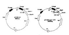


Landscapes
- Peptides Or Proteins (AREA)
- Preparation Of Compounds By Using Micro-Organisms (AREA)
Abstract
Description
Translated fromChinese相关申请related application
[0001]本发明申请要求源自2006年1月26日递交的中国发明申请200610002312.1的优先权,该申请的全部内容包含于本发明申请之中。[0001] The application of the present invention requires priority from the Chinese invention application 200610002312.1 submitted on January 26, 2006, and the entire content of this application is included in the application for the present invention.
发明领域field of invention
[0002]本发明涉及可特异性结合H5亚型禽流感病毒血凝素(HA)蛋白之单克隆抗体,及其保守性变异体或活性片段,或其多肽或多肽类似物的相关编码序列,或产生所述单克隆抗体的细胞株,及应用该抗体或片段用于诊断与治疗的方法。The present invention relates to the monoclonal antibody that can specifically bind H5 subtype avian influenza virus hemagglutinin (HA) protein, and its conservative variant or active fragment, or the relevant coding sequence of its polypeptide or polypeptide analogue, Or a cell line producing the monoclonal antibody, and a method for using the antibody or fragment for diagnosis and treatment.
背景技术Background technique
[0003]自H5型禽流感于1996年首先在我国广东省农场的鹅群暴发以来(Xu X et al.,1999,Virology),由此病毒演生出来的另一株H5病毒在香港的禽鸟农场(1997年4月)及市场(1997年11月)暴发,引起了历史上禽流感病毒第一次直接由禽传人事件,前后共18例确诊病例中有6人死亡。2003年以来,H5病毒株在整个东亚及东南亚国家的相继暴发,WHO及流感专家预测H5禽流感病毒将最有可能成为下一次人类大流感暴发的流行株。2004年初,H5型高致病性禽流感也先后在我国十几个省暴发,并在中国香港、泰国、荷兰等发现H5型禽流感传染鸡、鸭、苍鹭、虎、猫等多种动物的事件。更令人担忧的是,在泰国已经出现疑似人传染人事件,马来西亚也有多例报道。进入2005年,罗马尼亚、俄罗斯、土耳其等欧洲国家相继发现禽类感染H5型禽流感病毒致死事件,专家认为此乃带毒候鸟迁移的结果,这使得控制高致病性H5型禽流感病毒的进一步扩散传播变得更为困难。专家预测,随着侯鸟的传播,H5型禽流感病毒有可能通过欧亚、亚非大陆桥进一步传播到卫生条件极差的非洲国家,使得H5型禽流感病毒获得和其它人流感病毒充分重组的机会和时间,届时将很可能形成一种对人类高度致命的全新流感病毒,其给人类带来的各种损失将难以估计。根据WHO统计结果,截止2006年1月19日,全球因感染H5N1病毒死亡的人类个案已达80例,给全球公共卫生安全带来极大的考验。Since the H5 type avian influenza first broke out in the geese of farms in Guangdong Province, my country in 1996 (Xu X et al., 1999, Virology), another strain of H5 virus derived from this virus has been infected in poultry in Hong Kong. The bird farm (April 1997) and the market (November 1997) broke out, causing the first time in history that the avian influenza virus was directly transmitted from poultry to humans. There were 6 deaths in a total of 18 confirmed cases. Since 2003, H5 virus strains have broken out successively throughout East Asia and Southeast Asian countries. WHO and influenza experts predict that H5 avian influenza virus will most likely become the epidemic strain of the next human influenza outbreak. At the beginning of 2004, H5 type highly pathogenic avian influenza also broke out in more than a dozen provinces of my country, and H5 type avian influenza was found to infect chickens, ducks, herons, tigers, cats and other animals in Hong Kong, Thailand, and the Netherlands. event. What is even more worrying is that there have been suspected cases of human-to-human transmission in Thailand, and many cases have been reported in Malaysia. Entering 2005, Romania, Russia, Turkey and other European countries successively discovered fatal incidents of poultry infected with H5 avian influenza virus. Experts believe that this is the result of migration of infected migratory birds, which makes it necessary to control the further spread of highly pathogenic H5 avian influenza virus Dissemination becomes more difficult. Experts predict that with the spread of migratory birds, the H5 avian influenza virus may further spread to African countries with extremely poor sanitation conditions through the Eurasian and Asian-African land bridges, so that the H5 avian influenza virus can fully recombine with other human influenza viruses At that time, a new influenza virus highly lethal to humans will be formed, and the various losses it will cause to humans will be difficult to estimate. According to the statistics of WHO, as of January 19, 2006, 80 human cases worldwide have died due to H5N1 virus infection, which has brought great challenges to global public health security.
[0004]最新研究结果(Li,K.S.et al.,2004,Nature)表明,华南水禽(家鸭)为H5型禽流感病毒的主要携带者及传播者,H5型禽流感的爆发有明显的季节性,并伴随着生物多态型(多种基因型)的演变。然而,分子流行病学研究表明,目前鸭群种大约有30%的阳性感染并无任何症状,鸡群中也有达10%的流行且无症状带毒。这些受感染的动物又能够不断使人受到新的感染,对人类的健康构成巨大的威胁。有关专家一致认为要控制好H5型高致病性禽流感病毒在整个东亚、东南亚以及欧洲各国的流行,早期诊断是先决条件,然后才能做到早隔离、早处理,对人做到早治疗。The latest research results (Li, K.S.et al., 2004, Nature) show that the South China water fowl (domestic duck) is the main carrier and disseminator of H5 type avian influenza virus, and the outbreak of H5 type avian influenza has obvious season Sex, accompanied by the evolution of biological polymorphisms (multiple genotypes). However, molecular epidemiological studies have shown that about 30% of the positive infections in ducks are asymptomatic, and 10% of chickens are prevalent and asymptomatic. These infected animals can continuously infect humans, posing a huge threat to human health. Relevant experts agree that to control the spread of H5 highly pathogenic avian influenza virus throughout East Asia, Southeast Asia, and European countries, early diagnosis is a prerequisite, and then early isolation, early treatment, and early treatment of humans can be achieved.
[0005]采用传统的病毒分离及血清诊断方法诊断禽流感病毒需时4-5天,且目前大部分人及动物疾控系统实验室缺乏三级生物安全实验室,故东南亚各国及地区对H5暴发的诊断明显滞后。常见的情况是大量鸡只死亡和扑杀行动完成后仍未有实验室的确诊报告,给控制病毒的暴发带来很大不便。另外,因少数禽类(特别是水禽,如家鸭)存在着无症状带毒情况,检疫系统又无有效的检测手段,这种情况的持续发展,引起了该病毒在多个国家地区反复暴发,迁延不断的局面。It takes 4-5 days to diagnose avian influenza virus by adopting traditional virus isolation and serological diagnosis method, and at present most of human and animal disease control system laboratory lack three-level biosafety laboratory, so Southeast Asian countries and regions are to H5 Diagnosis of outbreaks is significantly delayed. A common situation is that a large number of chickens die and the culling operation is completed, but there is still no laboratory confirmation report, which brings great inconvenience to the control of the outbreak of the virus. In addition, because a small number of poultry (especially waterfowl, such as domestic ducks) have asymptomatic virus transmission, and the quarantine system has no effective detection means, the continuous development of this situation has caused repeated outbreaks of the virus in many countries and regions. constant situation.
[0006]同时,由于H5型禽流感病毒(其中Goose/Guangdong/1/96为代表株)属高致病性病毒,对目前常用的动物模型均有致死性,而采用基因工程方法表达的血凝素蛋白(HA)又不能完整取得抗原性,世界上多个著名实验室先后尝试制备针对该病毒系的单克隆抗体均未获成功。目前对该病毒抗原性分析不得不采用特异性及反应性均明显达不到诊断试剂要求的由A/chicken/Pennsylvania/1370/83(H5N2)和A/chicken/Pennsylvania/8125/83(H5N2)制备的单克隆抗体。Simultaneously, because H5 type avian influenza virus (wherein Goose/Guangdong/1/96 is representative strain) belongs to highly pathogenic virus, animal model commonly used at present all has lethality, and the blood that adopts genetic engineering method to express The lectin protein (HA) cannot completely obtain the antigenicity, and many famous laboratories in the world have tried to prepare monoclonal antibodies against this virus line without success. At present, the antigenicity analysis of the virus has to use A/chicken/Pennsylvania/1370/83 (H5N2) and A/chicken/Pennsylvania/8125/83 (H5N2), which obviously cannot meet the requirements of diagnostic reagents in terms of specificity and reactivity. Prepared monoclonal antibodies.
[0007]鉴于上述情形,目前迫切期望能有一种方便、快捷、实时的诊断方法和手段,从而及时把第一代跨种族传染的病人隔离治疗,防止病毒向人传人发展,在病毒未适应人类之前就打断其传播链,从而从根本上消除该病毒对人类的大流感威胁。In view of above-mentioned situation, urgently expect to have a kind of convenient, quick, real-time diagnostic method and means at present, thus in time the patient of first generation interracial infection is isolated and treated, prevents virus from developing from person to person, when virus is not adapted to human Its chain of transmission was interrupted before, thereby fundamentally eliminating the threat of the virus to humans as a pandemic.
[0008]中国国内,有关抗禽流感病毒H5亚型检测研究已有文献报道。扬州大学畜牧兽医学院秦爱建等(秦爱建,邵红霞,钱琨等,中国预防兽医学报,2003,03期)研制了抗禽流感病毒H5和H9亚型血凝素特异性单克隆抗体,应用这些单克隆抗体进行间接免疫荧光试验被证明能在24小时内迅速检测出相应的禽流感病毒。北京出入境检验检疫局利用荧光RT-PCR快速检测高致病力禽流感病毒H5亚型,检测时间缩短为4小时,于2005年12月10日通过临床验证。郭元吉在“人禽流感研究现状”一文中综述了H 5亚型毒株抗体检测需用微量中和实验或特异性高的ELISA(郭元吉,中华实验和临床病毒学杂志,2004,03期),但有关利用ELISA检测H5亚型的研究性文献却未见相关报道。[0008] In China, relevant anti-avian influenza virus H5 subtype detection research has been reported in the literature. Qin Aijian, College of Animal Husbandry and Veterinary Medicine, Yangzhou University, et al. (Qin Aijian, Shao Hongxia, Qian Kun, etc., Chinese Journal of Preventive Veterinary Medicine, 2003, 03) have developed anti-avian influenza virus H5 and H9 subtype hemagglutinin-specific monoclonal antibodies. Antibody indirect immunofluorescence test proved to be able to rapidly detect the corresponding avian influenza virus within 24 hours. The Beijing Entry-Exit Inspection and Quarantine Bureau used fluorescent RT-PCR to quickly detect the H5 subtype of highly pathogenic avian influenza virus, and the detection time was shortened to 4 hours. It passed the clinical verification on December 10, 2005. Guo Yuanji summed up the detection of H5 subtype strain antibody in the "Current Situation of Human Avian Influenza Research" by microneutralization experiment or ELISA with high specificity (Guo Yuanji, Chinese Journal of Experimental and Clinical Virology, 2004, No. 03 ), but there is no relevant report on the research literature on the detection of H5 subtypes by ELISA.
[0009]国外有关利用ELISA检测H5N1抗体的研究已有报道。Rowe等报道了应用重组血凝素蛋白作为抗原包被,利用间接ELISA检测H5N1抗体,其ELISA的灵敏度为80%,特异度为62%(Rowe T.et al.,J.Clin.Microbiol.,April 1999;37(4):937-43),但该文献并非针对H5N1亚型血凝素HA基因特异的单克隆抗体。Zhou等(Zhou,E.M.et al.,Avian Dis.,1998,42(4):757-61)、Shafer等(Shafer,A.L.et al.,Avian Dis.,1998,42(1):28-34)采用竞争ELISA法检测禽流感病毒抗核心蛋白抗体,但检测对象均为A型禽流感所有H1-H16亚型的NP蛋白抗体,不能确定亚型。Lu报道了基于单克隆抗体的Dot-ELISA检测禽流感病毒(AIV),该方法直接检测AIV抗原,其特异性在于不会与其它禽类病毒发生交叉反应(Lu H.,Avian Dis.,2003,47(2):361-9)。虽然Sala等建立了基于H7亚型表面糖蛋白特异的单克隆抗体的ELISA,但其亚型为H7;单克隆抗体为表面糖蛋白特异的单克隆抗体,并非H5亚型血凝素HA基因特异的单克隆抗体(Sala G,Cordioli P,Moreno-Martinet al.Avian Dis.2003,47(3Suppl):1057-9)。[0009] There have been reports abroad on the use of ELISA to detect H5N1 antibodies. Rowe et al reported the application of recombinant hemagglutinin protein as antigen coating, using indirect ELISA to detect H5N1 antibody, the sensitivity of the ELISA was 80%, and the specificity was 62% (Rowe T. et al., J.Clin.Microbiol., April 1999; 37(4): 937-43), but this document is not specific to the H5N1 subtype hemagglutinin HA gene monoclonal antibody. Zhou et al. (Zhou, E.M. et al., Avian Dis., 1998, 42 (4): 757-61), Shafer et al. (Shafer, A.L. et al., Avian Dis., 1998, 42 (1): 28-34 ) using the competitive ELISA method to detect anti-core protein antibodies of avian influenza virus, but the detection objects are all H1-H16 subtypes of NP protein antibodies of type A avian influenza, and the subtype cannot be determined. Lu reported the detection of avian influenza virus (AIV) by Dot-ELISA based on monoclonal antibody. This method directly detects AIV antigen, and its specificity is that it will not cross-react with other avian viruses (Lu H., Avian Dis., 2003, 47(2):361-9). Although Sala et al established an ELISA based on a monoclonal antibody specific to the surface glycoprotein of the H7 subtype, the subtype is H7; the monoclonal antibody is a monoclonal antibody specific to the surface glycoprotein, not to the H5 subtype hemagglutinin HA gene (Sala G, Cordioli P, Moreno-Martin et al. Avian Dis. 2003, 47(3Suppl): 1057-9).
[0010]遗憾的是,现有禽流感病毒免疫学诊断手段使用的单克隆抗体大部分针对的是核蛋白(NP蛋白),因而其检测的是A型(也称甲型)流感病毒,但实际上A型流感病毒包括H1~H16共16个亚型,其中相当大的亚型均无致病性或有低致病性,只有H5亚型禽流感病毒为危害最大的高致病性禽流感病毒。因而现有技术远不能满足临床检测的需要。Unfortunately, most of the monoclonal antibodies used by the existing avian influenza virus immunological diagnosis means are aimed at nucleoprotein (NP protein), thus what it detects is type A (also claiming type A) influenza virus, but In fact, type A influenza virus includes 16 subtypes from H1 to H16, among which quite large subtypes have no pathogenicity or low pathogenicity, and only H5 subtype avian influenza virus is the most harmful and highly pathogenic bird flu virus. flu virus. Therefore, the existing technology is far from meeting the needs of clinical testing.
[0011]本发明的根本目的在于克服现有禽流感病毒免疫检测技术的缺陷,所采用的单克隆抗体针对的为H5亚型的HA蛋白,因而能够特异的检测具有高致病性的H5亚型禽流感病毒。Fundamental purpose of the present invention is to overcome the defective of existing avian influenza virus immunoassay technique, what adopted monoclonal antibody is aimed at is the HA protein of H5 subtype, thereby can specific detection have the H5 subtype of high pathogenicity type of avian influenza virus.
发明内容Contents of the invention
[0012]本发明提供了可特异性结合H5亚型禽流感病毒血凝素(HA)蛋白之单克隆抗体,及可阻断至少50%的单克隆抗体与H5亚型禽流感病毒血凝素(HA)蛋白结合活性之单克隆抗体.本发明也提供了相关的杂交瘤细胞株,分离的核酸分子和短肽,以及其含该单克隆抗体的药物组合物和医药诊断设备和试剂盒。本发明也提供了利用该单克隆抗体探测,诊断,预防,和治疗禽流感病毒,特别是H5亚型禽流感病毒的方法.The present invention provides the monoclonal antibody that can specifically bind H5 subtype avian influenza virus hemagglutinin (HA) protein, and can block at least 50% monoclonal antibody and H5 subtype avian influenza virus hemagglutinin (HA) Monoclonal antibody with protein binding activity. The present invention also provides related hybridoma cell lines, isolated nucleic acid molecules and short peptides, and pharmaceutical compositions, medical diagnostic equipment and kits containing the monoclonal antibody. The present invention also provides methods for detecting, diagnosing, preventing, and treating avian influenza virus, especially H5 subtype avian influenza virus, using the monoclonal antibody.
附图说明Description of drawings
[0013]图1为金标法H5亚型流感病毒HA抗原检测试剂盒的测定结果,其中″a″出现两条红色线,认定为阳性;″b″只出现质控线,认定为阴性;″c″不出现红色线,则认定为无效。Fig. 1 is the measurement result of gold standard method H5 subtype influenza virus HA antigen detection kit, wherein " a " appears two red lines, is identified as positive; " b " only occurs quality control line, is identified as negative; If "c" does not have a red line, it is considered invalid.
[0014]图2为金标法H5亚型流感病毒anti-HA抗体检测试剂盒的测定结果,其中″a″只出现质控线,认定为阳性;″b″出现两条红色线,认定为阴性;″c″不出现红色线,则认定为无效。Fig. 2 is the measurement result of gold label method H5 subtype influenza virus anti-HA antibody detection kit, and wherein " a " only occurs quality control line, is identified as positive; " b " occurs two red lines, is identified as Negative; if "c" does not have a red line, it is considered invalid.
[0015]图3为H5亚型流感病毒HA抗原检测试剂盒的测定结果,其中,“+”表示出现椭圆形显色区域,认定为阳性;“-”表示未出现椭圆形显色区域,认定为阴性。Fig. 3 is the measurement result of H5 subtype influenza virus HA antigen detection kit, and wherein, "+" expression occurs oval chromogenic region, is identified as positive; "-" expression oval chromogenic region does not occur, and is determined is negative.
[0016]图4为6种嵌合抗体的表达质粒图:pcDNA3.1-Ak8H5、pcDNA3.1-AH8H5、pcDNA3.1-Ak10F7、pcDNA3.1-AH 10F7、pcDNA3.1-Ak4D1和pcDNA3.1-AH4D1。Fig. 4 is the expression plasmid figure of 6 kinds of chimeric antibodies: pcDNA3.1-Ak8H5, pcDNA3.1-AH8H5, pcDNA3.1-Ak10F7, pcDNA3.1-AH 10F7, pcDNA3.1-Ak4D1 and pcDNA3.1 -AH4D1.
[0017]图5为三种嵌合抗体对Ck/HK/Yu22/02毒株的血凝抑制实验,其中,第1、2、3列:PBS对照物;第4和5列:10F7cAb;第6列:10F7mAb;第7和8列:4D1cAb;第9列:4D1mAb;第10和11列:8H5cAb;第12列:8H5mAb。Fig. 5 is the hemagglutination inhibition experiment of three kinds of chimeric antibodies to Ck/HK/Yu22/02 strain, wherein, the 1st, 2nd, 3rd row: PBS control; The 4th and 5th row: 10F7cAb; Column 6: 10F7 mAb;
[0018]图6为嵌合抗体与表达H5血凝素的细胞反应的免疫荧光检测实验结果,其中A为cAb 4D1(DAPI);B为cAb 4D1(FITC);C为cAb 10F7(DAPI);D为cAb 10F7(FITC);E为抗-HBV cAb(DAPI);F为抗-HBV cAb(FITC)。Fig. 6 is the immunofluorescence detection experimental result of the cell reaction of chimeric antibody and expression H5 hemagglutinin, and wherein A is cAb 4D1 (DAPI); B is cAb 4D1 (FITC); C is cAb 10F7 (DAPI); D is cAb 10F7 (FITC); E is anti-HBV cAb (DAPI); F is anti-HBV cAb (FITC).
[0019]图7为噬菌体多肽的ELISA检测值OD(450/620)的矩形图。Fig. 7 is the histogram of the ELISA detection value OD (450/620) of phage polypeptide.
[0020]图8为pTO-T7与pTO-T7-239-123的质粒示意图。[0020] Fig. 8 is a schematic diagram of the plasmids of pTO-T7 and pTO-T7-239-123.
[0021]图9为pTO-T7与pTO-T7-239-125的质粒示意图。[0021] Fig. 9 is a schematic diagram of the plasmids of pTO-T7 and pTO-T7-239-125.
[0022]图10为融合蛋白239-123纯化样品的SDS-PAGE电泳图,其中,1:分子重量标签;2:表达239-123的E.coli全部溶胞产物;3:239-123之超声溶胞产物的上清;4:缓冲液I中的239-123;5:2M尿素中的239-123纯化融合蛋白;6:4M尿素中的239-123的纯化融合蛋白;7:8M尿素中的239-123的纯化融合蛋白。Fig. 10 is the SDS-PAGE electrophoresis figure of fusion protein 239-123 purification sample, wherein, 1: molecular weight label; 2: express the whole lysate of E.coli of 239-123; 3: the ultrasound of 239-123 Supernatant of lysate; 4: 239-123 in buffer I; 5: 239-123 purified fusion protein in 2M urea; 6: purified fusion protein of 239-123 in 4M urea; 7: 8M urea Purified fusion protein of 239-123.
[0023]图11为融合蛋白239-125纯化样品的SDS-PAGE电泳图,其中,1:分子重量标签;2:表达239-125的E.coli全部溶胞产物;3:全部溶胞产物之离心分离上清;4:缓冲液I中的239-125;5:2M尿素中的239-125的纯化融合蛋白;6:4M尿素中的239-125的纯化融合蛋白;7:8M尿素中的239-125的纯化融合蛋白。Fig. 11 is the SDS-PAGE electrophoresis figure of fusion protein 239-125 purification sample, wherein, 1: molecular weight label; 2: express the whole lysate of E.coli of 239-125; 3: the whole lysate Centrifuge the supernatant; 4: 239-125 in buffer I; 5: the purified fusion protein of 239-125 in 2M urea; 6: the purified fusion protein of 239-125 in 4M urea; 7: the purified fusion protein of 239-125 in 8M urea Purified fusion protein of 239-125.
[0024]图12显示了239-123融合蛋白的结合特性:其ELISA检测值OD(450/620)的色彩强度矩形图,其中,239-123融合蛋白结合到水平轴所标识的各种毒株上。Fig. 12 has shown the binding characteristic of 239-123 fusion protein: the color intensity histogram of its ELISA detection value OD (450/620), wherein, 239-123 fusion protein is bound to the various strains indicated by horizontal axis superior.
[0025]图13显示了239-125融合蛋白的结合特性:其ELISA检测值OD(450/620)的色彩强度矩形图,其中,239-125融合蛋白结合到水平轴所标识的各种毒株上。Fig. 13 has shown the binding characteristic of 239-125 fusion protein: the color intensity histogram of its ELISA detection value OD (450/620), wherein, 239-125 fusion protein is bound to the various strains indicated by horizontal axis superior.
[0026]图14为在各种mAb稀释下,239-123融合蛋白结合到8H5mAb(三角形点线)或8C11mAb(方点线)的ELISA检测值OD(450/620)的色彩强度。Fig. 14 is under various mAb dilutions, the color intensity of the ELISA detection value OD (450/620) of 239-123 fusion protein binding to 8H5mAb (triangular dotted line) or 8C11mAb (square dotted line).
[0027]图15为pC149-mut与pC149-mut-123的质粒图。[0027] Figure 15 is a plasmid map of pC149-mut and pC149-mut-123.
[0028]图16为pC149-mut与pC149-mut-125的质粒图。[0028] Figure 16 is a plasmid map of pC149-mut and pC149-mut-125.
[0029]图17为小量表达的重组蛋白的全胞溶胞产物的SDS-PAGE电泳图,其中,1:D123;2:T123;3:F123;4:Q123;5:D125;6:T125;7:F125;8:Q125。Fig. 17 is the SDS-PAGE electrophoresis figure of the whole cell lysate of the recombinant protein expressed in small quantities, wherein, 1: D123; 2: T123; 3: F123; 4: Q123; 5: D125; 6: T125 ;7: F125; 8: Q125.
[0030]图18为纯化重组蛋白的SDS-PAGE电泳图,其中,1:D123;2:T123;3:F123;4:Q123;5:D125;6:T125;7:F125;8:Q125。Fig. 18 is the SDS-PAGE electrophoresis figure of purified recombinant protein, wherein, 1: D123; 2: T123; 3: F123; 4: Q123; 5: D125; 6: T125; 7: F125; 8: Q125.
[0031]图19为类病毒颗粒的电镜图,该类病毒颗粒是由HBV cAg片段的融合蛋白和结合抗体的多肽组装而成的。Fig. 19 is the electron micrograph of virus-like particle, and this virus-like particle is assembled by the fusion protein of HBV cAg fragment and the polypeptide of binding antibody.
[0032]图20为显示融合蛋白HBc-123/125与单抗8H5之间结合亲和力的ELISA检测值OD(450/620)的色彩强度矩形图。[0032] FIG. 20 is a color intensity histogram showing the ELISA detection value OD (450/620) of the binding affinity between the fusion protein HBc-123/125 and the monoclonal antibody 8H5.
[0033]图21为显示融合蛋白HBc-Q123或HBc-D125与各种单抗之间结合亲和力的ELISA检测值OD(450/620)的色彩强度矩形图;结果表明,融合蛋白HBc-Q123和HBc-D125T特异性地结合单抗8H5。Fig. 21 shows the color intensity histogram of the ELISA detection value OD (450/620) of binding affinity between fusion protein HBc-Q123 or HBc-D125 and various monoclonal antibodies; The results show that fusion protein HBc-Q123 and HBc-D125T specifically binds mAb 8H5.
[0034]图22显示了ELISA检测HBc-123融合蛋白免疫鼠血清抗体滴度上升曲线,其中,时间为0-4周,抗体滴度通过ELISA检测。[0034] Figure 22 shows the ELISA detection HBc-123 fusion protein immune mouse serum antibody titer rising curve, wherein, the time is 0-4 weeks, the antibody titer is detected by ELISA.
[0035]图23显示了ELISA检测HBc-125融合蛋白免疫鼠血清抗体滴度上升曲线,其中,时间为0-4周,抗体滴度通过ELISA检测。[0035] Figure 23 shows the ELISA detection HBc-125 fusion protein immune mouse serum antibody titer rising curve, wherein, the time is 0-4 weeks, the antibody titer is detected by ELISA.
[0036]图24显示了免疫荧光检测免疫鼠血清与SF21细胞中表达的HA蛋白的反应。[0036] Fig. 24 shows the reaction of immunofluorescence detection of HA protein expressed in immune mouse serum and SF21 cells.
[0037]图25为HBc-122、HBc-124、HBc-128和HBc-129重组蛋白组装的类病毒颗粒的电镜图。[0037] FIG. 25 is an electron micrograph of virus-like particles assembled by HBc-122, HBc-124, HBc-128 and HBc-129 recombinant proteins.
[0038]图26为显示融合蛋白HBc-122、HBc-124、HBc-128和HBc-129与各种单抗之间的结合亲和力的ELISA检测值OD(450/620)的色彩强度矩形图;结果表明,融合蛋白HBc-122、HBc-124、HBc-128和HBc-129特异性地结合到单抗8H5。Fig. 26 shows the color intensity histogram of the ELISA detection value OD (450/620) of the binding affinity between fusion protein HBc-122, HBc-124, HBc-128 and HBc-129 and various monoclonal antibodies; The results showed that fusion proteins HBc-122, HBc-124, HBc-128 and HBc-129 specifically bound to Mab 8H5.
[0039]图27显示了由12肽融合蛋白组装的的类病毒颗粒与H5N1病毒竞争结合8H5酶标抗体的结果,其中,纵轴是色彩强度(以OD(450/620)值表示),水平轴是实验中所用的各种类病毒颗粒和PBS对照物。Fig. 27 shows the result that the virus-like particle assembled by 12 peptide fusion proteins competes with the H5N1 virus in conjunction with the 8H5 enzyme-labeled antibody, wherein, the vertical axis is the color intensity (expressed in OD (450/620) value), the level Axes are the various virus-like particles and PBS controls used in the experiments.
具体实施方式Detailed ways
[0040]本发明申请中涉及的有关术语的定义如下:The definition of relevant terms involved in the application of the present invention is as follows:
[0041]本发明中的“血凝素”一词指禽流感病毒的包膜糖蛋白。血凝素蛋白介导流感病毒针对宿主细胞的吸附和进入。禽流感病毒的血凝素蛋白有16个血清学亚型,HA1-HA16,分别对应于16个病毒亚型,即H1-H16。[0041] The term "hemagglutinin" in the present invention refers to the envelope glycoprotein of avian influenza virus. The hemagglutinin protein mediates the adsorption and entry of influenza virus to host cells. The hemagglutinin protein of avian influenza virus has 16 serological subtypes, HA1-HA16, corresponding to 16 virus subtypes, namely H1-H16.
[0042]本发明中的“抗体”一词指任意一种免疫球蛋白,包括能结合特异性抗原的单抗、多抗、双特异性或多特异性抗体。一个完整的抗体包含两条重链和两条轻链。每条重链含有一个可变区和第一、第二、第三等三个恒定区;每条轻链包含一个可变区和一个恒定区。抗体呈“Y”型,“Y”型结构的颈部含有两条重链的第二和第三恒定区,其通过二硫键结合形成。“Y”型结构的每条臂含有其中一条重链的第一恒定区和可变区,和一条轻链的可变区和恒定区。轻链和重链的可变区决定抗原的结合;每条链的可变区均含有三个高变区,称互补决定区(CDR)(轻链(L)的CDR包含LCDR1、LCDR2、LCDR3,重链(H)的CDR包含HCDR1、HCDR2、HCDR3(其由Kabat等人命名,见Sequencesof Proteins of Immunological Interest,Fifth Edition(1991),第1-3卷,NIHPublication 91-3242,Bethesda Md)。其中,三个CDR由构架区(FR)间隔开。构架区比CDR区更保守并形成一个架子状结构支撑超变区。重链和轻链的恒定区与抗原结合无关,但具有多种效应功能。抗体依据重链恒定区的氨基酸序列可以分成几类,主要是:IgA、IgD、IgE、IgG和IgM,其中有些类还进一步分成亚类,如IgG1、IgG2、IgG3、IgG4、IgA1或IgA2等。[0042] The term "antibody" in the present invention refers to any immunoglobulin, including monoclonal, polyclonal, bispecific or multispecific antibodies that can bind to specific antigens. A complete antibody contains two heavy chains and two light chains. Each heavy chain contains a variable region and three constant regions; each light chain contains a variable region and a constant region. Antibodies are in the shape of a "Y", with the neck of the "Y" structure containing the second and third constant domains of the two heavy chains, formed by disulfide bonds. Each arm of the "Y" structure contains the first constant and variable regions of one of the heavy chains, and the variable and constant regions of one of the light chains. The variable regions of the light and heavy chains determine the binding of antigens; the variable regions of each chain contain three hypervariable regions, called complementarity determining regions (CDRs) (the CDRs of the light chain (L) include LCDR1, LCDR2, LCDR3 , the CDRs of the heavy chain (H) comprise HCDR1, HCDR2, HCDR3 (named by Kabat et al., see Sequences of Proteins of Immunological Interest, Fifth Edition (1991), Volumes 1-3, NIH Publication 91-3242, Bethesda Md). Among them, the three CDRs are separated by the framework region (FR). The framework region is more conservative than the CDR region and forms a shelf-like structure to support the hypervariable region. The constant regions of the heavy and light chains have nothing to do with antigen binding, but have multiple effects Function. Antibodies can be divided into several classes according to the amino acid sequence of the heavy chain constant region, mainly: IgA, IgD, IgE, IgG and IgM, some of which are further divided into subclasses, such as IgG1, IgG2, IgG3, IgG4, IgA1 or IgA2 wait.
[0043]本发明中的“抗体”一词,除特指完整的免疫球蛋白外,也指免疫球蛋白的片段(如至少是免疫球蛋白分子的一个免疫活性区段),如Fab、Fab′、F(ab′)2、Fv片段、单链抗体分子或由含有一个或多个CDR区的免疫球蛋白分子的任意片段形成的多特异性抗体。另外,本发明涉及的抗体也可以是由一个特定的人免疫球蛋白中的一个或多个CDR区结合一个或多个不同的人免疫球蛋白的构架区形成的抗体。The term "antibody" in the present invention, except specifically referring to the complete immunoglobulin, also refers to fragments of the immunoglobulin (such as at least an immunologically active segment of the immunoglobulin molecule), such as Fab, Fab ', F(ab')2, Fv fragments, single chain antibody molecules or multispecific antibodies formed from any fragment of an immunoglobulin molecule containing one or more CDR regions. In addition, the antibody involved in the present invention can also be an antibody formed by combining one or more CDR regions of a specific human immunoglobulin with one or more framework regions of different human immunoglobulins.
[0044]抗体相关的“Fab”片段是指含有一条轻链的可变区和恒定区和一条重链的可变区和恒定区经二硫键结合起来的抗体分子的一部分。[0044] An antibody-related "Fab" fragment refers to a portion of an antibody molecule that contains the variable and constant regions of a light chain and the variable and constant regions of a heavy chain joined by disulfide bonds.
[0045]“Fab’”片段是指包含了部分铰链区的Fab片段。[0045] "Fab'" fragment refers to a Fab fragment comprising part of the hinge region.
[0046]F(ab′)2指的是Fab’的二聚体。[0046] F(ab')2 refers to a dimer of Fab'.
[0047]抗体的“Fc”指的是第一重链的第二、第三恒定区与第二重链的第二、第三恒定区经二硫键结合成的抗体的一部分。抗体的Fc段有多种不同的功能,但不参与抗原的结合。[0047] The "Fc" of an antibody refers to a part of the antibody formed by disulfide bonding of the second and third constant regions of the first heavy chain and the second and third constant regions of the second heavy chain. The Fc segment of an antibody has a variety of different functions, but is not involved in antigen binding.
[0048]抗体的“Fv”段指的是能结合完整的抗原结合位点的抗体的最小片段。一个Fv片段包括一条轻链的可变区结合到一条重链的可变区。[0048] The "Fv" fragment of an antibody refers to the smallest fragment of an antibody that binds the complete antigen combining site. An Fv fragment consists of the variable region of a light chain joined to the variable region of a heavy chain.
[0049]本发明中的“单链抗体”或“scFv”指的是由轻链可变区与重链可变区直接相连或通过一个肽链连接而成的工程抗体(Houston 1988)"Single-chain antibody" or "scFv" in the present invention refers to an engineered antibody (Houston 1988) that is directly connected to the variable region of the light chain and the variable region of the heavy chain or connected by a peptide chain
[0050]本发明中的“单链抗体Fv-Fc”或“scFv-Fc”也包括scFv连接抗体的Fc段形成的工程抗体。[0050] The "single-chain antibody Fv-Fc" or "scFv-Fc" in the present invention also includes an engineered antibody formed by connecting scFv to the Fc segment of an antibody.
[0051]本发明中的“抗原决定簇”(或称表位)指的是抗原分子中与抗体结合的那部分氨基酸或原子基团。[0051] The "antigenic determinant" (or epitope) in the present invention refers to the part of amino acids or atomic groups in the antigen molecule that binds to the antibody.
[0052]本发明中的“单抗”一词指的是来自一群高度同源的抗体分子中的一个抗体或抗体的一个片断,也即除仅在少数情况下可能出现的自然突变外,一群完全相同的抗体分子。单抗对抗原上的单一表位具有高特异性。单抗与多抗不同,多抗是包含了识别抗原上的不同表位的抗体分子。虽然传统的单抗是由杂交瘤细胞分泌的,但本发明涉及的单抗并不仅限于此制备方法。如,本发明涉及的单抗可采用Kohler等首次报道的杂交瘤技术获得(Nature,256:495,1975),也可采用重组DNA技术获得(如参见U.S.P 4,816,567)。The term "mAb" in the present invention refers to an antibody or a fragment of an antibody from a group of highly homologous antibody molecules, that is, except for natural mutations that may occur only in a few cases, a group The exact same antibody molecule. mAbs are highly specific for a single epitope on an antigen. Monoclonal antibodies are different from polyclonal antibodies, which are antibody molecules that recognize different epitopes on antigens. Although traditional monoclonal antibodies are secreted by hybridoma cells, the monoclonal antibodies involved in the present invention are not limited to this preparation method. For example, the monoclonal antibody involved in the present invention can be obtained by the hybridoma technology first reported by Kohler et al. (Nature, 256:495, 1975), or by recombinant DNA technology (see U.S.P 4,816,567).
[0053]本发明中的“嵌合抗体”一词指的是抗体轻链或/和重链的一部分是源自某一特定物种或属某一特定抗体类或亚类的序列相同或同源的抗体,而抗体轻链或/和重链的另一部分是源自另一物种或属于另一抗体类或亚类的序列相同或同源的抗体。不管怎样,这种抗体片段仍保留了对目标抗原的结合活性(U.S.P 4,816,567to Cabilly et al.;Morrison et al.,Proc.Natl.Acad.Sci.USA,81:68516855(1984))。The term "chimeric antibody" in the present invention refers to a part of the antibody light chain or/and heavy chain that is derived from a specific species or belongs to a specific antibody class or subclass sequence identical or homologous antibody, and another part of the light chain or/and heavy chain of the antibody is an antibody with the same or homologous sequence derived from another species or belonging to another antibody class or subclass. However, such antibody fragments still retain the binding activity for the target antigen (U.S.P 4,816,567 to Cabilly et al.; Morrison et al., Proc. Natl. Acad. Sci. USA, 81:68516855 (1984)).
[0054]本申请中,“人源化抗体”一词指的是人源免疫球蛋白(受体抗体)的全部或部分CDR区被一非人源抗体(供体抗体)的CDR区替换后得到抗体或抗体片段,其中的供体抗体可以是有预期特异性、亲和性和反应性的小鼠、大鼠或兔抗体。另外,人源免疫球蛋白的构架区(FR)的氨基酸序列也可被相应的非人源抗体的氨基酸序列所替换。而且,人源化抗体的氨基酸残基也可以既非来源于受体抗体,也非来源于供体抗体的CDR区或构架区序列。这些人工修饰的目的是进一步完善或优化抗体性能。总之,人源化抗体是指含有至少一个、通常是两个几乎完整的可变区,其中对应的所有或几乎所有的CDR区是来自非人源抗体,其中的全部或几乎全部的FR区是来自人源抗体。理想的人源化抗体至少含有免疫球蛋白的Fc区的一部分,通常是人源免疫球蛋白的Fc区。更多详细内容请参阅:Jones et al.,Nature,321:522525(1986);Reichmann et al.,Nature,332:323329(1988);Presta,Curr.Op.Struct.Biol.,2:593596(1992);and Clark,Immunol.Today 21:397402(2000).In the present application, the term "humanized antibody" refers to the replacement of all or part of the CDR region of a human immunoglobulin (recipient antibody) by a CDR region of a non-human antibody (donor antibody) To obtain antibodies or antibody fragments, the donor antibody can be a mouse, rat or rabbit antibody with the desired specificity, affinity and reactivity. In addition, the amino acid sequences of the framework regions (FRs) of human immunoglobulins may also be replaced by corresponding amino acid sequences of non-human antibodies. Furthermore, the amino acid residues of a humanized antibody may not be derived from the CDR region or framework region sequences of neither the recipient antibody nor the donor antibody. The purpose of these artificial modifications is to further refine or optimize antibody performance. In short, a humanized antibody is one that contains at least one, usually two, almost complete variable regions, wherein all or almost all of the corresponding CDR regions are from non-human antibodies, and all or almost all of the FR regions are from human antibodies. Ideally, a humanized antibody contains at least a portion of the Fc region of an immunoglobulin, usually that of a human immunoglobulin. For more details, please refer to: Jones et al., Nature, 321: 522525 (1986); Reichmann et al., Nature, 332: 323329 (1988); Presta, Curr.Op.Struct.Biol., 2: 593596 ( 1992); and Clark, Immunol. Today 21:397402 (2000).
[0055]本申请涉及的“被分离”一词,指的是天然状态下经人工手段获得的。如果自然界中出现某一种“被分离”的物质或成分,那么可能是其所处的天然环境发生了改变或从天然环境下离分出该物质,或二者情况均有发生。比如,某一活体动物体内天然存在的某种未被分离的多聚核苷酸或多肽,而从这种天然状态下分离出来的高纯度的相同的多聚核苷酸或多肽即称之为被分离。这里的“被分离”不排除混有人工或合成的物质,也不排除存在不影响物质活性的其它不纯物质。[0055] The term "separated" referred to in the present application refers to the natural state obtained by artificial means. If an "isolated" substance or component occurs in nature, it may be that its natural environment has changed or the substance has been isolated from its natural environment, or both. For example, a certain unisolated polynucleotide or polypeptide naturally existing in a living animal, and the same polynucleotide or polypeptide isolated from this natural state with high purity is called be separated. The "separated" here does not exclude the mixing of artificial or synthetic substances, nor does it exclude the existence of other impure substances that do not affect the activity of the substance.
[0056]本发明中“载体”一词指的是,可将编码某蛋白的多聚核苷酸插入其中并使蛋白获得表达的一种核酸运载工具。载体可通过转化、转导或转染宿主细胞,使其携带的遗传物质元件在宿主细胞内表达得以表达。举例来说,载体包括:质粒;噬菌粒;柯斯质粒;人工染色体如酵母人工染色体(YAC)、细菌人工染色体(BAC)或P1来源的人工染色体(PAC);噬菌体如λ噬菌体或M13噬菌体及动物病毒等。用作载体的动物病毒种类有逆转录酶病毒(包括慢病毒)、腺病毒、腺相关病毒、疱疹病毒(如单纯疱疹病毒)、痘病毒、杆状病毒、乳头瘤病毒、乳头多瘤空泡病毒(如SV40)。一种载体可能含有多种控制表达的元件,包括启动子序列、转录起始序列、增强子序列、选择元件及报告基因。另外,载体还可含有复制起始位点。载体还有可能包括有协助其进入细胞的成分,如病毒颗粒、脂质体或蛋白外壳,但不仅仅只有这些物质。[0056] The term "vector" in the present invention refers to a nucleic acid delivery tool in which a polynucleotide encoding a protein can be inserted and the protein can be expressed. The vector can be expressed by transforming, transducing or transfecting the host cell, so that the genetic material elements carried by it can be expressed in the host cell. For example, vectors include: plasmids; phagemids; cosmids; artificial chromosomes such as yeast artificial chromosome (YAC), bacterial artificial chromosome (BAC) or P1-derived artificial chromosome (PAC); phage such as lambda phage or M13 phage and animal viruses. Types of animal viruses used as vectors include retroviruses (including lentiviruses), adenoviruses, adeno-associated viruses, herpesviruses (such as herpes simplex virus), poxviruses, baculoviruses, papillomaviruses, papillary polyoma vacuoles Viruses (such as SV40). A vector may contain a variety of elements that control expression, including promoter sequences, transcription initiation sequences, enhancer sequences, selection elements, and reporter genes. In addition, the vector may also contain an origin of replication. Vectors may also include components that facilitate their entry into cells, such as viral particles, liposomes, or protein coats, but not only.
[0057]本发明中“宿主细胞”一词指的是导入载体的细胞,包括如下许多细胞类型,如大肠杆菌或枯草菌等原核细胞,如酵母细胞或曲霉菌等真菌细胞,如S2果蝇细胞或Sf9等昆虫细胞,或者如纤维原细胞,CHO细胞,COS细胞,NSO细胞,HeLa细胞,BHK细胞,HEK 293细胞或人细胞的动物细胞。Among the present invention, " host cell " term refers to the cell of introducing carrier, comprises following many cell types, as prokaryotic cells such as escherichia coli or subtilis, fungal cells such as yeast cells or aspergillus, such as S2 fruit fly cells or insect cells such as Sf9, or animal cells such as fibroblasts, CHO cells, COS cells, NSO cells, HeLa cells, BHK cells, HEK 293 cells or human cells.
[0058]“中和抗体”一词指的是能清除或显著降低目标病毒抗原结合毒力的抗体或抗体片段。[0058] The term "neutralizing antibody" refers to an antibody or antibody fragment capable of clearing or significantly reducing the binding virulence of a target viral antigen.
[0059]“序列相同百分比”一词指的是候选序列中的核酸或氨基酸分别与对应的核酸或多肽序列中核酸或氨基酸相同性的百分比。这里涉及到的与核酸序列或者多肽序到有关的术语“序列相似百分比”定义为候选核酸序列或氨基酸残基序列分别与目的核酸序列或氨基酸序列的相似百分比。对于某一个序列,将它和目的序列进行比对,必要时可跳过突变缺口,以达到最大的基因相似百分比,而不去考虑相似序列的任意保守性突变。本领域的多种比对方法可用于确定核酸或氨基酸序列的相似性,如可用的计算机软件包括BLAST,BLAST-2,ALIGN,ALIGN-2或Megalign(DNASTAR)等。熟悉本领域技术的工作人员懂得给比对设定合适的测量参数,包括为使用最大可比性的一些运算法则达到而全长序列的比较。[0059] The term "percentage of sequence identity" refers to the percentage of nucleic acid or amino acid identity in a candidate sequence to that of a corresponding nucleic acid or polypeptide sequence. The term "percentage of sequence similarity" related to nucleic acid sequence or polypeptide sequence is defined as the percentage of similarity between a candidate nucleic acid sequence or amino acid residue sequence and a target nucleic acid sequence or amino acid sequence, respectively. For a certain sequence, it is compared with the target sequence, and mutation gaps can be skipped if necessary to achieve the maximum percentage of gene similarity, regardless of any conservative mutations in similar sequences. Various alignment methods in the art can be used to determine the similarity of nucleic acid or amino acid sequences, such as available computer software including BLAST, BLAST-2, ALIGN, ALIGN-2 or Megalign (DNASTAR) and the like. Those skilled in the art will be able to set appropriate measurement parameters for alignments, including comparisons of full-length sequences to be achieved using algorithms for maximum comparability.
[0060]“特异性结合”一词指的是,指两分子间的非随机结合反应,如抗体和产生该抗体的抗原间的反应。此处,结合第一种抗原的抗体对第二种抗原的结合亲和力是检测不到或很弱。在某些实施方式中,一个某抗原特异性抗体是指以亲和力(KD)≤10-5M(如10-6M、10-7M、10-8M、10-9M、10-10M等)结合该抗原,其中KD指解离率与结合率的比值(koff/kon),其可以采用本领域技术人员熟悉的方法进行测定。[0060] The term "specific binding" refers to a non-random binding reaction between two molecules, such as the reaction between an antibody and the antigen from which it was produced. Here, an antibody that binds a first antigen has no detectable or weak binding affinity for the second antigen. In some embodiments, an antigen-specific antibody refers to an antibody with an affinity (KD) ≤ 10-5 M (such as 10-6 M, 10-7 M, 10-8 M, 10-9 M, 10-10 M, etc.) bind to the antigen, wherein KD refers to the ratio of the off-rate to the on-rate (koff/kon), which can be determined by methods familiar to those skilled in the art.
[0061]抗体Antibody
[0062]本发明所述单抗能够特异性结合H5亚型禽流感病毒。本发明的一个方面涉及能特异性结合H5亚型禽流感病毒血凝素蛋白的单抗及其相应的抗原结合片段。[0062] The monoclonal antibody of the present invention can specifically bind to the H5 subtype avian influenza virus. One aspect of the present invention relates to a monoclonal antibody capable of specifically binding to the hemagglutinin protein of H5 subtype avian influenza virus and its corresponding antigen-binding fragment.
[0063]本发明所述抗H5单抗是由小鼠杂交瘤细胞株8H5,3C8,10F7,4D1,3G4和2F2所分泌的。这些单抗的名称以其相应的杂交瘤细胞株进行命名。也就是,这些抗H5单抗分别由杂交瘤细胞株8H5、3C8、10F7、4D1、3G4和2F2产生,并分别命名为8H5、3C8、10F7、4D1、3G4和2F2。单抗8H5、3C8、10F7、4D1、3G4和2F2能特异性结合H5亚型禽流感病毒血凝素蛋白。小鼠杂交瘤细胞株8H5、3C8、10F7、4D1、3G4和2F2已在2006年1月17日于中国典型培养物保藏中心(CCTCC,Wuhan University,Wuhan,China)进行保藏,保藏号是CCTCC-C200607(杂交瘤细胞株8H5),CCTCC-C200605(杂交瘤细胞株3C8)、CCTCC-C200608(杂交瘤细胞株10F7)、CCTCC-C200606(杂交瘤细胞株4D1)、CCTCC-C200604(杂交瘤细胞株3G4)和CCTCC-C200424(杂交瘤细胞株2F2)。[0063] The anti-H5 monoclonal antibody of the present invention is secreted by mouse hybridoma cell lines 8H5, 3C8, 10F7, 4D1, 3G4 and 2F2. The names of these monoclonal antibodies are named after their corresponding hybridoma cell lines. That is, these anti-H5 mAbs were produced by hybridoma cell lines 8H5, 3C8, 10F7, 4D1, 3G4, and 2F2, respectively, and were named 8H5, 3C8, 10F7, 4D1, 3G4, and 2F2, respectively. Monoclonal antibodies 8H5, 3C8, 10F7, 4D1, 3G4 and 2F2 can specifically bind to H5 subtype avian influenza virus hemagglutinin protein. Mouse hybridoma cell lines 8H5, 3C8, 10F7, 4D1, 3G4 and 2F2 have been preserved in China Center for Type Culture Collection (CCTCC, Wuhan University, Wuhan, China) on January 17, 2006, and the preservation number is CCTCC- CCTCC-C200607 (hybridoma cell line 8H5), CCTCC-C200605 (hybridoma cell line 3C8), CCTCC-C200608 (hybridoma cell line 10F7), CCTCC-C200606 (hybridoma cell line 4D1), CCTCC-C200604 (hybridoma cell line 3G4) and CCTCC-C200424 (hybridoma cell line 2F2).
[0064]本发明涉及单抗还包括能阻断单抗8H5,3C8,10F7,4D1,3G4或2F2结合H5亚型禽流感病毒血凝素蛋白的单抗。这些单抗结合的血凝素上的表位可以与单抗8H5、3C8、10F7、4D1、3G4或2F2所识别的表位相同。这些单抗所识别的表位也可以和单抗8H5、3C8、10F7、4D1、3G4或2F2所识别的表位在空间上有重叠。这样的单抗可以降低单抗8H5、3C8、10F7、4D1、3G4或2F2与H5亚型禽流感病毒血凝素蛋白的结合力至少50%,或至少60%,或优选至少70%,或更优选至少75%,或更优选至少80%,或更优选至少85%,或更优选至少90%,或更优选95%,或最优选99%。[0064] The present invention relates to monoclonal antibodies that also include monoclonal antibodies capable of blocking monoclonal antibodies 8H5, 3C8, 10F7, 4D1, 3G4 or 2F2 from binding to H5 subtype avian influenza virus hemagglutinin protein. The epitope on the hemagglutinin bound by these mAbs may be the same as the epitope recognized by mAbs 8H5, 3C8, 10F7, 4D1, 3G4 or 2F2. The epitopes recognized by these mAbs may also spatially overlap with the epitopes recognized by mAbs 8H5, 3C8, 10F7, 4D1, 3G4 or 2F2. Such monoclonal antibody can reduce the binding force of monoclonal antibody 8H5, 3C8, 10F7, 4D1, 3G4 or 2F2 to H5 subtype avian influenza virus hemagglutinin protein by at least 50%, or at least 60%, or preferably at least 70%, or more Preferably at least 75%, or more preferably at least 80%, or more preferably at least 85%, or more preferably at least 90%, or more preferably 95%, or most preferably 99%.
[0065]可以采用常规方法如Antibodies:A Laboratory Manual,ColdSpring Harbor Laboratory,Ed Harlow and David Lane(1988)中描述的方法,测定某一未知单抗降低某一已知单抗结合H5血凝素蛋白的能力。例如,先把抗原预包被在微孔板上,然后把系列稀释的未标记的待测抗体和特定浓度的标记后的已知单抗共同加入上述预包被后的微孔板中孵育,洗涤后测定不同稀释度的待测抗体下已知抗体结合到板上的数量。待测抗体竞争已知抗体结合抗原的能力越强,已知抗体结合抗原的能力就越弱。通常,抗原是预包被在96孔微孔板上,并利用放射标记法或酶标记法测定未标记单抗阻断已标记单抗的能力。Conventional methods can be used such as Antibodies: A Laboratory Manual, Cold Spring Harbor Laboratory, the method described in Ed Harlow and David Lane (1988), to determine that a certain unknown monoclonal antibody reduces the binding of a certain known monoclonal antibody to the H5 hemagglutinin protein Ability. For example, the antigen is pre-coated on a microwell plate, and then serially diluted unlabeled antibody to be tested and a specific concentration of labeled known monoclonal antibody are added to the above-mentioned pre-coated microwell plate for incubation. After washing, the amount of known antibody bound to the plate under different dilutions of the antibody to be tested was determined. The greater the ability of the test antibody to compete with the binding of the known antibody to the antigen, the less able the known antibody is to bind the antigen. Typically, the antigen is pre-coated on a 96-well microplate, and the ability of the unlabeled mAb to block the labeled mAb is determined by radiolabeling or enzymatic labeling.
[0066]可以采用Kohler等在Nature 256:495(1975)中报道的杂交瘤制备方法来制备单抗。首先将免疫原(必要时候添加佐剂)免疫注射小鼠或其它合适的宿主动物。免疫原或佐剂的注射方式通常为皮下多点注射或腹腔注射。免疫原预先藕联到某些已知蛋白,如血清白蛋白或大豆姨酶抑制剂上,可能会有助于增强抗原在宿主内的免疫原性。佐剂可以利用福氏佐剂或MPL-TDM等。动物受免疫后,体内会有分泌特异性结合免疫原的抗体的淋巴细胞产生。另外,淋巴细胞也可以利用体外免疫获得。收集目的淋巴细胞与骨髓瘤细胞并用合适的融合剂,如PEG,进行融合以获得杂交瘤细胞(Goding,MonoclonalAntibodies:Principles and Practice,pp.59-103,Academic Press,1996)。[0066] The hybridoma preparation method reported in Nature 256: 495 (1975) by Kohler et al. can be used to prepare monoclonal antibodies. Firstly, the immunogen (adding an adjuvant if necessary) is immunized into mice or other suitable host animals. The immunogen or adjuvant is usually injected subcutaneously at multiple points or intraperitoneally. Pre-coupling of the immunogen to certain known proteins, such as serum albumin or soybean enzyme inhibitors, may help to enhance the immunogenicity of the antigen in the host. As an adjuvant, Freund's adjuvant, MPL-TDM, etc. can be used. After the animal is immunized, lymphocytes that secrete antibodies that specifically bind to the immunogen will be produced in the body. In addition, lymphocytes can also be obtained by in vitro immunization. The target lymphocytes and myeloma cells are collected and fused with a suitable fusion agent, such as PEG, to obtain hybridoma cells (Goding, Monoclonal Antibodies: Principles and Practice, pp.59-103, Academic Press, 1996).
[0067]上述制备的杂交瘤细胞可以接种到合适的培养液中生长,培养液中最好含有一种或多种能够抑制未融合的、母体骨髓瘤细胞生长的物质。例如,对缺乏次黄嘌呤鸟嘌呤磷酸转移酶(HGPRT or HPRT)的母体骨髓瘤细胞,在培养液中添加次黄嘌呤、氨基喋呤和胸腺嘧啶(HAT培养基)等物质将可以抑制HGPRT-缺陷细胞的生长。[0067] The hybridoma cells prepared above can be inoculated into a suitable culture medium to grow, and the culture medium preferably contains one or more substances capable of inhibiting the growth of unfused, parental myeloma cells. For example, for maternal myeloma cells lacking hypoxanthine guanine phosphotransferase (HGPRT or HPRT), adding substances such as hypoxanthine, aminopterin and thymine (HAT medium) to the culture medium will inhibit HGPRT- Defective cell growth.
[0068]优选的骨髓瘤细胞应该具有融合率高,抗体分泌能力稳定,对HAT培养液敏感等能力。其中,骨髓瘤细胞首选鼠源骨髓瘤,如MOP-21 and MC-11小鼠肿瘤衍生株(THE Salk Institute Cell Distribution Center,San Diego,Calif.USA),和SP-2/0 or X63-Ag8-653细胞株(American Type Culture Collection,Rockville,Md.USA)。另外也有研究报道利用人骨髓瘤和人鼠异源骨髓瘤细胞株制备人单抗(Kozbor,J.Immunol.,133:3001(1984);Brodeur et al.,Monoclonal Antibody Production Techniques and Applications,pp.51-63,Marcel Dekker,Inc.,New York,1987)。[0068] Preferred myeloma cells should have a high fusion rate, stable antibody secretion, and sensitivity to HAT culture fluid. Among them, murine myeloma is the first choice for myeloma cells, such as MOP-21 and MC-11 mouse tumor derivative strains (THE Salk Institute Cell Distribution Center, San Diego, Calif.USA), and SP-2/0 or X63-Ag8 -653 cell line (American Type Culture Collection, Rockville, Md. USA). In addition, there are also research reports on the use of human myeloma and human mouse heteromyeloma cell lines to prepare human monoclonal antibodies (Kozbor, J. Immunol., 133: 3001 (1984); Brodeur et al., Monoclonal Antibody Production Techniques and Applications, pp. 51-63, Marcel Dekker, Inc., New York, 1987).
[0069]杂交瘤细胞生长的培养液用于检测针对特异抗原的单抗的产生。测定杂交瘤细胞产生的单抗的结合特异性有免疫沉淀或体外结合试验,如放射免疫试验(RIA)、酶联免疫吸附试验(ELISA)。例如,利用Munson等在Anal.Biochem.107:220(1980)描述的Scatchard分析法可用来测定单抗的亲和力。[0069] The culture medium grown by hybridoma cells was used to detect the production of monoclonal antibodies against specific antigens. Immunoprecipitation or in vitro binding assays, such as radioimmunoassay (RIA) and enzyme-linked immunosorbent assay (ELISA), are used to determine the binding specificity of monoclonal antibodies produced by hybridoma cells. For example, the affinity of mAbs can be determined using the Scatchard assay described by Munson et al., Anal. Biochem. 107:220 (1980).
[0070]当杂交瘤产生的抗体的特异性、亲和力和反应性确定之后,目的细胞株可以通过(Goding,Monoclonal Antibodies:Principles and Practice,pp.59-103,Academic Press,1996)所描述的标准的有限稀释法进行亚克隆化。合适的培养液可以是DMEM或RPMI-1640等。另外,杂交瘤细胞还可以腹水瘤的形式在动物体内生长。After the specificity, affinity and reactivity of the antibody produced by the hybridoma are determined, the target cell line can pass the standard described in (Goding, Monoclonal Antibodies: Principles and Practice, pp.59-103, Academic Press, 1996) Subcloning by limiting dilution method. A suitable culture medium can be DMEM or RPMI-1640, etc. Alternatively, hybridoma cells can also be grown in animals in the form of ascites tumors.
[0071]利用传统的免疫球蛋白纯化方法,如蛋白A琼脂糖凝胶、羟基磷灰石层析、凝胶电泳、透析或亲和层析等,可以将亚克隆细胞分泌的单抗从细胞培养液、腹水或血清中分离出来。Utilize traditional immunoglobulin purification method, such as protein A sepharose, hydroxyapatite chromatography, gel electrophoresis, dialysis or affinity chromatography etc., the monoclonal antibody secreted by subcloning cell can be obtained from cell Separated from culture medium, ascitic fluid or serum.
[0072]本发明所述单抗还可以是通过基因工程重组技术获得。利用特异性结合单抗重链和轻链基因的核酸引物进行PCR扩增,可以从杂交瘤细胞中分离得到编码单抗重链和轻链基因的DNA分子。所得DNA分子插入表达载体内,然后转染宿主细胞,如E.coli细胞、猿猴COS、CHO细胞、或其它不产生免疫球蛋白的骨髓瘤细胞。转染后的宿主细胞在特定条件下培养并表达目标抗体。[0072] The monoclonal antibody of the present invention can also be obtained through genetic engineering recombination technology. The DNA molecules encoding the heavy chain and light chain genes of the monoclonal antibody can be isolated from hybridoma cells by using nucleic acid primers that specifically bind to the heavy chain and light chain genes of the monoclonal antibody to carry out PCR amplification. The resulting DNA molecules are inserted into expression vectors, and then transfected into host cells, such as E. coli cells, simian COS, CHO cells, or other myeloma cells that do not produce immunoglobulins. The transfected host cells are cultured under specific conditions and express the target antibody.
[0073]本发明的抗体对H5血凝素蛋白结合具有高特异性和高亲和力。这些抗体对其它亚型的血凝素蛋白可能会有弱的交叉反应性,优选地,这些抗体对其它亚型的血凝素蛋白完全没有交叉反应性。一方面,本发明的抗体结合H5血凝素的KD值小于1×10⁻⁵M;优选地,KD值小于1×10⁻⁶M;更优选地,KD值小于1×10⁻⁷M,最优选地,KD值小于1×10⁻⁸M。[0073] The antibodies of the present invention have high specificity and high affinity for H5 hemagglutinin protein binding. These antibodies may have weak cross-reactivity to other subtypes of hemagglutinin proteins, preferably, these antibodies have no cross-reactivity to other subtypes of hemagglutinin proteins at all. In one aspect, the KD value of the antibody of the present invention binding to H5 hemagglutinin is less than 1×10⁻⁵ M; preferably, the KD value is less than 1×10⁻⁶ M; more preferably, the KD value is less than 1×10⁻⁷ M, Most preferably, the KD value is less than 1×10⁻⁸ M.
[0074]本发明的单抗可以是包含两条重链和两条轻链的传统的“Y”型结构状的抗体。另外,所述抗体也可以是保持了对血凝素蛋白亲和力的传统的“Y”型结构状的抗体上的Fab片段、Fab′、F(ab)2、Fv、或其它类型的部分片段,其结合血凝素蛋白的亲和力可以高于或低于传统的“Y”型结构状的抗体。[0074] The monoclonal antibody of the present invention may be an antibody with a traditional "Y" structure comprising two heavy chains and two light chains. In addition, the antibody can also be a Fab fragment, Fab', F(ab)2 , Fv, or other types of partial fragments on the antibody with the traditional "Y" structure that maintains the affinity for the hemagglutinin protein, Its binding affinity to the hemagglutinin protein can be higher or lower than that of traditional "Y"-shaped antibodies.
[0075]本发明的抗体片段可以利用水解完整的抗体分子获得(参见Morimoto et al.,J.Biochem.Biophys.Methods 24:107-117(1992)andBrennan et al.,Science 229:81(1985))。另外,这些抗体片段也可以直接由重组宿主细胞产生(reviewed in Hudson,Curr.Opin.Immunol.11:548-557(1999);Little et al.,Immunol.Today,21:364-370(2000))。比如,Fab′片段可以直接从E.coli细胞中获得或化学藕联形成F(ab′)2片段(Carter et al.,Bio/Technology,10:163-167(1992))。再如,F(ab′)2片段可以用亮氨酸拉链GCN4连接获得。另外,Fv、Fab或F(ab′)2片段也可以直接从重组宿主细胞培养液中直接分离得到。本领域的普通技术人员完全知晓制备抗体片段的其它技术。[0075] Antibody fragments of the present invention can be obtained by hydrolyzing intact antibody molecules (see Morimoto et al., J. Biochem. Biophys. Methods 24: 107-117 (1992) and Brennan et al., Science 229: 81 (1985) ). Alternatively, these antibody fragments can also be produced directly by recombinant host cells (reviewed in Hudson, Curr. Opin. Immunol. 11:548-557 (1999); Little et al., Immunol. Today, 21:364-370 (2000) ). For example, Fab' fragments can be obtained directly from E. coli cells or chemically coupled to form F(ab')2 fragments (Carter et al., Bio/Technology, 10:163-167 (1992)). As another example, F(ab')2 fragments can be obtained by ligation with leucine zipper GCN4. In addition, Fv, Fab or F(ab')2 fragments can also be directly isolated from the culture medium of recombinant host cells. Other techniques for preparing antibody fragments are well known to those of ordinary skill in the art.
[0076]抗体核酸序列Antibody nucleic acid sequence
[0077]本发明涉及特异性结合H5血凝素蛋白的抗体或抗体片段的编码核酸分子。编码抗体的核酸分子可以从杂交瘤细胞中分离得到。本领域的普通技术人员完全知晓利用常规技术可以测定这些分子的核酸序列。本发明涉及的抗体核酸分子也可以利用传统的基因工程重组技术或化学合成方法获得。一方面,本发明涉及的抗体核酸分子的序列包含了抗H5抗体的重链可变区或抗体分子的部分核酸序列。另一方面,本发明涉及的抗体核酸分子的序列也包括抗H5抗体的轻链可变区或抗体分子的部分核酸序列。另一方面,本发明涉及的抗体核酸分子的序列还包括重链或轻链可变区的CDR序列。[0077] The present invention relates to nucleic acid molecules encoding antibodies or antibody fragments that specifically bind the H5 hemagglutinin protein. Nucleic acid molecules encoding antibodies can be isolated from hybridoma cells. Those of ordinary skill in the art are well aware that the nucleic acid sequences of these molecules can be determined using routine techniques. The antibody nucleic acid molecules involved in the present invention can also be obtained by traditional genetic engineering recombination techniques or chemical synthesis methods. In one aspect, the sequence of the antibody nucleic acid molecule involved in the present invention comprises the heavy chain variable region of the anti-H5 antibody or a partial nucleic acid sequence of the antibody molecule. On the other hand, the sequence of the antibody nucleic acid molecule involved in the present invention also includes the light chain variable region of the anti-H5 antibody or a partial nucleic acid sequence of the antibody molecule. On the other hand, the sequence of the antibody nucleic acid molecule involved in the present invention also includes the CDR sequence of the heavy chain or light chain variable region.
[0078]本发明的一方面涉及编码单抗8H5、3C8、10F7、4D1、3G4和2F2重链和轻链可变区序列的核酸分子。单抗8H5、3C8、10F7、4D1、3G4和2F2重链可变区序列的核酸分子分别对应于SEQ ID NO:1、SEQ ID NO:5、SEQ ID NO:9、SEQ ID NO:16、SEQ ID NO:20和SEQ ID NO:24。单抗8H5、3C8、10F7、4D1和2F2轻链可变区序列的核酸分子分别对应于SEQ ID NO:3、SEQ ID NO:7、SEQ ID NO:11、SEQ ID NO:18和SEQ ID NO:26。本发明还涉及包含了单抗8H5、3C8、10F7、4D1、3G4和2F2重链和轻链可变区序列的核酸分子变异体或类似体。[0078] One aspect of the invention pertains to nucleic acid molecules encoding the heavy and light chain variable region sequences of mAbs 8H5, 3C8, 10F7, 4D1, 3G4, and 2F2. The nucleic acid molecules of the heavy chain variable region sequences of monoclonal antibodies 8H5, 3C8, 10F7, 4D1, 3G4 and 2F2 correspond to SEQ ID NO: 1, SEQ ID NO: 5, SEQ ID NO: 9, SEQ ID NO: 16, SEQ ID NO: ID NO: 20 and SEQ ID NO: 24. The nucleic acid molecules of the light chain variable region sequences of monoclonal antibodies 8H5, 3C8, 10F7, 4D1 and 2F2 correspond to SEQ ID NO: 3, SEQ ID NO: 7, SEQ ID NO: 11, SEQ ID NO: 18 and SEQ ID NO :26. The present invention also relates to nucleic acid molecule variants or analogues comprising the heavy chain and light chain variable region sequences of monoclonal antibodies 8H5, 3C8, 10F7, 4D1, 3G4 and 2F2.
[0079]另一方面,本发明还涉及各种分离的核酸分子的变异体,其序列与如下核酸序列相同SEQ ID NO:1、SEQ ID NO:3、SEQ ID NO:5、SEQ ID NO:7、SEQ ID NO:9、SEQ ID NO:11、SEQ ID NO:16、SEQ ID NO:18、SEQ IDNO:20、SEQ ID NO:24或SEQ ID NO:26。具体地,核酸变异体的序列与如下核酸序列SEQ ID NO:1、SEQ ID NO:3、SEQ ID NO:5、SEQ ID NO:7、SEQ ID NO:9、SEQ ID NO:11、SEQ ID NO:16、SEQ ID NO:18、SEQ IDNO:20、SEQ ID NO:24或SEQ ID NO:26的相同性至少达70%、优选至少达75%、更优选至少达80%、更优选至少达85%、更优选至少达90%、最优选至少达95%。On the other hand, the present invention also relates to the variant of various isolated nucleic acid molecules, its sequence is identical with following nucleic acid sequence SEQ ID NO: 1, SEQ ID NO: 3, SEQ ID NO: 5, SEQ ID NO: 7. SEQ ID NO: 9, SEQ ID NO: 11, SEQ ID NO: 16, SEQ ID NO: 18, SEQ ID NO: 20, SEQ ID NO: 24 or SEQ ID NO: 26. Specifically, the sequence of the nucleic acid variant is the same as the following nucleic acid sequences SEQ ID NO: 1, SEQ ID NO: 3, SEQ ID NO: 5, SEQ ID NO: 7, SEQ ID NO: 9, SEQ ID NO: 11, SEQ ID NO: 16, SEQ ID NO: 18, SEQ ID NO: 20, SEQ ID NO: 24 or SEQ ID NO: 26 are at least 70%, preferably at least 75%, more preferably at least 80%, more preferably at least Up to 85%, more preferably at least up to 90%, most preferably at least up to 95%.
[0080]本发明还提供能特异性结合H5亚型禽流感病毒的抗体片段的编码序列的核酸分子。[0080] The present invention also provides a nucleic acid molecule capable of specifically binding to the coding sequence of the antibody fragment of the H5 subtype avian influenza virus.
[0081]本发明更进一步还涉及编码抗体重链可变区氨基酸序列为SEQID NOs:28-30、SEQ ID NOs:34-36、SEQ ID NOs:40-42、SEQ ID NOs:46-48、SEQ ID NOs:52-54和SEQ ID NOs:58-60的所对应被分离的核酸分子。本发明还涉及编码抗体轻链可变区氨基酸序列为SEQ ID NOs:31-33、SEQID NOs:37-39、SEQ ID NOs:43-45、SEQ ID NOs:49-51和SEQ ID NOs:61-63的所对应的核酸分子。[0081] The present invention further relates to encoding antibody heavy chain variable region amino acid sequences as SEQ ID NOs: 28-30, SEQ ID NOs: 34-36, SEQ ID NOs: 40-42, SEQ ID NOs: 46-48, Isolated nucleic acid molecules corresponding to SEQ ID NOs: 52-54 and SEQ ID NOs: 58-60. The present invention also relates to amino acid sequences encoding antibody light chain variable regions as SEQ ID NOs: 31-33, SEQ ID NOs: 37-39, SEQ ID NOs: 43-45, SEQ ID NOs: 49-51 and SEQ ID NOs: 61 The nucleic acid molecule corresponding to -63.
[0082]本发明涉及含有所述核酸分子的重组表达载体,也涉及转化了这些分子的宿主细胞。而且,本发明还涉及利用包含了所述核酸分子的宿主细胞在特定条件下培养并分离得到发明所述抗体的方法。[0082] The present invention relates to recombinant expression vectors containing said nucleic acid molecules, as well as host cells transformed with these molecules. Moreover, the present invention also relates to a method for obtaining the antibody of the invention by culturing and isolating the host cell containing the nucleic acid molecule under specific conditions.
[0083]抗体多肽序列Antibody polypeptide sequence
[0084]单抗8H5、3C8、10F7、4D1、3G4和2F2重链和轻链可变区氨基酸序列可以从对应的核酸序列中推导得到。单抗8H5、3C8、10F7、4D1、3G4和2F2重链可变区氨基酸序列分别是SEQ ID NO:2、SEQ ID NO:6、SEQ ID NO:10、SEQ ID NO:17、SEQ ID NO:21和SEQ ID NO:25。单抗8H5、3C8、10F7、4D1和2F2轻链可变区氨基酸序列分别是SEQ ID NO:4、SEQ ID NO:8、SEQID NO:12、SEQ ID NO:19和SEQ ID NO:27。一方面,本发明提供的抗H5单抗包含的重链可变区氨基酸序列为SEQ ID NO:2、SEQ ID NO:6、SEQ ID NO:10、SEQ ID NO:17、SEQ ID NO:21和SEQ ID NO:25。另一方面,本发明提供的抗H5单抗包含的轻链可变区氨基酸序列为SEQ ID NO:4、SEQ ID NO:8、SEQ ID NO:12、SEQ ID NO:19和SEQ ID NO:27。[0084] Monoclonal antibody 8H5, 3C8, 10F7, 4D1, 3G4 and 2F2 heavy chain and light chain variable region amino acid sequence can be deduced from the corresponding nucleic acid sequence. The amino acid sequences of the heavy chain variable regions of monoclonal antibodies 8H5, 3C8, 10F7, 4D1, 3G4, and 2F2 are SEQ ID NO: 2, SEQ ID NO: 6, SEQ ID NO: 10, SEQ ID NO: 17, and SEQ ID NO: 21 and SEQ ID NO:25. The amino acid sequences of the light chain variable regions of monoclonal antibodies 8H5, 3C8, 10F7, 4D1 and 2F2 are SEQ ID NO: 4, SEQ ID NO: 8, SEQ ID NO: 12, SEQ ID NO: 19 and SEQ ID NO: 27, respectively. In one aspect, the amino acid sequence of the heavy chain variable region contained in the anti-H5 monoclonal antibody provided by the present invention is SEQ ID NO: 2, SEQ ID NO: 6, SEQ ID NO: 10, SEQ ID NO: 17, SEQ ID NO: 21 and SEQ ID NO:25. On the other hand, the amino acid sequences of the light chain variable region contained in the anti-H5 monoclonal antibody provided by the present invention are SEQ ID NO: 4, SEQ ID NO: 8, SEQ ID NO: 12, SEQ ID NO: 19 and SEQ ID NO: 27.
[0085]另一方面,本发明提供的抗体的重链可变区的氨基酸序列与SEQID NO:2、SEQ ID NO:6、SEQ ID NO:10、SEQ ID NO:17、SEQ ID NO:21或SEQ ID NO:25的序列相似性至少达70%,优选是至少75%,优选是至少80%,优选是85%,再优选是至少90%,最好的是至少95%。On the other hand, the amino acid sequence of the heavy chain variable region of the antibody provided by the invention is identical to that of SEQ ID NO: 2, SEQ ID NO: 6, SEQ ID NO: 10, SEQ ID NO: 17, SEQ ID NO: 21 Or the sequence similarity of SEQ ID NO: 25 is at least 70%, preferably at least 75%, preferably at least 80%, preferably 85%, more preferably at least 90%, most preferably at least 95%.
[0086]另一方面,本发明提供的抗体的轻链可变区的氨基酸序列与SEQID NO:4、SEQ ID NO:8、SEQ ID NO:12、SEQ ID NO:19或SEQ ID NO:27的序列相似性至少达70%,优选是至少75%,优选是至少80%,优选是85%,再优选是至少90%,最好的是至少95%。On the other hand, the amino acid sequence of the light chain variable region of the antibody provided by the invention is the same as SEQ ID NO: 4, SEQ ID NO: 8, SEQ ID NO: 12, SEQ ID NO: 19 or SEQ ID NO: 27 The sequence similarity is at least 70%, preferably at least 75%, preferably at least 80%, preferably 85%, more preferably at least 90%, and most preferably at least 95%.
[0087]单抗8H5、3C8、10F7、4D1、3G4和2F2的重链和轻链可变区的CDR的氨基酸序列确定如下:The amino acid sequence of the heavy chain of monoclonal antibody 8H5, 3C8, 10F7, 4D1, 3G4 and 2F2 and the CDR of light chain variable region is determined as follows:
单抗8H5重链的CDR1、CDR2和CDR3的氨基酸序列分别为SEQ IDNos:28-30。单抗8H5轻链的CDR1、CDR2和CDR3的氨基酸序列分别为SEQ ID Nos:31-33。The amino acid sequences of CDR1, CDR2 and CDR3 of the heavy chain of monoclonal antibody 8H5 are SEQ ID Nos: 28-30, respectively. The amino acid sequences of CDR1, CDR2 and CDR3 of the light chain of monoclonal antibody 8H5 are SEQ ID Nos: 31-33, respectively.
单抗3C8重链的CDR1、CDR2和CDR3的氨基酸序列分别为SEQ IDNos:34-36。单抗3C8轻链的CDR1、CDR2和CDR3的氨基酸序列分别为SEQ ID Nos:37-39。The amino acid sequences of CDR1, CDR2 and CDR3 of the heavy chain of monoclonal antibody 3C8 are SEQ ID Nos: 34-36, respectively. The amino acid sequences of CDR1, CDR2 and CDR3 of the light chain of monoclonal antibody 3C8 are SEQ ID Nos: 37-39, respectively.
单抗10F7重链的CDR1、CDR2和CDR3的氨基酸序列分别为SEQ IDNos:40-42。单抗10F7轻链的CDR1、CDR2和CDR3的氨基酸序列分别为SEQ ID Nos:43-45。The amino acid sequences of CDR1, CDR2 and CDR3 of the heavy chain of monoclonal antibody 10F7 are SEQ ID Nos: 40-42, respectively. The amino acid sequences of CDR1, CDR2 and CDR3 of the light chain of monoclonal antibody 10F7 are SEQ ID Nos: 43-45, respectively.
单抗4D1重链的CDR1、CDR2和CDR3的氨基酸序列分别为SEQ IDNos:46-48。单抗4D1轻链的CDR1、CDR2和CDR3的氨基酸序列分别为SEQ ID Nos:49-51。The amino acid sequences of CDR1, CDR2 and CDR3 of the heavy chain of monoclonal antibody 4D1 are SEQ ID Nos: 46-48, respectively. The amino acid sequences of CDR1, CDR2 and CDR3 of the light chain of monoclonal antibody 4D1 are SEQ ID Nos: 49-51, respectively.
单抗3G4重链的CDR1、CDR2和CDR3的氨基酸序列分别为SEQ IDNos:52-54。The amino acid sequences of CDR1, CDR2 and CDR3 of the heavy chain of monoclonal antibody 3G4 are SEQ ID Nos: 52-54, respectively.
单抗2F2重链的CDR1、CDR2和CDR3的氨基酸序列分别为SEQ IDNos:58-60。单抗2F2轻链的CDR1、CDR2和CDR3的氨基酸序列分别为SEQ ID Nos:61-63。The amino acid sequences of CDR1, CDR2 and CDR3 of the heavy chain of mAb 2F2 are SEQ ID Nos: 58-60, respectively. The amino acid sequences of CDR1, CDR2 and CDR3 of the light chain of monoclonal antibody 2F2 are SEQ ID Nos: 61-63, respectively.
[0088]另一方面,本发明提供了抗H5单抗重链或片段,其含有如下CDR:(i)来自SEQ ID NOs:28-30的一个或多个CDR;(ii)来自SEQ ID NOs:34-36的一个或多个CDR;(iii)来自SEQ ID NOs:40-42的一个或多个CDR;(iv)来自SEQ ID NOs:46-48的一个或多个CDR;(v)来自SEQ ID NOs:52-54的一个或多个CDR;或(vi)来自SEQ ID NOs:58-60的一个或多个CDR。在一实施方式中,抗H5单抗重链或片段含有三个氨基酸序列为SEQ ID NOs:28-30的CDR。在另一实施方式中,抗H5单抗重链或片段含有三个氨基酸序列为SEQ ID NOs:34-36的CDR。在又一实施方式中,抗H5单抗重链或片段含有三个氨基酸序列为SEQ ID NOs:40-42的CDR。在又一实施方式中,抗H5单抗重链或片段含有三个氨基酸序列为SEQ ID NOs:46-48的CDR。在又一实施方式中,抗H5单抗重链或片段含有三个氨基酸序列为SEQ ID NOs:52-54的CDR。在又一实施方式中,抗H5单抗重链或片段含有三个其氨基酸序列为SEQ ID NOs:58-60的CDR。On the other hand, the present invention provides anti-H5 monoclonal antibody heavy chain or fragment, and it contains following CDR: (i) from SEQ ID NOs: 28-30 one or more CDRs; (ii) from SEQ ID NOs (iii) one or more CDRs from SEQ ID NOs: 40-42; (iv) one or more CDRs from SEQ ID NOs: 46-48; (v) One or more CDRs from SEQ ID NOs: 52-54; or (vi) one or more CDRs from SEQ ID NOs: 58-60. In one embodiment, the anti-H5 mAb heavy chain or fragment contains three CDRs whose amino acid sequences are SEQ ID NOs: 28-30. In another embodiment, the anti-H5 mAb heavy chain or fragment contains three CDRs whose amino acid sequences are SEQ ID NOs: 34-36. In yet another embodiment, the anti-H5 mAb heavy chain or fragment contains three CDRs whose amino acid sequences are SEQ ID NOs: 40-42. In yet another embodiment, the anti-H5 mAb heavy chain or fragment contains three CDRs whose amino acid sequences are SEQ ID NOs: 46-48. In yet another embodiment, the anti-H5 mAb heavy chain or fragment contains three CDRs whose amino acid sequences are SEQ ID NOs: 52-54. In yet another embodiment, the anti-H5 mAb heavy chain or fragment contains three CDRs whose amino acid sequences are SEQ ID NOs: 58-60.
[0089]另一方面,抗H5单抗的重链或片段的CDR含有的氨基酸序列可能是在SEQ ID NOs:28-30、34-36、40-42、46-48、52-54和58-60上出现一个或多个氨基酸的突变或增添或缺失。优选的是,突变或添加或缺失的氨基酸不超过3个氨基酸。更优选的是,突变或添加或缺失的氨基酸不超过2个氨基酸。最优选的是,突变或添加或缺失的氨基酸不超过1个氨基酸。On the other hand, the heavy chain of anti-H5 monoclonal antibody or the amino acid sequence that the CDR of fragment contains may be in SEQ ID NOs:28-30,34-36,40-42,46-48,52-54 and 58 Mutation or addition or deletion of one or more amino acids at -60. Preferably, no more than 3 amino acids are mutated or added or deleted. More preferably, no more than 2 amino acids are mutated or added or deleted. Most preferably, no more than 1 amino acid is mutated or added or deleted.
[0090]另一方面,本发明提供了抗H5单抗轻链或片段含有如下CDR:(i)来自SEQ ID NOs:31-33的一个或多个CDR;(ii)来自SEQ ID NOs:37-39的一个或多个CDR;(iii)来自SEQ ID NOs:43-45的一个或多个CDR;(iv)来自SEQ ID NOs:49-51的一个或多个CDR;(v)来自SEQ ID NOs:55-57的一个或多个CDR;或(vi)来自SEQ ID NOs:61-63的一个或多个CDR。在一实施方式中,抗H5单抗轻链或片段含有三个其氨基酸序列为SEQ ID NOs:31-33的CDR。在另一实施方式中,抗H5单抗轻链或片段含有三个其氨基酸序列为SEQ ID NOs:37-39的CDR。在又一实施方式中,抗H5单抗轻链或片段含有三个其氨基酸序列为SEQ ID NOs:43-45的CDR。在又一实施方式中,抗H5单抗轻链或片段含有三个其氨基酸序列为SEQ ID NOs:49-51的CDR。在又一实施方式中,抗H5单抗轻链或片段含有三个其氨基酸序列为SEQ ID NOs:55-57的CDR。在又一实施方式中,抗H5单抗轻链或片段含有三个其氨基酸序列为SEQ ID NOs:61-63的CDR。On the other hand, the present invention provides anti-H5 monoclonal antibody light chain or fragment containing following CDR: (i) from SEQ ID NOs: 31-33 one or more CDRs; (ii) from SEQ ID NOs: 37 - one or more CDRs of 39; (iii) one or more CDRs from SEQ ID NOs: 43-45; (iv) one or more CDRs from SEQ ID NOs: 49-51; (v) one or more CDRs from SEQ ID NOs: 49-51; One or more CDRs of ID NOs: 55-57; or (vi) one or more CDRs from SEQ ID NOs: 61-63. In one embodiment, the anti-H5 mAb light chain or fragment contains three CDRs whose amino acid sequences are SEQ ID NOs: 31-33. In another embodiment, the anti-H5 mAb light chain or fragment contains three CDRs whose amino acid sequences are SEQ ID NOs: 37-39. In yet another embodiment, the anti-H5 mAb light chain or fragment contains three CDRs whose amino acid sequences are SEQ ID NOs: 43-45. In yet another embodiment, the anti-H5 mAb light chain or fragment contains three CDRs whose amino acid sequences are SEQ ID NOs: 49-51. In yet another embodiment, the anti-H5 mAb light chain or fragment contains three CDRs whose amino acid sequences are SEQ ID NOs: 55-57. In yet another embodiment, the anti-H5 mAb light chain or fragment contains three CDRs whose amino acid sequences are SEQ ID NOs: 61-63.
[0091]另一方面,抗H5单抗的轻链或片段的CDR含有的氨基酸序列可能是在SEQ ID NOs:31-33,37-39,43-45,49-51和61-63上出现一个或多个氨基酸的突变、增添或缺失。优选的是,突变、添加或缺失的氨基酸不超过3个氨基酸。更优选的是,突变、添加或缺失的氨基酸不超过2个氨基酸。最优选的是,突变、添加或缺失的氨基酸不超过1个氨基酸。On the other hand, the light chain of anti-H5 monoclonal antibody or the aminoacid sequence contained in the fragment CDR may appear on SEQ ID NOs: 31-33, 37-39, 43-45, 49-51 and 61-63 A mutation, addition or deletion of one or more amino acids. Preferably, no more than 3 amino acids are mutated, added or deleted. More preferably, no more than 2 amino acids are mutated, added or deleted. Most preferably, no more than 1 amino acid is mutated, added or deleted.
[0092]上述抗体或CDR的可变区的氨基酸发生突变、添加或缺失之后的变异体仍然保留特异性结合H5亚型禽流感病毒的能力。本发明也包含这样的抗原结合片段的变异体。[0092] The variants after amino acid mutations, additions or deletions of the variable regions of the antibodies or CDRs still retain the ability to specifically bind to the H5 subtype avian influenza virus. The invention also encompasses variants of such antigen-binding fragments.
[0093]本发明的单抗变异体可以通过传统的基因工程方法获得。本领域的技术人员完全知晓利用核酸突变改造DNA分子的方法。另外,编码重链和轻链变异体的核酸分子也可以通过化学合成获得。[0093] The monoclonal antibody variants of the present invention can be obtained by traditional genetic engineering methods. Methods for engineering DNA molecules using nucleic acid mutations are well known to those skilled in the art. In addition, nucleic acid molecules encoding heavy and light chain variants can also be obtained by chemical synthesis.
[0094]嵌合抗体、人源化抗体合融合蛋白Chimeric antibody, humanized antibody and fusion protein
[0095]另一方面,本发明也提供了嵌合抗体,它是由鼠源单抗8H5,3C8,10F7,4D1,3G4或2F2或其变异体的重链和/或轻链完整的或部分的可变区结合人源单抗恒定区组成的。而且,本发明也包括了人源化抗体,它们是由鼠源单抗8H5,3C8,10F7,4D1,3G4或2F2或其变异体的的一个或多个CDR嫁接到人源抗体的框架上组成的。On the other hand, the present invention also provides chimeric antibody, it is made of murine monoclonal antibody 8H5, 3C8, 10F7, 4D1, 3G4 or 2F2 or its variant heavy chain and/or light chain whole or part The variable region is combined with the constant region of human monoclonal antibody. Moreover, the present invention also includes humanized antibodies, which are composed of one or more CDRs of murine monoclonal antibodies 8H5, 3C8, 10F7, 4D1, 3G4 or 2F2 or variants thereof grafted onto the framework of human antibodies of.
[0096]另一方面,本发明还提供了藕联了某种分子的完全或部分含有本发明所述单抗的一种融合蛋白。[0096] On the other hand, the present invention also provides a fusion protein that is coupled with a certain molecule and completely or partially contains the monoclonal antibody of the present invention.
[0097]嵌合抗体、人源化抗体和融合蛋白都可以利用传统的基因工程技术获得。如,编码单抗的DNA可以通过突变的方法把人源抗体的重链和轻链的恒定区的序列替换成同源性的鼠源序列而改造成(Morrison,et al.,Proc.Nat.Acad.Sci.81:6851(1984)),或通过把整个或部分免疫球蛋白编码序列与非免疫球蛋白编码序列进行共价藕联而获得嵌合或人源化抗体或融合蛋白。[0097] Chimeric antibodies, humanized antibodies, and fusion proteins can all be obtained using conventional genetic engineering techniques. For example, the DNA encoding a monoclonal antibody can be modified by replacing the sequences of the constant regions of the heavy and light chains of the human antibody with homologous mouse sequences by mutation (Morrison, et al., Proc. Nat. Acad. Sci. 81:6851 (1984)), or by covalently coupling all or part of an immunoglobulin coding sequence with a non-immunoglobulin coding sequence to obtain a chimeric or humanized antibody or fusion protein.
[0098]中和抗体Neutralizing antibody
[0099]另一方面,本发明提供了能够中和H5亚型禽流感病毒之病毒活性的抗H5抗体。在一实施方式中,这种中和抗体能够中和H5亚型禽流感病毒之病毒活性的至少60%,或至少70%,或优选至少75%,或优选至少80%,或优选至少85%,或优选至少90%,更优选至少95%,最优选至少99%。[0099] In another aspect, the present invention provides an anti-H5 antibody capable of neutralizing the viral activity of H5 subtype avian influenza virus. In one embodiment, the neutralizing antibody is capable of neutralizing at least 60%, or at least 70%, or preferably at least 75%, or preferably at least 80%, or preferably at least 85%, of the viral activity of the H5 subtype avian influenza virus , or preferably at least 90%, more preferably at least 95%, most preferably at least 99%.
[0100]本领域普通技术人员完全知晓利用传统的技术方法可以测定抗体中和H5亚型禽流感病毒的病毒活性。如本发明的实施例1中所描述的中和试验的方法即可用于测定本发明中的某一个特定H5单抗的中和活性。[0100] Those of ordinary skill in the art are well aware that traditional technical methods can be used to measure the viral activity of antibodies neutralizing H5 subtype avian influenza virus. The neutralization test method as described in Example 1 of the present invention can be used to determine the neutralizing activity of a specific H5 monoclonal antibody in the present invention.
短肽short peptide
本发明还提供了一种模拟单克隆抗体识别表位的短肽.The invention also provides a short peptide that mimics the epitope recognized by a monoclonal antibody.
十个含7个氨基酸的短肽由于其与单克隆抗体8H5、3C8结合而被筛选出来.其中,6个与8H5单克隆抗体结合的短肽有氨基酸序列SEQ ID NOS:64-69,4个与3C8单克隆抗体结合的短肽有氨基酸序列SEQ ID NOS:70-73.Ten short peptides containing 7 amino acids were screened out due to their binding to monoclonal antibodies 8H5 and 3C8. Among them, 6 short peptides binding to 8H5 monoclonal antibodies have the amino acid sequence SEQ ID NOS: 64-69, 4 The short peptide combined with the 3C8 monoclonal antibody has the amino acid sequence of SEQ ID NOS: 70-73.
七肽8H5A(SEQ ID NO:64)和8H5E(SEQ ID NO:68)显示了反应活性.8H5A七肽和8H5单克隆抗体结合得非常好,但与其他单克隆抗体结合得弱.8H5E七肽和8H5单克隆抗体结合得相对较差.The heptapeptides 8H5A (SEQ ID NO: 64) and 8H5E (SEQ ID NO: 68) showed reactivity. The 8H5A heptapeptide bound very well to the 8H5 mAb, but weakly to the other mAbs. The 8H5E heptapeptide Binds relatively poorly to 8H5 monoclonal antibody.
进一步地,本发明还提供了13个与单克隆抗体8H5结合有好而的特性的含12个氨基酸的短肽确(表16,SEQ ID NOS:74-97).Further, the present invention also provides 13 short peptides containing 12 amino acids that have good properties in combination with the monoclonal antibody 8H5 (Table 16, SEQ ID NOS: 74-97).
该12肽123或125可用于制成融合蛋白239-123和239-125,该融合蛋白分别和8C11及8H5单克隆抗体结合得很好.该12肽123或125可用于和HBVcAg制成融合蛋白,该融合蛋白和8H5单克隆抗体而非其他抗体结合有特性.融合蛋白HBc-122,HBc-124,HBc-128,HBc-129和8H5单克隆抗体而非其他抗体结合有特性.融合蛋白HBc-122,HBc-124,HBc-128,HBc-129还可模拟单克隆抗体识别表位而形成类似病毒颗粒的组装.The 12-
[0101]检测方法Detection method
[0102]本发明还提供了一种利用本发明所述单克隆抗体检测H5型禽流感病毒标本中的抗原和/或抗体的方法。[0102] The present invention also provides a method for utilizing the monoclonal antibody of the present invention to detect antigens and/or antibodies in H5 avian influenza virus specimens.
[0103]一方面,本发明提供了检测H5亚型禽流感病毒的方法,包括以下几个步骤:(i)将本发明的某一株单克隆抗体或其某个片段与上述样品中的病毒结合而成抗体病毒或抗体片段病毒复合物;(ii)检测该样品复合物以确定样品中是否有病毒。On the one hand, the present invention provides the method for detecting H5 subtype avian influenza virus, comprises the following steps: (i) a certain strain monoclonal antibody of the present invention or a certain fragment thereof and the virus in the above-mentioned sample Combine to form antibody virus or antibody fragment virus complex; (ii) detect the sample complex to determine whether there is virus in the sample.
[0104]另一方面,本发明提供了一种检测样品中H5亚型禽流感病毒的方法,包括以下几个步骤:(i)将第一抗体吸附到固相支持物上;(ii)在上述支持物中加入可能含有H5亚型禽流感病毒的可疑待测样品;(iii)在上述支持物中加入带有标记物的第二抗体;(iv)检测该标记物的存在从而判定H5亚型禽流感病毒是否存在。On the other hand, the present invention provides a kind of method that detects H5 subtype avian influenza virus in the sample, comprises the following steps: (i) first antibody is adsorbed on the solid phase support; (ii) in Adding a suspicious sample to be tested that may contain H5 subtype avian influenza virus to the above-mentioned support; (iii) adding a second antibody with a marker to the above-mentioned support; (iv) detecting the presence of the marker to determine the H5 subtype Presence of Type Avian Influenza Virus.
[0105]本发明的另一个方面,提供了一种检测样品中H5亚型禽流感病毒的检测方法,包括以下几步:(i)将抗体吸附到某一固相支持物;(ii)在该固相支持物中加入预混合H5血凝素标记物的可能含有H5亚型禽流感病毒的样品;(iii)检测是否存在H5血凝素标记物。Another aspect of the present invention, provides a kind of detection method of H5 subtype avian influenza virus in the detection sample, comprises the following steps: (i) antibody is adsorbed to certain solid support; (ii) in A sample that may contain the H5 subtype avian influenza virus that is premixed with the H5 hemagglutinin marker is added to the solid phase support; (iii) detecting whether there is an H5 hemagglutinin marker.
[0106]检测方法可以使用酶联免疫吸附(ELISA)、酶免疫检测、化学发光免疫检测、放射免疫检测、荧光免疫检测、免疫色谱法、竞争法及类似检测方法。利用竞争法或夹心法方式,上述检测方法可以用于检测目标抗原或抗体。[0106] Assays can use enzyme-linked immunosorbent assay (ELISA), enzyme immunoassay, chemiluminescent immunoassay, radioimmunoassay, fluorescent immunoassay, immunochromatography, competition, and the like. Using the competition method or the sandwich method, the above detection method can be used to detect the target antigen or antibody.
[0107]竞争法是比较样品中抗原和一种已知量的标记抗原竞争结合本发明所述单克隆抗体的数量关系。开展基于竞争法的免疫学检测是将含有未知数量的目标抗原的样品加入到事先用已知的物理或化学方法把本发明所述单抗包被到固相支持物上而得以进行的。同时加入预先定量的标记后的目标抗原进行反应。孵育后,冲洗固相支持物,检测结合到该支持物上的标记物的活性。[0107] The competition method is to compare the quantitative relationship between the antigen in the sample and a known amount of labeled antigen to compete for binding to the monoclonal antibody of the present invention. The immunological detection based on the competition method is carried out by adding a sample containing an unknown amount of the target antigen to a solid phase support coated with the monoclonal antibody of the present invention by a known physical or chemical method in advance. At the same time, a pre-quantified labeled target antigen is added for reaction. After incubation, the solid support is washed and the activity of the label bound to the support is detected.
[0108]在夹心法中,样品中的目标抗原被夹在包被单抗和标记单抗之间,然后再加入标记物比如酶的底物,通过底物颜色的变化检测并判定抗原的存在。开展基于夹心法的免疫学检测,例如,先把含有一种未知数量目标抗原的样品加入到用物理或化学方法预先包被了本发明中所述单克隆抗体的固相支持物上进行反应。然后,加入本发明所述的标记单抗进行反应。孵育后,冲洗该支持物,再对结合到该支持物上的标记物的活性进行检测。标记物可以是放射性同位素如125碘、酶、酶的底物、发光物质如异鲁米诺和吖啶酯、荧光物质如荧光素和罗丹明、生物素和有色物质如乳胶颗粒和胶体金等。标记用的酶可以是过氧化物酶(如辣根过氧化物酶HRP)、碱性磷酸酶、β半乳糖苷酶和葡萄糖氧化酶。对于这些反应中合适的底物有2,2′-连氮基-双(3-乙基苯并噻吡咯啉-6磺酸)、鲁米诺-过氧化氢、邻苯二胺-过氧化氢(针对过氧化物酶)、对硝基苯磷酸盐、4-甲基磷酸伞型酮、3-(2′-螺旋金刚烷)-4-甲氧基-4-(3″-磷酰基)苯基-1,2-二乙氧基烷(针对碱性磷酸酶)、对硝基苯-β-D-半乳糖和甲基伞形酮-β-D-半乳糖(针对β半乳糖苷酶)。其它的标记包括量子点标记、生色团标记、酶标记、亲和配体标记、电磁自旋标记、重原子标记、标记有纳米微粒光散射标记或其它纳米微粒的探针、异硫氰酸荧光素(FITC)、TRITC、罗丹明、四甲基罗丹明、R-藻红蛋白、Cy-3、Cy-5、Cy-7、得克萨斯红、Phar-Red、异藻红蛋白(APC)、表位标记如FLAG或HA表位、以及酶标记如碱性磷酸酶、辣根过氧化物酶、I2-半乳糖苷酶、碱性磷酸酶、β-半乳糖苷酶或乙酰胆碱酯酶和半抗原偶联物如洋地黄毒苷或二硝基苯酚、或能够形成配合物的结合配对如链霉抗生物素蛋白/生物素、抗生物素蛋白/生物素或抗原/抗体配合物如包括兔IgG和抗-兔IgG;荧光基团如伞形三糖(umbelliferone)、荧光素、异硫氰酸荧光素、罗丹明、四甲基罗丹明、伊红、绿荧光蛋白、藻红、香豆素、甲基香豆素、芘、孔雀绿、二苯乙烯、荧光黄、Cascade蓝、二氯三嗪基荧光素、丹磺酰氯、藻红蛋白、荧光镧系络合物如包括铕和铽、Cy3、Cy5、分子信标(molecular beacons)和其荧光衍生物、发光材料如鲁米诺;光散射或细胞质基因组共振材料如金或银颗粒或量子斑(quantum dot):或放射性材料如14C、123I、124I、131I、Tc99m、35S或3H;或球珠(spherical shell),以及标记有本领域已知的任何其它信号产生标记物的探针。例如,可检测的分子包括但不限于荧光基团以及前面所述其它已知的,如在Joseph R.Lakowicz(Editor)所编的Principles of Fluorescence Spectroscopy,Plenum Pub Corp,第二版(July1999)和Richard P.Hoagland的第六版的Molecular Probes Handbook所描述的。在某些实施方式中,标记物包括半导体纳米微晶如量子斑(即Qdots),参见U.S.P 6,207,392。Qdots可从Quantum Dot Corporation购得。用于本发明的半导体纳米微晶包括Group II-V半导体的纳米微晶如MgS、MgSe、MgTe、CaS、CaSe、CaTe、SrS、SrSe、SrTe、BaS、BaSe、BaTe、ZnS、ZnSe、ZnTe、CdS、CdSe、CdTe、HgS、HgSe、HgTe和其混合物以及Group III-V半导体的纳米微晶如GaAs、InGaAs、InP、InAs和其混合物。Group IV半导体如锗或硅的使用,或有机半导体的使用,在某些条件下可能是方便可行的。半导体纳米微晶也可以包括合金,其含有两种或多种选自于Group III-V化合物、Group II-VI化合物、Group IV元素和其组合物的半导体。[0108] In the sandwich method, the target antigen in the sample is sandwiched between the coated monoclonal antibody and the labeled monoclonal antibody, and then a marker such as an enzyme substrate is added, and the presence of the antigen is detected and determined by the change in the color of the substrate. To carry out immunological detection based on the sandwich method, for example, first add a sample containing an unknown amount of target antigen to the solid phase support pre-coated with the monoclonal antibody of the present invention by physical or chemical methods for reaction. Then, add the labeled monoclonal antibody of the present invention for reaction. After incubation, the support is washed and the activity of the label bound to the support is detected. Labels can be radioactive isotopes such as 125 iodine, enzymes, enzyme substrates, luminescent substances such as isoluminol and acridinium esters, fluorescent substances such as fluorescein and rhodamine, biotin and colored substances such as latex particles and colloidal gold, etc. . The enzymes used for labeling can be peroxidase (such as horseradish peroxidase HRP), alkaline phosphatase, β-galactosidase and glucose oxidase. Suitable substrates for these reactions are 2,2'-azino-bis(3-ethylbenzothiapyrroline-6sulfonic acid), luminol-hydroperoxide, o-phenylenediamine-peroxide Hydrogen (for peroxidase), p-nitrophenyl phosphate, umbelliferone 4-methylphosphate, 3-(2′-spiroadamantane)-4-methoxy-4-(3″-phosphoryl ) phenyl-1,2-diethoxyalkane (for alkaline phosphatase), p-nitrophenyl-β-D-galactose and methylumbelliferone-β-D-galactose (for β-galactose glycosidase). Other labels include quantum dot labels, chromophore labels, enzyme labels, affinity ligand labels, electromagnetic spin labels, heavy atom labels, probes labeled with nanoparticle light scattering labels or other nanoparticles, Fluorescein Isothiocyanate (FITC), TRITC, Rhodamine, Tetramethylrhodamine, R-Phycoerythrin, Cy-3, Cy-5, Cy-7, Texas Red, Phar-Red, Isophycoerythrin (APC), epitope tags such as FLAG or HA epitopes, and enzyme tags such as alkaline phosphatase, horseradish peroxidase, I2 -galactosidase, alkaline phosphatase, β-galactosidase or Acetylcholinesterase and hapten conjugates such as digoxigenin or dinitrophenol, or binding partners capable of complex formation such as streptavidin/biotin, avidin/biotin or antigen/antibody Complexes such as rabbit IgG and anti-rabbit IgG; fluorescent groups such as umbelliferone (umbelliferone), fluorescein, fluorescein isothiocyanate, rhodamine, tetramethylrhodamine, eosin, green fluorescent protein, Phycoerythrin, Coumarin, Methylcoumarin, Pyrene, Malachite Green, Stilbene, Fluorescent Yellow, Cascade Blue, Dichlorotriazinyl Fluorescein, Dansyl Chloride, Phycoerythrin, Fluorescent Lanthanide Complex Examples include europium and terbium, Cy3, Cy5, molecular beacons and their fluorescent derivatives, luminescent materials such as luminol; light scattering or cytoplasmic genomic resonance materials such as gold or silver particles or quantum dots: or radioactive materials such as14 C,123 I,124 I,131 I, Tc99m,35 S or3 H; or spherical shells, and probes labeled with any other signal producing label known in the art. For example, detectable molecules include, but are not limited to, fluorophores as well as others known as described above, such as in Principles of Fluorescence Spectroscopy, edited by Joseph R. Lakowicz (Editor), Plenum Pub Corp, Second Edition (July 1999) and As described in Richard P. Hoagland's Molecular Probes Handbook, 6th edition. In certain embodiments, labels include semiconductor nanocrystallites such as quantum dots (ie, Qdots), see USP 6,207,392. Qdots are commercially available from Quantum Dot Corporation. Semiconductor nanocrystallites used in the present invention include nanocrystallites of Group II-V semiconductors such as MgS, MgSe, MgTe, CaS, CaSe, CaTe, SrS, SrSe, SrTe, BaS, BaSe, BaTe, ZnS, ZnSe, ZnTe, CdS, CdSe, CdTe, HgS, HgSe, HgTe and their mixtures and nanocrystals of Group III-V semiconductors such as GaAs, InGaAs, InP, InAs and their mixtures. The use of Group IV semiconductors such as germanium or silicon, or the use of organic semiconductors, may be convenient and feasible under certain conditions. Semiconductor nanocrystallites may also include alloys containing two or more semiconductors selected from Group III-V compounds, Group II-VI compounds, Group IV elements, and combinations thereof.
[0109]在某些实施方式中,荧光能量受体连接到检测探针作为标记物。在一实施方式中,荧光能量受体可以通过化合物与单线态氧反应形成荧光化合物而形成,或通过化合物与一辅助化合物反应并将其转化为荧光化合物而形成。这类化合物可以包括于本发明装置中的缓冲液中。在其它的实施方式中,荧光能量受体可以是包括化学发光剂或基团的化合物的一部分,例如,荧光能量受体可以包括稀土金属如铕、钐、碲等金属络合物。这些材料因其具有尖锐的发光谱带而特别具有吸引力;而且,镧系标记物如铕(III)可以提供有效的长时间的信号发射,同时不易光漂白,因而可以使得含有处理/反应样品的测试装置在需要的情况下可以放置较长一段时间。在各种异源和同源免疫测定法中,已经使用长寿命的荧光铕(III)络合物纳米微粒作为标记物,例如可以参见Huhtinen et al.Clin.Chem.2004Oct;50(10):1935-6。但这些内部标记的纳米微粒与时间分辨荧光检测一起使用时,测定性能可以改善。在异源测定中,低浓度下测定的动态范围可以扩展;而且,通过使用检测抗体涂敷的高特异活性纳米微粒标记物,而不是使用常规标记的检测抗体,测定的动力学特性也可以改善。在同源测定中,对于荧光共振能量转移来说,铕(III)纳米微粒是很有效的供体,从而可以进行简单、快速和高效的筛选。在一实施方式中,标记物如此处披露的荧光标记物,包括与生物分子偶联的纳米微粒标记物。换句话说,纳米微粒可以用作检测或捕捉探针。例如,本发明中,可以利用连接到单抗或链霉抗生物素蛋白(SA)的铕(III)-标记的纳米微粒来检测样品中特定的分析物,如纳米微粒基的免疫测定。纳米微粒可以作为附着特定结合剂的底物,这些特定结合剂是针对分析物和检测(如标记物)或捕捉成分的。有关标记物的实例可以参见U.S.P4,695,554;4,863,875;4,373,932;和4,366,241。U.S.P4,313,734和4,373,932中则披露了胶体金属和染色颗粒。而U.S.P4,954,452中则披露了如何制备和使用非金属的胶体;U.S.P4,252,459中披露了用作标记物的有机聚合物乳胶颗粒。[0109] In certain embodiments, a fluorescent energy acceptor is attached to a detection probe as a label. In one embodiment, a fluorescent energy acceptor can be formed by reacting a compound with singlet oxygen to form a fluorescent compound, or by reacting a compound with an auxiliary compound and converting it to a fluorescent compound. Such compounds may be included in the buffer in the device of the invention. In other embodiments, the fluorescent energy acceptor may be part of a compound including a chemiluminescent agent or group, for example, the fluorescent energy acceptor may include metal complexes of rare earth metals such as europium, samarium, tellurium, and the like. These materials are particularly attractive due to their sharp emission bands; moreover, lanthanide labels such as europium(III) can provide efficient long-term signal emission while being less prone to photobleaching, thus allowing the treatment/reaction of samples containing The test device can be left for a long period of time if needed. Long-lived fluorescent europium(III) complex nanoparticles have been used as labels in various heterologous and homologous immunoassays, see for example Huhtinen et al. Clin. Chem. 2004 Oct; 50(10): 1935-6. However, assay performance can be improved when these internally labeled nanoparticles are used with time-resolved fluorescence detection. In heterogeneous assays, the dynamic range of the assay can be extended at low concentrations; moreover, the kinetic properties of the assay can be improved by using a highly specific activity nanoparticle label coated with a detection antibody instead of a conventionally labeled detection antibody . Europium(III) nanoparticles are efficient donors for fluorescence resonance energy transfer in homologous assays, allowing simple, fast and efficient screening. In one embodiment, the label is a fluorescent label as disclosed herein, including a nanoparticle label conjugated to a biomolecule. In other words, nanoparticles can be used as detection or capture probes. For example, in the present invention, europium(III)-labeled nanoparticles linked to mAbs or streptavidin (SA) can be used to detect specific analytes in samples, such as nanoparticle-based immunoassays. Nanoparticles can serve as substrates for the attachment of specific binding agents for analytes and detection (eg, labels) or capture components. Examples of markers can be found in U.S. Patents 4,695,554; 4,863,875; 4,373,932; and 4,366,241. Colloidal metals and dyed particles are disclosed in U.S. Patents 4,313,734 and 4,373,932. U.S.P4,954,452 discloses how to prepare and use non-metallic colloids; U.S.P4,252,459 discloses organic polymer latex particles used as markers.
[0110]把标记物结合到抗原或抗体上的方法,可以通过顺丁烯二酰亚胺法(J.Biochem.(1976),79,233)、生物素活化法(J.Am.Chem.Soc.(1978),100,3585)、疏水结合法、酯活化法或异氰酸酯法(″Enzyme免疫测定techniques″,published in 1987 by Igaku Shoin)。[0110] The method for binding the label to the antigen or antibody can be through the maleimide method (J.Biochem. (1976), 79, 233), the biotin activation method (J.Am.Chem. Soc. (1978), 100, 3585), hydrophobic binding method, ester activation method or isocyanate method ("Enzyme immunoassay techniques", published in 1987 by Igaku Shoin).
[0111]如果上述标记物为放射性同位素,则需使用好的防辐射的工作台面或液体防护设备。如果上述标记物为酶,需加入底物,酶的活性通过比色法或荧光计测定。如果上述标记物为荧光物质、发光物质或有色物质,测定方法可以相应的使用本领域熟知的方法进行测定。[0111] If the above-mentioned markers are radioactive isotopes, good radiation-proof work surfaces or liquid protective equipment should be used. If the above marker is an enzyme, a substrate needs to be added, and the activity of the enzyme is measured by a colorimetric method or a fluorometer. If the above-mentioned marker is a fluorescent substance, a luminescent substance or a colored substance, the determination method can be determined by a corresponding method well known in the art.
[0112]本发明中,用于检测H5亚型禽流感病毒的样品包括但不限于动物或病人的排泄物、口腔和鼻腔分泌物、鸡胚培养中的全病毒或裂解病毒等。[0112] In the present invention, samples for detecting H5 subtype avian influenza virus include but are not limited to animal or patient excreta, oral and nasal secretions, whole virus or lysed virus in chicken embryo culture, etc.
[0113]检测装置和试剂盒Detection device and test kit
[0114]本发明还涉及一种检测H5亚型禽流感病毒感染的诊断试剂盒,尤其是检测样品中H5亚型禽流感病毒抗原或抗体的诊断试剂盒。本发明所述诊断试剂盒包含至少一种本发明所述单克隆抗体。本发明所述诊断试剂所使用的本发明所述单克隆抗体并不做特别限制,可以是任何一株识别H5血凝素抗原的单抗,也可以是本发明所述的具抗原特异性的任一单抗的抗体片段,如F(ab′)2、Fab′、Fab等。[0114] The present invention also relates to a diagnostic kit for detecting H5 subtype avian influenza virus infection, especially a diagnostic kit for detecting H5 subtype avian influenza virus antigen or antibody in a sample. The diagnostic kit of the present invention comprises at least one monoclonal antibody of the present invention. The monoclonal antibody of the present invention used in the diagnostic reagent of the present invention is not particularly limited, and can be any monoclonal antibody that recognizes the H5 hemagglutinin antigen, or the antigen-specific antibody described in the present invention. Antibody fragments of any monoclonal antibody, such as F(ab')2 , Fab', Fab, etc.
[0115]一方面,本发明涉及两种检测H5亚型禽流感病毒的试剂盒,这些试剂至少包括一种单抗或者是它们的活性片段或者是突变体。优选地,本发明的试剂盒还包含适合检测抗原-抗体反应的检测试剂。[0115] On the one hand, the present invention relates to two kinds of kits for detecting H5 subtype avian influenza virus, these reagents include at least one monoclonal antibody or their active fragments or mutants. Preferably, the kit of the present invention further comprises a detection reagent suitable for detecting the antigen-antibody reaction.
[0116]另一方面,本发明涉及一种检测抗H5亚型禽流感病毒抗体的试剂盒,它包括至少一种本发明所述单抗或其活性片段或其突变体。优选地,本发明中所述试剂盒包含适合检测抗原-抗体反应的检测试剂。[0116] On the other hand, the present invention relates to a test kit for detecting anti-H5 subtype avian influenza virus antibody, which includes at least one monoclonal antibody or its active fragment or its mutant of the present invention. Preferably, the kits of the present invention comprise detection reagents suitable for detecting antigen-antibody reactions.
[0117]本发明的诊断试剂盒中涉及的固体支持物或固相支持物包括但不限于微孔板、磁微粒、免疫色谱用滤纸、聚合体例如聚苯乙烯、玻璃珠、玻璃滤器和其它不溶的载体。在一实施例中,一个包含多个隔间或区域的固相支持物中,至少有一个隔间用本发明的抗体所涂盖。优选的是,至少一个隔间(或第一个隔间)用本发明的抗体所涂盖,并且至少一个剩余的隔间(或第二个隔间)用可以和禽流感病毒除H5以外的亚型(例如,H1,H2,H3,H4,H6,H7,H9,H10,H11,H12,H13,H13,H14,H15orH16)可以特异性地结合的抗体所涂盖,优选的是亚型H1,H3,H7,H9。[0117] The solid support or solid phase support involved in the diagnostic kit of the present invention includes but is not limited to microwell plates, magnetic particles, filter paper for immunochromatography, polymers such as polystyrene, glass beads, glass filters and other insoluble carrier. In one embodiment, in a solid support comprising multiple compartments or regions, at least one compartment is coated with an antibody of the invention. Preferably, at least one compartment (or the first compartment) is coated with an antibody of the invention and at least one remaining compartment (or the second compartment) is coated with an antibody that can interact with an avian influenza virus other than H5. Subtypes (e.g., H1, H2, H3, H4, H6, H7, H9, H10, H11, H12, H13, H13, H14, H15orH16) can be coated with antibodies that specifically bind, preferably subtype H1 , H3, H7, H9.
[0118]本发明的诊断试剂还包含一些其它成分,包括但不限于标记用的酶、相应底物、放射性同位素、反光物质、荧光物质、有色物质、缓冲液和板条以及前面提到的诸如此类的物质。[0118] The diagnostic reagent of the present invention also includes some other components, including but not limited to enzymes for labeling, corresponding substrates, radioactive isotopes, reflective substances, fluorescent substances, colored substances, buffers and strips, and the aforementioned and the like substance.
[0119]本发明的诊断试剂中,所使用的发明单抗必须预先包被在固相支持物上。在一优选的实施方式中,对包被的单抗进行方向定位会有助于提高抗原抗体结合效率。TaeWoon Cha等(Proteomics 5,416-419(2005))已证实控制预包被蛋白分子的构象及设计固相支持物表面理想的化学环境将有助于保持和提高固相化蛋白的反应活性及效价。文献已报道过多种方法能够把抗体按预定构象方向结合在固相支持物上。Shawn Weng等(Proteomics 2,48-57(2002))曾报道过利用核酸连接蛋白的方法使蛋白分子以相同的空间定位结合在某个表面上。Soellner,M等(J.AM.CHEM.SOC.125,11790-11791(2003))报道了一种能把包括抗体和抗原在内的蛋白通过Staudinger连接反应而以一种相同的方式结合到某个表面上的方法。在Staudinger反应中,叠氮化合物和磷硫酯反应形成一种氨基化合物。Hairong Zhang等(Anal.Chem,78,609-616(2006))报道了一种通过抗体Fab′片段上的自由硫醇反应将镀金的磁珠上抗体定位到颗粒表面的方法,依据这个,所有抗体上的抗原结合位点都定位于一个理想的构象上。Hai Xu等(J.Phys.Chem.B,110,1907-1914(2006))曾报道将抗体吸附到亲水硅氧化物/水表面的方法。Seung-yong Seong等(Proteomics,3,2176-2189(2003))概括了定向地将蛋白固定到某个表面的方法以及在这些方法中所用到的蛋白分子。所有这些参考文献都被完整地归纳在这里。[0119] In the diagnostic reagent of the present invention, the monoclonal antibody of the invention used must be pre-coated on a solid support. In a preferred embodiment, orientation of the coated monoclonal antibody will help to improve the binding efficiency of antigen and antibody. TaeWoon Cha et al. (
[0120]本发明的诊断试剂盒中,所使用的单抗或抗原必须事先用上述提到的标记物标记。[0120] In the diagnostic kit of the present invention, the monoclonal antibody or antigen used must be previously labeled with the above-mentioned markers.
[0121]本发明还涉及一种可自动化检测样品中的禽流感病毒的自动化检测装置。[0121] The present invention also relates to an automatic detection device that can automatically detect avian influenza virus in a sample.
[0122]现有技术中已经公开了各种利用免疫化学检测生物液样品中是否存在分析物的装置。这类装置可以利用所谓的“夹心法”测定,例如,目标分析物如抗原夹在标记的抗体和固定在固相支持物的抗体之间。通过观察是否存在结合的抗原标记的抗体复合物和/或其数量而进行测定。这类装置也可以采用竞争法免疫测定,其中,将结合于固相表面的抗体与含有未知数量抗原分析物的样品以及与同样类型的标记抗原进行接触。然后,测定结合于固相表面的标记的抗原的数量,从而为样品中抗原分析物的数量提供间接的测量。不同的测定可以利用适于测定不同分析物的装置,例如,将不同的抗体或抗原结合于测试底物的设计区或可寻址区(如吸水或非吸水膜)。由于这些方法以及下面讨论的其它的方法既可以检测抗体,也可以检测抗原,它们通常称作免疫化学配体-受体测定或简称免疫测定法.[0122] Various devices for detecting the presence of analytes in samples of biological fluids using immunochemistry have been disclosed in the prior art. Such devices may utilize so-called "sandwich" assays, eg, the analyte of interest, such as an antigen, is sandwiched between a labeled antibody and an antibody immobilized on a solid support. The assay is performed by observing the presence and/or amount of bound antigen-labeled antibody complexes. Such devices can also employ competition immunoassays, in which antibodies bound to a solid surface are contacted with a sample containing an unknown amount of antigenic analyte and with labeled antigen of the same type. The amount of labeled antigen bound to the solid surface is then determined, thereby providing an indirect measure of the amount of antigenic analyte in the sample. Different assays may utilize devices adapted to measure different analytes, eg, different antibodies or antigens bound to engineered or addressable regions of the test substrate (eg, absorbent or non-absorbent membranes). Because these methods, and others discussed below, detect both antibodies and antigens, they are often referred to as immunochemical ligand-receptor assays or simply immunoassays.
[0123]固相免疫测定装置,无论是夹心法类型还是竞争法类型,都可以对生物液样品如血液或尿液中的分析物提供灵敏的检测。固相免疫测定装置包括固相支持物,其中,配体-受体对中的一员,通常是抗体、抗原或半抗原,结合于该固相支持物上。通常,早期的固相支持物的形式为平板、试管或聚苯乙烯珠,这在放射免疫测定和酶免疫测定中是熟知的。最近,人们采用各种多孔材料如尼龙、硝化纤维素、乙酸纤维素酯、玻璃纤维和其它的多孔聚合物作为固相支持物。人们也公开了许多自身带免疫测定的试剂盒,其采用多孔材料作为免疫化学成分如抗原、半抗原或抗体的固相载体。通常这些试剂盒在设计上可以采用纤维素试纸、流通或或迁移。本发明中可以采用任何常规的、已知的装置来完成免疫测定或特异性结合测定,以检测流感。[0123] Solid-phase immunoassay devices, whether of the sandwich or competition type, can provide sensitive detection of analytes in biological fluid samples such as blood or urine. Solid phase immunoassay devices comprise a solid support to which a member of a ligand-receptor pair, typically an antibody, antigen or hapten, is bound. Typically, early solid supports were in the form of plates, test tubes or polystyrene beads, which are well known in radioimmunoassays and enzyme immunoassays. Recently, various porous materials such as nylon, nitrocellulose, cellulose acetate, glass fibers and other porous polymers have been used as solid supports. A number of self-contained immunoassay kits have also been disclosed which employ porous materials as solid phase supports for immunochemical components such as antigens, haptens or antibodies. Often these kits are designed to use cellulose strips, flow-through or migration. Any conventional, known device may be used in the present invention to perform an immunoassay or specific binding assay to detect influenza.
[0124]在某些方面,本发明包括检测由各种流感病毒或其亚型引起的感染的装置。在某些实施方式中,将含有一种或多种流感病毒或抗流感病毒抗体的样品送入装置中,以测定样品是否被一种或多种流感病毒或其亚型感染的主体。包含固相支持物的装置可以包括置于其上的抗流感病毒抗体或流感病毒抗原,从而可以对怀疑含有流感病毒、流感病毒蛋白或抗流感病毒抗体的样品进行测定。在许多实施方式中,本发明装置中所使用的抗体包括但不限于:多抗、单抗(MAb)或其保守性或功能性变体、嵌合抗体、变形抗体、人源化抗体、其生物活性片段或这些抗体的任意组合;此处功能性抗体或其片段,从整体上讲就是指抗体。本发明的抗体可以适应任何装置,以检测流感病毒。例如,H5禽流感病毒可以通过靶定样品中的H5蛋白或抗H5亚型抗体而予以检测。在一实施方式中,H5是来自禽流感病毒(AIV)。[0124] In certain aspects, the invention includes devices for detecting infection by various influenza viruses or subtypes thereof. In certain embodiments, a sample containing one or more influenza viruses or anti-influenza virus antibodies is delivered to the device to determine whether the sample has infected a subject with one or more influenza viruses or subtypes thereof. A device comprising a solid support may include anti-influenza antibodies or influenza antigens disposed thereon so that samples suspected of containing influenza, influenza proteins, or anti-influenza antibodies may be assayed. In many embodiments, the antibodies used in the devices of the invention include, but are not limited to: polyclonal antibodies, monoclonal antibodies (MAbs) or conservative or functional variants thereof, chimeric antibodies, deformed antibodies, humanized antibodies, their Biologically active fragments or any combination of these antibodies; here, functional antibodies or fragments thereof refer to antibodies as a whole. Antibodies of the invention can be adapted to any device for the detection of influenza virus. For example, H5 avian influenza virus can be detected by targeting the H5 protein or anti-H5 subtype antibodies in a sample. In one embodiment, H5 is from avian influenza virus (AIV).
[0125]市场上购得的许多装置可以很容易地安装(或附加)此处所公开的抗体或抗原。这些装置可以包括检测方法中所使用的固相底物,包括但不限于微孔板、磁珠、免疫色谱用的滤纸、聚合物如聚苯乙烯、玻璃珠、玻璃滤器和其它不溶性的载体。底物通常可以是但不限于下列形状:带状、板状、芯片、球状、珠状、孔状如微滴定板上的孔、或任何其它合适的形状。而且,结合其配合搭档(即抗原或抗体)的可以是任何形状,例如,微滴定盘、试管、纤维素试纸、微离心管、珠、自旋盘等等。合适的材料包括玻璃、塑料(如聚乙烯、PVC、聚丙烯、聚苯乙烯等)、蛋白、纸、碳水化合物和其它的固相支持物。其它可以使用的材料包括陶瓷、金属、metalloids、半导体材料、水泥等。在某些实施方式中,免疫测定法(如ELISA)微滴定板包括96孔、384孔板或1536孔的形式,或更多的孔,如其它商用板。[0125] Many commercially available devices can be readily loaded with (or attached to) the antibodies or antigens disclosed herein. These devices may include solid phase substrates used in detection methods including, but not limited to, microwell plates, magnetic beads, filter paper for immunochromatography, polymers such as polystyrene, glass beads, glass filters, and other insoluble supports. Substrates can generally be, but are not limited to, the following shapes: ribbons, plates, chips, spheres, beads, wells such as wells in a microtiter plate, or any other suitable shape. Also, what binds its partner (ie, antigen or antibody) can be of any shape, for example, microtiter plates, test tubes, cellulose test papers, microcentrifuge tubes, beads, spinner disks, and the like. Suitable materials include glass, plastics (eg, polyethylene, PVC, polypropylene, polystyrene, etc.), proteins, paper, carbohydrates, and other solid supports. Other materials that may be used include ceramics, metals, metalloids, semiconducting materials, cements, and the like. In certain embodiments, immunoassay (eg, ELISA) microtiter plates comprise 96-well, 384-well, or 1536-well formats, or more wells, such as other commercially available plates.
[0126]一些可以使用的装置包括纤维素试纸、侧流装置、cartridge、多重装置、微滴定板、微流装置、板或阵列或高通量的平台,如在下列美国专利中所公开的:U.S.专利号:6,448,001;4,943,522;6,485,982;6,656,744;6,811,971;5,073,484;5,716,778;5,798,273;6,565,808;5,078,968;5,415,994;6,235,539;6,267,722;6,297,060;7,098,040;6,375,896;7,083,912;5,225,322;6,780,582;5,763,262;6,306,642;7,109,042;5,952,173和5,914,241。例如,微流装置可以是U.S.P 5,707,799和WO 2004/029221中所公开的。[0126] Some devices that can be used include cellulose dipsticks, lateral flow devices, cartridges, multiplex devices, microtiter plates, microfluidic devices, plates or arrays or high throughput platforms, as disclosed in the following U.S. patents: U.S.专利号:6,448,001;4,943,522;6,485,982;6,656,744;6,811,971;5,073,484;5,716,778;5,798,273;6,565,808;5,078,968;5,415,994;6,235,539;6,267,722;6,297,060;7,098,040;6,375,896;7,083,912;5,225,322;6,780,582;5,763,262;6,306,642;7,109,042;5,952,173和5,914,241. For example, the microfluidic device can be as disclosed in U.S.P. 5,707,799 and WO 2004/029221.
[0127]纤维素试纸Cellulose test paper
[0128]在一些很平常的纤维素试纸测定中,如家用怀孕和排卵检测试剂盒中,免疫化学成分如抗体是结合在固相上。检测装置在怀疑含有未知抗原分析物的样品中蘸一下,然后保温培育。或者也可将少量的样品置于样品接受区中。然后加入标记的抗体,并检测标记物,作为感兴趣的分析物是否存在的指示。在某些情况下,标记物是酶,这样,需要加入酶标记的抗体,其可同时加入或在保温培育后加入。然后,清洗装置,并将其再插入到含有酶的底物的第二溶液中。如果存在酶标记,其会与底物发生相互作用,使得产生带色的产物,该产物要么作为沉淀物沉积到固相上,要么在底物溶液中产生可见光的变化。Baxter等在EP-A 0125118中公开了这样的夹心形纤维素试纸的免疫测定.Kali et等在EP-A 0 282 192中公开了一种可以用于竞争法测定的纤维素试纸装置。T纤维素试纸的材料、形式以及标记物是已知的,而且可以用于流感检测中。纤维素试纸装置的实例包括U.S.P4,235,601、5,559,041、5,712,172和6,790,611中所描述的。在某些实施方式中,本发明的抗体可以置于纤维素试纸装置上。例如,可以使用固相支持物纤维素试纸,检测样品中的抗H5亚型AIV抗体,其中可以在一处或多处基质(matrix)点位安装该试纸。一处基质可附着有非特异性控制的抗体或其功能性片段。这些基质点位可以是蛋白结合位和/或抗原结合位,而且通常是由硝化纤维素制得的,但也可以使用本领域已知的任何适当的媒质,如某些尼龙和聚偏乙烯类。在某些实施方式中,可以在固相支持物附着多中基质,每一基质含有针对多种亚型流感病毒的抗原或抗体。[0128] In some very common cellulose dipstick assays, such as home pregnancy and ovulation test kits, immunochemical components such as antibodies are bound to a solid phase. The detection device is dipped in a sample suspected of containing an unknown antigen analyte and then incubated. Alternatively, a small amount of sample can be placed in the sample receiving area. A labeled antibody is then added and the label detected as an indication of the presence of the analyte of interest. In some cases, the label is an enzyme, and thus, an enzyme-labeled antibody needs to be added, either simultaneously or after the incubation. The device is then rinsed and reinserted into a second solution containing the enzyme's substrate. If an enzyme label is present, it interacts with the substrate resulting in a colored product that either deposits as a precipitate onto the solid phase or produces a visible light change in the substrate solution. Baxter et al. disclose in EP-A 0125118 the immunoassay of such sandwich-shaped cellulose test paper. Kaliet et al. disclose a kind of cellulose test paper device that can be used for competition assay in EP-
[0129]连续流[0129] Continuous flow
[0130]连续流型免疫测定装置的设计可以避免纤维素试纸测定中所需要的大量的培育和清洗。Valkirs等在U.S.P4,632,901中公开了一种包括抗体(对目标抗原分析物具有特异性)的装置,该抗体结合于多孔性膜或滤器中,而该装置中加入液体样品。由于液体流经或连续流过膜中,目标分析物结合到抗体上。在加入样品之后,可以再加入标记的抗体。标记抗体的目视检测可以指出样品中是否存在目标抗原分析物。Korom et等在EP-A 0 299 359中公开了一种在连续流装置上的改进,其中,标记的抗体附载到一膜中,该膜起到试剂传送体系的作用。这样的装置可以包括对样品中的成分起过滤器作用的层,并包括检测中所使用的试剂。当样品从一层流向另一层时,其与特定的结合试剂接触并发生反应,在某些情况下,标记体系中的组分可以提供是否存在目标分析物的指示。[0130] The design of the continuous flow immunoassay device avoids the extensive incubation and washing required in the cellulose dipstick assay. Valkirs et al. in U.S. Patent 4,632,901 disclose a device comprising antibodies (specific for an antigenic analyte of interest) bound in a porous membrane or filter to which a liquid sample is added. As a result of fluid flow or continuous flow through the membrane, the target analyte binds to the antibody. After the sample is added, the labeled antibody can be added. Visual detection of labeled antibodies can indicate the presence of the target antigen analyte in the sample. Koromet et al. in EP-
[0131]免疫过滤装置[0131] Immunofiltration device
[0132]免疫过滤装置可以从市场上购得(如Pierce,Rockford,IL),而且可以很容易地进行改造,以附载本发明的抗体。在酶联免疫流动测定法(E LIFA)中,在96孔样品板和真空室之间使用硝化纤维素膜。反应剂加到样品板上,真空使得反应剂通过该硝化纤维素膜。套管将没有结合的产物转移到收集室中。为了进行检测,在添加酶底物前,在收集室中放置一微量培养板(微孔板)。真空使得带色的产物转移到微量培养板的孔中,以便在自动化的微量培养板读取装置上进行分析。ELIFA体系包括精确切割的聚甲基丙烯酸甲酯有机玻璃,并带有密封垫圈,从而能够提供从一个孔到另一个孔之间恒定的流率。套管可以精确地将带色的产物转移到微量培养板的孔中,以便进行分析。本发明的捕捉抗体基本上是点在底物上(如微滴定板、膜或芯片)。施加怀疑带有流感病毒或流感病毒抗原的生物样品并进行孵育,使得捕捉抗体与其结合。随后,加入检测抗体。U.S.专利申请2003/0108949公开了一种高通量的免疫过滤装置。这样的装置可以包括起过滤器作用的层和/或包括检测中所用的试剂。当样品与特定的结合试剂发生反应,在某些情况下,标记体系中的组分就可以提供是否存在某一分析物的指示。[0132] Immunofiltration devices are commercially available (eg, Pierce, Rockford, IL) and can be readily adapted to carry antibodies of the invention. In enzyme-linked immunoassay flow assay (ELIFA), a nitrocellulose membrane is used between a 96-well sample plate and a vacuum chamber. Reagents are applied to the sample plate and a vacuum forces the reagents through the nitrocellulose membrane. The cannula transfers unbound product into the collection chamber. For the assay, a microplate (microplate) is placed in the collection chamber before the enzyme substrate is added. The vacuum transfers the colored product to the wells of the microplate for analysis on an automated microplate reader. The ELIFA system consists of precision cut polymethyl methacrylate plexiglass with ferrules to provide a constant flow rate from well to well. Cannulas allow for precise transfer of colored product into microplate wells for analysis. The capture antibodies of the present invention are essentially spotted on a substrate (such as a microtiter plate, membrane or chip). A biological sample suspected of containing influenza virus or influenza virus antigen is applied and incubated to allow the capture antibody to bind thereto. Subsequently, detection antibody is added. U.S. Patent Application 2003/0108949 discloses a high throughput immunofiltration device. Such devices may include layers that act as filters and/or include reagents used in detection. When a sample is reacted with a specific binding reagent, in some cases components of the labeling system can provide an indication of the presence or absence of an analyte.
[0133]侧流装置[0133] Sidestream device
[0134]在侧流测定中,利用某些或全部的检测用试剂来浸渍膜。提供分析物检测区,并在该区检测标记的分析物。例如,可以参见Tom等的U.S.P4,366,241和Zuk的EP-A 0 143 574。已知对于侧流检测装置可以进行许多改进。这类装置可以包含某些特异性结合检测中的试剂(样品在施加到侧流带之前可以与某些试剂反应,或者可以依次地在侧流带上添加额外的试剂),或者侧流带可以包含用于特异性结合检测的所有必须的试剂。侧流装置经常包含试剂,这些试剂可以附着在彩色标记上,从而可以直接观察到检测结果,而不需要进一步添加其它物质。例如,可以参见Bernstein的U.S.P 4,770,853、May等人的WO 88/08534和Ching等人的EP-A 0299428。这类装置通常如此构建,使其包括施加样品的位置、试剂区和检测区。此类装置通常是由吸水材料制得的,这样可以使得样品从样品施加区经试剂或反应区连续流至检测区。尽管某些反应在样品施加到带上之前可能就已发生,在某些实施方式中,反应区包括用于免疫测定的试剂。一种特异性结合试剂如抗体,其可以扩散性地结合到样品施加区或反应区的带上,使得其能够结合样品中的抗原,并与样品一起沿带流动。在检测区,可以直接用抗原或抗体的另一特异性结合搭档,捕捉抗原-抗体复合物,或者,也可以用另外的特异性结合搭档,如抗生物素蛋白或链霉抗生物素蛋白和生物素,间接地予以捕获。类似地,标记物可以直接地或间接地附着在抗原或抗体上。侧流装置的实例可参见U.S.P4,818,677,4,943,522,5,096,837(RE 35,306),5,096,837,5,118,428,5,118,630,5,221,616,5,223,220,5,225,328,5,415,994,5,434,057,5,521,102,5,536,646,5,541,069,5,686,315,5,763,262,5,766,961,5,770,460,5,773,234,5,786,220,5,804,452,5,814,455,5939,331和6,306,642。而U.S.P 4,703,017,6,187,598,6,352,862,6,485,982,6,534,320和6,767,714中则公开了其它的侧流装置,其可以改进,以便用于检测液体样品中的多个分析物。[0134] In lateral flow assays, the membrane is impregnated with some or all of the detection reagents. An analyte detection zone is provided and labeled analyte is detected in the zone. See, for example, U.S. Patent 4,366,241 by Tom et al. and EP-
[0135]常规技术中,可以在一单一的测试带中为每个分析物建立单独的检测区,从而可以检测一个样品中的多个分析物。通过使用不同的标记物或者在不同检测区检测同一标记物,可以区分不同的分析物。可以采用任何常规的装置,完成对多个分析物的测定。[0135] In conventional techniques, separate detection zones can be established for each analyte in a single test strip, so that multiple analytes in a sample can be detected. Different analytes can be distinguished by using different labels or by detecting the same label in different detection zones. Determination of multiple analytes can be accomplished using any conventional apparatus.
[0136]免疫测定利用免疫体系的机制,其中,当存在致病的抗原或外来异物时,肌体就会作出反应,产生抗体。这些抗体和抗原,也即免疫反应物,能够彼此结合,从而产生一种高特异性的反应机制,该机制可以用来确定测试样品中是否存在特定的抗原以及其浓度。[0136] Immunoassays utilize the mechanisms of the immune system whereby the body responds by producing antibodies in the presence of disease-causing antigens or foreign bodies. These antibodies and antigens, known as immunoreactants, are able to bind to each other, resulting in a highly specific reaction mechanism that can be used to determine the presence and concentration of a particular antigen in a test sample.
[0137]这样的侧流装置通常包括一多孔性膜,其任选地负载于刚性材料上。一般地,该多孔性膜可由许多材料制得,其能够允许流体通过即可。例如,用于形成多孔性膜的材料包括但不限于天然材料、合成材料、或自然存在但经合成改性的材料如多糖(如纤维素材料如纸、纤维素衍生物如乙酸纤维素酯、硝化纤维素);聚醚砜;聚乙烯;尼龙;PVDF;聚酯;聚丙烯;二氧化硅;无机材料如灭活氧化铝,硅藻土,亚硫酸镁,或其他无机微细材料,这些材料均匀地分布于多孔聚合物基体中,聚合物如氯乙烯,乙烯氯丙烯共聚物,乙烯氯乙烯醋酸盐共聚物,自然生成的织物(如棉花)或合成的(如尼龙或人造纤维);多孔渗水胶质,如硅胶,琼脂糖,右旋糖苷和凝胶;聚合薄膜,如聚丙烯酰胺或其类似物。在一个特别实施方式中,所述多孔渗水膜有硝化纤维素和/或聚醚砜材料形成。应该理解的是所述术语“硝化纤维素”指纤维素硝酸酯,它可以只是硝化纤维素或是它与硝酸酯或其他酸酯的混和物,如带有1-7个碳原子的脂族羧酸。[0137] Such lateral flow devices generally comprise a porous membrane, optionally supported on a rigid material. In general, the porous membrane can be made from any number of materials, as long as it is capable of allowing fluid to pass through. For example, materials used to form porous membranes include, but are not limited to, natural materials, synthetic materials, or naturally occurring but synthetically modified materials such as polysaccharides (e.g., cellulosic materials such as paper, cellulose derivatives such as cellulose acetate, nitrocellulose); polyethersulfone; polyethylene; nylon; PVDF; polyester; polypropylene; silica; inorganic materials such as inactivated alumina, diatomaceous earth, magnesium sulfite, or other inorganic fine materials, these materials Uniformly distributed in a porous polymer matrix, polymers such as vinyl chloride, ethylene chloride propylene copolymer, vinyl chloride vinyl acetate copolymer, naturally occurring fabrics (such as cotton) or synthetic (such as nylon or rayon); Porous hydrocolloids, such as silica gel, agarose, dextran, and gel; polymeric films, such as polyacrylamide or the like. In a particular embodiment, said porous membrane is formed of nitrocellulose and/or polyethersulfone material. It should be understood that the term "nitrocellulose" refers to cellulose nitrates, which may be nitrocellulose alone or mixtures thereof with nitrates or other esters, such as aliphatic nitrates having 1 to 7 carbon atoms. carboxylic acid.
[0138]这样的设备也可包括一个含有吸收衬垫的条带,此条带处于所述测试/检测或对照区的上游或下游。如本领域技术人员所熟知,所述吸收衬垫可帮助促进毛细管作用力式液体流通过所述膜。在某些实施方式中,吸收衬垫可含有可移动的免疫测定试剂(如,抗体)。当然,需要理解的是,所述可移动或固定的免疫测定试剂也可以被处理在所述测试/检测或对照区的上游的任何位置,就像在检测系统的单独组分中一样。[0138] Such devices may also include a strip containing an absorbent pad upstream or downstream of said test/test or control zone. As is well known to those skilled in the art, the absorbent pad can help facilitate capillary force flow of fluid through the membrane. In certain embodiments, an absorbent pad can contain mobilized immunoassay reagents (eg, antibodies). Of course, it will be understood that the removable or immobilized immunoassay reagents may also be disposed of anywhere upstream of the test/detection or control zone, as in separate components of the detection system.
[0139]在许多实施方式中,有些合适的材料可被用于形成所述样品衬垫,包括但不限于,硝化纤维素,纤维素,多孔聚乙烯衬垫,纤维玻璃试纸。如果需要,样品衬垫也可包括一种或多种预处理的分析试剂,它们或共价地或非共价地结合到衬垫上。所述待测样品从样品衬垫上转移到与样品衬垫一端连通的结合衬垫上。所述结合衬垫由一种可供液体流通的材料形成。例如,在一实施方式中,所述结合衬垫有纤维玻璃形成。应当理解的是其他结合衬垫也可用于本发明。可选择地,在某些实施方式中,结合物或其他免疫试剂可被包括在一个组分内,所述组分在应用到检测带之前与样品混合。[0139] In many embodiments, several suitable materials may be used to form the sample pad including, but not limited to, nitrocellulose, cellulose, porous polyethylene pads, fiberglass test paper. If desired, the sample pad may also include one or more pre-treated assay reagents, either covalently or non-covalently bound to the pad. The sample to be tested is transferred from the sample pad to a binding pad connected to one end of the sample pad. The bonding pad is formed from a fluid-permeable material. For example, in one embodiment, the bonding pad is formed of fiberglass. It should be understood that other bonding pads may also be used in the present invention. Alternatively, in certain embodiments, conjugates or other immunological reagents can be included in a component that is mixed with the sample prior to application to the detection zone.
[0140]为方便检测待测样品中被分析的存在与否,可在所述结合衬垫上应用多种检测探针。当这些检测探针在结合衬垫上,当被分析物从样品衬垫流到结合衬垫时,这些探针仍然可供被分析物结合。一点结合到被分析物,检测探针随后被用来识别被分析物的存在与否。检测探针可用于分析的检测或是标准。但在另一个实施方式中,单独的标准探针可被应用到结合衬垫上,以与所述检测探针结合以便同时标准化并检测,所以可以消除常规分析标准系统通常导致的错误。[0140] To facilitate detection of the presence or absence of an analyte in a test sample, a variety of detection probes may be applied to the binding pad. When the detection probes are on the binding pad, the probes remain available for analyte binding as the analyte flows from the sample pad to the binding pad. Once bound to the analyte, the detection probe is then used to identify the presence or absence of the analyte. Detection probes can be used in assays or standards for assays. But in another embodiment, separate standard probes can be applied to the binding pads to bind to the detection probes for simultaneous normalization and detection, so errors often caused by conventional analytical standard systems can be eliminated.
[0141]在一些情况下,可能需要以某种方式修饰所述检测探针以便它们更容易结合到被分析物上。在这种情况下,所述检测探针可被特定的特异性结合元件所修饰,所述特异性结合元件粘合到检测探针上以形成结合探针。特异性结合元件一般指特异性结合配对的一个元件,如,两种不同的分子,其中一个分子化学性和/或物理性地结合于另一个分子上。例如,免疫反应性特异结合元件可包括抗原、半抗原、配合体、抗体(初级的或二级的)和它们的腐恶货物,包括那些由重组DNA方法或合成肽方法形成的物质。抗体可以是单克隆抗体或多克隆抗体,重组蛋白或它们的混和物或片断,或抗体于其他特异性结合元件的混和物。准备这样的抗体的细节及它们作为特异性结合元件使用的合适性在此公开。其他普通特异性结合配对包括但不限于,生物素和抗生物素蛋白(或其衍生物),生物素和链锁状球菌抗生物蛋白,糖类和血凝素,补充的核苷酸序列(包括探针和在检测目标核酸序列的DNA杂交测定中使用的捕获核酸序列),补充的肽序列,包括那些由重组方法形成的肽序列,效应分子和受体分子,激素和激素结合蛋白,酶辅因子和酶,酶抑制物和酶,等等。而且,特异性结合配对包括与原始特异性元件相似的元件。例如,被分析物的衍生物或片断,例如,被分析物类似物,都可被应用,只要它含有至少一个与被分析物相同的表位。[0141] In some cases, it may be desirable to modify the detection probes in some way so that they bind more readily to the analyte. In this case, the detection probe may be modified with specific specific binding elements bound to the detection probe to form a binding probe. A specific binding element generally refers to a member of a specific binding pair, eg, two different molecules, one of which is chemically and/or physically bound to the other. For example, immunoreactive specific binding elements may include antigens, haptens, ligands, antibodies (primary or secondary) and their spoilage cargoes, including those formed by recombinant DNA methods or synthetic peptide methods. Antibodies may be monoclonal or polyclonal antibodies, recombinant proteins or mixtures or fragments thereof, or mixtures of antibodies and other specific binding elements. Details of the preparation of such antibodies and their suitability for use as specific binding elements are disclosed herein. Other common specific binding pairs include, but are not limited to, biotin and avidin (or derivatives thereof), biotin and streptavidin, carbohydrates and hemagglutinin, complementary nucleotide sequences ( Includes probes and capture nucleic acid sequences used in DNA hybridization assays for detection of target nucleic acid sequences), supplemental peptide sequences, including those formed by recombinant methods, effector and receptor molecules, hormones and hormone-binding proteins, enzymes Cofactors and enzymes, enzyme inhibitors and enzymes, etc. Furthermore, a specific binding pair includes similar elements to the original specificity elements. For example, derivatives or fragments of the analyte, eg, analyte analogs, can be used as long as it contains at least one epitope identical to the analyte.
[0142]例如,在一实施方式中,含有检测样品的流体被运送到金标垫,在金标垫处分析物和被一种特殊结合元件修饰过的检测探针混合以形成分析物复合物。因为金标垫与多孔渗水膜之间流动畅通,所以,所述复合物可以从金标垫出转移至多孔渗水膜上的检测区。或者,可以通过整合不同抗原(如,不同流感病毒或来自不同流感病毒的病毒抗原)的特异抗体来利用多个检测区域。所述检测区域可含有固定剂,所述固定剂一般可以和分析物和/或分析物复合物(如,分析物和检测探针的复合物)形成化学或物力连接。在某些实施方式中,所述试剂可以是一种生物试剂,如本文公开的抗体。其他生物试剂已为本领域的技术人员所熟知,它们包括但不限于,抗原、半抗原、抗体、蛋白质A或G、抗生物素蛋白、链霉抗生素蛋白或它们的复合物。在某些情况,人们希望这些生物试剂具有和分析物和/或分析物与检测探针的复合物结合的能力。[0142] For example, in one embodiment, a fluid containing a test sample is transported to a gold-labeled pad where the analyte mixes with a detection probe modified with a specific binding element to form an analyte complex . Because the flow between the gold standard pad and the porous water-permeable membrane is unimpeded, the complex can be transferred from the gold-labeled pad to the detection area on the porous water-permeable membrane. Alternatively, multiple detection regions can be utilized by incorporating antibodies specific for different antigens (eg, different influenza viruses or viral antigens from different influenza viruses). The detection zone may contain an immobilizing agent that generally forms a chemical or physical bond with the analyte and/or analyte complex (eg, a complex of an analyte and a detection probe). In certain embodiments, the agent may be a biological agent, such as an antibody disclosed herein. Other biological agents are well known to those skilled in the art and include, but are not limited to, antigens, haptens, antibodies, protein A or G, avidin, streptavidin, or complexes thereof. In some cases, it is desirable for these biological reagents to have the ability to bind the analyte and/or the complex of the analyte and the detection probe.
[0143]这些试剂用于检测探针/分析物复合物的固定结合位点。在某些情况下,分析物,如抗体、抗原等,含有两个结合位点。一旦到达检测区,其中的一个结合位点被复合探针的特异结合元件占据。但是,分析物的未被占据的位点可以和固定剂结合。一且和固定剂结合,复合探针就形成一种新的三重夹心式复合物。[0143] These reagents are used to detect immobilized binding sites of probe/analyte complexes. In some cases, analytes, such as antibodies, antigens, etc., contain two binding sites. Once in the detection zone, one of the binding sites is occupied by the specific binding element of the composite probe. However, unoccupied sites of the analyte can be bound by the immobilizer. Once bound to the fixative, the composite probe forms a new triple-sandwich complex.
[0144]检测或测试区一般可以提供任意数量的不同检测区域,以便使用者可以更好地决定待测样品中特定分析物的存在与否。每个区域都含有相同的试剂或含有固定多种分析物的不同试剂。例如,检测区可能包括两个或更多的不同检测区域(如,线状、点状等)。检测区域可能被处理成线状的形式,其方向与待测样品流动通过分析装置的方向实质上垂直。同样,在某些实施方式中,检测区域可能被处理成线状的形式,其方向与待检测样品流动通过分析装置的方向实质上平行。[0144] The detection or testing zone can generally provide any number of different detection zones so that the user can better determine the presence or absence of a particular analyte in the sample being tested. Each zone contains either the same reagents or different reagents that immobilize multiple analytes. For example, a detection zone may include two or more different detection areas (eg, lines, dots, etc.). The detection zone may be configured in the form of a line substantially perpendicular to the flow of the sample to be tested through the analytical device. Also, in some embodiments, the detection zone may be configured in the form of a line substantially parallel to the flow of the sample to be detected through the assay device.
[0145]在某些情况下,所述膜也可以确定一个对照区(未示),它可用来给使用者一个分析正常进行的信号。例如,所述对照区(未示)可以含有一种固定剂,所述固定剂一般具有和探针或固定在探针上的试剂化学和/物理结合的能力。这种试剂包括但不限于,例如,抗原、半抗原、抗体、蛋白质A或G、抗生物素蛋白、链霉抗生素蛋白、二级抗体或它们的复合物。另外,也可能希望使用各种不同的非生物材料作为对照区试剂。例如,在某些实施方式中,所述对照区试剂可能包括一种聚合电解质,如上所述的,可能结合到未被固定的探针上。因为所述对照区的试剂只对探针是特异性的,无论分析物是否存在都会有信号形成。所述对照区可以被放置在沿着膜的任何位置,但优选位于检测区的上游。[0145] In some cases, the membrane may also define a control zone (not shown), which may be used to signal to the user that the assay is proceeding normally. For example, the control zone (not shown) may contain a fixative that generally has the ability to chemically and/physically bind to the probes or reagents immobilized on the probes. Such reagents include, but are not limited to, for example, antigens, haptens, antibodies, protein A or G, avidin, streptavidin, secondary antibodies, or complexes thereof. Additionally, it may also be desirable to use various non-biological materials as control zone reagents. For example, in certain embodiments, the control zone reagent may include a polyelectrolyte, as described above, that may bind to unimmobilized probes. Since the reagents in the control zone are specific only for the probe, a signal will be formed regardless of the presence or absence of analyte. The control zone may be placed anywhere along the membrane, but is preferably upstream of the detection zone.
[0146]使用这种分析装置检测某种分析物是否存在可以使用各种不同的形式。例如,在上述实施方式中,利用了一种夹心式的方式。其他利用此夹心式测定的例子参见Grubb等人的美国专利第4,168,146号,以及Tom的美国专利第4,366,241号,这些文献在此被完全饮引用。另外,也可使用其他方式,如竞争式。在竞争式分析中,标记探针通常和一个与分析物相同或相似的分子配对。所以,标记探针就和待测分析物竞争可以利用的试剂。在检测分析物如半抗原时通常用竞争式测定,每个单价半抗原只可结合一个抗体分子。竞争式免疫测定的例子参见Deutsch等人美国专利第4,235,601号,Liotta的第4,442,204号及Buechler的第5,208,535号专利,在此这些文献被完全引用。其他形式的设备参见Lambofte等人的第5,395,754号专利、Jou等人的第5,670,381号专利及Malick等人的第6,194,220号专利。这些专利文献在此被完全引用。[0146] The detection of the presence or absence of an analyte using such an analytical device can take a variety of different formats. For example, in the above-mentioned embodiment, a sandwich type method is utilized. Other examples utilizing this sandwich assay are found in US Patent No. 4,168,146 to Grubb et al., and US Patent No. 4,366,241 to Tom, which are hereby incorporated by reference in their entirety. In addition, other methods, such as competition, can also be used. In competitive assays, the labeled probe is usually paired with a molecule that is the same as or similar to the analyte. Thus, the labeled probe competes with the analyte to be tested for available reagents. Competitive assays are commonly used when detecting analytes such as haptens, where each monovalent hapten binds only one antibody molecule. Examples of competitive immunoassays are described in Deutsch et al. US Patent No. 4,235,601, Liotta Patent No. 4,442,204 and Buechler Patent No. 5,208,535, which are incorporated herein by reference in their entirety. See Patent No. 5,395,754 to Lambofte et al., Patent No. 5,670,381 to Jou et al., and Patent No. 6,194,220 to Malick et al. for other forms of devices. These patent documents are fully incorporated herein.
[0147]微流控制装置[0147] Microfluidic control device
[0148]在本发明的某些方面,本文所公开的抗体可被整合到一个微流控器件中。此设备是一个微流体流动系统,可以结合一个或更多分析物。结合上的分析物可直接在设备上分析或从设备上分离下来,例如作更进一步的分析或处理。可选择的,未结合到设备上的分析物可被收集起来,例如,作进一步的处理或分析。[0148] In certain aspects of the invention, the antibodies disclosed herein can be incorporated into a microfluidic device. The device is a microfluidic flow system that can bind one or more analytes. Bound analytes can be analyzed directly on the device or separated from the device for further analysis or processing, for example. Alternatively, analytes that do not bind to the device can be collected, eg, for further processing or analysis.
[0149]示范性的设备是一种带有可供样品流通的平板通道的流动式仪器。此类设备参见美国专利第5,837,115号。样品可以通过重力、毛细管或一种主动力被转移到此设备中,如通过输液泵将一种样品如血液灌输到微流控器件中。其他已知输送方法也可以使用。微流控器件可任意地依靠设备中的一排结构体来固定分析物。所述结构体可通过多种方法制造,包括但不限于,激光照射、压纹、x光微影(LIGA)、电镀、电铸、影印石版术、活性离子蚀刻、离子束处理法、压缩模塑法、铸造、动力喷射造型法、喷射造型法、显微机械加工材料。需了解的是,制造本发明设备所使用的方法不是关键的,只要所用方法导致大量的同样的结构体和设备。而且,这种方法必须导致所述结构体的大表面积且相互排列紧密以产生一个狭窄的通道。所述通道允许分析物在液体中分散以提高在固定位点固定分析物和/或标记试剂的效率。[0149] An exemplary device is a flow instrument with a flat plate channel through which a sample can flow. Such devices are described in US Patent No. 5,837,115. Samples can be transferred into the device by gravity, capillary, or an active force, such as an infusion pump that infuses a sample, such as blood, into a microfluidic device. Other known delivery methods can also be used. Microfluidic devices optionally rely on an array of structures within the device to immobilize analytes. The structures can be fabricated by a variety of methods including, but not limited to, laser irradiation, embossing, x-ray lithography (LIGA), electroplating, electroforming, photolithography, reactive ion etching, ion beam processing, compression molding Molding, casting, power injection molding, injection molding, micromachining materials. It is to be understood that the method used to fabricate the devices of the invention is not critical so long as the method used results in a large number of identical structures and devices. Furthermore, this approach must result in a large surface area of the structures and be closely aligned to each other to create a narrow channel. The channels allow the analyte to disperse in the liquid to increase the efficiency of immobilizing the analyte and/or labeling reagent at the immobilization site.
[0150]所述大批量生产的结构体优选由任意数量的聚合材料制成。这些材料包括,但不限于,聚烯烃类如聚丙烯、聚乙稀,聚酯如聚乙稀对苯二酸酯,含聚合物的苯乙烯如聚苯乙烯、苯乙烯丙烯腈、丙烯腈丁二烯乙烯苯均聚物,聚碳酸酯,丙烯酸聚合物如聚甲基丙烯酸甲酯和聚丙烯酸腈,含聚合物的氯化物如乙稀聚合物的氯化物和聚偏二氯乙烯,乙缩醛同质聚合物和共聚物,纤维素及其酯类,纤维素硝酸盐,含聚合物的氟化物如聚偏二氟乙烯,聚四氟乙烯,聚酰胺,酰亚胺,聚醚醚酮,含聚合物的硫化物如聚亚苯基硫化物和聚醚硫化物,聚亚安酯,含聚合物的硅化物如聚二烷基硅氧烷。另外,所述结构体可由共聚物、上述材料的混合物和/或碾压物、金属箔如铝箔、金属处理的膜或沉积在上述材料的金属制成,也可由玻璃或陶瓷材料制成。在其中的一种方法中,一种激光如受激准分子激光器可被用来照射光掩膜,以便从光掩膜穿过的光融化底层材料以在材料基层形成通道(Sercel,J.,et al.,SPIE Proceedings,Vol.998,September,1988)。[0150] The mass-produced structures are preferably made from any number of polymeric materials. These materials include, but are not limited to, polyolefins such as polypropylene, polyethylene, polyesters such as polyethylene terephthalate, polymer-containing styrenes such as polystyrene, styrene acrylonitrile, acrylonitrile butadiene Diene vinyl benzene homopolymers, polycarbonates, acrylic polymers such as polymethyl methacrylate and polyacrylonitrile, chloride containing polymers such as vinyl polymer chloride and polyvinylidene chloride, acetal Aldehyde homopolymers and copolymers, cellulose and its esters, cellulose nitrates, fluorinated compounds containing polymers such as polyvinylidene fluoride, polytetrafluoroethylene, polyamides, imides, polyetheretherketone , Polymer-containing sulfide such as polyphenylene sulfide and polyether sulfide, polyurethane, polymer-containing silicide such as polydialkylsiloxane. In addition, the structures may be made of copolymers, mixtures and/or laminates of the aforementioned materials, metal foils such as aluminum foils, metallized films or metals deposited on the aforementioned materials, glass or ceramic materials. In one of these methods, a laser such as an excimer laser can be used to illuminate a photomask so that the light passing through the photomask melts the underlying material to form channels in the base layer of the material (Sercel, J., et al., SPIE Proceedings, Vol.998, September, 1988).
[0151]通常,微流控器件可包括一个供待测样品进入的入口。通常所述通道为毛细管,用来转移所述待测样品从进口到设备中,还包括一排结构体用来提供固定位点,以及一个通口,如排放气体的出口。另外,定制此设备时还可以增加反应槽(chambers)和额外的毛细管。通常,待测样品依靠毛细管作用力移动通过设备。另外,可以使用一个或多个毛细管带动待测样品从进口到通道中。此外,可以使用一个或多个毛细管脱离设备的结构体面积(structuresarea)。但是,也可使用不同压力驱动液体在设备中流动,以此来代替或增加毛细管动力。[0151] Typically, the microfluidic device may include an inlet for the sample to be tested. Typically the channel is a capillary for transferring the sample to be tested from the inlet to the device, and includes an array of structures to provide a fixed location, and a port, such as an outlet for exhaust gas. In addition, chambers and additional capillaries can be added to customize the device. Typically, the sample to be tested moves through the device by capillary forces. In addition, one or more capillaries can be used to drive the sample to be tested from the inlet to the channel. In addition, one or more capillary detachment device's structure area can be used. However, different pressures can be used to drive fluid flow through the device, instead of or in addition to capillary dynamics.
[0152]可供液体流动的通道由相邻结构体之间产生。通道和结构体设计对于优化结构体表面和流体分子的接触是重要的。代表性地,通道的深度约从1μm到1mm。通道的平均宽度一般约从0.02μm到20μm。通道可以包括各种形状的结构体,包括菱形、六边形、环形或方形,其高度一般约从1mu.m到1mm,平均宽度约从1μm到1mm。[0152] Channels for fluid flow are created between adjacent structures. Channel and structure design is important to optimize the contact between the surface of the structure and the fluid molecules. Typically, the depth of the channel is from about 1 μm to 1 mm. The average width of the channels is generally from about 0.02 μm to 20 μm. Channels may comprise structures of various shapes, including rhomboid, hexagonal, circular or square, with a height typically from about 1 mu.m to 1 mm and an average width from about 1 μm to 1 mm.
[0153]固定剂可以像在毛细管和/或反应槽里一样共价地或非共价地结合到结构体的表面。所述试剂可以是限时释放试剂,空间隔离试剂,或被包被和干燥于表面。放置固定剂到所述表面的技术已为本领域的技术人员所熟知。在一个实施方式中,所述固定剂是本文所公开的以流感病毒抗原(如H5 AIV)为靶标的抗体。[0153] Fixatives may be covalently or non-covalently bound to the surface of the structure as in capillaries and/or reaction tanks. The reagents may be time-released reagents, spatially isolated reagents, or coated and dried on a surface. Techniques for placing fixatives on such surfaces are well known to those skilled in the art. In one embodiment, the fixative is an antibody disclosed herein that targets an influenza virus antigen, such as H5 AIV.
[0154]使用本发明设备的方法包括利用特异性结合元件。检测的方法包括与有色标记物入荧光染色剂获有色颗粒的结合。可选择地,检测步骤可包括与一种可产生有色产物的酶的结合。[0154] Methods of using the devices of the invention include the use of specific binding elements. Methods of detection include binding of colored markers, fluorescent dyes, or colored particles. Alternatively, the detection step may involve binding to an enzyme that produces a colored product.
[0155]本发明的设备中可以使用一个或多个代替的流动途径。毛细管从进口运输待测样品后分支成不同的通路:通向结构体的主要通路和代替通路。代替通路可以允许多个固定位点的存在,且允许在单个测试位点上同时实现分析物的存在与否或多个分析物的数量。在一个优选实施方式中,在检测设备中多个分析物(不同流感亚型)被确定。[0155] One or more alternative flow paths may be used in the devices of the present invention. After transporting the sample to be tested from the inlet, the capillary branches into different pathways: the main pathway to the structure and the alternative pathway. Alternative pathways may allow the presence of multiple fixed sites and allow the presence or absence of an analyte or the quantity of multiple analytes to be achieved simultaneously on a single test site. In a preferred embodiment, multiple analytes (different influenza subtypes) are determined in the detection device.
[0156]替代通路可包括混合所述试剂盒待测样品的区域。例如,反应槽可作为添加试剂的区域。另外,此设备通道中也可包括诱捕设备(trapping device)用以清除一定尺寸以上的流体成分。例如,在本发明的设备中可包括一个分离器,例如从全血中分离出血浆或血清。例如,一种疏水烧结多孔渗水材料的粘合物质(matrix)可使一种血红细胞粘合剂应用到其表面上。所述粘合物质可被放置在设备中所述结构体的前面。全血样品中的血红细胞陷入到所述粘合物质的空隙中,而实质性的血细胞自由的血浆和血清穿过所述粘合物质,并被毛细管作用力输送到设备中的结构体上。此处全盘引用美国专利第4933092号。[0156] Alternative pathways may include areas for mixing the samples to be tested by the kit. For example, a reaction tank can serve as an area for adding reagents. In addition, the device channel may also include a trapping device to remove fluid components above a certain size. For example, a separator may be included in the device of the invention, eg to separate plasma or serum from whole blood. For example, a binding matrix of hydrophobic sintered porous material may have a red blood cell adhesive applied to its surface. The adhesive substance may be placed in front of the structure in the device. Red blood cells in a whole blood sample are trapped in the voids of the binding mass, while substantially blood cell-free plasma and serum pass through the binding mass and are transported by capillary forces onto structures in the device. US Patent No. 4,933,092 is incorporated herein by reference in its entirety.
[0157]自动化[0157] Automation
[0158]本发明的抗体很容易适应自动化免疫化学分析仪。为方便本发明方法的自动化,以及减少转变时间,本发明的免疫测定中的一个固定抗体需和磁性颗粒结合。[0158] The antibodies of the invention are readily adaptable to automated immunochemical analyzers. In order to facilitate the automation of the method of the present invention, and to reduce the transition time, an immobilized antibody in the immunoassay of the present invention needs to be bound to magnetic particles.
[0159]通过可获得的商业方法将抗体与这样的磁性珠结合,如Dynal公司[Lake Success,N.Y.(USA)]的M-280羊抗兔IgG包被的Dynabeads和目标蛋白的兔抗体,或使用Dynal公司的M-450 Tosylactivated Dynabeads,公家结合上一个相关抗体。可选择地,试剂如戊二醛可用来使目标抗体共价结合到一个固体支持物上,优选磁性珠。代表性的结合试剂可包括有机化合物如硫脂,碳化二酰亚胺,琥珀酰亚胺酯,二异氰酸酯,戊二醛,重氮苯,六亚甲基二胺。Binding the antibody to such magnetic beads by commercially available methods, such as M-280 goat anti-rabbit IgG coated Dynabeads from Dynal Corporation [Lake Success, N.Y. (USA)] and a rabbit antibody to the protein of interest, or Using Dynal's M-450 Tosylactivated Dynabeads, the public is combined with a related antibody. Alternatively, reagents such as glutaraldehyde can be used to covalently bind the antibody of interest to a solid support, preferably magnetic beads. Representative binding reagents may include organic compounds such as thioesters, carbodiimides, succinimidyl esters, diisocyanates, glutaraldehyde, diazobenzene, and hexamethylenediamine.
[0160]优选的自动化的/免疫分析系统是ACS:180.RTM.自动化学发光系统[美国纽约塔里敦和马萨诸塞州马菲尔德拜耳公司;包括ACS:180 PLUS系统;ACS:180SE系统和ACS:CENTAUR.RTM.系统]。ACS:180.RTM.自动免疫测定系统参见文献Dudley,B.S.,J.Clin.immunoassay,14(2):77(Summer 1991)。此系统利用化学发光标记物作为示踪剂以及顺磁性颗粒(PMP)作为固相试剂。ACS:180系统提供竞争式结合和夹心式结合两种测定方式,其中每个步骤都是自动化的。ACS:180系统使用微米级顺磁性颗粒以使可用的表面积最大化,而且提供一种不需离心就可从未结合的示踪剂中快速磁分离已结合的示踪剂的方法。试剂可被同时加入或稍后加入。其他标记物,如酶标物,也可代替如吖啶酯等化学发光标记物使用。优选用光度计检测发光信号。优选使用拜耳公司的免疫1.TM.免疫测定系统。其他可适应使用本发明的抗体的运行免疫测定法的示范性自动化设备包括美国专利第5,807,522号和第6,907,722号。A preferred automated/immunoassay system is the ACS: 180.RTM. Automated Chemiluminescent System [Bayer Corporation, Tarrytown, New York, USA and Marfield, Massachusetts; including the ACS: 180 PLUS system; the ACS: 180SE system and the ACS: CENTAUR.RTM.system]. ACS: 180. RTM. Automated immunoassay system See Dudley, B.S., J. Clin. immunoassay, 14(2):77 (Summer 1991). This system utilizes chemiluminescent labels as tracers and paramagnetic particles (PMPs) as solid phase reagents. The ACS:180 system provides both competitive binding and sandwich binding assays, where each step is automated. The ACS:180 system uses micron-sized paramagnetic particles to maximize the available surface area and provides a method for rapid magnetic separation of bound tracer from unbound tracer without centrifugation. Reagents can be added simultaneously or later. Other labels, such as enzyme labels, can also be used instead of chemiluminescent labels such as acridinium esters. The luminescent signal is preferably detected photometrically. The Immuno1.TM. immunoassay system from Bayer is preferably used. Other exemplary automated equipment that can be adapted to run immunoassays using the antibodies of the invention include US Patent Nos. 5,807,522 and 6,907,722.
[0161]在另一个实施方式中,本发明的抗流感抗体可被整合到自动多孔板以便使用免疫测定方法。多孔分析模具(如平板)可适应于在多孔分析模具的一个或多个孔或反应槽(如,多孔分析板的孔)内的诱导的基于化学发光的测定。多孔分析板可包括几个元件,包括例如,平板顶部,平板底部,孔,工作电极,计算电极,参考电极,绝缘材料,电连接的接触面,电连接所述电极和接触面的传导通孔,粘合剂,分析试剂,识别标记或记号。平板上的孔可以是平板顶部的开口,开口的内壁可以是孔壁。平板底部可与顶部粘合在一起(可以直接粘合或借助其他成分粘合)作为孔的底部。[0161] In another embodiment, the anti-influenza antibodies of the invention can be incorporated into automated multiwell plates for use in immunoassay methods. A multiwell assay mold (eg, a plate) can be adapted for induced chemiluminescence-based assays within one or more wells or reaction wells of a multiwell assay mold (eg, wells of a multiwell assay plate). A multiwell assay plate may comprise several elements including, for example, a plate top, a plate bottom, wells, a working electrode, a counting electrode, a reference electrode, insulating material, contact surfaces for electrical connections, conductive vias that electrically connect said electrodes and contact surfaces , Adhesives, analytical reagents, identification marks or marks. The holes in the plate may be openings at the top of the plate, and the inner walls of the openings may be the walls of the holes. The bottom of the plate can be bonded to the top (either directly or with the aid of other components) as the bottom of the well.
[0162]所述多孔分析模具(如平板)可含有任意数量、任意形状或尺寸、以任意形状或构象排列及有多种不同材料组成的孔和/或反应槽。在本发明的一个优选实时方式中,所述多孔分析板为利用工业标准生产的多孔板,具有标准形式的平板和孔的数量、大小、形状和构象。标准式样实例包括96-,384-,1536-和9600-孔板,所述孔以二维排列。其他式样包括单孔,双孔,6-孔和24-孔和6144-孔板。优选地,所述孔和/或反应槽至少有一个整合在其中的第一电极,更优选地,包括至少一个第二电极。根据优选的实时方式,所述孔和/或反应槽至少含有一个整合在其中的工作电极,更优选地也包括至少一个计算电极。根据一个特别优选的实施方式,工作电极、计算电极,和任选地,参考电极都被整合到孔和/或反应槽中。所述分析平板优选平坦的,但也可以是弯曲的(不平坦的)。[0162] The porous analytical mold (such as a flat plate) may contain any number of wells and/or reaction tanks of any shape or size, arranged in any shape or conformation, and composed of a variety of different materials. In a preferred real-time mode of the present invention, the multi-well assay plate is a multi-well plate produced using industry standards, having standard formats of plates and wells in number, size, shape and configuration. Examples of standard formats include 96-, 384-, 1536- and 9600-well plates with the wells arranged in two dimensions. Other formats include single-well, dual-well, 6-well and 24-well and 6144-well plates. Preferably, said well and/or reaction tank has at least one first electrode integrated therein, more preferably at least one second electrode. According to a preferred real-time mode, said well and/or reaction tank has at least one working electrode integrated therein, more preferably also at least one counting electrode. According to a particularly preferred embodiment, the working electrode, the counting electrode, and optionally the reference electrode are all integrated into the well and/or reaction well. The assay plate is preferably flat, but may also be curved (not flat).
[0163]而且,孔中、反应槽中和/或分析模具的分析区域中(例如,在多孔分析板的孔中)可包含一种或多种分析试剂。例如,在微滴板的不同区域可使用含有不同流感病毒抗体或一种流感病毒多肽的不同表型抗体的分析试剂。这些分析试剂可以被固定或被放置在一个或多个孔和/或反应槽的表面(优选在电极的表面,最优选在工作电极的表面),并且可被固定于或被放置在一个或多个分析区域中(例如,以一定式样排列的试剂被固定于或被放置在一个或多个孔和/或反应槽的表面,优选在工作电极和/或计算电极的表面,最优选在工作电极的表面)。所述分析试剂也可通过所述孔和/或反应槽的轮廓而被包含或定位在其内。例如,一定式样的绝缘材料可限定或定位流体。[0163] Furthermore, one or more assay reagents may be contained in the wells, in the reaction wells, and/or in the assay region of the assay die (eg, in the wells of a multiwell assay plate). For example, assay reagents containing different antibodies to influenza virus or antibodies to different phenotypes of an influenza virus polypeptide can be used in different areas of a microtiter plate. These analytical reagents can be immobilized or placed on the surface of one or more wells and/or reaction chambers (preferably on the surface of the electrode, most preferably on the surface of the working electrode), and can be fixed on or placed on one or more In each analysis region (for example, reagents arranged in a certain pattern are immobilized or placed on the surface of one or more wells and/or reaction wells, preferably on the surface of the working electrode and/or counting electrode, most preferably on the surface of the working electrode s surface). The assay reagents may also be contained or located within the contours of the wells and/or reaction wells. For example, a pattern of insulating material can confine or locate fluid.
[0164]在一实施方式中,本发明的仪器可被用于促使并测量在分析模具中的发光,优选的是多孔分析板。可以整合在一起的有,例如,一个或多个光子侦测器;不透光外壳;用于接触分析模具的电连接器;传送多孔分析模具进出仪器的机械装置(更具体地,是进出不透光外壳的机械装置);联合并定位多孔分析模具和光子侦测器以及电连接器的机械装置。观察和辨别模具的装置(如,一个或多个条形码扫描器(如,一个条形码扫描器扫描平板或模具的一侧,另一个扫描平板或模具的另一侧));方位传感器;使与模具有电连接器的机械设备,一个或多个引导在模具中发光的电源;和合适的电子装置和软件。[0164] In one embodiment, the apparatus of the present invention may be used to induce and measure luminescence in an assay mold, preferably a multi-well assay plate. Can be integrated, for example, one or more photon detectors; a light-tight housing; electrical connectors for contacting the assay mold; a mechanism for transporting the porous assay mold into and out of the instrument (more specifically, into and out of the instrument). Mechanisms for light-transmitting housings); Mechanisms for combining and positioning porous analytical molds and photon detectors and electrical connectors. Devices to view and identify molds (e.g., one or more barcode scanners (e.g., one barcode scanner scans one side of the plate or mold and the other scans the other side of the plate or mold)); orientation sensors; A mechanical device with electrical connectors, one or more power sources leading to light in the mould; and suitable electronics and software.
[0165]所述仪器也可包括储存、堆放、移动和/或分布一个或多个分析模具的机械设备(如,多孔板栈式存储器)。所述仪器可以方便地使用光子侦测器阵列(例如,光电二极管阵列)或成像光子侦测器阵列(例如,CCD照相机)以测量发光。这些探测器允许这些仪器测量从多孔(和/或反应槽)中发出的光,同时也/或使从单个孔(和/或反应槽)中发出的光的强度和空间分布成像。[0165] The apparatus may also include a mechanical device (eg, a multi-well plate stacker) for storing, stacking, moving, and/or distributing one or more assay molds. The instrument may conveniently use an array of photon detectors (eg, a photodiode array) or an array of imaging photon detectors (eg, a CCD camera) to measure luminescence. These detectors allow these instruments to measure light emanating from wells (and/or reaction wells) while also/or imaging the intensity and spatial distribution of light emanating from individual wells (and/or reaction wells).
[0166]优选地,所述仪器可以测量从分析模具的一个或多个区发出的光,优选的分析模具为多孔分析板。在某些实施方式中,一个区包括一群孔(和/或反应槽),数量从一个到少于分析模具总孔(和/或反应槽)数(例如,一个多孔板中孔的一行,一列或二维子阵)。在一个优选的实施方式中,一个区包括多孔板孔数的4%-50%的孔。在一个特别优选的实施方式中,多孔分析板被分成柱状部面(例如,一个区有一行或一列孔)或方形部面(例如,一个标准尺寸的多孔板杯分成六个等尺寸的方形部面)。在某些实施方式中,一个区包括一个或多个孔并在孔中有不止一个的流体容纳区域。所述仪器,优选地,可以在一个给定的模具(优选平板)中逐步诱导ECL并/或逐步测量从所述区中而来的ECL。[0166] Preferably, the instrument is capable of measuring light emitted from one or more regions of an assay mold, preferably a multiwell assay plate. In some embodiments, a zone includes a group of wells (and/or reaction wells) ranging from one to less than the total number of wells (and/or reaction wells) in the assay die (e.g., one row, one column of wells in a multiwell plate) or a two-dimensional subarray). In a preferred embodiment, a zone comprises 4% to 50% of the wells of the multiwell plate. In a particularly preferred embodiment, the multiwell assay plate is divided into columnar sections (e.g., a zone with a row or column of wells) or square sections (e.g., a standard-sized multiwell plate cup divided into six equally sized square sections). noodle). In certain embodiments, a zone includes one or more pores and more than one fluid containing region within the pores. The instrument, preferably, can induce stepwise ECL in a given mold (preferably a plate) and/or stepwise measure the ECL from said zone.
[0167]所述仪器也可整合微处理器和电脑来控制器具中的某些功能,以及帮助存储,分析和显示数据。这些微处理器和电脑可位于仪器之内,或位于远处而和仪器相连(例如通过网络连接)。[0167] The instrument may also incorporate a microprocessor and computer to control certain functions within the instrument, as well as to assist in storing, analyzing and displaying data. These microprocessors and computers can be located within the instrument, or remotely located and connected to the instrument (eg, via a network connection).
[0168]膜/表面[0168] Membrane/Surface
[0169]在本发明的诸多方面,整合了流感病毒抗原或抗流感病毒抗体的设备包括一个表面或膜。多种表面或膜可以为各种普通免疫测定设备提供一个表面,在其上抗体或抗原被固定或被处理。这样,膜可以提供包括检测区域和使用免疫试剂使检测结果(例如,无论样品含一中或多种病毒)可视化的对照区域。在多种实施方式中,含有流感病毒抗原或在其上处理有抗流感病毒抗体的膜被依次处理到固体支持物上(例如侧向流动或纤维素试纸装置)。[0169] In aspects of the invention, a device incorporating influenza antigens or anti-influenza antibodies comprises a surface or membrane. A variety of surfaces or membranes can provide a surface for various common immunoassay devices on which antibodies or antigens are immobilized or processed. In this way, the membrane can be provided to include a detection region and a control region using immunological reagents to visualize the detection result (eg, whether the sample contains one or more viruses). In various embodiments, membranes containing influenza antigens or having anti-influenza antibodies treated thereon are sequentially processed onto a solid support (eg, a lateral flow or cellulose dipstick device).
[0170]抗原/抗体可以结合到其上的膜或表面由一种材料组成,此材料包括但不限于纤维素、硝化纤维、尼龙、带有一个四价氨基的阳离子尼龙(Zata探针),被转换成DPT的氨苯基硫醚(aminophenylthioether,APT)试纸,叠氮衍生物(和酶检测标记物使用时不被染色)或亲水聚偏二乙烯氟化物(PVDF)(可由Mass.,Billerica,Millipore得到)。术语“消化纤维”指任何纤维素的硝酸酯。所以合适的材料可包括与纤维素的羧酸酯结合的硝化纤维。硝化纤维膜的孔径可有很大的变化,但通常是约在5-20微米,优选约8-15微米。但是,本领域技术人员可预期其他所熟知材料也可被使用。在某些实施方式中,所述测试区域包括由Millipore生产的被积压到一个光亮的聚酯薄膜背衬的硝化纤维卷制成的硝化纤维网状组件。在另一个实施方式中,此含有抗原/抗体区域(或“测试区域”)由尼龙制成。在另一个实施方式中,所述测试区域包括一种可以固定橡胶或其他可以携带另一个可特异性结合分析物的试剂的颗粒,因此可以确定一个测试区,例如压制的尼龙粉末,或玻璃纤维。在某些实施方式中,所述测试区域包括一种在干燥状态不透明而在潮湿状态透明的材料。[0170] The membrane or surface to which the antigen/antibody can bind is composed of a material including but not limited to cellulose, nitrocellulose, nylon, cationic nylon with a quaternary amino group (Zata probe), Aminophenylthioether (APT) paper converted to DPT, azide derivatives (and not stained when used with enzyme detection markers) or hydrophilic polyvinylidene fluoride (PVDF) (available from Mass., Billerica, Millipore obtained). The term "digestible fiber" refers to any nitrate ester of cellulose. Suitable materials may therefore include nitrocellulose combined with carboxylate esters of cellulose. The pore size of nitrocellulose membranes can vary widely, but is generally about 5-20 microns, preferably about 8-15 microns. However, those skilled in the art would expect that other well-known materials could also be used. In certain embodiments, the test area comprises a nitrocellulose mesh assembly manufactured from Millipore nitrocellulose rolls laminated to a clear mylar backing. In another embodiment, the antigen/antibody containing region (or "test region") is made of nylon. In another embodiment, the test area includes a particle that can immobilize rubber or other reagents that can carry another reagent that can specifically bind the analyte, thereby defining a test area, such as compressed nylon powder, or fiberglass . In certain embodiments, the test area comprises a material that is opaque in a dry state and transparent in a wet state.
[0171]测试和对照区[0171] Test and Control Areas
[0172]设备可包括含有测试和对照区的膜或表面,测试区域可由上述材料的任何一种构成。通常测试区和对照区可确定测试区域的成分。在一个实施方式中,所述测试和对照区包括和测试区域相同的材料。通常,术语“测试区域”在此用来解释在设备中/上包括至少一个测试和对照区的区域。在有些实施方式中,设备使用吸水材料而在某些实施方式中,为提供非吸水性的流体,这些材料要被终止液处理以打断造成吸水膜吸水性的作用力。合适的终止液包括牛血清白蛋白,甲基化的牛血清白蛋白,动物的全血清,酪蛋白,脱脂奶粉,大量的清洁剂和聚合物,例如,PEG,PVA及其类似物。在某些实施方式中,在未处理的吸水膜上的干扰位点由于终止液的存在而完全破坏已允许费吸水性流体可以通过。如此处指出的,本发明的测试设备是一个带有多个测试和对照区的设备。[0172] The device may include a membrane or surface containing test and control regions, and the test region may be constructed of any of the materials described above. Typically the test area and control area will determine the composition of the test area. In one embodiment, the test and control areas comprise the same material as the test area. In general, the term "test area" is used herein to denote an area in/on a device comprising at least one test and control area. In some embodiments, the device uses absorbent materials and in some embodiments, to provide a non-absorbent fluid, these materials are treated with a stop fluid to break the forces that make the absorbent film absorbent. Suitable stop solutions include bovine serum albumin, methylated bovine serum albumin, whole animal serum, casein, nonfat dry milk, a variety of detergents and polymers such as PEG, PVA and the like. In certain embodiments, the interference sites on the untreated water-absorbent membrane are completely disrupted by the presence of the stop solution to allow the passage of non-absorbent fluid. As noted herein, the test device of the present invention is a device with multiple test and control zones.
[0173]检测区域一般包括一个或多个对照区,这样对于验证样品流动是否是预期的很有用处。每一个对照区都包括一个空间上不同的区域,在此区域里常有一个特异性结合配对的固定元件,该元件可和标记的对照试剂反应。在某个实施方式中,程序上的对照区包括一个待测分析物的可靠样品,或样品的一个片断。在这个实施例中,使用了一种标记试剂,所述的液态样本携带标记试剂到达测试和对照区;没有被结合到被分析物上的标记试剂就将结合到定位在对照区的待测被分析物的可靠样品上。在另一个实施例中,对照线含有抗体,该抗体对标记试剂是特异性的或者为了固定的标记试剂而提供的。在操作中,标记试剂被限制在每一个单个的或多个的对照区,即使在待测样本中任何一个或者所有感兴趣的分析物都不存在。[0173] The detection zone typically includes one or more control zones, which are useful for verifying that sample flow is as expected. Each control zone includes a spatially distinct region in which there is usually an immobilization element of a specific binding partner reactive with a labeled control reagent. In a certain embodiment, the procedural control zone includes an authentic sample, or a fraction of the sample, of the analyte to be tested. In this embodiment, a labeled reagent is used, and the liquid sample carries the labeled reagent to the test and control areas; the labeled reagent that is not bound to the analyte will bind to the test sample located in the control area. analytes on reliable samples. In another embodiment, the control line contains antibodies specific for or provided for immobilized labeling reagents. In operation, the labeled reagent is confined to each single or multiple control zone even if any or all of the analytes of interest are absent from the test sample.
[0174]在某些实施方式中,固体支持物包括一定式样的区域,所述区域包括抗原/抗体结合粘合物质领域,此领域可被设计成任何想要的形状(例如,方形,椭圆形,圆形,垂线或水平线)。例如,抗原结合粘合物质(matrix)领域被处理到一个固体支持纤维素试纸上,所述纤维素试纸可由塑料或聚酯薄膜材料制成。通过使用本发明,通过整合每个在单个测试带或单个固相支持物纤维素试纸的不同位置上含有不同特异性抗原的多个方形粘合物质,有可能在一个单独的测试中测定到多个抗H5AIV抗体亚型。[0174] In certain embodiments, a solid support comprises a patterned area comprising a field of antigen/antibody binding adhesive material which can be designed into any desired shape (e.g., square, oval , circle, vertical or horizontal line). For example, antigen-binding matrix fields are processed onto a solid supported cellulose test paper, which may be made of plastic or mylar material. By using the present invention, it is possible to measure multiple values in a single test by integrating multiple square adhesive substances each containing different specific antigens at different locations on a single test strip or a single solid support cellulose test paper. anti-H5AIV antibody subtypes.
[0175]在各种实施方式中,含有本发明的抗体并应用在免疫分析中的装置可被包括在一个试剂盒中。为了特殊的形式的免疫分析应用,该试剂盒包括了必要的反应试剂。该试剂盒包括一个纤维素试纸和单独的随其应用的试剂,其上固定了分析流感多种亚型所需的抗体的横向流动设备,或者任何带有必要试剂的常规装置。例如,通过一个纤维素试纸频繁的浸沾一系列的在试剂盒中提供的试剂的过程,在样本中有或没有特定的抗-亚型流感病毒(例如,H5AIV抗体)或者流感病毒抗原(例如,H5抗原)可以被简单快速地确定。该试剂盒适合专家或业内人员使用。利用本发明的试剂盒可以快速进行从体液获得的流感病毒(例如,AIV)的血清诊断,体液可以是血液、尿液、唾液、精液、粪便,唾液、胆汁、脑脊液,鼻涕、泌尿生殖器的液体、鼻呼气,脊髓液,等等。[0175] In various embodiments, devices containing antibodies of the invention for use in immunoassays can be included in a kit. For specific format immunoassay applications, the kit includes the necessary reagents. The kit consists of a cellulose dipstick and separate accompanying reagents, on which are immobilized the lateral flow device for the antibodies required for the analysis of the various subtypes of influenza, or any conventional device with the necessary reagents. For example, the presence or absence of specific anti-subtype influenza viruses (e.g., H5AIV antibodies) or influenza virus antigens (e.g., , H5 antigen) can be determined simply and quickly. This kit is suitable for use by experts or practitioners. Utilize the test kit of the present invention to quickly carry out the serodiagnosis of influenza virus (for example, AIV) obtained from body fluid, body fluid can be blood, urine, saliva, semen, feces, saliva, bile, cerebrospinal fluid, nasal mucus, genitourinary liquid , nasal exhalation, spinal fluid, etc.
[0176]在另一实施方式中,固相的支持纤维素试纸或者横向流动装置安置在一个测试试管或者类似的容器中,该测试管或者类似容器添加了被怀疑感染了流感病毒(例如,AIV)的病人或者动物样本。样本和结合在纤维素试纸中的抗原/抗体反应。然后取出纤维素试纸并轻轻地清洗。固相支持纤维素试纸被从清洗液中取出并被放入另一个试管中,该试管含有高度稀释的,经亲和分离纯化的免疫球蛋白,该免疫球蛋白对从其中获得样本的物种是特异性的,并可和碱性磷酸酶或者其他适宜的酶结合。接着纤维素试纸从第二抗体溶液取出并放入一个带有清洗液的容器中。一旦纤维素试纸从清洗液中取出就被放入带有显色剂或者其他合适的底物溶液的最终的试管中。阳性反应可以通过简单的观测对照标准(上面的线)和阳性线(下面的线)的比较而评定出来。如果阳性线颜色比标准线深,那么检测结果就是阳性。共价连接到经亲和纯化的免疫球蛋白的酶和底物反应,所述底物在酶-底物反应的结束时产生一个有色反应产物。通过这种方式连接的密切相关的纯化的免疫球蛋白就可以很容易的检测出来,该检测显示在样品中存在对流感病毒(如,H5抗体)特异性的抗体。这个技术整合了已知的ELISA技术,在此处也有揭露。[0176] In another embodiment, a solid-phase supported cellulose dipstick or lateral flow device is housed in a test tube or similar container to which a suspected influenza virus (e.g., AIV) is added. ) patient or animal samples. The sample reacts with the antigen/antibody bound in the cellulose test paper. Then remove the cellulose test paper and wash it gently. The solid-supported cellulose paper is removed from the wash and placed into another tube containing highly diluted, affinity-isolated purified immunoglobulins that are specific for the species from which the sample was obtained. Specific, and can be combined with alkaline phosphatase or other suitable enzymes. The cellulose paper is then removed from the secondary antibody solution and placed in a container with washing solution. Once the cellulose paper is removed from the wash solution it is placed in the final test tube with the chromogenic reagent or other suitable substrate solution. A positive response can be assessed by simply observing a comparison of the control standard (upper line) and the positive line (lower line). If the positive line is darker than the standard line, the test result is positive. Enzymes covalently attached to affinity-purified immunoglobulin react with a substrate that produces a colored reaction product at the end of the enzyme-substrate reaction. Closely related purified immunoglobulins linked in this manner can be readily detected, which indicates the presence in the sample of antibodies specific for influenza virus (eg, H5 antibodies). This technique incorporates known ELISA techniques, also disclosed herein.
[0177]应到理解的是,选择合适的酶和底物和合适的反应条件的技术已为本领域的技术人员所熟知。这些酶在和免疫球蛋白结合后要仍然有活性。每个酶-底物配对的化学反应都要得到一个有颜色的产物。另外,有多种可选择的配对方式,其中酶和底物都可以和经亲和纯化的免疫球蛋白结合,但是只有在纯化的免疫球蛋白已经连接到样本抗体之后,酶和底物的反应才能产生有颜色的反应产物。[0177] It will be appreciated that techniques for selecting suitable enzymes and substrates and suitable reaction conditions are well known to those skilled in the art. These enzymes must remain active after binding to the immunoglobulin. Each enzyme-substrate pairing chemical reaction results in a colored product. In addition, there are several alternative pairings in which both the enzyme and the substrate can bind to the affinity-purified immunoglobulin, but the enzyme-substrate reaction occurs only after the purified immunoglobulin has been linked to the sample antibody. To produce colored reaction products.
[0178]当然,通过使用不同的抗体/抗原,样本可以很容易的筛选出一组不同的病毒和不同的亚型。本领域的技术人员应当明白,通过这里描述的免疫测定法需分析不同的组。参见CURRENT PROTOCOLS INIMMUNOLOGY(Coligan,John E.et.al.,eds.1999)。[0178] Of course, by using different antibodies/antigens, samples can be easily screened for a different set of viruses and different subtypes. Those skilled in the art will appreciate that different groups need to be analyzed by the immunoassays described herein. See CURRENT PROTOCOLS INIMMUNOLOGY (Coligan, John E. et. al., eds. 1999).
[0179]阵列[0179] array
[0180]本发明的抗体可以很容易的被应用在设备中,用该设备检测分析物的方法是一种快速检测方法,该方法包括检测样本中的一个或多个流感病毒蛋白(例如,H5)或者一个或多个的抗-流感病毒抗体。这种方法包括抗体以含其他抗体的阵列的形式展示的实施方式,所述其他抗体其以多种不同的病毒如其他的流感病毒亚型(如,AIV)为靶标。在其他的实施方式中,抗体可以以相同的抗原为目标或者特异性的和一个既定的多肽上的不同的抗原决定基结合。Antibody of the present invention can be easily applied in the device, and the method for detecting analyte with this device is a kind of rapid detection method, and this method comprises detection sample in one or more influenza virus protein (for example, H5 ) or one or more anti-influenza antibodies. Such methods include embodiments in which antibodies are displayed in arrays containing other antibodies that target a variety of different viruses, such as other influenza virus subtypes (eg, AIV). In other embodiments, antibodies may target the same antigen or specifically bind to a different epitope on a given polypeptide.
[0181]在本发明进一步的实施方式中,抗体的阵列包括底物,大量的离散排列的小片段(patch),在底物表面部分的已知区域上,其中(i)每一个片段包括固定在底物上的抗体,其中所述既定片段中抗体可结合特定的病毒表达产物、产物的片断,或者宿主蛋白,例如抗病毒抗体,和(ii)阵列包括很多不同的抗体,每一个抗体都可以结合不同的病毒表达产物、病毒产物片断,或者宿主蛋白,如抗病毒抗体。[0181] In a further embodiment of the present invention, the array of antibodies comprises a substrate, a large number of discretely arranged small fragments (patch), on a known region of the surface portion of the substrate, wherein (i) each patch comprises an immobilized antibodies on a substrate, wherein the antibody in the given fragment binds to a particular viral expression product, a fragment of a product, or a host protein, such as an antiviral antibody, and (ii) the array includes a number of different antibodies, each of which Can bind different viral expression products, viral product fragments, or host proteins, such as antiviral antibodies.
[0182]优选地,抗体共价地固定在阵列的片断上,可以直接固定也可以间接固定。在多数情况下,阵列至少包括10个片断。在一个优选的实施例中,阵列至少包括50个片段。在一个特别优选实施例中,阵列包括至少100个片段。在一个选择性的优选实施例中,抗体阵列可以包括超过103,104,或者105个片段。[0182] Preferably, the antibodies are covalently immobilized on segments of the array, either directly or indirectly. In most cases, the array includes at least 10 segments. In a preferred embodiment, the array comprises at least 50 segments. In a particularly preferred embodiment, the array comprises at least 100 segments. In an alternative preferred embodiment, the antibody array may comprise more than 103 , 104 , or 105 fragments.
[0183]被每一个片段覆盖的底物表面区域优选不超过0.25mm2。优选地,每个片段覆盖的底物表面区域在大约1μm2到1000μm2。在一个特别优选实施例中,每个片段覆盖的底物表面区域在大约1μm2到2500μm2。在一个可选择的实施例中,在阵列上的一个片段可覆盖底物表面的面积有2500nm2这么小,虽然如此小的片段在阵列的应用当中一般是没有必要的。[0183] The surface area of the substrate covered by each fragment preferably does not exceed0.25mm2 . Preferably, each fragment covers a substrate surface area of from about 1 μm2 to 1000 μm2 . In a particularly preferred embodiment, each fragment covers a substrate surface area of between about 1 μm2 and 2500 μm2 . In an alternative embodiment, a segment on the array may cover an area of as little as 2500nm2 of the substrate surface, although such small segments are generally not necessary for array applications.
[0184]阵列中的片段可以是任何几何形状。例如,片段可以是矩形的,圆形的。阵列中的片段也可以是不规则的形状。片段也可以随意的从下层底物中间布局(median plan)开始增加。[0184] The segments in the array can be of any geometric shape. For example, fragments can be rectangular, circular. Fragments in an array can also be irregularly shaped. Fragments can also be added at will starting from the median plan of the underlying substrate.
[0185]隔开阵列上的片段的距离可以是不同的。优选地,阵列上的片段间相邻之间的距离大约1μm到500μm。典型地,如果片段的直径大于10μm,隔开阵列上片段的距离约与片段的直径或侧面长度成比例。如果片段比较小,这时隔开片段的距离一般都比片段本身的直径要大。[0185] The distance separating segments on the array may vary. Preferably, the distance between adjacent segments on the array is about 1 [mu]m to 500 [mu]m. Typically, if the diameter of the segments is greater than 10 [mu]m, the distance separating the segments on the array is approximately proportional to the diameter or lateral length of the segments. If the fragments are relatively small, then the distance separating the fragments is generally greater than the diameter of the fragments themselves.
[0186]在阵列的一特别实施方式中,阵列的所有片段都被包含在底物表面上大约1cm2或更小的区域。因此在一个优选的阵列的实施例中,阵列在底物表面上总面积大约1cm2或更小的区域内包括了100个或更多片段。可选择地,一个特别优选的阵列在底物表面上总面积大约1cm2或更小的区域内包括了103个或更多的片段。优选的阵列在底物表面上总面积大约1cm2或更小的区域内,甚至可以随意包括104或者105或者更多的片段。在本发明的其他实施例中,所有的阵列的片段都包含在底物的表面上面积大约为1mm2或更小的区域内。[0186] In a particular embodiment of the array, all segments of the array are contained within an area of about 1cm2 or less on the surface of the substrate. Thus in a preferred embodiment of the array, the array comprises 100 or more fragments in a total area of about 1cm2 or less on the substrate surface. Alternatively, a particularly preferred array comprises103 or more fragments in a total area of about 1cm2 or less on the substrate surface. Preferred arrays may even optionally include104 or105 or more fragments over a total area of about 1cm2 or less on the substrate surface. In other embodiments of the invention, all of the array's segments are contained within an area of about 1mm2 or less on the surface of the substrate.
[0187]代表性地,在阵列的一个片段上只有一个抗体。如果在一个片段上不止一个的抗体,那么在那个片段上的所有的抗体必须共用一个普通的结合伙伴。例如,一个片段可包括很多种流感病毒蛋白的抗体(虽然,抗体潜在地可以结合既定流感病毒上的不同抗原决定基)。在优选实施例中,所述流感病毒蛋白/抗原是H5且流感病毒是AIV。[0187] Typically, there is only one antibody on a segment of the array. If there is more than one antibody on a fragment, then all antibodies on that fragment must share a common binding partner. For example, a fragment may include antibodies to a variety of influenza virus proteins (although, potentially, antibodies may bind to different epitopes on a given influenza virus). In preferred embodiments, the influenza virus protein/antigen is H5 and the influenza virus is AIV.
[0188]本发明中的阵列可以含有日呢以数量的不同的抗体。典型地,所述阵列包括至少10种不同的抗体。优选地,所述阵列包括至少50种不同抗体。更优选地,所述阵列包括至少100种抗体。可选择的优选阵列包括超过103种不同抗体或者超过104种不同抗体。所述阵列甚至可随意包括超过105种不同抗体。[0188] Arrays of the invention may contain any number of different antibodies. Typically, the array includes at least 10 different antibodies. Preferably, the array comprises at least 50 different antibodies. More preferably, the array comprises at least 100 antibodies. Alternative preferred arrays include more than103 different antibodies or more than104 different antibodies. The array can even optionally include more than105 different antibodies.
[0189]在阵列的一个实施例中,阵列的每一个片段包括一种不同的抗体。例如,一个阵列包括大约100个片段可以包括大约100个不同的抗体。类似地,一个含约10000个片段的阵列溘包括大约10000种不同抗体。然而,在一个可选择的实施例中,每一个不同的抗体被固定在阵列的多个片段上。例如,每一个不同的抗体可以随意的在2-6个不同的片段中出现。因此,本发明的一个阵列,可包括大约3000抗体片段,但仅包括大约1000种不同的抗体,因为每一个不同的抗体在3个不同的片段中出现。[0189] In one embodiment of the array, each fragment of the array comprises a different antibody. For example, an array comprising about 100 fragments may comprise about 100 different antibodies. Similarly, an array of about 10,000 fragments would include about 10,000 different antibodies. However, in an alternative embodiment, each different antibody is immobilized on multiple segments of the array. For example, each different antibody can optionally appear in 2-6 different fragments. Thus, an array of the invention may include about 3000 antibody fragments, but only about 1000 different antibodies because each different antibody occurs in 3 different fragments.
[0190]典型地,可被阵列上的大量不同抗体结合的不同蛋白的数量至少有10个。然而,优选地,在阵列上的大量不同的抗体可以用来结合大量数量的不同蛋白,例如,至少大约50或者至少大约100个。在进一步优选的实施例中,在阵列上大量不同抗体可以结合至少大约103个蛋白。[0190] Typically, the number of different proteins that can be bound by a large number of different antibodies on the array is at least 10. Preferably, however, a large number of different antibodies on the array can be used to bind a large number of different proteins, eg, at least about 50 or at least about 100. In further preferred embodiments, the plurality of different antibodies can bind at least about103 proteins on the array.
[0191]利用本实施例中的抗体阵列可随意包含在流动反应槽中以每25mm2表面积1-10uL液体体积的形式放置二维阵列。覆盖在流动反应槽中的阵列上的罩壳可以是透明或半透明的。在一个实时方式中,所述罩壳可包括耐热玻璃或石英玻璃。在其他的实施方式中,所述罩壳可能是检测系统的一部分,以监测固定在阵列上的抗体与溶液,如细胞抽提液中蛋白质之间的反应。所述流动反应槽应该保持含有适当的液体溶液以保护抗体。盐度、温度和其他条件优选与正常生理条件保持相似。在流体溶液中的样品可能渗入上述流动反应槽中,它们肯定和固定的抗体反应。需要提供足够的时间以允许抗体及其结合伙伴发生结合。这所需的时间取决于抗体对其结合伙伴的亲和力。流体输送到阵列中不需要专门的微流泵、阀门或混合技术。[0191] The antibody arrays utilized in this example can optionally be included in a flow reaction chamber to place a two-dimensional array in a liquid volume of 1-10 uL per 25mm2 surface area. The cover covering the array in the flow reaction tank can be transparent or translucent. In one real form, the enclosure may comprise pyrex or quartz glass. In other embodiments, the housing may be part of a detection system to monitor the reaction between antibodies immobilized on the array and proteins in a solution, such as a cell extract. The flow reaction tank should be kept containing an appropriate liquid solution to protect the antibodies. Salinity, temperature and other conditions are preferably kept similar to normal physiological conditions. Samples in fluid solution may seep into the above-mentioned flow reaction chamber where they must react with the immobilized antibodies. Sufficient time needs to be provided to allow binding of the antibody and its binding partner. The time this takes depends on the antibody's affinity for its binding partner. Fluid delivery into the array does not require specialized microfluidic pumps, valves, or mixing techniques.
[0192]检测方法Detection method
[0193]使用本发明的抗体适用的任何装置中,可获得很多类的检测成分来检测结合伙伴的存在。检测可以是数量上的也可以是质量上的。本发明的阵列可和光学检测方法如可视光或紫外光的吸收、化学发光和荧光(包括有效期、偏振、荧光关联能谱法(FCS)、荧光共振能量转移法(FRET))合用。而且,其他检测模式,如基于光波的,参见PCT公开文本(WO 96/26432和美国专利第5,677,196号),基于表面胞质团共振分析技术(surface plasmonresonance)的,基于表面电荷传感器的,基于表面作用力传感器的都可以和本发明的许多实施方式兼容。可选择地,基于Brewster角度显微镜方法(Brewster Angle microscopy )(BAM)的(Schaaf et al.,Langmuir,3:1131-1135(1987))和椭圆光度法(U.S.Pat.Nos.5,141,311and 5,116,121;Kim,Macromolecules,22:2682-2685(1984))的科技也可被应用。水晶微平衡作用(microbalances)和解吸附过程(参见例如美国专利第5,719,060号)也提供了其他适合本发明阵列某些实施方式的检测方法。参见美国专利第5,313,264号可见一种光学生物传感器系统可与本发明的一些矩阵相兼容,也可与多种未标记的检测方法如表面胞质团共振分析技术、内反射荧光显微镜(TIRF)、Brewster角度显微镜方法、光波发光模式光谱法(OWLS)相兼容。[0193] In any device suitable for use with the antibodies of the invention, a wide variety of detection components are available to detect the presence of a binding partner. Testing can be quantitative or qualitative. The arrays of the invention can be used in conjunction with optical detection methods such as absorption of visible or ultraviolet light, chemiluminescence and fluorescence (including expiration, polarization, fluorescence correlation spectroscopy (FCS), fluorescence resonance energy transfer (FRET)). Also, other detection modalities, such as based on light waves, see PCT publications (WO 96/26432 and U.S. Patent No. 5,677,196), based on surface plasmon resonance, based on surface charge sensors, based on surface Force sensors are compatible with many embodiments of the invention. Alternatively, Brewster Angle microscopy (BAM) based (Schaaf et al., Langmuir, 3:1131-1135 (1987)) and ellipsometry (U.S.Pat.Nos.5,141,311and 5,116,121; Kim , Macromolecules, 22:2682-2685 (1984)) technology can also be applied. Crystal microbalances and desorption processes (see, eg, US Patent No. 5,719,060) also provide other detection methods suitable for certain embodiments of arrays of the invention. See U.S. Patent No. 5,313,264 to see that an optical biosensor system can be compatible with some matrices of the present invention, and can also be used with various unlabeled detection methods such as surface plasmon resonance analysis technology, internal reflection fluorescence microscopy (TIRF), Compatible with Brewster angle microscopy method and optical wave luminescence spectroscopy (OWLS).
[0194]在某些实施方式中,所述整合了本发明抗体的设备可被整合到一个系统中,所述系统包括阅读器(reader),特别是设置在电脑中的阅读器,如基于反射和/或荧光的阅读器,以及包括一个使用数据还原(data reduction)和曲线拟合算法的数据处理软件,优选地与被训练的神经网络(trained neuralnetwork)连用以准确地确定在生物样品中分析物的存在与否及其浓度。如这里所使用的阅读器是指一种检测和/或定量数据的工具,如在包括在使用本发明抗体的测试设备中测试带(test strips)上。数据应是裸眼可视的,但不需要如此。所述方法包括在病人样品上执行免疫测定的步骤,使用基于反射和/或荧光的阅读器读取数据的步骤,使用利用数据还原的数据处理软件来处理所得数据。优选的软件包括曲线拟合算法,可任意地与被训练的神经网络连用,以确定在待测样品中被分析物的存在与否及其浓度。从阅读器中获得的数据可以被进一步地通过医学诊断系统处理输出数据以提供风险评估或医学条件的诊断。在可择实施方式中,所述输出数据可被用于输入到后续的结论支持系统,如神经网络,它可被训练以评估这些数据。[0194] In some embodiments, the device incorporating the antibody of the present invention can be integrated into a system, the system comprising a reader (reader), especially a reader arranged in a computer, such as a reflection-based and/or fluorescent readers, and data processing software including a data reduction (data reduction) and curve fitting algorithm, preferably in conjunction with a trained neural network (trained neural network) to accurately determine the presence and concentration of substances. A reader as used herein refers to a means of detecting and/or quantifying data, such as on test strips included in test devices using the antibodies of the invention. Data should be visible to the naked eye, but doesn't need to be. The method comprises the steps of performing an immunoassay on a patient sample, reading the data using a reflectance and/or fluorescence based reader, and processing the resulting data using data processing software utilizing data reduction. Preferred software includes a curve fitting algorithm, optionally used in conjunction with a trained neural network, to determine the presence and concentration of an analyte in a test sample. The data obtained from the reader can be further processed output data by the medical diagnostic system to provide risk assessment or diagnosis of medical conditions. In alternative embodiments, the output data may be used as input to a subsequent conclusion support system, such as a neural network, which may be trained to evaluate the data.
[0195]在多各种实施方式中,所述阅读器可以是反射阅读器,发射阅读器,荧光阅读器,化学生物发光阅读器,磁力阅读器或电流测定阅读器(或两个或更多的结合),这取决于要从设备中检测的信号。而且,某些检测方法一般用于传统的免疫测定法,该法需要使用标记物,这些检测方法可应用到本发明的阵列中。这些技术包括非竞争式免疫测定法,竞争式免疫测定法,双标记物法,比值免疫测定法。这些特定的技术主要适用在当带有不同特异性的不同抗体的数目很小(约小于100)时和抗体阵列一起使用的时候。在所述竞争式方法中,结合位点的占据是间接确定的。在这种方法中,阵列的抗体被暴露在标记的显影试剂中,所述显影试剂通常是带有标记的分析物或其类似物。所述显影试剂与分析物竞争结合在抗体上的位点。显影试剂结合到单个片断的抗体上可形成在不同片断上抗体的少量占据。[0195] In various embodiments, the reader may be a reflection reader, an emission reader, a fluorescence reader, a chemibioluminescence reader, a magnetic reader or an amperometric reader (or two or more combination), depending on the signal to be detected from the device. Furthermore, certain detection methods commonly used in traditional immunoassays, which require the use of labels, can be applied to the arrays of the present invention. These techniques include noncompetitive immunoassays, competitive immunoassays, dual-label methods, and ratiometric immunoassays. These particular techniques are primarily applicable when the number of different antibodies with different specificities is small (approximately less than 100) when used with antibody arrays. In the competitive approach, the occupancy of the binding site is determined indirectly. In this method, the antibodies of the array are exposed to a labeled imaging reagent, typically a labeled analyte or analogue thereof. The imaging reagent competes with the analyte for binding sites on the antibody. Binding of the imaging reagent to a single fragment of the antibody can result in a minor occupancy of the antibody on a different fragment.
[0196]在非竞争式方法中,结合位点的占据是直接确定的。在这种方法中,所述列阵的片断暴露于标记的显影液中,所述显影液可以结合到被结合的分析物上或在蛋白固定试剂中的以被占据的结合位点上。例如,所述显影液可以是与被占据位点直接对应的被标记的抗体(例如,“夹心式分析”)。可选择地,双标记法,当所述抗体被一种标记物标记,显影液背另一个标记物标记时采用比值法(Ekins,et al.,Clinica Chimica Acta.,194:91-114,1990)。在前述的技术中可以使用很多不同的标记方法,包括放射性同位素法、酶法、化学发光法和荧光法。在某些实施方式中,优选荧光检测法。检测的方法包括但不限于,颜色的改变、光吸收或光传输的改变,PH的改变,传导性的改变,荧光性的改变,物理相的变化或类似方法。[0196] In a non-competitive approach, the occupancy of the binding site is determined directly. In this method, segments of the array are exposed to a labeled developer that can bind to bound analytes or to occupied binding sites in protein immobilization reagents. For example, the imaging solution can be a labeled antibody that directly corresponds to the occupied site (eg, a "sandwich assay"). Alternatively, a double labeling method, using a ratio method when the antibody is labeled with one marker and the developer is labeled with the other (Ekins, et al., Clinica Chimica Acta., 194:91-114, 1990 ). Many different labeling methods can be used in the foregoing techniques, including radioisotopic, enzymatic, chemiluminescent, and fluorescent methods. In certain embodiments, fluorescence detection is preferred. Methods of detection include, but are not limited to, changes in color, changes in light absorption or transmission, changes in pH, changes in conductivity, changes in fluorescence, changes in physical phase, or the like.
[0197]待测样品应提供检测系统的可检测成分或者这种成分可被加入。所述成分的差异很大,这取决于检测系统的性质。其中一种检测方法包括使用颗粒,颗粒被用于提供散光或改变流动速度。所述颗粒可以是,但不限于,细胞、不能融合于液体系统的多聚颗粒、橡胶颗粒、陶瓷颗粒、核酸颗粒、粘合颗粒或其类似物。颗粒的选择取决于检测方法、在流体变化时分散的分布情况和稳定性,活跃性,参与度等诸如此类。在固定位点分析物与特异性结合元件结合可通过检测设备中待测样品的压力来随意检测。例如,与待测样品连接的压力检测器进入或退出通道时将能检测到压力降低,所述压力降低是由于分析物的结合,它会导致通道内流速放缓。[0197] The sample to be tested should provide a detectable component of the detection system or such a component can be added. The composition varies widely, depending on the nature of the detection system. One method of detection involves the use of particles, which are used to provide light scattering or to alter flow velocity. The particles can be, but are not limited to, cells, polymeric particles that cannot fuse in liquid systems, rubber particles, ceramic particles, nucleic acid particles, adhesive particles, or the like. The choice of particles depends on the detection method, distribution and stability of dispersion in fluid changes, activity, participation, etc. Binding of an analyte to a specific binding element at a fixed site can optionally be detected by detecting the pressure of the sample to be tested in the device. For example, a pressure detector coupled to a sample to be tested will detect a drop in pressure as it enters or exits a channel due to analyte binding, which causes a slowing of flow in the channel.
[0198]例如,为确定已存在的可检测的标记物(例如,抗体结合)的量,及抗原存在的量,可以使用Hazelgrove等人的方法和仪器(参见Anal.Biochem150:449-456,1985)。这种方法是基于与电脑连接的电视摄影机。所述标记通过电视摄影机成像而显示在一个发光盒子上,且被一种数字化平板(Techmar公司)数字化。数字化后,电脑会读取出每个标记的位置、宽度、高度和相对面积。光密度测量也用于测定蛋白质的绝对浓度。[0198] For example, to determine the amount of detectable marker (e.g., antibody binding) present, and the amount of antigen present, the methods and apparatus of Hazelgrove et al. (see Anal.Biochem 150:449-456, 1985 ). This method is based on a TV camera connected to a computer. The markers were imaged by a television camera and displayed on a lighted box and digitized by a digitizer tablet (Techmar). After digitization, the computer reads out the position, width, height and relative area of each marker. Densitometry is also used to determine the absolute concentration of protein.
[0199]在另一实施方式中,将包括Dot-ELISA测试的装置用于检测直接来自任何样品的目标蛋白。因而,本发明的抗体可以用于包括下列步骤的方法中:[0199] In another embodiment, a device comprising a Dot-ELISA test is used to detect a protein of interest directly from any sample. Thus, antibodies of the invention can be used in methods comprising the following steps:
(a)提供一固相支持物,以便进行单抗的测定;(a) providing a solid support for the determination of monoclonal antibodies;
(b)将怀疑带有流感病毒的样品施加到该固相支持物上;(b) applying a sample suspected of containing influenza virus to the solid support;
(c)将含有有机酸如柠檬酸或乳酸的溶液施加到固相支持物上;(c) applying a solution containing an organic acid such as citric acid or lactic acid to the solid support;
(d)将含有溶粘液剂(mucolytic agent)和去污剂的溶液施加到该固相支持物上;(d) applying a solution containing a mucolytic agent (mucolytic agent) and a detergent to the solid support;
(e)将该固相支持物与初级单抗(primary Mab)、嵌合单抗、其变体或片段接触是够的时间,使得初级单抗、嵌合单抗、其变体或片段和H5AIV蛋白结合在一起,形成结合抗原的初级单抗;(e) contacting the solid phase support with primary monoclonal antibody (primary Mab), chimeric monoclonal antibody, variant or fragment thereof for a time sufficient to allow primary monoclonal antibody, chimeric monoclonal antibody, variant or fragment thereof and H5AIV proteins bind together to form primary mAbs that bind the antigen;
(f)将上述与酶标记的抗-单抗缀合物(conjugate)接触足够的时间,使得该结合抗原的初级单抗便于结合到该缀合物上;以及(f) contacting the aforementioned enzyme-labeled anti-mAb conjugate (conjugate) for a sufficient time to allow the antigen-binding primary mAb to readily bind to the conjugate; and
(g)将显色试剂施加到该固相支持物上,其中,该显色试剂被酶催化而形成带色的标志,从而使得能够目视检测样品中是否存在H5AIV蛋白。(g) Applying a chromogenic reagent to the solid support, wherein the chromogenic reagent is catalyzed by an enzyme to form a colored marker, thereby enabling visual detection of the presence or absence of H5AIV protein in the sample.
美国专利申请2006/0246429中公开了一种Dot-ELISA方法,此处将其整体作为对比文献。A Dot-ELISA method is disclosed in US Patent Application 2006/0246429, which is hereby taken as a reference in its entirety.
[0200]在某些实施方式中,本发明的装置或试剂盒可以用于护理(caresetting)领域,其中,可以利用显色检测系统如采用碱性磷酸酶的系统。例如,分离出若干小瓶,其禽有在缓冲液、NBT或5-溴-4-氯-3-吲哚基磷酸酯(BCIP)中的链霉抗生物素蛋白-碱性磷酸酶缀合物,并将其设计成可依次达到所期望的色彩,从而使其可作为试剂盒的组分。利用显色检测系统,其结果可以用肉眼观察而不需要任何设备的帮助。在一实施方式中,检测装置可以利用碱性磷酸酶定量确定存在的AIV量。当与光密度计一起使用时,这种包含碱性磷酸酶的装置可以给出准确的定量结果。[0200] In certain embodiments, the devices or kits of the invention may be used in the field of care setting, where chromogenic detection systems such as those employing alkaline phosphatase may be utilized. For example, separate vials with streptavidin-alkaline phosphatase conjugate in buffer, NBT, or 5-bromo-4-chloro-3-indolyl phosphate (BCIP) , and designed to sequentially achieve the desired color, so that it can be used as a kit component. Using a chromogenic detection system, the results can be observed with the naked eye without the aid of any equipment. In one embodiment, the detection device may utilize alkaline phosphatase to quantitatively determine the amount of AIV present. This alkaline phosphatase-containing device gives accurate quantitative results when used with a densitometer.
[0201]在一实施方式中,检测H5AIV蛋白或抗H5亚型AIV抗体的试剂盒可以包括:检测标记物如结合在其上的链霉抗生物素蛋白-碱性磷酸酶缀合物;含NBT或5-溴4-氯-3-吲哚基磷酸酯(BCIP)的试剂;以及参比标准。In one embodiment, the test kit that detects H5AIV albumen or anti-H5 subtype AIV antibody can comprise: detection marker such as streptavidin-alkaline phosphatase conjugate bound thereon; Reagents for NBT or 5-bromo-4-chloro-3-indolyl phosphate (BCIP); and reference standards.
[0202]在本申请描述的任一实施方式中,测试样品可能是来自但不限于生理来源。可以用于本发明装置的测试样品包括怀疑带有抗原或抗体的样品,这些样品可以是来自非人类的动物体或者人体;可以用于本发明装置的测试样品包括但不限于:生理体液如血液、血清、血浆、唾液、眼睛分泌物、脑脊液、脓、渗出液、乳汁、汗液、眼泪、耳流出物、痰、淋巴液、尿液、粪便、口鼻分泌物;组织如肺、脾和肾;鸡胚培养中的全病毒或裂解病毒的液;以及其它怀疑含有流感病毒蛋白或抗流感病毒抗体的样品,只有其是可溶的或可悬浮于适当的流体中。测试样品可以经过预先处理,如萃取、添加、分离、稀释、浓缩、过滤、蒸馏、透析等等。除了生理液之外,也可以使用其它的液体测试样品,而且感兴趣的成分既可以是液体,也可以是固体,只要该固体能被溶解或可以悬浮于一液体介质中即可。在一实施方式中,样品取自鼻腔。本发明的装置通常带有可以附着一种或多种抗原或抗体的表面。[0202] In any of the embodiments described herein, the test sample may be from, but is not limited to, a physiological source. The test samples that can be used for the device of the present invention include samples suspected of having antigens or antibodies, and these samples can be from non-human animal bodies or human bodies; the test samples that can be used for the device of the present invention include but are not limited to: physiological body fluids such as blood , serum, plasma, saliva, eye secretions, cerebrospinal fluid, pus, exudate, breast milk, sweat, tears, ear discharge, phlegm, lymph, urine, feces, oronasal secretions; tissues such as lung, spleen and Kidney; fluids of whole or lysed virus from chicken embryo cultures; and other samples suspected of containing influenza virus proteins or anti-influenza virus antibodies, only if they are soluble or suspendable in an appropriate fluid. Test samples can be pre-treated, such as extraction, addition, separation, dilution, concentration, filtration, distillation, dialysis, etc. In addition to physiological fluids, other liquid test samples can also be used, and the component of interest can be either a liquid or a solid, as long as the solid can be dissolved or suspended in a liquid medium. In one embodiment, the sample is taken from the nasal cavity. Devices of the invention typically have a surface to which one or more antigens or antibodies can be attached.
[0203]治疗方法和药物组合物Methods of treatment and pharmaceutical compositions
[0204]本发明提供了一种预防和治疗禽流感病毒相关病毒感染患者的方法,包括对患者干预一定量的包含了一种或多种本发明所述单抗的有药物活性的药物成分。本发明还提供了一种含有本发明所述的一种或多种单抗的药物成分或在此基础上得到的盐类药物。[0204] The present invention provides a method for preventing and treating patients with avian influenza virus-associated virus infection, including intervening a certain amount of pharmaceutically active pharmaceutical ingredients containing one or more monoclonal antibodies of the present invention. The present invention also provides a pharmaceutical composition containing one or more monoclonal antibodies described in the present invention or a salt medicine obtained on the basis thereof.
[0205]本发明所述药物成分的干预方式可以是传统的干预途径,包括口服、口腔、舌下、眼球、局部、肠胃外、直肠、叶鞘内、内胞浆网槽内、腹股沟、膀胱内、局部(如,粉剂、药膏或滴剂),或鼻腔途径,但不仅局限于此。[0205] The intervention mode of the pharmaceutical composition of the present invention can be a traditional intervention approach, including oral, oral, sublingual, eyeball, local, parenteral, rectal, in the leaf sheath, in the inner cytoplasmic reticulum, in the groin, in the bladder , topical (eg, powder, ointment, or drops), or nasal route, but not limited to.
[0206]适合肠胃外途径注射的药物成分可能含有符合药物制备要求的无菌水或非水溶液、气雾剂、悬浮液或乳剂,可在临用时重悬成可注射的溶液或气雾剂的无菌粉剂。如适合的水性和非水性载体,工具和各种稀释液如水、乙醇、多羟基化合物(如丙烯乙二醇、聚乙烯二醇、丙三醇及其类似物),合适的混合物,菜油(如橄榄油),和可用于注射的有机脂,如乙烷油酸。如使用卵磷脂衣壳维持药物的合适流动性。如使用气雾剂、表面活性剂以维持合适的颗粒尺寸。[0206] The pharmaceutical composition suitable for parenteral injection may contain sterile water or non-aqueous solution, aerosol, suspension or emulsion that meets the requirements for pharmaceutical preparation, and can be resuspended into an injectable solution or aerosol immediately before use. Sterile powder. Such as suitable aqueous and non-aqueous vehicles, tools and various diluents such as water, ethanol, polyols (such as propylene glycol, polyethylene glycol, glycerol and the like), suitable mixtures, vegetable oils (such as olive oil), and injectable organic lipids such as ethaneoleic acid. Such as the use of lecithin capsid to maintain the appropriate fluidity of the drug. Such as the use of aerosols, surfactants to maintain a suitable particle size.
[0207]本发明所述的药物组合物还可含有一些起保护性、保湿、乳化和气雾化的佐剂,也可以含有预防微生物污染的速溶成分,如各种抗细菌试剂、抗真菌试剂,如parabens,chlorobutanol,苯酚,山梨酸及类似物。也可以包括维持渗透压的试剂,如糖、NaCl及其类似物。可使用延长吸附的试剂来延长注射用药物成分吸附时间,如单硬脂酸盐和凝胶等。[0207] The pharmaceutical composition of the present invention can also contain some protective, moisturizing, emulsifying and aerosolized adjuvants, and can also contain instant ingredients to prevent microbial contamination, such as various antibacterial agents, antifungal agents, Such as parabens, chlorobutanol, phenol, sorbic acid and the like. Agents that maintain osmotic pressure, such as sugars, NaCl, and the like, may also be included. Adsorption-prolonging agents can be used to prolong the adsorption time of pharmaceutical ingredients for injection, such as monostearate and gel.
[0208]口服固相剂型包括胶囊、片剂、粉剂、颗粒剂等。这些固相剂型中的活性成分至少混有一种传统的惰性药物赋形剂(或载体)如柠檬酸钠、磷酸钙,或(a)填充剂或添加剂如淀粉、乳糖、蔗糖、甘露糖和硅酸;(b)粘合剂,如羧甲基纤维素、藻酸盐、明胶、聚乙烯吡咯烷酮、蔗糖和阿拉伯树胶;(C)湿润剂,如甘油;(d)碎裂剂,如琼脂,碳酸钙,马铃薯粉或木薯粉;(e)缓凝剂,如石蜡;(f)促吸收剂,如四氨基混合物;(g)保湿剂,如十六烷基醇和单硬脂酸甘油酯;(h)吸附剂,如高岭土和斑脱土;(i)润滑剂,如滑石,硬脂酸钙,硬脂酸镁,固体聚乙二醇,硫酸十二烷醇钠,或其上述物质的混合物。在片剂和胶囊剂型中,可能还含有缓冲剂。[0208] Oral solid dosage forms include capsules, tablets, powders, granules and the like. The active ingredient in these solid dosage forms is mixed with at least one traditional inert pharmaceutical excipient (or carrier) such as sodium citrate, calcium phosphate, or (a) fillers or additives such as starch, lactose, sucrose, mannose and silicon acids; (b) binders, such as carboxymethylcellulose, alginate, gelatin, polyvinylpyrrolidone, sucrose, and gum arabic; (c) humectants, such as glycerin; (d) disintegrating agents, such as agar, Calcium carbonate, potato flour or tapioca flour; (e) retarders, such as paraffin; (f) absorption enhancers, such as tetraamino mixture; (g) humectants, such as cetyl alcohol and glyceryl monostearate; (h) Adsorbents, such as kaolin and bentonite; (i) Lubricants, such as talc, calcium stearate, magnesium stearate, solid polyethylene glycol, sodium lauryl sulfate, or combinations thereof mixture. In tablet and capsule forms, it may also contain buffering agents.
[0209]固相剂型可以通过做成改良释放或脉冲释放剂型,是在上述提到的各种直接释放赋形剂中添加一些能改变药物释放速率的赋形剂而形成,可以包含在剂型中也可以做成外衣的形式。速率释放改造剂包括羧丙基甲基纤维素,甲基纤维素,碳甲基纤维素钠,纤维素乙烷,醋酸纤维素,聚乙烯氧化物,黄原胶糖,异丙烯酸氨共聚物,氢化调味油,巴西棕榈蜡,石蜡,邻苯二酸醋酸纤维素,邻苯二甲酸羧丙基甲基纤维素,甲基丙烯酸共聚物或上述物质的混合物。改良释放和脉冲释放剂型可能含有一种或一组具有改良释放速率的赋形剂。[0209] The solid phase dosage form can be made into a modified release or pulse release dosage form, which is formed by adding some excipients that can change the drug release rate in the various direct release excipients mentioned above, and can be included in the dosage form It can also be made into the form of a coat. Rate-release modifying agents include carboxypropylmethylcellulose, methylcellulose, sodium carboxymethylcellulose, cellulose ethane, cellulose acetate, polyethylene oxide, xanthan gum, ammonium isoacrylate copolymer, Hydrogenated flavor oils, carnauba wax, paraffin wax, cellulose acetate phthalate, carboxypropylmethylcellulose phthalate, methacrylic acid copolymer or mixtures of the above. Modified-release and pulsatile-release dosage forms may contain one or a combination of excipients with a modified release rate.
[0210]本发明所述药物成分还可由快速的雾化剂或消溶剂(FDDFs)组成,包含如下成分:天冬氨酰苯丙氨酸甲酯,磺胺钾,柠檬酸,croscarmellosesodium,crospovidone,抗坏血酸,乙烷基丙烯酸盐,乙烷基纤维素,明胶,氢氧基丙基甲基纤维素,硬脂酸镁,甘露醇,甲基乙丁烯酸盐,调味薄荷,聚乙二醇,气化硅胶,二氧化硅,乙醇酸淀粉钠,硬脂酸延胡索酸钠,山梨醇,木糖醇。这里用于描述FDDFs的“雾化和消溶”一词,依赖于所用药物的溶解性,如药物是不可溶的,可制成快速的雾化剂型,如药物是可溶的,则可制成快速的溶剂型。[0210] The pharmaceutical composition of the present invention can also be composed of fast nebulizers or dissolving agents (FDDFs), comprising the following components: aspartame, potassium sulfonamide, citric acid, croscarmellosesodium, crospovidone, ascorbic acid , Ethyl Acrylate, Ethyl Cellulose, Gelatin, Hydroxy Propyl Methyl Cellulose, Magnesium Stearate, Mannitol, Methyl Ethacrylate, Peppermint Flavor, Polyethylene Glycol, Gas Silica gel, silicon dioxide, sodium starch glycolate, sodium stearate fumarate, sorbitol, xylitol. The term "aerosolization and dissolving" used here to describe FDDFs depends on the solubility of the drug used. If the drug is insoluble, it can be made into a rapid aerosolized dosage form. If the drug is soluble, it can be made into Fast solvent-borne.
[0211]类似形式的固相成分也使用诸如乳糖或牛奶糖或其它高分子量的聚乙二醇及类似的赋形剂制成软明胶或硬明胶的填充剂型。[0211] Similar forms of solid phase compositions are also prepared in filled dosage forms of soft or hard gelatin using excipients such as lactose or milk sugar or other high molecular weight polyethylene glycols and similar excipients.
[0212]诸如片剂、糖衣剂、胶囊剂和颗粒剂等之类的固体剂型可以通过诸如肠衣或其它本领域普通人员均知晓的外包衣壳的方式制成。也可以是含有乳浊剂、也可以是含有能起缓慢、延迟、控制活性药物释放的类似的成分。也可以使用多聚物和石蜡等成分进行包埋。如果合适,也可用上述一种或多种赋形剂把活性成分制成微囊的形式的剂型。[0212] Solid dosage forms such as tablets, dragees, capsules, and granules, etc., may be formed by such means as enteric coatings or other outer coatings known to those of ordinary skill in the art. It may also contain opacifying agents, or similar components capable of slowing, delaying, and controlling the release of the active drug. Components such as polymers and paraffins can also be used for embedding. The active ingredient can also be in microencapsulated form, if appropriate, with one or more of the above-mentioned excipients.
[0213]用于口服的液体剂型,包括符合药物要求的乳状剂、溶液、悬浮液、糖浆和西也剂等。除了活性成分,液体剂型也可含有本领域常用的一些惰性溶液,如水或其它溶剂,可溶性试剂和乳化剂,如乙烷基醇,异丙基醇,乙烷基碳酸盐,苯基安息香酸盐,丙稀乙二醇,1,3-丁烯乙二醇,油,特别是,棉籽油,落花生油,玉米油,橄榄油,调味油和芝麻油,甘油,氢糠基醇,聚乙二醇和脂肪酸山梨醇酯,以及上述物质的混合物或类似的物质。[0213] For oral liquid dosage forms, including emulsions, solutions, suspensions, syrups and medicaments etc. meeting the requirements of medicine. In addition to the active ingredient, liquid dosage forms may also contain some inert solutions commonly used in the art, such as water or other solvents, soluble agents and emulsifiers, such as ethyl alcohol, isopropyl alcohol, ethyl carbonate, phenylbenzoic acid Salt, propylene glycol, 1,3-butene glycol, oils, especially, cottonseed oil, groundnut oil, corn oil, olive oil, seasoning and sesame oils, glycerin, hydrofurfuryl alcohol, macrogol Alcohols and sorbitan esters of fatty acids, and mixtures of the foregoing or similar substances.
[0214]除了这些惰性稀释液,药物成分也可包括保湿剂、乳化剂、悬浮剂、糖化剂、调味剂和香味剂等佐剂。另外,药物成分还可包括乙氧基化均聚乙醇,聚氧乙烯烷基山梨醇和山梨聚糖脂,微晶纤维素,间氢氧化铝,膨润土,琼脂聚合物和黄芪胶,或这些物质的混合物之类的悬浮剂。[0214] In addition to these inert diluents, the pharmaceutical composition may also include adjuvants such as humectants, emulsifying agents, suspending agents, saccharifying agents, flavoring agents, and perfuming agents. In addition, the pharmaceutical ingredients may also include ethoxylated homopolyethylene glycol, polyoxyethylene alkyl sorbitol and sorbitan lipids, microcrystalline cellulose, aluminum metahydroxide, bentonite, agar polymer and tragacanth, or combinations thereof Suspending agents such as mixtures.
[0215]本发明所述药物成分也制成适合兽用治疗的混合物,或符合兽用的盐类,或符合兽用的溶剂或初成药,并根据普通兽医和兽医从业者的要求制成一种最适合某种特定动物的给药剂量和途径药物的合适剂型。[0215] The pharmaceutical composition of the present invention is also made into a mixture suitable for veterinary treatment, or meets veterinary salts, or meets veterinary solvents or primary drugs, and is made into a mixture according to the requirements of ordinary veterinarians and veterinary practitioners. An appropriate dosage form of a drug that is most suitable for the dose and route of administration in a particular animal.
[0216]本发明所述一个或多个单抗可以结合其它抗病毒试剂用于预防和或治疗H5禽流感病毒感染相关疾病。单抗可以和这些抗病毒试剂同时、分开或连续给药。其它抗病毒试剂包括利巴韦林,金刚烷,羧基脲,IL-2,IL-12和五羧链胞酸,但不仅限于这些。[0216] The one or more monoclonal antibodies of the present invention can be used in combination with other antiviral agents to prevent and or treat diseases related to H5 avian influenza virus infection. Monoclonal antibodies can be administered simultaneously, separately or sequentially with these antiviral agents. Other antiviral agents include, but are not limited to, ribavirin, adamantane, carboxyurea, IL-2, IL-12, and pentacarboxylic acid.
[0217]多肽筛选方法和抗体识别的多肽及疫苗Polypeptide Screening Method and Antibody Recognized Polypeptide and Vaccine
[0218]本发明提供了一种筛选本发明所述单抗识别的模拟表位多肽的检测方法。而且,本发明也提供了本发明所述单抗识别的表位模拟多肽。一方面,本发明公布的短肽含有氨基酸序列SEQ ID Nos:64-68、70-73、74、76、78、80、82、84、86、88、90、92、94和96。这些多肽能够结合本发明所述单抗。因此,这些多肽拥有和H5血凝素相同的抗原特异性。这些多肽也可用于制备H5亚型禽流感疫苗,也可以用于诊断抗H5血凝素抗体的存在。[0218] The present invention provides a detection method for screening the mimotope polypeptide recognized by the monoclonal antibody of the present invention. Moreover, the present invention also provides the epitope mimetic polypeptide recognized by the monoclonal antibody of the present invention. On the one hand, the short peptide disclosed by the present invention contains the amino acid sequence of SEQ ID Nos: 64-68, 70-73, 74, 76, 78, 80, 82, 84, 86, 88, 90, 92, 94 and 96. These polypeptides can bind to the monoclonal antibodies of the present invention. Therefore, these peptides possess the same antigenic specificity as H5 hemagglutinin. These polypeptides can also be used to prepare H5 subtype avian influenza vaccines, and can also be used to diagnose the existence of anti-H5 hemagglutinin antibodies.
[0219]另一方面,本发明所述的筛选方法包括如下步骤:(i)在特定条件下培养一个适合多肽表达的多肽展示文库;(ii)把培养溶液和本发明的单抗混合;(iii)筛选特异性结合上述单抗的噬菌体克隆。用于筛选的单克隆抗体不仅限于单抗8H5,3C8,10F7,4D1,3G4和2F2。本申请实施例11-13中详细描述了一种利用噬菌体展示多肽文库成功筛选结合本发明所述单抗的短肽检测方法。On the other hand, the screening method of the present invention comprises the following steps: (i) cultivating a polypeptide display library suitable for polypeptide expression under specific conditions; (ii) mixing the culture solution with the monoclonal antibody of the present invention; ( iii) screening phage clones that specifically bind to the above monoclonal antibodies. The mAbs used for screening are not limited to mAbs 8H5, 3C8, 10F7, 4D1, 3G4, and 2F2. Examples 11-13 of the present application describe in detail a detection method for using a phage display polypeptide library to successfully screen short peptides that bind to the monoclonal antibody of the present invention.
实施例Example
[0220]下面结合具体实施例与附图,对本发明进一步加以描述,但这些描述并不构成对本发明的限制。[0220] Below in conjunction with specific embodiments and accompanying drawings, the present invention is further described, but these descriptions do not constitute limitation of the present invention.
[0221]实施例1:抗H5亚型禽流感病毒HA基因单克隆抗体的制备Embodiment 1: the preparation of anti-H5 subtype avian influenza virus HA gene monoclonal antibody
[0222]抗原的制备:The preparation of antigen:
[0223]以病毒株Ck/HK/Yu22/02(H5N1)(简称Yu22),接种9天龄受精鸡胚,30□孵育2天后,收集鸡胚液,得到扩增后的Yu22病毒。收集活病毒,在4□下以0.03%福马林福尔马林灭活,灭活病毒经HA检测,确定灭活病毒液的滴度(注:HA滴度测定和HI检测的具体方法参见WHO操作指南,我们选择HA=1024,该病毒株由香港大学微生物系提供)。[0223] With virus strain Ck/HK/Yu22/02 (H5N1) (abbreviated as Yu22), inoculate 9-day-old fertilized chicken embryos, and after incubation for 2 days at 30°C, collect chicken embryo fluid to obtain amplified Yu22 virus. Collect the live virus, inactivate it with 0.03% formalin formalin at 4°C, and detect the inactivated virus by HA to determine the titer of the inactivated virus solution (note: refer to the WHO operation for the specific methods of HA titer determination and HI detection guideline, we chose HA=1024, which was provided by the Department of Microbiology, University of Hong Kong).
[0224]小白鼠Small white mouse
[0225]6周龄雌性Balb/c鼠购自厦门大学抗癌中心,并在该中心饲养实验。[0225] 6-week-old female Balb/c mice were purchased from the Anti-Cancer Center of Xiamen University, and were raised in the center for experiments.
[0226]杂交瘤的制备:The preparation of hybridoma:
[0227]我们使用标准的体内免疫方式和PEG融合方法获得单克隆抗体,详细方法参见Ed Harlow et al.,“Antibodies A Laboratory Manual”,ColdSpring Harbor Laboratory 1988.简要过程如下:We use standard in vivo immunization method and PEG fusion method to obtain monoclonal antibody, detailed method is referring to Ed Harlow et al., "Antibodies A Laboratory Manual", ColdSpring Harbor Laboratory 1988. Brief process is as follows:
[0228]小鼠免疫:将上述预处理的病毒液与福氏完全佐剂(CFA)等体积混合乳化,经四肢肌肉多点注射,每只每次注射300ul。首次免疫后15d和29d,分别用同样剂量的病毒液加弗氏不完全佐剂(IFA)进行加强免疫。第二加强后采血检测HI的抑制效价,当效价达到1∶640后,取小鼠脾脏做融合。融合前72hr再次加强免疫,经尾静脉注射病毒液1次,50ul/只。制备10块融合板。[0228] Mice immunization: the above-mentioned pretreated virus liquid was mixed and emulsified with equal volumes of Freund's complete adjuvant (CFA), and injected into muscles of limbs at multiple points, each with 300ul per injection. 15d and 29d after the first immunization, booster immunization was carried out with the same dose of virus solution plus incomplete Freund's adjuvant (IFA). Blood was collected after the second boost to detect the inhibitory titer of HI. When the titer reached 1:640, the mouse spleen was taken for fusion. Immunization was boosted again 72 hours before the fusion, and the virus solution was injected once through the tail vein, 50ul per mouse. Prepare 10 fusion plates.
[0229]融合:取血清HI滴度最高的小鼠脾脏细胞与小鼠骨髓瘤细胞相融合,先把脾脏研磨得到脾细胞悬液,然后与细胞数低十倍的处于对数生长期的SP2/0小鼠骨髓瘤细胞混合,经PEG1500作用1min将两种细胞融合一起,然后把融合细胞液100ml分装到10块96孔板中培养。融合培养基为含HAT和20%FBS的RPMI1640完全筛选培养基。抗原特异性克隆通过血凝抑制(HI)实验筛选,经3次克隆化后,得到稳定的单克隆抗体细胞株。Fusion: get the mouse spleen cell with the highest serum HI titer to fuse with the mouse myeloma cell, first the spleen is ground to obtain the spleen cell suspension, and then the SP2 that is in the logarithmic growth phase with the number of cells lower than ten times /0 mouse myeloma cells were mixed, and the two kinds of cells were fused together by PEG1500 for 1 min, and then 100 ml of the fusion cell solution was divided into ten 96-well plates for culture. The fusion medium is RPMI1640 complete selection medium containing HAT and 20% FBS. Antigen-specific clones were screened by hemagglutination inhibition (HI) tests, and stable monoclonal antibody cell lines were obtained after cloning three times.
[0230]杂交瘤的筛选:融合后细胞在96孔细胞板上培养10天后,吸取细胞上清做血凝抑制(HI)检测,阳性孔继续克隆化,直至细胞株所分泌的抗体能够稳定抑制Yu22株病毒与鸡血发生凝集为止。The screening of hybridoma: After fusion, cells are cultured on 96-well cell plate for 10 days, absorb cell supernatant and do hemagglutination inhibition (HI) detection, and positive wells continue to be cloned until the antibody secreted by the cell line can stably inhibit Until the Yu22 strain virus agglutinates with chicken blood.
[0231]筛选结果:获得六株单克隆抗体2F2、3G4、3C8、4D1、8H5、10F7。[0231] Screening results: six monoclonal antibodies 2F2, 3G4, 3C8, 4D1, 8H5, and 10F7 were obtained.
[0232]杂交瘤的培养:稳定的杂交瘤单克隆抗体细胞株先在二氧化碳培养箱中扩增培养,经96孔转移至24孔,再转移至50ml细胞瓶经扩增培养。然后收集细胞瓶内的细胞注射到小鼠腹腔内,7-10天后从小鼠腹腔中吸取腹水。[0232] The cultivation of hybridoma: the stable hybridoma monoclonal antibody cell strain was first amplified and cultivated in a carbon dioxide incubator, transferred to 24 wells through 96 wells, and then transferred to a 50ml cell bottle for amplified culture. Then the cells in the cell bottle were collected and injected into the peritoneal cavity of the mouse, and the ascites was sucked from the peritoneal cavity of the mouse after 7-10 days.
[0233]单克隆抗体的纯化:Purification of monoclonal antibodies:
[0234]腹水先用50%的硫铵沉淀处理,然后对PBS,pH7.2透析,之后用DEAE柱在HPLC下纯化,得到纯化后的单克隆抗体,经SDS-PAGE鉴定纯化后的单克隆抗体纯度。Ascites is first treated with 50% ammonium sulfate precipitation, then PBS, pH7.2 dialyzed, then purified under HPLC with DEAE column, obtains the monoclonal antibody after purification, the monoclonal antibody after SDS-PAGE identification purification Antibody purity.
[0235]单克隆抗体的血凝抑制实验活性验证:The hemagglutination inhibition experiment activity verification of monoclonal antibody:
[0236]选用34株来源于越南、印尼、马来西亚、泰国、香港、中国和欧洲等地的属于不同变异亚系的(Chen et al.,PNAS,103:2845,2006)H5N1病毒和14株非H5病毒(H1~H13,鸡NDV),利用血凝抑制试验方法(HI)鉴定所述单抗与病毒的反应性,结果如表1和2,可见5株单抗均具有很好的特异性,与非H5病毒株均无反应;而对H5病毒株的反应,不同单抗株对不同病毒株的反应性存在差异,除了3G4反应谱最小外,其余的四株单抗和病毒的反应谱基本都接近或达到100%。Select 34 (Chen et al., PNAS, 103: 2845, 2006) H5N1 viruses and 14 non H5 virus (H1~H13, chicken NDV), using the hemagglutination inhibition test method (HI) to identify the reactivity of the monoclonal antibody with the virus, the results are shown in Tables 1 and 2, it can be seen that the five monoclonal antibodies all have good specificity , had no reaction with non-H5 virus strains; for the response to H5 virus strains, the reactivity of different monoclonal antibody strains to different virus strains was different, except for 3G4 with the smallest reaction spectrum, the response spectrum of the other four monoclonal antibodies and viruses Basically all close to or reach 100%.
[0237]表1单抗对H5和非H5病毒的血凝抑制试验阳性反应比The hemagglutination inhibition test positive response ratio of table 1 monoclonal antibody to H5 and non-H5 virus

[0238]表2单抗对34株H5型病毒的血凝抑制滴度The hemagglutination inhibition titer of table 2 monoclonal antibody to 34 strains of H5 type virus


注:<,滴度小于100.Note: <, the titer is less than 100.
[0239]单抗对病毒的中和实验:The neutralization experiment of monoclonal antibody to virus:
[0240]利用微孔中和实验检测所述单抗对H5N 1病毒的中和活性(Hulse-Post et al.PNAS,102:10682-7(,2005)),结果见表3,可见单抗8H5对所有H5N1病毒株均显示出很好的中和活性。Utilize the neutralization activity (Hulse-Post et al.PNAS, 102:10682-7 (, 2005)) of described monoclonal antibody to H5N1 virus detected by micropore neutralization experiment, the results are shown in Table 3, visible monoclonal antibody 8H5 showed good neutralizing activity against all H5N1 virus strains.
[0241]表3单抗对H5N1病毒的中和实验滴度The neutralization test titer of table 3 monoclonal antibody to H5N1 virus

注:/,无数据;<,滴度小于100Note: /, no data; <, titer less than 100
[0242]实施例2:H5亚型流感病毒HA抗原检测试剂盒(酶联免疫法,ELISA)的组装Embodiment 2: the assembly of H5 subtype influenza virus HA antigen detection kit (enzyme-linked immunoassay, ELISA)
[0243]本试剂盒使用双抗体夹心法检测标本中的H5亚型流感病毒的HA抗原。首先在试剂盒的聚乙烯微孔板条上预包被抗H5型流感病毒HA基因的单克隆抗体,当裂解后的H5型流感病毒HA抗原加到微孔后,预包被的单克隆抗体能将其捕获,随后加入的酶标单克隆抗体也能与之结合,而后通过酶催化底物显色程度判断结果。当标本中不含流感病毒抗原或不是H5型流感病毒时,底物不会显色。可检测的标本包括排泄物、口鼻腔分泌物、鸡胚培养的完整病毒或裂解病毒等。[0243] The kit uses the double-antibody sandwich method to detect the HA antigen of the H5 subtype influenza virus in the specimen. First, the monoclonal antibody against H5 influenza virus HA gene is pre-coated on the polyethylene microwell strip of the kit. When the cleaved H5 influenza virus HA antigen is added to the microwell, the pre-coated monoclonal antibody It can be captured, and then the enzyme-labeled monoclonal antibody added can also bind to it, and then the result can be judged by the degree of color development of the enzyme-catalyzed substrate. When the specimen does not contain influenza virus antigen or H5 influenza virus, the substrate will not develop color. Detectable specimens include excreta, oral and nasal secretions, whole virus or lysed virus cultured in chicken embryos, etc.
[0244]酶标板的制备:The preparation of microtiter plate:
[0245]在试剂盒的聚乙烯微孔板条上预包被抗H5型流感病毒HA基因的单克隆抗体隆抗体,单克隆抗体包被使用10mM磷酸盐缓冲液(PB,pH7.4),在37□下包被过夜;然后使用PBST(10mM PBS+0.05%Tween 20)洗涤一次,扣干后再加入封闭液(10mMPBS+2%明胶),37□封闭2hr,再扣干并真空抽干包装成成品试剂盒酶标板(8×12孔)。The monoclonal antibody clone antibody that pre-coats anti-H5 type influenza virus HA gene on the polyethylene microwell plate strip of test kit, monoclonal antibody coating uses 10mM phosphate buffer saline (PB, pH7.4), Coating overnight at 37□; then wash once with PBST (10mM PBS+0.05% Tween 20), button dry and then add blocking solution (10mMPBS+2% gelatin), block at 37□ for 2hr, then button dry and vacuum dry Packaged into a finished kit microtiter plate (8×12 wells).
[0246]试剂盒其它成分的配置:The configuration of other components of test kit:
[0247]病毒裂解液组成:Virus lysate consists of:
裂解液A(LB-A):6%CHAPS+2%Tween-20+1%Tween-80。Lysate A (LB-A): 6% CHAPS+2% Tween-20+1% Tween-80.
裂解液B(LB-B):100mM PMSF,使用异丙醇溶解,工作终浓度为2mM。Lysis solution B (LB-B): 100mM PMSF, dissolved in isopropanol, the final working concentration is 2mM.
裂解液C(LB-C):10mM PBS,pH7.4Lysis buffer C (LB-C): 10mM PBS, pH7.4
[0248]酶标试剂:以HRP标记抗H5型流感病毒HA基因的单克隆抗体隆抗体,得到稀释度合适的酶标试剂Enzyme-labeled reagent: the monoclonal antibody clone antibody of anti-H5 type influenza virus HA gene with HRP labeling, obtains the suitable enzyme-labeled reagent of dilution
[0249]阳性对照:使用合适滴度的H5N1-Yu22株灭活病毒作为阳性对照Positive control: use the H5N1-Yu22 strain inactivated virus of suitable titer as positive control
[0250]阴性对照:以裂解液A为阴性对照。Negative control: take lysate A as negative control.
[0251]显色剂A液:13.4g/L Na2HPO4.12H2O+4.2g/L柠檬酸.H2O+0.3g/L过氧化氢脲Color developer A liquid: 13.4g/L Na2 HPO4.12H2 O+4.2g/L citric acid.H2 O+0.3g/L urea hydrogen peroxide
[0252]显色剂B液:0.2mM/L四甲基联苯胺3,3’,5,5’-Tetramethylbenzidine(TMB)+20mM/L二甲基甲酰胺Chromogen B liquid: 0.2mM/
[0253]终止液:2M浓硫酸Termination solution: 2M concentrated sulfuric acid
[0254]浓缩洗涤液:20x PBSTConcentrated wash solution: 20x PBST
[0255]封板膜:2张Sealing film: 2 sheets
[0256]自封袋:1个Ziplock bag: 1
[0257]说明书:1份Instruction manual: 1 part
[0258]检测过程:Detection process:
[0259]配液:将50ml浓缩液(20×)用蒸馏水或去离子水稀释至1000ml备用Dosing: 50ml concentrate (20×) is diluted to 1000ml with distilled water or deionized water for subsequent use
[0260]编号:将样品对应微孔板按序编号,每板应设阴性对照3孔,阳性对照2孔和空白对照1孔(空白对照孔不加样品及酶标试剂,其余各步相同)。Numbering: the corresponding micropore plate of sample is numbered in sequence, and every plate should establish
[0261]样品处理及加样:Sample processing and sample addition:
[0262]当样品为液体时(包括原始标本、鸡胚培养标本、细胞培养标本):按每孔需要100ul LB-A+4ul LB-B预配适量的LB-A与LB-B的混合液并振荡混合。然后在微孔上每孔先加入100ul待测标本,再加入100ul配好的上述裂解液进行反应。When sample is liquid (comprising original specimen, chick embryo culture specimen, cell culture specimen): need the mixed solution of 100ul LB-A+4ul LB-B pre-allocated appropriate amount of LB-A and LB-B by every hole and shake to mix. Then add 100ul of the sample to be tested to each well of the microwell, and then add 100ul of the above-mentioned lysate for reaction.
[0263]当样品为干拭子原始标本时:取1ml LB-A+40ul LB-B+1ml PBS混合后,加入一个标本管中,振荡溶解标本,室温静置30min后,重新振悬后,6000rpm离心5min,吸取上清检测,每孔加100ul进行反应。When sample is the original specimen of dry swab: after getting 1ml LB-A+40ul LB-B+1ml PBS to mix, add in a specimen tube, shake and dissolve specimen, after leaving standstill at room temperature 30min, after re-suspension, Centrifuge at 6000rpm for 5min, absorb the supernatant for detection, and add 100ul to each well for reaction.
[0264]当样品为干粪便原始标本时:取1ml LB-A+40ul LB-B+1ml PBS混合后,加入一个标本管中,将干粪便配置成10%(w/v)的粪悬液标本,振荡溶解标本,室温静置30min后,重新振悬,6000rpm离心5min,吸取上清检测,每孔加100ul进行反应。When sample is the original specimen of dry ight soil: after getting 1ml LB-A+40ul LB-B+1ml PBS to mix, add in a sample tube, dry ight soil is configured into the excrement suspension of 10% (w/v) Samples, shake to dissolve the samples, let stand at room temperature for 30 minutes, re-suspend, centrifuge at 6000rpm for 5 minutes, absorb the supernatant for detection, add 100ul to each well for reaction.
[0265]每次检测均需要设置阴阳性对照孔,每孔加100ul对照液。[0265] Each detection needs to be provided with negative and positive control wells, and every hole adds 100ul contrast solution.
[0266]孵育:用封口膜封板后置微量振荡器上中等速度室温(25~28□)震荡60min。[0266] Incubation: seal the plate with a parafilm and place it on a micro shaker at a moderate speed at room temperature (25-28 □) for 60 min.
[0267]洗涤:小心揭掉封板膜,用洗板机洗涤5遍,最后一次尽量扣干。[0267] Washing: carefully peel off the sealing film, wash 5 times with a plate washer, and dry as much as possible for the last time.
[0268]加酶:分别在相应孔中加入酶标试剂100ul。Add enzyme: add enzyme-labeled reagent 100ul in corresponding hole respectively.
[0269]孵育:用封口膜封板后,置37度温育30分钟。[0269] Incubation: After sealing the plate with a parafilm, put it at 37 degrees and incubate for 30 minutes.
[0270]重复步骤6[0270]
[0271]显色:每孔加入显色剂A、B液各50ul,轻轻振荡混匀,37□避光显色30分钟。[0271] Color development: add each 50ul of color developer A and liquid B to each hole, shake and mix gently, and develop color at 37° in the dark for 30 minutes.
[0272]测定:每孔加终止液1滴(50ul),轻轻振荡混匀,用酶标仪单波长450nm(需设空白对照孔)或双波长450nm/630nm测定各孔OD值。Measure: add
[0273]结果判定:Result judgment:
[0274]a)阴性对照的正常范围:正常情况下,阴性对照孔≤0.1(阴性对照孔OD值若大于0.1应舍弃,如果所有阴性对照孔OD值都大于0.1,应重复实验。若阴性对照孔小于0.03,则按0.03计算)。A) the normal range of negative control: under normal circumstances, negative control hole≤0.1 (negative control hole OD value should abandon if greater than 0.1, if all negative control hole OD values are all greater than 0.1, should repeat experiment. If negative control If the hole is less than 0.03, it will be calculated as 0.03).
[0275]b)阳性对照的正常范围:正常情况下,阳性对照孔OD值≥0.50。[0275] b) normal range of positive control: under normal circumstances, positive control hole OD value >= 0.50.
[0276]c)临界值(CUTOFF)计算:阴性对照孔OD均值+0.15。[0276] c) critical value (CUTOFF) calculation: negative control well OD mean+0.15.
[0277]d)阳性判定:样品OD值≥临界值(CUTOFF)者为禽流感病毒H5型HA抗原反应阳性。[0277] d) Positive determination: the sample OD value ≥ critical value (CUTOFF) is positive for avian influenza virus H5 type HA antigen.
[0278]e)阴性判定:样品OD值<临界值(CUTOFF)者为禽流感病毒H5型HA抗原反应阴性。[0278] E) Negative judgment: the sample OD value<cutoff value (CUTOFF) is negative for avian influenza virus H5 type HA antigen.
[0279]临床标本测定实验:Clinical specimen determination experiment:
[0280]利用本试剂盒检测各种H5和非H5病毒标本,结果见表4,可以看出本试剂盒具有很好的检测灵敏度和特异性。Utilize this kit to detect various H5 and non-H5 virus samples, the results are shown in Table 4, it can be seen that this kit has good detection sensitivity and specificity.
[0281]表4利用H5抗原ELISA检测试剂盒检测H5和非H5病毒标本Table 4 utilizes H5 antigen ELISA detection kit to detect H5 and non-H5 virus specimen

注:-,阴性;a,检出数量;b,检测样本数量;HA,HA unit.Note: -, negative; a, detected quantity; b, detected sample quantity; HA, HA unit.
[0282]实施例3:H5亚型流感病毒anti-HA抗体检测试剂盒(酶联免疫法,ELISA)的组装Embodiment 3: the assembly of H5 subtype influenza virus anti-HA antibody detection kit (enzyme-linked immunoassay, ELISA)
[0283]本试剂盒采用竞争法检测血清标本中的H5型流感病毒HA抗原特异性抗体。首先在试剂盒中微孔板条上一次包被抗H5亚型流感病毒HA的单克隆抗体,二次包被H5亚型流感病毒HA基因的重组表达抗原。在加入血清标本和酶标单克隆抗体后,标本中的特异性抗体与酶标单克隆抗体竞争结合酶标板上的抗原,如果加入的血清标本能明显抑制酶标单克隆抗体与抗原的结合,则表明标本中含流感病毒H5型HA抗原的特异性抗体。当标本中不含流感病毒抗体或不是H5型流感病毒抗体,不会抑制酶标单克隆抗体与抗原的反应。[0283] This kit adopts the competition method to detect the H5 influenza virus HA antigen-specific antibody in the serum specimen. Firstly, the monoclonal antibody against H5 subtype influenza virus HA is coated once on the microwell strip in the kit, and the recombinant expression antigen of H5 subtype influenza virus HA gene is coated twice. After adding the serum sample and the enzyme-labeled monoclonal antibody, the specific antibody in the sample competes with the enzyme-labeled monoclonal antibody to bind to the antigen on the enzyme-labeled plate. If the added serum sample can significantly inhibit the binding of the enzyme-labeled monoclonal antibody to the antigen , it indicates that the specimen contains specific antibodies to influenza virus H5 type HA antigen. When the specimen does not contain influenza virus antibody or H5 influenza virus antibody, it will not inhibit the reaction of the enzyme-labeled monoclonal antibody with the antigen.
[0284]酶标板的制备:The preparation of elisa plate:
[0285]在试剂盒的聚乙烯微孔板条上一次预包被抗H5型流感病毒HA基因的单克隆抗体隆抗体,单克隆抗体包被使用10mM磷酸盐缓冲液(PB,pH7.4),在37□下包被过夜;然后使用PBST(10mM PBS+0.05%Tween 20)洗涤一次,扣干后再加入封闭液(10mMPBS+2%明胶),37℃封闭2hr。扣干后进行二次包被,使用10mM PBS,pH7.4溶液稀释重组表达抗原后按每孔100ul加入到一次包被处理后的微孔板上,37℃包被2hr,洗涤一次后再37□封闭2hr,最后扣干并真空抽干包装成成品试剂盒酶标板(8×12孔)。The monoclonal antibody clone antibody that pre-coats anti-H5 type influenza virus HA gene once on the polyethylene microwell strip of test kit, monoclonal antibody coating uses 10mM phosphate buffer saline (PB, pH7.4) , coated overnight at 37□; then washed once with PBST (10mM PBS+0.05% Tween 20), buckled dry and then added blocking solution (10mM PBS+2% gelatin), blocked at 37°C for 2hr. After drying, perform secondary coating, use 10mM PBS, pH 7.4 solution to dilute the recombinant expressed antigen, add 100ul per well to the microwell plate after the primary coating treatment, coat at 37°C for 2hr, wash once and then wash again at 37°C □Sealed for 2 hours, finally buckled and vacuum-dried and packaged into a finished kit microtiter plate (8×12 wells).
[0286]试剂盒其它成分的配置:The configuration of other components of test kit:
[0287]a)酶标试剂:以HRP标记抗H5型流感病毒HA基因的单克隆抗体,得到稀释度合适的酶标试剂。[0287] a) enzyme-labeled reagent: mark the monoclonal antibody against the H5 influenza virus HA gene with HRP to obtain an enzyme-labeled reagent with a suitable dilution.
[0288]b)阳性对照:使用合适浓度的抗H5型流感病毒HA基因的单克隆抗体作为阳性对照。[0288] b) Positive control: use a monoclonal antibody against the H5 influenza virus HA gene at a suitable concentration as a positive control.
[0289]c)阴性对照:使用100%小牛血清(NBS)作为阴性对照。[0289] c) Negative control: 100% bovine bovine serum (NBS) was used as a negative control.
[0290]d)显色剂A液:13.4g/L Na2HPO4.12H2O+4.2g/L柠檬酸.H2O+0.3g/L过氧化氢脲D) developer A liquid: 13.4g/L Na2 HPO.12H2 O+4.2g/L citric acid.H2 O+0.3g/L urea hydrogen peroxide
[0291]显色剂B液:0.2mM/L四甲基联苯胺3,3’,5,5’-Tetramethylbenzidine(TMB)+20mM/L二甲基甲酰胺Chromogen B liquid: 0.2mM/
[0292]终止液:2M浓硫酸Termination solution: 2M concentrated sulfuric acid
[0293]g)浓缩洗涤液:20x PBSTg) concentrated washing solution: 20x PBST
[0294]h)封板膜:2张H) sealing film: 2 sheets
[0295]i)自封袋:1个I) Ziplock bag: 1
[0296]j)说明书:1份J) instructions: 1 part
[0297]检测过程:Detection process:
[0298]a)配液:将50ml浓缩液(20×)用蒸馏水或去离子水稀释至1000ml备用。[0298] a) dosing: 50ml concentrated solution (20×) was diluted to 1000ml with distilled water or deionized water for subsequent use.
[0299]b)编号:将样品对应微孔板按序编号,每板应设阴性对照3孔,阳性对照2孔和空白对照1孔(空白对照孔不加样品及酶标试剂,其余各步相同)。B) numbering: the corresponding micropore plate of sample is numbered in sequence, every plate should establish
[0300]c)加样:分别在相应孔中加入待测样品或阴性、阳性对照50ul。[0300] c) sample addition: add the sample to be tested or negative and positive control 50ul in corresponding wells respectively.
[0301]d)加酶:在相应孔中加入酶标试剂50ul。D) add enzyme: add enzyme-labeled reagent 50ul in corresponding hole.
[0302]e)孵育:混匀后用封口膜封板,置37度温育60分钟。[0302] e) incubation: seal the plate with parafilm after mixing, and incubate at 37 degrees for 60 minutes.
[0303]f)洗涤:小心揭掉封板膜,用洗板机洗涤5遍,最后一次尽量扣干。[0303] f) washing: carefully peel off the sealing film, wash 5 times with a plate washer, and dry it as much as possible for the last time.
[0304]g)显色:每孔加入显色剂A、B液各50ul,轻轻振荡混匀,37□避光显色15分钟。[0304] g) color development: each hole was added with 50 ul of color developer A and liquid B, shaken and mixed gently, and developed color for 15 minutes at 37 □ in the dark.
[0305]h)测定:每孔加终止液1滴(50ul),轻轻振荡混匀,用酶标仪单波长450nm(需设空白对照孔)或双波长450nm/630nm测定各孔OD值。H) measure: add
[0306]结果判定:Result judgment:
[0307]a)阴性对照的正常范围:正常情况下,阴性对照孔≥0.1。[0307] a) normal range of negative control: under normal circumstances, negative control well > 0.1.
[0308]b)阳性对照的正常范围:正常情况下,阳性对照孔OD值≤0.1。[0308] b) normal range of positive control: under normal circumstances, positive control hole OD value≤0.1.
[0309]c)临界值(Cutoff值)计算:Cutoff值=阴性对照孔OD均值/2。C) critical value (Cutoff value) calculation: Cutoff value=negative control hole OD mean value/2.
[0310]d)阳性判定:样品OD值<Cutoff值判H5型流感病毒HA抗原特异性抗体阳性。[0310] d) Positive judgment: the sample OD value < Cutoff value is judged to be positive for H5 influenza virus HA antigen-specific antibody.
[0311]e)阴性判定:样品OD值≥Cutoff值判H5型流感病毒HA抗原特异性抗体阴性。[0311] E) Negative judgment: sample OD value >= Cutoff value is judged as H5 type influenza virus HA antigen-specific antibody negative.
[0312]临床标本测定实验:Clinical specimen determination experiment:
[0313]利用本H5抗体试剂盒检测人血清和鸡血清标本,结果见表5,说明本试剂盒具有很好的灵敏度和特异性。Utilize this H5 antibody test kit to detect human serum and chicken serum specimen, the results are shown in Table 5, illustrate that this test kit has good sensitivity and specificity.
[0314]表5利用H5抗体ELISA试剂盒测定血清标本Table 5 utilizes H5 antibody ELISA kit to measure serum specimen
注:-,阴性;a,检出数量;b,检测样本数量.Note: -, negative; a, detected number; b, number of tested samples.
[0315]实施例4:H5亚型流感病毒HA抗原检测试剂盒(金标法)的组装Embodiment 4: the assembly of H5 subtype influenza virus HA antigen detection kit (gold standard method)
[0316]本试纸条是采用胶体金免疫层析技术研制的新一代诊断试剂,可检测的标本包括排泄物、口鼻腔分泌物、鸡胚培养的完整病毒或裂解病毒等。本品设计精巧,一次性使用,操作简便、安全、可靠、无污染,自带质控对照,不需任何附加试剂,显示结果明确,反应迅速,整个操作时间仅需30分钟。This test strip is a new generation of diagnostic reagents developed by colloidal gold immunochromatography technology, and detectable specimens include excreta, oral and nasal secretions, complete viruses or lysed viruses cultivated in chicken embryos, etc. This product is exquisite in design, one-time use, easy to operate, safe, reliable, and pollution-free. It comes with a quality control control without any additional reagents. It shows clear results and responds quickly. The entire operation time only takes 30 minutes.
[0317]本试纸条分别在硝酸纤维素膜上检测区包被anti-HA的单克隆抗体,对照区包被羊抗鼠IgG。检测时样品中H5型流感病毒与标记胶体金的anti-HA单克隆抗体(Ab--Au)形成复合物(Ag-Ab-Au),由于层析作用复合物沿膜带移动,可与包被在检测区的anti-HA的单克隆抗体形成双抗体夹心免疫复合物。如为阳性样品,则可分别在检测区及对照区各凝集形成一条红色线;如为阴性样品,则只在对照区形成一条红色线。[0317] The test strips are respectively coated with the monoclonal antibody of anti-HA in the detection zone on the nitrocellulose membrane, and the control zone is coated with goat anti-mouse IgG. During the detection, the H5 influenza virus in the sample forms a complex (Ag-Ab-Au) with the anti-HA monoclonal antibody (Ab--Au) labeled with colloidal gold. Due to the chromatographic action, the complex moves along the membrane band and can be combined with the included The anti-HA monoclonal antibody in the detection area forms a double-antibody sandwich immune complex. If it is a positive sample, it can agglutinate to form a red line in the detection area and the control area respectively; if it is a negative sample, it will only form a red line in the control area.
[0318]试剂盒测试条的制备:同常规方法。The preparation of test kit test strip: with routine method.
[0319]试剂盒组成:测试条、裂解液、说明书。The kit consists of: test strips, lysate, instructions.
[0320]操作过程:Operation process:
[0321]a)样品处理及加样:A) sample processing and sample addition:
[0322]i.样品为液体时(包括原始标本、鸡胚培养标本、细胞培养标本):When i. sample is liquid (comprising original specimen, chick embryo culture specimen, cell culture specimen):
[0323]按每孔需要100ul LB-A+4ul LB-B预配适量的LB-A与LB-B的混合液并振荡混合。然后在微孔上每孔先加入100ul待测标本,再加入70ul配好的上述裂解液进行反应。100ul LB-A+4ul LB-B is pre-prepared an appropriate amount of mixed solution of LB-A and LB-B according to each hole and shakes and mixes. Then add 100ul of the sample to be tested to each well of the microwell, and then add 70ul of the above lysate prepared for reaction.
[0324]ii.样品为干拭子原始标本时:ii. When the sample is the original specimen of dry swab:
[0325]取1ml LB-A+40ul LB-B+1ml PBS混合后,加入一个标本管中,振荡溶解标本,室温静置30min后,重新振悬后,6000rpm离心5min,吸取上清检测,每孔加70μl进行反应。After getting 1ml LB-A+40ul LB-B+1ml PBS to mix, add in a sample tube, shake and dissolve sample, after leaving standstill at room temperature 30min, after re-vibration suspension, 6000rpm centrifugal 5min, draw supernatant detection, every Add 70 μl to the well for reaction.
[0326]iii.样品为干粪便原始标本时:iii. When the sample is the original specimen of dry stool:
取1ml LB-A+40ul LB-B+1ml PBS混合后,加入一个标本管中,将干粪便配置成10%(w/v)的粪悬液标本,振荡溶解标本,室温静置30min后,重新振悬,6000rpm离心5min,吸取上清检测,每孔加70ul进行反应。Take 1ml LB-A + 40ul LB-B + 1ml PBS and mix them into a sample tube, make dry feces into a 10% (w/v) fecal suspension sample, shake to dissolve the sample, and let it stand at room temperature for 30 minutes. Re-suspend, centrifuge at 6000rpm for 5min, absorb the supernatant for detection, and add 70ul to each well for reaction.
[0327]b)在加样处缓慢加样70ul,平置于室温。[0327] b) Slowly add 70ul of sample at the sample application place, and place it flat at room temperature.
[0328]结果判定:30分钟内观察结果。Result judgment: observe the result within 30 minutes.
[0329]出现两条红色线,认定为阳性;只出现质控线,认定为阴性;不出现红色线,则认定为无效,结果参见图1。Occur two red lines, be identified as positive; Only quality control line occurs, be identified as negative; Red line does not occur, then be identified as invalid, the results are referring to Fig. 1.
[0330]实施例5:H5亚型流感病毒抗-HA抗体检测试剂盒(金标法)的组装Embodiment 5: the assembly of H5 subtype influenza virus anti-HA antibody detection kit (gold standard method)
[0331]本试纸条是采用胶体金免疫层析技术研制的新一代诊断试剂,可检测血清标本中的H5型流感病毒抗体。本品设计精巧,一次性使用,操作简便、安全、可靠、无污染,自带质控对照,不需任何附加试剂,显示结果明确,反应迅速,整个操作时间仅需30分钟。适用于H5型HA抗体的初筛。本试纸条分别在硝酸纤维素膜上检测区包被anti-HA的单克隆抗体,对照区包被羊抗鼠IgG。玻璃纤维上冻干有anti-HA单克隆抗体标记胶体金和H5型流感病毒重组HA表达抗原,采用竞争法来检测待测样品中的H5型流感病毒anti-HA抗体。如样品中含anti-H5的抗体则与anti-HA单克隆抗体标记胶体金竞争从而阻断复合物的形成不能显色;若为阴性,则形成复合物显色。[0331] This test strip is a new generation of diagnostic reagents developed by colloidal gold immunochromatography, which can detect H5 influenza virus antibodies in serum samples. This product is exquisite in design, one-time use, easy to operate, safe, reliable, and pollution-free. It comes with a quality control control without any additional reagents. It shows clear results and responds quickly. The entire operation time only takes 30 minutes. It is suitable for the primary screening of H5 type HA antibody. The test strips are coated with anti-HA monoclonal antibody on the detection area of the nitrocellulose membrane, and goat anti-mouse IgG on the control area. Anti-HA monoclonal antibody labeled colloidal gold and H5 influenza virus recombinant HA expression antigen were lyophilized on the glass fiber, and the competition method was used to detect the H5 influenza virus anti-HA antibody in the sample to be tested. For example, the antibody containing anti-H5 in the sample will compete with the anti-HA monoclonal antibody-labeled colloidal gold to block the formation of the complex and cannot develop color; if it is negative, the complex will be formed to develop color.
[0332]试剂盒测试条的制备:同常规方法The preparation of test kit test strip: with routine method
[0333]试剂盒组成:测试条、说明书The kit consists of: test strips, instructions
[0334]检测过程:Detection process:
[0335]将密封袋打开,取出所需试纸条,在加样处缓慢加入血清样品70微升,平置于室温,30分钟内观察结果,超出30分钟,结果无效。[0335] The sealed bag is opened, the required test strip is taken out, and 70 microliters of serum samples are slowly added to the place where the sample is added, placed flat at room temperature, and the results are observed within 30 minutes. If it exceeds 30 minutes, the result is invalid.
[0336]结果判定:Result judgment:
只出现质控线,认定为阳性;出现两条红色线,认定为阴性;不出现红色线,则认定为无效,参见图2。If only the quality control line appears, it is regarded as positive; if two red lines appear, it is regarded as negative; if no red line appears, it is regarded as invalid, see Figure 2.
[00337]实施例6:H5亚型流感病毒HA抗原检测试剂盒的组装Embodiment 6: the assembly of H5 subtype influenza virus HA antigen detection kit
[0338]本试剂盒利用免疫渗滤技术研制的新一代酶免诊断试剂,可检测标本中的H5亚型流感病毒的HA抗原,可检测的标本包括排泄物、口鼻腔分泌物、鸡胚培养的完整病毒或裂解病毒等。本试剂盒的渗虑检测装置为一次性使用,操作简便、安全、可靠、无污染,可以进行快速检测,显示结果明确、反应迅速,整个操作时间约需45分钟左右。本试剂盒在渗虑检测装置的硝酸纤维素膜上的样品检测区预包被anti-H5(HA)的单克隆抗体和在对照区包被羊抗鼠IgG。加入经裂解的含H5亚型流感病毒HA标本后,预包被的单抗体将其捕获,形成抗原抗体复合物(Ab-Ag),随后加入酶标单抗(Ab-HRP)与抗原抗体复合物结合,最终形成抗体-抗原-酶标抗体复合物(Ab-Ag-Ab-HRP),而后通过酶催化底物显色进行结果判定。当标本中不含H5亚型流感病毒时,检测区不显色,只在对照区形成一个显色点。This test kit utilizes the new generation of enzyme immunodiagnostic reagent developed by immune diafiltration technology, the HA antigen of the H5 subtype influenza virus in the detectable specimen, detectable specimen comprises excrement, oral and nasal cavity secretion, chicken embryo culture whole virus or split virus, etc. The permeation detection device of this kit is for one-time use, easy to operate, safe, reliable, and pollution-free. It can perform rapid detection with clear results and rapid response. The entire operation time takes about 45 minutes. The kit is pre-coated with anti-H5 (HA) monoclonal antibody on the sample detection area on the nitrocellulose membrane of the permeation detection device and coated with goat anti-mouse IgG on the control area. After adding the lysed HA specimen containing H5 subtype influenza virus, the pre-coated monoclonal antibody captures it to form an antigen-antibody complex (Ab-Ag), and then adds enzyme-labeled monoclonal antibody (Ab-HRP) to complex with the antigen-antibody The antibody-antigen-enzyme-labeled antibody complex (Ab-Ag-Ab-HRP) is finally formed, and then the result is judged by the color development of the enzyme-catalyzed substrate. When the sample does not contain H5 subtype influenza virus, the detection area will not develop color, and only one color point will be formed in the control area.
[0339]渗漏检测装置的制备:Preparation of Leak Detection Device:
[0340]硝酸纤维素膜和吸水滤纸置于一平的底部支持上。一个形状合适的盖子盖在底部支持之上,后而盖子中部有一个开口。一个形状合适盖子中部开口的流量控制单位插入开口。该流量控制单位有二个孔分别为装载样品和对照。流量控制单位的底部紧紧地被按在底部支持之上以限制样品流动使之保持在硝化纤维素膜和滤纸上。抗-H5(HA)单克隆抗体被涂在流量控制单位的测试区,而goat anti-mouse IgG被涂在流量控制单位的对照区。试纸在空气中风干1小时,然后包装在真空成为渗漏检测装置.[0340] The nitrocellulose membrane and absorbent filter paper are placed on a flat bottom support. A suitably shaped cover is placed over the bottom support and has an opening in the middle of the cover. A flow control unit shaped to fit the opening in the middle of the lid is inserted into the opening. The flow control unit has two wells for loading sample and control respectively. The bottom of the flow control unit is pressed tightly against the bottom support to restrict sample flow to the nitrocellulose membrane and filter paper. Anti-H5 (HA) monoclonal antibody was applied to the test area of the flow control unit, and goat anti-mouse IgG was applied to the control area of the flow control unit. The test strips are air-dried for 1 hour, then packaged in a vacuum to become a leak detection device.
[0341]试剂盒组成:The test kit consists of:
[0342]a)渗虑检测装置a) percolation detection device
[0343]b)样品处理装置:一个瓶子由过滤器盖帽拧紧;过滤器盖帽中部包含过滤器使得溶液可以解过滤通过.[0343] b) Sample processing device: a bottle screwed on by a filter cap; the middle part of the filter cap contains a filter so that the solution can be de-filtered through.
[0344]c)酶标试剂:以HRP标记抗H5型流感病毒HA基因的单克隆抗体隆抗体,得到稀释度合适的酶标试剂。C) enzyme-labeled reagent: the monoclonal antibody clone antibody of anti-H5 type influenza virus HA gene is marked with HRP, obtains the suitable enzyme-labeled reagent of dilution.
[0345]d)裂解液:3%NP40+1%Triton X-100+40mM PBS,pH7.4。D) lysate: 3%NP40+1%Triton X-100+40mM PBS, pH7.4.
[0346]e)洗涤液:2%Triton X-100+20mM EDTA+0.25%Tween 20+0.1%Proclin 300+150mM NaCl+5mM PBS,pH7.4。E) washing liquid: 2%Triton X-100+20mM EDTA+0.25%Tween 20+0.1%Proclin 300+150mM NaCl+5mM PBS, pH7.4.
[0347]f)显色液:3,3’,5,5’-Tetramethylbenzidine(TMB)Liquid底物System for Membranes 。F) chromogenic solution: 3,3',5,5'-Tetramethylbenzidine (TMB) Liquid substrate System for Membranes.
[0348]g)终止液:50mM柠檬酸水溶液G) stop liquid: 50mM citric acid aqueous solution
[0349]h)说明书h) instructions
[0350]检测过程:Detection process:
[0351]a)样品处理及加样:A) sample processing and sample addition:
每个样品处理装置中加入200μl待测样品,随后滴加8滴裂解缓冲液,充分混匀.然后,所有裂解样品被在样品处理器中紧压通过过滤器盖帽装入检测孔。待样品完全吸收后(约25分钟),摘掉流量控制单位[0352]b)洗涤:滴加5滴洗液,让洗液完全吸收;Add 200 μl of the sample to be tested in each sample processing device, then add 8 drops of lysis buffer, and mix well. Then, all the lysed samples are tightly pressed in the sample processor and loaded into the detection well through the filter cap. After the sample is completely absorbed (about 25 minutes), remove the flow control unit [0352] b) Washing: add 5 drops of lotion to allow the lotion to be completely absorbed;
[0352]b)洗涤:加入5滴洗涤缓冲液,使之完全吸收.B) washing: add 5 drops of washing buffer, make it absorb completely.
[0353]c)加酶:滴加4滴酶标试剂,待酶完全渗虑干净后反应2分钟;C) add enzyme: drop 4 drops of enzyme-labeled reagents, react 2 minutes after enzyme is infiltrated completely;
[0354]d)洗涤:分2次洗涤,第一次滴入8滴洗涤液,洗液完全渗虑干净后再滴入5滴洗涤液,让洗涤液渗虑干净;D) washing: divide 2 times of washings, drip into 8 washing liquids for the first time, drip into 5 washing liquids again after the washing liquids are fully infiltrated, and let the washing liquids infiltrate clean;
[0355]e)显色:滴加2滴显色液,待显色液吸干后2分钟进行结果判定,超过5分钟的结果无临床意义,结果判定示意图参见图3。E) color development:
[0356]临床标本测定实验:利用本H5快速检测试剂盒测定临床标本,获得表6的数据结果,显示本试剂盒具有很好的灵敏度和特异性。Clinical specimen measurement experiment: Utilize this H5 rapid detection kit to measure clinical specimen, obtain the data result of table 6, show that this test kit has good sensitivity and specificity.
[0357]表6利用H5抗原免疫渗虑法对临床病毒标本的测定Table 6 utilizes H5 antigen immune infiltration method to the mensuration of clinical virus specimen

注:-,阴性;a,检出数量;b,检测样本数量.Note: -, negative; a, detected number; b, number of tested samples.
[0358]实施例7:单克隆抗体轻链基因和重链基因可变区的分离Example 7: Isolation of Monoclonal Antibody Light Chain Gene and Heavy Chain Gene Variable Region
[0359]半贴璧培养107个杂交瘤细胞,吹管吹起贴壁细胞使悬浮,转移到新的4ml离心管中,1500rpm离心3min,收集沉淀的细胞,重悬于100ul无菌PBS(pH7.45)中,转移到一新的1.5ml离心管中。加入800ul Trizol(Roche,Germany),轻轻颠倒混匀,静置10min。加入200ul氯仿,剧烈振荡15s,静置10min,4℃12000rpm离心15min,转移上层液体至一新的1.5ml离心管中,加入等体积的异丙醇,混匀,静置10min。4℃12000rpm离心10min,弃上清,加入600ul 75%乙醇洗涤,4℃12000rpm离心5min,弃上清,沉淀于60□真空抽干5min。透明的沉淀溶于70ul DEPC H2O中,分装成两管。每管加入1ul反转录引物,其中一管加入的反转录引物为MVJkR(5’-CCg TTT(T/g)AT(T/C)TC CAg CTT ggT(g/C)CC-3’),用于扩增轻链可变区基因,另一管加入的反转录引物为MVDJhR(5’-C ggT gAC Cg(T/A)ggT(C/g/T)CCTTg(g/A)CC CCA-3’),用于扩增重链可变区基因。每管再加入1ul dNTP(上海生工),置72℃水浴10min,立即放到冰浴中置5min,加入10ul 5x反转录缓冲液,1ul AMV(10u/ul,Pormega),1ul Rnasin(40u/ul,Promega),混匀后于42□将RNA反转录成cDNA。Semi-adhered to the
[0360]抗体基因可变区的分离采用聚合酶链式反应(PCR)法,使用根据Novagen公司的Ig-Prime kits合成的引物组以及另外设计合成的两条下游引物MVJkR、MVDJhR(上海博亚公司合成),MVJkR为轻链可变区基因扩增的下游引物,MVDJhR为重链可变区基因扩增的下游引物。模板即为以上合成的两种cDNA。PCR条件为:94℃5min,94℃40s 53℃1min 72℃50s 35cycles,72℃15min。回收目的片段并克隆至pMD 18-T载体,送至上海博亚公司测序,序列经blast比对后确定抗体可变区序列,并推测出相应的氨基酸序列。The isolation of antibody gene variable region adopts polymerase chain reaction (PCR) method, uses the primer set synthesized according to the Ig-Prime kits of Novagen company and two downstream primers MVJkR, MVDJhR (Shanghai Boya of Shanghai Boya) that design and synthesize in addition Company synthesis), MVJkR is the downstream primer for the amplification of the light chain variable region gene, and MVDJhR is the downstream primer for the amplification of the heavy chain variable region gene. The templates were the two cDNAs synthesized above. The PCR conditions are: 94°C for 5min, 94°C for 40s, 53°C for 1min, 72°C for 50s for 35cycles, and 72°C for 15min. The target fragment was recovered and cloned into the pMD 18-T vector, and sent to Shanghai Boya Company for sequencing. After the sequence was compared by blast, the sequence of the variable region of the antibody was determined, and the corresponding amino acid sequence was deduced.
[0361]依上述方法从6株禽流感单抗杂交瘤细胞株中克隆出其抗体可变区基因,并推测出相应的氨基酸序列。表7所示为所用的上游引物序列,表8所示6株单克隆抗体可变区核甘酸和氨基酸序列编号。互补决定区(complementary determinant region,CDR)通过I MGT/V-QUEST(http://imgt.cines.fr/textes/vquest/)进行确定(表9)。[0361] The antibody variable region genes were cloned from 6 avian influenza monoclonal antibody hybridoma cell lines according to the above method, and the corresponding amino acid sequences were deduced. Table 7 shows the sequences of the upstream primers used, and Table 8 shows the nucleotide and amino acid sequence numbers of the variable regions of the six monoclonal antibodies. The complementary determining region (complementary determinant region, CDR) was determined by I MGT/V-QUEST (http://imgt.cines.fr/textes/vquest/) (Table 9).
[0362]表7扩增禽流感单抗可变区基因的上游引物序列Table 7 amplifies the upstream primer sequence of avian influenza monoclonal antibody variable region gene


表8 6株单克隆抗体可变区核甘酸和氨基酸序列编号Table 8 Nucleic acid and amino acid sequence numbers of variable regions of 6 monoclonal antibodies

表9 6株单克隆抗体CDR氨基酸序列Table 9 CDR amino acid sequences of 6 monoclonal antibodies

[0363]实施例88H5单链抗体的表达及活性检测Expression and active detection of embodiment 88H5 single chain antibody
[0364]将8H5抗体基因的重链和轻链可变区通过(GGGGS)3短肽连接成单链抗体DNA片段。以8H5 HF1/8H5 HR1为引物对扩增出8H5重链可变区片段,以8H5 KF1/8H5 KR1为引物对扩增出8H5轻链可变区片段,引物序列见表10。分别回收两个片段,再以这两个片段互为引物及模板在新的PCR系统中进行重叠延伸,得到少量完整单链抗体片段,然后以完整片段为模板,以8H5 HF1/8H5 KR1为引物进行大量扩增,回收单链抗体片段,以BamHI/Sal I酶切回收到单链抗体片段,克隆到相同酶切的pTO-T7原核表达载体中。以ER2566大肠杆菌作为表达菌株,采用常规方法表达,结果表达的蛋白质是以不溶性的包涵体形式存在。以常规方法洗涤纯化包涵体,结果单链抗体主要溶解于8M尿素中。将溶解于8M尿素中的单链抗体蛋白逐步对1×PBS透析复性,12000rpm离心10分钟去除沉淀,将最终得到的初步纯化的单链抗体溶液进行活性测定。[0364] The heavy chain and light chain variable regions of the 8H5 antibody gene were linked into single-chain antibody DNA fragments by (GGGGS)3 short peptides. The 8H5 heavy chain variable region fragment was amplified with 8H5 HF1/8H5 HR1 as the primer pair, and the 8H5 light chain variable region fragment was amplified with 8H5 KF1/8H5 KR1 as the primer pair. The primer sequences are shown in Table 10. Recover the two fragments separately, and then use the two fragments as primers and templates for overlapping extension in a new PCR system to obtain a small amount of complete single-chain antibody fragments, then use the complete fragments as templates and 8H5 HF1/8H5 KR1 as primers A large amount of amplification was performed to recover the single-chain antibody fragment, which was digested with BamHI/Sal I to recover the single-chain antibody fragment, and cloned into the pTO-T7 prokaryotic expression vector with the same enzyme digestion. ER2566 Escherichia coli was used as the expression strain and expressed by conventional methods, and the expressed protein was in the form of insoluble inclusion bodies. The inclusion bodies were washed and purified by conventional methods. As a result, the single-chain antibody was mainly dissolved in 8M urea. The single-chain antibody protein dissolved in 8M urea was gradually refolded by dialysis against 1×PBS, centrifuged at 12,000 rpm for 10 minutes to remove the precipitate, and the activity of the finally obtained preliminary purified single-chain antibody solution was determined.
[0365]表10 单链抗体及嵌合抗体克隆用引物Table 10 Single-chain antibody and chimeric antibody cloning primers


[0366]采用竞争ELISA法检测初步纯化出的8H5单链抗体的活性。在聚苯乙烯板条的孔中包被禽流感多克隆抗体,并用BSA封闭,待测孔中加入50μl上述单链抗体溶液以及50μl禽流感H5病毒,阴性对照孔中则加入50μl1×PBS溶液及50μl禽流感H5病毒,阳性对照孔中加入50μl禽流感多克隆抗体及50μl禽流感H5病毒,三孔重复。各孔稍混匀后于37℃反应1小时后,以HRP标记的禽流感多克隆抗体为二抗再反应半小时,加入A、B显色液于37℃显色15分钟,终止反应后用酶标仪读数。结果阴性对照平均数值为1.871,阳性对照平均数值为0.089,待测孔平均数值为0.597,说明初步纯化出的8H5单链抗体蛋白具有较高的活性。[0366] The activity of the initially purified 8H5 single-chain antibody was detected by a competition ELISA method. Coat polyclonal antibody against avian influenza in the wells of polystyrene slats and block with BSA. Add 50 μl of the above single-chain antibody solution and 50 μl of avian influenza H5 virus to the wells to be tested, and add 50 μl of 1×PBS solution and 50 μl of avian influenza H5 virus, 50 μl of avian influenza polyclonal antibody and 50 μl of avian influenza H5 virus were added to the positive control wells, and repeated in three wells. Mix the wells slightly and react at 37°C for 1 hour, then react with HRP-labeled avian influenza polyclonal antibody as the secondary antibody for another half an hour, add color developing solution A and B to develop color at 37°C for 15 minutes, stop the reaction and use Microplate reader reading. Results The average value of the negative control was 1.871, the average value of the positive control was 0.089, and the average value of the wells to be tested was 0.597, indicating that the preliminarily purified 8H5 single-chain antibody protein had higher activity.
[0367]选用26株H5N 1病毒,利用血凝抑制试验方法(HI)鉴定所述8H5单链抗体与病毒的反应性。在96孔血凝板中,每孔加入25μL PBS,在第一孔中加入25ul 8H5单链抗体溶液(0.08mg/ml)并混匀,取25μL到第二孔,如此向后倍比稀释。取25μl病毒加入单链抗体中,室温孵育30min,再加入0.5%鸡红细胞50μL,室温孵育30min,观察红细胞凝集。结果8H5单链抗体对其中的16株病毒都有HI活性(表11)。Select 26 strains of H5N1 virus for use, utilize hemagglutination inhibition test method (HI) to identify the reactivity of described 8H5 single-chain antibody and virus. In a 96-well hemagglutination plate, add 25μL PBS to each well, add 25ul 8H5 single-chain antibody solution (0.08mg/ml) to the first well and mix well, take 25μL to the second well, and then doubly dilute backwards. Take 25 μl of virus and add it to the single-chain antibody, incubate at room temperature for 30 minutes, then add 50 μL of 0.5% chicken red blood cells, incubate at room temperature for 30 minutes, and observe erythrocyte agglutination. Results The 8H5 scFv had HI activity against 16 strains of the viruses (Table 11).
[0368]实施例9 10F7、4D1单链抗体的表达及活性检测The expression and active detection of
[0369]方法如实施例8所述,将抗体基因的重链和轻链可变区通过(GGGGS)3短肽连接成单链抗体DNA片段。以10F7VHF/10F7VHR为引物对扩增出10F7重链可变区片段,以10F7VKF/10F7VKR为引物对扩增出10F7轻链可变区片段。以4D1VHF/4D1VHR为引物对扩增出4D1重链可变区片段,以4D1VKF/4D1VKR为引物对扩增出4D1轻链可变区片段。[0369] The method is as described in Example 8, the heavy chain and light chain variable regions of antibody genes are linked into single-chain antibody DNA fragments by (GGGGS)3 short peptides. The 10F7 heavy chain variable region fragment was amplified by using 10F7VHF/10F7VHR as a primer pair, and the 10F7 light chain variable region fragment was amplified by using 10F7VKF/10F7VKR as a primer pair. The 4D1 heavy chain variable region fragment was amplified by using 4D1VHF/4D1VHR as a primer pair, and the 4D1 light chain variable region fragment was amplified by using 4D1VKF/4D1VKR as a primer pair.
[0370]以10F7VHF/10F7VKR为引物进行大量扩增重叠的10F7单链抗体片段,以4D1VHF/4D1VKR为引物进行大量扩增重叠的4D1单链抗体片段。以BamH I/SalI酶切回收到单链抗体片段,克隆到相同酶切的pT0-T7原核表达载体中。同样以ER2566大肠杆菌作为表达菌株,表达过程同上,表达的蛋白质以不溶性的包涵体形式存在。超声沉淀经相同纯化过程,结果单链抗体主要溶解于8M尿素中。将溶解于8M尿素中的单链抗体蛋白逐步对1×PBS透析复性,12000rpm离心10分钟去除沉淀,将最终得到的初步纯化的单链抗体溶液进行活性测定。[0370] Use 10F7VHF/10F7VKR as primers to amplify a large amount of overlapping 10F7 single-chain antibody fragments, and use 4D1VHF/4D1VKR as primers to amplify a large amount of overlapping 4D1 single-chain antibody fragments. The single-chain antibody fragment was recovered by digestion with BamHI/SalI, and cloned into the pT0-T7 prokaryotic expression vector with the same digestion. Escherichia coli ER2566 was also used as the expression strain, the expression process was the same as above, and the expressed protein existed in the form of insoluble inclusion bodies. Ultrasonic precipitation went through the same purification process, and as a result, the single-chain antibody was mainly dissolved in 8M urea. The single-chain antibody protein dissolved in 8M urea was gradually refolded by dialysis against 1×PBS, centrifuged at 12,000 rpm for 10 minutes to remove the precipitate, and the activity of the finally obtained preliminary purified single-chain antibody solution was measured.
[0371]选用26株H5N1病毒,利用血凝抑制试验方法(HI)鉴定所述初步纯化的10F7、4D1单链抗体的活性。方法同上,单链抗体10F7的浓度为1.06mg/ml,4D1的浓度为0.34mg/ml。结果4D1单链抗体对其中的23株病毒都有HI活性,10F7单链抗体对其中的14株病毒有HI活性(表11)。[0371] Select 26 strains of H5N1 virus, and use the hemagglutination inhibition test method (HI) to identify the activity of the initially purified 10F7, 4D1 single-chain antibody. The method is the same as above, the concentration of the single-chain antibody 10F7 is 1.06 mg/ml, and the concentration of 4D1 is 0.34 mg/ml. Results The 4D1 scFv had HI activity against 23 of them, and the 10F7 scFv had HI activity against 14 of them (Table 11).
[0372]表11 三种单链抗体对25株H5N1禽流感病毒的HI结果The HI result of table 11 three kinds of single-chain antibodies to 25 strains of H5N1 avian influenza virus


[0373]注:HI滴度为稀释2的n次方,n为表中数值Note: HI titer is the nth power of diluting 2, and n is numerical value in the table
[0374]采用中和实验测定初步纯化的10F7单链抗体的活性。选取7株2002年至2006年期间,在香港、印尼、青海等地,从鸡、鸭及数种野生鸟类中分离到的毒株,利用血凝抑制实验鉴定所述10F7单链抗体于病毒的反应性,结果该单链抗体对其中5株病毒都表现出了较好的中和活性(见表12)。针对Ck/HK/Yu22/02毒株,scFv经64倍稀释后仍然能够抑制病毒感染细胞。[0374] The activity of the initially purified 10F7 single-chain antibody was determined by a neutralization experiment. Select 7 strains isolated from chickens, ducks and several wild birds in Hong Kong, Indonesia, Qinghai and other places from 2002 to 2006, and use the hemagglutination inhibition test to identify the 10F7 single-chain antibody in the virus As a result, the single-chain antibody showed better neutralizing activity to five strains of the virus (see Table 12). For the Ck/HK/Yu22/02 strain, the scFv can still inhibit the virus from infecting cells after being diluted 64 times.
[0375]表12 10F7scFv中和实验结果The neutralization experiment result of table 12 10F7scFv

[0377]实施例10嵌合抗体的表达及活性检测Expression and active detection of
[0378]将4D1、10F7和8H5单抗的重链可变区和轻链可变区基因,分别加上信号肽序列,再克隆到含有人gamma 1重链和kappa轻链恒定区序列的真核表达质粒中。其中pcDNA3.1-AH含有人gamma 1重链恒定区序列,pcDNA3.1-Ak含有人轻链kappa恒定区序列。The heavy chain variable region and the light chain variable region gene of 4D1, 10F7 and 8H5 monoclonal antibody, add signal peptide sequence respectively, clone again to the true gene that contains
[0379]以8H58CHF1/8H5VHR为引物对扩增出8H5部分重链信号肽序列及可变区片段,以该PCR产物为模板,8H58CHF2/8H5VHR为引物对,扩增出带完整信号肽的8H5重链可变区序列,克隆至Bam HI/Xho I双酶切的pcDNA3.1-AH质粒,从而获得表达人鼠嵌合重链的表达质粒pcDNA3.1-AH8H5。以8H58CKF1/8H5VKR1为引物对扩增出8H5部分轻链信号肽序列及可变区片段,以该PCR产物为模板,8H58CKF2/8H5VKR1为引物对扩增出带完整信号肽序列的8H5轻链可变区片段,克隆至EcoR I/Xho I双酶切的pcDNA3.1-Ak质粒,从而获得表达人鼠嵌合轻链的表达质粒pcDNA3.1-Ak8H5。Take 8H58CHF1/8H5VHR as primer pair to amplify 8H5 part heavy chain signal peptide sequence and variable region fragment, take this PCR product as template, 8H58CHF2/8H5VHR is primer pair, amplify the 8H5 heavy chain with complete signal peptide The chain variable region sequence was cloned into the pcDNA3.1-AH plasmid digested with Bam HI/Xho I to obtain the expression plasmid pcDNA3.1-AH8H5 expressing the human-mouse chimeric heavy chain. 8H58CKF1/8H5VKR1 was used as a primer pair to amplify the 8H5 light chain signal peptide sequence and variable region fragment, and the PCR product was used as a template, and 8H58CKF2/8H5VKR1 was used as a primer pair to amplify the 8H5 light chain variable region with a complete signal peptide sequence. The region fragment was cloned into the EcoR I/Xho I double-digested pcDNA3.1-Ak plasmid to obtain the expression plasmid pcDNA3.1-Ak8H5 expressing the human-mouse chimeric light chain.
[0380]以10F78CHF1/10F7VHR为引物对扩增出10F7部分重链信号肽序列及可变区片段,以该PCR产物为模板,10F78CHF2/10F7VHR为引物对,扩增出带完整信号肽的10F7重链可变区序列,克隆至Bam HI/Xho I双酶切的pcDNA3.1-AH质粒,从而获得表达人鼠嵌合重链的表达质粒pcDNA3.1-AH10F7。以10F78CKF1/10F7VKR为引物对扩增出10F7部分轻链信号肽序列及可变区片段,以该PCR产物为模板,10F78CKF2/10F7VKR为引物对扩增出带完整信号肽序列的10F7轻链可变区序列,克隆至EcoR I/Xho I双酶切的pcDNA3.1-Ak质粒,从而获得表达人鼠嵌合轻链的表达质粒pcDNA3.1-Ak10F7。Using 10F78CHF1/10F7VHR as a primer pair to amplify the 10F7 partial heavy chain signal peptide sequence and variable region fragments, using the PCR product as a template, and 10F78CHF2/10F7VHR as a primer pair to amplify the 10F7 heavy chain with a complete signal peptide The chain variable region sequence was cloned into the pcDNA3.1-AH plasmid cleaved with Bam HI/Xho I to obtain the expression plasmid pcDNA3.1-AH10F7 expressing the human-mouse chimeric heavy chain. Using 10F78CKF1/10F7VKR as a primer pair to amplify the 10F7 light chain signal peptide sequence and variable region fragment, using the PCR product as a template, 10F78CKF2/10F7VKR as a primer pair to amplify the 10F7 light chain variable region with a complete signal peptide sequence The region sequence was cloned into the EcoR I/Xho I double-digested pcDNA3.1-Ak plasmid to obtain the expression plasmid pcDNA3.1-Ak10F7 expressing the human-mouse chimeric light chain.
[0381]以4D1VHF1/4D1VHR为引物对扩增出4D1部分重链信号肽序列及可变区片段,以该PCR产物为模板,4D1VHF2/4D1VHR为引物对,扩增出带完整信号肽的4D1重链可变区序列,克隆至Bam HI/Xho I双酶切的pcDNA3.1-AH质粒,获得表达人鼠嵌合重链的表达质粒pcDNA3.1-AH4D1。以4D1VKF/4D1VKR为引物对扩增出4D1轻链信号肽序列及可变区片段,克隆至EcoR I/Xho I双酶切的pcDNA3.1-Ak质粒,从而获得表达人鼠嵌合轻链的表达质粒pcDNA3.1-Ak4D1。Take 4D1VHF1/4D1VHR as primer pair to amplify 4D1 part heavy chain signal peptide sequence and variable region fragment, take this PCR product as template, 4D1VHF2/4D1VHR is primer pair, amplify the 4D1 heavy chain with complete signal peptide The chain variable region sequence was cloned into the pcDNA3.1-AH plasmid digested with Bam HI/Xho I to obtain the expression plasmid pcDNA3.1-AH4D1 expressing the human-mouse chimeric heavy chain. Using 4D1VKF/4D1VKR as a primer pair, the 4D1 light chain signal peptide sequence and variable region fragments were amplified, and cloned into EcoR I/Xho I double-enzyme-digested pcDNA3.1-Ak plasmids to obtain human-mouse chimeric light chain expression Expression plasmid pcDNA3.1-Ak4D1.
[0382]图4为3种嵌合抗体的表达质粒图。Fig. 4 is the expression plasmid figure of 3 kinds of chimeric antibodies.
[0383]通过磷酸钙转染法将含嵌合重链和嵌合轻链的双质粒共转染293FT细胞,收集培养上清,经饱和硫铵沉淀获得初步纯化的嵌合抗体(cAb)。调整嵌合抗体和鼠单抗(mAb)的初始浓度至0.7ug/ml,用Ck/HK/Yu22/02毒株进行血凝抑制HI活性检测,结果表明,三种嵌合抗体的HI活性均与相应鼠单抗一致(图5)。[0383] By calcium phosphate transfection method, double plasmids containing chimeric heavy chain and chimeric light chain were co-transfected into 293FT cells, the culture supernatant was collected, and the chimeric antibody (cAb) was preliminarily purified by saturated ammonium sulfate precipitation. The initial concentration of chimeric antibody and mouse monoclonal antibody (mAb) was adjusted to 0.7ug/ml, and Ck/HK/Yu22/02 strain was used to detect hemagglutination inhibition HI activity. The results showed that the HI activity of the three chimeric antibodies were all Consistent with the corresponding mouse monoclonal antibody (Figure 5).
[0384]选用23株H5N1病毒,利用血凝抑制试验方法(HI)鉴定所述初步纯化的10F7、4D1嵌合抗体的活性,方法同上。结果两种嵌合抗体对这23株病毒都有HI活性(表13)。[0384] Select 23 strains of H5N1 virus, and use the hemagglutination inhibition test method (HI) to identify the activity of the preliminary purified 10F7, 4D1 chimeric antibody, and the method is the same as above. Results Both chimeric antibodies had HI activity against these 23 viruses (Table 13).
[0385]表13两种嵌合抗体对23株H5N1禽流感病毒的HI结果The HI result of two kinds of chimeric antibodies of table 13 to 23 strains of H5N1 avian influenza virus

[0387]进一步采用免疫荧光方法测定cAb的活性。在24孔细胞培养板中放置盖玻片,在其中培养昆虫细胞SF21,通过昆虫细胞-杆状病毒表达系统,在SF21细胞中表达禽流感病毒的HA蛋白。表达后的细胞经过PBS洗涤,4%多聚甲醛固定,山羊多抗血清封闭后,与4D1或10F7嵌合抗体室温孵育1小时,以HBV特异的人鼠嵌合抗体为阴性对照。以荧光标记的羊抗人抗体(Sigma,St.Louis,MO,USA)为二抗,室温反应半小时,取DAPI(Sigma,St.Louis,MO,USA)染细胞核,室温10分钟,然后制片于正置荧光显微镜(Nikon)下观察。由图6的结果可知,4D1、10F7嵌合抗体均能特异地与SF21细胞中表达禽流感病毒的HA蛋白结合。[0387] The activity of cAb was further determined by immunofluorescence method. A cover glass is placed in a 24-hole cell culture plate, and insect cell SF21 is cultivated therein, and the HA protein of the avian influenza virus is expressed in the SF21 cell through an insect cell-baculovirus expression system. The expressed cells were washed with PBS, fixed with 4% paraformaldehyde, blocked with goat polyantibody serum, and incubated with 4D1 or 10F7 chimeric antibody for 1 hour at room temperature, and HBV-specific human-mouse chimeric antibody was used as a negative control. Fluorescence-labeled goat anti-human antibody (Sigma, St.Louis, MO, USA) was used as the secondary antibody, reacted at room temperature for half an hour, stained with DAPI (Sigma, St.Louis, MO, USA) for nuclei, room temperature for 10 minutes, and then prepared The slices were observed under an upright fluorescence microscope (Nikon). It can be known from the results in Figure 6 that both 4D1 and 10F7 chimeric antibodies can specifically bind to the HA protein of avian influenza virus expressed in SF21 cells.
[0388]实施例11从噬菌体7肽库中筛选模拟单克隆抗体识别表位的短肽
[0389]选用New England Biolabs公司的7肽噬菌体展示文库(ph.D.C7C peptide library)筛选与单克隆抗体8H5、3C8结合的7肽,按操作说明书进行。大体如下:[0389] The 7-peptide phage display library (ph.D.C7C peptide library) of New England Biolabs was selected to screen the 7-peptide combined with monoclonal antibodies 8H5 and 3C8, and carried out according to the operating instructions. Generally as follows:
[0390]吸取50μl Protein A-琼脂糖介质(50%的水悬液)于微量离心管中,加1ml TBS+0.1%Tween(TBST)溶液。轻弹管壁或温和震荡重悬介质。低速离心30秒,沉淀介质,小心吸去上清。介质重悬于1ml封阻缓冲液中,4℃作用60分钟,偶尔混匀。在此期间,用TBS缓冲液将2×10¹¹个噬菌体粒子(相当于10μl的原始文库)和300ng抗体稀释至终体积200μl,抗体终浓度为10nM。室温作用20分钟。封阻反应后,低速离心沉淀介质,并用1mlTBS洗4次,每次均需沉淀介质。将噬菌体-抗体混合物加入洗过的介质中,温和混匀,室温作用15分钟并不时混匀。低速离心沉淀介质,弃上清,用1mlTBTS洗10次。重悬沉淀介质于1ml 0.2M的Glycine-HCI(pH2.2),1mg/mlBSA中,室温作用10分钟,洗脱结合的噬菌体。离心洗脱混合液1分钟,小心将上清转入另一新的微量离心管中。立即用150μl 1M Tris-HCI,pH9.1的缓冲液中和洗脱液。取出约1μL滴定噬菌体滴度。将剩余的噬菌体溶液加入20mL对数生长前期的ER2738宿主菌液中,37℃震荡培养4.5h。将菌液移入50mL离心管中,10000rpm离心10min。吸取上部80%上清,加入1/6体积的PEG/NaCl(20%PEG-8000,2.5M NaCI)溶液,4℃静置过夜。10000rpm 4℃离心15min。倒掉上清,用1mL PBS溶解沉淀噬菌体。4℃离心5min,吸取上清,加入1/6体积的PEG/NaCI溶液,4℃静置1h。10000rpm 4℃离心15min。倒掉上清,用200μL PBS溶解沉淀噬菌体,4℃保存。重复上述步骤,进行再一轮的筛选。[0390] Pipette 50 μl of Protein A-agarose medium (50% aqueous suspension) into a microcentrifuge tube, add 1 ml of TBS+0.1% Tween (TBST) solution. Resuspend the medium by flicking the tube wall or gently vortexing. Centrifuge at low speed for 30 seconds to pellet the medium and carefully aspirate the supernatant. The medium was resuspended in 1ml blocking buffer, and reacted at 4°C for 60 minutes, mixing occasionally. During this period, 2 ×10 phage particles (equivalent to 10 μl of the original library) and 300 ng of antibody were diluted to a final volume of 200 μl with TBS buffer, and the final antibody concentration was 10 nM. 20 minutes at room temperature. After the blocking reaction, centrifuge the precipitation medium at low speed, and wash 4 times with 1ml TBS, each time the precipitation medium is required. Add the phage-antibody mixture to the washed medium, mix gently, and incubate at room temperature for 15 minutes with occasional mixing. Centrifuge the pellet medium at low speed, discard the supernatant, and wash 10 times with 1ml TBTS. Resuspend the precipitation medium in 1ml 0.2M Glycine-HCl (pH 2.2), 1mg/ml BSA, and act at room temperature for 10 minutes to elute the bound phage. Centrifuge the elution mixture for 1 minute and carefully transfer the supernatant to a new microcentrifuge tube. The eluate was immediately neutralized with 150 µl of 1M Tris-HCl, pH 9.1 buffer. Remove about 1 µL to titrate the phage titer. The remaining phage solution was added to 20 mL of the ER2738 host bacterial solution in the pre-logarithmic growth stage, and cultured with shaking at 37°C for 4.5 hours. Transfer the bacterial solution into a 50mL centrifuge tube and centrifuge at 10000rpm for 10min. Aspirate the upper 80% supernatant, add 1/6 volume of PEG/NaCl (20% PEG-8000, 2.5M NaCl) solution, and let stand overnight at 4°C. Centrifuge at 10000rpm at 4°C for 15min. Pour off the supernatant and dissolve the pelleted phage with 1 mL PBS. Centrifuge at 4°C for 5min, absorb the supernatant, add 1/6 volume of PEG/NaCI solution, and let stand at 4°C for 1h. Centrifuge at 10000rpm at 4°C for 15min. Pour off the supernatant, dissolve the precipitated phage with 200 μL PBS, and store at 4°C. Repeat the above steps for another round of screening.
[0391]将过夜培养的ER2738宿主菌按照1∶10的比例稀释到LB培养基中,并将菌液分装到培养管中(1mL/管)。每种单克隆抗体挑取10个经过3轮筛选的LB/IPTG/Xgal培养板上兰色的噬菌斑单克隆,接种到上述菌液中。37℃震荡培养4.5h。将菌液移到1.5mL离心管中,10000g离心后,取200ul上清保存,余下的按照小量M13抽提纯化试剂盒(上海华舜生物工程有限公司)操作手册提取ssDNA。经上海博亚生物技术有限公司测序,获知插入的7肽序列如表14。[0391] The ER2738 host bacterium cultivated overnight was diluted into LB medium according to a ratio of 1:10, and the bacterial solution was dispensed into culture tubes (1 mL/tube). For each monoclonal antibody, pick 10 single clones of blue phage plaques on the LB/IPTG/Xgal culture plate after 3 rounds of screening, and inoculate them into the above bacterial solution. Incubate with shaking at 37°C for 4.5h. Transfer the bacterial solution to a 1.5mL centrifuge tube, centrifuge at 10,000g, take 200ul of the supernatant for storage, and extract the remaining ssDNA according to the operation manual of the small volume M13 extraction and purification kit (Shanghai Huashun Bioengineering Co., Ltd.). After sequencing by Shanghai Boya Biotechnology Co., Ltd., the sequence of the inserted 7-peptide is shown in Table 14.
[0392]表14 与单克隆抗体8H5、3C8结合的7肽序列The 7 peptide sequence that table 14 is combined with monoclonal antibody 8H5, 3C8

[0393]实施例12噬菌体7肽的活性检测The active detection of
[0394]将含有8H5A、8H5E、3C8A 7肽的三种噬菌体进行大量扩增,经PEG沉淀后,溶于PBS,测定滴度为1011-1012之间。以5ug/ml浓度分别包被4种鼠单抗8H5、4A1、9N7、4D11,5%脱脂奶PBS封闭。将分别加入进行梯度稀释后的三种噬菌体,反应1小时后,洗涤5次,加入1∶5000羊抗M13酶标抗体,反应半小时后显色15分钟,终止后读数。结果如表15所示,可知8H5A与8H5单抗的反应具有较好的特异性,而与其它三种单抗的反应性很弱。8H5E对8H5单抗的特异反应性较差。[0394] Three kinds of phages containing 8H5A, 8H5E, and
[0395]表15噬菌体7肽的特异性结合活性检测结果The specific binding activity detection result of table 15

[0396]实施例13从噬菌体12肽库中筛选模拟8H5识别表位的短肽
[0397]选用New England Biolabs公司的12肽噬菌体展示文库(ph.D.12peptide library)筛选与单克隆抗体8H5结合的12肽,按操作说明书进行筛选。具体步骤参照实施例11。[0397] The 12 peptide phage display library (ph.D.12peptide library) of New England Biolabs was selected to screen the 12 peptides combined with the monoclonal antibody 8H5, and the screening was carried out according to the operating instructions. For specific steps, refer to Example 11.
[0398]第三轮筛选后,取出约1ul洗脱下来的噬菌体进行滴定。挑取单一噬菌体空斑至对数期的ER2738菌中,37℃培养4.5~5h,离心收集噬菌体上清进行ELISA检测。包被10ug/ml浓度的鼠单抗8H5,以噬菌体上清为一抗,Anti-M 13/HRP抗体(Amersham Phmarcia Biotech,UK)以1∶5000稀释为二抗,取禽流感相关抗体4D1mAb、10F7mAb以及抗HEV E2蛋白的不相关抗体8C11mAb为鼠单抗对照。图7显示其中较好的12个噬菌体多肽的检测结果。由图中结果可知,大部分噬菌体肽针对目标抗体8H5的读值高于对照抗体3倍以上,有较好的特异性。[0398] After the third round of screening, about 1 ul of phage eluted was taken out for titration. Pick a single phage plaque and put it in the logarithmic phase of ER2738 bacteria, incubate at 37°C for 4.5-5 hours, and collect the phage supernatant by centrifugation for ELISA detection. Mouse monoclonal antibody 8H5 was coated at a concentration of 10ug/ml, phage supernatant was used as the primary antibody, Anti-M 13/HRP antibody (Amersham Phmarcia Biotech, UK) was diluted 1:5000 as the secondary antibody, and avian influenza-related antibody 4D1mAb, 10F7 mAb and 8C11 mAb, an irrelevant antibody against HEV E2 protein, were used as mouse monoclonal antibody controls. Figure 7 shows the detection results of 12 better phage polypeptides. It can be seen from the results in the figure that the reading value of most phage peptides against the target antibody 8H5 is more than 3 times higher than that of the control antibody, showing good specificity.
[0399]根据噬菌体ssDNA抽提试剂盒(Omega,USA)的常规操作抽提DNA,以该DNA模板进行测序(Invitrogen,Shanghai,China),获得以上12株噬菌体展示12肽的核苷酸及氨基酸序列(见表16)。Extract DNA according to the routine operation of phage ssDNA extraction kit (Omega, USA), carry out sequencing (Invitrogen, Shanghai, China) with this DNA template, obtain the nucleotide and the amino acid of above 12 strains of
[0400]表16与单抗隆抗体8H5结合的12肽序列The 12 peptide sequences that table 16 combines with monoclonal antibody 8H5


[0401]实施例14.12肽123和125与239蛋白融合表达及活性检测Embodiment 14.12
[0402]239-123和239-125融合表达载体的构建Construction of 239-123 and 239-125 fusion expression vector
[0403]本实验室在大肠杆菌中表达了HEV ORF2的一个片段(a.a.368-606),获得的重组蛋白239可组装成为类病毒颗粒,具有良好的免疫原性。我们通过PCR方法,构建出239序列的C端融合12肽序列的原核表达载体pTO-T7-239-123(图8)和pTO-T7-239-125(图9)。首先,针对239序列和12肽序列设计引物(表17),以239基因为模板,应用引物239-123F/239-123R1和239-125F/239-125R1进行第一轮PCR扩增。产物回收、纯化后作为模板,应用引物239-123F/239-123R2和239-125F/239-125R2进行第二轮PCR扩增,扩增出239-123和239-125片段。然后,分别回收PCR产物239-123和239-125,Ndel和EcoRI双酶切后克隆入载体pTO-T7中。连接物转化大肠杆菌ER2566,小量表达及质粒酶切鉴定,阳性克隆即为重组原核表达载体pTO-T7-239-123和pTO-T7-239-125。This laboratory has expressed a fragment (a.a.368-606) of HEV ORF2 in escherichia coli, and the recombinant protein 239 that obtains can be assembled into virus-like particle, has good immunogenicity. We constructed the prokaryotic expression vectors pTO-T7-239-123 ( FIG. 8 ) and pTO-T7-239-125 ( FIG. 9 ) in which the C-terminus of the 239 sequence was fused with 12 peptide sequences by PCR method. First, primers (Table 17) were designed for the 239 sequence and 12 peptide sequences, and the 239 gene was used as a template to perform the first round of PCR amplification using primers 239-123F/239-123R1 and 239-125F/239-125R1. The recovered and purified product was used as a template, and the primers 239-123F/239-123R2 and 239-125F/239-125R2 were used for the second round of PCR amplification to amplify fragments 239-123 and 239-125. Then, the PCR products 239-123 and 239-125 were respectively recovered, digested with Ndel and EcoRI and cloned into the vector pTO-T7. The linker was transformed into Escherichia coli ER2566, expressed in a small amount and identified by restriction enzyme digestion, and the positive clones were the recombinant prokaryotic expression vectors pTO-T7-239-123 and pTO-T7-239-125.
[0404]表17 239-123和239-125克隆引物序列Table 17 239-123 and 239-125 clone primer sequences

[0405]239-123和239-125融合蛋白的表达和纯化Expression and Purification of 239-123 and 239-125 Fusion Proteins
[0406]挑取转化pTO-T7-239-123或pTO-T7-239-125质粒的E R2566单菌落,于2mL含有Kn抗性的LB培养基中,37℃震荡培养至OD600约0.5,然后按照1∶1000的比例转接扩大培养至500mL LB(含有Kn抗性)中,培养至OD600约1.0时,加入IPTG 500μL,37℃诱导4h。4℃下收集菌体,8000rpm离心10min。弃上清,菌体沉淀用20mL/瓶的裂解液重悬。冰水浴,采用超声破碎仪处理破碎细胞。超声条件如下:工作时间:10min;脉冲:打2sec,停5sec;输出功率:70%。12000rpm离心10min,保留上清,沉淀用20mL 2%Triton重悬,震荡30min。12000rpm离心10min,保留上清,沉淀用20mL Bufferl重悬,震荡30min。重复Triton/Bufferl处理一次。12000rpm离心10min,保留上清,沉淀用20mL 2M Urea/Bufferl重悬,震荡30min。12000rpm离心10min,保留上清,沉淀用20mL 4M Urea/Bufferl重悬,震荡30min。12000rpm离心10min,保留上清,沉淀用20mL 8MUrea/Bufferl重悬,震荡30min。12000rpm离心10min,保留上清。各保留上清制成蛋白上样样品以进行SDS-PAGE分析(图10、图11)。由SDS-PAGE分析得知,蛋白主要溶解于8M Urea中,纯度可达90%。将8M Urea中的蛋白梯度透析至PBS中(8M Urea-4M Urea-2M Urea-PBS)。Pick the ER2566 single bacterium colony of transforming pTO-T7-239-123 or pTO-T7-239-125 plasmid, in the LB culture medium that contains Kn resistance in 2mL, 37 ℃ of shaking cultures to OD600 about 0.5, then According to the ratio of 1:1000, transfer and expand the culture to 500 mL LB (containing Kn resistance), and when the OD600 is about 1.0, add 500 μL of IPTG and induce for 4 hours at 37 °C. The cells were collected at 4°C and centrifuged at 8000rpm for 10min. Discard the supernatant, and resuspend the cell pellet with 20 mL/bottle of lysate. Cells were broken in an ice-water bath and sonicated. Ultrasound conditions are as follows: working time: 10min; pulse: on for 2sec, off for 5sec; output power: 70%. Centrifuge at 12000rpm for 10min, retain the supernatant, resuspend the pellet with
[0407]239-123和239-125融合蛋白的活性检测The activity detection of 239-123 and 239-125 fusion protein
[0408]直接ELISA法检测Direct ELISA method detects
[0409]初步纯化的239-123和239-125融合蛋白以10μg/mL的浓度包被96孔板,37℃2h。洗板1次,ED封闭液封闭非特异性结合位点,37℃2h后4℃过夜。去封闭液后,每孔加入100μL不同的鼠单抗,包括:8C11、7H8、3C8、8H5、1A6、13E1、1D8、1G2、3G41、13A2、11H8、4D1、10HD4、14H12、6CF3、7D1、7E8、10DE2、16A12、3FC1、8E2、3D2、10D122、13E7共24株单抗。其中8C11为239蛋白特异单抗,8H5为12肽筛选所用单抗,其余22株单抗为针对禽流感病毒(AIV)上血凝蛋白(HA)单抗。37℃孵育1h。PBST洗板5次,每孔加入GAM-HRP(1∶10000)100μL,37℃孵育30min。PBST洗板5次,加入显色液显色10min,终止液终止,酶标仪读值。由图12、图13结果可知,239-123和239-125融合蛋白只与抗体8C11和8H5反应,与其它单抗均无反应。说明239-123和239-125融合蛋白的反应特异性非常好。[0409] The initially purified 239-123 and 239-125 fusion proteins were coated on a 96-well plate at a concentration of 10 μg/mL, and kept at 37° C. for 2 h. Wash the plate once, block non-specific binding sites with ED blocking solution, and then overnight at 4°C after 2h at 37°C. After removing the blocking solution, add 100 μL of different mouse monoclonal antibodies to each well, including: 8C11, 7H8, 3C8, 8H5, 1A6, 13E1, 1D8, 1G2, 3G41, 13A2, 11H8, 4D1, 10HD4, 14H12, 6CF3, 7D1, 7E8 , 10DE2, 16A12, 3FC1, 8E2, 3D2, 10D122, 13E7, a total of 24 monoclonal antibodies. Among them, 8C11 is a monoclonal antibody specific to 239 protein, 8H5 is a monoclonal antibody used for 12 peptide screening, and the remaining 22 monoclonal antibodies are monoclonal antibodies against hemagglutinin (HA) on avian influenza virus (AIV). Incubate at 37°C for 1h. The plate was washed 5 times with PBST, 100 μL of GAM-HRP (1:10000) was added to each well, and incubated at 37° C. for 30 min. Wash the
初步纯化的239-123和239-125融合蛋白分别以10μg/mL的浓度包被96孔板,37℃2h。洗板1次,ED封闭液封闭非特异性结合位点,37℃2h后4℃过夜。每孔加入100μL倍比稀释的8H5抗体(同时带有阳性对照8C11抗体),37℃孵育1h。PBST洗板5次,每孔加入GAM-HRP(1∶10000)100μL,37℃孵育30min。PBST洗板5次,加入显色液显色10min,终止液终止,酶标仪读值。图14的结果表明,随着8H5抗体浓度的降低,8H5与239-125融合蛋白的结合活性也随之降低。这与阳性对照抗体8C11与239蛋白的结合活性变化一致。239-123融合蛋白检测结果与此一致。进一步说明239-123和239-125这两种融合蛋白与抗体8H5的结合是特异性反应。The preliminarily purified 239-123 and 239-125 fusion proteins were coated on 96-well plates at a concentration of 10 μg/mL, respectively, and kept at 37°C for 2h. Wash the plate once, block non-specific binding sites with ED blocking solution, and then overnight at 4°C after 2h at 37°
[0410]实施例1512肽123和125与HBV cAg蛋白融合表达Embodiment 1512
[0411]融合表达载体的构建The construction of fusion expression vector
[0412]利用HBcAg的a.a.1-149在大肠杆菌中能以类病毒颗粒形式表达的性质,本实验室将该149个氨基酸插入大肠杆菌表达载体pTO-T7中,并以连接子替代HBcAg B细胞优势表位区段的第79、80两个氨基酸,构建了突变型HBc表达质粒pC149-mut。HBcAg有着很强的T细胞免疫原性,外源多肽在HBcAg内部MIR(mayor immunodominant region,a.a.78-83)的融合不会改变其颗粒的多聚特性,且可使外源表位暴露于颗粒表面。Utilize the a.a.1-149 of HBcAg can be expressed in Escherichia coli with the character of virus-like particle form, this laboratory inserts this 149 amino acids in the Escherichia coli expression vector pTO-T7, and replaces HBcAg B cell with linker The 79th and 80th amino acids of the dominant epitope segment were used to construct the mutant HBc expression plasmid pC149-mut. HBcAg has strong T cell immunogenicity, and the fusion of exogenous polypeptides in the MIR (mayor immunodominant region, a.a.78-83) inside HBcAg will not change the multimerization characteristics of its particles, and can expose exogenous epitopes to the particles surface.
[0413]将123和125分别以1至5个拷贝插入HBcAg的第79和80位氨基酸,得到系列融合蛋白,分别统称为HBc-123、HBc-125。根据12肽序列,以及pC149-mut的载体序列,分别设计含12肽序列的正向引物及通用反向引物149MRP(表18,下划线表示插入的多肽序列)。以pC149-mut为模板,应用引物HBc123F2/HBcR和HBc123F2/HBcR进行第一轮PCR扩增。产物回收、纯化后作为模板,应用引物HBc123F1/HBcR和HBc123F1/HBcR进行第二轮PCR扩增,扩增出连有12肽序列的C149a.a.81-149,经Bgl II和EcoR I酶切后回收纯化得到片段。同时以BamH I和EcoR I双酶切载体pC149-MUT,回收载体后与片段连接。连接物转化大肠杆菌ER2566进行表达鉴定及质粒酶切鉴定,鉴定正确的质粒即插入了一个12肽,分别命名为pC149-mut-123和pC149-mut-125。此质粒再用BamH I和EcoR I双酶切得载体,与之前经Bgl II和EcoR I酶切的含有12肽序列的片断连接,阳性克隆即为含有2个12肽拷贝数的重组原核表达质粒,分别命名为pC149-mut-D123和pC149-mut-D125。用类似方法,构建出分别含有3个拷贝、4个拷贝及5个拷贝数的重组原核表达载体pC149-mut-T123、pC149-mut-F123、pC149-mut-Q123和pC149-mut-T125、pC149-mut-F125、pC149-mut-Q125。构建质粒图如图15、图16所示(仅以pC149-mut-123和pC149-mut-125为例)。[0413] 1 to 5 copies of 123 and 125 were inserted into the 79th and 80th amino acids of HBcAg respectively to obtain a series of fusion proteins, collectively referred to as HBc-123 and HBc-125 respectively. According to the 12-peptide sequence and the vector sequence of pC149-mut, the forward primer containing the 12-peptide sequence and the universal reverse primer 149MRP were respectively designed (Table 18, the underline indicates the inserted polypeptide sequence). Using pC149-mut as a template, primers HBc123F2/HBcR and HBc123F2/HBcR were used for the first round of PCR amplification. After recovery and purification, the product was used as a template, and the primers HBc123F1/HBcR and HBc123F1/HBcR were used for the second round of PCR amplification, and C149a.a.81-149 with 12 peptide sequences was amplified, which was digested by Bgl II and EcoR I Afterwards, the fragments were recovered and purified. At the same time, the vector pC149-MUT was digested with BamH I and EcoR I, and the vector was recovered and ligated with the fragment. The linker was transformed into Escherichia coli ER2566 for expression identification and plasmid digestion identification. The identified correct plasmid was inserted with a 12-peptide, which were named pC149-mut-123 and pC149-mut-125 respectively. This plasmid was then digested with BamH I and EcoR I to obtain the vector, and ligated with the fragment containing the 12-peptide sequence that had been digested with Bgl II and EcoR I before. The positive clone was a recombinant prokaryotic expression plasmid containing two copies of the 12-peptide , named pC149-mut-D123 and pC149-mut-D125, respectively. Using a similar method, construct recombinant prokaryotic expression vectors pC149-mut-T123, pC149-mut-F123, pC149-mut-Q123 and pC149-mut-T125, pC149 containing 3 copies, 4 copies and 5 copies respectively -mut-F125, pC149-mut-Q125. The constructed plasmid maps are shown in Figure 15 and Figure 16 (only pC149-mut-123 and pC149-mut-125 are taken as examples).
[0414]表18123、125与HBV cAg融合蛋白表达的克隆引物序列The cloning primer sequence expressed by table 18123,125 and HBV cAg fusion protein


(下划线为12肽序列)(12 peptide sequences are underlined)
[0416]融合蛋白的表达及纯化Expression and Purification of Fusion Proteins
[0417]将pC149-mut-D123、pC149-mut-T123、pC149-mut-F123、pC149-mut-Q123、pC149-mut-D125、pC149-mut-T125、pC149-mut-F125、pC149-mut-Q125表达质粒表达出来的融合蛋白分别称为D123、T123、F123、Q123、D125、T125、F125、Q125。分别将含有八种表达质粒的ER2566菌种,接种于2mL含有Kn抗性的LB培养基中,37℃震荡培养至OD600约0.5,然后按照1∶1000的比例转接扩大培养至500mL LB(含有Kn抗性)中,培养至OD600约0.8时,加入IPTG 500μL,18℃诱导20h。4℃下收集菌体,8000rpm离心10min。弃上清,菌体沉淀用20mL/瓶的裂解液重悬。冰水浴,采用超声破碎仪处理破碎细胞。超声条件如下:工作时间:10min;脉冲:打2sec,停5sec;输出功率:70%。12000rpm离心10min,保留上清。经SDS-PAGE鉴定,所有融合蛋白主要表达在上清中(图17)。pC149-mut-D123, pC149-mut-T123, pC149-mut-F123, pC149-mut-Q123, pC149-mut-D125, pC149-mut-T125, pC149-mut-F125, pC149-mut- The fusion proteins expressed by the Q125 expression plasmid are called D123, T123, F123, Q123, D125, T125, F125, and Q125, respectively. The ER2566 strains containing eight expression plasmids were inoculated in 2 mL of LB medium containing Kn resistance, cultured with shaking at 37 °C until the OD600 was about 0.5, and then transferred to 500 mL LB (containing Kn resistance), cultured to OD600 of about 0.8, added 500 μL of IPTG, and induced for 20 h at 18°C. The cells were collected at 4°C and centrifuged at 8000rpm for 10min. Discard the supernatant, and resuspend the cell pellet with 20 mL/bottle of lysate. Cells were broken in an ice-water bath and sonicated. Ultrasound conditions are as follows: working time: 10min; pulse: on for 2sec, off for 5sec; output power: 70%. Centrifuge at 12000rpm for 10min and keep the supernatant. As identified by SDS-PAGE, all fusion proteins were mainly expressed in the supernatant ( FIG. 17 ).
[0418]由于上清表达蛋白比较杂,故需要对其进行纯化。并且此类蛋白在适宜的条件下可以自发组装形成颗粒,所以对蛋白进行纯化的同时,应考虑其自发组装形成颗粒的条件。我们采用以下方法对蛋白进行纯化以及促进蛋白自发组装形成颗粒:以总体积20%的量加入饱和硫酸铵进行蛋白沉淀,冰浴反应30min。12000rpm离心10min,弃上清。沉淀用含有5%β-巯基乙醇的CBBuffer吹起,37℃震荡30min,12000rpm离心10min,上清透析至PB5.8(含有300mM NaCl和50mM EDTA)的缓冲液体系中。每8h换液一次,换液6次后收集透析液,12000rpm离心10min,上清样品进行SDS-PAGE鉴定纯度。此方法中,先以20%饱和硫酸铵对蛋白进行初步纯化,再以含有5%β-巯基乙醇的CB Buffer促使蛋白形成颗粒组装引发二聚体,然后在低PH值及高盐的条件下促进蛋白自发组装形成颗粒。同时此条件下,目的蛋白可以得到进一步的纯化(图18)。[0418] Since the supernatant expressed protein is more complicated, it needs to be purified. And such proteins can spontaneously assemble to form particles under suitable conditions, so when purifying proteins, the conditions for their spontaneous assembly to form particles should be considered. We used the following method to purify the protein and promote the spontaneous assembly of the protein to form particles: add saturated ammonium sulfate in an amount of 20% of the total volume for protein precipitation, and react in ice bath for 30 minutes. Centrifuge at 12000rpm for 10min, discard the supernatant. The precipitate was blown with CBBuffer containing 5% β-mercaptoethanol, shaken at 37°C for 30min, centrifuged at 12000rpm for 10min, and the supernatant was dialyzed into the buffer system of PB5.8 (containing 300mM NaCl and 50mM EDTA). The medium was changed every 8 hours, and the dialysate was collected after changing the medium 6 times, centrifuged at 12,000 rpm for 10 minutes, and the supernatant samples were tested for purity by SDS-PAGE. In this method, the protein is initially purified with 20% saturated ammonium sulfate, and then the CB Buffer containing 5% β-mercaptoethanol is used to promote the protein to form particle assembly to initiate dimerization, and then under the conditions of low pH value and high salt Promote the spontaneous assembly of proteins to form particles. At the same time, under this condition, the target protein can be further purified (Figure 18).
[0419]以上表达的8种融合蛋白均以上述方法初步纯化后,样品用2%磷酸钨负染,直接进行电镜观察(图19)。组装成功的颗粒呈均匀的空心球形(部分颗粒为实心状),有大小两种,直径分别约为35nm及20nm。[0419] After the above-mentioned 8 kinds of fusion proteins were preliminarily purified by the above-mentioned method, the samples were negatively stained with 2% tungsten phosphate, and directly observed by electron microscope (Fig. 19). The successfully assembled particles are uniform hollow spheres (some particles are solid), and have two sizes, with diameters of about 35nm and 20nm respectively.
[0420]实施例16ELISA检测HBc-123和HBc-125融合蛋白活性Embodiment 16ELISA detects HBc-123 and HBc-125 fusion protein activity
[0421]将八种融合蛋白分别以10μg/mL的浓度包被96孔板,37℃2h。洗板1次,ED封闭液封闭非特异性结合位点,37℃ 2h后4℃过夜。每孔加入100μL 8H5抗体,37℃孵育1h。PBST洗板5次,每孔加入GAM-HRP(1∶10000)100μL,37℃孵育30min。PBST洗板5次,加入显色液显色10min,终止液终止,酶标仪读值。由图20的结果可知,八种融合蛋白均具有与8H5抗体结合的活性。[0421] The eight fusion proteins were coated on a 96-well plate at a concentration of 10 μg/mL, and kept at 37° C. for 2 h. Wash the plate once, block non-specific binding sites with ED blocking solution, and then overnight at 4°C after 2 hours at 37°
[0422]以Q123和D125蛋白为例,进行抗体特异性的检测。10μg/mL的蛋白浓度包被96孔板,37℃2h。洗板1次,ED封闭液封闭非特异性结合位点,37℃2h后4℃过夜。每孔加入100μL不同抗体,包括:8H5、8G9、3C8、4D1、10F7、1G2、3D2、3CF1、7D1、6CF3、7H8、10DE2、13E7、16A12共14株单抗。其中8H5为12肽筛选所用单抗,其余13株单抗为针对禽流感病毒(AIV)上血凝蛋白(HA)单抗,37℃孵育1h。PBST洗板5次,每孔加入GAM-HRP(1∶10000)100μL,37℃孵育30min。PBST洗板5次,加入显色液显色10min,终止液终止,酶标仪读值。对E LISA结果分析(图21),Q123和D125融合蛋白只与抗体8H5反应,与其它禽流感单抗均无反应。说明Q123和D125融合蛋白的反应特异性非常好。[0422] Taking the Q123 and D125 proteins as an example, the specificity of the antibody was detected. Coat the 96-well plate with a protein concentration of 10 μg/mL and store at 37° C. for 2 hours. Wash the plate once, block non-specific binding sites with ED blocking solution, and then overnight at 4°C after 2h at 37°
[0423]实施例17HBc-123和HBc-125融合蛋白的免疫源性分析The immunogenic analysis of embodiment 17HBc-123 and HBc-125 fusion protein
[0424]将上述经过纯化的八种HBc-123和HBc-125融合蛋白分别与等体积的福氏佐剂混合(初次免疫时与完全福氏佐剂混合,加强免疫时与不完全福氏佐剂混合),以肌肉多点注射的方式,免疫BALB/c小鼠,每只小鼠的免疫量为100μg蛋白,3-4只小鼠为一组。初次免疫后,每隔一周加强免疫一次,同时采取眼球血。进行抗血清分离:将血悬液置37℃2hr,再移入4℃过夜,令血球凝集。4000g离心10min,吸取上清,即得到抗血清。存放于-20℃备用。The above-mentioned eight kinds of purified HBc-123 and HBc-125 fusion proteins are mixed with equal volume of Freund's adjuvant respectively (mixed with complete Freund's adjuvant during initial immunization, mixed with incomplete Freund's adjuvant during booster immunization) Mixture of agents), immunize BALB/c mice by multi-point intramuscular injection, the immune amount of each mouse is 100 μg protein, 3-4 mice are a group. After the initial immunization, booster immunization was given every other week, and eye blood was taken at the same time. Separation of antiserum: place the blood suspension at 37°C for 2 hours, and then transfer it to 4°C overnight to allow the blood cells to agglutinate. Centrifuge at 4000 g for 10 min, absorb the supernatant, and obtain the antiserum. Store at -20°C for later use.
[0425]以1μg/孔包被239-123和239-125融合蛋白,HRP标记的羊抗鼠IgG作为酶标二抗,建立抗12肽123或125特异抗体检测的间接法ELISA,用来检测动物抗血清的123肽或125肽特异抗体与12肽反应性以及抗体产生滴度情况。HBc-123的各融合蛋白免疫血清以1∶1000稀释后,ELISA检测结果如图22。HBc-125的各融合蛋白免疫血清以1∶2000稀释后,ELISA检测结果如图23。Coating 239-123 and 239-125 fusion protein with 1 μ g/well, the goat anti-mouse IgG of HRP label is used as enzyme-labeled secondary antibody, the indirect method ELISA of setting up
[0426]实施例18免疫小鼠血清的免疫荧光检测The immunofluorescence detection of embodiment 18 immune mouse serum
[0427]在24孔细胞培养板中放置盖玻片,在其中培养昆虫细胞SF21,通过昆虫细胞-杆状病毒表达系统,在SF21细胞中表达禽流感病毒的HA蛋白。表达后的细胞经过PBS洗涤,4%多聚甲醛固定,山羊多抗血清封闭后,与1∶20稀释的T123和F125免疫鼠血清室温孵育1小时,以HBc免疫鼠血清为阴性对照。以荧光标记的羊抗鼠抗体(Sigma,St.Louis,MO,USA)为二抗,室温反应半小时,取DAPI(Sigma,St.Louis,MO,USA)染细胞核,室温10分钟,然后制片于正置荧光显微镜(Nikon)下观察。由图24的结果可知,T123和F125免疫鼠血清均能特异地与SF21细胞中表达禽流感病毒的HA蛋白结合。进一步表明123和125较好的模拟了HA的抗原表位。[0427] Cover glass was placed in a 24-well cell culture plate, and insect cells SF21 were cultivated therein, and the HA protein of avian influenza virus was expressed in SF21 cells by the insect cell-baculovirus expression system. The expressed cells were washed with PBS, fixed with 4% paraformaldehyde, blocked with goat polyantiserum, and incubated with T123 and F125 immune mouse serum diluted 1:20 at room temperature for 1 hour, and HBc immune mouse serum was used as a negative control. Fluorescence-labeled goat anti-mouse antibody (Sigma, St.Louis, MO, USA) was used as the secondary antibody, reacted at room temperature for half an hour, stained with DAPI (Sigma, St.Louis, MO, USA) for nuclei, room temperature for 10 minutes, and then prepared The slices were observed under an upright fluorescent microscope (Nikon). From the results in Figure 24, it can be known that the sera of T123 and F125 immunized mice can specifically bind to the HA protein of avian influenza virus expressed in SF21 cells. It further showed that 123 and 125 better simulated the epitope of HA.
[0428]实施例1912肽122、124、128、129与HBV cAg蛋白融合表达及活性检测Embodiment 1912 peptide 122,124,128,129 and HBV cAg protein fusion expression and activity detection
[0429]4种12肽122、124、128、129,分别以两个拷贝插入HBV cAg蛋白中,得到的融合蛋白分别称为HBc-122、HBc-124、HBc-128和HBc-129。4 kinds of 12
[0430]HBc-122、HBc-124、HBc-128和HBc-129融合蛋白表达载体的构建方法与HBc-123、HBc-125融合蛋白表达载体的构建方法相同,所用引物如表19。第一轮的上游引物为F3,第二轮的上游引物为F2,第三轮的上游引物为F1,下游引物均为HBcR。通过3轮PCR,将目的12肽片断与C 149-mut片段相连,并以Bgl II和EcoR I双酶切后,与BamH I和EcoR I双酶切的pC 149-mut载体连接,构建成表达质粒(具体方法详见实施例15)。The construction method of HBc-122, HBc-124, HBc-128 and HBc-129 fusion protein expression vector is identical with the construction method of HBc-123, HBc-125 fusion protein expression vector, and used primers are as table 19. The upstream primer of the first round is F3, the upstream primer of the second round is F2, the upstream primer of the third round is F1, and the downstream primers are all HBcR. Through 3 rounds of PCR, the
[0431]表19 HBc-122、HBc-124、HBc-128和HBc-129融合蛋白克隆引物序列Table 19 HBc-122, HBc-124, HBc-128 and HBc-129 fusion protein cloning primer sequence


[0432]HBc-122、HBC-124、HBc-128和HBc-129融合蛋白表达和纯化与上述HBc-123、HBc-125融合蛋白的表达、纯化方法相同(具体方法详见实施例15)。HBc与12肽融合蛋白的表达情况以及颗粒组装情况如表20所示。颗粒的电镜观察图片如图25。[0432] The expression and purification of HBc-122, HBC-124, HBc-128 and HBc-129 fusion proteins are the same as the expression and purification methods of the above-mentioned HBc-123 and HBc-125 fusion proteins (see Example 15 for details). The expression of HBc and 12-peptide fusion protein and particle assembly are shown in Table 20. The electron microscope observation pictures of the particles are shown in Figure 25.
[0433]表20HBc与12肽融合蛋白的表达及VLP形成The expression of table 20HBc and 12 peptide fusion proteins and VLP formation

[0434]间接ELISA检测HBc-122、HBc-124、HBc-128和HBc-129融合蛋白活性Indirect ELISA detects HBc-122, HBc-124, HBc-128 and HBc-129 fusion protein activity
[0435]HBc-122、HBc-124、HBc-128和HBc-129融合蛋白以10μg/mL的浓度包被96孔板,37℃2h。洗板1次,ED封闭液封闭非特异性结合位点,37℃2h后4℃过夜。每孔加入100μL不同抗体,包括:8H5、8G9、3C8、1G2、3D2、3CF1、7D1、6CF3、7H8、10DE2、13E7、16A12共12株单抗。其中8H5为12肽筛选所用单抗,其余11株单抗为针对禽流感病毒(AIV)上血凝蛋白(HA)单抗,37℃孵育1h。PBST洗板5次,每孔加入GAM-HRP(1∶10000)100μL,37℃孵育30min。PBST洗板5次,加入显色液显色10min,终止液终止,酶标仪读值。由图26的结果可知,HBc-122、HBc-124、HBc-128和HBc-129融合蛋白只与抗体8H5反应,与其它禽流感单抗均无反应,说明HBc-122、HBc-124、HBc-128和HBc-129融合蛋白与8H5单抗的反应特异性非常好。[0435] The HBc-122, HBc-124, HBc-128 and HBc-129 fusion proteins were coated on a 96-well plate at a concentration of 10 μg/mL, and kept at 37° C. for 2 h. Wash the plate once, block non-specific binding sites with ED blocking solution, and then overnight at 4°C after 2h at 37°
[0436]实施例20展示12肽的类病毒颗粒与禽流感病毒的竞争ELISA实验Embodiment 20 shows the competition ELISA experiment of virus-like particles and avian influenza virus of 12 peptides
[0437]取禽流感病毒H5N1特异鼠单抗2F2以10ug/ml包板,病毒株Ck/HK/Yu22/02以1∶40稀释后加入孔板中孵育,37℃,1h;弃去孔中病毒,将10ug初纯的类病毒颗粒与1∶1000稀释的8H5/HRP共同加入孔板中孵育,37℃,30min。126和127并不与H5单抗结合,但同样展示于VLP上作为阴性对照,另又以PBS为不加VLP的阴性对照。结果如图27所示,上述5个待测的类病毒颗粒均能与病毒竞争结合8H5酶标抗体,进一步提示这5个12肽均有可能模拟了8H5抗原表位的部分空间结构。Get avian influenza virus H5N1 specific murine monoclonal antibody 2F2 with 10ug/ml cladding plate, virus strain Ck/HK/Yu22/02 adds in the orifice plate with 1: 40 dilution and hatches, 37 ℃, 1h; Discard in the hole For virus, add 10ug of primary pure virus-like particles and 1:1000 diluted 8H5/HRP into the well plate and incubate at 37°C for 30min. 126 and 127 did not bind to H5 monoclonal antibody, but were also displayed on VLP as a negative control, and PBS was used as a negative control without VLP. The results are shown in Figure 27. All of the above five tested virus-like particles could compete with the virus for binding to the 8H5 enzyme-labeled antibody, which further suggested that the five 12-peptides might simulate part of the spatial structure of the 8H5 epitope.
序 列 表Sequence List
SEQ ID NO:1(8H5 Vh Nucleotide sequence)SEQ ID NO: 1 (8H5 Vh Nucleotide sequence)
CAGGTTCAGCTGCAGCAGTCTGGAGCTGAGCTGATGAAGCCTGGGGCCTCAGTGAAGATATCCTGCAAGGCTACTGGCTACACTTTCAGTAACTACTGGATAGAGTGGATAAAGCAGAGGCCTGGACATGGCCTTGAGTGGATTGGAGAGATTTTACCTGGAAGCGATAGAACAAACTACAATGGGAAGTTCAAGGGCAAGGCCACATTCACTGCAGATACATCCTCCAACACAGCCCACATGCAACTCAGTAGCCTGACATCTGAGGACTCTGCCGTCTATTACTGTGCAAATAGATACGACGGGTATTATTTTGGTTTGGATTACTGGGGTCAAGGAACCTCAGTCGCCGTCTCCTCAGCCCAGGTTCAGCTGCAGCAGTCTGGAGCTGAGCTGATGAAGCCTGGGGCCTCAGTGAAGATATCCTGCAAGGCTACTGGCTACACTTTCAGTAACTACTGGATAGAGTGGATAAAGCAGAGGCCTGGACATGGCCTTGAGTGGATTGGAGAGATTTTACCTGGAAGCGATAGAACAAACTACAATGGGAAGTTCAAGGGCAAGGCCACATTCACTGCAGATACATCCTCCAACACAGCCCACATGCAACTCAGTAGCCTGACATCTGAGGACTCTGCCGTCTATTACTGTGCAAATAGATACGACGGGTATTATTTTGGTTTGGATTACTGGGGTCAAGGAACCTCAGTCGCCGTCTCCTCAGCC
SEQ ID NO:2(8H5 Vh Amino Acid sequence)SEQ ID NO: 2 (8H5 Vh Amino Acid sequence)
QVQLQQSGAELMKPGASVKISCKATGYTFSNYWIEWIKQRPGHGLEWIGEILPGSDRTNYNGKFKGKATFTADTSSNTAHMQLSSLTSEDSAVYYCANRYDGYYFGLDYWGQGTSVAVSSAQVQLQQSGAELMKPGASVKISCKATGYTFSNYWIEWIKQRPGHGLEWIGEILPGSDRTNYNGKFKGKATFTADTSSNTAHMQLSLTSEDSAVYYCANRYDGYYFGLDYWGQGTSVAVSSA
SEQ ID NO:3(8H5 Vk Nucleotide sequence)SEQ ID NO: 3 (8H5 Vk Nucleotide sequence)
GAAATCGTGCTCACCCAGTCTCCAGCAATCATGTCTGCATCTCTAGGGGAGAAGGTCACCATGAGCTGCAGGGCCAGCTCAAGTGTAAATTTCGTTTACTGGTACCAGCAGAGGTCAGATGCCTCCCCCAAACTATTGATTTACTATTCATCCAACCTGGCTCCTGGAGTCCCACCTCGCTTCAGTGGCAGTGGGTCTGGGAACTCTTATTCTCTCACAATCAGCGGCTTGGAGGGTGAAGATGCTGCCACTTATTACTGCCAGCACTTTACTAGTTCCCCGTACACGTTCGGAGGGGGGACCAACCTGGAAATAAAACGGGAAATCGTGCTCACCCAGTCTCCAGCAATCATGTCTGCATCTCTAGGGGAGAAGGTCACCATGAGCTGCAGGGCCAGCTCAAGTGTAAATTTCGTTTACTGGTACCAGCAGAGGTCAGATGCCTCCCCCAAACTATTGATTTACTATTCATCCAACCTGGCTCCTGGAGTCCCACCTCGCTTCAGTGGCAGTGGGTCTGGGAACTCTTATTCTCTCACAATCAGCGGCTTGGAGGGTGAAGATGCTGCCACTTATTACTGCCAGCACTTTACTAGTTCCCCGTACACGTTCGGAGGGGGGACCAACCTGGAAATAAAACGG
SEQ ID NO:4(8H5 Vk Amino Acid sequence)SEQ ID NO: 4 (8H5 Vk Amino Acid sequence)
EIVLTQSPAIMSASLGEKVTMSCRASSSVNFVYWYQQRSDASPKLLIYYSSNLAPGVPPRFSGSGSGNSYSLTISGLEGEDAATYYCQHFTSSPYTFGGGTNLEIKREIVLTQSPAIMSASLGEKVTMSCRASSSVNFVYWYQQRSDASPKLLIYYSSNLAPGVPPRFSGSGSGNSYSLTISGLEGEDAATYYCQHFTSSPYTFGGGTNLEIKR
SEQ ID NO:5(3C8 Vh Nucleotide sequence)SEQ ID NO: 5 (3C8 Vh Nucleotide sequence)
CAGATCCAGTTGGTGCAGTCTGGACCTGAGCTGAAGAAGCCTGGAGAGACAGTCAAGATCTCCTGCAAGGCCTCTGGGTACAGCTTCACAAACTATGGAATGAACTGGGTGAAGCAGGCTCCAGGAAAGGGTCTAAAGTGGATGGGCTGGATAAACACCTACACCGGAGAGCCAGCCTATGCTGATGACTTCAAGGGACGGTTTGCCTTCTCTCTGGAAACCTCTGCCAGCACTGCCTATTTGCAGATCAACAACCTCAAAAATGAGGACACGGCTACATATTTCTGTGCAAGATGGAATAGAGATGCTATGGACTACTGGGGTCAAGGAACCTCGGTCACCGTATCTAGCCAGATCCAGTTGGTGCAGTCTGGACCTGAGCTGAAGAAGCCTGGAGAGACAGTCAAGATCTCCTGCAAGGCCTCTGGGTACAGCTTCACAAACTATGGAATGAACTGGGTGAAGCAGGCTCCAGGAAAGGGTCTAAAGTGGATGGGCTGGATAAACACCTACACCGGAGAGCCAGCCTATGCTGATGACTTCAAGGGACGGTTTGCCTTCTCTCTGGAAACCTCTGCCAGCACTGCCTATTTGCAGATCAACAACCTCAAAAATGAGGACACGGCTACATATTTCTGTGCAAGATGGAATAGAGATGCTATGGACTACTGGGGTCAAGGAACCTCGGTCACCGTATCTAGC
SEQ ID NO:6(3C8 Vh Amino Acid sequence)SEQ ID NO: 6 (3C8 Vh Amino Acid sequence)
QIQLVQSGPELKKPGETVKISCKASGYSFTNYGMNWVKQAPGKGLKWMGWINTYTGEPAYADDFKGRFAFSLETSASTAYLQINNLKNEDTATYFCARWNRDAMDYWGQGTSVTVSSQIQLVQSGPELKKPGETVKISCKASGYSFTNYGMNWVKQAPGKGLKWMGWINTYTGEPAYADDFKGRFAFSLETSASTAYLQINNLKNEDTATYFCARWNRDAMDYWGQGTSVTVSS
SEQ ID NO:7(3C8 VK Nucleotide sequence)SEQ ID NO: 7 (3C8 VK Nucleotide sequence)
GACATTGTGCTGACCCAATCTCCAGCTTCTTTGGCTGTGTCTCTTGGGCAGAGGGCCACCATATCCTGCAGAGCCAGTGAAAGTGTTGATAGTTCTGACAATAGTCTTATGCACTGGTACCAGCAGAAACCAGGACAGCCACCCAAACTCCTCATCTATCGTGCATCCAACCTAGAATCTGGGATCCCTGCCAGGTTCAGTGGCAGTGGGTCTAGGACAGACTTCACCCTCACCATTAATCCTGTGGAGGCTGATGATGTTGCAACCTATTACTGTCAGCAAAGTATTGGGGATCCTCCGTACACGTTCGGAGGGGGGACCAAGCTGGAAATAAAACGGGACATTGTGCTGACCCAATCTCCAGCTTCTTTGGCTGTGTCTCTTGGGCAGAGGGCCACCATATCCTGCAGAGCCAGTGAAAGTGTTGATAGTTCTGACAATAGTCTTATGCACTGGTACCAGCAGAAACCAGGACAGCCACCCAAACTCCTCATCTATCGTGCATCCAACCTAGAATCTGGGATCCCTGCCAGGTTCAGTGGCAGTGGGTCTAGGACAGACTTCACCCTCACCATTAATCCTGTGGAGGCTGATGATGTTGCAACCTATTACTGTCAGCAAAGTATTGGGGATCCTCCGTACACGTTCGGAGGGGGGACCAAGCTGGAAATAAAACGG
SEQ ID NO:8(3C8VK Amino Acid sequence)SEQ ID NO: 8 (3C8VK Amino Acid sequence)
DIVLTQSPASLAVSLGQRATISCRASESVDSSDNSLMHWYQQKPGQPPKLLIYRASNLESGIPARFSGSGSRTDFTLTINPVEADDVATYYCQQSIGDPPYTFGGGTKLEIKRDIVLTQSPASLAVSLGQRATISCRASEVDSSDNSLMHWYQQKPGQPPKLLIYRASNLESGIPARFSGSGSRTDFLTINPVEADDVATYYCQQSIGDPPPYTFGGGTKLEIKR
SEQ ID NO:9(10F7 Vh Nucleotide sequence)SEQ ID NO: 9 (10F7 Vh Nucleotide sequence)
CAGGTCCAACTGCAGCAGCCTGGGGCTGAACTTGTGAAGCCTGGGGCTTCAGTGAAGCTGTCCTGCAAGGCTTCTGGCTACACCTTCACCAGCTACTGGATGCACTGGGTGAAGCAGAGGCCTGGACAGGGCCTTGAGTGGATCGGAGAGATTGATCCTTCTGATTCTTATACTAACTACAATCAGAAGTTCAAGGGCAAGGCCACATTGACTGTAGACAAATCCTCCAGCACAGCCTACATGCAGCTCAGCAGCCTGACATCTGAGGACTCTGCGGTCTATTACTGTGCAAGGGGGGGTACAGGAGACTTTCACTATGCTATGGACTACTGGGGTCAAGGCACCTCGGTCACCGTATCATCGCAGGTCCAACTGCAGCAGCCTGGGGCTGAACTTGTGAAGCCTGGGGCTTCAGTGAAGCTGTCCTGCAAGGCTTCTGGCTACACCTTCACCAGCTACTGGATGCACTGGGTGAAGCAGAGGCCTGGACAGGGCCTTGAGTGGATCGGAGAGATTGATCCTTCTGATTCTTATACTAACTACAATCAGAAGTTCAAGGGCAAGGCCACATTGACTGTAGACAAATCCTCCAGCACAGCCTACATGCAGCTCAGCAGCCTGACATCTGAGGACTCTGCGGTCTATTACTGTGCAAGGGGGGGTACAGGAGACTTTCACTATGCTATGGACTACTGGGGTCAAGGCACCTCGGTCACCGTATCATCG
SEQ ID NO:10(10F7 Vh Amino Acid sequence)SEQ ID NO: 10 (10F7 Vh Amino Acid sequence)
QVQLQQPGAELVKPGASVKLSCKASGYTFTSYWMHWVKQRPGQGLEWIGEIDPSDSYTNYNQKFKGKATLTVDKSSSTAYMQLSSLTSEDSAVYYCARGGTGDFHYAMDYWGQGTSVTVSSQVQLQQPGAELVKPGASVKLSCKASGYTFTSYWMHWVKQRPGQGLEWIGEIDPSDSYTNYNQKFKGKATLTVDKSSSTAYMQLSLTSEDSAVYYCARGGTGDFHYAMDYWGQGTSVTVSS
SEQ ID NO:11(10F7 VK Nucleotide sequence)SEQ ID NO: 11 (10F7 VK Nucleotide sequence)
GACATCCTGATGACCCAATCTCCATCCTCCATGTCTGTATCTCTGGGAGACACAGTCAGCATCACTTGCCATGCAAGTCAGGGCATTAGCAGTAATATAGGGTGGTTGCAGCAGAAACCAGGGAAATCATTTAAGGGCCTGATCTATCATGGAACCAACTTGGAAGATGGAGTTCCATCAAGGTTCAGTGGCAGTGGATCTGGAGCAGATTATTCTCTCACCATCAGCAGCCTGGAATCTGAAGATTTTGCAGACTATTACTGTGTACAGTATGTTCAGTTCCCGTACACGTTCGGAGGGGGCACCAAGCTGGAAATCAAACGGGACATCCTGATGACCCAATCTCCATCCTCCATGTCTGTATCTCTGGGAGACACAGTCAGCATCACTTGCCATGCAAGTCAGGGCATTAGCAGTAATATAGGGTGGTTGCAGCAGAAACCAGGGAAATCATTTAAGGGCCTGATCTATCATGGAACCAACTTGGAAGATGGAGTTCCATCAAGGTTCAGTGGCAGTGGATCTGGAGCAGATTATTCTCTCACCATCAGCAGCCTGGAATCTGAAGATTTTGCAGACTATTACTGTGTACAGTATGTTCAGTTCCCGTACACGTTCGGAGGGGGCACCAAGCTGGAAATCAAACGG
SEQ ID NO:12(10F7 VK Amino Acid sequence)SEQ ID NO: 12 (10F7 VK Amino Acid sequence)
DILMTQSPSSMSVSLGDTVSITCHASQGISSNIGWLQQKPGKSFKGLIYHGTNLEDGVPSRFSGSGSGADYSLTISSLESEDFADYYCVQYVQFPYTFGGGTKLEIKRDILMTQSPSSMSVSLGDTVSITCHASQGISSNIGWLQQKPGKSFKGLIYHGTNLEDGVPSRFSGSGSGADYSLTISSLESEDFADYYCVQYVQFPYTFGGGTKLEIKR
SEQ ID No:13SEQ ID No: 13
CATGGGATGCTGCCGGTGTATCATGGGATGCTGCCGGTGTAT
SEQ ID No:14SEQ ID No: 14
AATTCTGGGCCTTGGCTGACGAATTCTGGGCCTTGGCTGACG
SEQ ID No:15SEQ ID No: 15
TGGCCGCCTCTGTCGAAGAAGTGGCCGCCTCTGTCGAAGAAG
SEQ ID NO:16(4D1 VH Nucleotide sequence)SEQ ID NO: 16 (4D1 VH Nucleotide sequence)
CAGGTCCAACTGCAGCAGCCTGGGGCTGAGCTTGTGAAGCCTGGGGCTCAGGTCCAACTGCAGCAGCCTGGGGCTGAGCTTGTGAAGCCTGGGGCT
TCAGTGAACCTGTCCTGTAAGGCTTCTGGCTACACCTTCACCAGCTACTTCAGTGAACCTGTCCTGTAAGGCTTCTGGCTACACCTTCACCAGCTACT
GGATGCACTGGGTGAAGCAGAGGCCTGGACAAGGCCTTGAGTGGATCGGATGCACTGGGTGAAGCAGAGGCCTGGACAAGGCCTTGAGTGGATC
GGAGAGATTGATCCTTCTGATAGTTTTACTACCTACAATCAAAACTTCAGGAGAGATTGATCCTTCTGATAGTTTTACTACCTACAATCAAAACTTCA
AAGACAGGGCCACATTGACTGTAGACAAATCATCCAGCACAGCCTACAAGACAGGGCCACATTGACTGTAGACAAATCATCCAGCACAGCCTAC
ATGCAGCTCAGAAGTCTGACATCTGAGGACTCTGCGGTCTATTACTGTATGCAGCTCAGAAGTCTGACATCTGAGGACTCTGCGGTCTATTACTGT
GCCAGGGGGGGTCCAGGAGACTTTCGCTATGCTATGGATTACTGGGGCGCCAGGGGGGGTCCAGGAGACTTTCGCTATGCTATGGATTACTGGGGC
CAAGGCACCTCGGTCACCGTCTCCTCACAAGGCACCTCGGTCACCGTCTCCTCA
SEQ ID NO:17(4D1 VH Amino Acid sequence)SEQ ID NO: 17 (4D1 VH Amino Acid sequence)
QVQLQQPGAELVKPGASVNLSCKASGYTFTSYWMHWVKQRPGQGLEWIQVQLQQPGAELVKPGASVNLSCKASGYTFTSYWMHWVKQRPGQGLEWI
GEIDPSDSFTTYNQNFKDRATLTVDKSSSTAYMQLRSLTSEDSAVYYCARGEIDPSDSFTTYNQNFKDRATLTVDKSSSTAYMQLRSLTSEDSAVYYCAR
GGPGDFRYAMDYWGQGTSVTVSSGGPGDFRYAMDYWGQGTSVTVSS
SEQ ID NO:18(4D1 VK Nucleotide sequence)SEQ ID NO: 18 (4D1 VK Nucleotide sequence)
GACATCCTGATGACCCAATCTCCATCCTCCATGTCTGTATCTCTGGGAGGACATCCTGATGACCCAATCTCCATCCTCCATGTCTGTATCTCTGGGAG
ACACAGTCAGCATCACTTGCCATGCAAGTCAGGGCATTAGCAGTAATAACACAGTCAGCATCACTTGCCATGCAAGTCAGGGCATTAGCAGTAATA
TAGGGTGGTTGCAGCAGAAACCAGGGAAATCATTTAAGGGCCTGATCTATCATGGAACCAACTTGGAAGATGGAGTTCCATCAAGGTTCAGTGGCAGTGGATCTGGAGCAGATTATTCTCTCACCATCAGCAGCCTGGAATCCGAAGACTTTGCAGACTATTACTGTGTACAGTATGTTCAGTTTCCCTACACGTTCGGAGGGGGGACCAAGCTGGAAATAAAACGGGCTTAGGGTGGTTGCAGCAGAAACCAGGGAAATCATTTAAGGGCCTGATCTATCATGGAACCAACTTGGAAGATGGAGTTCCATCAAGGTTCAGTGGCAGTGGATCTGGAGCAGATTTTCTCCACCATCAGCAGCAGCCTGGAATCCGAAGACTTTGCAGACTATTACTGTGTACAGTATGTTTCAGTTCCCTACACGTTCGGAGGGGGGACCAACCTGGGAAACT
SEQ ID NO:19(4D1 Vk Amino Acid sequence)SEQ ID NO: 19 (4D1 Vk Amino Acid sequence)
DILMTQSPSSMSVSLGDTVSITCHASQGISSNIGWLQQKPGKSFKGLIYHGTNLEDGVPSRFSGSGSGADYSLTISSLESEDFADYYCVQYVQFPYTFGGGTKLEIKRADILMTQSPSSMSVSLGDTVSITCHASQGISSNIGWLQQKPGKSFKGLIYHGTNLEDGVPSRFSGSGSGADYSLTISSLESEDFADYYCVQYVQFPYTFGGGTKLEIKRA
SEQ ID NO:20(3G4VH Nucleotide sequence)SEQ ID NO: 20 (3G4VH Nucleotide sequence)
CAGGTCCAACTGCAGCAGTCTGGGGCTGAGCTGGTGAGGCCTGGGGTCTCAGTGAAGATTTCCTGCAAGGGTTCTGGCTACACATTCACTGATTATGCTATGCATTGGGTGAAGCAGAGTCATGCAAAGAGTCTAGAGTGGATTGGACTTATTAATACTGACTATGGTGATACTACTTACAACCAGAAGTTCAAGGGCAAGGCCACAATGACTGTAGACAAATCCTCCAACACAGCCTATATGGAACTTGCCAGACTGACATCTGAGGATTCTGCCATCTATTACTGTGCAAGATCGGACTATGATTACTATTTCTGTGGTATGGACTACTGGGGTCAAGGAACCACGGTCACCGAATCTCTACAGGTCCAACTGCAGCAGTCTGGGGCTGAGCTGGTGAGGCCTGGGGTCTCAGTGAAGATTTCCTGCAAGGGTTCTGGCTACACATTCACTGATTATGCTATGCATTGGGTGAAGCAGAGTCATGCAAAGAGTCTAGAGTGGATTGGACTTATTAATACTGACTATGGTGATACTACTTACAACCAGAAGTTCAAGGGCAAGGCCACAATGACTGTAGACAAATCCTCCAACACAGCCTATATGGAACTTGCCAGACTGACATCTGAGGATTCTGCCATCTATTACTGTGCAAGATCGGACTATGATTACTATTTCTGTGGTATGGACTACTGGGGTCAAGGAACCACGGTCACCGAATCTCTA
SEQ ID NO:21(3G4 VH Amino Acid sequence)SEQ ID NO: 21 (3G4 VH Amino Acid sequence)
QVQLQQSGAELVRPGVSVKISCKGSGYTFTDYAMHWVKQSHAKSLEWIGLINTDYGDTTYNQKFKGKATMTVDKSSNTAYMELARLTSEDSAIYYCARSDYDYYFCGMDYWGQGTTVTESLQVQLQQSGAELVRPGVSVKISCKGSGYTFTDYAMHWVKQSHAKSLEWIGLINTDYGDTTYNQKFKGKATMTVDKSSNTAYMELARLTSEDSAIYYCARSDYDYYFCGMDYWGQGTTVTESL
SEQ ID NO:22SEQ ID NO: 22
SEQ ID NO:23SEQ ID NO: 23
SEQ ID NO:24(2F2 VH Nucleotide sequence)SEQ ID NO: 24 (2F2 VH Nucleotide sequence)
CAGGTGCAGCTGAAGGAGTCAGGACCTGGCCTGGTGGCGCCCTCACAGCGCCTGTCCATCACATGCACCGTCTCAGGGTTCTCATTAACCGGCTATGGTGTACACTGGATTCGCCAGTCTCCAGGAAAGGGTCTGGAGTGGCTGGGAATGATATGGGCTGAGGGAAGAACCGACTATAATTCAGTTCTCAAATCCAGACTGAGCATCAATAAGGACAATTCCAGGAGCCAAGTTTTCTTAGAAATGAACAGTCTGCAAACTGATGACACAGCCAGGTACTACTGTGCCAGAGAGGTGATTACTACGGAAGCCTGGTACTTCGATGTCTGGGGCCAAGGAACCTCGGTCACCGAATCTCAGGTGCAGCTGAAGGAGTCAGGACCTGGCCTGGTGGCGCCCTCACAGCGCCTGTCCATCACATGCACCGTCTCAGGGTTCTCATTAACCGGCTATGGTGTACACTGGATTCGCCAGTCTCCAGGAAAGGGTCTGGAGTGGCTGGGAATGATATGGGCTGAGGGAAGAACCGACTATAATTCAGTTCTCAAATCCAGACTGAGCATCAATAAGGACAATTCCAGGAGCCAAGTTTTCTTAGAAATGAACAGTCTGCAAACTGATGACACAGCCAGGTACTACTGTGCCAGAGAGGTGATTACTACGGAAGCCTGGTACTTCGATGTCTGGGGCCAAGGAACCTCGGTCACCGAATCT
SEQ ID NO:25(2F2 VH Amino Acid sequence)SEQ ID NO: 25 (2F2 VH Amino Acid sequence)
QVQLKESGPGLVAPSQRLSITCTVSGFSLTGYGVHWIRQSPGKGLEWLGMIWAEGRTDYNSVLKSRLSINKDNSRSQVFLEMNSLQTDDTARYYCAREVITTEAWYFDVWGQGTSVTESQVQLKESGPGLVAPSQRLSITCTVSGFSLTGYGVHWIRQSPGKGLEWLGMIWAEGRTDYNSVLKSRLSINKDNSRSQVFLEMNSLQTDDTARYYCAREVITTEAWYFDVWGQGTSVTES
SEQ ID NO:26(2F2 VK Nucleotide sequence)SEQ ID NO: 26 (2F2 VK Nucleotide sequence)
GACATTGTGATGACTCAGTCTCCAGCCACCCTGTCTGTGACTCCAGGAGATAGAGTCTCTCTTTCCTGCAGGGCCAGCCAGAGTATTAGCGACTACTTATACTGGTATCAACAAAAATCACATGAGTCTCCAAGGCTTCTCATCAAATATGCTTCCCAATCCATCTCTGGGATCCCCTCCAGATTCAGTGGCAGTGGATCAGGGTCAGATTTCACTCTCACTATCAACAGTGTGGAACCTGAAGATGTTGGAATGTATTACTGTCAAAATGGTCACACCTTTCCGCTCACGTTCGGTGCTGGCACCAAGCTGGAAATCAAACGGGACATTGTGATGACTCAGTCTCCAGCCACCCTGTCTGTGACTCCAGGAGATAGAGTCTCTCTTTCCTGCAGGGCCAGCCAGAGTATTAGCGACTACTTATACTGGTATCAACAAAAATCACATGAGTCTCCAAGGCTTCTCATCAAATATGCTTCCCAATCCATCTCTGGGATCCCCTCCAGATTCAGTGGCAGTGGATCAGGGTCAGATTTCACTCTCACTATCAACAGTGTGGAACCTGAAGATGTTGGAATGTATTACTGTCAAAATGGTCACACCTTTCCGCTCACGTTCGGTGCTGGCACCAAGCTGGAAATCAAACGG
SEQ ID NO:27(2F2 VK Amino Acid sequence)SEQ ID NO: 27 (2F2 VK Amino Acid sequence)
DIVMTQSPATLSVTPGDRVSLSCRASQSISDYLYWYQQKSHESPRLLIKYASQSISGIPSRFSGSGSGSDFTLTINSVEPEDVGMYYCQNGHTFPLTFGAGTKLEIKRDIVMTQSPATLSVTPGDRVSLSCRASQSISDYLYWYQQKSHESPRLLIKYASQSISGIPSRFSGSGSGSDFLTINSVEPEDVGMYYCQNGHTFPLTFGAGTKLEIKR
SEQ ID NO:28(8H5 Vh CDR1 Amino Acid sequence)SEQ ID NO: 28 (8H5 Vh CDR1 Amino Acid sequence)
GYTFSNYWGYTFSNYW
SEQ ID NO:29(8H5 Vh CDR2 Amino Acid sequence)SEQ ID NO: 29 (8H5 Vh CDR2 Amino Acid sequence)
ILPGSDRTILPGSDRT
SEQ ID NO:30(8H5 Vh CDR3 Amino Acid sequence)SEQ ID NO: 30 (8H5 Vh CDR3 Amino Acid sequence)
ANRYDGYYFGLDYANRYDGYYFGLDY
SEQ ID NO:31(8H5 Vk CDR1 Amino Acid sequence)SEQ ID NO: 31 (8H5 Vk CDR1 Amino Acid sequence)
SSVNFSSVNF
SEQ ID NO:32(8H5 Vk CDR2 Amino Acid sequence)SEQ ID NO: 32 (8H5 Vk CDR2 Amino Acid sequence)
YSSYSS
SEQ ID NO:33(8H5 Vk CDR3 Amino Acid sequence)SEQ ID NO: 33 (8H5 Vk CDR3 Amino Acid sequence)
QHFTSSPYTQHFTSSPYT
SEQ ID NO:34(3C8 Vh CDR1 Amino Acid sequence)SEQ ID NO: 34 (3C8 Vh CDR1 Amino Acid sequence)
GYSFTNYGGYSFTNYG
SEQ ID NO:35(3C8 Vh CDR2 Amino Acid sequence)SEQ ID NO: 35 (3C8 Vh CDR2 Amino Acid sequence)
INTHTGEPINTHTGEP
SEQ ID NO:36(3C8 Vh CDR3 Amino Acid sequence)SEQ ID NO: 36 (3C8 Vh CDR3 Amino Acid sequence)
ARWNRDAMDYARWNRDAMDY
SEQ ID NO:37(3C8 Vk CDR1 Amino Acid sequence)SEQ ID NO: 37 (3C8 Vk CDR1 Amino Acid sequence)
ESVDSSDNSLESVDSSDNSL
SEQ ID NO:38(3C8 Vk CDR2 Amino Acid sequence)SEQ ID NO: 38 (3C8 Vk CDR2 Amino Acid sequence)
RASRAS
SEQ ID NO:39(3C8 Vk CDR3 Amino Acid sequence)SEQ ID NO: 39 (3C8 Vk CDR3 Amino Acid sequence)
QQSIGDPPYTQQSIGDPPPYT
SEQ ID NO:40(10F7 Vh CDR1 Amino Acid sequence)SEQ ID NO: 40 (10F7 Vh CDR1 Amino Acid sequence)
GYTFTSYWGYTFTSYW
SEQ ID NO:41(10F7 Vh CDR2 Amino Acid sequence)SEQ ID NO: 41 (10F7 Vh CDR2 Amino Acid sequence)
IDPSDSYTIDPSDSYT
SEQ ID NO:42(10F7 Vh CDR3 Amino Acid sequence)SEQ ID NO: 42 (10F7 Vh CDR3 Amino Acid sequence)
ARGGTGDFHYAMDYARGGTGDFHYAMDY
SEQ ID NO:43(10F7 Vk CDR1 Amino Acid sequence)SEQ ID NO: 43 (10F7 Vk CDR1 Amino Acid sequence)
QGISSNQGISSN
SEQ ID NO:44(10F7 Vk CDR2 Amino Acid sequence)SEQ ID NO: 44 (10F7 Vk CDR2 Amino Acid sequence)
HGTHGT
SEQ ID NO:45(10F7 Vk CDR3 Amino Acid sequence)SEQ ID NO: 45 (10F7 Vk CDR3 Amino Acid sequence)
QYVQFPYTQYVQFPYT
SEQ ID NO:46(4D1 Vh CDR1 Amino Acid sequence)SEQ ID NO: 46 (4D1 Vh CDR1 Amino Acid sequence)
GYTFTSYWGYTFTSYW
SEQ ID NO:47(4D1 Vh CDR2 Amino Acid sequence)SEQ ID NO: 47 (4D1 Vh CDR2 Amino Acid sequence)
IDPSDSFTIDPSDSFT
SEQ ID NO:48(4D1 Vh CDR3 Amino Acid sequence)SEQ ID NO: 48 (4D1 Vh CDR3 Amino Acid sequence)
ARGGPGDFRYAMDYARGGPGDFRYAMDY
SEQ ID NO:49(4D1 Vk CDR1 Amino Acid sequence)SEQ ID NO: 49 (4D1 Vk CDR1 Amino Acid sequence)
QGISSNQGISSN
SEQ ID NO:50(4D1 Vk CDR2 Amino Acid sequence)SEQ ID NO: 50 (4D1 Vk CDR2 Amino Acid sequence)
HGTHGT
SEQ ID NO:51(4D1 Vk CDR3 Amino Acid sequence)SEQ ID NO: 51 (4D1 Vk CDR3 Amino Acid sequence)
VQYVQFPYTVQYVQFPYT
SEQ ID NO:52(3G4 Vh CDR1 Amino Acid sequence)SEQ ID NO: 52 (3G4 Vh CDR1 Amino Acid sequence)
GYTFTDYAGYTFTDYA
SEQ ID NO:53(3G4 Vh CDR2 Amino Acid sequence)SEQ ID NO: 53 (3G4 Vh CDR2 Amino Acid sequence)
INTDYGDTINTDYGDT
SEQ ID NO:54(3G4 Vh CDR3 Amino Acid sequence)SEQ ID NO: 54 (3G4 Vh CDR3 Amino Acid sequence)
ARSDYDYYFCGMDYARSDYDYYFCGMDY
SEQ ID NO:55(3G4 Vk CDR1 Amino Acid sequence)SEQ ID NO: 55 (3G4 Vk CDR1 Amino Acid sequence)
SEQ ID NO:56(3G4 Vk CDR2 Amino Acid sequence)SEQ ID NO: 56 (3G4 Vk CDR2 Amino Acid sequence)
SEQ ID NO:57(3G4 Vk CDR3 Amino Acid sequence)SEQ ID NO: 57 (3G4 Vk CDR3 Amino Acid sequence)
SEQ ID NO:58(2F2 Vh CDR1 Amino Acid sequence)SEQ ID NO: 58 (2F2 Vh CDR1 Amino Acid sequence)
GFSLTGYGGFSLTGYG
SEQ ID NO:59(2F2 Vh CDR2 Amino Acid sequence)SEQ ID NO: 59 (2F2 Vh CDR2 Amino Acid sequence)
IWAEGRTIWAEGRT
SEQ ID NO:60(2F2 Vh CDR3 Amino Acid sequence)SEQ ID NO: 60 (2F2 Vh CDR3 Amino Acid sequence)
AREVITTEAWYFDVAREVITTE AWYFDV
SEQ ID NO:61(2F2 Vk CDR1 Amino Acid sequence)SEQ ID NO: 61 (2F2 Vk CDR1 Amino Acid sequence)
QSISDYQSISDY
SEQ ID NO:62(2F2 Vk CDR2 Amino Acid sequence)SEQ ID NO: 62 (2F2 Vk CDR2 Amino Acid sequence)
YASYAS
SEQ ID NO:63(2F2 Vk CDR3 Amino Acid sequence)SEQ ID NO: 63 (2F2 Vk CDR3 Amino Acid sequence)
QNGHTFPLTQNGHTFPLT
SEQ ID NO:64(Antibody-binding peptide-8H5)SEQ ID NO: 64 (Antibody-binding peptide-8H5)
HGMLPVYHGMLPVY
SEQ ID NO:65(Antibody-binding peptide-8H5)SEQ ID NO: 65 (Antibody-binding peptide-8H5)
PPSNYGRPPSNYGR
SEQ ID NO:66(Antibody-binding peptide-8H5)SEQ ID NO: 66 (Antibody-binding peptide-8H5)
PPSNFGKPPSNFGK
SEQ ID NO:67(Antibody-binding peptide-8H5)SEQ ID NO: 67 (Antibody-binding peptide-8H5)
GDPWFTSGDPWFTS
SEQ ID NO:68(Antibody-binding peptide-8H5)SEQ ID NO: 68 (Antibody-binding peptide-8H5)
NSGPWLTNSGPWLT
SEQ ID NO:69(Antibody-binding peptide-8H5)SEQ ID NO: 69 (Antibody-binding peptide-8H5)
SEQ ID NO:70(Antibody-binding peptide-3C8)SEQ ID NO: 70 (Antibody-binding peptide-3C8)
WPPLSKKWPPLSKK
SEQ ID NO:71(Antibody-binding peptide-3C8)SEQ ID NO: 71 (Antibody-binding peptide-3C8)
NTFRTPINTFRTPI
SEQ ID NO:72(Antibody-binding peptide-3C8)SEQ ID NO: 72 (Antibody-binding peptide-3C8)
NTFRDPNNTFRDPN
SEQ ID NO:73(Antibody-binding peptide-3C8)SEQ ID NO: 73 (Antibody-binding peptide-3C8)
NPIWTKLNPIWTKL
SEQ ID NO:74(Antibody-binding peptide)SEQ ID NO: 74 (Antibody-binding peptide)
MEPVKKYPTRSPMEPVKKYPTRSP
SEQ ID NO:75(Antibody-binding peptide)SEQ ID NO: 75 (Antibody-binding peptide)
ATGGAGCCGGTGAAGAAGTATCCGACGCGTTCTCCTATGGAGCCGGTGAAGAAGTATCCGACGCGTTCTCCT
SEQ ID NO:76(Antibody-binding peptide)SEQ ID NO: 76 (Antibody-binding peptide)
ETQLTTAGLRLLETQLTTAGLRLL
SEQ ID NO:77(Antibody-binding peptide)SEQ ID NO: 77 (Antibody-binding peptide)
GAGACTCAGCTGACTACGGCGGGTCTTCGGCTGCTTGAGACTCAGCTGACTACGGCGGGTCTTCGGCTGCTT
SEQ ID NO:78(Antibody-binding peptide)SEQ ID NO: 78 (Antibody-binding peptide)
ETPLTETALKWHETPLTETALKWH
SEQ ID NO:79(Antibody-binding peptide)SEQ ID NO: 79 (Antibody-binding peptide)
GAGACGCCTCTTACGGAGACGGCTTTGAAGTGGCATGAGACGCCTCTTACGGAGACGGCTTTGAAGTGGCAT
SEQ ID NO:80(Antibody-binding peptide)SEQ ID NO: 80 (Antibody-binding peptide)
QTPLTMAALELFQTPLTMAALELF
SEQ ID NO:81(Antibody-binding peptide)SEQ ID NO: 81 (Antibody-binding peptide)
CAGACGCCGCTGACTATGGCTGCTCTTGAGCTTTTTCAGACGCCGCTGACTATGGCTGCTCTTGAGCTTTTT
SEQ ID NO:82(Antibody-binding peptide)SEQ ID NO: 82 (Antibody-binding peptide)
DTPLTTAALRLVDTPLTTAALRLV
SEQ ID NO:83(Antibody-binding peptide)SEQ ID NO: 83 (Antibody-binding peptide)
GATACTCCGCTGACGACGGCGGCTCTTCGGCTGGTTGATACTCCGCTGACGACGGCGGCTCTTCGGCTGGTT
SEQ ID NO:84(Antibody-binding peptide)SEQ ID NO: 84 (Antibody-binding peptide)
TPLTLWALSGLRTPLTLWALSGLR
SEQ ID NO:85(Antibody-binding peptide)SEQ ID NO: 85 (Antibody-binding peptide)
ACGCCGCTTACGCTTTGGGCTCTTTCTGGGCTGAGGACGCCGCTTACGCTTTGGGCTCTTTCTGGGCTGAGG
SEQ ID NO:86(Antibody-binding peptide)SEQ ID NO: 86 (Antibody-binding peptide)
QTPLTETALKWHQTPLTTALKWH
SEQ ID NO:87(Antibody-binding peptide)SEQ ID NO: 87 (Antibody-binding peptide)
CAGACGCCTCTTACGGAGACGGCTTTGAAGTGGCATCAGACGCCTCTTACGGAGACGGCTTTGAAGTGGCAT
SEQ ID NO:88(Antibody-binding peptide)SEQ ID NO: 88 (Antibody-binding peptide)
QTPLTMAALELLQTPLTMAALLELL
SEQ ID NO:89(Antibody-binding peptide)SEQ ID NO: 89 (Antibody-binding peptide)
CAGACGCCTCTGACTATGGCGGCTCTTGAGCTTCTTCAGACGCCTCTGACTATGGCGGCTCTTGAGCTTCTT
SEQ ID NO:90(Antibody-binding peptide)SEQ ID NO: 90 (Antibody-binding peptide)
HLQDGSPPSSPHHLQDGSPPSSPH
SEQ ID NO:91(Antibody-binding peptide)SEQ ID NO: 91 (Antibody-binding peptide)
CAGACGCCTCTGACTATGGCGGCTCTTGAGCTTCTTCAGACGCCTCTGACTATGGCGGCTCTTGAGCTTCTT
SEQ ID NO:92(Antibody-binding peptide)SEQ ID NO: 92 (Antibody-binding peptide)
GHVTTLSLLSLRGHVTTLSLSLLR
SEQ ID NO:93(Antibody-binding peptide)SEQ ID NO: 93 (Antibody-binding peptide)
GGGCATGTGACGACTCTTTCTCTTCTGTCGCTGCGGGGGCATGTGACGACTCTTTTCTCTTCTGTCGCTGCGG
SEQ ID NO:94(Antibody-binding peptide)SEQ ID NO: 94 (Antibody-binding peptide)
FPNFDWPLSPWTFPNFDWPLSPWT
SEQ ID NO:95(Antibody-binding peptide)SEQ ID NO: 95 (Antibody-binding peptide)
TTTCCGAATTTTGATTGGCCTCTGTCTCCGTGGACGTTTCCGAATTTTGATTGGCCTCTGTCTCCGTGGACG
SEQ ID NO:96(Antibody-binding peptide)SEQ ID NO: 96 (Antibody-binding peptide)
ETPLTEPAFKRHETPLTEPAFKRH
SEQ ID NO:97(Antibody-binding peptide)SEQ ID NO: 97 (Antibody-binding peptide)
GAGACGCCTCTTACGGAGCCGGCTTTTAAGCGGCATGAGACGCCTCTTACGGAGCCGGCTTTTAAGCGGCAT
Claims (61)
Translated fromChinesePriority Applications (1)
| Application Number | Priority Date | Filing Date | Title |
|---|---|---|---|
| CNA2007100060981ACN101220097A (en) | 2007-01-26 | 2007-01-26 | Monoclonal antibody to H5 subtype avian influenza virus hemagglutinin protein or its binding active fragment and use thereof |
Applications Claiming Priority (1)
| Application Number | Priority Date | Filing Date | Title |
|---|---|---|---|
| CNA2007100060981ACN101220097A (en) | 2007-01-26 | 2007-01-26 | Monoclonal antibody to H5 subtype avian influenza virus hemagglutinin protein or its binding active fragment and use thereof |
Publications (1)
| Publication Number | Publication Date |
|---|---|
| CN101220097Atrue CN101220097A (en) | 2008-07-16 |
Family
ID=39630197
Family Applications (1)
| Application Number | Title | Priority Date | Filing Date |
|---|---|---|---|
| CNA2007100060981APendingCN101220097A (en) | 2007-01-26 | 2007-01-26 | Monoclonal antibody to H5 subtype avian influenza virus hemagglutinin protein or its binding active fragment and use thereof |
Country Status (1)
| Country | Link |
|---|---|
| CN (1) | CN101220097A (en) |
Cited By (9)
| Publication number | Priority date | Publication date | Assignee | Title |
|---|---|---|---|---|
| WO2008154813A1 (en)* | 2007-06-15 | 2008-12-24 | Xiamen University | Monoclonal antibodies binding to avian influenza virus subtype h5 haemagglutinin and uses thereof |
| WO2010040281A1 (en)* | 2008-10-09 | 2010-04-15 | 厦门大学 | Humanized antibodies binding to avian influenza virus subtype h5 haemagglutinin and uses thereof |
| CN101481406B (en)* | 2007-07-27 | 2013-07-03 | 厦门大学 | H5 subtype avian influenza virus neutralization epitope peptide mimics and uses thereof |
| CN103613664A (en)* | 2013-10-18 | 2014-03-05 | 广东省疾病预防控制中心 | Avian influenza virus H5N1 antibody molecule, and detection kit and application thereof |
| CN103713134A (en)* | 2013-12-17 | 2014-04-09 | 武汉大学 | Detection kit for visibly detecting virus and virus detection method |
| CN101851289B (en)* | 2008-10-09 | 2015-01-07 | 厦门大学 | Humanized antibody of H5 subtype avian influenza virus hemagglutinin and application thereof |
| CN101979404B (en)* | 2008-12-09 | 2015-01-14 | 厦门大学 | Conserved neutralizing epitope mimic peptide of H5 subtype avian influenza viruses and use thereof |
| CN113121680A (en)* | 2021-04-12 | 2021-07-16 | 华南农业大学 | An anti-H5 subtype avian influenza nanobody protein and its encoding gene and application |
| CN114778846A (en)* | 2022-03-23 | 2022-07-22 | 北京利德曼生化股份有限公司 | Kidney injury molecule (KIM-1) determination kit and detection method thereof |
- 2007
- 2007-01-26CNCNA2007100060981Apatent/CN101220097A/enactivePending
Cited By (13)
| Publication number | Priority date | Publication date | Assignee | Title |
|---|---|---|---|---|
| CN101607995A (en)* | 2007-06-15 | 2009-12-23 | 厦门大学 | Monoclonal antibody or binding active fragment thereof of H5 subtype avian influenza virus hemagglutinin protein and application thereof |
| US8603467B2 (en) | 2007-06-15 | 2013-12-10 | Xiamen University | Monoclonal antibodies binding to avian influenza virus subtype H5 haemagglutinin and uses thereof |
| WO2008154813A1 (en)* | 2007-06-15 | 2008-12-24 | Xiamen University | Monoclonal antibodies binding to avian influenza virus subtype h5 haemagglutinin and uses thereof |
| CN101481406B (en)* | 2007-07-27 | 2013-07-03 | 厦门大学 | H5 subtype avian influenza virus neutralization epitope peptide mimics and uses thereof |
| CN101851289B (en)* | 2008-10-09 | 2015-01-07 | 厦门大学 | Humanized antibody of H5 subtype avian influenza virus hemagglutinin and application thereof |
| WO2010040281A1 (en)* | 2008-10-09 | 2010-04-15 | 厦门大学 | Humanized antibodies binding to avian influenza virus subtype h5 haemagglutinin and uses thereof |
| CN101979404B (en)* | 2008-12-09 | 2015-01-14 | 厦门大学 | Conserved neutralizing epitope mimic peptide of H5 subtype avian influenza viruses and use thereof |
| CN103613664B (en)* | 2013-10-18 | 2015-08-12 | 广东省疾病预防控制中心 | A kind of antibody molecule of avian influenza virus H 5 N 1 and detection kit thereof and purposes |
| CN103613664A (en)* | 2013-10-18 | 2014-03-05 | 广东省疾病预防控制中心 | Avian influenza virus H5N1 antibody molecule, and detection kit and application thereof |
| CN103713134A (en)* | 2013-12-17 | 2014-04-09 | 武汉大学 | Detection kit for visibly detecting virus and virus detection method |
| CN103713134B (en)* | 2013-12-17 | 2015-09-02 | 武汉大学 | A kind of detection kit of Visual retrieval virus and detection method thereof |
| CN113121680A (en)* | 2021-04-12 | 2021-07-16 | 华南农业大学 | An anti-H5 subtype avian influenza nanobody protein and its encoding gene and application |
| CN114778846A (en)* | 2022-03-23 | 2022-07-22 | 北京利德曼生化股份有限公司 | Kidney injury molecule (KIM-1) determination kit and detection method thereof |
Similar Documents
| Publication | Publication Date | Title |
|---|---|---|
| CN103254308B (en) | Monoclonal antibody of haemagglutinin protein of H5 subtype of avian influenza virus, or binding activity segment thereof and application of monoclonal antibody or binding activity segment | |
| US20090068637A1 (en) | Monoclonal antibodies binding to avian influenza virus subtype h5 haemagglutinin and use thereof | |
| CN101220097A (en) | Monoclonal antibody to H5 subtype avian influenza virus hemagglutinin protein or its binding active fragment and use thereof | |
| JP5597128B2 (en) | H5 subtype-specific binding protein useful for diagnosis and monitoring of H5 avian influenza | |
| JP5432453B2 (en) | Detection method of influenza A virus virulent strain | |
| CN105837686B (en) | Monoclonal antibody and application thereof | |
| JP5435844B2 (en) | Immunodetection method for influenza virus H5 subtype | |
| CN110272488A (en) | Feline calicivirus monoclonal antibody and its application | |
| KR101821956B1 (en) | Monoclonal antibody specific to nucleoprotein of influenza A virus and rapid fluorescence-linked immunochromatographic diagnostic kit using the same | |
| KR102339334B1 (en) | Specific monoclonal antibodies of the antigen m of the human metapneumovirus (hmpv) and use thereof in a diagnostic method | |
| JP2011069800A (en) | Method of immunologically discriminating and detecting human influenza virus h1 subtype | |
| KR102196159B1 (en) | Monoclonal antibody against hemagglutinin of avian influenza virus subtype H7, hybridoma cell line producing the same, and uses thereof | |
| KR20240026885A (en) | Antibodies against nucleocapsid protein of SARS-CoV-2 and their uses | |
| US11186651B2 (en) | Monoclonal antibody for the detection of the antiretroviral drug emtricitabine (FTC, 2′,3′-dideoxy-5-fluoro-3′-thiacytidine) | |
| KR20230084469A (en) | Assays to detect SARS-COV-2 | |
| KR20220092744A (en) | Monoclonal Antibody specific for Equine influenza virus H3N8 and Composition for detecting Equine influenza virus using the same | |
| HK1128956A (en) | Monoclonal antibodies binding to avian influenza virus subtype h5 haemagglutinin and uses thereof | |
| WO2015069718A2 (en) | Pre-haptoglobin-2 monoclonal antibodies and uses thereof | |
| KR101287602B1 (en) | Antibody recognizing kidney type and respiratory type infectious bronchitis virus and use thereof | |
| KR20110064174A (en) | Swine Flu Virus A / H1N1-specific Monoclonal Antibodies and Their Uses for Diagnosis of Swine Flu | |
| CN120699139A (en) | An antibody or antigen-binding fragment thereof and related products and applications | |
| CN120647751A (en) | Antibody against human papilloma virus E1 protein or kit and related application | |
| CN117178190A (en) | Assay for the detection of SARS-COV-2 |
Legal Events
| Date | Code | Title | Description |
|---|---|---|---|
| C06 | Publication | ||
| PB01 | Publication | ||
| C02 | Deemed withdrawal of patent application after publication (patent law 2001) | ||
| WD01 | Invention patent application deemed withdrawn after publication | Open date:20080716 |