CN101212926A - Portable Image Processing Device - Google Patents
Portable Image Processing DeviceDownload PDFInfo
- Publication number
- CN101212926A CN101212926ACN200680020002.0ACN200680020002ACN101212926ACN 101212926 ACN101212926 ACN 101212926ACN 200680020002 ACN200680020002 ACN 200680020002ACN 101212926 ACN101212926 ACN 101212926A
- Authority
- CN
- China
- Prior art keywords
- image
- endoscope
- image processing
- processing device
- portable
- Prior art date
- Legal status (The legal status is an assumption and is not a legal conclusion. Google has not performed a legal analysis and makes no representation as to the accuracy of the status listed.)
- Pending
Links
Images


Classifications
- A—HUMAN NECESSITIES
- A61—MEDICAL OR VETERINARY SCIENCE; HYGIENE
- A61B—DIAGNOSIS; SURGERY; IDENTIFICATION
- A61B1/00—Instruments for performing medical examinations of the interior of cavities or tubes of the body by visual or photographical inspection, e.g. endoscopes; Illuminating arrangements therefor
- A61B1/307—Instruments for performing medical examinations of the interior of cavities or tubes of the body by visual or photographical inspection, e.g. endoscopes; Illuminating arrangements therefor for the urinary organs, e.g. urethroscopes, cystoscopes
- A—HUMAN NECESSITIES
- A61—MEDICAL OR VETERINARY SCIENCE; HYGIENE
- A61B—DIAGNOSIS; SURGERY; IDENTIFICATION
- A61B1/00—Instruments for performing medical examinations of the interior of cavities or tubes of the body by visual or photographical inspection, e.g. endoscopes; Illuminating arrangements therefor
- A61B1/00002—Operational features of endoscopes
- A61B1/00043—Operational features of endoscopes provided with output arrangements
- A61B1/00045—Display arrangement
- A61B1/00048—Constructional features of the display
- A—HUMAN NECESSITIES
- A61—MEDICAL OR VETERINARY SCIENCE; HYGIENE
- A61B—DIAGNOSIS; SURGERY; IDENTIFICATION
- A61B1/00—Instruments for performing medical examinations of the interior of cavities or tubes of the body by visual or photographical inspection, e.g. endoscopes; Illuminating arrangements therefor
- A61B1/00064—Constructional details of the endoscope body
- A61B1/00071—Insertion part of the endoscope body
- A61B1/0008—Insertion part of the endoscope body characterised by distal tip features
- A61B1/00082—Balloons
- A—HUMAN NECESSITIES
- A61—MEDICAL OR VETERINARY SCIENCE; HYGIENE
- A61B—DIAGNOSIS; SURGERY; IDENTIFICATION
- A61B1/00—Instruments for performing medical examinations of the interior of cavities or tubes of the body by visual or photographical inspection, e.g. endoscopes; Illuminating arrangements therefor
- A61B1/00064—Constructional details of the endoscope body
- A61B1/00105—Constructional details of the endoscope body characterised by modular construction
- A—HUMAN NECESSITIES
- A61—MEDICAL OR VETERINARY SCIENCE; HYGIENE
- A61B—DIAGNOSIS; SURGERY; IDENTIFICATION
- A61B1/00—Instruments for performing medical examinations of the interior of cavities or tubes of the body by visual or photographical inspection, e.g. endoscopes; Illuminating arrangements therefor
- A61B1/00163—Optical arrangements
- A61B1/00165—Optical arrangements with light-conductive means, e.g. fibre optics
- A—HUMAN NECESSITIES
- A61—MEDICAL OR VETERINARY SCIENCE; HYGIENE
- A61B—DIAGNOSIS; SURGERY; IDENTIFICATION
- A61B1/00—Instruments for performing medical examinations of the interior of cavities or tubes of the body by visual or photographical inspection, e.g. endoscopes; Illuminating arrangements therefor
- A61B1/04—Instruments for performing medical examinations of the interior of cavities or tubes of the body by visual or photographical inspection, e.g. endoscopes; Illuminating arrangements therefor combined with photographic or television appliances
- A61B1/042—Instruments for performing medical examinations of the interior of cavities or tubes of the body by visual or photographical inspection, e.g. endoscopes; Illuminating arrangements therefor combined with photographic or television appliances characterised by a proximal camera, e.g. a CCD camera
- A—HUMAN NECESSITIES
- A61—MEDICAL OR VETERINARY SCIENCE; HYGIENE
- A61B—DIAGNOSIS; SURGERY; IDENTIFICATION
- A61B1/00—Instruments for performing medical examinations of the interior of cavities or tubes of the body by visual or photographical inspection, e.g. endoscopes; Illuminating arrangements therefor
- A61B1/06—Instruments for performing medical examinations of the interior of cavities or tubes of the body by visual or photographical inspection, e.g. endoscopes; Illuminating arrangements therefor with illuminating arrangements
- A61B1/07—Instruments for performing medical examinations of the interior of cavities or tubes of the body by visual or photographical inspection, e.g. endoscopes; Illuminating arrangements therefor with illuminating arrangements using light-conductive means, e.g. optical fibres
- G—PHYSICS
- G02—OPTICS
- G02B—OPTICAL ELEMENTS, SYSTEMS OR APPARATUS
- G02B23/00—Telescopes, e.g. binoculars; Periscopes; Instruments for viewing the inside of hollow bodies; Viewfinders; Optical aiming or sighting devices
- G02B23/24—Instruments or systems for viewing the inside of hollow bodies, e.g. fibrescopes
- G02B23/2407—Optical details
Landscapes
- Health & Medical Sciences (AREA)
- Life Sciences & Earth Sciences (AREA)
- Surgery (AREA)
- Physics & Mathematics (AREA)
- Optics & Photonics (AREA)
- Biomedical Technology (AREA)
- Animal Behavior & Ethology (AREA)
- Radiology & Medical Imaging (AREA)
- Nuclear Medicine, Radiotherapy & Molecular Imaging (AREA)
- Engineering & Computer Science (AREA)
- Biophysics (AREA)
- Heart & Thoracic Surgery (AREA)
- Medical Informatics (AREA)
- Molecular Biology (AREA)
- Pathology (AREA)
- General Health & Medical Sciences (AREA)
- Public Health (AREA)
- Veterinary Medicine (AREA)
- Urology & Nephrology (AREA)
- Astronomy & Astrophysics (AREA)
- General Physics & Mathematics (AREA)
- Endoscopes (AREA)
- Instruments For Viewing The Inside Of Hollow Bodies (AREA)
Abstract
Description
Translated fromChinese技术领域technical field
本发明是关于一种可携式影像处理装置,用来影像处理物体的内部区域,譬如人类或动物的体腔、器官或通道。The present invention relates to a portable image processing device, which is used for image processing the internal area of an object, such as body cavities, organs or passages of humans or animals.
背景技术Background technique
通常地物体内部区域的影像,特别是身体内部区域,是由内视镜伸入需观察的内部区域而撷取到的。一个典型的内视镜包含一条可伸缩的外接软管或硬管,其中一个镜头系统配置于末端处。此内视镜的末端伸入需观察的身体或其他物体之洞口。此内部区域的影像透过这个末端的镜头系统,传送至此管的另一端而为照相机或电子光学影像感应器,譬如电荷耦合元件(charge coupled device,CCD),所接收。此照相机产生需观察物体之内部光学影像的一个视讯信号。经过适当的讯号处理程序,此视讯信号可显示于远端的显示元件如CRT终端器或视讯终端器。Generally, the images of the internal area of the object, especially the internal area of the body, are captured by the endoscope extending into the internal area to be observed. A typical endoscope consists of a flexible external hose or tube with a lens system at the end. The end of the endoscope is inserted into the opening of the body or other objects to be observed. The image of the internal area is transmitted to the other end of the tube through the lens system at this end and is received by a camera or an electro-optical image sensor, such as a charge coupled device (CCD). The camera generates a video signal of the internal optical image of the object to be observed. After appropriate signal processing procedures, this video signal can be displayed on a remote display device such as a CRT terminal or a video terminal.
目前市面上使用的内视镜系统,其使用如CRT终端机或视讯终端器为远端显示元件,有几个不受欢迎的因素。其中最不受欢迎的因素为:(1)设备需要体积庞大以建立及显示影像,且它们需靠近操作台;以及(2)操作地点与互相连线单元的数目。传统的内视镜需要使用一些电子元件的组合,通称为视讯塔。这个装配式的设备包括几个电子元件来提供下列功能:处理从照相机的视讯信号,供应电力给设备与照相机,供应可视光的能量给内视镜及显示视讯影像,给使用此设备的医生,护士或手术人员。There are several unpopular factors in endoscope systems currently on the market that use CRT terminals or video terminals as remote display components. Among the most unfavorable factors are: (1) the large size of the equipment required to create and display the images, and their proximity to the operating table; and (2) the number of operating locations and interconnected units. Traditional endoscopes require the use of a combination of electronic components known as video towers. This fabricated device consists of several electronic components to provide the following functions: processing video signals from the camera, supplying power to the device and camera, supplying visible light energy to the endoscope and displaying video images, to the doctor using the device , nurse or surgical staff.
在世界专利号WO 2003/082075之标题为『一个整合的视算系统(AnIntegrated Visualization System)』的文献中,公开一种视讯内视镜系统,其包含一个内视镜及一个照相机与灯光单元。此照相机与灯光单元有一个内视镜转接器,用来连接此单元至内视镜;一个照相机,用来接收从内视镜传来的光学影像;一个光源及一个电源。从照相机接收的影像转换为视讯信号,而传送至一个远端接收器,以提供给使用者观察。此系统因而有一个远端影像观察元件,来观察影像,此点使得使用者在现场观察影像需要的元件数目增加了。In the document titled "An Integrated Visualization System (An Integrated Visualization System)" of World Patent No. WO 2003/082075, a video endoscope system is disclosed, which includes an endoscope, a camera and a lighting unit. The camera and light unit has an endoscope adapter for connecting the unit to the endoscope; a camera for receiving optical images from the endoscope; a light source and a power supply. The image received from the camera is converted into a video signal and sent to a remote receiver for viewing by the user. The system therefore has a remote image observation component to observe the image, which increases the number of components required for the user to observe the image on site.
因此一个可携式影像处理装置是需要的,可让使用者直接在现场观察影像,而不用将影像传送至一个远端影像观察元件。Therefore, a portable image processing device is needed, which allows users to directly observe images on site without transmitting the images to a remote image viewing device.
发明内容Contents of the invention
本发明提供一种可携式影像处理装置,用来影像处理物体的内部区域,譬如人类或动物的体腔、器官或通道。The present invention provides a portable image processing device, which is used for image processing the internal area of an object, such as body cavities, organs or passages of humans or animals.
本发明的可携式影像处理装置包含:The portable image processing device of the present invention comprises:
一个内视镜包含一个影像撷取光学线路及一个照明光学线路;及an endoscope comprising an image capture optic and an illumination optic; and
一个容易接合又容易松脱于内视镜尖端的手握式影像观察元件。A hand-held video observation unit that is easily engaged and easily released from the tip of the endoscope.
该影像观察元件进一步包含:The image viewing element further includes:
一个可光学式连接于影像撷取光学线路的影像侦测器,用来侦测从内视镜末端传来的影像;an image detector optically connected to the image capture optical circuit for detecting the image transmitted from the end of the endoscope;
一个与侦测器相通的显示器,用来显示侦测器侦测到的影像;及a display in communication with the detector for displaying images detected by the detector; and
一个可光学式连接于照明光学线路的光源,用来传送光至内视镜末端。A light source optically connectable to the illumination optics for delivering light to the endoscope end.
本发明之影像处理装置的影像观察元件之较佳实施例为包括一个可充电的电源。此影像观察元件的光源之较佳实施例为包含一个发光启动器系统,其包含一发光二极体(LED)、一个镜头及一个可程式化晶片互相通讯。A preferred embodiment of the image viewing element of the image processing device of the present invention includes a rechargeable power source. A preferred embodiment of the light source for the image viewing device includes a light actuator system comprising a light emitting diode (LED), a lens and a programmable chip in communication with each other.
本发明之影像处理装置的一个较佳实施例为其影像观察元件包含一个手机或一个个人数位助理(PDA)。A preferred embodiment of the image processing device of the present invention includes a mobile phone or a personal digital assistant (PDA) as its image observation component.
本发明之影像处理装置的内视镜中,影像撷取光学线路的较佳实施例为包含多个影像撷取光纤,及照明光学线路的较佳实施例为包含多个照明光纤。此照明光纤的较佳实施例为环绕影像撷取光纤于内视镜之至少末端部分。此内视镜的尖端的较佳实施例为包含一个转接器而且其容易接合又容易松脱于影像观察元件。此转接器的较佳实施例为包含第一个连接器,用来与照明光学线路做光通讯,及第二个连接器用来与影像撷取光学线路做光通讯。In the endoscope of the image processing device of the present invention, a preferred embodiment of the image capture optical circuit includes a plurality of image capture optical fibers, and a preferred embodiment of the illumination optical circuit includes a plurality of illumination optical fibers. A preferred embodiment of this illumination fiber is to wrap around the image capture fiber at least at the end portion of the endoscope. The preferred embodiment of the tip of the endoscope includes an adapter and it is easy to connect and loosen to the image viewing element. A preferred embodiment of the adapter includes a first connector for optical communication with the illumination optics and a second connector for optical communication with the image capture optics.
本发明之影像处理装置的影像观察元件的较佳实施例为包括一个连接埠,而且其容易接合又容易松脱于内视镜的尖端。此连接埠的较佳实施例为包含一个影像连接器,光学耦合于内视镜的影像撷取线路;及一个照明连接器,光学耦合于内视镜的照明线路。A preferred embodiment of the image viewing component of the image processing device of the present invention includes a connecting port, and it is easy to connect and loosen to the tip of the endoscope. A preferred embodiment of the connection port includes an image connector optically coupled to the image capture circuit of the endoscope; and an illumination connector optically coupled to the illumination circuit of the endoscope.
本发明之可携式影像处理装置的较佳实施例为包括一个特制的可移动护套,来保护内视镜的末端部分。此护套包含一个具有封口末端的导管,且其开口尖端用来承接内视镜的末端。此护套的较佳实施例为一个本质为液体的不透水材料、或是一个本质为气体与液体的不透水材料、或是一个本质为透明的乳胶材料。A preferred embodiment of the portable image processing device of the present invention includes a special removable sheath to protect the end portion of the endoscope. The sheath consists of a catheter with a sealed end and an open tip for receiving the end of an endoscope. Preferred embodiments of the sheath are an inherently liquid impermeable material, or an inherently gas and liquid impermeable material, or an inherently transparent latex material.
本发明的其中一个目的为,可携式影像处理装置的护套包括一个膨胀的球形短颈玻璃容器,靠近护套的末端。此球形短颈玻璃容器经由一条膨胀导管连接至一个加压器,其中此球形短颈玻璃容器透过膨胀导管,从加压器传送液体来而膨胀。此加压器的较佳实施例为包含一个注射器连接至膨胀导管。It is an object of the present invention that the housing of the portable image processing device includes an inflated spherical short-necked glass container near the end of the housing. The spherical short-neck glass container is connected to a pressurizer via an expansion conduit, wherein the spherical short-neck glass container is inflated by delivering liquid from the pressurizer through the expansion conduit. A preferred embodiment of the pressurizer comprises a syringe connected to the inflation catheter.
本发明的另一目的为,可携式影像处理装置的护套包括一个吸引导管,其具有一开放末端接于或相连于护套的末端及一尖端连接至吸引工具。此吸引工具的较佳实施例为一个注射器。Another object of the present invention is that the sheath of the portable image processing device includes a suction catheter having an open end connected to or connected to the end of the sheath and a tip connected to the suction tool. A preferred embodiment of the suction tool is a syringe.
本发明的又一目的为,可携式影像处理装置的护套包括一个抓取器,在末端用来抓取物体。Another object of the present invention is that the sheath of the portable image processing device includes a grabber at the end for grabbing objects.
本发明亦提供一种手握式影像观察元件,包含有:The present invention also provides a hand-held image observation device, comprising:
一个连接埠容易接合又容易松脱于内视镜的尖端,此连接埠包含一个影像连接器光学耦合于内视镜的影像撷取线路,及一个照明连接器光学耦合于内视镜的照明线路;A port that is easily engaged and easily released on the tip of the endoscope, the port includes an image connector optically coupled to the image capture circuit of the endoscope, and an illumination connector optically coupled to the illumination circuit of the endoscope ;
一个影像侦测器,光学耦合于影像连接器;an image detector optically coupled to the image connector;
一个显示器,与侦测器互相通讯;及a display that communicates with the detector; and
一个光源,光学耦合于照明连接器。A light source is optically coupled to the lighting connector.
本发明的影像观察元件的较佳实施例为,包括一个可充电的电源。此光源的较佳实施例为包含一个发光启动器系统,其包含一发光二极体(LED)、一个镜头及一个可程式化晶片互相通讯。A preferred embodiment of the image viewing device of the present invention includes a rechargeable power source. A preferred embodiment of the light source includes a light actuator system comprising a light emitting diode (LED), a lens and a programmable chip in communication with each other.
本发明的一个较佳实施例的目的为,此影像观察元件包含一个手机或一个个人数位助理(PDA)。It is an object of a preferred embodiment of the invention that the image viewing unit comprises a mobile phone or a personal digital assistant (PDA).
本发明更提供一种可携式影像处理装置,用来对物体内部区域做影像处理,其中内视镜的末端伸入需摄影的内部区域,光从影像观察元件传至内视镜末端,以照明内部区域而其影像则从内视镜末端传至影像观察元件,其中影像由侦测器所侦测并显示于显示器上。The present invention further provides a portable image processing device for performing image processing on the internal area of an object, wherein the end of the endoscope extends into the internal area to be photographed, and the light is transmitted from the image observation element to the end of the endoscope to The inner area is illuminated and its image is transmitted from the end of the endoscope to the image observation unit, where the image is detected by the detector and displayed on the display.
本发明更提供一种可携式影像处理装置,用来对人类或动物的身体内部区域做影像处理,其中内视镜的末端伸入需摄影的身体内部区域,光从影像观察元件传至内视镜末端,以照明身体内部区域而其影像则从内视镜末端传至影像观察元件,其中影像由侦测器所侦测并显示于显示器上。The present invention further provides a portable image processing device, which is used to perform image processing on the internal area of the body of a human or animal, wherein the end of the endoscope extends into the internal area of the body to be photographed, and the light is transmitted from the image observation element to the internal area of the body. The end of the endoscope is used to illuminate the internal area of the body and its image is transmitted from the end of the endoscope to the image observation unit, where the image is detected by the detector and displayed on the display.
本发明更提供一种可携式影像处理方法,对物体内部区域的影像做观察。此方法包含将内视镜的末端伸入需摄影的内部区域,光从影像观察元件传至内视镜末端,以照明内部区域而其影像则从内视镜末端传至影像观察元件,其中影像由侦测器所侦测并显示于显示器上。The invention further provides a portable image processing method for observing the image of the inner region of the object. This method includes extending the end of the endoscope into the internal area to be photographed, light is passed from the image viewing element to the end of the endoscope to illuminate the internal area and its image is transmitted from the end of the endoscope to the image viewing element, where the image Detected by the detector and displayed on the display.
本发明更提供一种可携式影像处理方法,对人类或动物的身体内部区域的影像做观察。此方法包含将内视镜的末端伸入需摄影的身体内部区域,光从影像观察元件传至内视镜末端,以照明身体内部区域而其影像则从内视镜末端传至影像观察元件,其中影像由侦测器所侦测并显示于显示器上。The present invention further provides a portable image processing method for observing the images of the internal regions of the human or animal body. This method includes extending the end of the endoscope into the internal area of the body to be photographed, the light is transmitted from the image observation unit to the end of the endoscope to illuminate the internal area of the body and its image is transmitted from the end of the endoscope to the image observation unit, The image is detected by the detector and displayed on the display.
此处所描述之本发明内容,不限于该发明的所有特点。The content of the invention described here is not limited to all features of the invention.
附图说明Description of drawings
通过参考下列详细描述,结合附图,对本发明的这些和其它的特点将变得更加易于理解,其中:These and other features of the present invention will become more readily understood by reference to the following detailed description, taken in conjunction with the accompanying drawings, in which:
图1为本发明的可携式影像处理装置之一个实施例,其包含一个手提的影像观察元件连接至一个内视镜。FIG. 1 is an embodiment of the portable image processing device of the present invention, which includes a hand-held image observation unit connected to an endoscope.
图2A、图2B、图2C为图1中影像观察元件的一个例子。图2A为此影像观察元件的一个透视图,图2B为此影像观察元件的正视图,及第图2C为此影像观察元件的后视图。FIG. 2A , FIG. 2B , and FIG. 2C are examples of the image viewing device in FIG. 1 . FIG. 2A is a perspective view of the image viewing element, FIG. 2B is a front view of the image viewing element, and FIG. 2C is a rear view of the image viewing element.
图3A、图3B为显示图2A到图2C影像观察元件中提出的发光启动系统之一个例子。图3A为此发光启动系统的侧视图,及图3B为此发光启动系统的正视图。3A and 3B show an example of the light-emitting activation system proposed in the image observation device shown in FIGS. 2A to 2C . FIG. 3A is a side view of the light activation system, and FIG. 3B is a front view of the light activation system.
图4为图3A、图3B中发光启动系统之头端的一个透视图。Figure 4 is a perspective view of the head end of the light activation system of Figures 3A and 3B.
图5为第图4发光启动系统头端之组成部分的一个分解透视图。FIG. 5 is an exploded perspective view of the components of the head end of the light-emitting activation system of FIG. 4. FIG.
图6为图1可携式影像处理装置的一个透视图,说明一个转接器用来连接内视镜至影像观察元件。FIG. 6 is a perspective view of the portable image processing device in FIG. 1 , illustrating an adapter used to connect the endoscope to the image observation unit.
图7为图6中之的转接器。图7A为图6中之的转接器的透视图。图7B为图6中之的转接器连至影像观察元件的一个侧视图。FIG. 7 is the adapter in FIG. 6 . FIG. 7A is a perspective view of the adapter in FIG. 6 . FIG. 7B is a side view of the adapter in FIG. 6 connected to the image viewing unit.
图8为图6中之转接器的一个后视图。FIG. 8 is a rear view of the adapter in FIG. 6 .
图9为图6中之内视镜的一个放大侧视图。Fig. 9 is an enlarged side view of the endoscope in Fig. 6 .
图10为图6中之内视镜的一个放大俯视图。Fig. 10 is an enlarged top view of the endoscope in Fig. 6 .
图11为本发明的影像处理装置之潜在影像处理的一个虚拟立体(3D)图。FIG. 11 is a virtual stereoscopic (3D) diagram of potential image processing of the image processing device of the present invention.
图12为本发明的影像处理装置之潜在影像处理的一个虚拟立体(3D)图。FIG. 12 is a virtual stereoscopic (3D) diagram of potential image processing of the image processing device of the present invention.
图13A、图13B为本发明影像观察元件撷取的两张照片影像的一个比较。图13A为没有使用发光启动系统之元件所得到的照片影像,而图13B为有使用发光启动系统之元件所得到的照片影像。13A and 13B are a comparison of two photographic images captured by the image observation device of the present invention. FIG. 13A is a photographic image obtained for a device without a light-emitting activation system, and FIG. 13B is a photographic image obtained for a device with a light-emitting activation system.
图14为本发明装置的另一实施例,其中内视镜包括一个可移动的球形短颈玻璃容器护套在内视镜的末端。Fig. 14 is another embodiment of the device of the present invention, wherein the endoscope includes a movable spherical short-neck glass container sheath at the end of the endoscope.
图15为图14中护套之球形短颈玻璃容器的一个放大侧面图。Fig. 15 is an enlarged side view of the spherical short-neck glass container of the sheath in Fig. 14.
图16为图14中护套之球形短颈玻璃容器的一个放大俯视图。Fig. 16 is an enlarged top view of the spherical short-necked glass container of the sheath in Fig. 14.
图17为图14中所示内视镜的一个透视图,备有接合处连接加压器的放大图,用来膨胀及收缩此球形短颈玻璃容器。Figure 17 is a perspective view of the endoscope shown in Figure 14, with an enlarged view of the joint attached to the pressurizer used to expand and contract the spherical short-necked glass container.
图18为图14所示球形短颈玻璃容器护套的一个透视图,其中内视镜的末端伸入里面。Figure 18 is a perspective view of the spherical short-neck glass container jacket shown in Figure 14 with the end of the endoscope protruding therein.
图19为本发明装置又一实施例,其中内视镜包括一个可移动的吸引护套,在内视镜的末端。Figure 19 is yet another embodiment of the device of the present invention, wherein the endoscope includes a movable suction sheath at the end of the endoscope.
图20为图19所示吸引护套的一个透视图,其中内视镜的末端伸入里面。Fig. 20 is a perspective view of the suction sheath shown in Fig. 19 with the tip of the endoscope protruding therein.
图21A、图21B、图21C、图21D为图14所示装置的使用例子。图21A为使用装置于男性泌尿系统之影像处理的正视图,而图21B为其侧视图。图21C为使用装置于女性泌尿系统之影像处理的正视图,而图21D为其侧面图。Fig. 21A, Fig. 21B, Fig. 21C, Fig. 21D are usage examples of the device shown in Fig. 14 . Fig. 21A is a front view of image processing using the device in the male urinary system, and Fig. 21B is a side view thereof. Fig. 21C is a front view of image processing using the device in female urinary system, and Fig. 21D is a side view thereof.
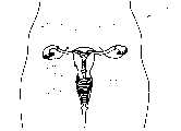
图22A、22B为显示图14所示装置的使用例子。图22A为将装置放在适当位置以得到女性生殖系统的虚拟影像。图22B为女性生殖系统的概要图,而图22C为内视镜之球形短颈玻璃容器膨胀于子宫的放大图。22A and 22B show examples of use of the device shown in FIG. 14 . Figure 22A is a virtual image of the female reproductive system with the device in place. Fig. 22B is a schematic diagram of the female reproductive system, and Fig. 22C is an enlarged view of the spherical short-necked glass container of the endoscope inflated in the uterus.
图23为本发明装置之内视镜末端的一个剖面放大图,说明光的流动与通过内视镜之光学影像的流向。Fig. 23 is an enlarged cross-sectional view of the endoscope end of the device of the present invention, illustrating the flow of light and the flow direction of the optical image through the endoscope.
图24A、图24B为本发明装置的又一实施例,其中内视镜包括一个可移动的护套,在内视镜的末端,此护套包括一个抓取器用来抓取需观察之内部区域的物体。Figure 24A and Figure 24B are yet another embodiment of the device of the present invention, wherein the endoscope includes a movable sheath at the end of the endoscope, and the sheath includes a grasper for grasping the internal area to be observed objects.
图24A为此护套的一个透视图,而Figure 24A is a perspective view of this sheath, while
图24B为此抓取器的一个放大图。Figure 24B is an enlarged view of this gripper.
图25为一个护套例子之用来包住本发明影像处理装置之内视镜末端的一个透视图。Fig. 25 is a perspective view of an example of a sheath used to enclose the endoscope end of the image processing device of the present invention.
图26为本发明影像处理装置之内视镜的另一实施例之一个侧视图。Fig. 26 is a side view of another embodiment of the endoscope of the image processing device of the present invention.
图27A、图27B为图6所示转接器的另一步实施例。图27A为此转接器的末端图,而图27B为此转接器连接至影像观察元件的一个侧视图。27A and 27B are another embodiment of the adapter shown in FIG. 6 . FIG. 27A is an end view of the adapter, and FIG. 27B is a side view of the adapter connected to the video viewing unit.
具体实施方式Detailed ways
本发明提供一种可携式影像处理装置,用来影像处理物体的内部区域,譬如人类或动物的体腔、器官或通道。The present invention provides a portable image processing device, which is used for image processing the internal area of an object, such as body cavities, organs or passages of humans or animals.
本发明的可携式影像处理装置包含:The portable image processing device of the present invention comprises:
一个内视镜,包含一个影像撷取光学线路及一个照明光学线路;及一个容易接合又容易松脱于内视镜尖端的手握式影像观察元件,An endoscope comprising an image capturing optical circuit and an illuminating optical circuit; and a hand-held image viewing element that is easily coupled and easily released from the tip of the endoscope,
该影像观察元件进一步包含:The image viewing element further includes:
一个可光学式连接于影像撷取光学线路的影像侦测器,用来侦测从内视镜末端传来的影像;an image detector optically connected to the image capture optical circuit for detecting the image transmitted from the end of the endoscope;
一个与侦测器相通的显示器,用来显示侦测器侦测到的影像;及a display in communication with the detector for displaying images detected by the detector; and
一个可光学式连接于照明光学线路的光源,用来传送光至内视镜末端。A light source optically connectable to the illumination optics for delivering light to the endoscope end.
『内视镜』这个名词的意涵为柔软或坚硬的观察工具其具有可诊断或甚至治疗的功能。此类观察工具有时亦称做『导管』,如果其包含一个影像撷取光学线路与一个照明光学线路,则也是本发明的范围。The term "endoscope" means a soft or hard viewing instrument with diagnostic or even therapeutic functions. Such viewing tools, sometimes referred to as "catheters," are also within the scope of the present invention if they include an image capture optic and an illumination optic.
本发明的影像处理装置是可携式的,且允许使用者看见显示于影像观察元件之显示器上物体内部区域的光学影像。因而不需要将影像传至一个外部观察器,如电视机(TV)或视讯监视器或电脑才能观察此影像。物体内部区域的快速虚拟立即可视影像可以从本发明的可携式影像处理装置得到。此影像观察元件足够小以致于可以握在手上,此元件易于使用、储存与携带。本发明的可携式影像处理装置特别适用于医护人员,譬如医生与护士,可以容易且快速地得到身体内部区域的可视影像作为诊断用。The image processing device of the present invention is portable and allows the user to see the optical image of the internal area of the object displayed on the display of the image viewing device. Therefore, there is no need to transmit the image to an external viewer, such as a television (TV) or video monitor or computer, to observe the image. The fast virtual instant visual image of the inner area of the object can be obtained from the portable image processing device of the present invention. The image viewing unit is small enough to be held in the hand, and the unit is easy to use, store and carry. The portable image processing device of the present invention is particularly suitable for medical personnel, such as doctors and nurses, who can easily and quickly obtain visual images of the internal regions of the body for diagnosis.
本发明的一个较佳实施例中,此影像观察元件是一只手机其可容易接合又容易松脱地转接于内视镜。In a preferred embodiment of the present invention, the image viewing element is a mobile phone, which can be easily connected and easily detached to be transferred to the endoscope.
参考图1的示意图,为说明本发明之可携式影像处理装置10的一个例子。其包含一只手机20容易接合又容易松脱地连结道内视镜30。此内视镜的末端35,透过身体的通道如尿道,可伸入体腔或器官。从内视镜的末端35发出光来照明体腔内部而体腔的影像则经由内视镜30传至影像观察元件20,允许医护人员,譬如医生与护士,可以立即观察此影像。Referring to the schematic diagram of FIG. 1 , it illustrates an example of a portable
在这些图中,此影像观察元件是一个改造的手机20,其可容易地握在手掌内5。然而,本发明的范围亦扩及其他影像观察元件,譬如一个改造的手持式个人数位助理(PDA),一个改造的数位相机,或类似的元件。本发明的一个较佳实施例为,此影像观察元件包含一个个人数位助理(PDA)。In these figures, the video viewing unit is a modified
如图2A、图2B、图2C所示,此手机20包含一个液晶显示荧幕(LCD)21来显示影像;一个耳机22与麦克风23,使得此手机仍可作为通讯工具;可程式化的软体可处理与储存影像;控制按钮24;一个电池25;及一个未展示的通用序列汇流排(USB)来使得此话机可连至电脑以传送其储存的影像。如同标准的手机,此电池25是可充电的使得此充电满载的元件可以工作一段时间(一般而言是八小时的正常通话时间与一百四十小时的待机时间)。As shown in Fig. 2A, Fig. 2B, and Fig. 2C, the
此手机20可以添加其他附件,譬如附着工具来附着此话机至一条皮带或手腕或类似处,头戴式耳机,独立的麦克风及类似元件。Other accessories can be added to the
此手机20的背面有一个连接埠来50承接如图6所示内视镜30的转接器40。此连接埠50有一基本的椭圆形且包括一个电荷耦合元件(CCD)影像感应器连60至一个光学聚焦镜片70及一个发光启动系统连接器80。The back side of the
一个发光启动系统85结合于此手机20的机身而包含一个圆柱形的头82与一个细长的机身81,就如图3A、图3B至图4、图5所示。此启动系统85一般而言高度大约6mm且宽度大约2mm。此细长的机身81包含一个FPC可程式化晶片。此圆柱形的头82包含一个位于FPC晶片81上的发光二极体(LED)83与一个微型光学聚焦镜片84,其将LED的发光聚焦于方向A(参考图4与图5)。此微型光学聚焦镜片84可用塑胶射出模具制造出来。这头的上层表面齐平于手机20的机身而提供连接埠50之发光启动系统的连结器80。此FPC晶片可预先程式为允许使用者修正从发光启动系统85亦即从内视镜30末端35发出的光亮度。使用者按下手机20的某一按钮24即可调整亮度。A light-emitting
图13A、图13B为本发明可携式影像处理装置10之影像观察元件20具有与不具有发光启动系统85所得到的光学影像显现之比较。此影像观察元件20具有发光启动系统85所得到的光学影像17示于图13B,其相当地较细致于及较清晰于此影像观察元件20不具有发光启动系统85所得到的光学影像,如图13A所示。此发光启动系统85因而有效地提供更聚焦的光用来照明需观察的影像,造成更清晰的影像显现于影像观察元件20。本发明装置只需要一个NICHIA NSCW 455之定闪的白光LED,即可提供足够光来照明身体的内部区域。此LED83一般由电池25提供电力。13A and 13B are comparisons of the optical image display obtained by the
使用者可立即地观察内视镜30所撷取且显示于LCD荧幕21的可视影像17。此手机20可有摄影机功能及静态照相功能,来提供体腔或器官内部之连续的视讯影像。此外,此手机20可储存撷取的影像,加上一般照相手机的标准功能,且影像可稍后透过话机的USB连接埠传送至另一个通讯设备或者电脑;不过,其它形式的传输电子资料,例如无线或红外线传输,也是众所周知的技术。此手机20亦可插入电视机(TV)监视器,以使得更大的影像显现于电视机(TV)监视器上。The user can immediately observe the
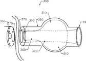
如图6、图7B及图27所示内视镜30的转接器40,其容易接合又容易松脱地连结到手机20的连接埠50。当内视镜转接器40连接至连接埠50,此发光启动系统连结器80配合一个光源导管90而将光依照方向A传送至内视镜的末端35。此外,光学聚焦镜片70配合一个光学影像导管95,而将影像依照方向B从内视镜的末端35传输至CCD影像感应器60。图27所示的转接器中,光学影像导管95大约是连接于连接埠50之光源导管90的两倍大。而此光学聚焦镜片70相对地大约是发光启动系统连结器80的两倍大。The
如图7A、图7B所示,此光源导管90包含一束光纤92由外层套管圈住且光学影像导管95包含一束光纤93由外层套管圈住。每一束光纤92,93可能有超过7200条的光纤。如图7所示的实施例,光源导管90与光学影像导管95接合于接合处91,而且光源导管90在至内视镜末端35的剩余长度上是圈住光学影像导管95的。如图9所示,在其末端35有一微型的光学聚焦镜片31,其包含一个内部光学聚焦镜片33及一个外部光学聚焦镜片32。此内部光学聚焦镜片33将内部区域的光学影像聚焦。聚焦的影像则沿着光纤93传输至手机20的CCD影像感应器60。此外部光学聚焦镜片32用来聚焦发光启动系统85产生的发光,然后沿着光纤92传至内视镜30末端35的镜片32。As shown in FIG. 7A and FIG. 7B , the light source guide 90 includes a bundle of
如图26所示之另一个实施例,光源导管90与光学影像导管95是分开的且沿着内视镜的整个长度。在光学影像导管95的末端放置一个光学影像聚焦镜片36,用来聚焦内部区域的光学影像。一个轻薄的聚焦镜片37放置于光源导管90的末端,用来聚焦发光启动系统85产生的发光,然后沿着光纤92传至内视镜30末端35的镜片32。In another embodiment shown in Figure 26, the
外部光学聚焦镜片32或轻薄的聚焦镜片37的发光照明体腔或通道的内部区域。由内部光学聚焦镜片33或光学影像聚焦镜片36聚焦的影像沿着光纤93传输至手机20的CCD影像感应器60。如图23所示,从发光启动系统80的发光依照全向内部反射原理跳跃于光纤92的管壁中而沿着光纤92行走。然后光由外部光学聚焦镜片32或光学影像聚焦镜片36所聚焦而射入体腔或通道的内部区域。同样地,光学影像依照全向内部反射原理跳跃于光纤93的管壁中而沿着光纤93传输。The illumination of the external optical focusing
图11与图12说明本发明之可携式影像处理装置10的影像撷取能力。当内视镜30的末端35透过虚拟体腔12之小孔15伸入时即显现其虚拟3D画面。如图12所示,测量距离内视镜30的末端35为50cm与100cm的光强度发现其分别可涵盖直径为250mm一直到500mm的范围。11 and 12 illustrate the image capture capability of the portable
本发明之可携式影像处理装置的较佳实施例为包括一个特制的可移动护套,来保护内视镜的末端部分。此护套包含一个具有封口末端的导管,且其开口尖端用来承接内视镜的末端。此护套的较佳实施例为包含一个本质为液体的不透水材料,也可以是一个本质为气体与液体的不透水材料,或是一个本质为透明的乳胶材料。A preferred embodiment of the portable image processing device of the present invention includes a special removable sheath to protect the end portion of the endoscope. The sheath consists of a catheter with a sealed end and an open tip for receiving the end of an endoscope. The preferred embodiment of the sheath comprises an inherently liquid impermeable material, may also be an inherently gas and liquid impermeable material, or an inherently transparent latex material.
图25说明一个护套600的实施例,其可放置于内视镜30的末端35。此护套600一般而言是消毒过的且可丢弃式的,也就是说它是用后即丢然后更换另一个消毒的护套以为下次使用。这意味着在每次应用之间不需要对内视镜做消毒,而是提供一个消毒的护套来至少保护内视镜的末端,其用来伸入体腔或通道。此护套600的较佳实施例为液体与气体混合的不透水材料,亦可以透明的乳胶材料且做出许多不同长度与大小的护套,作为不同大小的内视镜及不同的程序所使用。此护套600可以是不透明的材料而只要其末端是透明的材料来允许内视镜30的光学影像导管95撷取影像。FIG. 25 illustrates an embodiment of a
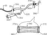
在本发明的另一实施例中,可携式影像处理装置的护套包括一个可膨胀的球形短颈玻璃容器靠近护套的末端。图14至图18说明本发明之可携式影像处理装置的一个实施例。其内视镜30具有一个球形短颈玻璃容器护套390其包含一个可膨胀的球形短颈玻璃容器310由一膨胀的导管360连接至一个注射器320。此注射器320的主体321保有流体如空气或液体,及一个活塞322。此注射器320的主体321尾端连接至膨胀导管360以致于当活塞322按照方向A推进时,流体从主体321流出通过膨胀导管360而膨胀球形短颈玻璃容器310。压缩球形短颈玻璃容器310则是活塞以反方向拉出而球形短颈玻璃容器310内的流体吸回注射器320的主体321。In another embodiment of the present invention, the housing of the portable image processing device includes an inflatable spherical short-necked glass container near the end of the housing. 14 to 18 illustrate an embodiment of the portable image processing device of the present invention. Its
球形短颈玻璃容器护套390有一条内视镜管370用来承接内视镜30。此内视镜30的末端35伸入内视镜管370直到其抵达护套的封口末端395,以致于护套390围绕住内视镜30的末端部分。在护套390的接点330,膨胀导管360接上内视镜管370,其用来如图17所示承接内视镜30。此护套390的较佳实施例为本质是液体与气体混合的不透水材料,且作为一个屏障来保护内视镜30,特别是内视镜30末端35的镜片31。The spherical short-neck
此内视镜300的末端被球形短颈玻璃容器护套390所围绕着其可用来影像处理如图22A、图22B、图22C所示之女性生殖系统200。内视镜300的末端395由医生或护士伸入女性的阴道210。此内视镜300可熟练地操作来通过子宫颈道220而进入子宫颈230,此程序辅以内视镜300的柔软弹性特质来完成。当其末端395进入子宫240时医生或护士压下注射器320的活塞322来膨胀球形短颈玻璃容器310,如上文更详细的叙述。此膨胀的球形短颈玻璃容器310撑开子宫颈230壁而保持内视镜300的末端395位于子宫240内。光从内视镜300的末端395发出来照明子宫240与输卵管250的开口。连接于卵巢260的输卵管250影像传输至影像观察元件20。医生或护士可立即地观察LCD显示荧幕21上的影像。The end of the
在本发明的另一个较佳实施例中,可携式影像处理装置的护套包括一条吸引导管,其具有一开放的末端位于或接于护套的末端,及一个尖端连接于吸引工具。图19与图20说明本发明之影像处理装置的一个实施例,其内视镜400有一个吸引护套490包含一条吸引导管460连接至有主体421与活塞422的注射器420。注射器420的主体421尾端连接至吸引导管460以致于当活塞422依照方向B拉出时,一个吸引力量产生于吸引导管460内。此吸引力量可用来从内视镜400伸入的体腔或通道内收集细胞组织样本或其他样本,例如用来取得如图22B所示之输卵管250的切片组织。此内视镜400可同时用来收集样本与撷取输卵管250的影像。本发明装置的此实施例可用来吸引受伤的创口或类似部位。此装置提供受伤创口的影像而让目标区的吸引可以执行。In another preferred embodiment of the present invention, the sheath of the portable image processing device includes a suction catheter, which has an open end located at or connected to the end of the sheath, and a tip connected to the suction tool. 19 and 20 illustrate an embodiment of the image processing device of the present invention, in which an
吸引护套490有一条内视镜管470用来承接内视镜30。此内视镜30的末端35伸入内视镜管470直到其抵达护套的封口末端495,以致于护套490围绕住内视镜30的末端部分。在护套490的接点430,吸引导管460接上内视镜管470,其用来承接内视镜30。此护套490的较佳实施例为本质是液体与气体混合的不透水材料,且作为一个屏障来保护内视镜30,特别是内视镜30末端35的镜片31。The
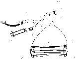
在本发明之影像处理装置的另一较佳实施例中,此护套末端包括一个抓取器用来抓取物体。图24说明本发明之影像处理装置的一个实施例。其内视镜500有一个抓取器护套590,包含一条抓取器导管560,连接至有主体521与活塞522的注射器520。此抓取器导管560有一个抓取器,其包含抓取器爪子510在它的末端,及一个连结器将抓取器爪子510,连至注射器活塞522。注射器主体521连接至抓取器导管560,而注射器活塞522则用来操作位于内视镜500末端595的抓取器爪子510。此抓取器爪子510可用来抓取物体,譬如从内视镜500伸入的体腔或通道中抓取异物或组织样本。此内视镜500可同时用来抓取物体及撷取身体内部区域的影像。In another preferred embodiment of the image processing device of the present invention, the end of the sheath includes a grabber for grabbing objects. Fig. 24 illustrates an embodiment of the image processing apparatus of the present invention. Its
抓取器护套590有一条内视镜管570用来承接内视镜30。此内视镜30的末端35伸入内视镜管570直到其抵达护套的封口末端595,以致于护套590围绕住内视镜30的末端部分。在护套590的接点530,抓取器导管560接上内视镜管570,其用来承接内视镜30。此护套590的较佳实施例为本质是液体与气体混合的不透水材料,且作为一个屏障来保护内视镜30,特别是内视镜30末端35的镜片31。The
图21为本发明装置的不同使用例子。在图21中,此内视镜300包括一个如上文与图14至图18有关之叙述的球形短颈玻璃容器护套390。不过,其他内视镜例子譬如具有吸引护套490的内视镜400或上文详细叙述具有防护之护套的内视镜可以替代使用。Figure 21 shows different examples of use of the device of the present invention. In Fig. 21, the
图21A、图21B、图21C、图21D说明男性500与女性600泌尿系统,其包含两颗肾脏520经由两条输尿管530连至膀胱540。尿液从膀胱540通过尿道550而流出体外。为了观察泌尿系统的内部区域,此柔软有弹性之内视镜的末端395由医生或护士伸入男性500或女性600的尿道550并操作其进入膀胱540。一旦于膀胱,球形短颈玻璃容器310就膨胀来保持此末端395于定位。光从内视镜300的末端395发出来照明膀胱540的内部区域而将其影像传输回至影像观察元件20的电荷耦合元件(CCD)影像感应器60。医生或护士可观察LCD显示荧幕21上的影像。此影像也可储存或进一步由影像观察元件处理,例如影像可放大而聚焦于特别感兴趣的区域。影像也可透过影像观察元件20的USB连接埠传送至电脑或类似的元件。此球形短颈玻璃容器310亦可作为扩张器之功能,而允许医生或护士来取得膀胱的切片组织样本。21A, 21B, 21C, and 21D illustrate male 500 and female 600 urinary systems, which include two
本发明之可携式影像处理装置可用来观察任何无法从外部观察之内部区域的影像,譬如人类或动物的内部体腔或器官。假使内视镜可伸入物体,则此影像观察元件可显示物体内部区域的影像。其并不需要将影像传送至外部影像显示元件如电脑等。此影像观察元件可以握于人的手掌中,因此本发明之影像处理装置易于使用与携带。此内视镜可伸入任何身体通道或管道,例如泌尿道、耳朵、鼻子、食道、肛门或生殖系统,或伸入开刀手术或受伤的伤口。因此可以观察到身体内部体腔、器官或通道。立即可视的影像常常使得可以侦测出先前诊断程序未检查出的病变。一旦诊断确定,则内视镜手术可以进行。内视镜手术的好处很多,包括较小的切口造成较少的疤痕,较轻微的手术后疼痛,及较短的住院期。此外,本发明之装置也可用来从身体内部体腔、器官或通道中收集组织切片样本。The portable image processing device of the present invention can be used to observe images of any internal areas that cannot be observed from the outside, such as internal body cavities or organs of humans or animals. If the endoscope can be extended into the object, the image viewing element can display the image of the inner area of the object. It does not need to transmit the image to an external image display device such as a computer. The image viewing element can be held in the palm of a person, so the image processing device of the present invention is easy to use and carry. The endoscope can be inserted into any body passage or duct, such as the urinary tract, ear, nose, esophagus, anus or reproductive system, or into surgical or wound wounds. It is thus possible to observe internal body cavities, organs or passages. Immediately visible images often allow the detection of lesions not detected by previous diagnostic procedures. Once the diagnosis is confirmed, endoscopic surgery can be performed. The benefits of endoscopic surgery are many, including smaller incisions resulting in less scarring, less post-operative pain, and a shorter hospital stay. In addition, the device of the present invention can also be used to collect tissue section samples from internal body cavities, organs or passages.
本发明之可携式影像处理装置特别适合用于泌尿科学领域来对泌尿道的感染做影像处理与诊断,及用于生育与妇科临床特别是早期诊断出卵巢癌或类似病症。The portable image processing device of the present invention is particularly suitable for image processing and diagnosis of urinary tract infection in the field of urology, and for fertility and gynecology clinics, especially for early diagnosis of ovarian cancer or similar diseases.
本发明之影像处理装置的应用不限制于医学的或兽医的临床,此装置可用于任何情况下,其内部区域的影像无法从外面得到适当的观察,其中内视镜用来取得与传输内部区域的影像。例如本发明的装置可用来检查机器的零件内部或类似地方。The application of the image processing device of the present invention is not limited to medical or veterinary clinics, the device can be used in any situation where the image of the internal area cannot be properly observed from the outside, wherein the endoscope is used to obtain and transmit the internal area of the image. For example the device of the invention can be used to inspect the interior of parts of machines or the like.
以上所述者,仅为本发明之较佳实施例而已,当不能以此限定本发明实施之范围。然而,本领域技术的人员将意识到,本发明可作出各种可能的变化和修改,而不背离本发明的范围和精神。The above descriptions are only preferred embodiments of the present invention, and should not limit the implementation scope of the present invention. However, those skilled in the art will appreciate that various possible changes and modifications may be made to the present invention without departing from the scope and spirit of the invention.
Claims (28)
Translated fromChineseApplications Claiming Priority (2)
| Application Number | Priority Date | Filing Date | Title |
|---|---|---|---|
| CA2,509,590 | 2005-06-06 | ||
| CA002509590ACA2509590A1 (en) | 2005-06-06 | 2005-06-06 | Portable imaging apparatus |
Publications (1)
| Publication Number | Publication Date |
|---|---|
| CN101212926Atrue CN101212926A (en) | 2008-07-02 |
Family
ID=37498067
Family Applications (1)
| Application Number | Title | Priority Date | Filing Date |
|---|---|---|---|
| CN200680020002.0APendingCN101212926A (en) | 2005-06-06 | 2006-06-06 | Portable Image Processing Device |
Country Status (4)
| Country | Link |
|---|---|
| US (1) | US20080207996A1 (en) |
| CN (1) | CN101212926A (en) |
| CA (1) | CA2509590A1 (en) |
| WO (1) | WO2006130967A1 (en) |
Cited By (6)
| Publication number | Priority date | Publication date | Assignee | Title |
|---|---|---|---|---|
| CN103181154A (en)* | 2010-10-29 | 2013-06-26 | 加利福尼亚大学董事会 | Mobile phone microscope apparatus and method for imaging |
| CN103860130A (en)* | 2012-12-13 | 2014-06-18 | 蒋小华 | Novel ultrasonic probe gastroscope exploring tube |
| CN104000551A (en)* | 2013-02-21 | 2014-08-27 | 邱立国 | Portable internal viewing device and image capturing method thereof |
| CN104323757A (en)* | 2014-11-13 | 2015-02-04 | 王卫东 | Endoscope with flash lamp and camera for portable intelligent equipment |
| CN104434007A (en)* | 2014-11-26 | 2015-03-25 | 南京春辉科技实业有限公司 | Fiber-endoscope matched with cell phone |
| TWI612935B (en)* | 2017-01-13 | 2018-02-01 | Cai yi qiu | Illuminated disposable medical device |
Families Citing this family (64)
| Publication number | Priority date | Publication date | Assignee | Title |
|---|---|---|---|---|
| US8858425B2 (en) | 2004-09-24 | 2014-10-14 | Vivid Medical, Inc. | Disposable endoscope and portable display |
| US8556806B2 (en)* | 2004-09-24 | 2013-10-15 | Vivid Medical, Inc. | Wavelength multiplexing endoscope |
| US8602971B2 (en)* | 2004-09-24 | 2013-12-10 | Vivid Medical. Inc. | Opto-Electronic illumination and vision module for endoscopy |
| US8827899B2 (en)* | 2004-09-24 | 2014-09-09 | Vivid Medical, Inc. | Disposable endoscopic access device and portable display |
| US9033870B2 (en) | 2004-09-24 | 2015-05-19 | Vivid Medical, Inc. | Pluggable vision module and portable display for endoscopy |
| US8878924B2 (en) | 2004-09-24 | 2014-11-04 | Vivid Medical, Inc. | Disposable microscope and portable display |
| US8480566B2 (en)* | 2004-09-24 | 2013-07-09 | Vivid Medical, Inc. | Solid state illumination for endoscopy |
| US9788790B2 (en) | 2009-05-28 | 2017-10-17 | Avinger, Inc. | Optical coherence tomography for biological imaging |
| US9125562B2 (en) | 2009-07-01 | 2015-09-08 | Avinger, Inc. | Catheter-based off-axis optical coherence tomography imaging system |
| US8696695B2 (en) | 2009-04-28 | 2014-04-15 | Avinger, Inc. | Guidewire positioning catheter |
| US20110009694A1 (en)* | 2009-07-10 | 2011-01-13 | Schultz Eric E | Hand-held minimally dimensioned diagnostic device having integrated distal end visualization |
| US20100121139A1 (en) | 2008-11-12 | 2010-05-13 | Ouyang Xiaolong | Minimally Invasive Imaging Systems |
| WO2011003006A2 (en) | 2009-07-01 | 2011-01-06 | Avinger, Inc. | Atherectomy catheter with laterally-displaceable tip |
| TWI578949B (en)* | 2009-07-15 | 2017-04-21 | Medical Intubation Tech Corp | Endoscopic device and its image processing method |
| EP2485079B1 (en) | 2009-09-29 | 2016-12-21 | Olympus Corporation | Endoscope system |
| DE102009056108A1 (en)* | 2009-11-30 | 2011-06-01 | Karl Storz Gmbh & Co. Kg | Medical device for supporting an endoscopic examination |
| US8535265B2 (en)* | 2009-12-22 | 2013-09-17 | Kimberly-Clark Worldwide, Inc. | Tracheal catheter with suction lumen port in close proximity to the cuff |
| US11382653B2 (en) | 2010-07-01 | 2022-07-12 | Avinger, Inc. | Atherectomy catheter |
| JP6173917B2 (en)* | 2010-12-21 | 2017-08-02 | ザ ユニバーシティ オブ ユタ リサーチ ファウンデイション | Assembly and use of optically guided medical tubes and control units |
| US9949754B2 (en) | 2011-03-28 | 2018-04-24 | Avinger, Inc. | Occlusion-crossing devices |
| EP2691038B1 (en) | 2011-03-28 | 2016-07-20 | Avinger, Inc. | Occlusion-crossing devices, imaging, and atherectomy devices |
| WO2012154578A1 (en)* | 2011-05-06 | 2012-11-15 | The Trustees Of The University Of Pennsylvania | Ped - endoscope image and diagnosis capture system |
| US9345406B2 (en) | 2011-11-11 | 2016-05-24 | Avinger, Inc. | Occlusion-crossing devices, atherectomy devices, and imaging |
| US9693759B2 (en) | 2011-11-16 | 2017-07-04 | Coloplast A/S | Operating device with a control handle and a flexible element connected to the control handle |
| US9557156B2 (en) | 2012-05-14 | 2017-01-31 | Avinger, Inc. | Optical coherence tomography with graded index fiber for biological imaging |
| WO2013172970A1 (en) | 2012-05-14 | 2013-11-21 | Avinger, Inc. | Atherectomy catheters with imaging |
| EP2849660B1 (en) | 2012-05-14 | 2021-08-25 | Avinger, Inc. | Atherectomy catheter drive assemblies |
| US11284916B2 (en) | 2012-09-06 | 2022-03-29 | Avinger, Inc. | Atherectomy catheters and occlusion crossing devices |
| WO2014073950A1 (en)* | 2012-11-08 | 2014-05-15 | Erasmus University Medical Center Rotterdam | An adapter for coupling a camera unit to an endoscope, a method of recording image and a computer program product |
| WO2014143064A1 (en) | 2013-03-15 | 2014-09-18 | Avinger, Inc. | Chronic total occlusion crossing devices with imaging |
| US11096717B2 (en) | 2013-03-15 | 2021-08-24 | Avinger, Inc. | Tissue collection device for catheter |
| KR20220097541A (en) | 2013-03-15 | 2022-07-07 | 버터플라이 네트워크, 인크. | Monolithic ultrasonic imaging devices, systems and methods |
| CN105228514B (en) | 2013-03-15 | 2019-01-22 | 阿维格公司 | Optical Pressure Sensor Assembly |
| US9667889B2 (en)* | 2013-04-03 | 2017-05-30 | Butterfly Network, Inc. | Portable electronic devices with integrated imaging capabilities |
| CN104116483A (en)* | 2013-04-29 | 2014-10-29 | 赵盾 | Portable endoscope |
| EP3019096B1 (en) | 2013-07-08 | 2023-07-05 | Avinger, Inc. | System for identification of elastic lamina to guide interventional therapy |
| EP3024594A2 (en) | 2013-07-23 | 2016-06-01 | Butterfly Network Inc. | Interconnectable ultrasound transducer probes and related methods and apparatus |
| US9370295B2 (en) | 2014-01-13 | 2016-06-21 | Trice Medical, Inc. | Fully integrated, disposable tissue visualization device |
| US10342579B2 (en) | 2014-01-13 | 2019-07-09 | Trice Medical, Inc. | Fully integrated, disposable tissue visualization device |
| US11547446B2 (en) | 2014-01-13 | 2023-01-10 | Trice Medical, Inc. | Fully integrated, disposable tissue visualization device |
| WO2015191954A1 (en)* | 2014-06-12 | 2015-12-17 | Endoluxe Inc. | Encasement platform for smartdevice for attachment to endoscope |
| US10357277B2 (en) | 2014-07-08 | 2019-07-23 | Avinger, Inc. | High speed chronic total occlusion crossing devices |
| WO2016019235A1 (en)* | 2014-07-31 | 2016-02-04 | The University Of Akron | A smartphone endoscope system |
| US9848110B2 (en)* | 2014-08-18 | 2017-12-19 | Gadget Support LLC | Inspection scope devices and methods for mobile electronic devices |
| US10568520B2 (en) | 2015-07-13 | 2020-02-25 | Avinger, Inc. | Micro-molded anamorphic reflector lens for image guided therapeutic/diagnostic catheters |
| WO2017027749A1 (en) | 2015-08-11 | 2017-02-16 | Trice Medical, Inc. | Fully integrated, disposable tissue visualization device |
| US20170086650A1 (en)* | 2015-09-25 | 2017-03-30 | Save My Scope Inc. | Adapter Device for Securing an Eyepiece to a Mobile Phone |
| JP6927986B2 (en) | 2016-01-25 | 2021-09-01 | アビンガー・インコーポレイテッドAvinger, Inc. | OCT imaging catheter with delay compensation |
| US10499792B2 (en) | 2016-03-25 | 2019-12-10 | University Of Washington | Phone adapter for flexible laryngoscope and rigid endoscopes |
| EP3435892B1 (en) | 2016-04-01 | 2024-04-03 | Avinger, Inc. | Atherectomy catheter with serrated cutter |
| USD798443S1 (en) | 2016-05-03 | 2017-09-26 | Coloplast A/S | Videoscope handle |
| US11344327B2 (en) | 2016-06-03 | 2022-05-31 | Avinger, Inc. | Catheter device with detachable distal end |
| WO2018006041A1 (en) | 2016-06-30 | 2018-01-04 | Avinger, Inc. | Atherectomy catheter with shapeable distal tip |
| NL2018494B1 (en)* | 2017-03-09 | 2018-09-21 | Quest Photonic Devices B V | Method and apparatus using a medical imaging head for fluorescent imaging |
| US10470645B2 (en)* | 2017-05-22 | 2019-11-12 | Gustav Lo | Imaging system and method |
| CN107290257A (en)* | 2017-07-29 | 2017-10-24 | 山东诺方电子科技有限公司 | One kind automation atmosphere particle monitoring equipment |
| US10750061B2 (en) | 2017-09-15 | 2020-08-18 | Omnivision Technologies, Inc. | Endoscope having camera module and LED on flexible printed circuit |
| US10772488B2 (en) | 2017-11-10 | 2020-09-15 | Endoluxe Inc. | System and methods for endoscopic imaging |
| EP3773235B1 (en) | 2018-03-29 | 2023-07-19 | Trice Medical, Inc. | Fully integrated endoscope with biopsy capabilities |
| US12167867B2 (en) | 2018-04-19 | 2024-12-17 | Avinger, Inc. | Occlusion-crossing devices |
| CN113260846B (en)* | 2018-08-16 | 2025-01-17 | 上海宜晟生物科技有限公司 | Surface color and liquid contact angle imaging |
| CN114746033B (en) | 2019-10-18 | 2025-01-10 | 阿维格公司 | Blocking crossing device |
| KR102813371B1 (en)* | 2019-11-19 | 2025-05-27 | 삼성전자주식회사 | Dual camera module, electronic apparatus including the same and method of operating electronic apparatus |
| US12376731B2 (en) | 2022-01-10 | 2025-08-05 | Endoluxe Inc. | Systems, apparatuses, and methods for endoscopy |
Family Cites Families (15)
| Publication number | Priority date | Publication date | Assignee | Title |
|---|---|---|---|---|
| GB1548864A (en)* | 1975-06-06 | 1979-07-18 | Plessey Co Ltd | Connectors for coaxially coupling the end of a linear optical waveguide element to a photoelectric transducer |
| US4470407A (en)* | 1982-03-11 | 1984-09-11 | Laserscope, Inc. | Endoscopic device |
| US4742819A (en)* | 1987-03-23 | 1988-05-10 | George Gordon P | Intubating scope with camera and screen |
| US4994910A (en)* | 1989-07-06 | 1991-02-19 | Acuimage Corporation | Modular endoscopic apparatus with probe |
| FR2690066B1 (en)* | 1992-04-21 | 1998-08-07 | Jean Marc Inglese | IMAGE ACQUISITION DEVICE USING A SEMICONDUCTOR LIGHT SOURCE |
| US5540711A (en)* | 1992-06-02 | 1996-07-30 | General Surgical Innovations, Inc. | Apparatus and method for developing an anatomic space for laparoscopic procedures with laparoscopic visualization |
| US5865727A (en)* | 1995-08-25 | 1999-02-02 | Asahi Kogaku Kogyo Kabushiki Kaisha | Portable endoscope system |
| US6554765B1 (en)* | 1996-07-15 | 2003-04-29 | East Giant Limited | Hand held, portable camera with adaptable lens system |
| US5879289A (en)* | 1996-07-15 | 1999-03-09 | Universal Technologies International, Inc. | Hand-held portable endoscopic camera |
| US6091453A (en)* | 1996-07-29 | 2000-07-18 | Coan; Steven | Hand held remote camera |
| US6066090A (en)* | 1997-06-19 | 2000-05-23 | Yoon; Inbae | Branched endoscope system |
| US6452626B1 (en)* | 1997-10-06 | 2002-09-17 | Edwin L. Adair | Communication devices incorporating reduced area imaging devices |
| US6793622B2 (en)* | 2001-09-05 | 2004-09-21 | Olympus Optical Co., Ltd. | Electric bending endoscope |
| TW581668B (en)* | 2003-10-15 | 2004-04-01 | Der-Yang Tien | Endoscopic device |
| US20050107665A1 (en)* | 2003-11-18 | 2005-05-19 | Nady Nady E. | Device for sealing a body canal and method of use |
- 2005
- 2005-06-06CACA002509590Apatent/CA2509590A1/ennot_activeAbandoned
- 2006
- 2006-06-06CNCN200680020002.0Apatent/CN101212926A/enactivePending
- 2006-06-06WOPCT/CA2006/000916patent/WO2006130967A1/enactiveApplication Filing
- 2006-06-06USUS11/916,323patent/US20080207996A1/ennot_activeAbandoned
Cited By (8)
| Publication number | Priority date | Publication date | Assignee | Title |
|---|---|---|---|---|
| US9325884B2 (en) | 2008-01-02 | 2016-04-26 | The Regents Of The University Of California | Cellscope apparatus and methods for imaging |
| US10616457B2 (en) | 2008-01-02 | 2020-04-07 | The Regents Of The University Of California | Cellscope apparatus and methods for imaging |
| CN103181154A (en)* | 2010-10-29 | 2013-06-26 | 加利福尼亚大学董事会 | Mobile phone microscope apparatus and method for imaging |
| CN103860130A (en)* | 2012-12-13 | 2014-06-18 | 蒋小华 | Novel ultrasonic probe gastroscope exploring tube |
| CN104000551A (en)* | 2013-02-21 | 2014-08-27 | 邱立国 | Portable internal viewing device and image capturing method thereof |
| CN104323757A (en)* | 2014-11-13 | 2015-02-04 | 王卫东 | Endoscope with flash lamp and camera for portable intelligent equipment |
| CN104434007A (en)* | 2014-11-26 | 2015-03-25 | 南京春辉科技实业有限公司 | Fiber-endoscope matched with cell phone |
| TWI612935B (en)* | 2017-01-13 | 2018-02-01 | Cai yi qiu | Illuminated disposable medical device |
Also Published As
| Publication number | Publication date |
|---|---|
| WO2006130967A1 (en) | 2006-12-14 |
| US20080207996A1 (en) | 2008-08-28 |
| CA2509590A1 (en) | 2006-12-06 |
Similar Documents
| Publication | Publication Date | Title |
|---|---|---|
| CN101212926A (en) | Portable Image Processing Device | |
| CN103298391B (en) | disposable endoscopic access device and portable display | |
| US5584796A (en) | Apparatus and method for retracting and viewing bodily tissues on remote display device | |
| EP1399201B1 (en) | Device for in-vivo procedures | |
| US9033870B2 (en) | Pluggable vision module and portable display for endoscopy | |
| JP5893124B2 (en) | Laparoscopic system | |
| JP4472069B2 (en) | Medical capsule endoscope | |
| US7419467B2 (en) | Medical inspection device | |
| US11096557B2 (en) | Endoscopy system having a miniature closed head | |
| US20200221936A1 (en) | Measurement Device | |
| US20060161048A1 (en) | Flexible video scope extension and methods | |
| JP2004202252A (en) | Endoscope device | |
| US20050085690A1 (en) | Endoscope apparatus | |
| CN101621957A (en) | Arthroscopy without cables | |
| CN113795187A (en) | Single use endoscope, cannula and obturator with integrated vision and illumination | |
| CN105725955A (en) | Gun type endoscope system capable of being connected with intelligent mobile device | |
| CN105169540A (en) | Brightness-adjustable double positioning video light stick for tracheal cannula | |
| KR100471653B1 (en) | Endoscope system | |
| CN205597881U (en) | Joinable intelligent mobile device's gun type endoscope system | |
| CN112998626A (en) | Disposable medical endoscope | |
| CN207785128U (en) | Big channel straight cutting enters endoscope | |
| CN100421613C (en) | Video Intubation Assist Device | |
| HK1120716A (en) | Portable imaging apparatus | |
| CN106343943A (en) | Arthroscopic device | |
| CN222032312U (en) | Handheld endoscope |
Legal Events
| Date | Code | Title | Description |
|---|---|---|---|
| C06 | Publication | ||
| PB01 | Publication | ||
| C10 | Entry into substantive examination | ||
| SE01 | Entry into force of request for substantive examination | ||
| REG | Reference to a national code | Ref country code:HK Ref legal event code:DE Ref document number:1120716 Country of ref document:HK | |
| C02 | Deemed withdrawal of patent application after publication (patent law 2001) | ||
| WD01 | Invention patent application deemed withdrawn after publication | Open date:20080702 | |
| REG | Reference to a national code | Ref country code:HK Ref legal event code:WD Ref document number:1120716 Country of ref document:HK |