CN101193595A - System for guiding a probe over the skin surface of a patient or animal - Google Patents
System for guiding a probe over the skin surface of a patient or animalDownload PDFInfo
- Publication number
- CN101193595A CN101193595ACNA2006800203847ACN200680020384ACN101193595ACN 101193595 ACN101193595 ACN 101193595ACN A2006800203847 ACNA2006800203847 ACN A2006800203847ACN 200680020384 ACN200680020384 ACN 200680020384ACN 101193595 ACN101193595 ACN 101193595A
- Authority
- CN
- China
- Prior art keywords
- patient
- probe
- blood vessel
- track
- blood
- Prior art date
- Legal status (The legal status is an assumption and is not a legal conclusion. Google has not performed a legal analysis and makes no representation as to the accuracy of the status listed.)
- Pending
Links
- 0CC1CC(*C2NCC2)C1Chemical compoundCC1CC(*C2NCC2)C10.000description1
Images


Classifications
- A—HUMAN NECESSITIES
- A61—MEDICAL OR VETERINARY SCIENCE; HYGIENE
- A61B—DIAGNOSIS; SURGERY; IDENTIFICATION
- A61B5/00—Measuring for diagnostic purposes; Identification of persons
- A61B5/48—Other medical applications
- A61B5/4887—Locating particular structures in or on the body
- A61B5/489—Blood vessels
- A—HUMAN NECESSITIES
- A61—MEDICAL OR VETERINARY SCIENCE; HYGIENE
- A61B—DIAGNOSIS; SURGERY; IDENTIFICATION
- A61B5/00—Measuring for diagnostic purposes; Identification of persons
- A61B5/0059—Measuring for diagnostic purposes; Identification of persons using light, e.g. diagnosis by transillumination, diascopy, fluorescence
- A61B5/0062—Arrangements for scanning
- A61B5/0066—Optical coherence imaging
- A—HUMAN NECESSITIES
- A61—MEDICAL OR VETERINARY SCIENCE; HYGIENE
- A61B—DIAGNOSIS; SURGERY; IDENTIFICATION
- A61B5/00—Measuring for diagnostic purposes; Identification of persons
- A61B5/0093—Detecting, measuring or recording by applying one single type of energy and measuring its conversion into another type of energy
- A61B5/0095—Detecting, measuring or recording by applying one single type of energy and measuring its conversion into another type of energy by applying light and detecting acoustic waves, i.e. photoacoustic measurements
- A—HUMAN NECESSITIES
- A61—MEDICAL OR VETERINARY SCIENCE; HYGIENE
- A61B—DIAGNOSIS; SURGERY; IDENTIFICATION
- A61B8/00—Diagnosis using ultrasonic, sonic or infrasonic waves
- A61B8/06—Measuring blood flow
- A—HUMAN NECESSITIES
- A61—MEDICAL OR VETERINARY SCIENCE; HYGIENE
- A61B—DIAGNOSIS; SURGERY; IDENTIFICATION
- A61B8/00—Diagnosis using ultrasonic, sonic or infrasonic waves
- A61B8/08—Clinical applications
- A61B8/0833—Clinical applications involving detecting or locating foreign bodies or organic structures
- A61B8/0841—Clinical applications involving detecting or locating foreign bodies or organic structures for locating instruments
- A—HUMAN NECESSITIES
- A61—MEDICAL OR VETERINARY SCIENCE; HYGIENE
- A61B—DIAGNOSIS; SURGERY; IDENTIFICATION
- A61B8/00—Diagnosis using ultrasonic, sonic or infrasonic waves
- A61B8/42—Details of probe positioning or probe attachment to the patient
- A61B8/4209—Details of probe positioning or probe attachment to the patient by using holders, e.g. positioning frames
- A61B8/4227—Details of probe positioning or probe attachment to the patient by using holders, e.g. positioning frames characterised by straps, belts, cuffs or braces
- A—HUMAN NECESSITIES
- A61—MEDICAL OR VETERINARY SCIENCE; HYGIENE
- A61B—DIAGNOSIS; SURGERY; IDENTIFICATION
- A61B8/00—Diagnosis using ultrasonic, sonic or infrasonic waves
- A61B8/44—Constructional features of the ultrasonic, sonic or infrasonic diagnostic device
- A61B8/4444—Constructional features of the ultrasonic, sonic or infrasonic diagnostic device related to the probe
- A61B8/4461—Features of the scanning mechanism, e.g. for moving the transducer within the housing of the probe
- A—HUMAN NECESSITIES
- A61—MEDICAL OR VETERINARY SCIENCE; HYGIENE
- A61B—DIAGNOSIS; SURGERY; IDENTIFICATION
- A61B90/00—Instruments, implements or accessories specially adapted for surgery or diagnosis and not covered by any of the groups A61B1/00 - A61B50/00, e.g. for luxation treatment or for protecting wound edges
- A61B90/36—Image-producing devices or illumination devices not otherwise provided for
- A61B2090/364—Correlation of different images or relation of image positions in respect to the body
- A61B2090/366—Correlation of different images or relation of image positions in respect to the body using projection of images directly onto the body
- A—HUMAN NECESSITIES
- A61—MEDICAL OR VETERINARY SCIENCE; HYGIENE
- A61B—DIAGNOSIS; SURGERY; IDENTIFICATION
- A61B5/00—Measuring for diagnostic purposes; Identification of persons
- A61B5/0059—Measuring for diagnostic purposes; Identification of persons using light, e.g. diagnosis by transillumination, diascopy, fluorescence
- A61B5/0073—Measuring for diagnostic purposes; Identification of persons using light, e.g. diagnosis by transillumination, diascopy, fluorescence by tomography, i.e. reconstruction of 3D images from 2D projections
- A—HUMAN NECESSITIES
- A61—MEDICAL OR VETERINARY SCIENCE; HYGIENE
- A61B—DIAGNOSIS; SURGERY; IDENTIFICATION
- A61B5/00—Measuring for diagnostic purposes; Identification of persons
- A61B5/103—Measuring devices for testing the shape, pattern, colour, size or movement of the body or parts thereof, for diagnostic purposes
- A61B5/107—Measuring physical dimensions, e.g. size of the entire body or parts thereof
- A61B5/1075—Measuring physical dimensions, e.g. size of the entire body or parts thereof for measuring dimensions by non-invasive methods, e.g. for determining thickness of tissue layer
- A—HUMAN NECESSITIES
- A61—MEDICAL OR VETERINARY SCIENCE; HYGIENE
- A61M—DEVICES FOR INTRODUCING MEDIA INTO, OR ONTO, THE BODY; DEVICES FOR TRANSDUCING BODY MEDIA OR FOR TAKING MEDIA FROM THE BODY; DEVICES FOR PRODUCING OR ENDING SLEEP OR STUPOR
- A61M5/00—Devices for bringing media into the body in a subcutaneous, intra-vascular or intramuscular way; Accessories therefor, e.g. filling or cleaning devices, arm-rests
- A61M5/42—Devices for bringing media into the body in a subcutaneous, intra-vascular or intramuscular way; Accessories therefor, e.g. filling or cleaning devices, arm-rests having means for desensitising skin, for protruding skin to facilitate piercing, or for locating point where body is to be pierced
- A61M5/427—Locating point where body is to be pierced, e.g. vein location means using ultrasonic waves, injection site templates
Landscapes
- Health & Medical Sciences (AREA)
- Life Sciences & Earth Sciences (AREA)
- Physics & Mathematics (AREA)
- Molecular Biology (AREA)
- Animal Behavior & Ethology (AREA)
- Pathology (AREA)
- Engineering & Computer Science (AREA)
- Biomedical Technology (AREA)
- Heart & Thoracic Surgery (AREA)
- Medical Informatics (AREA)
- Veterinary Medicine (AREA)
- Surgery (AREA)
- Biophysics (AREA)
- General Health & Medical Sciences (AREA)
- Public Health (AREA)
- Nuclear Medicine, Radiotherapy & Molecular Imaging (AREA)
- Radiology & Medical Imaging (AREA)
- Vascular Medicine (AREA)
- Hematology (AREA)
- Acoustics & Sound (AREA)
- Ultra Sonic Daignosis Equipment (AREA)
- Measurement Of The Respiration, Hearing Ability, Form, And Blood Characteristics Of Living Organisms (AREA)
- Infusion, Injection, And Reservoir Apparatuses (AREA)
Abstract
Description
Translated fromChinese技术领域technical field
本发明涉及对皮肤以及皮肤以下的组织和结构的医学诊断,并且尤其涉及插管领域,因而涉及将套管或针插入人或动物的脉管系统中。The present invention relates to medical diagnosis of the skin and tissues and structures below the skin, and in particular to the field of cannulation, thus the insertion of a cannula or needle into the vasculature of a human or animal.
技术背景technical background
为了使用超声系统等的皮肤或皮肤下的结构的医学诊断,需要将测量探针定位于皮肤之上,从而超声波可以穿透患者身体。然后可以使用超声以产生已扫描的身体部分的二维(2D)或三维(3D)图像,从而产生例如血管、肾脏或肝脏的图像。For medical diagnosis of the skin or subcutaneous structures using ultrasound systems or the like, it is necessary to position the measurement probe above the skin so that the ultrasound waves can penetrate the patient's body. Ultrasound can then be used to produce two-dimensional (2D) or three-dimensional (3D) images of the scanned body part, producing images of blood vessels, kidneys or liver, for example.
尤其当将执行对于诸如血管的较小对象的成像时,探针相对于所扫描的对象的移动变得更加关键。这种移动减少了图像的有效空间分辨率,并且因而应当避免。然而,手动移动并固定探针在合适的地方以及最优化其方位是困难的任务,尤其当需要执行附加任务时。Especially when imaging of smaller objects such as blood vessels is to be performed, the movement of the probe relative to the scanned object becomes more critical. Such movement reduces the effective spatial resolution of the image and should therefore be avoided. However, manually moving and securing the probe in place and optimizing its orientation is a difficult task, especially when additional tasks need to be performed.
为了避免这种移动,在许多情况下不允许患者移动,甚至需要屏住呼吸。然而,当患者仍然移动过多时,结果是不满意的,这尤其发生在小孩子的情况下。To avoid this movement, in many cases the patient is not allowed to move and even needs to hold their breath. However, the results are unsatisfactory when the patient still moves too much, which happens especially in the case of small children.
US 6,478,740 B2公开了一种具有手持卫星器件(satellite)的超声成像系统。该卫星器件包括机动化换能器,其跨过皮肤移动并且可以用于定位血管。US 6,478,740 B2 discloses an ultrasound imaging system with a handheld satellite. The satellite device includes a motorized transducer that moves across the skin and can be used to locate blood vessels.
US 6,530,886 B1公开了一种使用超声测量皮下脂肪的装置。超声探针可滑动地安装在借助于皮带紧固在患者身体上的装置。US 6,530,886 B1 discloses a device for measuring subcutaneous fat using ultrasound. The ultrasound probe is slidably mounted on a device fastened to the patient's body by means of a strap.
发明内容Contents of the invention
本发明的一个目的是避免现有技术的上述问题,以及提供一种用于在患者皮肤上导引探针的系统,借助于该系统可以以非常精确的方式控制探针的移动。An object of the present invention is to avoid the above-mentioned problems of the prior art and to provide a system for guiding a probe on the skin of a patient by means of which the movement of the probe can be controlled in a very precise manner.
该目的和其它目的由独立权利要求的特征所解决。本发明的其它实施例由从属权利要求的特征所描述。应当强调的是,权利要求中的任何附图标记不应当理解为限制本发明的范围。This and other objects are solved by the features of the independent claims. Other embodiments of the invention are described by the features of the dependent claims. It should be emphasized that any reference signs in the claims should not be construed as limiting the scope of the invention.
该系统包括探针支持器,探针可以刚性地连接到其上。此外,使用至少一个挠性轨道,其可以缠绕在患者或患者身体的一部分之上,其中所述探针支持器被可移动地安装或者可以可移动地安装在所述轨道上。The system includes a probe holder to which a probe can be rigidly attached. Furthermore, at least one flexible track is used, which can be wrapped around the patient or a part of the patient's body, wherein the probe holder is movably mounted or can be movably mounted on the track.
本说明书中使用的术语“患者”应当理解为包括人类以及动物。The term "patient" used in this specification should be understood to include humans as well as animals.
探针可以借助于诸如螺钉、咬合底座等的常规紧固装置而紧固到探针支持器。这样,在探针支持器和探针之间存在固定的空间关系。轨道环绕患者,并且然后坚固地连接到患者。而且,由于将探针支持器连接到至少一个轨道,不存在探针相对于患者身体的不想要的移动。由于探针支持器被可移动地安装或可以可移动地安装在轨道上,沿着轨道的精确移动是可能的,以搜索探针的最优方位。The probe can be fastened to the probe holder by means of conventional fastening means such as screws, snap-in mounts and the like. In this way, there is a fixed spatial relationship between the probe holder and the probe. The rails surround the patient and are then firmly attached to the patient. Furthermore, due to the connection of the probe holder to at least one rail, there is no unwanted movement of the probe relative to the patient's body. Since the probe holder is movably mounted or can be movably mounted on the rail, precise movement along the rail is possible in order to search for an optimal orientation of the probe.
上一段落中所陈述的方法允许手动、半自动或自动地精确移动探针支持器以及探针。为了更好地提高该精确性,可以将探针支持器的方位紧固到上轨道。在该紧固过程之前,可以容易地改变轨道相对于患者身体的方位,以便于最优化探针的测量方位。The method set out in the previous paragraph allows precise movement of the probe holder as well as the probe manually, semi-automatically or automatically. To further improve this accuracy, the bearing of the probe holder can be fastened to the upper rail. Before this fastening process, the orientation of the rail relative to the patient's body can be easily changed in order to optimize the measurement orientation of the probe.
在最简单的情况中,探针支持器仅是一块板,其被可移动地安装或者可以可移动地安装在至少一个轨道上,以使得探针支持器可以容易地沿着轨道平移。In the simplest case the probe holder is just a plate which is movably mounted or can be movably mounted on at least one rail so that the probe holder can be easily translated along the rail.
该至少一个轨道含有挠性材料,以使得其可以缠绕患者或患者的身体器官。因而其可以缠绕腿、手、臂或身体其它部分。The at least one track contains a flexible material such that it can be wrapped around the patient or an organ of the patient's body. It can thus be wrapped around a leg, hand, arm or other part of the body.
探针支持器可以容纳轨道上部的至少一部分,例如借助于凹窝。如果凹窝附近的支持器的材料是刚性的,轨道端部可以插入凹窝中。如果材料是弹性的,存在将探针支持器卡合(clicking)在轨道上的另外的可能性。然后,轨道咬合在探针支持器的凹窝中,并且在探针支持器和轨道之间建立可移动关系。The probe holder may receive at least a part of the upper part of the rail, for example by means of a recess. If the material of the holder near the recess is rigid, the end of the rail can be inserted into the recess. If the material is elastic, there is a further possibility of clicking the probe holder on the rail. The rail then snaps into the recess of the probe holder and establishes a movable relationship between the probe holder and the rail.
在优选实施例中,该系统包括至少两个挠性轨道,其布置成基本上互相平行,并且探针可移动地安装在所述轨道之间。这有利于系统更牢固地连接到身体部位。尤其,这一选择禁止探针支持器围绕垂直于轨道的轴的旋转运动。而且,由于具有两个轨道的方法最小化了轨道上和/或上述用于收纳轨道的凹窝的剪切力,可以将较大的探针更牢固地连接到探针支持器。In a preferred embodiment, the system comprises at least two flexible rails arranged substantially parallel to each other and the probe is movably mounted between said rails. This facilitates a stronger connection of the system to the body part. In particular, this option prohibits rotational movement of the probe holder about an axis perpendicular to the track. Also, since the approach of having two rails minimizes shear forces on the rails and/or the aforementioned dimples for receiving the rails, larger probes can be more securely attached to the probe holder.
在又一实施例中,轨道安装在可连接至患者的皮带上。为此目的,皮带由例如聚合物的挠性材料制成,从而挠性皮带可以缠绕在患者或动物或患者身体器官周围。皮带便于佩戴具有两个或以上轨道的系统且加速佩戴过程,这是由于仅需将单一皮带连接到人体部位,而代替多个部件。而且,佩戴皮带比佩戴单一轨道更方便。因而,皮带允许更方便和更快速地使用该系统。In yet another embodiment, the track is mounted on a belt attachable to the patient. For this purpose, the belt is made of a flexible material such as a polymer, so that the flexible belt can be wrapped around a patient or an animal or an organ of the patient's body. The strap facilitates donning of systems with two or more tracks and speeds up the donning process since only a single strap needs to be attached to the body part instead of multiple components. Also, wearing a belt is more convenient than wearing a single track. Thus, the belt allows for easier and faster use of the system.
在又一实施例中,探针支持器可基本平行地移动以及基本上垂直于至少一个轨道。如上所述,探针支持器可以包括凹窝,其容纳轨道的上部,以使得探针支持器可以沿着轨道移动。而且,轨道自身可以在基本上垂直于轨道的方向上移动。为此目的,该至少一个轨道可以滑动地安装在皮带上,例如通过将轨道安装在板上,其中板安装在第二轨道上,该第二轨道的定向垂直于第一轨道的定向。该实施例允许探针在皮肤上的二维移动。In yet another embodiment, the probe holder is movable substantially parallel and substantially perpendicular to the at least one track. As mentioned above, the probe holder may include a dimple that receives the upper portion of the track so that the probe holder may move along the track. Furthermore, the track itself can move in a direction substantially perpendicular to the track. For this purpose, the at least one track can be slidably mounted on the belt, for example by mounting the track on a plate, wherein the plate is mounted on a second track, the orientation of which is perpendicular to the orientation of the first track. This embodiment allows two-dimensional movement of the probe on the skin.
在又一实施例上,该皮带包括Velcro紧固件。该Velcro紧固件帮助调整强度的长度(length of strength),并且确保皮带紧密地适于身体部位。而且,这允许补偿不同的身体尺寸以及不同的身体部分所需的不同的皮带尺寸。然而,在后者情况下,优选对不同的身体部位采用不同的皮带。In yet another embodiment, the strap includes Velcro fasteners. The Velcro fasteners help adjust the length of strength and ensure that the strap fits snugly to the body part. Also, this allows compensating for different body sizes and different belt sizes required for different body parts. In the latter case, however, it is preferable to use different belts for different body parts.
在又一实施例中,该系统包括用于相对于至少一个轨道而固定探针支持器的方位的装置。该固定装置可能是压靠轨道作为制动器的元件。当探针已经处于执行测量的最优方位时,用上述制动器固定探针相对于皮肤的方位。固定装置具有这样的优点,即无需手动固定该方位,允许自动操作,从而使用该系统的医护人员可以方便地执行其它任务。In yet another embodiment, the system includes means for fixing the orientation of the probe holder relative to the at least one rail. The fixing means may be an element that presses against the rail as a brake. When the probe is already in an optimal orientation for performing measurements, the above-mentioned stop is used to fix the orientation of the probe relative to the skin. The immobilization device has the advantage that there is no need to manually fix the orientation, allowing automatic operation so that the medical staff using the system can easily perform other tasks.
在本发明另一方面,该系统提供一种穿刺系统,用于将套管或针插入患者血管中。然后,探针支持器包含适于该目的的探针,同时探针采用诸如近红外成像、光学相干断层摄影、光声成像或超声技术的技术。而且,可以使用基于多普勒信号的技术,例如多普勒超声或多普勒光学相干断层摄影。同样,可以实施基于多普勒和基于成像的信号采集技术的组合。In another aspect of the invention, the system provides a puncture system for inserting a cannula or needle into a blood vessel of a patient. The probe holder then contains a probe suitable for the purpose, while the probe employs techniques such as near infrared imaging, optical coherence tomography, photoacoustic imaging or ultrasound techniques. Also, techniques based on Doppler signals, such as Doppler ultrasound or Doppler optical coherence tomography, may be used. Likewise, a combination of Doppler-based and imaging-based signal acquisition techniques can be implemented.
与在上一段落中所述的穿刺系统协作的导引系统的实施例,提供了执行抽血、灌输、导管插入、透析应用等的可能性。与不具有该系统的操作相比,对于患者可以更安全地执行这些任务,并且更可能真正穿刺血管。这对于经验较少的医护人员而言,尤其有效。因而,可以指派这组人员以更低地成本执行这些任务。而且,增加的安全性和舒适感允许患者在家中使用系统,其与合适的血管参数分析工具一同使得可以更频繁地测量。Embodiments of the guide system, in cooperation with the puncture system described in the previous paragraph, offer the possibility to perform blood draws, infusions, catheterizations, dialysis applications, etc. These tasks are safer for the patient to perform and more likely to actually puncture the blood vessel than would be the case without the system. This is especially effective for medical staff with less experience. Thus, this group of personnel can be assigned to perform these tasks at a lower cost. Furthermore, the increased safety and comfort allow the patient to use the system at home, which together with appropriate vascular parameter analysis tools enables more frequent measurements.
穿刺系统允许操作者在执行上述任务之一时手动、半自动或甚至自动地工作。The piercing system allows the operator to work manually, semi-automatically or even automatically while performing one of the tasks mentioned above.
在本发明另一实施例中,该系统包括致动装置,其适于在患者皮肤上移动探针支持器。这优选响应于穿刺系统的输出而实现。然后,该移动可能在平行于和/或垂直于轨道的方向上。这样,该系统可以自主地确定探针测量的最优方位,并且确定插入套管的最优方位。这提供了更高的自动化程度并对操作者提供了增加的舒适度。In another embodiment of the invention, the system comprises actuating means adapted to move the probe holder over the patient's skin. This is preferably done in response to the output of the piercing system. The movement may then be in a direction parallel and/or perpendicular to the track. In this way, the system can autonomously determine the optimal orientation for probe measurements and determine the optimal orientation for insertion of the cannula. This provides a higher degree of automation and increased comfort for the operator.
在另一实施例中,穿刺系统包括用于确定血管的至少一个位置的位置确定装置以及用于响应于位置确定装置的输出而确定血管的穿刺位置的处理装置。位置确定装置通过使用上述探针执行测量,并且诸如计算实体和软件的处理装置相应地分析测量值。处理装置可以将结果显示在屏幕上,例如作为示出血管的2D图像和3D图像。In another embodiment, the puncturing system comprises position determining means for determining at least one position of the blood vessel and processing means for determining the puncture position of the blood vessel in response to an output of the position determining means. The position determining means perform measurements by using the above-mentioned probes, and processing means such as computing entities and software analyze the measured values accordingly. The processing means may display the results on a screen, for example as a 2D image and a 3D image showing blood vessels.
通常,位置确定装置适于提供血管的多个几何形状数据。这允许确定诸如血管直径、血管尺寸以及皮肤下深度的参数。此外,位置确定装置有效地确定血管路线。对血管的这种几何形状和位置信息的有效使用,允许高精度和可靠地确定最优穿刺位置,这最终允许最小化损伤血管壁的危险。因此,可以最小化产生和严重出血、血肿或炎症。同样通过有效使用获得的血管的几何形状和位置数据,可以防止多次尝试将针或套管插入,因为在插入针或套管之前对血管可靠和精确的检查几乎确保可以正确地一次将针或套管插入或引入脉管系统。尤其,在紧急情况下明显的是,与完全手动插管相比,该受导引的穿刺高度有利。Typically, the position determining means are adapted to provide a plurality of geometrical data of the blood vessel. This allows parameters such as vessel diameter, vessel size and subcutaneous depth to be determined. Furthermore, the location determining means effectively determines the course of the blood vessel. The effective use of this geometrical and positional information of the blood vessel allows the optimal puncture position to be determined with high precision and reliability, which ultimately allows minimizing the risk of damaging the vessel wall. Thus, generation and severe bleeding, hematoma or inflammation can be minimized. Also by effective use of the acquired geometry and position data of the vessel, multiple attempts to insert the needle or cannula can be prevented, as a reliable and precise inspection of the vessel prior to insertion of the needle or cannula almost ensures that the needle or cannula can be inserted correctly the first time. The cannula is inserted or introduced into the vasculature. In particular, it becomes apparent in emergency situations that this guided puncture is highly advantageous compared to fully manual cannulation.
当该系统装配有如上所述的穿刺系统时,该穿刺系统可以包括光源和与光源相关的投影装置。在这种情况下,投影装置适于用灯光在患者皮肤上指示穿刺位置和血管路线。该实施例有利于手动或半自动插管的情况,因为由光导引将套管定位在皮肤上并且在定位之后对准套管。When the system is equipped with a piercing system as described above, the piercing system may comprise a light source and a projection device associated with the light source. In this case, the projection device is adapted to indicate the puncture location and the blood vessel course with light on the patient's skin. This embodiment is advantageous in the case of manual or semi-automatic intubation, as the cannula is positioned on the skin by the light guide and aligned after positioning.
作为范例,穿刺位置可以由十字或者另一图案标记,并且血管路线可以由箭头或线显示。而且,也可以指示套管关于皮肤的角度。投影装置可以包括可倾斜镜体,反射由光源发射的光。光可以是激光,例如激光指示器,发光二极管的光。该光优选为绿光,因为绿光在所有皮肤类型上,例如在浅色皮肤和暗色皮肤上易于可见。As an example, the puncture location may be marked by a cross or another pattern, and the vessel route may be shown by an arrow or a line. Furthermore, the angle of the cannula with respect to the skin can also be indicated. The projection device may include a tiltable mirror reflecting light emitted by the light source. The light can be a laser, such as a laser pointer, light from a light emitting diode. The light is preferably green light, since green light is easily visible on all skin types, eg light and dark skin.
根据本发明又一实施例,位置确定装置还适于在插入针或套管期间追踪针或套管远端的位置。穿刺系统还具有控制装置,用于响应于对针或套管远端的追踪而控制针或套管的移动。这样,穿刺系统装配有反馈,允许监视和核查是否正确地插入了针或套管的远端。该功能有效地表示了穿刺系统的安全机构,并且帮助阻止不考虑精确血管检查,而可能错误地引入套管,这可能对患者健康有严重后果。According to a further embodiment of the invention, the position determining means is further adapted to track the position of the distal end of the needle or cannula during insertion of the needle or cannula. The piercing system also has control means for controlling movement of the needle or cannula in response to tracking of the distal end of the needle or cannula. In this way, the piercing system is equipped with feedback allowing monitoring and verification of correct insertion of the distal end of the needle or cannula. This function effectively represents a safety mechanism of the puncture system and helps to prevent the possible incorrect introduction of the cannula without regard to precise vessel inspection, which may have serious consequences for the patient's health.
通常,位置确定装置提供了血管的路线和位置确定,并且以足够的重复率追踪针或套管远端,其允许在套管引入偏离预定路径或进度的情况下快速做出反应。同样,位置确定装置允许核查是否已经将针或套管的远端正确地插入人脉管系统中。因而,位置确定装置不仅在插入针或套管期间提供了控制机构,而且允许在已经终止脉管内插入之后核查针或套管的最终方位。Typically, the position determination device provides the course and position determination of the blood vessel and tracks the needle or cannula distal end with a sufficient repetition rate that allows rapid response in the event that cannula introduction deviates from the intended path or schedule. Also, the position determining means allow checking whether the distal end of the needle or cannula has been correctly inserted into the human vasculature. Thus, the position determination device not only provides a control mechanism during insertion of the needle or cannula, but also allows verification of the final orientation of the needle or cannula after the intravascular insertion has been terminated.
代替追踪针或套管远端,也可以在插入期间监视和跟随血管的方位、尺寸或移动。原则上,这应当也提供足够的信息,并且稍微简单的方法。如果已知针或套管需要停止之处并且如果已经确定插入参数,可以在插入期间监视血管的位置和尺寸。然而,如果血管未停留在合适的地方,插入就出错。Instead of tracking the distal end of the needle or cannula, the position, size or movement of the vessel may also be monitored and followed during insertion. In principle, this should also provide enough information, and a slightly simpler approach. If it is known where the needle or cannula needs to stop and if the insertion parameters have been determined, the position and size of the vessel can be monitored during insertion. However, if the blood vessel does not stay in place, the insertion goes wrong.
血管识别装置适于识别血管是动脉或静脉。通过附加功能,套管插入系统得到更安全的使用,因为许多应用需要穿刺正确的血管类型。这允许较少经验的医护人员使用套管插入系统,这节省了昂贵健康系统中的花费。甚至可以想象的是,不具有医护知识的患者在医护人员的监督下使用套管插入系统。然后,例如抽血的程序变得特别容易。The blood vessel identifying means is adapted to identify whether the blood vessel is an artery or a vein. With additional features, the cannula insertion system is safer to use, as many applications require the correct vessel type to be punctured. This allows less experienced medical staff to use the cannula insertion system, which saves money in expensive healthcare systems. It is even conceivable that patients without medical knowledge use the cannula insertion system under the supervision of medical personnel. Procedures such as drawing blood then become particularly easy.
识别血管类型的第一种可能性在于应用具有或不具有成像的常规多普勒技术,尤其超声或光学多普勒系统。在该情况下,超声和光学信号耦合到包含血管的组织中,而然后由血液中的粒子吸收。然后,超声或光能由粒子发射、并且由传感器作为多普勒信号检测。从传感器流走的血液发射的超声或光波的频率,低于耦合到组织的波的频率。多普勒信号因而产生血流的方向,其区分动脉和静脉,因为动脉中血液远离心脏流动,而静脉中血液流向心脏。血管的该流动方向可以用颜色标记。血流的方向可以赋予红色或者蓝色,指示流向超声换能器或光学探针或者远离其。这是该技术称为彩色多普勒(超声)技术的原因。A first possibility to identify the vessel type consists in applying conventional Doppler techniques with or without imaging, especially ultrasound or optical Doppler systems. In this case, ultrasound and optical signals are coupled into tissue containing blood vessels, which are then absorbed by particles in the blood. Ultrasound or light energy is then emitted by the particles and detected by the sensor as a Doppler signal. Blood flowing away from the sensor emits ultrasound or light waves at a lower frequency than the waves coupled into tissue. The Doppler signal thus yields the direction of blood flow, which distinguishes arteries from veins, since blood flows away from the heart in arteries and toward the heart in veins. This flow direction of the blood vessel can be marked with color. The direction of blood flow can be given a red or blue color, indicating flow towards or away from the ultrasound transducer or optical probe. This is why the technique is called color Doppler (ultrasound).
相似于第一种可能性的第二种可能性是将多普勒信号的频移确定为时间的函数。可以使用该结果计算作为时间的函数的血流。静脉中血流在时间上相当恒定,然而动脉中血流实质上脉动的,脉搏的频率表示心率。因而,可以使用血流的脉动或非脉动特性区分动脉和静脉。A second possibility, similar to the first possibility, is to determine the frequency shift of the Doppler signal as a function of time. The results can be used to calculate blood flow as a function of time. Blood flow in veins is fairly constant in time, whereas blood flow in arteries is pulsating in nature, with the frequency of the pulse indicating the heart rate. Thus, arteries and veins can be distinguished using the pulsatile or non-pulsatile nature of blood flow.
第三种可能性是执行机械触诊。如果包含血管的组织受到机械压力,静脉趋向于萎陷,然而由于不同的血管壁特性动脉不萎陷。因而,可以使用静脉和动脉关于机械压力的不同性能而区分动脉和静脉。通过将成像探针推到皮肤上并且跟随相应信号可以执行机械触诊。A third possibility is to perform mechanical palpation. Veins tend to collapse if the tissue containing the blood vessels is subjected to mechanical stress, whereas arteries do not collapse due to different vessel wall properties. Thus, the different properties of veins and arteries with respect to mechanical pressure can be used to distinguish arteries from veins. Mechanical palpation can be performed by pushing the imaging probe onto the skin and following the corresponding signal.
区分动脉和静脉的第四种可能性是确定血液中的含氧量,其可以由吸收技术测量。在第一步,血管经受第一波长的光,其被静脉中所发现的低氧血较好地吸收。在第二步,血管经受第二波长的光,其被动脉中所发现的高氧血较好地吸收。因而通过测量和分析两种波长的吸收可以区分动脉和静脉。A fourth possibility to distinguish arteries from veins is to determine the oxygen content in the blood, which can be measured by absorption techniques. In a first step, blood vessels are subjected to light of a first wavelength, which is better absorbed by the hypoxic blood found in veins. In a second step, the blood vessel is subjected to light of a second wavelength, which is better absorbed by the hyperoxygenated blood found in the artery. Arteries and veins can thus be distinguished by measuring and analyzing the absorption at the two wavelengths.
根据又一优选实施例,针和套管可应用于抽血和/或药物灌输和/或输血,和/或导管插入和/或透析应用。因而,本发明可以广泛地应用于需要将针或套管插入人脉管系统的各种不同的医疗目的。用于固定针或套管的各个套管插入装置通常通过使用组件概念而实现,允许快速和牢固的使针或套管插入系统适于多个不同的目的。According to yet another preferred embodiment, the needle and the cannula are applicable for blood withdrawal and/or drug infusion and/or blood transfusion, and/or catheterization and/or dialysis applications. Thus, the present invention can be widely applied to various medical purposes requiring the insertion of a needle or cannula into the human vasculature. The individual cannula insertion devices for securing a needle or cannula are usually realized by using an assembly concept, allowing a quick and secure adaptation of the needle or cannula insertion system for a number of different purposes.
附图说明Description of drawings
从下文中所述的实施例,本发明的这些和其它方面将是显然的,并且将参考所述实施例而说明。These and other aspects of the invention will be apparent from and elucidated with reference to the embodiments described hereinafter.
图1示出了连接到患者手臂的导引系统,Figure 1 shows the guidance system attached to the patient's arm,
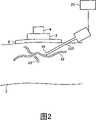
图2示出了连接到患者手臂的导引系统的侧视图,Figure 2 shows a side view of the guidance system attached to the patient's arm,
图3示出了允许在一个方向上移动的探针支持器,Figure 3 shows a probe holder that allows movement in one direction,
图4示出了允许在两个方向上移动的第二支持器,Figure 4 shows a second holder allowing movement in two directions,
图5示出了本发明穿刺系统的示意性结构图,Figure 5 shows a schematic structural diagram of the puncture system of the present invention,
图6示出了穿刺系统确定的穿刺位置和插入方位的示意图。Fig. 6 shows a schematic diagram of the puncture position and insertion orientation determined by the puncture system.
附图标记reference sign
01:系统01: System
02:探针支持器02: Probe Holder
03:患者03: Patient
04:探针04: Probe
05:轨道05: Orbit
05’:轨道05': track
06:轨道06: Orbit
06’:轨道06': track
07:身体的部分/手臂07: Body Parts/Arms
08:皮带08: belt
09:固定装置09: Fixtures
10:Velcro紧固件10: Velcro fasteners
13:致动装置13: Actuating device
17:光源17: light source
18:投影装置18: Projection device
19:远端19: remote
20:控制装置20: Control device
21:血管识别装置21: Vessel identification device
22:动脉22: Arteries
23:静脉23: veins
24:箭头24: Arrow
24’:箭头24': Arrow
25:箭头25: Arrow
25’:箭头25': Arrow
26:凹窝26: Dimple
26’:凹窝26': Dimple
26”:凹窝26": Dimple
26:凹窝26: Dimple
100:穿刺系统100: piercing system
102:血管102: Blood vessels
104:皮肤104: skin
106:组织106: Organization
108:采集模块108: Acquisition module
110:检测系统110: Detection system
112:控制单元112: Control unit
114:套管控制器114: casing controller
116:套管载体116: casing carrier
117:套管117: Casing
118:平行于皮肤的方向118: Direction parallel to the skin
119:角度119: angle
120:插入方向120: Insertion direction
122:套管远端122: the distal end of the cannula
124:穿刺位置124: Puncture position
126:插入方位126: insert orientation
128:插入路径128: Insert path
具体实施方式Detailed ways
图1示出了根据本发明的导引系统1。该系统1缠绕在患者3的手臂7周围。其具有矩形形状的皮带8,宽15cm,并且长度可借助于Velcro紧固件7调整。通过合适地使用Velcro紧固件10,可以确保皮带8紧密地适于患者3的手臂。Figure 1 shows a guidance system 1 according to the invention. The system 1 is wrapped around the
两个挠性轨道5、5’位于皮带8之上。Two
轨道5、5’由聚丙烯制成,并且以平行结构间隔开约7cm。探针支持器2可移动地安装在轨道5、5’之间,并且承载探针4。探针4旋在探针支持器2上。探针支持器2可以如箭头24和24’所示平行于轨道5、5’行进。第一轨道5、5’可移动地安装在第二轨道6、6’上,同时轨道6、6’的对准方向垂直于轨道5、5’的对准方向。因而,探针支持器2可平行,以及垂直第一轨道5、5’移动。图1的实施例中选择两个平行轨道,确定系统不摇晃,并且有效地防止探针支持器围绕轨道旋转。而且,探针支持器的稳定安装使得可以将更重和/或更大的探针牢固地连接到探针支持器。The
根据图1的系统容易地连接到患者,由此Velcro紧固件10允许根据患者或其被连接的身体部分的尺寸而独立调整皮带8的长度。而且,其允许分别在方向24、24’和25、25’上非常精确地移动探针2。为了更多地提高精确性,探针支持器可以紧固到轨道上。在该紧固过程之前,轨道的方位可以容易地改变,以便于最优化探针的测量方位。The system according to FIG. 1 is easily attached to the patient, whereby the
图2中示出了探针支持器2与手臂7的连接。探针支持器2搁置在皮带8上,其中为了简单,未示出轨道。探针4在探针支持器2之上,并且扫描以寻找诸如动脉22和静脉23的血管。如下面更详细讨论地,血管识别装置21用于区分静脉和动脉。而且,血管识别装置21还适于监视和/或导引套管117的远端19移动进入动脉22或静脉23。The connection of the
图3更详细地示出了探针支持器2。探针支持器2基本上包括带纵向凹窝26、26’的矩形板,轨道5、5’插入所述凹窝中。板相对于轨道的移动可以这样建立,即致动装置13包括具有齿的大齿轮(未示出),由于板具有用于此目的的长圆开口,由此齿与轨道中的相应开口啮合。致动装置13还包括用于确定轨道中探针支持器2的安全方位的装置9。Figure 3 shows the
图4中使用相同的方法,图4示出了具有凹窝26、26’的探针支持器2,所述凹窝用于容纳在第一方向上基本平行的第一对轨道。而且,探针支持器2具有附加凹窝26”和26,所述附加凹窝与凹窝26、26’相比,布置有高度偏移量O,如双箭头所示。这些附加凹窝26”和26用于容纳在垂直于第一方向的第二方向上基本上平行的第二对轨道。在两个方向上的移动,通过沿着由凹窝26、26’容纳的轨道5、5’(未示出)滑动探针支持器2和/或通过沿着由凹窝26”和26容纳的轨道6、6’(未示出)的滑动而实现。The same approach is used in Figure 4, which shows a
图5示出了穿刺系统100的示意性结构图,其可安装在探针支持器2上,其中为了简化,未示出探针支持器2。穿刺系统100具有采集模块108、检测系统110、控制单元112、套管控制器114以及套管载体116。套管117自身可以刚性地连接到套管载体116,其表示用于固定套管的紧固装置以及用于移动和对准套管117的装置,如套管控制单元114所控制的。套管117和套管载体116可以沿着插入方向120以及沿着基本上平行于皮肤104表面的方向118而移动。原则上,方向118可以是平行于皮肤表面的平面中的任何方向。通常,套管117和套管载体116借助于套管控制器114在所有三个空间方向上可移动。同样,插入方向120和皮肤104表面之间的角度α119可以借助于套管控制器114以这样的方式任意修改,即以借助于检测系统110和控制单元112确定的方式。Fig. 5 shows a schematic block diagram of a piercing
图6借助于人皮肤104的横截面图示,示出了将穿刺系统应用于人。在皮肤104的表面下是组织106所围绕的血管102。当穿刺系统100安装在探针支持器2上时,其在人皮肤104上。采集模块108适于从组织106和血管102采集光、光声和声学数据,其允许分类至少一个血管参数,诸如血管位置、血管直径、血管尺寸、皮肤104表面下的深度、血管的几何形状、血流或类似参数。FIG. 6 shows the application of the piercing system to a human by means of a cross-sectional illustration of
优选地,采集模块108借助于允许产生提供对血管102的识别的信号的超声、近红外成像、光学相干断层摄影、多普勒超声、多普勒光学相干断层摄影或光声技术。由采集模块108采集的信号应用于检测系统110,其依次产生血管102的信号。因此,检测系统110以及采集模块108在某种意义上协作,即检测系统110适于对从采集模块108获得的信号执行信号处理。通过使用光学、光声或超声检测,甚至在皮肤104表面下相当深度处,也可以精确地定位血管102。附加地或作为选择,也可以应用包括例如允许检测例如血管102中血流的多普勒超声技术的多普勒技术。同样地,可以相应地应用多普勒光学相干断层摄影术。Preferably, the acquisition module 108 is by means of ultrasound, near-infrared imaging, optical coherence tomography, Doppler ultrasound, Doppler optical coherence tomography or photoacoustic techniques allowing the generation of signals providing identification of the
也可以在不进行对血管成像的情况下,获得位置数据、几何形状数据以及与血管102的路线相关的数据的采集。因而,成像系统110并非必须提供可视图像。代替地,可以允许成像系统110从采集模块108采集的信号中直接提取血管参数。因而,可以借助于检测系统110或由控制单元112执行对血管参数的提取。Acquisition of positional data, geometry data, and data related to the course of the
控制单元112具有处理单元,其允许处理从检测系统110获得的数据。根据检测系统110提供的数据类型,控制单元112的处理单元还可以处理血管参数以便从血管102的信号中提取所需的血管参数。而且,允许控制单元112执行组织分析,以核查穿刺位置附近的组织是否适于穿刺。The
控制单元112用于处理血管参数,以便寻找和确定血管102的穿刺位置,其理想地适于插入套管117。在基本实施例中,可以关于血管102的位置和路线确定该穿刺位置。更精密复杂的实施方式还考虑了预期穿刺位置附近的血管几可形状以及血管直径和皮肤104表面下的深度。The
通常,可以将穿刺位置确定为考虑了所有类型的血管参数的最优化过程的结果。例如,通常借助于控制单元112的处理单元执行的最优化过程可以指定穿刺位置不应在血管102的分叉或交汇处附近。此外,穿刺位置可能需要一定直径的血管102。同样,可以关于皮肤104表面之下血管102的最小可能深度而确定穿刺位置。附加地,控制单元也可以确定插入方向120,其指定套管117需要引入皮肤104和组织106的角度α119。In general, the puncture position can be determined as a result of an optimization process that takes into account all types of vessel parameters. For example, an optimization process, typically performed by means of a processing unit of the
已经确定穿刺位置之后,控制单元112还适于指定套管117的插入方位。如可从图6获得的,插入方位指定套管117的方位以及对准或方向,套管117需要从其沿着插入方向偏移、即沿着符合套管纵向方向的方向移动,以便用其远端在确定的穿刺位置撞击血管。After the puncture position has been determined, the
在指定穿刺位置之后,组织分析装置核查围绕插入方位126的组织106是否适于穿刺。在备选方案中,选择相反顺序:在第一步中,分析皮肤表面,并且如果这是可以的,那么确定血管。组织分析装置可以是分离的装置,或者设置为控制单元112的附加功能。对于后者的情况,需要相应地补充控制单元112的固件。然后,控制单元112需要分析检测系统110的输出,所述检测系统110适于提供对穿刺位置124的测量。After specifying the puncture position, the tissue analysis device checks whether the
在确定了穿刺位置124和插入方位126之后,开始插管,由此或者由操作者触发或者由穿刺系统自主地触发该开始。一旦套管117向血管102推进,采集模块108也采集套管117远端的方位数据。特别地,当套管117已经穿刺皮肤104时,对其远端的检测允许控制套管117通过组织的移动。一旦采集模块108检测到套管117的远端不适当地碰撞血管102,套管插入的整个过程可以中断并且可以抽回套管117。这样,血管相关数据和套管117的远端的方位数据的同时采集,允许有效地实现自主穿刺系统的反馈和安全机制。After the
代替追踪针或套管的远端122,也可以在插入期间监视和跟随血管102的方位或移动,这是比上一段所述的方法更简单的方法。如果已知针或套管117必须停止之处或如果已经确定插入参数,可以在插入期间监视血管102的位置。然而,如果血管移动,该插入出错。Instead of tracking the
从下列事实中产生一个特别的优点,所述事实是探针支持器2容纳致动装置13(比较图3),并且上述穿刺系统100与其协作。该致动装置适于响应于穿刺系统100的输出而操作,从而穿刺系统100成为自动血管探测器和穿刺位置探测器。A particular advantage arises from the fact that the
如从图6中可获得的,穿刺系统100包括激光器17和投影装置18,后者包括可倾斜镜体,以将光投影在穿刺位置124上。这对内科医生有帮助,因为其视觉地导引医生将套管117精确地插入患者身体中。As can be obtained from FIG. 6 , the piercing
而且,穿刺系统包括血管识别装置,其与采集模块108相同,即采集模块108适于区分血管是动脉22或静脉23。Moreover, the puncture system includes a blood vessel identification device, which is the same as the collection module 108 , that is, the collection module 108 is adapted to distinguish whether the blood vessel is an
Claims (13)
Applications Claiming Priority (2)
| Application Number | Priority Date | Filing Date | Title |
|---|---|---|---|
| EP05105115 | 2005-06-10 | ||
| EP05105115.9 | 2005-06-10 |
Publications (1)
| Publication Number | Publication Date |
|---|---|
| CN101193595Atrue CN101193595A (en) | 2008-06-04 |
Family
ID=37216018
Family Applications (1)
| Application Number | Title | Priority Date | Filing Date |
|---|---|---|---|
| CNA2006800203847APendingCN101193595A (en) | 2005-06-10 | 2006-06-06 | System for guiding a probe over the skin surface of a patient or animal |
Country Status (5)
| Country | Link |
|---|---|
| US (1) | US20080221519A1 (en) |
| EP (1) | EP1893093A1 (en) |
| JP (1) | JP2008545502A (en) |
| CN (1) | CN101193595A (en) |
| WO (1) | WO2006131881A1 (en) |
Cited By (6)
| Publication number | Priority date | Publication date | Assignee | Title |
|---|---|---|---|---|
| CN103028185A (en)* | 2011-09-30 | 2013-04-10 | Ge医疗系统环球技术有限公司 | Automatic vessel intervention device, system and method based on real-time volume ultrasonic waves |
| CN103720457A (en)* | 2012-10-10 | 2014-04-16 | 美国科视数字系统公司 | Catheter discrimination and guidance system |
| CN104248790A (en)* | 2014-09-04 | 2014-12-31 | 肖程午 | Intelligent transfusion device and operation method thereof |
| CN106061386A (en)* | 2014-01-29 | 2016-10-26 | 贝克顿·迪金森公司 | Wearable electronics for enhanced visualization during insertion of interventional devices |
| WO2017067055A1 (en)* | 2015-10-21 | 2017-04-27 | 深圳市前海康启源科技有限公司 | Injection apparatus having automatic positioning and shooting function |
| WO2017067056A1 (en)* | 2015-10-21 | 2017-04-27 | 深圳市前海康启源科技有限公司 | Semi-automatic injection apparatus with ejection function |
Families Citing this family (55)
| Publication number | Priority date | Publication date | Assignee | Title |
|---|---|---|---|---|
| US8838210B2 (en)* | 2006-06-29 | 2014-09-16 | AccuView, Inc. | Scanned laser vein contrast enhancer using a single laser |
| US8038622B2 (en)* | 2007-08-03 | 2011-10-18 | Innoscion, Llc | Wired and wireless remotely controlled ultrasonic transducer and imaging apparatus |
| US8152751B2 (en) | 2007-02-09 | 2012-04-10 | Baxter International Inc. | Acoustic access disconnection systems and methods |
| US10463778B2 (en) | 2007-02-09 | 2019-11-05 | Baxter International Inc. | Blood treatment machine having electrical heartbeat analysis |
| DE102007015553B3 (en)* | 2007-03-29 | 2008-11-06 | Pajunk Gmbh & Co. Kg Besitzverwaltung | Device for locating a cannula pierced into a body |
| JP4407714B2 (en)* | 2007-04-06 | 2010-02-03 | セイコーエプソン株式会社 | Biometric authentication device and biometric authentication method |
| JP5274829B2 (en)* | 2007-12-27 | 2013-08-28 | シスメックス株式会社 | Non-invasive living body measurement device |
| JP5521172B2 (en)* | 2008-09-22 | 2014-06-11 | 公益財団法人ヒューマンサイエンス振興財団 | Image printing device |
| US9480455B2 (en) | 2009-06-18 | 2016-11-01 | Quanta Fluid Solutions, Ltd. | Vascular access monitoring device |
| EP2442851B1 (en)* | 2009-06-18 | 2013-09-04 | Quanta Fluid Solutions Ltd | Vascular access monitoring device |
| JP5428795B2 (en)* | 2009-11-19 | 2014-02-26 | 学校法人早稲田大学 | Ultrasonic diagnostic system, robot for ultrasonic diagnostic apparatus, and program |
| EP2386248B1 (en)* | 2010-05-14 | 2018-06-27 | Samsung Medison Co., Ltd. | Ultrasonic diagnostic apparatus |
| KR101133465B1 (en)* | 2010-05-14 | 2012-04-09 | 삼성메디슨 주식회사 | Ultrasonic diagnosis apparatus |
| CN103228219B (en)* | 2010-08-09 | 2016-04-27 | C·R·巴德股份有限公司 | Support and Covering Structures for Ultrasound Probe Heads |
| JP5847490B2 (en) | 2011-08-25 | 2016-01-20 | キヤノン株式会社 | Subject information acquisition device |
| EP2844316B1 (en)* | 2012-04-30 | 2021-05-05 | ivWatch, LLC | Appliance for an electromagnetic spectrum sensor monitoring an intravascular infusion |
| US9486276B2 (en) | 2012-10-11 | 2016-11-08 | Tva Medical, Inc. | Devices and methods for fistula formation |
| CN105228683B (en) | 2013-03-14 | 2022-06-10 | Tva医疗公司 | Fistula-forming device and method for forming fistula |
| JP2014195499A (en)* | 2013-03-29 | 2014-10-16 | セイコーエプソン株式会社 | Ultrasonic measurement system, ultrasonic probe and sheet |
| US20140323904A1 (en)* | 2013-04-30 | 2014-10-30 | Elwha Llc | Stabilized device for remote palpation of tissue |
| WO2015151516A1 (en)* | 2014-04-03 | 2015-10-08 | 凸版印刷株式会社 | Puncture injection instrument |
| FR3019724A1 (en)* | 2014-04-14 | 2015-10-16 | Agece Ecole Centrale D Electronique | DEVICE FOR MAINTAINING A VEIN OF A USER IN POSITION AND DEVICE FOR PUNCTURING OR INJECTING IN A VEIN OF A USER |
| EP3236858B1 (en)* | 2014-12-22 | 2020-09-09 | Koninklijke Philips N.V. | A system and a method for measuring arterial parameters |
| US10603040B1 (en) | 2015-02-09 | 2020-03-31 | Tva Medical, Inc. | Methods for treating hypertension and reducing blood pressure with formation of fistula |
| US10548528B2 (en)* | 2015-08-07 | 2020-02-04 | Ryan James Appleby | Smartphone device for body analysis |
| CN105877780B (en)* | 2015-08-25 | 2019-05-31 | 上海深博医疗器械有限公司 | Fully-automatic ultrasonic scanner and scanning detection method |
| DE102015016138A1 (en)* | 2015-12-14 | 2017-06-14 | Westfälische Wilhelms-Universität Münster | Ultrasound probe guide belt, system and method of use |
| JP6952696B2 (en)* | 2015-12-16 | 2021-10-20 | キヤノン ユーエスエイ, インコーポレイテッドCanon U.S.A., Inc | Medical guidance device |
| WO2017124062A1 (en) | 2016-01-15 | 2017-07-20 | Tva Medical, Inc. | Devices and methods for forming a fistula |
| CN114042224B (en) | 2016-01-15 | 2024-09-17 | Tva医疗公司 | Device and method for advancing a wire |
| US10874422B2 (en) | 2016-01-15 | 2020-12-29 | Tva Medical, Inc. | Systems and methods for increasing blood flow |
| JP7219090B2 (en) | 2016-01-15 | 2023-02-07 | ティーブイエー メディカル, インコーポレイテッド | Systems and methods for gluing vessels |
| US20170203053A1 (en)* | 2016-01-19 | 2017-07-20 | Joseph Choate Burkett | Visual-Assisted Insertion Device |
| JP7529565B2 (en) | 2017-03-31 | 2024-08-06 | インナバスク メディカル インコーポレイテッド | Apparatus and method for cannulation of vascular access grafts - Patents.com |
| CN109247910B (en)* | 2017-07-12 | 2020-12-15 | 京东方科技集团股份有限公司 | Blood vessel display device and blood vessel display method |
| US11583249B2 (en)* | 2017-09-08 | 2023-02-21 | Biosense Webster (Israel) Ltd. | Method and apparatus for performing non-fluoroscopic transseptal procedure |
| CN112752544B (en)* | 2018-08-24 | 2023-08-15 | 美多力医疗器械私人有限公司 | Device for guiding placement of ultrasonic probe auxiliary equipment |
| US11925781B2 (en) | 2018-10-30 | 2024-03-12 | InnAVasc Medical, Inc. | Apparatus and method for cannulation of vascular access vessel |
| KR102238250B1 (en)* | 2018-12-07 | 2021-04-09 | 인제대학교 산학협력단 | Laser guide arm for ultra sound guided nerve block and vessel access |
| NL2022350B1 (en)* | 2019-01-07 | 2020-08-13 | Vitestro Holding B V | Cannula insertion system |
| CA3140626A1 (en) | 2019-05-31 | 2020-12-03 | Tva Medical, Inc. | Systems, methods, and catheters for endovascular treatment of a blood vessel |
| US11723687B2 (en) | 2019-12-11 | 2023-08-15 | Medline Industries, Lp | Window dressing for use with ultrasonic aid in venipuncture |
| KR102425133B1 (en)* | 2020-05-18 | 2022-07-26 | 주식회사 뉴퐁 | Apparatus for processing ultrasound image |
| KR102556390B1 (en)* | 2020-09-24 | 2023-07-17 | 주식회사 에어스메디컬 | Automatic Invasion Device to Human Body and Method for Controlling the Same Device |
| CN116568222A (en) | 2020-12-17 | 2023-08-08 | 富士胶片株式会社 | Ultrasonic diagnostic apparatus and control method for ultrasonic diagnostic apparatus |
| WO2022153725A1 (en)* | 2021-01-18 | 2022-07-21 | テルモ株式会社 | Blood vessel position indicator |
| WO2023002933A1 (en)* | 2021-07-21 | 2023-01-26 | テルモ株式会社 | Vascular puncture device and vascular puncture system |
| US12059292B2 (en) | 2022-01-31 | 2024-08-13 | GE Precision Healthcare LLC | Systems and methods for ultrasound probe positioning |
| JP2024027020A (en)* | 2022-08-16 | 2024-02-29 | 富士フイルムヘルスケア株式会社 | Photoacoustic signal measuring instrument and photoacoustic signal measuring system |
| WO2024070933A1 (en)* | 2022-09-30 | 2024-04-04 | テルモ株式会社 | Vascular puncture device and control method thereof |
| US20240139433A1 (en)* | 2022-10-27 | 2024-05-02 | Becton, Dickinson And Company | Vascular access system and method for continuous ultrasound monitoring |
| NL2034438B1 (en)* | 2023-03-27 | 2024-10-02 | Vitestro Holding B V | Cannula insertion system |
| US20240390605A1 (en)* | 2023-05-25 | 2024-11-28 | Becton, Dickinson And Company | Imaging system and method for integrated vascular access device indwell assessment and data integration |
| US20250000456A1 (en)* | 2023-06-29 | 2025-01-02 | Bard Access Systems, Inc. | Device for Detecting a Blood Vessel |
| WO2025036889A1 (en)* | 2023-08-15 | 2025-02-20 | Sanofi | Imaging arrangement and method of imaging veins |
Family Cites Families (13)
| Publication number | Priority date | Publication date | Assignee | Title |
|---|---|---|---|---|
| JPH0428562Y2 (en)* | 1988-02-23 | 1992-07-10 | ||
| US5167630A (en)* | 1991-09-26 | 1992-12-01 | Paul Kamaljit S | Blood vessel cannulation device |
| CA2240757C (en)* | 1997-07-14 | 2001-08-28 | Matsushita Electric Industrial Co., Ltd. | Blood vessel puncturing device |
| US6171245B1 (en)* | 1998-03-12 | 2001-01-09 | Siemens Medical Systems, Inc. | Method of imaging scatterers based on acoustically stimulated changes of their acoustic properties |
| JP2000070264A (en)* | 1998-08-28 | 2000-03-07 | Ge Yokogawa Medical Systems Ltd | Ultrasonic probe holding appliance |
| US6132379A (en)* | 1998-11-04 | 2000-10-17 | Patacsil; Estelito G. | Method and apparatus for ultrasound guided intravenous cannulation |
| US6530886B1 (en)* | 1999-10-08 | 2003-03-11 | Tanita Corporation | Method and apparatus for measuring subcutaneous fat using ultrasonic wave |
| US6478740B2 (en)* | 2000-09-26 | 2002-11-12 | Sean Souney | Portable hand-carry satellite diagnostic ultrasound system for general and cardiac imaging |
| US6695786B2 (en)* | 2001-03-16 | 2004-02-24 | U-Systems, Inc. | Guide and position monitor for invasive medical instrument |
| US6443928B1 (en)* | 2001-04-02 | 2002-09-03 | Raymond Francis | Vein scope and injection system |
| US6755789B2 (en)* | 2002-02-05 | 2004-06-29 | Inceptio Medical Technologies, Llc | Ultrasonic vascular imaging system and method of blood vessel cannulation |
| US7166075B2 (en)* | 2002-03-08 | 2007-01-23 | Wisconsin Alumni Research Foundation | Elastographic imaging of in vivo soft tissue |
| US20050119546A9 (en)* | 2002-07-31 | 2005-06-02 | Connell Reynolds | Systems and methods for locating blood vessels |
- 2006
- 2006-06-06CNCNA2006800203847Apatent/CN101193595A/enactivePending
- 2006-06-06USUS11/916,823patent/US20080221519A1/ennot_activeAbandoned
- 2006-06-06EPEP06745066Apatent/EP1893093A1/ennot_activeWithdrawn
- 2006-06-06WOPCT/IB2006/051797patent/WO2006131881A1/ennot_activeApplication Discontinuation
- 2006-06-06JPJP2008515358Apatent/JP2008545502A/ennot_activeWithdrawn
Cited By (9)
| Publication number | Priority date | Publication date | Assignee | Title |
|---|---|---|---|---|
| CN103028185A (en)* | 2011-09-30 | 2013-04-10 | Ge医疗系统环球技术有限公司 | Automatic vessel intervention device, system and method based on real-time volume ultrasonic waves |
| CN103028185B (en)* | 2011-09-30 | 2017-04-12 | Ge医疗系统环球技术有限公司 | Automatic vessel intervention device, system and method based on real-time volume ultrasonic waves |
| US9731066B2 (en) | 2011-09-30 | 2017-08-15 | General Electric Company | Device, system and method of automatic vessel access based on real time volumetric ultrasound |
| CN103720457A (en)* | 2012-10-10 | 2014-04-16 | 美国科视数字系统公司 | Catheter discrimination and guidance system |
| CN106061386A (en)* | 2014-01-29 | 2016-10-26 | 贝克顿·迪金森公司 | Wearable electronics for enhanced visualization during insertion of interventional devices |
| CN106061386B (en)* | 2014-01-29 | 2019-07-26 | 贝克顿·迪金森公司 | Wearable electronic device for enhanced visualization during insertion of an interventional device |
| CN104248790A (en)* | 2014-09-04 | 2014-12-31 | 肖程午 | Intelligent transfusion device and operation method thereof |
| WO2017067055A1 (en)* | 2015-10-21 | 2017-04-27 | 深圳市前海康启源科技有限公司 | Injection apparatus having automatic positioning and shooting function |
| WO2017067056A1 (en)* | 2015-10-21 | 2017-04-27 | 深圳市前海康启源科技有限公司 | Semi-automatic injection apparatus with ejection function |
Also Published As
| Publication number | Publication date |
|---|---|
| US20080221519A1 (en) | 2008-09-11 |
| WO2006131881A1 (en) | 2006-12-14 |
| JP2008545502A (en) | 2008-12-18 |
| EP1893093A1 (en) | 2008-03-05 |
Similar Documents
| Publication | Publication Date | Title |
|---|---|---|
| CN101193595A (en) | System for guiding a probe over the skin surface of a patient or animal | |
| EP1874378B1 (en) | Cannula inserting system having tissue analysis means | |
| EP1883436B1 (en) | Cannula inserting system | |
| US20080275396A1 (en) | Cannula Inserting System | |
| US20090275823A1 (en) | Cannula inserting system | |
| JP7182240B2 (en) | Puncture system and puncture control device | |
| US10448850B2 (en) | Photoacoustic flowmetry systems and methods | |
| US20150065916A1 (en) | Fully automated vascular imaging and access system | |
| CN104856649B (en) | It is a kind of that the device with puncture vessel is identified by infrared imaging and pressure change | |
| CN109044498B (en) | Arterial puncture system and method for determining arterial puncture position | |
| KR20220050146A (en) | Systems and Methods for Portable Ultrasound Guided Cannulation | |
| Lomas et al. | Duplex Doppler measurements of the portal vein in portal hypertension | |
| CN107913072A (en) | Automatic identification blood vessel extracts the device of blood | |
| US20120133752A1 (en) | Apparatus for positioning a percutaneous access device | |
| JP7754505B2 (en) | Systems and methods for portable ultrasound-guided cannulation | |
| EP4230157A1 (en) | Mountable device for autonomous blood vessel interfacing and instrument actuation | |
| US20240415537A1 (en) | Mountable device for autonomous blood vessel interfacing and instrument actuation | |
| Woolsey et al. | Surgical Navigation Probe Utilizing Optical Coherence Tomography and Laser Doppler |
Legal Events
| Date | Code | Title | Description |
|---|---|---|---|
| C06 | Publication | ||
| PB01 | Publication | ||
| C10 | Entry into substantive examination | ||
| SE01 | Entry into force of request for substantive examination | ||
| C02 | Deemed withdrawal of patent application after publication (patent law 2001) | ||
| WD01 | Invention patent application deemed withdrawn after publication | Open date:20080604 |