CN101074914B - Urine Particle Analyzer and Method - Google Patents
Urine Particle Analyzer and MethodDownload PDFInfo
- Publication number
- CN101074914B CN101074914BCN2007101020962ACN200710102096ACN101074914BCN 101074914 BCN101074914 BCN 101074914BCN 2007101020962 ACN2007101020962 ACN 2007101020962ACN 200710102096 ACN200710102096 ACN 200710102096ACN 101074914 BCN101074914 BCN 101074914B
- Authority
- CN
- China
- Prior art keywords
- urine
- sample
- scattered light
- mensuration
- fluorescence
- Prior art date
- Legal status (The legal status is an assumption and is not a legal conclusion. Google has not performed a legal analysis and makes no representation as to the accuracy of the status listed.)
- Active
Links
Images


Classifications
- G—PHYSICS
- G01—MEASURING; TESTING
- G01N—INVESTIGATING OR ANALYSING MATERIALS BY DETERMINING THEIR CHEMICAL OR PHYSICAL PROPERTIES
- G01N33/00—Investigating or analysing materials by specific methods not covered by groups G01N1/00 - G01N31/00
- G01N33/48—Biological material, e.g. blood, urine; Haemocytometers
- G01N33/483—Physical analysis of biological material
- G01N33/487—Physical analysis of biological material of liquid biological material
- G01N33/493—Physical analysis of biological material of liquid biological material urine
Landscapes
- Health & Medical Sciences (AREA)
- Engineering & Computer Science (AREA)
- Life Sciences & Earth Sciences (AREA)
- Biomedical Technology (AREA)
- Urology & Nephrology (AREA)
- Physics & Mathematics (AREA)
- Chemical & Material Sciences (AREA)
- Molecular Biology (AREA)
- Hematology (AREA)
- Biophysics (AREA)
- Food Science & Technology (AREA)
- Medicinal Chemistry (AREA)
- Analytical Chemistry (AREA)
- Biochemistry (AREA)
- General Health & Medical Sciences (AREA)
- General Physics & Mathematics (AREA)
- Immunology (AREA)
- Pathology (AREA)
- Investigating Or Analysing Biological Materials (AREA)
Abstract
Translated fromChinese
Description
Translated fromChinese技术领域:Technical field:
本发明涉及尿中粒子分析仪及其分析方法。更具体地说是涉及应用流式细胞术光学测定、分析尿中所含成份的尿中粒子分析装置及其分析方法。 The invention relates to a particle analyzer in urine and an analysis method thereof. More specifically, it relates to a urine particle analysis device for optically measuring and analyzing components contained in urine by flow cytometry and an analysis method thereof. the
背景技术:Background technique:
尿中粒子的一般测定项目有红细胞、白细胞、上皮细胞、管型及细菌,但其中所谓管型是指Tomm-Horsfall粘蛋白在少量血浆蛋白(白朊)存在的环境下在肾小管内凝集,以此为基质,血液细胞和肾小管上皮细胞等包含其中形成的圆柱状物。管型有的大小可达数十μm以上,从其形状上又被称为圆柱体尿(cylinder),由于以肾小管为铸模而形成,故被称为管型(cast)(管型的存在意味着肾小管曾被一时性堵塞,是暗示肾实质有病理性变化的重要依据,特别是血液细胞和上皮管型等包入其中的管型很有临床意义)。 The general measurement items of particles in urine include red blood cells, white blood cells, epithelial cells, casts and bacteria, but the so-called casts refer to the aggregation of Tomm-Horsfall mucin in the renal tubules in the presence of a small amount of plasma protein (albumin). Using this as a matrix, blood cells, renal tubular epithelial cells, etc. contain the cylinders formed therein. The size of some casts can reach more than tens of μm, and they are also called cylinders in terms of their shape. Since they are formed with renal tubules as molds, they are called casts (the existence of casts) It means that the renal tubules have been temporarily blocked, which is an important basis for implying pathological changes in the renal parenchyma, especially the blood cells and epithelial casts, which are wrapped in it, which are of clinical significance). the
上皮细胞由偏平上皮细胞和移行上皮细胞组成。偏平上皮细胞是圆形或多角形的极薄的细胞,是尿道部分剥离而形成。而移行上皮细胞则有梨形和纺织形等多种形状,是构成肾盂、尿道膀胱甚至内尿道口的细胞。大小从数十μm到表层型100μm以上不等。 Epithelial cells are composed of flat epithelial cells and transitional epithelial cells. Flattened epithelial cells are round or polygonal, extremely thin cells that result from the peeling off of parts of the urethra. The transitional epithelial cells have various shapes such as pear-shaped and textile-shaped, and are the cells that constitute the renal pelvis, urethral bladder, and even the internal urethral orifice. The size ranges from tens of μm to more than 100 μm in the superficial type. the
红细胞的测定对判断肾小球到尿道有无出血十分重要,特别是肾泌尿系统疾病、出血性疾病、白血病等患者的尿液中多见。红细胞大小一般为8μm左右,两面呈凹形圆盘状,但在尿中往往被破坏。特别是来源于肾小球的红细胞都变形,大小也变小。被破坏的红细胞溶血、有析出物。The determination of red blood cells is very important to judge whether there is bleeding from the glomerulus to the urethra, especially in the urine of patients with renal and urinary system diseases, hemorrhagic diseases, and leukemia. The size of red blood cells is generally about 8 μm, and the two sides are concave discs, but they are often destroyed in urine. In particular, red blood cells derived from glomeruli are deformed and become smaller in size. Destroyed red blood cells are hemolyzed and have precipitates.
白细胞多见于肾、尿道感染症、肾结核等患者的尿液中。因此,通过测定尿中白细胞可以早期发现炎症和感染症。白细胞大小为6~14μm。测定细菌是调查有无感染的检查。该细菌有球菌和杆菌。球菌是0.5~2μm左右的球形菌,杆菌是长径为2~10μm左右的菌。球菌增殖则形成单列排列呈念珠状的连锁型和不规则排列呈葡萄房状的葡萄型等集块。 White blood cells are more common in the urine of patients with kidney, urinary tract infection, and renal tuberculosis. Therefore, inflammation and infection can be detected early by measuring white blood cells in urine. The size of white blood cells is 6-14 μm. Determination of bacteria is a test to investigate the presence or absence of infection. The bacteria are cocci and bacilli. Cocci are spherical bacteria with a diameter of about 0.5 to 2 μm, and bacilli are bacteria with a long diameter of about 2 to 10 μm. Cocci proliferate to form clusters such as chain type arranged in a single row in the shape of bead and grape type arranged irregularly in the shape of grape house. the
一直以来,尿中粒子的分析一般在检查室主要用显微镜目测。这种方法首先离心分离尿液,对其浓缩,视情况染色后,将其沉渣置于显微镜载玻片上,在显微镜下进行分类和计数。这种镜检首先是在100倍的低倍率视野(LPF)中把握尿中有无粒子和尿液标本的状态,再在400倍的高倍率视野(HPF)进行成份分类。检测项目中,管型即使出现,个数也很少,但其检出在临床上非常有用,因此,以低倍率视野(LPF)寻找。其他粒子以高倍率视野(HPF)分类,红细胞和白细胞个数以高倍率视野(HPF)报告。这种尿中粒子检查具备定性检查(比如:关于细菌“++”)、定量检查(比如:关于红细胞“5个/HPF”)和形态检查(比如:关于红细胞“确认存在异形细胞”)三个要素。 Traditionally, the analysis of particles in urine has generally been performed visually with a microscope in an examination room. In this method, the urine is first centrifuged, concentrated, stained as appropriate, and the sediment is placed on a microscope slide for classification and counting under a microscope. This kind of microscopic examination first grasps whether there are particles in the urine and the state of the urine specimen in the low power field (LPF) of 100 times, and then classifies the components in the high power field (HPF) of 400 times. Among the detection items, the number of casts is small even if they appear, but their detection is very useful clinically, so they are searched with a low power field (LPF). Other particles are classified by high power field (HPF), and the number of red blood cells and white blood cells is reported by high power field (HPF). This kind of urine particle inspection has three types: qualitative inspection (for example: "++" about bacteria), quantitative inspection (for example: "5/HPF" about red blood cells) and morphological inspection (for example: "confirm the presence of abnormal cells" about red blood cells) elements. the
作为尿中粒子检查的自动化发明了自动显微镜装置。流式自动显微镜分析装置使用UA-2000(SYSMEX公司制),这种装置不必浓缩尿液标本即可将尿液标本流入扁平形流动池,拍摄在流动池流动的状态并存储下来。存储的影像以粒子大小区分,用户根据该影像对各种粒子进行分类。 The automatic microscope device was invented as the automation of urine particle inspection. UA-2000 (manufactured by SYSMEX Co., Ltd.) was used as the flow cytometry automatic microscope analysis device. This device can flow the urine sample into the flat flow cell without concentrating the urine sample, and photograph and store the state of the flow in the flow cell. The stored image is differentiated by particle size, and the user can classify various particles based on this image. the
近年,在这种自动显微镜方式的基础上又提出了自动分类粒子的装置,但是尿中粒子形态多种多样,被破坏的粒子也很多,高精度地分类影像有困难。特别是小型粒子红细胞(尤其红细胞碎片)、细菌和结晶精确分类很难,使用户不得不再分类。 In recent years, based on this automatic microscope method, a device for automatically classifying particles has been proposed. However, there are various forms of particles in urine and many damaged particles, so it is difficult to classify images with high precision. In particular, the precise classification of small particles of red blood cells (especially red blood cell fragments), bacteria and crystals is difficult, so that users have to reclassify. the
作为检查尿中粒子的自动分类装置,有应用流式细胞仪的全自动尿有形成份分析装置UF-100(SYSMEX公司生产)。此装置用染色剂对尿中粒 子染色,结合散射光信号和荧光信号,对红细胞、白细胞、上皮细胞、管型和细菌进行分类。分类试剂由使用对各种粒子的膜和核进行染色的色素和保持尿中粒子形态的组分构成(比如参照专利公开平成8-170960号公报)。如上所述,尿中粒子形态多样,被破坏的粒子也很多,仅靠将流式细胞仪测得的散射光信号强度和荧光信号强度组合起来很难精确分类。于是,通过将散射光信号的强度和脉冲幅度、荧光信号强度和脉冲幅度组合起来分别对尿中粒子进行分类(如可参照美国专利No.5,325,168)。这种采用流式细胞仪的尿中粒子测定装置想尽办法提供尿中粒子的分类和形态报告。比如,通过对红细胞散射光信号的分析,提供尿中红细胞来源(来源于肾小球或非肾小球)的信息(如可参照美国专利No.6,118,522)。用此装置可以自动分类尿中粒子,对尿液检查的自动化作出了巨大贡献。 As an automatic sorting device for detecting particles in urine, there is a fully automatic urine formed component analyzer UF-100 (manufactured by SYSMEX Corporation) using a flow cytometer. This device stains urine particles with a dye, combines scattered light signals and fluorescent signals, and classifies red blood cells, white blood cells, epithelial cells, casts and bacteria. The classification reagent consists of pigments that stain the membranes and nuclei of various particles and components that maintain the particle shape in urine (for example, refer to Patent Publication No. Heisei 8-170960). As mentioned above, the particles in urine have various shapes and many damaged particles, and it is difficult to accurately classify them only by combining the scattered light signal intensity and fluorescence signal intensity measured by flow cytometry. Then, the urine particles are classified separately by combining the intensity and pulse amplitude of the scattered light signal, and the intensity and pulse amplitude of the fluorescence signal (for example, refer to US Patent No. 5,325,168). This urine particle measurement device using flow cytometry tries to provide classification and form report of urine particles. For example, the source of red blood cells in urine (from glomerulus or non-glomerulus) can be provided by analyzing the scattered light signal of red blood cells (for example, refer to US Patent No. 6,118,522). Urine particles can be automatically classified with this device, making a great contribution to the automation of urine testing. the
有些标本即使用如此想尽办法分析散射光信号和荧光信号的装置也会妨碍高精度的测定。其原因之一是有的标本难以对白细胞和上皮细胞正确分类。大多数上皮细胞是扁平上皮细胞和移行上皮细胞中来自表层的细胞。他们比白细胞大。但是,移行上皮细胞中来自中层或深层的细胞以及尿管上皮细胞中存在有被称为小管型上皮细胞的小型上皮细胞。他们个头与白细胞差不多,且与白细胞一样为有核细胞,因此,受荧光色素染色的程度也一样,出现区域重叠。 Some specimens, even with devices that try to analyze the scattered light signal and the fluorescent signal in such a way, will prevent high-precision determination. One reason for this is that some specimens are difficult to correctly classify leukocytes and epithelial cells. Most epithelial cells are cells from the superficial layer of squamous and transitional epithelium. They are larger than white blood cells. However, cells from the middle or deep layers of the transitional epithelium and cells of the urothelium have small epithelial cells called tubular epithelial cells. They are about the same size as leukocytes, and are nucleated cells like leukocytes, so they are stained to the same extent by fluorochromes, with overlapping areas. the
有的尿标本中含有结晶。结晶一般不被染色,故大部分分布于比红细胞荧光强度低的区域。但是也有的结晶比如尿酸结晶因自身带有荧光,会分布到荧光强度高的区域。尿中的结晶数有时会比红细胞数等多得多,因此,有的标本难以正确测定红细胞。于是,比如,专利公开平成11-23446号公报中建议检测红细胞出现区域的侧向散射光信号来评价该区域红细胞数的可靠性。 Some urine samples contain crystals. Crystals are generally not stained, so most of them are distributed in areas with lower fluorescence intensity than red blood cells. However, some crystals such as uric acid crystals will be distributed in areas with high fluorescence intensity due to their own fluorescence. The number of crystals in urine may be much more than the number of red blood cells, so it is difficult to accurately measure red blood cells in some samples. Thus, for example, Patent Publication No. Heisei 11-23446 proposes to detect the side scattered light signal of the area where red blood cells appear to evaluate the reliability of the number of red blood cells in the area. the
另一方面,尿的细菌检查根据临床目的也要求更高灵敏度的检查。但 是,用显微镜的目测检查很难检出数量少的细菌特别是小型细菌,没有用于高灵敏度的检查。在这种情况下,培养试样检测细菌的培养检查与尿中粒子检查分开,另行在细菌检验室进行。培养检查需要培养天数,因此,人们呼唤不培养即可进行高灵敏度细菌检查。 On the other hand, the bacterial test of urine requires a test with higher sensitivity due to clinical purposes. However, it is difficult to detect a small number of bacteria, especially small bacteria, by visual inspection with a microscope, and it is not used for high-sensitivity inspection. In this case, the culture test for the detection of bacteria in the culture sample is separated from the urine particle test and is performed separately in the bacteria test room. Culture inspection requires days of culture, therefore, people call for high-sensitivity bacterial inspection without culture. the
于是,用流式细胞仪分析细菌、即用染色剂对细菌染色、用散射光信号和荧光信号测定细菌的方法应运而生。比如,欧洲专利申请公告No.EP1136563和美国专利申请公告No.US2002/0076743记载的方法是用含阳离子型表面活性剂的染色剂以便溶解细菌外的杂质,即使试样含有和细菌同样大小的杂质也能精确测定。 Therefore, the method of analyzing bacteria with flow cytometer, that is, staining bacteria with staining agent, and measuring bacteria with scattered light signal and fluorescent signal came into being. For example, the methods described in European Patent Application Publication No.EP1136563 and U.S. Patent Application Publication No.US2002/0076743 use a staining agent containing cationic surfactants to dissolve impurities outside bacteria, even if the sample contains impurities of the same size as bacteria can also be accurately measured. the
发明内容:Invention content:
本发明的范围只由后附权利要求书所规定,在任何程度上都不受这一节发明内容的陈述所限。 The scope of the present invention is defined only by the appended claims and is not limited in any way by the statements in this summary. the
本发明首先涉及的尿中粒子分析仪包括以下部分: The particle analyzer in urine that the present invention firstly relates to comprises the following parts:
用尿液标本和染色剂制备测定试样的制样系统; A sample preparation system for preparing test samples with urine specimens and dyes;
光学检测系统,其包括照射上述所制测定试样的光源、检测该试样产生的前向散射光的前向散射光采集器、检测该试样产生的侧向散射光的侧向散射光采集器和检测该试样产生的荧光的荧光采集器;以及 An optical detection system, which includes a light source for irradiating the prepared measurement sample, a forward scattered light collector for detecting the forward scattered light generated by the sample, and a side scattered light collector for detecting the side scattered light generated by the sample detector and a fluorescence collector for detecting the fluorescence produced by the sample; and
根据上述光学检测系统检测出的前向散射光、侧向散射光和荧光测定尿中白细胞的测定系统。 A measurement system for measuring leukocytes in urine based on forward scattered light, side scattered light and fluorescence detected by the above optical detection system. the
其中所述测定系统根据所述前向散射光和所述荧光对尿中白细胞和上皮细胞进行第一分析,根据所述前向散射光和所述侧向散射光对尿中白细胞和上皮细胞进行第二分析;及所述测定系统根据上述第一分析结果和上述第二分析结果区分白细胞和上皮细胞。 Wherein the measurement system performs a first analysis on white blood cells and epithelial cells in urine according to the forward scattered light and the fluorescence, and performs a first analysis on white blood cells and epithelial cells in urine according to the forward scattered light and the side scattered light a second analysis; and the assay system distinguishes between leukocytes and epithelial cells based on the results of the first analysis above and the results of the second analysis above. the
其中所述测定系统进一步包括:在上述第一分析中划定尿中白细胞和上皮细胞出现区域的第一特定系统;以及在第二分析中划定尿中白细胞和上皮细胞出现区域的第二特定系统。Wherein the measurement system further comprises: a first specific system for delimiting the area where leukocytes and epithelial cells appear in urine in the above-mentioned first analysis; and a second specific system for delimiting the area where leukocytes and epithelial cells appear in urine in the second analysis system.
其中所述测定系统在所述第一分析中,根据所述前向散射光和所述荧光生成第一散点图;所述测定系统在所述第二分析中,根据所述前向散射光和所述侧向散射光生成第二散点图。 wherein the assay system generates a first scattergram based on the forward scattered light and the fluorescence in the first analysis; the assay system generates a first scattergram based on the forward scattered light in the second analysis A second scatterplot is generated from the side scattered light. the
其中所述测定系统测定尿中红细胞。 wherein said assay system measures red blood cells in urine. the
其中所述测定系统测定尿中细菌。 wherein said assay system measures bacteria in urine. the
其中所述制样系统将第一染色剂和第二染色剂添加进所述尿液标本中制备所述测定试样;所述测定系统具有:根据所述光学检测系统检测出来的前向散射光、侧向散射光和荧光测定至少含白细胞的尿中粒子的第一测定系统;根据所述光学检测系统检测出来的前向散射光和侧向散射光之一及荧光测定尿中细菌的第二测定系统。 Wherein the sample preparation system adds the first dye and the second dye to the urine sample to prepare the measurement sample; the measurement system has: according to the forward scattered light detected by the optical detection system 1. A first measurement system for measuring urine particles containing at least leukocytes by side scattered light and fluorescence; a second system for measuring bacteria in urine according to one of forward scattered light and side scattered light and fluorescence detected by the optical detection system Determination system. the
其中所述制样系统具有将尿液标本分配为第一等分和第二等分的标本分配器;所述制样系统还具有在上述第一等分混入第一染色剂制备第一试样的第一制样系统和在上述第二等分混入第二染色剂制备第二试样的第二制样系统;所述第一测定系统根据上述光学检测系统从上述第一测定试样检测到的前向散射光、侧向散射光和荧光测定上述至少含白细胞的尿中粒子;及所述第二测定系统根据上述光学检测系统从上述第二测定试样检测到的前向散射光和侧向散射光之一及荧光测定上述尿中细菌。 Wherein the sample preparation system has a sample dispenser for distributing the urine sample into the first aliquot and the second aliquot; the sample preparation system also has the function of mixing the first staining agent in the first aliquot to prepare the first sample The first sample preparation system and the second sample preparation system for preparing the second sample by mixing the second dye in the above second aliquot; the first measurement system detects from the above first measurement sample according to the above optical detection system The forward scattered light, side scattered light and fluorescence measure the above-mentioned urine particles containing at least leukocytes; One of the scattered light and fluorescence determination of the bacteria in the urine. the
其中所述第二制样系统在所述第二等分中混入表面活性剂制备所述第二测定试样。进一步包括将上述制样系统所制测定试样提供给上述光学检测系统的供样系统,其中上述供样系统向上述光学检测系统供应所述第一测定试样后,向上述光学检测系统供应所述第二测定试样。 Wherein the second sample preparation system mixes a surfactant into the second aliquot to prepare the second measurement sample. It further includes a sample supply system that provides the measurement sample prepared by the above sample preparation system to the above optical detection system, wherein the above sample supply system supplies the first measurement sample to the above optical detection system, and then supplies the above optical detection system with the first measurement sample. Describe the second measurement sample. the
本发明还提供一种尿中粒子分析方法,包括: The present invention also provides a method for analyzing particles in urine, comprising:
a)混合尿液标本和染色剂,制备测定试样; a) Mix the urine specimen and the dye to prepare the test sample;
b)用光束照射上述制备的测定试样,检测该试样产生的前向散射光、侧向散射光和荧光;以及 b) irradiate the measurement sample prepared above with a light beam, and detect the forward scattered light, side scattered light and fluorescence generated by the sample; and
c)根据上述检测出的前向散射光、侧向散射光和荧光,测定尿液中的白细胞。 c) Determination of white blood cells in the urine based on the detected forward scattered light, side scattered light and fluorescence. the
其中所述步骤c)包含以下步骤: Wherein said step c) comprises the following steps:
根据所述前向散射光和所述荧光对尿中白细胞和上皮细胞进行第一分析的第一分析步骤; a first analysis step of performing a first analysis of leukocytes and epithelial cells in urine based on said forward scattered light and said fluorescence;
根据所述前向散射光和所述侧向散射光对尿中白细胞和上皮细胞进行第二分析的第二分析步骤;及 a second analysis step of performing a second analysis of white blood cells and epithelial cells in urine based on said forward scattered light and said side scattered light; and
根据上述第一分析步骤的分析结果和上述第二分析步骤的分析结果区分白细胞和上皮细胞的区分步骤。 A step of distinguishing leukocytes from epithelial cells based on the analysis results of the above-mentioned first analysis step and the analysis results of the above-mentioned second analysis step. the
其中上述第一分析步骤包含划定尿中白细胞和上皮细胞出现区域的第一特定步骤;及上述第二分析步骤包含划定尿中白细胞和上皮细胞出现区域的第二特定步骤。 Wherein the above-mentioned first analysis step includes a first specific step of delineating the area where leukocytes and epithelial cells appear in urine; and the above-mentioned second analysis step includes a second specific step of delimiting the area where leukocytes and epithelial cells appear in urine. the
其中所述步骤c)包含测定尿中红细胞的步骤。 Wherein said step c) comprises the step of measuring red blood cells in urine. the
其中所述步骤c)包含测定尿中细菌的步骤。 Wherein said step c) comprises the step of measuring bacteria in urine. the
其中所述步骤a)包含在所述尿液标本加入第一染色剂和第二染色制备所述测定试样的步骤;所述步骤c)包括:根据在所述步骤b)检测出的前向散射光、侧向散射光和荧光测定至少含白细胞的尿中粒子的第一测定步骤,以及根据在所述步骤b)检测出的前向散射光和侧向散射光之一及荧光测定尿中细菌的第二测定步骤。 Wherein said step a) comprises the step of adding the first staining agent and second staining to the urine specimen to prepare the assay sample; said step c) comprises: according to the forward direction detected in said step b) A first measuring step of measuring at least leukocyte-containing particles in urine by scattered light, side scattered light and fluorescence, and measuring urine urine particles based on one of forward scattered light, side scattered light and fluorescence detected in said step b). Second assay step for bacteria. the
其中所述步骤a)包含将所述尿液标本分配为第一等分和第二等分的标本分配步骤;所述步骤a)还包含在上述第一等分加入第一染色剂混合制备第一试样的第一制样步骤和在上述第二等分加入第二染色剂混合制备第二试样的第二制样步骤;所述步骤b)还包括:从所述第一测定试样检测前向散射光、侧向散射光和荧光的第一检测步骤和从所述第二测定试样检测前向散射光和侧向散射光之一以及荧光的第二检测步骤;所述第一测定步骤含根据在上述第一检测步骤从所述第一测定试样检测出的前向散射光、侧向散射光和荧光测定上述至少含白细胞的尿中粒子的步骤;及所述第二测定步骤含根据在上述第二检测步骤从所述第二测定试样检测出的前向散射光和侧向散射光之一及荧光测定尿中细菌的步骤。 Wherein said step a) includes the sample distribution step of distributing said urine sample into a first aliquot and a second aliquot; said step a) also includes adding a first staining agent to the above first aliquot and mixing to prepare the second aliquot The first sample preparation step of a sample and the second sample preparation step of adding a second dye to the above-mentioned second aliquot and mixing to prepare a second sample; said step b) also includes: a first detection step of detecting forward scattered light, side scattered light and fluorescence and a second detection step of detecting one of forward scattered light and side scattered light and fluorescence from said second measurement sample; said first The measuring step includes a step of measuring the above-mentioned urine particles containing at least leukocytes based on the forward scattered light, side scattered light and fluorescence detected from the first measuring sample in the above-mentioned first detecting step; and the second measuring The step includes a step of measuring bacteria in urine based on one of forward scattered light and side scattered light detected from the second measurement sample in the second detecting step and fluorescence. the
其中在所述第二制样步骤向所述第二等分中添加表面活性剂。 Wherein the surfactant is added to the second aliquot in the second sample preparation step. the
其中所述第一检测步骤在所述第二检测步骤之前进行 Wherein said first detection step is carried out before said second detection step
附图说明:Description of drawings:
图1为本发明尿中粒子分析仪一个实施方式及其附属个人电脑的斜视示意图; Fig. 1 is a schematic diagram of an oblique view of an embodiment of the urine particle analyzer of the present invention and its attached personal computer;
图2为尿中粒子分析仪的制样系统及光学检测系统的功能略图; Figure 2 is a functional sketch of the sample preparation system and optical detection system of the urine particle analyzer;
图3为光学检测系统的结构图; Fig. 3 is the structural diagram of optical detection system;
图4为显示第一染色剂一例的吸收波长和吸光度关系的附图; Fig. 4 is the accompanying drawing showing the relationship between absorption wavelength and absorbance of an example of the first staining agent;
图5为显示第二染色剂一例的吸收波长和吸光度关系的附图; Fig. 5 is the accompanying drawing showing the relationship between the absorption wavelength and the absorbance of an example of the second staining agent;
图6为图1所示尿中粒子分析仪的整体结构框图; Fig. 6 is the overall structural block diagram of particle analyzer in urine shown in Fig. 1;
图7为尿中粒子分析仪的定量机构及制样系统的斜视略图; Figure 7 is a schematic oblique view of the quantitative mechanism and sample preparation system of the urine particle analyzer;
图8为尿中粒子分析仪的定量机构及制样系统的说明图; Figure 8 is an explanatory diagram of the quantitative mechanism and sample preparation system of the urine particle analyzer;
图9为使用本发明一个实施方式的尿中粒子分析仪分析尿液的步骤流程图(前半部分); Fig. 9 is a flow chart (first half) of steps for analyzing urine using a urine particle analyzer according to an embodiment of the present invention;
图10为使用本发明一个实施方式的尿中粒子分析仪分析尿液的步骤流程图(后半部分); Fig. 10 is a flow chart (second half) of steps for analyzing urine using a urine particle analyzer according to an embodiment of the present invention;
图11(a)-图11(e)为在本发明一个实施方式的尿中粒子分析仪获得的散射图一例的例示图; Fig. 11 (a)-Fig. 11 (e) is the illustration figure of an example of the scatter pattern obtained in the urine particle analyzer of an embodiment of the present invention;
图12为本在发明一个实施方式的尿中粒子分析仪获得的细菌类的散 射图一例的例示图; Figure 12 is an illustration of an example of the scatter diagram of bacteria obtained by the urine particle analyzer according to an embodiment of the invention;
图13为本在发明一个实施方式的尿中粒子分析仪获得的细菌类的散射图一例的例示图。 FIG. 13 is an illustration of an example of a scattergram of bacteria obtained by the urine particle analyzer according to one embodiment of the present invention. the
具体实施方式:Detailed ways:
下面根据附图,详细说明本发明尿中粒子分析仪的实施方式。图1为本发明一个实施方式的尿中粒子分析仪及其附属个人电脑的斜视说明图。图1中,为简单明了,部分省略了收纳尿中粒子分析仪组件的机壳。 The implementation of the urine particle analyzer of the present invention will be described in detail below with reference to the accompanying drawings. Fig. 1 is an explanatory oblique view of a urine particle analyzer and its attached personal computer according to one embodiment of the present invention. In FIG. 1 , for the sake of simplicity and clarity, the housing housing the components of the urine particle analyzer is partially omitted. the
[装置的结构] [Structure of device]
在图1中,尿中粒子分析仪U具有制备试样的制样系统2、运送样品架(试管架)3的样品架台4、从试样中检测尿中粒子和细菌信息的光学检测系统5和电路部分14。机壳侧面通过旋臂15装配操作台16,上面放置有个人电脑13。个人电脑13与尿中粒子分析仪U的电路部分14局域网连接。 In Fig. 1, the urine particle analyzer U has a
图2是上述制样系统2及光学检测系统5的功能概要图。图中,放入试管T内的尿液(标本)被无图示的注射式泵通过吸移管17抽取,用标本分配器1注入制样系统。本实施方式的制样系统由制样系统(第一制样系统)2U和制样系统(第二制样系统)2b组成,标本分配器1将尿液(标本)等分定量,分配给制样系统2U和制样系统2b。 FIG. 2 is a functional schematic diagram of the
制样系统2U的尿液等分部分与稀释液19U和染色液(染色剂)18U混合后被该染色液(染色剂)18U中所含色素染色。此染色试样成为分析红细胞、白细胞、上皮细胞和管型等较大尿中粒子(尿沉渣)的悬浮液。另一方面,制样系统2b的尿液等分部分与稀释液19b和染色液(染色剂)18b混合后被该染色液(染色剂)18b中所含色素染色。此染色试样成为分析细菌的悬浮液。 The urine aliquot of the sample preparation system 2U is mixed with the diluent 19U and the staining solution (staining agent) 18U, and is then stained with the pigment contained in the staining solution (staining agent) 18U. This stained sample becomes a suspension for analysis of large urine particles (urinary sediment) such as erythrocytes, leukocytes, epithelial cells, and casts. On the other hand, the urine aliquot of the
如上制备的2种悬浮液(试样)中,首先制样系统2U的悬浮液(第 一试样)被导入光学检测系统5,在鞘液流动池51中形成被鞘液包裹的细流,受激光照射。然后同样,制样系统2b的悬浮液(第二试样)被导入光学检测系统5,在鞘液流动池51中形成被鞘液包裹的细流,受激光照射。这种动作由后述微处理器11(控制装置)控制无图示的驱动器和电磁阀等自动进行。 Among the two kinds of suspensions (sample) prepared as above, first the suspension (first sample) of the sample preparation system 2U is introduced into the optical detection system 5, and a thin stream wrapped by the sheath liquid is formed in the sheath
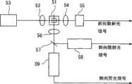
图3是光学检测系统5的结构示意图。图中聚光镜52将光源半导体激光器53发出的激光聚光到鞘液流动池51,聚光镜54将尿中粒子的前向散射光聚光到作为散射光采集器的发光二极管55。另一个聚光镜56将上述粒子的侧向散射光和侧向荧光聚光至分色镜57。分色镜57将侧向散射光反射到作为散射光采集器的光电倍增管58,滤出侧向荧光输送到荧光采集器光电倍增管59。这些光信号反映了尿中粒子的特征。发光二极管55、光电倍增管58和光电倍增管59将光信号转换成电信号,再分别输出前向散射光信号(FSC)、侧向散射光信号(SSC)和侧向荧光信号(SFL)。这些输出信号被无图示的前置放大器放大后,供下一步处理。 FIG. 3 is a schematic structural diagram of the optical detection system 5 . In the figure, the
作为光源也可以用气体激光器取代半导体激光器,但从低成本、小型化且低耗电量来说,还是采用半导体激光器为好,采用半导体激光器不仅可以降低产品成本,还可期待装置的小型化和节电化。半导体激光器中,以红色半导体激光器为宜,因为红色半导体激光器成本低、寿命长、厂家供货稳定。 Gas lasers can also be used as light sources instead of semiconductor lasers, but in terms of low cost, miniaturization and low power consumption, it is better to use semiconductor lasers. Using semiconductor lasers can not only reduce product costs, but also expect miniaturization and Power saving. Among the semiconductor lasers, the red semiconductor laser is suitable, because the red semiconductor laser has low cost, long life and stable supply from the manufacturer. the
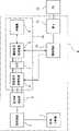
图6为显示尿中粒子分析仪U的整体结构的框图。图中,尿中粒子分析仪U具有:前述标本分配器1、制样系统2和光学检测系统5、对经前置放大器放大的光学检测系统5的输出信号进行放大和滤波处理的模拟信号处理电路6、将模拟信号电路6的输出转换为数字信号的A/D转换器7、对数字信号进行所定波形处理的数字信号处理电路8、连接数字信号处理电路8的存储器9、与模拟信号处理电路6及数字信号处理电路8连接的 微处理器11和接在微处理器11上的网卡12。外部的个人电脑13(分析器)通过网卡12与尿中粒子分析仪U形成局域网连接,用此个人电脑13可以对在尿中粒子分析仪U取得的数据进行分析。上述模拟信号处理电路6、A/D转换器7、数字信号处理电路8和存储器9构成了对光学检测系统5输出的电信号进行信号处理的信号处理电路10。 FIG. 6 is a block diagram showing the overall structure of the urine particle analyzer U. In the figure, the urine particle analyzer U has: the aforementioned sample dispenser 1, the
尿中粒子分析仪U的结构为:从尿液标本和染色剂混合而成的测定试样中采集2种散射光(前向散射光和侧向散向光)和荧光,再根据这些前向散射光、侧向散向光和荧光测定尿中白细胞。一般认为,前向散射光反映粒子(尿中有形成份)的大小,侧向散向光反映粒子的内容。即,这些散射光强度可以作为显示粒子不同特性的参数用于尿中粒子的分析。另一方面,荧光强度反映白细胞的核的状态。这样,通过分别使用三种不同的信息(前向散射光强度、侧向散向光强度和荧光强度)就可非常精确地对白细胞进行分类。 The structure of the Urine Particle Analyzer U is to collect two kinds of scattered light (forward scattered light and side scattered light) and fluorescence from a measurement sample mixed with a urine sample and a dye, and then based on these forward scattered light Determination of white blood cells in urine by scattered light, side scattered light and fluorescence. It is generally believed that forward scattered light reflects the size of particles (formed components in urine), and side scattered light reflects the content of particles. That is, these scattered light intensities can be used in the analysis of particles in urine as parameters showing different characteristics of particles. On the other hand, the fluorescence intensity reflects the state of the nucleus of the leukocyte. Thus, leukocytes can be classified very accurately by using three different pieces of information (forward scattered light intensity, side scattered light intensity, and fluorescence intensity) respectively. the
(尿中粒子测定试剂) (Reagent for determination of particles in urine)
关于测定这些尿中粒子的试剂,专利公告平成8-170960有详细说明。此试剂的一个实施方式是染料选择的是能对膜染色的色素,以便也能对无核的粒子染色。作为试剂,为了达到防止红细胞溶血的目的和取得稳定的荧光强度,应加入渗透压补偿剂,将浓度调整为100~600mOSm/kg,以使渗透压适于分类测定。尿中粒子的细胞膜和核(核膜)被此试剂染色。作为含可对膜染色的色素的染色剂,可以使用缩合苯衍生物,比如可使用花青系色素NK-529(商品名。林原生物化学研究所制)。此染色剂不仅染色细胞膜,还对核膜染色。使用这种染色剂,白细胞和上皮等有核细胞细胞质(细胞膜)中的染色强度和核(核膜)中的染色强度重合,以此可以将白细胞和上皮等有核细胞与红细胞等无核尿中粒子区别开。 The reagents for measuring these particles in urine are described in detail in Patent Publication No. 8-170960. One embodiment of this reagent is that the dye is chosen to be a pigment that can stain membranes, so that it can also stain non-nucleated particles. As a reagent, in order to achieve the purpose of preventing red blood cell hemolysis and obtain stable fluorescence intensity, an osmotic pressure compensator should be added, and the concentration should be adjusted to 100-600mOSm/kg, so that the osmotic pressure is suitable for classification determination. Cell membranes and nuclei (nuclear membranes) of particles in urine are stained with this reagent. A condensed benzene derivative, such as cyanine-based dye NK-529 (trade name: manufactured by Hayashibara Biochemical Research Institute), can be used as a dye containing a dye capable of dyeing a membrane. This stain not only stains the cell membrane, but also the nuclear membrane. With this staining agent, the staining intensity in the cytoplasm (cell membrane) of nucleated cells such as leukocytes and epithelium and the staining intensity in the nucleus (nuclear membrane) are superimposed, so that nucleated cells such as leukocytes and epithelium can be separated from anucleated cells such as red blood cells. Particles are distinguished. the
(细菌分析用试剂)(Reagents for bacterial analysis)
欧洲专利申请文件No.EP1136563对能从含有同样大小的杂质的试样中精确测定细菌的试剂有详细说明。此试剂的一个实施方式是使用能染色核酸的色素作为染料。作为含有核染色色素的染色剂比如可以使用以下结构式(化1)中所示美国专利申请公告No.US2002/0076743记载的花青类色素。 European Patent Application No. EP1136563 describes in detail a reagent capable of accurately determining bacteria from a sample containing impurities of the same size. One embodiment of this reagent is to use a dye capable of staining nucleic acid as a dye. As a dye containing a nuclear dye, for example, a cyanine dye described in US Patent Application Publication No. US2002/0076743 shown in the following structural formula (Chem. 1) can be used. the

其中,特别是以下结构式(化2)所示花青类色素更为适用。 Among them, especially the cyanine pigment represented by the following structural formula (Chem. 2) is more suitable. the

在这种情况下,最好在用于测定至少含红细胞的尿中粒子的尿液标本中加入的染色剂(第一染色剂)含有膜染色色素,而在用于测定细菌的尿液标本中加入的染色剂(第二染色剂)最好含有染色核酸的色素。尿中粒子中含有象红细胞那样没有核的细胞,由于第一染色剂含有膜染色的色素,包括这种无核的细胞在内,尿中粒子都可以检测出来。又由于第二染色剂含有核染色的色素,细菌的核被有效地染色,即使很小的细菌也可以很精确地测定。 In this case, it is preferable that the staining agent (first staining agent) added to the urine specimen for the determination of urine particles containing at least erythrocytes contain a membrane-staining pigment, and the staining agent for the determination of bacteria in the urine specimen The added staining agent (second staining agent) preferably contains a pigment for staining nucleic acid. Urine particles contain non-nucleated cells such as erythrocytes, and since the first staining agent contains membrane-stained pigments, urine particles can be detected including such non-nucleated cells. Also, since the second staining agent contains a pigment for nuclear staining, the nuclei of bacteria are efficiently stained, and even small bacteria can be accurately measured. the
上述第一染色剂和第二染色剂其吸收波长的峰值最好存在于上述半导体激光器的发光波长附近。通过选择靠近上述半导体激光器的发光波长的第一染色剂和上述第二染色剂的吸收波长峰值,可以通过半导体激光器测 定被染色的尿中粒子和细菌。图4显示了这种第一染色剂一例(含有NK-529(商品名,林原生物化学研究所制)的染色剂)的吸收波长和吸光度的关系,其吸收波长的峰值存在于红色半导体激光器发光波长(635nm)附近的640nm处。图5显示了第二染色剂一例(含上述(化2)所示美国专利申请公告No.US2002/0076743上记载的花青系列色素的染色剂))的吸收波长和吸光度的关系,其吸收波长的峰值存在于红色半导体激光器发光波长(635nm)附近的636nm处。此外,图5还显示了在试剂温度为15℃和35℃的情况下吸光度的变化,但可以知道,只要是常温左右的温度,试剂的吸光度没有大的变化。 Preferably, the peaks of the absorption wavelengths of the first dye and the second dye exist near the emission wavelength of the semiconductor laser. By selecting the first dye near the emission wavelength of the semiconductor laser and the absorption wavelength peak of the second dye, the particles and bacteria in urine stained can be measured by the semiconductor laser. Figure 4 shows the relationship between the absorption wavelength and absorbance of an example of this first staining agent (staining agent containing NK-529 (trade name, manufactured by Hayashibara Biochemical Research Institute)), and the peak of the absorption wavelength exists in the red semiconductor laser light emission At 640nm near the wavelength (635nm). Figure 5 shows the relationship between the absorption wavelength and the absorbance of an example of the second coloring agent (the coloring agent containing the cyanine series pigments described in the US Patent Application Publication No. US2002/0076743 shown in the above-mentioned (Chem. 2))), and the absorption wavelength The peak exists at 636nm near the emission wavelength (635nm) of the red semiconductor laser. In addition, Fig. 5 also shows the change in absorbance when the reagent temperature is 15°C and 35°C, but it can be seen that as long as the temperature is around room temperature, the absorbance of the reagent does not change significantly. the
细菌测定用试剂含有阳离子型表面活性剂,其目的是使染料透过膜尽快染色以及收缩粘液丝和红细胞碎片等杂质。在此情况下,由于表面活性剂可以破损细菌的细胞膜,因此通过使用第二染色剂所含色素可以高效地对细菌的核酸染色。其结果是经过短时间的染色处理即可更精确地进行上述细菌的测定。在第二等分部分加入表面活性剂混合的情况下,最好细菌测定在尿中粒子测定结束后进行。第二试样含表面活性剂,如果在测定细菌后测定尿中粒子,由于试样携带,表面活性剂会混入第一试样,有可能破坏含红细胞的尿中粒子细胞膜,影响该尿中粒子的测定。而测定完尿中粒子再测定细菌,可以防止第一试样中混入表面活性剂,精确地测定尿中粒子。 Reagents for bacteria measurement contain cationic surfactants, the purpose of which is to allow the dye to permeate the membrane to stain as quickly as possible and to shrink impurities such as mucus filaments and red blood cell fragments. In this case, since the surfactant can damage the cell membrane of the bacteria, the nucleic acid of the bacteria can be efficiently stained by using the dye contained in the second staining agent. As a result, the above-mentioned bacteria can be more accurately measured after a short staining process. In the case where the second aliquot is mixed with surfactant, it is best to perform the bacterial determination after the urine particle determination. The second sample contains surfactant. If the urine particles are measured after the bacteria are measured, the surfactant will be mixed into the first sample due to the sample carrying, which may destroy the urine particle cell membrane containing red blood cells and affect the urine particles. determination. By measuring the bacteria in the urine after measuring the particles in the urine, it is possible to prevent the surfactant from being mixed into the first sample and accurately measure the particles in the urine. the
本实施方式的结构是,从一个尿液标本分别制备测定至少含红细胞的尿中粒子用的第一试样和测定细菌用第二试样,用上述第一试样测定至少含红细胞的尿中粒子,用上述第二试样测定细菌。这样,一个分析装置可以分别精确地测定至少含红细胞的尿中粒子和细菌。又因第一试样和第二试样共用一个光学检测系统,可以简化装置结构,可以在降低产品成本的同时,实现装置小型化。The structure of this embodiment is that a first sample for measuring urine particles containing at least red blood cells and a second sample for measuring bacteria are prepared separately from one urine sample, and the urine containing at least red blood cells is measured using the first sample. Particles, bacteria were measured with the above-mentioned second sample. Thus, one analysis device can accurately measure at least erythrocyte-containing particles and bacteria in urine, respectively. In addition, because the first sample and the second sample share an optical detection system, the structure of the device can be simplified, and the miniaturization of the device can be realized while reducing the product cost.
图7为本实施方式尿中粒子分析仪的定量机构及制样系统的斜视略图;图8是其说明图。本实施方式采用常用的进样阀30作为将一定量的尿液标本分配到制样系统(第一制样系统)2U和制样系统(第二试样设备器)2b的定量机构。此进样阀30由2个圆盘形的固定元件和两个固定元件夹着的可动元件组成,上述可动元件受电动机31操作转动。 Fig. 7 is a schematic oblique view of the quantitative mechanism and sample preparation system of the urine particle analyzer according to this embodiment; Fig. 8 is an explanatory diagram thereof. In this embodiment, a commonly used
上述进样阀30有互相重叠的2个矾土陶瓷制的圆盘30A、30b。圆盘30A、30b内部形成供试样流通的流路,其中一个圆盘30b以其中心轴为旋转中心旋转,上述流路被分断,以此定量试样。此进样阀30通过内部有试样用流路32的流体盒33与上述制样系统2b连为一体。即,进样阀30、流体盒33和制样系统2b互相紧密地配置,使之在热度上成为一体,进样阀30的温度与制样系统2b的温度基本一致。与此相反,制样系统2U则留有一定空隙S用螺钉35固定在固定于机壳上的安装板34,因此,制样系统2U处于与上述进样阀30和制样系统2b在热度上基本隔离的状态。 The
上述制样系统2U和制样系统2b分别由构成调温系统的加热器36U、36b加热。以此将制备上述第一试样的制样系统2U的温度调节为第一温度,将制备上述第二试样的制样系统2b的温度调节为比第一温度略高的第二温度。具体而言,制样系统2U调为35±2℃,制样系统2b调为比此略高的42±2℃。试样温度越高,该试样所含红细胞和细菌等所定部位(膜和核等)越能快速染色,从而缩短测定时间,但另一方面,红细胞易因高温受损,温度过高,可能无法正确测定。因此,将用于测定比其他尿中粒子更耐热的细菌的第二试样的温度调节得高于用于测定尿中粒子的第一试样,换言之,通过将制样系统2U和制样系统2b分别调节到适于各自的测定温度,即可精确地测定含红细胞的尿中粒子和细菌。制样系统2U和制样系统2b的温度比如可以用电热调节器测定。然后根据此测定结果开关加热器36U、36b,即可将制样系统2U和制样系统2b调节到上述所定范围的温度。The above-mentioned sample preparation system 2U and
由于进样阀30和制样系统2b连为一体,热度一样,可以防止在进样阀30调温的试样在运送到制样系统2b时冷却。如此可以减少调温的浪费。此时,供应给温度低于制样系统2b的制样系统2U的试样由于从进样阀30运送时试样流路通过上述空隙S,可以使其自然下降。 Since the
[分析步骤] [Analysis steps]
下面按照图9~10所示流程,说明使用本发明一实施方式的尿中粒子分析仪进行尿液分析的步骤。 The steps of urine analysis using the urine particle analyzer according to one embodiment of the present invention will be described below according to the flowcharts shown in FIGS. 9 to 10 . the
首先,预先从主计算机中取得在该机中管理的标本号、与该标本号相关的患者的姓名、年龄、性别和门诊科等患者信息以及测定项目等标本信息(步骤S1)。接着,通过个人电脑13的键盘和鼠标等输入手段下达实施测定的指示(步骤S2)。根据这一指示,装有试样的试管T的样品架3被样品架台4运送至所定的吸移位置(步骤S3)。在此吸移位置,上述试管T旋转,读取该试管T外面贴的ID标签上的条形码(步骤S4)。以此可知道试样的标本号,通过与在步骤S1取得标本号的标本信息作比照,即可特定该试样的测定项目。 First, the sample number managed by the machine, patient information such as name, age, sex, and outpatient department of the patient related to the sample number, and sample information such as measurement items are acquired in advance from the host computer (step S1). Next, an instruction to execute the measurement is given through input means such as a keyboard and a mouse of the personal computer 13 (step S2). According to this instruction, the
然后,吸移管17下降,其前端插入试管T内的试样中,在这种状态下反复轻轻抽吐上述试样,使试样充分搅拌(步骤S5)。搅拌后,吸样所定量(800μL),用进样阀30分别向制备测定至少含红细胞的尿中粒子(SED)用试样的制样系统2U和制备测定尿中细菌(BAC)用试样的制样系统2b各以150μL和62.5μL频率加样(步骤S7和步骤S11)。 Then, the
制样系统2U中一定量染色液(染色剂)和稀释液与上述试样一道定量加入(步骤S8和步骤S9)。另一方面,制样系统2b中也同样,一定量染色液(染色剂)和稀释液与上述试样一道定量加入(步骤S12和步骤S13)。制样系统2U和制样系统2b分别用加热器36U、36b加温至一定温度,在此状态下用螺旋桨状的搅拌器(无图示)搅拌试样(步骤S10和步骤S14)。 另外,在步骤S9中加入制样系统2U的稀释液含有表面活性剂,可以破坏细菌膜,从而高效对细菌的核染色。 In the sample preparation system 2U, a certain amount of staining solution (staining agent) and diluent are quantitatively added together with the above-mentioned sample (step S8 and step S9). On the other hand, also in the
接下来,向光学检测系统5的鞘液流动池51中加入鞘液(步骤S15),然后,先将测定尿中粒子(SED)用试样导入光学检测系统5,在上述鞘液流动池51中形成被鞘液包裹的细流(鞘液流)(步骤S16)。再用半导体激光器53发出的激光束照射形成的鞘液流(步骤S17)。之所以先测定尿中粒子是因为细菌测定用试样中含有表面活性剂,如果测定细菌后再测定尿中粒子,试样携带会使表面活性剂混入尿中粒子测定用试样,从而破坏含红细胞的尿中粒子细胞膜,影响该尿中粒子的测定。 Next, add the sheath fluid to the sheath
上述激光束照射产生的尿中粒子的前向散射光、荧光和侧向散射光分别被光电二极管55、光电倍增管59和光电倍增管58采集并转换为电信号,作为前向散射光信号(FSC)、荧光信号(FL)和侧向散射光信号(SSC)输出(步骤S18~20)。这些输出信号通过前置放大器放大(步骤S21~23)。 The forward scattered light, fluorescence and side scattered light of the urine particles produced by the laser beam irradiation are respectively collected by the
另一方面,用测定尿中粒子(SED)用试样测定结束后,继续用在步骤S14制备的试样测定尿中细菌。此时,用测定尿中粒子时使用的光学检测系统5与上述步骤S15~23同样操作,输出前向散射光信号(FSC)和荧光信号(FL)并放大。 On the other hand, after the measurement with the sample for measuring urine particles (SED) is completed, the measurement of bacteria in urine is continued using the sample prepared in step S14. At this time, the optical detection system 5 used for the measurement of urine particles is operated in the same manner as the above-mentioned steps S15 to 23, and the forward scattered light signal (FSC) and the fluorescence signal (FL) are output and amplified. the
放大的上述前向散射光信号(FSC)、荧光信号(FL)和侧向散射光信号(SSC)在上述信号处理电路10(参照图6)被转换为数字信号并施以一定的波形处理(步骤S24~27),通过网卡12输送到个人电脑13。另外,步骤S25中的“FLH”是用高增益放大荧光信号(FL)的高灵敏度物质,步骤S26的“FLL”是同样荧光信号(FL)用低增益放大的低灵敏度物质。 The amplified forward scattered light signal (FSC), fluorescent signal (FL) and side scattered light signal (SSC) are converted into digital signals in the above signal processing circuit 10 (refer to FIG. 6 ) and subjected to certain waveform processing ( Steps S24-27), sending to the
个人电脑13汇总尿中粒子(SED)原始数据(步骤S28),同时根据这些数据绘制散射图(步骤S29)。再通过运算分析对散射图进行分类(步骤S30),统计每类各粒子的数量(步骤S31)。The
关于细菌也同样,放大的上述前向散射光信号(FSC)和荧光信号(FL)在上述信号处理电路10被转换为数字信号并施以一定的波形处理(步骤S32~34)。步骤S32中的“FSCH”是用高增益放大前向散射光信号(FSC)的高灵敏度物质,步骤S33的“FSCL”是同样前向散射光信号(FSC)用低增益放大的低灵敏度物质。 Similarly for bacteria, the amplified forward scattered light signal (FSC) and fluorescence signal (FL) are converted into digital signals by the
然后,通过网卡12输送到个人电脑13。在个人电脑13算出细菌(BAC)的原始数据(步骤S35)同时根据这些数据绘出散射图(步骤S36)。再通过计算分析对上述绘制的散射图进行分类(步骤S37),统计每类各粒子的数量(步骤S38)。如上得出的测定结果显示在个人电脑13的显示器荧光屏上(步骤S39)。 Then, it is sent to the
作为尿中粒子(SED)的测定结果,根据前向散射光、侧向散射光和荧光各信号绘出散射图。图11A是以荧光强度(低灵敏度)(FLL)为横轴、以前向散射光强度(FSC)为纵轴时的散射图。荧光信号强度大的区域出现有核的大细胞--上皮细胞(EC)和白细胞(WBC)。大半上皮细胞比白细胞大,比白细胞出现在荧光强度大的区域,但小型上皮细胞也有的会出现在和白细胞交迭的区域。为了识别二者使用侧向散射光信号。图11b为以侧向散射光强度(SSC)为横轴、以前向散射光强度(FSC)为纵轴时的散射图。上皮细胞出现在比白细胞侧向散射光强度大的区域,因此从此散射图可以识别上皮细胞。 As the measurement result of urine particles (SED), a scatter diagram was drawn from each signal of forward scattered light, side scattered light, and fluorescence. FIG. 11A is a scatter diagram with fluorescence intensity (low sensitivity) (FLL) as the horizontal axis and forward scattered light intensity (FSC) as the vertical axis. Nucleated large cells—epithelial cells (EC) and white blood cells (WBC)—appeared in areas of high fluorescence signal intensity. Most of the epithelial cells are larger than white blood cells and appear in areas with higher fluorescence intensity than white blood cells, but some small epithelial cells also appear in areas overlapping with white blood cells. To distinguish between the two the side scattered light signal is used. Fig. 11b is a scattering diagram when the lateral scattered light intensity (SSC) is taken as the horizontal axis and the forward scattered light intensity (FSC) is taken as the vertical axis. Epithelial cells appear in regions of greater side-scattered light intensity than leukocytes, so epithelial cells can be identified from this scatter pattern. the
图11c为以荧光强度(高灵敏度)(FLH)为横轴、以前向散射光强度(FSC)为纵轴时的散射图,显示荧光强度低的区域。红细胞(RBC)无核,故分布于荧光强度弱的区域。结晶(X′TAL)有的出现在红细胞出现的区域,因此使用侧向散射光信号确认结晶的出现。图11b为以侧向散射光强度(SSC)为横轴、以前向散射光强度(FSC)为纵轴时的散射图。结晶不一定分布于侧向散射光强度中心,也会出现在强度大的区域,因此 通过此散射图可以将其与红细胞区分开。 Fig. 11c is a scatter diagram when the fluorescence intensity (high sensitivity) (FLH) is taken as the horizontal axis and the forward scattered light intensity (FSC) is taken as the vertical axis, showing areas with low fluorescence intensity. Red blood cells (RBC) have no nuclei, so they are distributed in areas with weak fluorescence intensity. Crystals (X'TAL) sometimes appeared in the area where red blood cells appeared, so the presence of crystals was confirmed using the side scattered light signal. Fig. 11b is a scattering diagram when the lateral scattered light intensity (SSC) is taken as the horizontal axis and the forward scattered light intensity (FSC) is taken as the vertical axis. Crystals are not necessarily distributed in the center of the side-scattered light intensity, but also appear in areas of high intensity, so they can be distinguished from red blood cells by this scatter pattern. the
图11d是以荧光宽度(FLLW)为横轴、以荧光宽度2(FLLW2)为纵轴时的散射图。FLLW表示捕捉到细胞膜被染色的粒子的荧光信号宽度,FLLW2表示强度高于核的荧光信号的宽度。如图所示,管型(CAST)的FLLW大,有内容物的管型(P.CAST)FLLW2大。无内容物的管型(CAST)出现在FLLW2低的区域。在此,信号的宽度指在以信号强度为纵轴、以时间为横轴的脉冲形信号波形中反映测出光信号的时间长短。 Fig. 11d is a scattering diagram when the fluorescence width (FLLW) is taken as the horizontal axis and the fluorescence width 2 (FLLW2) is taken as the vertical axis. FLLW indicates the width of the fluorescent signal that captures the stained particles of the cell membrane, and FLLW2 indicates the width of the fluorescent signal whose intensity is higher than that of the nucleus. As shown in the figure, the cast (CAST) has a large FLLW, and the cast (P.CAST) with contents has a large FLLW2. Casts without contents (CAST) occur in areas of low FLLW2. Here, the signal width refers to the length of time for reflecting the measured optical signal in a pulse-shaped signal waveform with signal intensity as the vertical axis and time as the horizontal axis. the
另一个细菌测定结果是从前向散射光信号和荧光信号生成的散射图。图11e是以荧光强度(B-FLH)为横轴、前向散射光信号强度(高灵敏度)(B-FSC)为纵轴的散射图。在尿中粒子测定中如图11c所示,细菌的出现区域与粘液丝(MUCUS)、酵母样真菌(YLC)和精子(SPERM)的出现区域重叠。但在细菌测定中由于细菌测定试剂将粘液丝和红细胞碎片等杂质收缩,显示的区域内只有细菌独立出现,同时比在尿中粒子测定时灵敏度提高了大约10倍,因此,小型细菌也能精确地检测出来,从而能够提出正确的细菌检测报告。 Another bacterial assay result is the scatter plot generated from the forward scattered light signal and the fluorescent signal. Fig. 11e is a scattering diagram with fluorescence intensity (B-FLH) as the horizontal axis and forward scattered light signal intensity (high sensitivity) (B-FSC) as the vertical axis. As shown in Figure 11c in the urine particle assay, the area of appearance of bacteria overlapped with that of mucus filaments (MUCUS), yeast-like fungi (YLC) and spermatozoa (SPERM). However, in the bacteria determination, because the bacteria determination reagent shrinks impurities such as mucus filaments and red blood cell fragments, only bacteria appear independently in the displayed area, and the sensitivity is about 10 times higher than that of urine particles. Therefore, small bacteria can also be accurately measured. Detected out, so as to be able to put forward the correct bacteria detection report. the
在本实施例中,细菌是单独测定的,由此可阻止起因于细菌自动分类装置中的可信度低(细菌由大到小个头不一,且分布无规则可循,因此画面上会和其他白细胞和红细胞等成份混淆,从而难以正确区分成份)的情况。而且,在判断是否需要指示用显微镜复检(Review)时,在发出复检指令时,能够附以测定结果可信度低的理由。 In the present embodiment, the bacteria are determined separately, which can prevent the low reliability caused by the bacteria automatic classification device (the bacteria are different from big to small, and the distribution is irregular, so the screen will be different from that of Other components such as white blood cells and red blood cells are confused, making it difficult to correctly distinguish the components). In addition, when judging whether or not it is necessary to instruct a microscopic re-examination (Review), when issuing a re-examination order, it is possible to add a reason that the reliability of the measurement result is low. the
图11e显示了细菌(BACT)的标准出现区域,但实际出现区域因细菌而异。图13是球菌大量连锁型出现的标本的测定例。该散射图中,细菌(BACT)出现的区域以对横轴(荧光强度)约呈45度分布。即,细菌(BACT)出现在前向散射光强度(FSC)高的区域。在这种标本中,测定尿中粒子(SED)时细菌广泛出现,一直到红细胞出现区域中的前向散射光强度 (FSC)低的区域都有细菌出现。对于这种标本,红细胞测定的可信度较低。另一方面,图12是含杆菌的标本的测定例。在此散射图中,细菌(BACT)出现区域以对横轴(荧光强度)呈低角度(约5~10度)分布。即,细菌(BACT)出现在前向散射光强度(FSC)低的区域。这种标本即使含大量杆菌,细菌出现的区域是比红细胞出现区域前向散射光强度(FSC)低的区域,红细胞的测定不会受细菌的影响。同样,在尿中粒子(SED)测定中细菌对白细胞(WBC)出现区域的影响也可以从细菌测定(BAC)中的细菌分布确认。这种根据细菌分布倾向判断有无影响其他粒子测定结果工作通过个人电脑13(分析器)中的计算分析进行,该判断结果在上述步骤S39中与其他结果一起显示在屏幕上。 Figure 11e shows the standard appearance area of bacteria (BACT), but the actual appearance area varies from bacteria to bacteria. Fig. 13 is an example of measurement of a specimen in which a large number of linked types of cocci appear. In this scatter diagram, areas where bacteria (BACT) appear are distributed at approximately 45 degrees to the horizontal axis (fluorescence intensity). That is, bacteria (BACT) appear in an area where the forward scattered light intensity (FSC) is high. In this specimen, bacteria were widely present in the measurement of urine particles (SED) up to the area where the forward scattered light intensity (FSC) was low in the area where erythrocytes appeared. For such specimens, red blood cell assays are less reliable. On the other hand, Fig. 12 is a measurement example of a sample containing bacilli. In this scatter diagram, areas where bacteria (BACT) appear are distributed at low angles (approximately 5-10 degrees) to the horizontal axis (fluorescence intensity). That is, bacteria (BACT) appear in an area where the forward scattered light intensity (FSC) is low. Even if such a specimen contains a large number of bacilli, the area where the bacteria appear is an area with a lower forward scattered light intensity (FSC) than the area where the red blood cells appear, and the determination of the red blood cells will not be affected by the bacteria. Similarly, the influence of bacteria on the area where white blood cells (WBC) appear in the urine particle (SED) assay can also be confirmed from the bacterial distribution in the bacteria assay (BAC). This judgment based on the distribution tendency of the bacteria affects other particle measurement results through calculation and analysis in the personal computer 13 (analyzer), and the judgment result is displayed on the screen together with other results in the above-mentioned step S39. the
如此从细菌测定(BAC)获得细菌分布信息,以此推断细菌在尿中粒子(SED)测定中的出现区域,即可确认其他粒子测定的可信度。因此,根据细菌测定(BAC)的分布可以判断是否需要指示用显微镜复检(Review),可以减少假阳性判断,下达适宜的复检指令。还可以减少由于无法进行可信度高的区分而作出复检判断的次数。 In this way, the bacteria distribution information obtained from the bacteria assay (BAC) can be used to infer the occurrence area of bacteria in the urine particle (SED) assay, and the reliability of other particle assays can be confirmed. Therefore, according to the distribution of the bacteria assay (BAC), it can be judged whether a microscopic re-examination (Review) is required, which can reduce false positive judgments and issue appropriate re-examination instructions. It is also possible to reduce the number of times of re-inspection judgments due to the inability to make highly reliable distinctions. the
本实施方式当尿中的细菌浓度超过5×10³个/mL时,即可测定该尿液中的细菌。具体而言,将测定时间设长一些、多设定测定试样的量,即可测定5×103个/mL这种低浓度的细菌。由于尿中细菌浓度超过5×103个/mL时即可测定该尿液细菌,因此,可以根据尿检中求得的敏感度测定细菌。 In this embodiment, when the concentration of bacteria in the urine exceeds 5×10³ /mL, the bacteria in the urine can be measured. Specifically, by setting the measurement time longer and the amount of the measurement sample larger, bacteria at a low concentration of 5×10³ /mL can be measured. Since the urine bacteria can be detected when the concentration of bacteria in the urine exceeds 5×10³ cells/mL, the bacteria can be measured according to the sensitivity obtained in the urine test.
如上所示,本发明一个实施方式的尿中粒子分析仪U从尿液标本和染色剂混合而成的测定试样中采集2种散射光(前向散射光和侧向散向光)和荧光,再根据这些前向散射光、侧向散向光和荧光测定尿中白细胞。如此,通过分别使用三种不同的信息(前向散射光强度、侧向散向光强度和荧光强度)可以对白细胞进行精确分类。 As described above, the urine particle analyzer U according to one embodiment of the present invention collects two types of scattered light (forward scattered light and side scattered light) and fluorescent light from a measurement sample obtained by mixing a urine sample and a dye. , and then measure white blood cells in urine according to these forward scattered light, side scattered light and fluorescence. In this way, leukocytes can be accurately classified by using three different pieces of information (forward scattered light intensity, side scattered light intensity, and fluorescence intensity) respectively. the
在尿中粒子分析仪U中,附属电脑13是分析从尿液各粒子测得的数 据进行测定的测定系统。电脑13还具备以下功能:作为第一特定系统根据上述前向散射光和上述荧光信号,划定尿中白细胞和上皮细胞的出现区域;作为第二特定系统,根据上述前向散射光和上述侧向散射光划定尿中白细胞和上皮细胞的出现区域。以此即可根据上述第一特定系统划定的结果和上述第二特定系统划定的结果高精度地区分白细胞和上皮细胞。 In the urine particle analyzer U, the attached
尿中粒子分析仪U在一个尿液标本可以混合第一染色剂和第二染色剂制备试样,通过上述第一测定系统测定至少含白细胞的尿中粒子,通过上述第二测定系统测定细菌。因此,用一个分析仪器即可高精度地分别测定至少含白细胞的尿中粒子和细菌。 The urine particle analyzer U can prepare a sample by mixing the first staining agent and the second staining agent in one urine sample, measure urine particles containing at least leukocytes by the above-mentioned first measurement system, and measure bacteria by the above-mentioned second measurement system. Therefore, urine particles and bacteria containing at least leukocytes can be separately measured with high precision using a single analyzer. the
尿中粒子分析仪U还具备将尿液标本分为第一等分和第二等分的标本分配器。以此分别制备用于测定至少含白细胞的尿中粒子的第一试样和用于测定细菌的第二试样,用上述第一试样测定至少含白细胞的尿中粒子,用上述第二试样测定细菌。因此,可以更精确地测定尿中粒子和细菌。且,第一试样和第二试样共用一个光学检测系统,能够简化仪器结构,降低产品成本,并实现仪器的小型化。 The urine particle analyzer U also has a sample dispenser for dividing the urine sample into the first aliquot and the second aliquot. In this way, a first sample for measuring urine particles containing at least leukocytes and a second sample for measuring bacteria were respectively prepared, and the above-mentioned first sample was used to measure urine particles containing at least leukocytes, and the above-mentioned second sample was used to measure urine particles containing at least leukocytes. Bacteria were tested. Therefore, particles and bacteria in urine can be determined more accurately. Moreover, the first sample and the second sample share one optical detection system, which can simplify the structure of the instrument, reduce the product cost, and realize the miniaturization of the instrument. the
前述的详细说明及附图是通过文字解释和图示来进行的,其目的不在于限定权利要求的保护范围。本说明书中的具体实施方式的各个变种对于普通技术人员来说显而易见,并处于权利要求及其等同技术的保护范围内。 The above-mentioned detailed description and drawings are done by means of text explanation and illustration, and the purpose is not to limit the scope of protection of the claims. Variations of the specific implementations in this specification are obvious to those skilled in the art, and are within the protection scope of the claims and their equivalent technologies. the
本实施方式检测在照射测定试样的激光光束的光轴延长线附近散射的光作为前向散射光。将对于照射测定试样的激光光束的光轴略呈直角地散射的散射光作为侧向散射光检测。但是本发明中的前向散射光和侧向散射光并非严格限定于上述方向散射的散射光。在上述实施方式中,前向散射光强度是作为反映粒子(尿中有形成份)大小的信息使用的,只要能达到此目的,稍微偏离上述光轴延长线某种程度散射的散射光也可以作为前向散射光使用。另外,在上述实施方式中,侧向散射光强度是作为反映粒子 内容的信息使用的,只要能达到此目的,某种程度偏离对上述光轴略呈直角方向散射的散射光也可以作为侧向散射光使用。 In this embodiment, light scattered near the optical axis extension of the laser beam irradiating the measurement sample is detected as forward scattered light. Scattered light scattered approximately at right angles to the optical axis of the laser beam irradiating the measurement sample is detected as side scattered light. However, the forward scattered light and side scattered light in the present invention are not strictly limited to the scattered light scattered in the above directions. In the above-mentioned embodiment, the intensity of forward scattered light is used as information reflecting the size of particles (formed components in urine). Forward scattered light is used. In addition, in the above-mentioned embodiment, the intensity of side scattered light is used as information reflecting the content of the particles. As long as this purpose can be achieved, the scattered light scattered in a direction slightly at right angles to the above-mentioned optical axis can also be used as the information of the side scattered light. Scattered light is used. the
前向散射光和侧向散射光的性质不一定要限定于上述说明的内容。比如侧向散射光强度不仅可以反映粒子的内容,有时也能反映粒子的大小和粒子表面状态。因此,本实施方式虽然在细菌测定(BAC)中使用前向散射光和荧光,也可以用侧向散射光代替上述前向散射光。 The properties of forward scattered light and side scattered light are not necessarily limited to those described above. For example, the intensity of side scattered light can not only reflect the content of the particle, but also sometimes reflect the size of the particle and the surface state of the particle. Therefore, in the present embodiment, although forward scattered light and fluorescence are used for bacteria measurement (BAC), side scattered light may be used instead of the above forward scattered light. the
本实施方式在测定粒子时绘制散点图。但散点图也可以不绘制。尿中粒子分析仪U绘制的散点图是以从尿中各种粒子相对应的信号数据中抽取的数个参数为座标轴绘制的分布图。这是作为算法分析的一种手法绘制的,它具有用户可以亲眼确认测定结果的优点。但是,只要用各粒子对应的信号数据进行分析,不绘制散点图也可以对粒子进行分类和计数。只要决定各粒子相应的信号数据在什么范围内该粒子应归类于哪种粒子即可。在本说明书中,不管是否绘制散点图,均将从各粒子所得数据分布的范围称为出现区域。 In this embodiment, a scattergram is drawn when measuring particles. But the scatter plot can also not be drawn. The scatter diagram drawn by the Urine Particle Analyzer U is a distribution diagram drawn with several parameters extracted from the signal data corresponding to various particles in urine as the coordinate axis. This is drawn as a means of algorithm analysis, and it has the advantage that users can confirm the measurement results with their own eyes. However, as long as the signal data corresponding to each particle is used for analysis, the particles can be classified and counted without drawing a scatter diagram. It only needs to decide what kind of particle the particle should be classified into within what range the corresponding signal data of each particle is. In this specification, regardless of whether a scattergram is drawn, the range of data distribution obtained from each particle is referred to as an appearance area. the
在本实施方式的尿中粒子分析仪U中,分析数据用电脑13与尿中粒子分析仪U主机的机壳是分开的,但也可以将这些都物理性地连为一体。In the urine particle analyzer U of the present embodiment, the
Claims (16)
Applications Claiming Priority (3)
| Application Number | Priority Date | Filing Date | Title |
|---|---|---|---|
| JP2006-138557 | 2006-05-18 | ||
| JP2006138557 | 2006-05-18 | ||
| JP2006138557AJP4918281B2 (en) | 2006-05-18 | 2006-05-18 | Urine component analyzer |
Publications (2)
| Publication Number | Publication Date |
|---|---|
| CN101074914A CN101074914A (en) | 2007-11-21 |
| CN101074914Btrue CN101074914B (en) | 2012-02-29 |
Family
ID=38712439
Family Applications (1)
| Application Number | Title | Priority Date | Filing Date |
|---|---|---|---|
| CN2007101020962AActiveCN101074914B (en) | 2006-05-18 | 2007-05-17 | Urine Particle Analyzer and Method |
Country Status (3)
| Country | Link |
|---|---|
| US (1) | US8333926B2 (en) |
| JP (1) | JP4918281B2 (en) |
| CN (1) | CN101074914B (en) |
Families Citing this family (35)
| Publication number | Priority date | Publication date | Assignee | Title |
|---|---|---|---|---|
| US8243272B2 (en)* | 2005-09-19 | 2012-08-14 | Jmar Llc | Systems and methods for detecting normal levels of bacteria in water using a multiple angle light scattering (MALS) instrument |
| EP2105724B1 (en)* | 2008-03-28 | 2019-02-27 | Sysmex Corporation | Biological sample analyzer and computer program product |
| US20090248318A1 (en)* | 2008-03-28 | 2009-10-01 | Takaaki Nagai | Sample analyzer, sample analyzing method and computer program product |
| US20100136609A1 (en)* | 2008-10-31 | 2010-06-03 | Biomerieux, Inc. | Method for separation, characterization and/or identification of microorganisms using raman spectroscopy |
| CA2741019C (en) | 2008-10-31 | 2018-08-28 | Biomerieux, Inc. | Methods for separation, characterization and/or identification of microorganisms using mass spectrometry |
| KR101759995B1 (en)* | 2008-10-31 | 2017-07-31 | 바이오메리욱스, 인코포레이티드. | Separation device for use in the separation, characterization and/or identification of microorganisms |
| WO2010062356A1 (en)* | 2008-10-31 | 2010-06-03 | Biomerieux, Inc. | Methods for separation, characterization and/or identification of microorganisms using spectroscopy |
| EP2364356B1 (en)* | 2008-10-31 | 2019-06-19 | Biomerieux, Inc | Methods for detection and identification of microorganisms |
| US8647835B2 (en) | 2008-10-31 | 2014-02-11 | BIO MéRIEUX, INC. | Methods for separation, characterization and/or identification of microorganisms using spectroscopy |
| JP2012507710A (en)* | 2008-10-31 | 2012-03-29 | バイオメリュー・インコーポレイテッド | Method for the separation, characterization and / or identification of microorganisms using identification agents |
| BRPI0920114B1 (en)* | 2008-10-31 | 2021-08-17 | Biomerieux, Inc. | METHODS FOR ISOLATION AND IDENTIFICATION OF MICROORGANISMS |
| JP5232611B2 (en)* | 2008-12-05 | 2013-07-10 | シスメックス株式会社 | Sample analyzer, sample analysis method, and computer program |
| BRPI1012879B1 (en)* | 2009-05-15 | 2019-10-29 | Bio Merieux Inc | system and method for automatic ventilation and sampling of a culture specimen container |
| US9783839B2 (en)* | 2009-05-15 | 2017-10-10 | BIOMéRIEUX, INC. | Automated container management device for microbial detection apparatus |
| JP5685378B2 (en)* | 2010-01-08 | 2015-03-18 | シスメックス株式会社 | Sample analyzer and sample analysis method |
| EP2348301B1 (en) | 2010-01-08 | 2013-08-21 | Sysmex Corporation | Sample analyzer |
| JP2013190206A (en)* | 2010-05-31 | 2013-09-26 | Arkray Inc | Urine test device |
| CN102087198A (en)* | 2010-11-18 | 2011-06-08 | 苏州生物医学工程技术研究所 | Flow cytometry |
| JP5748782B2 (en)* | 2011-01-21 | 2015-07-15 | 株式会社日立ハイテクノロジーズ | Automatic analyzer |
| CN102564927B (en)* | 2011-12-30 | 2013-08-28 | 北京普利生仪器有限公司 | Urine holographic detecting method and equipment |
| EP4220158A3 (en)* | 2013-02-28 | 2023-08-09 | Sysmex Corporation | Urine specimen analysis device and urine specimen analysis method |
| WO2015129870A1 (en)* | 2014-02-28 | 2015-09-03 | シスメックス株式会社 | Urine sample analysis method and reagent kit for urine sample analysis |
| KR20180023061A (en)* | 2014-02-28 | 2018-03-06 | 시스멕스 가부시키가이샤 | Method for urine sample analysis, reagent for urine sample analysis, and reagent kit for urine sample analysis |
| CN103940709A (en)* | 2014-05-06 | 2014-07-23 | 南京中科神光科技有限公司 | Real-time microbial particle counter |
| JP6238856B2 (en) | 2014-08-25 | 2017-11-29 | シスメックス株式会社 | Urine atypical cell analysis method, urine analyzer, and body fluid atypical cell analysis method |
| CN112326538B (en)* | 2015-03-31 | 2024-12-24 | 希森美康株式会社 | Urine analysis system, imaging device, cell imaging device, urine analysis method, management device and information processing method |
| CN104819981B (en)* | 2015-05-16 | 2017-11-21 | 山东耀华医疗器械股份有限公司 | A urine composition measuring instrument |
| JP6352871B2 (en)* | 2015-08-28 | 2018-07-04 | シスメックス株式会社 | Urine sample analyzer and urine sample analysis method |
| GB2543288B (en) | 2015-10-13 | 2017-11-01 | Cook Medical Technologies Llc | Tiltable implantable medical device |
| EP3449251A1 (en) | 2016-04-25 | 2019-03-06 | Aperture Bio, LLC | Systems, devices and methods for sequential analysis of complex matrix samples for high confidence bacterial detection and drug susceptibility prediction using a flow cytometer |
| CN106093364A (en)* | 2016-05-27 | 2016-11-09 | 长春迪瑞医疗科技股份有限公司 | A kind of physiology crystallizes in-vitro simulated and verification method |
| CN109612911B (en)* | 2018-12-26 | 2023-07-21 | 深圳天依生命健康科技有限公司 | Full-automatic sperm cell detector |
| US11959908B2 (en)* | 2019-12-17 | 2024-04-16 | Arkray, Inc. | Measurement device and measurement method |
| CN111504869B (en)* | 2020-05-15 | 2021-06-08 | 中国计量科学研究院 | Flow type aggregate impurity analyzer |
| CN112834688A (en)* | 2021-01-07 | 2021-05-25 | 南京力创环境科技有限公司 | Calcium hardness detector and calcium hardness detection method |
Family Cites Families (17)
| Publication number | Priority date | Publication date | Assignee | Title |
|---|---|---|---|---|
| JP3213334B2 (en) | 1991-05-14 | 2001-10-02 | シスメックス株式会社 | Urine cell analyzer |
| JPH05322882A (en)* | 1992-05-21 | 1993-12-07 | Hitachi Ltd | Blood analyzer |
| ES2278566T3 (en) | 1994-10-20 | 2007-08-16 | Sysmex Corporation | REAGENT AND METHOD FOR ANALYZING SOLID COMPONENTS IN THE URINE. |
| JP3580615B2 (en) | 1994-10-20 | 2004-10-27 | シスメックス株式会社 | Urine particulate analysis reagent and particulate analysis method using the reagent |
| JP3347495B2 (en) | 1994-11-14 | 2002-11-20 | シスメックス株式会社 | Particle analyzer |
| JP3783808B2 (en) | 1997-05-19 | 2006-06-07 | シスメックス株式会社 | Leukocyte classification and counting reagent |
| JP3642658B2 (en) | 1997-06-30 | 2005-04-27 | シスメックス株式会社 | Urine component analyzer and analysis method |
| JPH1183849A (en)* | 1997-09-12 | 1999-03-26 | Toa Medical Electronics Co Ltd | Analyzing method for material component in urine and reagent for its analysis |
| JP3867880B2 (en)* | 1998-04-08 | 2007-01-17 | シスメックス株式会社 | Apparatus and method for distinguishing urine red blood cells |
| JP3837006B2 (en) | 2000-03-22 | 2006-10-25 | シスメックス株式会社 | Bacterial staining method and detection method |
| US7309581B2 (en) | 2000-11-01 | 2007-12-18 | Sysmex Corporation | Method of staining, detection and counting bacteria, and a diluent for bacterial stain |
| JP4659252B2 (en)* | 2001-03-29 | 2011-03-30 | シスメックス株式会社 | Flow cytometer |
| JP3871624B2 (en) | 2001-07-26 | 2007-01-24 | シスメックス株式会社 | Particle analyzer |
| JP4299597B2 (en)* | 2002-07-29 | 2009-07-22 | シスメックス株式会社 | Hematology analyzer and method |
| JP4503370B2 (en)* | 2004-06-30 | 2010-07-14 | シスメックス株式会社 | Formed component analysis apparatus and method |
| JP4413120B2 (en) | 2004-09-30 | 2010-02-10 | シスメックス株式会社 | Analytical particle analysis method and analyzer |
| US7625757B2 (en)* | 2006-01-27 | 2009-12-01 | Sysmex Corporation | Reagent for immature leukocyte analysis and reagent kit |
- 2006
- 2006-05-18JPJP2006138557Apatent/JP4918281B2/enactiveActive
- 2007
- 2007-05-10USUS11/798,113patent/US8333926B2/ennot_activeExpired - Fee Related
- 2007-05-17CNCN2007101020962Apatent/CN101074914B/enactiveActive
Also Published As
| Publication number | Publication date |
|---|---|
| US8333926B2 (en) | 2012-12-18 |
| JP2007309765A (en) | 2007-11-29 |
| US20070269897A1 (en) | 2007-11-22 |
| CN101074914A (en) | 2007-11-21 |
| JP4918281B2 (en) | 2012-04-18 |
Similar Documents
| Publication | Publication Date | Title |
|---|---|---|
| CN101074914B (en) | Urine Particle Analyzer and Method | |
| JP4759438B2 (en) | Urine component analyzer | |
| JP4078600B2 (en) | Hematology device based on flow cytometry | |
| CN105891090B (en) | Blood cell analyzer, body fluid analysis method, and control system therefor | |
| US20170336386A1 (en) | Method and apparatus for analyzing individual cells or particulates using fluorescent quenching and/or bleaching | |
| CN105026928B (en) | Urine sample analysis device and urine sample analysis method | |
| JP2941041B2 (en) | Classification of leukocytes by flow cytometry | |
| JP2012525589A (en) | Method for discriminating red blood cells from white blood cells by using forward scattering from a laser in an automated hematology analyzer | |
| CN120569621A (en) | Hematology flow system | |
| EP1857805B1 (en) | Apparatus for analyzing particles in urine and method thereof | |
| Ma et al. | Hematology Analyzers and Reagents | |
| JP4825033B2 (en) | Urine analyzer | |
| KR20200117458A (en) | Multi-system for conducting biochemistry and Blood test, and Multi-Disc therefor | |
| AU2001263405B2 (en) | Flow cytometry-based hematology system | |
| AU2001263405A1 (en) | Flow cytometry-based hematology system |
Legal Events
| Date | Code | Title | Description |
|---|---|---|---|
| C06 | Publication | ||
| PB01 | Publication | ||
| C10 | Entry into substantive examination | ||
| SE01 | Entry into force of request for substantive examination | ||
| C14 | Grant of patent or utility model | ||
| GR01 | Patent grant |