CN101008641B - Reagent for immature leukocyte analysis and reagent kit - Google Patents
Reagent for immature leukocyte analysis and reagent kitDownload PDFInfo
- Publication number
- CN101008641B CN101008641BCN2007100036821ACN200710003682ACN101008641BCN 101008641 BCN101008641 BCN 101008641BCN 2007100036821 ACN2007100036821 ACN 2007100036821ACN 200710003682 ACN200710003682 ACN 200710003682ACN 101008641 BCN101008641 BCN 101008641B
- Authority
- CN
- China
- Prior art keywords
- reagent
- immature
- leukocytes
- kit
- sample
- Prior art date
- Legal status (The legal status is an assumption and is not a legal conclusion. Google has not performed a legal analysis and makes no representation as to the accuracy of the status listed.)
- Active
Links
Images


Classifications
- G—PHYSICS
- G01—MEASURING; TESTING
- G01N—INVESTIGATING OR ANALYSING MATERIALS BY DETERMINING THEIR CHEMICAL OR PHYSICAL PROPERTIES
- G01N33/00—Investigating or analysing materials by specific methods not covered by groups G01N1/00 - G01N31/00
- G01N33/48—Biological material, e.g. blood, urine; Haemocytometers
- G01N33/50—Chemical analysis of biological material, e.g. blood, urine; Testing involving biospecific ligand binding methods; Immunological testing
- G01N33/5005—Chemical analysis of biological material, e.g. blood, urine; Testing involving biospecific ligand binding methods; Immunological testing involving human or animal cells
- G01N33/5094—Chemical analysis of biological material, e.g. blood, urine; Testing involving biospecific ligand binding methods; Immunological testing involving human or animal cells for blood cell populations
- G—PHYSICS
- G01—MEASURING; TESTING
- G01N—INVESTIGATING OR ANALYSING MATERIALS BY DETERMINING THEIR CHEMICAL OR PHYSICAL PROPERTIES
- G01N15/00—Investigating characteristics of particles; Investigating permeability, pore-volume or surface-area of porous materials
- G01N15/10—Investigating individual particles
- G01N15/14—Optical investigation techniques, e.g. flow cytometry
- G01N15/1468—Optical investigation techniques, e.g. flow cytometry with spatial resolution of the texture or inner structure of the particle
- G—PHYSICS
- G01—MEASURING; TESTING
- G01N—INVESTIGATING OR ANALYSING MATERIALS BY DETERMINING THEIR CHEMICAL OR PHYSICAL PROPERTIES
- G01N15/00—Investigating characteristics of particles; Investigating permeability, pore-volume or surface-area of porous materials
- G01N15/01—Investigating characteristics of particles; Investigating permeability, pore-volume or surface-area of porous materials specially adapted for biological cells, e.g. blood cells
- G01N2015/016—White blood cells
- Y—GENERAL TAGGING OF NEW TECHNOLOGICAL DEVELOPMENTS; GENERAL TAGGING OF CROSS-SECTIONAL TECHNOLOGIES SPANNING OVER SEVERAL SECTIONS OF THE IPC; TECHNICAL SUBJECTS COVERED BY FORMER USPC CROSS-REFERENCE ART COLLECTIONS [XRACs] AND DIGESTS
- Y10—TECHNICAL SUBJECTS COVERED BY FORMER USPC
- Y10T—TECHNICAL SUBJECTS COVERED BY FORMER US CLASSIFICATION
- Y10T436/00—Chemistry: analytical and immunological testing
- Y10T436/10—Composition for standardization, calibration, simulation, stabilization, preparation or preservation; processes of use in preparation for chemical testing
- Y10T436/101666—Particle count or volume standard or control [e.g., platelet count standards, etc.]
- Y—GENERAL TAGGING OF NEW TECHNOLOGICAL DEVELOPMENTS; GENERAL TAGGING OF CROSS-SECTIONAL TECHNOLOGIES SPANNING OVER SEVERAL SECTIONS OF THE IPC; TECHNICAL SUBJECTS COVERED BY FORMER USPC CROSS-REFERENCE ART COLLECTIONS [XRACs] AND DIGESTS
- Y10—TECHNICAL SUBJECTS COVERED BY FORMER USPC
- Y10T—TECHNICAL SUBJECTS COVERED BY FORMER US CLASSIFICATION
- Y10T436/00—Chemistry: analytical and immunological testing
- Y10T436/10—Composition for standardization, calibration, simulation, stabilization, preparation or preservation; processes of use in preparation for chemical testing
- Y10T436/107497—Preparation composition [e.g., lysing or precipitation, etc.]
- Y—GENERAL TAGGING OF NEW TECHNOLOGICAL DEVELOPMENTS; GENERAL TAGGING OF CROSS-SECTIONAL TECHNOLOGIES SPANNING OVER SEVERAL SECTIONS OF THE IPC; TECHNICAL SUBJECTS COVERED BY FORMER USPC CROSS-REFERENCE ART COLLECTIONS [XRACs] AND DIGESTS
- Y10—TECHNICAL SUBJECTS COVERED BY FORMER USPC
- Y10T—TECHNICAL SUBJECTS COVERED BY FORMER US CLASSIFICATION
- Y10T436/00—Chemistry: analytical and immunological testing
- Y10T436/25—Chemistry: analytical and immunological testing including sample preparation
- Y10T436/25125—Digestion or removing interfering materials
Landscapes
- Life Sciences & Earth Sciences (AREA)
- Health & Medical Sciences (AREA)
- Chemical & Material Sciences (AREA)
- Immunology (AREA)
- Engineering & Computer Science (AREA)
- Urology & Nephrology (AREA)
- Physics & Mathematics (AREA)
- Biomedical Technology (AREA)
- Pathology (AREA)
- Hematology (AREA)
- General Physics & Mathematics (AREA)
- Molecular Biology (AREA)
- General Health & Medical Sciences (AREA)
- Cell Biology (AREA)
- Biochemistry (AREA)
- Analytical Chemistry (AREA)
- Microbiology (AREA)
- Medicinal Chemistry (AREA)
- Food Science & Technology (AREA)
- Ecology (AREA)
- Tropical Medicine & Parasitology (AREA)
- Biotechnology (AREA)
- Dispersion Chemistry (AREA)
- Investigating Or Analysing Biological Materials (AREA)
Abstract
Translated fromChinese
Description
Translated fromChinese技术领域:Technical field:
本发明涉及一种试剂及其试剂盒,特别是用于对从生物上收集的试样中所含白细胞进行分类和计数的幼稚白细胞分析用试剂及试剂盒。The present invention relates to a reagent and its kit, especially a reagent and kit for immature leukocyte analysis for classifying and counting leukocytes contained in samples collected from organisms.
背景技术:Background technique:
血细胞在骨髓中制造出来,由未成熟细胞分化、成熟,移动到末梢血。健康人的末梢血液中不会出现幼稚白细胞(immature leukocyte),而患有白血病、癌症骨髓转移和重症感染症等病人的末梢血中会出现幼稚白细胞。因此,检查生物试样中的成熟白细胞和幼稚白细胞在诊断上述疾病上是极为重要的。Blood cells are produced in the bone marrow, differentiate and mature from immature cells, and move to the peripheral blood. Immature leukocytes will not appear in the peripheral blood of healthy people, but immature leukocytes will appear in the peripheral blood of patients with leukemia, cancer bone marrow metastasis and severe infection. Therefore, examination of mature leukocytes and immature leukocytes in a biological sample is extremely important in diagnosing the above-mentioned diseases.
作为测定白细胞的试剂,周知有美国发明专利说明书5958776上公开的试剂。此试剂和生物标本混合配制成测定用试样,将此测定用试样装入流式细胞仪(Flow cytometer),可根据特定波长光照所得光学信息,将检体分为成熟白细胞和幼稚白细胞,分别计数。还能将幼稚白细胞再细分为原始粒细胞(myeloblast)、幼稚粒细胞IG(Immature Granulocyte),并分别计数。然而,用此试剂处理含有幼稚白细胞的试样时,在某些处理条件下会发生原始粒细胞等幼稚白细胞受损伤,从而降低分选精度。因此,要想用此试剂准确地对试样中的幼稚白细胞进行分选和计数就必须严格控制反应温度和反应时间等处理条件。As a reagent for measuring leukocytes, a reagent disclosed in US Patent No. 5,958,776 is known. This reagent is mixed with a biological sample to prepare a test sample, and the test sample is loaded into a flow cytometer (Flow cytometer), and the sample can be divided into mature white blood cells and immature white blood cells according to the optical information obtained by light at a specific wavelength. Count separately. It is also possible to subdivide immature white blood cells into myeloblast and immature granulocyte IG (Immature Granulocyte), and count them separately. However, when a sample containing immature leukocytes is treated with this reagent, immature leukocytes such as myeloblasts may be damaged under certain processing conditions, thereby reducing the sorting accuracy. Therefore, in order to use this reagent to accurately sort and count the immature leukocytes in the sample, it is necessary to strictly control the processing conditions such as reaction temperature and reaction time.
发明内容:Invention content:
本发明的范围只由后附权利要求书所规定,在任何程度上都不受这一节发明内容的陈述所限。The scope of the present invention is defined only by the appended claims and is not limited in any way by the statements in this summary.
本发明提供一种用于分析试样中所含幼稚白细胞(immature leukocyte)的试剂,即幼稚白细胞分析用试剂,其含有:破坏试样中红细胞及成熟白细胞细胞膜的表面活性剂(surfactant)、收缩受损血细胞的增溶剂(solubilizing agent)、糖和对核酸染色的色素。The present invention provides a reagent for analyzing immature leukocytes (immature leukocytes) contained in a sample, that is, a reagent for analyzing immature leukocytes, which contains: a surfactant (surfactant) that destroys the cell membranes of erythrocytes and mature leukocytes in a sample; Solubilizing agents for damaged blood cells, sugars, and pigments that stain nucleic acids.
其中所述表面活性剂为有以下结构式1的聚氧乙烯型非离子型表面活性剂。。Wherein said surfactant is a polyoxyethylene type nonionic surfactant having the following structural formula 1. .
R1—R2—(CH2CH2O)n-HR1 —R2 —(CH2 CH2 O)n -H
(式中、R1为碳数是10~25的烷基、链烯基或炔基,(wherein, R1 is an alkyl, alkenyl or alkynyl group with 10 to 25 carbon atoms,
R2为
结构式1Structural formula 1
其中所述增溶剂至少选自以下之一:肌氨酸(sarcosine)衍生物、肌氨酸衍生物的盐、胆酸(cholic acid)衍生物和甲基葡糖酰胺(methylglucamide)等。Wherein the solubilizing agent is selected from at least one of the following: sarcosine derivatives, salts of sarcosine derivatives, cholic acid derivatives, methylglucamide and the like.
其中所述糖至少选自:木糖醇、树胶醛醣、葡萄糖、甘露醇、山梨糖醇和核醣醇。其中所述糖含量为10~75g/l。Wherein said sugar is at least selected from: xylitol, arabinose, glucose, mannitol, sorbitol and ribitol. Wherein the sugar content is 10-75g/l.
其中所述试剂中不含氯化钠。其中试剂的pH值为5.0~9.0。其中试剂的渗透压为150~600mOsm/kg。Wherein said reagent does not contain sodium chloride. The pH value of the reagent is 5.0-9.0. The osmotic pressure of the reagent is 150-600 mOsm/kg.
本发明还提供一种分析试样中所含幼稚白细胞的试剂盒,即幼稚白细胞分析用试剂盒,其包括第一试剂和第二试剂,其中第一试剂含有破坏红细胞及成熟白细胞细胞膜的表面活性剂、收缩受损伤血细胞的增溶剂、渗透压调整剂,其渗透压为150~600mOsm/kg,电传导度为低于6ms/cm;以及第二试剂含有对核酸染色的色素。The present invention also provides a kit for analyzing immature leukocytes contained in a sample, that is, a kit for analyzing immature leukocytes, which includes a first reagent and a second reagent, wherein the first reagent contains a surface active substance that destroys the cell membranes of erythrocytes and mature leukocytes. An agent, a solubilizer for shrinking damaged blood cells, and an osmotic pressure regulator, the osmotic pressure of which is 150-600 mOsm/kg, and the electrical conductivity is lower than 6 ms/cm; and the second reagent contains a pigment for staining nucleic acid.
其中所述第一试剂的渗透压为3ms/cm以下。所述渗透压调整剂至少选自糖和氨基酸。其中所述氨基酸至少选自糖胶和丙胺酸中一种。其中含有1~50g/l所述氨基酸。其中所述第一试剂的氯化钠浓度为3g/l以下。Wherein the osmotic pressure of the first reagent is below 3ms/cm. The osmotic pressure regulator is at least selected from sugars and amino acids. Wherein said amino acid is selected from at least one of sugar gum and alanine. It contains 1-50 g/l of said amino acid. Wherein the sodium chloride concentration of the first reagent is below 3g/l.
一种分析试样中所含幼稚白细胞的试剂盒,即幼稚白细胞分析用试剂盒,其包括第一试剂和第二试剂,其中第一试剂含有破坏红细胞及成熟白细胞细胞膜的表面活性剂、收缩受损伤血细胞的增溶剂、糖,以及第二试剂含有对核酸染色的色素A kit for analyzing immature leukocytes contained in a sample, i.e., a kit for immature leukocyte analysis, which includes a first reagent and a second reagent, wherein the first reagent contains a surfactant that destroys the cell membranes of red blood cells and mature white blood cells, a shrinkage receptor Solubilizers that damage blood cells, sugars, and second reagents that contain pigments that stain nucleic acids
本发明提供的幼稚白细胞分析用试剂及其试剂盒能够精确地分析幼稚白细胞和成熟白细胞,且试样的处理条件的允许范围广于以往。The reagent for analyzing immature leukocytes and the kit thereof provided by the present invention can accurately analyze immature leukocytes and mature leukocytes, and the allowable range of sample processing conditions is wider than before.
附图说明:Description of drawings:
图1:流式细胞仪。Figure 1: Flow Cytometry.
图2:实施例1中的第一二维分布图。Figure 2: First two-dimensional profile in Example 1.
图3:实施例1中的第二二维分布图。Figure 3: The second two-dimensional profile in Example 1.
图4:实施例2中的第一二维分布图。Figure 4: First two-dimensional profile in Example 2.
图5:实施例2中的第二二维分布图。Figure 5: The second two-dimensional profile in Example 2.
图6:比较例1中的第一二维分布图。FIG. 6 : The first two-dimensional profile in Comparative Example 1. FIG.
图7:比较例1中的第二二维分布图。FIG. 7 : Second two-dimensional profile in Comparative Example 1. FIG.
图8:比较例2中的第一二维分布图。FIG. 8 : The first two-dimensional profile in Comparative Example 2. FIG.
图9:比较例2中的第二二维分布图。FIG. 9 : Second two-dimensional profile in Comparative Example 2. FIG.
图10:实施例3~6和比较例3的结果的显示图。FIG. 10 : Display diagrams of the results of Examples 3 to 6 and Comparative Example 3. FIG.
图11:实施例7中的第一二维分布图。Figure 11: First two-dimensional profile in Example 7.
图12:实施例7中的第二二维分布图。Figure 12: Second two-dimensional profile in Example 7.
图13:实施例8中的第一二维分布图。Figure 13: The first two-dimensional profile in Example 8.
图14:实施例8中的第二二维分布图。Figure 14: Second two-dimensional profile in Example 8.
图15:实施例9中的第一二维分布图。Figure 15: The first two-dimensional profile in Example 9.
图16:实施例9中的第二二维分布图。Figure 16: Second two-dimensional profile in Example 9.
符号说明:Symbol Description:
6:喷嘴;21:光源;22:准直仪透镜;23:流动室;24:聚光镜;25:小孔板;26:前向散射光检测器;27:聚光镜;28:二向色镜;29:侧向散射光检测器;30:小孔板;31:侧向荧光检测器;32:放大器;33:放大器;34:放大器;35:分析器6: nozzle; 21: light source; 22: collimator lens; 23: flow chamber; 24: condenser; 25: small hole plate; 26: forward scattered light detector; 27: condenser; 28: dichroic mirror; 29: side scatter light detector; 30: small hole plate; 31: side fluorescence detector; 32: amplifier; 33: amplifier; 34: amplifier; 35: analyzer
具体实施方式:Detailed ways:
使用本实施方式的幼稚白细胞分析用试剂(以下也简称为试剂),可以将试样中所含白细胞分为成熟白细胞和幼稚白细胞,并分别计数。还可以进一步将成熟白细胞细分为淋巴细胞、单核细胞和粒细胞,并分别计数。特别是使用本试剂可以将幼稚白细胞细分为幼稚粒细胞IG和原始粒细胞,并分别非常精确地计数。Using the reagent for analyzing immature leukocytes (hereinafter also simply referred to as reagent) of this embodiment, leukocytes contained in a sample can be classified into mature leukocytes and immature leukocytes, and can be counted separately. Mature white blood cells can also be further subdivided into lymphocytes, monocytes, and granulocytes and counted separately. In particular, immature white blood cells can be subdivided into immature granulocytes IG and myeloblasts by using this reagent, and can be counted very accurately.
在本说明书中,所谓幼稚白细胞指普通健康人末梢血中不存在、而存在于骨髓中的未成熟白细胞。比如:指原始粒细胞、前骨髓细胞、骨髓细胞和后骨髓细胞等。前骨髓细胞、骨髓细胞和后骨髓细胞有时也称为幼稚粒细胞IG。原始粒细胞也包括骨髓系干细胞(CFU-GEMN)、中性粒-巨噬细胞系干细胞(CFU-GMN、嗜酸性粒细胞系干细胞(CFU-EOS)等白细胞系造血前驱细胞。In this specification, the so-called immature leukocytes refer to immature leukocytes that do not exist in the peripheral blood of ordinary healthy people but exist in the bone marrow. For example: refers to myeloblasts, pre-myeloid cells, myeloid cells and post-myeloid cells. Premyeloid cells, myeloid cells, and postmyeloid cells are also sometimes referred to as immature granulocytic IGs. Myeloblasts also include myeloid stem cells (CFU-GEMN), neutrophil-macrophage stem cells (CFU-GMN, eosinophil stem cells (CFU-EOS) and other leukocyte hematopoietic precursor cells.
作为供测定的生物试样,只要是含白细胞的试样即可,没有特别限定,比如可以是血液、尿液、骨髓穿刺液和通过单采血液成份技术(体外血脂分离术)收集的试样等。The biological sample to be measured is not particularly limited as long as it is a sample containing leukocytes, and examples include blood, urine, bone marrow aspiration fluid, and samples collected by apheresis (extracorporeal apheresis) wait.
本实施方式的试剂包含破坏红细胞和成熟白细胞细胞膜的表面活性剂、使受破坏血细胞收缩的增溶剂和对核酸染色的色素。将生物试样与试剂混合,凭借表面活性剂的作用,试样中的血细胞细胞膜受损伤。此表面活性剂可以破坏红细胞和成熟白细胞细胞膜,但实质上不会使幼稚白细胞的细胞膜受损。红细胞和成熟白细胞等受损伤的血细胞因增溶剂的作用而收缩。因幼稚白细胞的细胞膜几乎没有损伤,所以与红细胞和成熟白细胞相比,很难因增溶剂而引起细胞的收缩。受损伤的血细胞的核在色素的作用下被染色,而幼稚白细胞几乎不被染色。The reagent of this embodiment contains a surfactant for destroying cell membranes of erythrocytes and mature leukocytes, a solubilizer for shrinking damaged blood cells, and a pigment for staining nucleic acid. When the biological sample is mixed with the reagent, the blood cell membrane in the sample is damaged by the action of the surfactant. This surfactant can damage the cell membranes of erythrocytes and mature leukocytes, but does not substantially damage the cell membranes of immature leukocytes. Injured blood cells such as erythrocytes and mature leukocytes shrink due to the action of solubilizers. Since the cell membrane of immature leukocytes is hardly damaged, it is less likely to cause cell shrinkage by solubilizers than erythrocytes and mature leukocytes. The nuclei of injured blood cells are stained by pigment, while immature leukocytes are hardly stained.
作为表面活性剂,比如可以用聚氧乙烯型非离子型表面活性剂。具体而言,可以使用有以下结构式1的东西。As the surfactant, for example, a polyoxyethylene type nonionic surfactant can be used. Specifically, what has the following structural formula 1 can be used.
R1-R2—(CH2CH2O)n-HR1 -R2 —(CH2 CH2 O)n -H
(式中、R1为碳数是10~25的烷基、链烯基或炔基,(wherein, R1 is an alkyl, alkenyl or alkynyl group with 10 to 25 carbon atoms,
R2为或-COO-;n为10-40。)R2 isor -COO-; n is 10-40. )
结构式1Structural formula 1
特别推荐使用聚氧乙烯(16)油酰醚(polyoxyethylene(16)oleyl ether)、聚氧乙烯(20)十二烷醚、聚氧乙烯(15)油酰醚等。It is especially recommended to use polyoxyethylene (16) oleyl ether, polyoxyethylene (20) lauryl ether, polyoxyethylene (15) oleyl ether, etc.
试剂中含有的表面活性剂的理想浓度因表面活性剂种类而异,比如使用聚氧乙烯(16)油酰醚的话,1000~50000ppm比较合适,10000~35000ppm更好。表面活性剂既可以单独使用,也可以二种以上并用。The ideal concentration of the surfactant contained in the reagent varies depending on the type of surfactant. For example, if polyoxyethylene (16) oleoyl ether is used, 1000-50000 ppm is more suitable, and 10000-35000 ppm is more preferable. Surfactants may be used alone or in combination of two or more.
作为增溶剂,比如可以使用肌氨酸(sarcosine)衍生物或其盐、胆酸(cholic acid)衍生物、甲基葡糖酰胺(methylglucamide)等。肌氨酸衍生物有以下结构式:As the solubilizing agent, for example, sarcosine derivatives or salts thereof, cholic acid derivatives, methylglucamide and the like can be used. Sarcosine derivatives have the following structural formula:

(式中、R1为C10-22的烷基:n为1-5。)(In the formula, R1 is an alkyl group of C10-22: n is 1-5.)
结构式2Structural formula 2
胆酸衍生物结构式如下:The structural formula of cholic acid derivatives is as follows:

(式中、R1为氢原子或羟基)(In the formula, R1 is a hydrogen atom or a hydroxyl group)
结构式3Structural formula 3
甲基葡糖酰胺结构式如下:The structural formula of methyl glucamide is as follows:

(式中、n为5-7。)(In the formula, n is 5-7.)
结构式4Structural formula 4
使用肌氨酸衍生物或其盐时,试剂中的理想浓度为200~3000ppm。使用胆酸衍生物时,则为100~10000ppm,使用甲基葡糖酰胺时则为1000~8000ppm。When using a sarcosine derivative or a salt thereof, the ideal concentration in the reagent is 200-3000 ppm. When using a cholic acid derivative, it is 100-10000 ppm, and when using methyl glucamide, it is 1000-8000 ppm.
就肌氨酸衍生物或其盐具体举例来说,有N-月桂酰肌氨酸钠盐(N-Lauroylsarcosine sodium salt)、月桂甲β-丙氨酸钠、十二烷基肌氨等。胆酸衍生物或其盐具体举例有:CHAPS{3-(3-(胆酰氨基丙基)二甲氨基)丙磺酸(3—[(3—Cholamidopropyl)dimethyl—ammonio]propanesulfonicacid)}、胆汁酸诱导体胶束CHAPSO(3-(3-(胆酰氨基丙基)二甲氨基)-2-羟基-1-丙磺酸3-[(3-Chloamidopropyl)dimethyl-ammonio]-2-hydroxy-1-propanesulfonate)等。甲基葡糖酰胺比如有:MEGA8(N一辛酰基一N一甲基葡萄糖)、MEGA9(N一壬酰基一N一甲基葡萄糖)、MEGA10(N一癸酰基一N一甲基葡萄糖)等。Specific examples of sarcosine derivatives or salts thereof include N-lauroyl sarcosine sodium salt (N-Lauroylsarcosine sodium salt), sodium lauryl β-alanine, lauryl sarcosine, and the like. Specific examples of bile acid derivatives or their salts include: CHAPS {3-(3-(cholamidopropyl) dimethylamino)propanesulfonic acid (3-[(3-Cholamidopropyl)dimethyl-ammonio]propanesulfonicacid)}, bile Acid inducer micelles CHAPSO(3-(3-(cholamidopropyl)dimethylamino)-2-hydroxy-1-propanesulfonic acid 3-[(3-Chloamidopropyl)dimethyl-ammonio]-2-hydroxy- 1-propanesulfonate) and so on. Methylglucamides include, for example: MEGA8 (N-octanoyl-N-methylglucose), MEGA9 (N-nonanoyl-N-methylglucose), MEGA10 (N-decanoyl-N-methylglucose), etc. .
此外,作为增溶剂,还可以使用辛基葡萄糖苷(OG)、蔗糖单癸酸酯(Sucrose monocaprate)、N-甲酰甲基亮氨酰丙胺酸(N—formylmethylleucylalanine)等。使用这些增溶剂时,其在试剂中的浓度为10~50000PPM。增溶剂既可单独使用,也可二种以上一起用。In addition, as a solubilizer, octyl glucoside (OG), sucrose monocaprate, N-formylmethylleucylalanine, and the like can also be used. When these solubilizers are used, their concentration in the reagent is 10-50000PPM. Solubilizers can be used alone or in combination of two or more.
作为色素来说,没有特别的限制,只要能够特异性地对核酸染色即可,不过以荧光色素为好。使用这种色素,无核的红细胞几乎不染色,而有核的白细胞却重度染色。根据其染色强度的不同即可辨别红细胞和白细胞。而且,有受损程度足以色素穿透的细胞膜的成熟白细胞被此色素重度染色,而幼稚白细胞几乎不染色。根据此染色强度的不同即可分辨白细胞中的成熟白细胞和幼稚白细胞。色素的种类可根据照射光适当选择。比如使用氦氖激光和红色半导体激光为光源时,推荐使用有以下结构式的色素。The dye is not particularly limited as long as it can specifically stain nucleic acid, but fluorescent dyes are preferred. With this pigment, anucleated red blood cells are barely stained, while nucleated white blood cells are heavily stained. Red blood cells and white blood cells can be distinguished according to their staining intensity. Furthermore, mature leukocytes with cell membranes damaged enough for the pigment to penetrate are heavily stained by this pigment, whereas immature leukocytes are barely stained. According to the difference in staining intensity, mature leukocytes and immature leukocytes in leukocytes can be distinguished. The type of pigment can be appropriately selected according to the irradiation light. For example, when using a helium-neon laser or a red semiconductor laser as a light source, it is recommended to use a pigment with the following structural formula.
结构式5Structural formula 5

(式中,R11表示氢原子或低级烷基;R21和R31或相同或不同,表示氢原子、低级烷基或低级烷氧基;R41表示氢原子、酰基或低级烷基;R51表示氢原子或可置换的低级烷基;Z表示硫原子、氧原子或可用低级烷基置换的碳原子;n1表示1或2;X1-表示负离子。)(wherein, R11 represents a hydrogen atom or a lower alkyl group; R21 and R31 are either the same or different, representing a hydrogen atom, a lower alkyl group or a lower alkoxy group; R41 represents a hydrogen atom, an acyl group or a lower alkyl group; R51 represents a hydrogen atom or a replaceable lower alkyl; Z represents a sulfur atom, an oxygen atom or a carbon atom that can be replaced by a lower alkyl; n1 represents 1 or 2; X1- represents an anion.)
式中R11中的低级烷基为碳原子数1~6的正链或支链的烷基。例如:甲基、乙基、丙基、丁基、异丁基、仲丁基(sec-丁基)、叔丁基(ter-丁基)、戊基、己基等,其中以甲基和乙基为最好。In the formula, the lower alkyl group inR11 is a normal chain or branched chain alkyl group having 1 to 6 carbon atoms. For example: methyl, ethyl, propyl, butyl, isobutyl, sec-butyl (sec-butyl), tert-butyl (ter-butyl), pentyl, hexyl, etc., in which methyl and ethyl base is best.
R21和R31中的低级烷基与上述相同,作为低级烷氧基,意指碳数为1~6的烷氧。比如甲氧基、乙氧基、丙氧基等,其中以甲氧基和乙氧基为好。另外,R21和R31最好是氢原子。The lower alkyl group in R21 and R31 is the same as above, and the lower alkoxy group means an alkoxy group having 1 to 6 carbon atoms. For example, methoxy, ethoxy, propoxy, etc., among which methoxy and ethoxy are preferred. In addition,R21 andR31 are preferably hydrogen atoms.
R41中的酰基推荐由脂肪族羧酸衍生的酰基。具体而言,有乙酰基和丙酰基等,其中以乙酰基为好。低级烷基也与上述相同。The acyl group in R41 is preferably an acyl group derived from an aliphatic carboxylic acid. Specifically, there are acetyl group, propionyl group and the like, among which acetyl group is preferable. The lower alkyl group is also the same as above.
R51中的低级烷基同上述,可置换的低级烷基指可用1~3个羟基、卤原子(氟、氯、溴或碘)等置换的低级烷基。其中,以一个羟基置换的甲基和乙基较为理想。The lower alkyl group in R51 is the same as above, and the substitutable lower alkyl group refers to a lower alkyl group that can be replaced by 1 to 3 hydroxyl groups, halogen atoms (fluorine, chlorine, bromine or iodine). Among them, methyl and ethyl groups substituted with one hydroxyl group are preferable.
Z中的低级烷基同上,Z最好是硫原子。The lower alkyl group in Z is the same as above, and Z is preferably a sulfur atom.
X1-中的负离子可以是卤负离子(氟、氯、溴或碘)、卤化硼负离子(BF4-、BC14-、BBR4I-等)、磷化化合物负离子、卤氧酸盐负离子、氟硫酸负离子、甲基硫酸负离子以及以含有芳香环卤或卤的烷基作为置换基的四苯基硼化合物负离子等。其中优选溴负离子或BF4-。The anions in X1- can be halide anions (fluorine, chlorine, bromine or iodine), boron halide anions (BF4-, BC14-, BBR4I-, etc.), phosphide compound anions, oxyhalide anions, fluorosulfate anions, Methylsulfate negative ion and tetraphenyl boron compound negative ion with aromatic ring halogen or halogenated alkyl as substituent, etc. Among them, bromide anion or BF4- is preferred.
就上述(I)的色素具体举例来说,推荐以下色素:With regard to the specific example of the pigment of the above-mentioned (I), the following pigments are recommended:
色素APigment A
结构式6
色素BPigment B

结构式7
色素CPigment C

结构式8
除上述色素外,也可适当使用碘化丙锭、溴化乙锭(EtBr)、乙锭氮蒽杂二聚物、二迭氮乙锭(ethidium diazide)、乙锭均二聚物—1、乙锭均二聚物—2、一迭氮乙锭(ethidium monazide)、TOTO—1、TO—PRO—1、TOTO—3、TO—PRO—3、碘绿(Iodine Green)、NK—3975(林原生物学研究所)、NK—1570(林原生物学研究所)、NK—1049(林原生物学研究所)等。In addition to the above pigments, propidium iodide, ethidium bromide (EtBr), ethidium nitrogen anthracene dimer, ethidium diazide, ethidium homodimer-1, Ethidium homodimer—2, ethidium monazide, TOTO—1, TO—PRO—1, TOTO—3, TO—PRO—3, iodine green (Iodine Green), NK—3975( Hayashibara Institute of Biology), NK-1570 (Hayashibara Institute of Biology), NK-1049 (Hayashibara Institute of Biology), etc.
上述色素在试剂中的浓度可以为0.01~500PPM,优选0.1~200PPM。色素可单独使用,也可二种以上并用。The concentration of the above-mentioned pigment in the reagent may be 0.01-500PPM, preferably 0.1-200PPM. Pigments may be used alone or in combination of two or more.
试剂的渗透压最好调整为150~600MOsM/KG。为了使试剂的渗透压调整到所期望的范围,试剂中应含有糖。用糖调整试剂的渗透压可以避免反应时间延长或反应温度过高引起原始粒细胞受损,从而准确地对原始粒细胞和成熟白细胞进行分类和计数。The osmotic pressure of the reagent is preferably adjusted to 150-600 MOsM/KG. In order to adjust the osmotic pressure of the reagent to a desired range, the reagent should contain sugar. Using sugar to adjust the osmotic pressure of the reagent can avoid the damage of myeloblasts caused by prolonged reaction time or high reaction temperature, so as to accurately classify and count myeloblasts and mature leukocytes.
作为调整渗透压的物质(渗透压调整剂)也可以让试剂中含有氯化钠。但是试剂中含有大量氯化钠,会导致试剂和试样混合后反应时间越长或反应温度越高,原始粒细胞就越容易受损,使原始粒细胞的细胞核被色素染色。一旦原始粒细胞被染色,检测出的荧光强度就会与成熟白细胞的荧光强度等同,使成熟白细胞与原始粒细胞的分类精度下降。因此,试剂中的氯化钠浓度以不影响测定为宜,可以是0.01~3G/L,最好为0G/L(不含)。Sodium chloride may be contained in the reagent as a substance for adjusting osmotic pressure (osmotic pressure adjusting agent). However, the reagent contains a large amount of sodium chloride, which will cause the longer the reaction time or the higher the reaction temperature after the reagent and the sample are mixed, the easier the myeloblasts will be damaged, and the nucleus of the myeloblasts will be stained with pigment. Once myeloblasts are stained, the detected fluorescence intensity will be equal to that of mature leukocytes, which reduces the classification accuracy of mature leukocytes and myeloblasts. Therefore, the concentration of sodium chloride in the reagent should not affect the determination, it can be 0.01-3G/L, preferably 0G/L (not included).
试剂中所含糖类没有特别限定,可以使用单糖类、多糖类、糖醇等。单糖类有葡萄糖、果糖等,多糖类有树胶醛醣等,糖醇有木糖醇、山梨醇、甘露醇和核醣醇。试剂中的糖浓度可为10~75G/L,最好是20~50G/L。在这些糖中,可以使用一种,也可以使用二种以上。The saccharide contained in the reagent is not particularly limited, and monosaccharides, polysaccharides, sugar alcohols, and the like can be used. Monosaccharides include glucose and fructose, polysaccharides include arabinose, etc., and sugar alcohols include xylitol, sorbitol, mannitol and ribitol. The sugar concentration in the reagent can be 10-75G/L, preferably 20-50G/L. Among these sugars, one type may be used, or two or more types may be used.
为了调整试剂的PH值,最好在试剂中添加缓冲剂。缓冲剂可以用HEPES等良好缓冲剂、磷酸缓冲剂等以及苛性钠等PH值渗透压调整剂。试剂的PH值以5.0~9.0为宜。In order to adjust the pH value of the reagent, it is best to add a buffer to the reagent. The buffering agent can be a good buffering agent such as HEPES, a phosphate buffering agent, etc., and a pH value osmotic pressure adjusting agent such as caustic soda. The pH value of the reagent is preferably 5.0-9.0.
上述各成份可以放在同一个容器内,但最好放在二个以上容器内作为试剂盒。此试剂盒包括第一试剂和第二试剂,其中第一试剂含有破坏红细胞和成熟白细胞细胞膜的表面活性剂、收缩受损血细胞的增溶剂和糖;第二试剂含有色素。在这种情况下,为了提高色素的保存稳定性,最好将第二试剂的色素溶解在有机溶媒中。The above-mentioned components can be placed in the same container, but preferably placed in more than two containers as a kit. The kit includes a first reagent and a second reagent, wherein the first reagent contains a surfactant for destroying cell membranes of red blood cells and mature white blood cells, a solubilizer for shrinking damaged blood cells and sugar; the second reagent contains pigment. In this case, in order to improve the storage stability of the pigment, it is preferable to dissolve the pigment of the second reagent in an organic solvent.
本发明的其他实施方式的试剂盒包括含有破坏红细胞和成熟白细胞细胞膜的表面活性剂、收缩受损血细胞的增溶剂和渗透压调整剂的第一试剂和含有核酸染色色素的第二试剂。A kit according to another embodiment of the present invention includes a first reagent containing a surfactant for destroying cell membranes of erythrocytes and mature leukocytes, a solubilizer for shrinking damaged blood cells, and an osmotic pressure regulator, and a second reagent containing a nucleic acid staining pigment.
渗透压调整剂添加的目的是将第一试剂的渗透压和电传导度调整到所期望的值。使用此渗透压调整剂,第一试剂的渗透压将调整到150~600MOsM/KG,第一试剂的电传导度将调整到6MS/cM以下,优选0.01~3MS/cM,最好调整为0.1~2MS/cM。The purpose of adding the osmotic pressure regulator is to adjust the osmotic pressure and electrical conductivity of the first reagent to desired values. Using this osmotic pressure regulator, the osmotic pressure of the first reagent will be adjusted to 150-600 MOsM/KG, and the electrical conductivity of the first reagent will be adjusted to below 6 MS/cM, preferably 0.01-3 MS/cM, preferably 0.1-3 MS/cM. 2MS/cM.
渗透压调整剂可以使用糖和氨基酸等。作为氨基酸,可用缬氨酸、脯氨酸、氨基乙酸和丙胺酸等,但推荐使用氨基乙酸和丙胺酸其中的一个或两个并用。氨基酸的浓度以1~50G/L为宜,10~30G/L更好。如果试剂中添加氯化钠作为渗透压调整剂的话,如上所述,其在试剂中的浓度以0.01~3G/L为宜,最好为0G/L,以免影响对原始粒细胞的测定。Sugars, amino acids, and the like can be used as osmotic pressure regulators. As the amino acid, valine, proline, glycine, alanine and the like can be used, but it is recommended to use one or both of glycine and alanine in combination. The concentration of amino acid is preferably 1-50G/L, more preferably 10-30G/L. If sodium chloride is added to the reagent as an osmotic pressure regulator, as mentioned above, its concentration in the reagent is preferably 0.01-3G/L, preferably 0G/L, so as not to affect the determination of myeloblasts.
将含有上述成份的试剂和生物试样混合,制备测定用试样,即可用流式细胞仪对幼稚白细胞进行分析。The reagent containing the above-mentioned components is mixed with a biological sample to prepare a sample for measurement, and the immature white blood cells can be analyzed by a flow cytometer.
生物试样和试剂(如果是试剂盒,与所有试剂的混合试剂)混合比例为1:10~1:1000。生物试样中的血细胞和试剂的反应条件最好为3~15秒、20~40℃。反应温度高,则缩短反应时间即可,反应温度低时,则延长反应时间即可。The mixing ratio of biological samples and reagents (if it is a kit, mixed with all reagents) is 1:10 to 1:1000. The reaction conditions between the blood cells and the reagent in the biological sample are preferably 3 to 15 seconds and 20 to 40°C. When the reaction temperature is high, the reaction time may be shortened, and when the reaction temperature is low, the reaction time may be prolonged.
用流式细胞仪测定试样时,用光照射流经流动室的测定用试样中的血细胞,取得散射光和荧光等光学信息,根据这些信息划定血细胞的种类。When a sample is measured with a flow cytometer, light is irradiated on the blood cells in the measurement sample flowing through the flow cell to obtain optical information such as scattered light and fluorescence, and the blood cell type is defined based on this information.
具体可以使用图1所示流式细胞仪。下面根据图1,举例说明对原始粒细胞的测定。Specifically, the flow cytometer shown in Figure 1 can be used. In the following, according to Fig. 1, the determination of myeloblasts will be illustrated.
从喷嘴6喷出的测定用试样从流动室23的锐孔流过。此时,试样中的血细胞呈一列从锐孔通过。光源21发出的光透过准直仪透镜22射向流经流动室23的血细胞,因此产生侧向散射光、侧向荧光和前向散射光。侧向散射光透过聚光镜27和二向色镜28射入侧向散射光检测器(光电倍增管)29,侧向荧光透过聚光镜27、二向色镜28、滤膜、小孔板30射入侧向荧光检测器(光电倍增管)31。前向散射光透过聚光镜24和小孔板25,射入前向散射光检测器(光电二极管)26。The measurement sample ejected from the
前向散射光检测器26输出的前向散射光信号、侧向散射光检测器29输出的侧向散射光信号和侧向荧光检测器31的侧向荧光信号分别被放大器32、放大器33和放大器34放大,输送到分析器35。The forward scattered light signal output by the forward scattered
分析器35从收到的前向散射光信号、侧向散射光信号和侧向荧光信号计算出前向散射光强度、侧向散射光强度和荧光强度。分析器35绘制以前向散射光强度和荧光强度为两个轴的第一二维分布图,在此二维分布图上划定出现试样中的全白细胞的区域(全白细胞区域)。再就出现在全白细胞区域的细胞绘制以侧向散射光强度和荧光强度为二轴的第二二维分布图,在此二维分布图上设定成熟白细胞出现的区域(成熟白细胞区域)、淋巴细胞出现区域(淋巴细胞区域)、单核细胞出现区域(单核细胞区域)和粒细胞出现区域(粒细胞区域)。进而还特别设定原始粒细胞出现区域(原始粒细胞区域)和幼稚粒细胞出现区域(幼稚粒细胞区域)。对出现在原始粒细胞区域的细胞数计数,作为试样中所含原始粒细胞数,对出现在幼稚粒细胞区域的细胞数计数,作为试样中幼稚粒细胞数。另外,原始粒细胞个小、单核,因此前向散射光强度强,侧向散射光强度弱,而且,如上所述几乎不染色,故荧光强度弱。粒细胞个大且为分叶核,故前向散射光强度和侧向散射光强度弱,而如上所述几乎不染色,荧光强度也很弱。The analyzer 35 calculates the forward scattered light intensity, side scattered light intensity and fluorescence intensity from the received forward scattered light signal, side scattered light signal and side fluorescence signal. The analyzer 35 draws a first two-dimensional distribution graph with forward scattered light intensity and fluorescence intensity as two axes, and defines a region where all leukocytes appear in the sample (all leukocyte region) on the two-dimensional distribution graph. Then draw a second two-dimensional distribution map with side scattered light intensity and fluorescence intensity as the two axes for the cells appearing in the whole leukocyte area, set the area where mature leukocytes appear (mature leukocyte area), Areas where lymphocytes appear (lymphocyte area), areas where monocytes appear (monocyte area) and granulocytes appear area (granulocyte area). Furthermore, the area where myeloblasts appear (myeloblast area) and the area where immature granulocytes appear (immature granulocyte area) are specifically set. The number of cells appearing in the myeloblast region was counted as the number of myeloblasts contained in the sample, and the number of cells appearing in the region of immature granulocytes was counted as the number of immature granulocytes in the sample. In addition, myeloblasts are small and mononucleated, so the intensity of forward scattered light is strong and the intensity of side scattered light is weak, and, as described above, they are hardly stained, so the intensity of fluorescence is weak. Granulocytes are large and have lobulated nuclei, so the intensity of forward scattered light and side scattered light are weak, but as mentioned above, they are hardly stained, and the intensity of fluorescence is also very weak.
实例:Example:
(实例1)(Example 1)
制备以下成份的第一试剂A和第二试剂。Prepare the first reagent A and the second reagent of the following composition.
<第一试剂A><First Reagent A>
聚氧乙烯(16)油酰醚(日光化学)20000PPMPolyoxyethylene (16) oleoyl ether (Nikko Chemical) 20000PPM
N-月桂酰肌氨酸钠500PPMSodium N-lauroyl sarcosinate 500PPM
树胶醛醣39.6G/LGum sugar 39.6G/L
4-羟乙基哌嗪乙磺酸(HEPES)10MM4-Hydroxyethylpiperazineethanesulfonic acid (HEPES) 10MM
蒸馏水1LDistilled water 1L
将上述成份混合,再添加NAOH,将PH调整为7.0。第一试剂A的渗透压为280MOsM/KG,电传导度为0.59MS/cM。The above ingredients were mixed, and NAOH was added to adjust the pH to 7.0. The osmotic pressure of the first reagent A is 280 MOsM/KG, and the electrical conductivity is 0.59 MS/cM.
<第二试剂><Second Reagent>
色素A20PPM,乙二醇1L。Pigment A20PPM, ethylene glycol 1L.
将980μL第一试剂和20μL第二试剂与20μL含有原始粒细胞的血液试样A混合,33℃下反应7秒后,用图1所示流式细胞仪测定侧向散射光强度、前向散射光强度和荧光强度。光源使用的是红色半导体激光器。Mix 980 μL of the first reagent and 20 μL of the second reagent with 20 μL of blood sample A containing myeloblasts, react at 33°C for 7 seconds, and measure the side scattered light intensity and forward scattering light intensity with the flow cytometer shown in Figure 1 Light intensity and fluorescence intensity. The light source uses a red semiconductor laser.
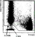
以所得侧向散射光强度和荧光强度为轴绘制第一二维分布图,划定全白细胞区域。此图如图2所示。对出现在全白细胞区域内的细胞计数作为全白细胞数。The first two-dimensional distribution map is drawn with the obtained side scattered light intensity and fluorescence intensity as the axis, and the whole white blood cell area is delineated. This diagram is shown in Figure 2. Count the cells appearing in the whole white blood cell area as the whole white blood cell number.
对于第一二维分布图中的全白细胞区域内出现的细胞,再以侧向散射光强度和荧光强度为轴,绘制第二二维分布图,将侧向散射光强度小、荧光强度也小的区域划定为原始粒细胞区域。此图如图3所示。对出现在原始粒细胞区域内的细胞计数,作为原始粒细胞数。For the cells appearing in the whole white blood cell area in the first two-dimensional distribution diagram, the second two-dimensional distribution diagram is drawn with the side scattered light intensity and fluorescence intensity as the axes, and the side scattered light intensity is small and the fluorescence intensity is also small The area defined as myeloblasts area. This diagram is shown in Figure 3. The number of cells present in the myeloblast region was counted as the number of myeloblasts.
在本实施例中,计算出原始粒细胞对全白细胞数的比率(原始粒细胞比率=原始粒细胞数/全白细胞数×100),原始粒细胞比率为45.8%。In this example, the ratio of myeloblasts to total leukocytes was calculated (ratio of myeloblasts=myeloblasts/total leukocytes×100), and the myeloblasts ratio was 45.8%.
(实施例2)(Example 2)
除将试剂与血样A在35℃反应外,其他均与实施例1相同,计算原始粒细胞比率,所得原始粒细胞比率为42.6%。实施例2中绘制的第一二维分布图如图4所示,第二二维分布图如图5所示。Except that the reagent was reacted with blood sample A at 35°C, the others were the same as in Example 1, and the ratio of myeloblasts was calculated, and the obtained ratio of myeloblasts was 42.6%. The first two-dimensional distribution diagram drawn in Example 2 is shown in FIG. 4 , and the second two-dimensional distribution diagram is shown in FIG. 5 .
(比较例1)(comparative example 1)
将含聚氧乙烯(16)油酰醚24.0G/L、N-月桂酰肌氨酸钠1.5G/L、DL-甲硫基丁氨酸20.0G/L、4-羟乙基哌嗪乙磺酸12.0G/L、1N—NAOH0.3G/L、NACL4.0G/L和色素A3.0MG/L的试剂1ML与血样B33μL混合反应,其他与实施例1相同,计算出原始粒细胞比率,所得原始粒细胞比率为25.1%。Contain polyoxyethylene (16) oleoyl ether 24.0G/L, N-lauroyl sarcosinate 1.5G/L, DL-methylthiobutyric acid 20.0G/L, 4-hydroxyethylpiperazine ethyl The reagent 1ML of sulfonic acid 12.0G/L, 1N-NAOH0.3G/L, NACL4.0G/L and pigment A3.0MG/L was mixed with blood sample B33μL for reaction, and the others were the same as in Example 1, and the ratio of myeloblasts was calculated. The resulting myeloblast ratio was 25.1%.
另,比较例1绘制的第一二维分布图如图6所示,第二二维分布图如图7所示。此试剂的渗透压为350MOsM/KG、电传导度为7.4MS/cM。In addition, the first two-dimensional distribution diagram drawn in Comparative Example 1 is shown in FIG. 6 , and the second two-dimensional distribution diagram is shown in FIG. 7 . The osmotic pressure of this reagent is 350MOsM/KG, and the electrical conductivity is 7.4MS/cM.
(比较例2)(comparative example 2)
除试剂和血样B在35℃反应外,其他与比较例1同,计算出原始粒细胞比率,所得原始粒细胞比率为7.2%。比较例2绘制的第一二维分布图如图8所示,第二二维分布图如图9所示。Except that the reagent and the blood sample B were reacted at 35° C., the others were the same as in Comparative Example 1. The ratio of myeloblasts was calculated, and the obtained ratio of myeloblasts was 7.2%. The first two-dimensional distribution diagram drawn in Comparative Example 2 is shown in FIG. 8 , and the second two-dimensional distribution diagram is shown in FIG. 9 .
从比较例1和2可以看出,尽管使用的是同一血样,但在35℃下反应时的原始粒细胞比率(比较例2)比在33℃下反应时的原始粒细胞比率(比较例1)低得多。这是因为35℃下反应时,应当出现在原始粒细胞区域的原始粒细胞出现在区域外。即,可以认为,使用比较例1和2制备的试剂,反应温度越高,原始粒细胞稳定性越差,在7秒钟的反应时间内原始粒细胞受到破坏。From Comparative Examples 1 and 2, it can be seen that although the same blood sample was used, the ratio of myeloblasts when reacted at 35°C (Comparative Example 2) was higher than the ratio of myelocytes when reacted at 33°C (Comparative Example 1 ) is much lower. This is because myeloblasts, which should appear in the myeloblast zone, appeared outside the zone when reacted at 35°C. That is, it can be considered that using the reagents prepared in Comparative Examples 1 and 2, the higher the reaction temperature, the worse the stability of myeloblasts, and the myeloblasts were destroyed within the reaction time of 7 seconds.
然而,从实施例1和2可以看出,在33℃下反应时的原始粒细胞比率(实施例1)与在35℃下反应时的原始粒细胞比率(实施例2)结果近似。这是因为在实施例中制备的试剂即使在高温下与原始粒细胞反应,实际上也不会破坏原始粒细胞,能够正确计数。即,使用实施例制备的试剂,原始粒细胞对温度的稳定性提高,不易受损。从上述可以断定,使用实施例制备的试剂可以正确地测定试样中的原始粒细胞。However, as can be seen from Examples 1 and 2, the myeloblast ratio when reacted at 33°C (Example 1) was similar to the myeloblast ratio when reacted at 35°C (Example 2). This is because even if the reagents prepared in Examples react with myeloblasts at high temperature, they will not actually destroy myeloblasts and can be counted correctly. That is, using the reagents prepared in the examples, myeloblasts are more stable to temperature and less likely to be damaged. From the above, it can be concluded that myeloblasts in the sample can be accurately measured using the reagents prepared in the examples.
(实施例3)(Example 3)
第一试剂B除了不是用树胶醛醣而是用木糖醇39.56G/L、聚氧乙烯(16)油酰醚不是20000PPM而是25000PPM、N-月桂酰肌氨酸钠不是500PPM而是750PPM之外,其他与第一试剂A一样制备。第一试剂B的渗透压为280MOsM/KG、电传导度为0.59MS/cM。The first reagent B is not aldose but xylitol 39.56G/L, polyoxyethylene (16) oleoyl ether is not 20000PPM but 25000PPM, N-lauroyl sarcosinate is not 500PPM but 750PPM Except, others are prepared in the same way as the first reagent A. The osmotic pressure of the first reagent B is 280 MOsM/KG, and the electrical conductivity is 0.59 MS/cM.
除试剂用的不是第一试剂A而是第一试剂B、血样用的不是血样A而是血样C外,其他与实施例1同,计算原始粒细胞比率。除试剂不是第一试剂A而是第一试剂B、血样不是血样A而是血样C、试剂与血样C反应不是7秒而是12秒外,其他与实施例1同,计算原始粒细胞比率。Except that the reagent is not the first reagent A but the first reagent B, and the blood sample is not the blood sample A but the blood sample C, the other is the same as that of Example 1, and the myeloblast ratio is calculated. Except that the reagent is not the first reagent A but the first reagent B, the blood sample is not blood sample A but blood sample C, and the reaction between the reagent and blood sample C is not 7 seconds but 12 seconds, others are the same as in Example 1, and the myeloblast ratio is calculated.
(实施例4)(Example 4)
第一试剂C除含有的不是木糖醇而是树胶醛醣39.52G/L外,其他与第一试剂B同样制备。第一试剂C的渗透压为280MOsM/KG、电传导度为0.62MS/cM。The first reagent C was prepared in the same way as the first reagent B except that it did not contain xylitol but arabinose 39.52 G/L. The osmotic pressure of the first reagent C is 280 MOsM/KG, and the electrical conductivity is 0.62 MS/cM.
除不是使用第一试剂B而是第一试剂C外,其他与实施例3相同,计算出原始粒细胞比率。Except that the first reagent C is used instead of the first reagent B, the ratio of myeloblasts is calculated in the same manner as in Example 3.
(实施例5)(Example 5)
第一试剂D除含有的不是木糖醇而是丙胺酸23.16G/L外,其他与第一试剂B同样制备。第一试剂D的渗透压为280MOsM/KG、电传导度为0.61MS/cM。The first reagent D was prepared in the same way as the first reagent B except that it did not contain xylitol but alanine 23.16 G/L. The osmotic pressure of the first reagent D is 280 MOsM/KG, and the electrical conductivity is 0.61 MS/cM.
除使用的不是第一试剂B而是第一试剂D外,其他与实施例3相同,计算出原始粒细胞比率。Except that the first reagent D is used instead of the first reagent B, the other is the same as in Example 3, and the myeloblast ratio is calculated.
(实施例6)(Example 6)
第一试剂E除含有的不是木糖醇而是糖胶19.52G/L外,其他与第一试剂B同样制备。第一试剂E的渗透压为280MOsM/KG、电传导度为0.63MS/cM。The first reagent E was prepared in the same way as the first reagent B except that it contained 19.52 G/L of sugar gum instead of xylitol. The osmotic pressure of the first reagent E is 280 MOsM/KG, and the electrical conductivity is 0.63 MS/cM.
除使用的不是第一试剂B而是第一试剂E外,其他与实施例3相同,计算出原始粒细胞比率。Except that instead of the first reagent B, the first reagent E was used, the ratio of myeloblasts was calculated in the same manner as in Example 3.
(比较例3)(comparative example 3)
除使用的不是血样A而是血样C外,其他与比较例1一样,计算原始粒细胞比率。另,除血样不是血样A而是血样C、试剂与血样C的反应时间不是7秒而是12秒外,其他与比较例1一样,计算原始粒细胞比率。Except that blood sample C was used instead of blood sample A, the ratio of myeloblasts was calculated as in Comparative Example 1. In addition, except that the blood sample is not blood sample A but blood sample C, and the reaction time between the reagent and blood sample C is not 7 seconds but 12 seconds, the ratio of myeloblasts is calculated as in Comparative Example 1.
实施例3~6以及比较例3的结果如图10所示。图10显示出试样与试剂反应12秒时的原始粒细胞比率比反应7秒时的原始粒细胞比率下降多少。The results of Examples 3 to 6 and Comparative Example 3 are shown in FIG. 10 . Figure 10 shows how much the myeloblast ratio decreased when the sample was reacted with the reagent for 12 seconds compared to when the sample was reacted for 7 seconds.
从图10可以看出,使用比较例3的试剂,让试剂与试样反应12秒,比反应7秒原始粒细胞比率下降20%以上。可以认为这是因为反应时间越长,受试剂的影响,原始粒细胞越容易受损。It can be seen from FIG. 10 that, using the reagent of Comparative Example 3, allowing the reagent to react with the sample for 12 seconds, the ratio of myeloblasts decreased by more than 20% compared to the reaction for 7 seconds. It can be thought that this is because the longer the reaction time, the more easily the myeloblasts are damaged by the influence of the reagent.
使用实施例3~6其中之一制备的试剂比使用比较例3的试剂,原始粒细胞比率下降控制在10%左右。即,由于使用实施例3~6的试剂,原始粒细胞的稳定性得以提高,在测定用试样中原始粒细胞不易受损。如上所述,可以判断,使用实施例3~6的试剂,原始粒细胞不易受损,得以正确地测定原始粒细胞。Using one of the reagents prepared in Examples 3-6 compared with the reagent of Comparative Example 3, the reduction of the ratio of myeloblasts was controlled at about 10%. That is, by using the reagents of Examples 3 to 6, the stability of myeloblasts is improved, and myeloblasts are less likely to be damaged in the measurement sample. As described above, it can be judged that myeloblasts are less likely to be damaged by using the reagents of Examples 3 to 6, and myeloblasts can be accurately measured.
(实施例7)(Example 7)
制备含以下成份的第一试剂F:Prepare the first reagent F containing the following ingredients:
聚氧乙烯(16)油酰醚25000PPMPolyoxyethylene (16) oleoyl ether 25000PPM
N-月桂酰肌氨酸钠750PPMSodium N-lauroyl sarcosinate 750PPM
木糖醇37.0G/LXylitol 37.0G/L
HEPES 10MMHEPES 10MM
蒸馏水1LDistilled water 1L
将上述成份混合,添加NAOH,使PH值调整为7.0。第一试剂F的渗透压为280MOsM/KG、电传导度为0.64MS/cM。第二试剂与实施例1制备的一样。Mix the above ingredients, add NAOH, and adjust the pH value to 7.0. The osmotic pressure of the first reagent F is 280 MOsM/KG, and the electrical conductivity is 0.64 MS/cM. The second reagent is the same as prepared in Example 1.
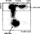
除使用的不是第一试剂A而是第一试剂F以及所用血样不是血样A而是血样D外,其他与实施例1同样,计算原始粒细胞比率,所得原始粒细胞比率45%。实施例7绘制的第一二维分布图如图11所示,第二二维分布图如图12所示。图12的第二二维分布图中也设定了成熟白细胞出现的区域(成熟白细胞区域)。Except that instead of the first reagent A, the first reagent F is used and the blood sample used is not blood sample A but blood sample D, other same as in Example 1, calculate the myeloblast ratio, and the obtained myeloblast ratio is 45%. The first two-dimensional distribution diagram drawn in Example 7 is shown in FIG. 11 , and the second two-dimensional distribution diagram is shown in FIG. 12 . The region where mature leukocytes appear (mature leukocyte region) is also set in the second two-dimensional distribution diagram in FIG. 12 .
用显微镜算出血样D中的原始粒细胞比率为58.5%。实施例7中测定的原始粒细胞比率与显微镜算出的原始粒细胞比率结果相近,因此可以断定,用实施例7的试剂可以正确地分辨血样中的成熟白细胞和原始粒细胞,正确地对原始粒细胞计数。The ratio of myeloblasts in blood sample D was calculated by microscope to be 58.5%. The ratio of myeloblasts measured in Example 7 is similar to the ratio of myeloblasts calculated by the microscope, so it can be concluded that the reagents in Example 7 can correctly distinguish the mature leukocytes and myeloblasts in the blood sample, and correctly identify myeloblasts. cell counts.
(实施例8)(Embodiment 8)
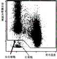
除使用的不是血样D而是血样G外,其他与实施例5同,计算原始粒细胞比率,所得原始粒细胞比率为5.5%。实施例8绘制的第一二维分布图如图13所示,第二二维分布图如图14所示。Except that blood sample G was used instead of blood sample D, the same as in Example 5, the myeloblast ratio was calculated, and the obtained myeloblast ratio was 5.5%. Figure 13 shows the first two-dimensional distribution chart drawn in Example 8, and Figure 14 shows the second two-dimensional distribution chart.
用显微镜算出血样E中的原始粒细胞比率,原始粒细胞为6.3%。实施例8中测定的原始粒细胞比率与显微镜算出的原始粒细胞比率结果相近,因此可以断定,用实施例8的试剂可以正确地分辨血样中的成熟白细胞和原始粒细胞,正确地对原始粒细胞计数。The ratio of myeloblasts in blood sample E was calculated with a microscope, and the myeloblasts were 6.3%. The ratio of myeloblasts measured in Example 8 is similar to the ratio of myeloblasts calculated by the microscope. Therefore, it can be concluded that the reagents in Example 8 can correctly distinguish the mature leukocytes and myeloblasts in the blood sample, and correctly identify the myeloblasts. cell counts.
还在图13所示第一二维分布图划定幼稚粒细胞出现的区域(幼稚粒细胞区域),对出现在此区域内的细胞计数,作为幼稚粒细胞数。根据此值,计算幼稚粒细胞对全白细胞数的比率(幼稚粒细胞比率),幼稚粒细胞比率为10%。Also, the area where immature granulocytes appeared (immature granulocyte area) was defined in the first two-dimensional distribution diagram shown in FIG. 13, and the cells appearing in this area were counted as the number of immature granulocytes. From this value, the ratio of immature granulocytes to the total leukocyte count (immature granulocyte ratio) was calculated, and the immature granulocyte ratio was 10%.
此外,用显微镜算出血样E中的幼稚粒细胞比率,幼稚粒细胞比率为13%。实施例8测定的幼稚粒细胞比率与显微镜算出的幼稚粒细胞比率结果相近。由此可以断定,用实施例8的试剂可以正确地分辨血样中的成熟白细胞、原始粒细胞和幼稚粒细胞,不仅对原始粒细胞,还能正确地对幼稚粒细胞计数。In addition, the ratio of immature granulocytes in blood sample E was calculated with a microscope, and the ratio of immature granulocytes was 13%. The ratio of immature granulocytes measured in Example 8 is close to the ratio of immature granulocytes calculated by microscope. From this, it can be concluded that the reagent of Example 8 can correctly distinguish mature leukocytes, myeloblasts and immature granulocytes in the blood sample, and can correctly count not only myeloblasts but also immature granulocytes.
(实施例9)(Example 9)
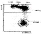
除使用血样F而不是血样D外,其他与实施例5同样,计算出幼稚粒细胞比率,所得幼稚粒细胞比率为12.4%。实施例9绘制的第一二维分布图如图15所示,第二二维分布图如图16所示。Except that blood sample F was used instead of blood sample D, the immature granulocyte ratio was calculated in the same manner as in Example 5, and the obtained immature granulocyte ratio was 12.4%. Figure 15 shows the first two-dimensional distribution chart drawn in Example 9, and Figure 16 shows the second two-dimensional distribution chart.
用显微镜算出血样E中的原始粒细胞比率和幼稚粒细胞比率,幼稚粒细胞比率为10.8%。实施例9测定的幼稚粒细胞比率与显微镜算出的幼稚粒细胞比率结果相近。由此可以断定,用实施例9的试剂可以正确地分辨血样中的成熟白细胞和幼稚粒细胞,能正确地对幼稚粒细胞计数。Calculate the ratio of primitive granulocytes and immature granulocytes in blood sample E with a microscope, and the ratio of immature granulocytes is 10.8%. The ratio of immature granulocytes measured in Example 9 is close to the ratio of immature granulocytes calculated by microscope. From this, it can be concluded that the reagent of Example 9 can correctly distinguish mature white blood cells and immature granulocytes in the blood sample, and can correctly count immature granulocytes.
前述的详细说明及附图是通过文字解释和图示来进行的,其目的不在于限定权利要求的保护范围。本说明书中的具体实施方式的各个变种对于普通技术人员来说显而易见,并处于权利要求及其等同技术的保护范围内。The above-mentioned detailed description and drawings are done by means of text explanation and illustration, and the purpose is not to limit the scope of protection of the claims. Variations of the specific implementation methods in this specification are obvious to those of ordinary skill, and are within the protection scope of the claims and their equivalent technologies.
Claims (13)
Translated fromChineseApplications Claiming Priority (3)
| Application Number | Priority Date | Filing Date | Title |
|---|---|---|---|
| JP2006-019895 | 2006-01-27 | ||
| JP2006019895 | 2006-01-27 | ||
| JP2006019895 | 2006-01-27 |
Publications (2)
| Publication Number | Publication Date |
|---|---|
| CN101008641A CN101008641A (en) | 2007-08-01 |
| CN101008641Btrue CN101008641B (en) | 2012-11-28 |
Family
ID=37907994
Family Applications (1)
| Application Number | Title | Priority Date | Filing Date |
|---|---|---|---|
| CN2007100036821AActiveCN101008641B (en) | 2006-01-27 | 2007-01-26 | Reagent for immature leukocyte analysis and reagent kit |
Country Status (4)
| Country | Link |
|---|---|
| US (1) | US7625757B2 (en) |
| EP (1) | EP1813942B1 (en) |
| CN (1) | CN101008641B (en) |
| AT (1) | ATE510212T1 (en) |
Families Citing this family (22)
| Publication number | Priority date | Publication date | Assignee | Title |
|---|---|---|---|---|
| JP4976038B2 (en)* | 2006-03-29 | 2012-07-18 | シスメックス株式会社 | Method for measuring hematological samples |
| JP4918281B2 (en)* | 2006-05-18 | 2012-04-18 | シスメックス株式会社 | Urine component analyzer |
| JP4806334B2 (en)* | 2006-11-27 | 2011-11-02 | シスメックス株式会社 | Method and apparatus for measuring hematological samples |
| US8859200B2 (en)* | 2007-06-25 | 2014-10-14 | Sysmex Corporation | Reagent and reagent kit for analysis of immature leukocyte |
| CN101680879B (en)* | 2007-06-25 | 2014-05-21 | 希森美康株式会社 | Analytical reagents and analytical kits for immature leukocytes |
| CN101349644B (en)* | 2007-07-20 | 2012-06-27 | 深圳迈瑞生物医疗电子股份有限公司 | Leukocytes classification agent and use method thereof |
| CN101475754A (en)* | 2008-01-04 | 2009-07-08 | 深圳迈瑞生物医疗电子股份有限公司 | Asymmetric cyanine fluorescent dye, composition and application in biological sample dyeing |
| US20110027826A1 (en)* | 2008-05-02 | 2011-02-03 | Arkray, Inc. | Leukocyte analysis method and analysis reagent for use in the method |
| CN101602762B (en)* | 2008-06-10 | 2013-10-16 | 深圳迈瑞生物医疗电子股份有限公司 | Asymmetric cyanine compound, preparation method and application thereof |
| JP5288970B2 (en)* | 2008-09-26 | 2013-09-11 | シスメックス株式会社 | Method for measuring mean red blood cell volume using a reagent for diluting blood samples |
| CN101726579B (en)* | 2008-10-17 | 2014-06-18 | 深圳迈瑞生物医疗电子股份有限公司 | Blood test reagent and method |
| CN101723874B (en)* | 2008-10-31 | 2013-09-11 | 深圳迈瑞生物医疗电子股份有限公司 | Cyanine compound and application thereof in dyeing biological samples |
| CN101750476B (en)* | 2008-12-08 | 2015-06-03 | 深圳迈瑞生物医疗电子股份有限公司 | Blood analysis reagent and use method thereof |
| CN101750274B (en)* | 2008-12-17 | 2014-06-25 | 深圳迈瑞生物医疗电子股份有限公司 | Differential blood count reagent, kit and method of differential blood count |
| JP5452058B2 (en)* | 2009-03-31 | 2014-03-26 | シスメックス株式会社 | Blood analyzer |
| EP2450706B1 (en)* | 2009-07-03 | 2017-07-26 | Sysmex Corporation | Blood analyzer and blood analyzing method |
| CN101988082B (en)* | 2009-07-31 | 2015-04-08 | 深圳迈瑞生物医疗电子股份有限公司 | Leukocyte classified counting reagent, kit and preparation method thereof and method of leukocyte classified counting |
| EP3104177B1 (en)* | 2011-12-21 | 2020-11-25 | Beckman Coulter, Inc. | Method for labeling intracellular and extracellular targets of leukocytes |
| CN102618060A (en)* | 2012-03-17 | 2012-08-01 | 江南大学 | Method for preparing asymmetrical cyanine dye and method for detecting bovine serum albumin by asymmetrical cyanine dye |
| US9746461B2 (en) | 2013-02-28 | 2017-08-29 | Sysmex Corporation | Urine sample analyzer and sample analyzing method |
| CN103398935B (en)* | 2013-08-23 | 2015-06-24 | 爱威科技股份有限公司 | Method and kit for leukocyte differential count |
| CN112195177B (en)* | 2020-10-28 | 2021-08-06 | 上海慕柏生物医学科技有限公司 | Nucleic acid extraction method and kit |
Citations (3)
| Publication number | Priority date | Publication date | Assignee | Title |
|---|---|---|---|---|
| US5094854A (en)* | 1988-03-04 | 1992-03-10 | Takeda Chemical Industries, Ltd. | Liposome composition useful for hypertheria therapy |
| CN1183559A (en)* | 1996-11-20 | 1998-06-03 | 东亚医用电子株式会社 | Differential proleukocyte count method |
| EP0867720B1 (en)* | 1997-03-28 | 2002-07-10 | Sysmex Corporation | Method of detecting hematopoietic progenitor cells |
Family Cites Families (11)
| Publication number | Priority date | Publication date | Assignee | Title |
|---|---|---|---|---|
| US5389549A (en)* | 1987-05-29 | 1995-02-14 | Toa Medical Electronics Co., Ltd. | Method for classifying leukocytes and a reagent used therefor |
| JP3048260B2 (en) | 1991-07-29 | 2000-06-05 | シスメックス株式会社 | Sample preparation method for leukocyte classification and counting |
| JP3301646B2 (en)* | 1993-03-19 | 2002-07-15 | シスメックス株式会社 | Reagent for immature cell measurement |
| US5639630A (en) | 1995-05-16 | 1997-06-17 | Bayer Corporation | Method and reagent composition for performing leukocyte differential counts on fresh and aged whole blood samples, based on intrinsic peroxidase activity of leukocytes |
| TW379284B (en) | 1996-04-12 | 2000-01-11 | Toa Medical Electronics | Agent for detecting reticulocyte |
| JP4042925B2 (en) | 1996-11-20 | 2008-02-06 | シスメックス株式会社 | Classification and counting method for immature leukocytes |
| US6790652B1 (en) | 1998-01-08 | 2004-09-14 | Bioimage A/S | Method and apparatus for high density format screening for bioactive molecules |
| US6916658B2 (en)* | 2001-07-27 | 2005-07-12 | Beckman Coulter, Inc. | Method for measurement of immature granulocytes |
| US7625730B2 (en) | 2002-06-24 | 2009-12-01 | Sysmex Corporation | Method for classifying and counting leukocytes |
| US7678578B2 (en) | 2005-02-07 | 2010-03-16 | Beckman Coulter, Inc. | Cell permeabilization and stabilization reagent and method of use |
| CN101078721B (en)* | 2006-05-23 | 2010-12-22 | 深圳迈瑞生物医疗电子股份有限公司 | Reagent and method for classifying leucocyte |
- 2007
- 2007-01-09USUS11/650,962patent/US7625757B2/enactiveActive
- 2007-01-25ATAT07001595Tpatent/ATE510212T1/ennot_activeIP Right Cessation
- 2007-01-25EPEP07001595Apatent/EP1813942B1/enactiveActive
- 2007-01-26CNCN2007100036821Apatent/CN101008641B/enactiveActive
Patent Citations (3)
| Publication number | Priority date | Publication date | Assignee | Title |
|---|---|---|---|---|
| US5094854A (en)* | 1988-03-04 | 1992-03-10 | Takeda Chemical Industries, Ltd. | Liposome composition useful for hypertheria therapy |
| CN1183559A (en)* | 1996-11-20 | 1998-06-03 | 东亚医用电子株式会社 | Differential proleukocyte count method |
| EP0867720B1 (en)* | 1997-03-28 | 2002-07-10 | Sysmex Corporation | Method of detecting hematopoietic progenitor cells |
Also Published As
| Publication number | Publication date |
|---|---|
| EP1813942B1 (en) | 2011-05-18 |
| US20070178597A1 (en) | 2007-08-02 |
| ATE510212T1 (en) | 2011-06-15 |
| EP1813942A1 (en) | 2007-08-01 |
| US7625757B2 (en) | 2009-12-01 |
| CN101008641A (en) | 2007-08-01 |
Similar Documents
| Publication | Publication Date | Title |
|---|---|---|
| CN101008641B (en) | Reagent for immature leukocyte analysis and reagent kit | |
| JP4324552B2 (en) | Leukocyte classification and counting method | |
| US7892841B2 (en) | Method and apparatus for measuring hematological sample | |
| JP4042925B2 (en) | Classification and counting method for immature leukocytes | |
| US8455209B2 (en) | Reagent and reagent kit for analysis of immature leukocyte | |
| JPH0534251A (en) | Method for preparing sample for classifying and counting leucocyte | |
| US20080131898A1 (en) | Method for measuring biological sample and measuring apparatus therefor | |
| CN101743469A (en) | Reagent kit for sample analysis and method for sample analysis | |
| US9222934B2 (en) | Reagent and reagent kit for analysis of immature leukocyte | |
| JP2002148261A (en) | Method for classifying and counting abnormal cell | |
| CN114270167A (en) | Blood testing method and blood analysis system | |
| Hübl et al. | Measurement of absolute concentration and viability of CD34+ cells in cord blood and cord blood products using fluorescent beads and cyanine nucleic acid dyes | |
| JP4806342B2 (en) | Reagent and reagent kit for immature leukocyte analysis | |
| JP2002223791A (en) | Method for classification counting of bone marrow nucleated cell | |
| JP4338206B2 (en) | Classification and counting method of erythroblasts | |
| JP4354982B2 (en) | Leukocyte classification and counting method | |
| JP4107441B2 (en) | Reagent for classification counting of erythroblasts | |
| JP2006300962A (en) | Method for classification and counting of leucocytes | |
| JP2012088336A (en) | Method and device for discriminating myeloblast from platelet aggregation | |
| JP2012103267A (en) | Hematological sample measuring instrument |
Legal Events
| Date | Code | Title | Description |
|---|---|---|---|
| C06 | Publication | ||
| PB01 | Publication | ||
| C10 | Entry into substantive examination | ||
| SE01 | Entry into force of request for substantive examination | ||
| C14 | Grant of patent or utility model | ||
| GR01 | Patent grant |