CN100998565A - Anti-angiogene compositions and methods of use - Google Patents
Anti-angiogene compositions and methods of useDownload PDFInfo
- Publication number
- CN100998565A CN100998565ACNA200610099887XACN200610099887ACN100998565ACN 100998565 ACN100998565 ACN 100998565ACN A200610099887X ACNA200610099887X ACN A200610099887XACN 200610099887 ACN200610099887 ACN 200610099887ACN 100998565 ACN100998565 ACN 100998565A
- Authority
- CN
- China
- Prior art keywords
- composition
- taxol
- polymer
- microspheres
- angiogenic
- Prior art date
- Legal status (The legal status is an assumption and is not a legal conclusion. Google has not performed a legal analysis and makes no representation as to the accuracy of the status listed.)
- Pending
Links
- 239000000203mixtureSubstances0.000titleclaimsabstractdescription223
- 238000000034methodMethods0.000titleabstractdescription100
- RCINICONZNJXQF-MZXODVADSA-NtaxolChemical compoundO([C@@H]1[C@@]2(C[C@@H](C(C)=C(C2(C)C)[C@H](C([C@]2(C)[C@@H](O)C[C@H]3OC[C@]3([C@H]21)OC(C)=O)=O)OC(=O)C)OC(=O)[C@H](O)[C@@H](NC(=O)C=1C=CC=CC=1)C=1C=CC=CC=1)O)C(=O)C1=CC=CC=C1RCINICONZNJXQF-MZXODVADSA-N0.000claimsabstractdescription230
- 229930012538PaclitaxelNatural products0.000claimsabstractdescription229
- 229960001592paclitaxelDrugs0.000claimsabstractdescription229
- 229920000642polymerPolymers0.000claimsabstractdescription139
- 239000004005microsphereSubstances0.000claimsdescription209
- -1poly(alkyl carbonate)Polymers0.000claimsdescription45
- 239000002245particleSubstances0.000claimsdescription37
- JVTAAEKCZFNVCJ-UHFFFAOYSA-Nlactic acidChemical compoundCC(O)C(O)=OJVTAAEKCZFNVCJ-UHFFFAOYSA-N0.000claimsdescription18
- 229920001610polycaprolactonePolymers0.000claimsdescription16
- 239000004632polycaprolactoneSubstances0.000claimsdescription15
- 150000001875compoundsChemical class0.000claimsdescription14
- 229920000747poly(lactic acid)Polymers0.000claimsdescription14
- AEMRFAOFKBGASW-UHFFFAOYSA-NGlycolic acidChemical compoundOCC(O)=OAEMRFAOFKBGASW-UHFFFAOYSA-N0.000claimsdescription12
- 229920001577copolymerPolymers0.000claimsdescription11
- 239000004626polylactic acidSubstances0.000claimsdescription11
- 229920000609methyl cellulosePolymers0.000claimsdescription10
- 239000001923methylcelluloseSubstances0.000claimsdescription10
- 239000004310lactic acidSubstances0.000claimsdescription9
- 235000014655lactic acidNutrition0.000claimsdescription9
- 239000002775capsuleSubstances0.000claimsdescription7
- 102000029749MicrotubuleHuman genes0.000claimsdescription4
- 108091022875MicrotubuleProteins0.000claimsdescription4
- 210000004688microtubuleAnatomy0.000claimsdescription4
- 108010088751AlbuminsProteins0.000claimsdescription3
- 102000009027AlbuminsHuman genes0.000claimsdescription3
- 229920001710PolyorthoesterPolymers0.000claimsdescription3
- 229920002807ThiomerPolymers0.000claimsdescription3
- JXLYSJRDGCGARV-WWYNWVTFSA-NVinblastineNatural productsO=C(O[C@H]1[C@](O)(C(=O)OC)[C@@H]2N(C)c3c(cc(c(OC)c3)[C@]3(C(=O)OC)c4[nH]c5c(c4CCN4C[C@](O)(CC)C[C@H](C3)C4)cccc5)[C@@]32[C@H]2[C@@]1(CC)C=CCN2CC3)CJXLYSJRDGCGARV-WWYNWVTFSA-N0.000claimsdescription3
- 239000002745poly(ortho ester)Substances0.000claimsdescription3
- 239000005020polyethylene terephthalateSubstances0.000claimsdescription3
- 229960003048vinblastineDrugs0.000claimsdescription3
- JXLYSJRDGCGARV-XQKSVPLYSA-NvincaleukoblastineChemical compoundC([C@@H](C[C@]1(C(=O)OC)C=2C(=CC3=C([C@]45[C@H]([C@@]([C@H](OC(C)=O)[C@]6(CC)C=CCN([C@H]56)CC4)(O)C(=O)OC)N3C)C=2)OC)C[C@@](C2)(O)CC)N2CCC2=C1NC1=CC=CC=C21JXLYSJRDGCGARV-XQKSVPLYSA-N0.000claimsdescription3
- FRPJXPJMRWBBIH-RBRWEJTLSA-NestramustineChemical compoundClCCN(CCCl)C(=O)OC1=CC=C2[C@H]3CC[C@](C)([C@H](CC4)O)[C@@H]4[C@@H]3CCC2=C1FRPJXPJMRWBBIH-RBRWEJTLSA-N0.000claimsdescription2
- 229960001842estramustineDrugs0.000claimsdescription2
- IAKHMKGGTNLKSZ-INIZCTEOSA-N(S)-colchicineChemical compoundC1([C@@H](NC(C)=O)CC2)=CC(=O)C(OC)=CC=C1C1=C2C=C(OC)C(OC)=C1OCIAKHMKGGTNLKSZ-INIZCTEOSA-N0.000claims2
- 229920002732PolyanhydridePolymers0.000claims2
- 229920000954PolyglycolidePolymers0.000claims2
- 239000000017hydrogelSubstances0.000claims2
- 229920000139polyethylene terephthalatePolymers0.000claims2
- 125000002456taxol groupChemical group0.000claims2
- LUEYTMPPCOCKBX-KWYHTCOPSA-NCuracin AChemical compoundC=CC[C@H](OC)CC\C(C)=C\C=C\CC\C=C/[C@@H]1CSC([C@H]2[C@H](C2)C)=N1LUEYTMPPCOCKBX-KWYHTCOPSA-N0.000claims1
- LUEYTMPPCOCKBX-UHFFFAOYSA-NCuracin ANatural productsC=CCC(OC)CCC(C)=CC=CCCC=CC1CSC(C2C(C2)C)=N1LUEYTMPPCOCKBX-UHFFFAOYSA-N0.000claims1
- 229960001338colchicineDrugs0.000claims1
- 239000000039congenerSubstances0.000claims1
- 229930013356epothiloneNatural products0.000claims1
- 150000003883epothilone derivativesChemical class0.000claims1
- 230000001772anti-angiogenic effectEffects0.000abstractdescription140
- 210000004204blood vesselAnatomy0.000abstractdescription73
- 239000003102growth factorSubstances0.000abstractdescription36
- 210000000013bile ductAnatomy0.000abstractdescription21
- 239000002870angiogenesis inducing agentSubstances0.000abstractdescription16
- 210000003437tracheaAnatomy0.000abstractdescription9
- 230000002137anti-vascular effectEffects0.000abstractdescription8
- 210000003238esophagusAnatomy0.000abstractdescription8
- 210000003708urethraAnatomy0.000abstractdescription8
- 210000000621bronchiAnatomy0.000abstractdescription7
- SHGAZHPCJJPHSC-YCNIQYBTSA-Nall-trans-retinoic acidChemical compoundOC(=O)\C=C(/C)\C=C\C=C(/C)\C=C\C1=C(C)CCCC1(C)CSHGAZHPCJJPHSC-YCNIQYBTSA-N0.000abstractdescription6
- 229930002330retinoic acidNatural products0.000abstractdescription6
- 229960001727tretinoinDrugs0.000abstractdescription6
- 230000001740anti-invasionEffects0.000abstractdescription5
- 206010028980NeoplasmDiseases0.000description177
- YMWUJEATGCHHMB-UHFFFAOYSA-NDichloromethaneChemical compoundClCClYMWUJEATGCHHMB-UHFFFAOYSA-N0.000description108
- 239000000243solutionSubstances0.000description79
- 239000003814drugSubstances0.000description68
- XLYOFNOQVPJJNP-UHFFFAOYSA-NwaterSubstancesOXLYOFNOQVPJJNP-UHFFFAOYSA-N0.000description66
- 229940079593drugDrugs0.000description65
- 239000011521glassSubstances0.000description59
- 239000005038ethylene vinyl acetateSubstances0.000description58
- 229920001200poly(ethylene-vinyl acetate)Polymers0.000description57
- 210000001519tissueAnatomy0.000description56
- DQXBYHZEEUGOBF-UHFFFAOYSA-Nbut-3-enoic acid;etheneChemical compoundC=C.OC(=O)CC=CDQXBYHZEEUGOBF-UHFFFAOYSA-N0.000description51
- 239000004372Polyvinyl alcoholSubstances0.000description45
- 229920002451polyvinyl alcoholPolymers0.000description45
- 239000010408filmSubstances0.000description44
- 208000037265diseases, disorders, signs and symptomsDiseases0.000description41
- 201000010099diseaseDiseases0.000description40
- 201000011510cancerDiseases0.000description39
- 210000004027cellAnatomy0.000description39
- 229920001244Poly(D,L-lactide)Polymers0.000description35
- 230000012010growthEffects0.000description34
- 230000002792vascularEffects0.000description32
- 230000000694effectsEffects0.000description28
- 238000002360preparation methodMethods0.000description28
- 238000011282treatmentMethods0.000description28
- 210000004185liverAnatomy0.000description25
- 210000001367arteryAnatomy0.000description24
- 239000012153distilled waterSubstances0.000description23
- 238000003780insertionMethods0.000description23
- 239000012071phaseSubstances0.000description23
- 238000002271resectionMethods0.000description23
- WEVYAHXRMPXWCK-UHFFFAOYSA-NAcetonitrileChemical compoundCC#NWEVYAHXRMPXWCK-UHFFFAOYSA-N0.000description21
- 241001465754MetazoaSpecies0.000description21
- HEMHJVSKTPXQMS-UHFFFAOYSA-MSodium hydroxideChemical compound[OH-].[Na+]HEMHJVSKTPXQMS-UHFFFAOYSA-M0.000description21
- 239000007921spraySubstances0.000description21
- 238000003756stirringMethods0.000description21
- 238000001356surgical procedureMethods0.000description21
- 230000015572biosynthetic processEffects0.000description20
- LOKCTEFSRHRXRJ-UHFFFAOYSA-Idipotassium trisodium dihydrogen phosphate hydrogen phosphate dichlorideChemical compoundP(=O)(O)(O)[O-].[K+].P(=O)(O)([O-])[O-].[Na+].[Na+].[Cl-].[K+].[Cl-].[Na+]LOKCTEFSRHRXRJ-UHFFFAOYSA-I0.000description20
- 230000002401inhibitory effectEffects0.000description20
- 239000002953phosphate buffered salineSubstances0.000description20
- 241000700159RattusSpecies0.000description19
- FAPWRFPIFSIZLT-UHFFFAOYSA-MSodium chlorideChemical compound[Na+].[Cl-]FAPWRFPIFSIZLT-UHFFFAOYSA-M0.000description19
- 230000010102embolizationEffects0.000description19
- 230000037431insertionEffects0.000description19
- FIAFUQMPZJWCLV-UHFFFAOYSA-NsuraminChemical compoundOS(=O)(=O)C1=CC(S(O)(=O)=O)=C2C(NC(=O)C3=CC=C(C(=C3)NC(=O)C=3C=C(NC(=O)NC=4C=C(C=CC=4)C(=O)NC=4C(=CC=C(C=4)C(=O)NC=4C5=C(C=C(C=C5C(=CC=4)S(O)(=O)=O)S(O)(=O)=O)S(O)(=O)=O)C)C=CC=3)C)=CC=C(S(O)(=O)=O)C2=C1FIAFUQMPZJWCLV-UHFFFAOYSA-N0.000description19
- 210000004087corneaAnatomy0.000description18
- 239000000725suspensionSubstances0.000description18
- 238000012360testing methodMethods0.000description18
- 208000027418Wounds and injuryDiseases0.000description17
- 238000002474experimental methodMethods0.000description17
- 238000002347injectionMethods0.000description17
- 239000007924injectionSubstances0.000description17
- LFQSCWFLJHTTHZ-UHFFFAOYSA-NEthanolChemical compoundCCOLFQSCWFLJHTTHZ-UHFFFAOYSA-N0.000description16
- 230000008569processEffects0.000description16
- 230000033115angiogenesisEffects0.000description15
- 230000006378damageEffects0.000description15
- 210000002889endothelial cellAnatomy0.000description15
- 230000001965increasing effectEffects0.000description15
- IJGRMHOSHXDMSA-UHFFFAOYSA-NAtomic nitrogenChemical compoundN#NIJGRMHOSHXDMSA-UHFFFAOYSA-N0.000description14
- 210000002257embryonic structureAnatomy0.000description14
- 210000001503jointAnatomy0.000description14
- 239000011780sodium chlorideSubstances0.000description14
- 230000004614tumor growthEffects0.000description14
- 241000287828Gallus gallusSpecies0.000description13
- 238000009472formulationMethods0.000description13
- 230000005764inhibitory processEffects0.000description13
- 239000011159matrix materialSubstances0.000description13
- 231100000241scarToxicity0.000description13
- 206010052428WoundDiseases0.000description12
- 238000005538encapsulationMethods0.000description12
- 239000000126substanceSubstances0.000description12
- 239000004094surface-active agentSubstances0.000description12
- 210000005166vasculatureAnatomy0.000description12
- 206010029113NeovascularisationDiseases0.000description11
- 238000004458analytical methodMethods0.000description11
- 206010003246arthritisDiseases0.000description11
- 210000000845cartilageAnatomy0.000description11
- 230000001419dependent effectEffects0.000description11
- 235000013601eggsNutrition0.000description11
- 239000010410layerSubstances0.000description11
- 230000003902lesionEffects0.000description11
- 238000012546transferMethods0.000description11
- 210000004881tumor cellAnatomy0.000description11
- 239000004037angiogenesis inhibitorSubstances0.000description10
- 238000013459approachMethods0.000description10
- 238000004090dissolutionMethods0.000description10
- 229920002959polymer blendPolymers0.000description10
- 238000005507sprayingMethods0.000description10
- 229960000621suramin sodiumDrugs0.000description10
- 229920003345Elvax®Polymers0.000description9
- 208000032843HemorrhageDiseases0.000description9
- OKKJLVBELUTLKV-UHFFFAOYSA-NMethanolChemical compoundOCOKKJLVBELUTLKV-UHFFFAOYSA-N0.000description9
- 208000034158bleedingDiseases0.000description9
- 230000000740bleeding effectEffects0.000description9
- 238000001727in vivoMethods0.000description9
- 238000011068loading methodMethods0.000description9
- 239000012528membraneSubstances0.000description9
- 239000008188pelletSubstances0.000description9
- 229920003023plasticPolymers0.000description9
- 239000004033plasticSubstances0.000description9
- 239000007787solidSubstances0.000description9
- 239000006228supernatantSubstances0.000description9
- 206010055665Corneal neovascularisationDiseases0.000description8
- 206010061218InflammationDiseases0.000description8
- 208000002260KeloidDiseases0.000description8
- 241001111421PannusSpecies0.000description8
- 229940121369angiogenesis inhibitorDrugs0.000description8
- 210000002159anterior chamberAnatomy0.000description8
- 230000015556catabolic processEffects0.000description8
- 239000003153chemical reaction reagentSubstances0.000description8
- 238000013270controlled releaseMethods0.000description8
- 201000000159corneal neovascularizationDiseases0.000description8
- 238000006731degradation reactionMethods0.000description8
- 238000001035dryingMethods0.000description8
- 238000002695general anesthesiaMethods0.000description8
- 230000002440hepatic effectEffects0.000description8
- 238000004128high performance liquid chromatographyMethods0.000description8
- 230000001969hypertrophic effectEffects0.000description8
- 230000004054inflammatory processEffects0.000description8
- 239000003112inhibitorSubstances0.000description8
- 210000001117keloidAnatomy0.000description8
- 239000007788liquidSubstances0.000description8
- 208000014018liver neoplasmDiseases0.000description8
- 230000036210malignancyEffects0.000description8
- 210000004379membraneAnatomy0.000description8
- 210000001525retinaAnatomy0.000description8
- 150000003431steroidsChemical class0.000description8
- 206010012689Diabetic retinopathyDiseases0.000description7
- 208000010412GlaucomaDiseases0.000description7
- 206010019695Hepatic neoplasmDiseases0.000description7
- 241000699670Mus sp.Species0.000description7
- 230000009471actionEffects0.000description7
- 210000003445biliary tractAnatomy0.000description7
- 230000036770blood supplyEffects0.000description7
- 210000004556brainAnatomy0.000description7
- 238000005266castingMethods0.000description7
- 239000003795chemical substances by applicationSubstances0.000description7
- 239000011248coating agentSubstances0.000description7
- 238000000576coating methodMethods0.000description7
- 230000034994deathEffects0.000description7
- 231100000517deathToxicity0.000description7
- 230000003247decreasing effectEffects0.000description7
- 238000009826distributionMethods0.000description7
- 230000003628erosive effectEffects0.000description7
- 239000003517fumeSubstances0.000description7
- 239000000499gelSubstances0.000description7
- 210000002767hepatic arteryAnatomy0.000description7
- 238000011534incubationMethods0.000description7
- 208000014674injuryDiseases0.000description7
- 210000000265leukocyteAnatomy0.000description7
- 238000004519manufacturing processMethods0.000description7
- 239000000463materialSubstances0.000description7
- 238000002844meltingMethods0.000description7
- 230000008018meltingEffects0.000description7
- 210000000214mouthAnatomy0.000description7
- 239000006199nebulizerSubstances0.000description7
- 229910052757nitrogenInorganic materials0.000description7
- 230000000717retained effectEffects0.000description7
- 239000011550stock solutionSubstances0.000description7
- 229960005314suraminDrugs0.000description7
- 230000006444vascular growthEffects0.000description7
- 210000003462veinAnatomy0.000description7
- 208000032544CicatrixDiseases0.000description6
- 206010027476MetastasesDiseases0.000description6
- 206010027457Metastases to liverDiseases0.000description6
- 241000283973Oryctolagus cuniculusSpecies0.000description6
- 230000003187abdominal effectEffects0.000description6
- 230000002776aggregationEffects0.000description6
- 238000004220aggregationMethods0.000description6
- 238000003556assayMethods0.000description6
- 210000004369bloodAnatomy0.000description6
- 239000008280bloodSubstances0.000description6
- 239000000969carrierSubstances0.000description6
- 208000006990cholangiocarcinomaDiseases0.000description6
- 210000002808connective tissueAnatomy0.000description6
- 210000003981ectodermAnatomy0.000description6
- 239000002077nanosphereSubstances0.000description6
- 201000003142neovascular glaucomaDiseases0.000description6
- 210000000056organAnatomy0.000description6
- 102000004169proteins and genesHuman genes0.000description6
- 108090000623proteins and genesProteins0.000description6
- 230000037387scarsEffects0.000description6
- 238000002560therapeutic procedureMethods0.000description6
- 229930190007BaccatinNatural products0.000description5
- 241000282414Homo sapiensSpecies0.000description5
- 102100026262Metalloproteinase inhibitor 2Human genes0.000description5
- 208000031481Pathologic ConstrictionDiseases0.000description5
- 239000004743PolypropyleneSubstances0.000description5
- 229920002125Sokalan®Polymers0.000description5
- 108010031374Tissue Inhibitor of Metalloproteinase-1Proteins0.000description5
- 102000005353Tissue Inhibitor of Metalloproteinase-1Human genes0.000description5
- 108010031372Tissue Inhibitor of Metalloproteinase-2Proteins0.000description5
- 210000001015abdomenAnatomy0.000description5
- 230000002159abnormal effectEffects0.000description5
- 208000009956adenocarcinomaDiseases0.000description5
- 230000002491angiogenic effectEffects0.000description5
- 238000002583angiographyMethods0.000description5
- QVGXLLKOCUKJST-UHFFFAOYSA-Natomic oxygenChemical compound[O]QVGXLLKOCUKJST-UHFFFAOYSA-N0.000description5
- 230000017531blood circulationEffects0.000description5
- 238000007906compressionMethods0.000description5
- 230000006835compressionEffects0.000description5
- 239000002872contrast mediaSubstances0.000description5
- 230000007423decreaseEffects0.000description5
- 238000009792diffusion processMethods0.000description5
- 239000006185dispersionSubstances0.000description5
- 210000001105femoral arteryAnatomy0.000description5
- 238000000338in vitroMethods0.000description5
- 230000002757inflammatory effectEffects0.000description5
- 230000007774longtermEffects0.000description5
- 210000004072lungAnatomy0.000description5
- 210000001161mammalian embryoAnatomy0.000description5
- 230000005012migrationEffects0.000description5
- 238000013508migrationMethods0.000description5
- 239000002105nanoparticleSubstances0.000description5
- 239000001301oxygenSubstances0.000description5
- 229910052760oxygenInorganic materials0.000description5
- 239000000047productSubstances0.000description5
- 230000002829reductive effectEffects0.000description5
- 210000002784stomachAnatomy0.000description5
- 230000008961swellingEffects0.000description5
- 238000005406washingMethods0.000description5
- 230000029663wound healingEffects0.000description5
- 208000022211Arteriovenous MalformationsDiseases0.000description4
- 206010006440Bronchial obstructionDiseases0.000description4
- CURLTUGMZLYLDI-UHFFFAOYSA-NCarbon dioxideChemical compoundO=C=OCURLTUGMZLYLDI-UHFFFAOYSA-N0.000description4
- LYCAIKOWRPUZTN-UHFFFAOYSA-NEthylene glycolChemical compoundOCCOLYCAIKOWRPUZTN-UHFFFAOYSA-N0.000description4
- 206010016654FibrosisDiseases0.000description4
- 108010010803GelatinProteins0.000description4
- 206010018338GliomaDiseases0.000description4
- 208000035346Margins of ExcisionDiseases0.000description4
- 241001529936MurinaeSpecies0.000description4
- 206010030178Oesophageal obstructionDiseases0.000description4
- 102000004179Plasminogen Activator Inhibitor 2Human genes0.000description4
- 108090000614Plasminogen Activator Inhibitor 2Proteins0.000description4
- NWGKJDSIEKMTRX-AAZCQSIUSA-NSorbitan monooleateChemical compoundCCCCCCCC\C=C/CCCCCCCC(=O)OC[C@@H](O)[C@H]1OC[C@H](O)[C@H]1ONWGKJDSIEKMTRX-AAZCQSIUSA-N0.000description4
- 241000202349Taxus brevifoliaSpecies0.000description4
- 206010044291Tracheal obstructionDiseases0.000description4
- 208000009443Vascular MalformationsDiseases0.000description4
- XTXRWKRVRITETP-UHFFFAOYSA-NVinyl acetateChemical compoundCC(=O)OC=CXTXRWKRVRITETP-UHFFFAOYSA-N0.000description4
- 239000002671adjuvantSubstances0.000description4
- 239000000443aerosolSubstances0.000description4
- 239000002246antineoplastic agentSubstances0.000description4
- 230000005744arteriovenous malformationEffects0.000description4
- 210000001188articular cartilageAnatomy0.000description4
- 210000000601blood cellAnatomy0.000description4
- 230000036760body temperatureEffects0.000description4
- 210000002302brachial arteryAnatomy0.000description4
- 230000001413cellular effectEffects0.000description4
- 238000002512chemotherapyMethods0.000description4
- 238000005520cutting processMethods0.000description4
- 238000001839endoscopyMethods0.000description4
- 230000003511endothelial effectEffects0.000description4
- 238000001704evaporationMethods0.000description4
- 230000008020evaporationEffects0.000description4
- 210000002950fibroblastAnatomy0.000description4
- 230000004761fibrosisEffects0.000description4
- 230000001497fibrovascularEffects0.000description4
- 239000012530fluidSubstances0.000description4
- 230000006870functionEffects0.000description4
- 239000008273gelatinSubstances0.000description4
- 229920000159gelatinPolymers0.000description4
- 235000019322gelatineNutrition0.000description4
- 235000011852gelatine dessertsNutrition0.000description4
- 208000002409gliosarcomaDiseases0.000description4
- 230000005484gravityEffects0.000description4
- 230000009545invasionEffects0.000description4
- 230000003211malignant effectEffects0.000description4
- 230000011278mitosisEffects0.000description4
- 238000002156mixingMethods0.000description4
- 201000008482osteoarthritisDiseases0.000description4
- 238000007458percutaneous transhepatic cholangiographyMethods0.000description4
- 229920001155polypropylenePolymers0.000description4
- 239000011148porous materialSubstances0.000description4
- 108090000765processed proteins & peptidesProteins0.000description4
- 230000000750progressive effectEffects0.000description4
- 230000035755proliferationEffects0.000description4
- 201000007914proliferative diabetic retinopathyDiseases0.000description4
- 206010039073rheumatoid arthritisDiseases0.000description4
- 239000002904solventChemical group0.000description4
- 229910001220stainless steelInorganic materials0.000description4
- 239000010935stainless steelSubstances0.000description4
- 230000036262stenosisEffects0.000description4
- 208000037804stenosisDiseases0.000description4
- 238000003860storageMethods0.000description4
- 230000004083survival effectEffects0.000description4
- 230000001225therapeutic effectEffects0.000description4
- 206010046459urethral obstructionDiseases0.000description4
- 210000000685uterine arteryAnatomy0.000description4
- 238000005303weighingMethods0.000description4
- 206010056375Bile duct obstructionDiseases0.000description3
- 201000004569BlindnessDiseases0.000description3
- 108091003079Bovine Serum AlbuminProteins0.000description3
- 208000026310Breast neoplasmDiseases0.000description3
- 206010008635CholestasisDiseases0.000description3
- 108010035532CollagenProteins0.000description3
- 102000008186CollagenHuman genes0.000description3
- 206010061818Disease progressionDiseases0.000description3
- 208000001976Endocrine Gland NeoplasmsDiseases0.000description3
- WSFSSNUMVMOOMR-UHFFFAOYSA-NFormaldehydeChemical compoundO=CWSFSSNUMVMOOMR-UHFFFAOYSA-N0.000description3
- HTTJABKRGRZYRN-UHFFFAOYSA-NHeparinChemical compoundOC1C(NC(=O)C)C(O)OC(COS(O)(=O)=O)C1OC1C(OS(O)(=O)=O)C(O)C(OC2C(C(OS(O)(=O)=O)C(OC3C(C(O)C(O)C(O3)C(O)=O)OS(O)(=O)=O)C(CO)O2)NS(O)(=O)=O)C(C(O)=O)O1HTTJABKRGRZYRN-UHFFFAOYSA-N0.000description3
- 206010023330Keloid scarDiseases0.000description3
- 102000008072LymphokinesHuman genes0.000description3
- 108010074338LymphokinesProteins0.000description3
- 206010025323LymphomasDiseases0.000description3
- 208000005890NeuromaDiseases0.000description3
- 208000034530PLAA-associated neurodevelopmental diseaseDiseases0.000description3
- 206010061902Pancreatic neoplasmDiseases0.000description3
- QGMRQYFBGABWDR-UHFFFAOYSA-MPentobarbital sodiumChemical group[Na+].CCCC(C)C1(CC)C(=O)NC(=O)[N-]C1=OQGMRQYFBGABWDR-UHFFFAOYSA-M0.000description3
- 108010022233Plasminogen Activator Inhibitor 1Proteins0.000description3
- 102100039418Plasminogen activator inhibitor 1Human genes0.000description3
- 206010038848Retinal detachmentDiseases0.000description3
- 208000007536ThrombosisDiseases0.000description3
- YXFVVABEGXRONW-UHFFFAOYSA-NTolueneChemical compoundCC1=CC=CC=C1YXFVVABEGXRONW-UHFFFAOYSA-N0.000description3
- 210000002945adventitial reticular cellAnatomy0.000description3
- 239000007864aqueous solutionSubstances0.000description3
- 230000010108arterial embolizationEffects0.000description3
- 229910052788bariumInorganic materials0.000description3
- DSAJWYNOEDNPEQ-UHFFFAOYSA-Nbarium atomChemical compound[Ba]DSAJWYNOEDNPEQ-UHFFFAOYSA-N0.000description3
- 230000008901benefitEffects0.000description3
- 230000002051biphasic effectEffects0.000description3
- 230000000903blocking effectEffects0.000description3
- 229940098773bovine serum albuminDrugs0.000description3
- 210000000481breastAnatomy0.000description3
- 239000000872bufferSubstances0.000description3
- 230000008355cartilage degradationEffects0.000description3
- 230000032823cell divisionEffects0.000description3
- 230000008859changeEffects0.000description3
- 210000003837chick embryoAnatomy0.000description3
- 210000003711chorioallantoic membraneAnatomy0.000description3
- 208000029742colonic neoplasmDiseases0.000description3
- 230000007797corrosionEffects0.000description3
- 238000005260corrosionMethods0.000description3
- 231100000433cytotoxicToxicity0.000description3
- 230000001472cytotoxic effectEffects0.000description3
- 229920006237degradable polymerPolymers0.000description3
- 230000008021depositionEffects0.000description3
- 238000010586diagramMethods0.000description3
- 238000010790dilutionMethods0.000description3
- 239000012895dilutionSubstances0.000description3
- 230000005750disease progressionEffects0.000description3
- 238000012377drug deliveryMethods0.000description3
- 210000001198duodenumAnatomy0.000description3
- 201000011523endocrine gland cancerDiseases0.000description3
- 238000007459endoscopic retrograde cholangiopancreatographyMethods0.000description3
- 239000000706filtrateSubstances0.000description3
- 238000002594fluoroscopyMethods0.000description3
- 239000007789gasSubstances0.000description3
- 210000001654germ layerAnatomy0.000description3
- 239000003292glueSubstances0.000description3
- PCHJSUWPFVWCPO-UHFFFAOYSA-NgoldChemical compound[Au]PCHJSUWPFVWCPO-UHFFFAOYSA-N0.000description3
- 229910052737goldInorganic materials0.000description3
- 239000010931goldSubstances0.000description3
- 229960002897heparinDrugs0.000description3
- 229920000669heparinPolymers0.000description3
- 210000005161hepatic lobeAnatomy0.000description3
- 238000010562histological examinationMethods0.000description3
- 230000002209hydrophobic effectEffects0.000description3
- 201000001371inclusion conjunctivitisDiseases0.000description3
- 210000004969inflammatory cellAnatomy0.000description3
- 230000028709inflammatory responseEffects0.000description3
- 238000001802infusionMethods0.000description3
- 238000007689inspectionMethods0.000description3
- 208000018937joint inflammationDiseases0.000description3
- 201000007270liver cancerDiseases0.000description3
- 230000035800maturationEffects0.000description3
- 230000007246mechanismEffects0.000description3
- 201000001441melanomaDiseases0.000description3
- 210000003716mesodermAnatomy0.000description3
- 230000000394mitotic effectEffects0.000description3
- 239000011259mixed solutionSubstances0.000description3
- 230000003287optical effectEffects0.000description3
- 210000000496pancreasAnatomy0.000description3
- 201000002528pancreatic cancerDiseases0.000description3
- 230000007170pathologyEffects0.000description3
- 229920006254polymer filmPolymers0.000description3
- 230000004264retinal detachmentEffects0.000description3
- 230000002207retinal effectEffects0.000description3
- 238000001878scanning electron micrographMethods0.000description3
- 230000037390scarringEffects0.000description3
- 210000003625skullAnatomy0.000description3
- 238000000935solvent evaporationMethods0.000description3
- 210000000952spleenAnatomy0.000description3
- 238000013268sustained releaseMethods0.000description3
- 239000012730sustained-release formSubstances0.000description3
- 210000001258synovial membraneAnatomy0.000description3
- 238000011521systemic chemotherapyMethods0.000description3
- 239000010409thin filmSubstances0.000description3
- 206010044325trachomaDiseases0.000description3
- 238000002054transplantationMethods0.000description3
- 239000003981vehicleSubstances0.000description3
- 230000004580weight lossEffects0.000description3
- VEEGZPWAAPPXRB-BJMVGYQFSA-N(3e)-3-(1h-imidazol-5-ylmethylidene)-1h-indol-2-oneChemical compoundO=C1NC2=CC=CC=C2\C1=C/C1=CN=CN1VEEGZPWAAPPXRB-BJMVGYQFSA-N0.000description2
- IADUEWIQBXOCDZ-VKHMYHEASA-N(S)-azetidine-2-carboxylic acidChemical compoundOC(=O)[C@@H]1CCN1IADUEWIQBXOCDZ-VKHMYHEASA-N0.000description2
- JTXMVXSTHSMVQF-UHFFFAOYSA-N2-acetyloxyethyl acetateChemical compoundCC(=O)OCCOC(C)=OJTXMVXSTHSMVQF-UHFFFAOYSA-N0.000description2
- BOCATKBAKISRLA-UHFFFAOYSA-N4-propyl-5-pyridin-4-yl-3h-1,3-oxazol-2-oneChemical compoundN1C(=O)OC(C=2C=CN=CC=2)=C1CCCBOCATKBAKISRLA-UHFFFAOYSA-N0.000description2
- RZVAJINKPMORJF-UHFFFAOYSA-NAcetaminophenChemical compoundCC(=O)NC1=CC=C(O)C=C1RZVAJINKPMORJF-UHFFFAOYSA-N0.000description2
- 208000003200AdenomaDiseases0.000description2
- 206010002091AnaesthesiaDiseases0.000description2
- 201000002862Angle-Closure GlaucomaDiseases0.000description2
- 208000003174Brain NeoplasmsDiseases0.000description2
- 206010006187Breast cancerDiseases0.000description2
- 206010009944Colon cancerDiseases0.000description2
- 208000028006Corneal injuryDiseases0.000description2
- AOJJSUZBOXZQNB-TZSSRYMLSA-NDoxorubicinChemical compoundO([C@H]1C[C@@](O)(CC=2C(O)=C3C(=O)C=4C=CC=C(C=4C(=O)C3=C(O)C=21)OC)C(=O)CO)[C@H]1C[C@H](N)[C@H](O)[C@H](C)O1AOJJSUZBOXZQNB-TZSSRYMLSA-N0.000description2
- 208000005171DysmenorrheaDiseases0.000description2
- 206010013935DysmenorrhoeaDiseases0.000description2
- 102000002322Egg ProteinsHuman genes0.000description2
- 108010000912Egg ProteinsProteins0.000description2
- 208000005189EmbolismDiseases0.000description2
- 206010014967EpendymomaDiseases0.000description2
- 102000009024Epidermal Growth FactorHuman genes0.000description2
- 206010015548EuthanasiaDiseases0.000description2
- 108010037362Extracellular Matrix ProteinsProteins0.000description2
- 102000010834Extracellular Matrix ProteinsHuman genes0.000description2
- 208000014061Extranodal ExtensionDiseases0.000description2
- GHASVSINZRGABV-UHFFFAOYSA-NFluorouracilChemical compoundFC1=CNC(=O)NC1=OGHASVSINZRGABV-UHFFFAOYSA-N0.000description2
- 208000004057Focal Nodular HyperplasiaDiseases0.000description2
- WZUVPPKBWHMQCE-UHFFFAOYSA-NHaematoxylinChemical compoundC12=CC(O)=C(O)C=C2CC2(O)C1C1=CC=C(O)C(O)=C1OC2WZUVPPKBWHMQCE-UHFFFAOYSA-N0.000description2
- 208000017604Hodgkin diseaseDiseases0.000description2
- 208000021519Hodgkin lymphomaDiseases0.000description2
- 208000010747Hodgkins lymphomaDiseases0.000description2
- 102000000588Interleukin-2Human genes0.000description2
- 108010002350Interleukin-2Proteins0.000description2
- 102000015696InterleukinsHuman genes0.000description2
- 108010063738InterleukinsProteins0.000description2
- 206010022773Intracranial pressure increasedDiseases0.000description2
- FBOZXECLQNJBKD-ZDUSSCGKSA-NL-methotrexateChemical compoundC=1N=C2N=C(N)N=C(N)C2=NC=1CN(C)C1=CC=C(C(=O)N[C@@H](CCC(O)=O)C(O)=O)C=C1FBOZXECLQNJBKD-ZDUSSCGKSA-N0.000description2
- QPJBONAWFAURGB-UHFFFAOYSA-LLobenzarit disodiumChemical compound[Na+].[Na+].[O-]C(=O)C1=CC=CC=C1NC1=CC(Cl)=CC=C1C([O-])=OQPJBONAWFAURGB-UHFFFAOYSA-L0.000description2
- 206010058467Lung neoplasm malignantDiseases0.000description2
- 102000005741MetalloproteasesHuman genes0.000description2
- 108010006035MetalloproteasesProteins0.000description2
- 241000699666Mus <mouse, genus>Species0.000description2
- 229920002274NalgenePolymers0.000description2
- 208000015914Non-Hodgkin lymphomasDiseases0.000description2
- 206010061535Ovarian neoplasmDiseases0.000description2
- KDLHZDBZIXYQEI-UHFFFAOYSA-NPalladiumChemical compound[Pd]KDLHZDBZIXYQEI-UHFFFAOYSA-N0.000description2
- 102000035195PeptidasesHuman genes0.000description2
- 108091005804PeptidasesProteins0.000description2
- 239000004698PolyethyleneSubstances0.000description2
- LOUPRKONTZGTKE-WZBLMQSHSA-NQuinineChemical compoundC([C@H]([C@H](C1)C=C)C2)C[N@@]1[C@@H]2[C@H](O)C1=CC=NC2=CC=C(OC)C=C21LOUPRKONTZGTKE-WZBLMQSHSA-N0.000description2
- 229910000831SteelInorganic materials0.000description2
- 208000005718Stomach NeoplasmsDiseases0.000description2
- NKANXQFJJICGDU-QPLCGJKRSA-NTamoxifenChemical compoundC=1C=CC=CC=1C(/CC)=C(C=1C=CC(OCCN(C)C)=CC=1)/C1=CC=CC=C1NKANXQFJJICGDU-QPLCGJKRSA-N0.000description2
- 239000004809TeflonSubstances0.000description2
- 229920006362Teflon®Polymers0.000description2
- 108090000435Urokinase-type plasminogen activatorProteins0.000description2
- 102000003990Urokinase-type plasminogen activatorHuman genes0.000description2
- 210000003815abdominal wallAnatomy0.000description2
- 150000001413amino acidsChemical class0.000description2
- 230000037005anaesthesiaEffects0.000description2
- 230000006427angiogenic responseEffects0.000description2
- 238000002399angioplastyMethods0.000description2
- 239000003242anti bacterial agentSubstances0.000description2
- 239000002260anti-inflammatory agentSubstances0.000description2
- 229940121375antifungal agentDrugs0.000description2
- 239000003429antifungal agentSubstances0.000description2
- 229940041181antineoplastic drugDrugs0.000description2
- 239000003904antiprotozoal agentSubstances0.000description2
- 239000003443antiviral agentSubstances0.000description2
- 239000011805ballSubstances0.000description2
- 235000021028berryNutrition0.000description2
- 239000012620biological materialSubstances0.000description2
- 238000001815biotherapyMethods0.000description2
- 239000007978cacodylate bufferSubstances0.000description2
- 229910002092carbon dioxideInorganic materials0.000description2
- 229910002091carbon monoxideInorganic materials0.000description2
- 210000003169central nervous systemAnatomy0.000description2
- 238000006243chemical reactionMethods0.000description2
- 230000010109chemoembolizationEffects0.000description2
- 229940044683chemotherapy drugDrugs0.000description2
- 229920001436collagenPolymers0.000description2
- 210000001072colonAnatomy0.000description2
- 238000011109contaminationMethods0.000description2
- 210000003683corneal stromaAnatomy0.000description2
- 208000002445cystadenocarcinomaDiseases0.000description2
- 230000007547defectEffects0.000description2
- 230000007850degenerationEffects0.000description2
- 238000011161developmentMethods0.000description2
- 230000018109developmental processEffects0.000description2
- 238000000502dialysisMethods0.000description2
- 210000003278egg shellAnatomy0.000description2
- 229940012466egg shell membraneDrugs0.000description2
- 230000003073embolic effectEffects0.000description2
- 210000001900endodermAnatomy0.000description2
- 210000004039endoderm cellAnatomy0.000description2
- 229940011871estrogenDrugs0.000description2
- 239000000262estrogenSubstances0.000description2
- 238000011156evaluationMethods0.000description2
- 230000005284excitationEffects0.000description2
- 210000002744extracellular matrixAnatomy0.000description2
- 239000000284extractSubstances0.000description2
- 238000000605extractionMethods0.000description2
- 239000000835fiberSubstances0.000description2
- 230000003480fibrinolytic effectEffects0.000description2
- 239000000834fixativeSubstances0.000description2
- 229960002949fluorouracilDrugs0.000description2
- 235000013305foodNutrition0.000description2
- 239000012634fragmentSubstances0.000description2
- 206010017758gastric cancerDiseases0.000description2
- 150000004676glycansChemical class0.000description2
- 229960000789guanidine hydrochlorideDrugs0.000description2
- PJJJBBJSCAKJQF-UHFFFAOYSA-Nguanidinium chlorideChemical compound[Cl-].NC(N)=[NH2+]PJJJBBJSCAKJQF-UHFFFAOYSA-N0.000description2
- JYGXADMDTFJGBT-VWUMJDOOSA-NhydrocortisoneChemical compoundO=C1CC[C@]2(C)[C@H]3[C@@H](O)C[C@](C)([C@@](CC4)(O)C(=O)CO)[C@@H]4[C@@H]3CCC2=C1JYGXADMDTFJGBT-VWUMJDOOSA-N0.000description2
- 230000007062hydrolysisEffects0.000description2
- 238000006460hydrolysis reactionMethods0.000description2
- WGCNASOHLSPBMP-UHFFFAOYSA-NhydroxyacetaldehydeNatural productsOCC=OWGCNASOHLSPBMP-UHFFFAOYSA-N0.000description2
- 206010020718hyperplasiaDiseases0.000description2
- 230000000642iatrogenic effectEffects0.000description2
- 210000000987immune systemAnatomy0.000description2
- 238000002513implantationMethods0.000description2
- 238000010348incorporationMethods0.000description2
- CGIGDMFJXJATDK-UHFFFAOYSA-NindomethacinChemical compoundCC1=C(CC(O)=O)C2=CC(OC)=CC=C2N1C(=O)C1=CC=C(Cl)C=C1CGIGDMFJXJATDK-UHFFFAOYSA-N0.000description2
- 230000006698inductionEffects0.000description2
- 238000011221initial treatmentMethods0.000description2
- NOESYZHRGYRDHS-UHFFFAOYSA-NinsulinChemical compoundN1C(=O)C(NC(=O)C(CCC(N)=O)NC(=O)C(CCC(O)=O)NC(=O)C(C(C)C)NC(=O)C(NC(=O)CN)C(C)CC)CSSCC(C(NC(CO)C(=O)NC(CC(C)C)C(=O)NC(CC=2C=CC(O)=CC=2)C(=O)NC(CCC(N)=O)C(=O)NC(CC(C)C)C(=O)NC(CCC(O)=O)C(=O)NC(CC(N)=O)C(=O)NC(CC=2C=CC(O)=CC=2)C(=O)NC(CSSCC(NC(=O)C(C(C)C)NC(=O)C(CC(C)C)NC(=O)C(CC=2C=CC(O)=CC=2)NC(=O)C(CC(C)C)NC(=O)C(C)NC(=O)C(CCC(O)=O)NC(=O)C(C(C)C)NC(=O)C(CC(C)C)NC(=O)C(CC=2NC=NC=2)NC(=O)C(CO)NC(=O)CNC2=O)C(=O)NCC(=O)NC(CCC(O)=O)C(=O)NC(CCCNC(N)=N)C(=O)NCC(=O)NC(CC=3C=CC=CC=3)C(=O)NC(CC=3C=CC=CC=3)C(=O)NC(CC=3C=CC(O)=CC=3)C(=O)NC(C(C)O)C(=O)N3C(CCC3)C(=O)NC(CCCCN)C(=O)NC(C)C(O)=O)C(=O)NC(CC(N)=O)C(O)=O)=O)NC(=O)C(C(C)CC)NC(=O)C(CO)NC(=O)C(C(C)O)NC(=O)C1CSSCC2NC(=O)C(CC(C)C)NC(=O)C(NC(=O)C(CCC(N)=O)NC(=O)C(CC(N)=O)NC(=O)C(NC(=O)C(N)CC=1C=CC=CC=1)C(C)C)CC1=CN=CN1NOESYZHRGYRDHS-UHFFFAOYSA-N0.000description2
- 229940047122interleukinsDrugs0.000description2
- 230000003834intracellular effectEffects0.000description2
- 201000009941intracranial hypertensionDiseases0.000description2
- 230000004410intraocular pressureEffects0.000description2
- 239000005367kimaxSubstances0.000description2
- 210000003041ligamentAnatomy0.000description2
- 230000000670limiting effectEffects0.000description2
- 201000005202lung cancerDiseases0.000description2
- 208000020816lung neoplasmDiseases0.000description2
- 229920002521macromoleculePolymers0.000description2
- 208000015486malignant pancreatic neoplasmDiseases0.000description2
- 238000005259measurementMethods0.000description2
- 239000002609mediumSubstances0.000description2
- 206010027191meningiomaDiseases0.000description2
- 230000004060metabolic processEffects0.000description2
- 229960000485methotrexateDrugs0.000description2
- 238000000386microscopyMethods0.000description2
- 210000003205muscleAnatomy0.000description2
- 230000001613neoplastic effectEffects0.000description2
- HLXZNVUGXRDIFK-UHFFFAOYSA-Nnickel titaniumChemical compound[Ti].[Ti].[Ti].[Ti].[Ti].[Ti].[Ti].[Ti].[Ti].[Ti].[Ti].[Ni].[Ni].[Ni].[Ni].[Ni].[Ni].[Ni].[Ni].[Ni].[Ni].[Ni].[Ni].[Ni].[Ni]HLXZNVUGXRDIFK-UHFFFAOYSA-N0.000description2
- 229910001000nickel titaniumInorganic materials0.000description2
- 229940021182non-steroidal anti-inflammatory drugDrugs0.000description2
- 231100000252nontoxicToxicity0.000description2
- 230000003000nontoxic effectEffects0.000description2
- 231100001221nontumorigenicToxicity0.000description2
- 235000015097nutrientsNutrition0.000description2
- 239000012074organic phaseSubstances0.000description2
- 208000008443pancreatic carcinomaDiseases0.000description2
- 210000000277pancreatic ductAnatomy0.000description2
- 230000001575pathological effectEffects0.000description2
- 210000003899penisAnatomy0.000description2
- 230000002093peripheral effectEffects0.000description2
- 210000003200peritoneal cavityAnatomy0.000description2
- 230000000649photocoagulationEffects0.000description2
- 230000000704physical effectEffects0.000description2
- 239000002797plasminogen activator inhibitorSubstances0.000description2
- 229920001992poloxamer 407Polymers0.000description2
- 229920000573polyethylenePolymers0.000description2
- 229920001282polysaccharidePolymers0.000description2
- 239000005017polysaccharideSubstances0.000description2
- 230000002980postoperative effectEffects0.000description2
- 239000000843powderSubstances0.000description2
- 230000035752proliferative phaseEffects0.000description2
- 150000003147proline derivativesChemical class0.000description2
- 239000005297pyrexSubstances0.000description2
- 230000005855radiationEffects0.000description2
- 150000003254radicalsChemical class0.000description2
- 238000001959radiotherapyMethods0.000description2
- 238000011084recoveryMethods0.000description2
- 230000000306recurrent effectEffects0.000description2
- 238000010992refluxMethods0.000description2
- 238000011160researchMethods0.000description2
- 229920005989resinPolymers0.000description2
- 239000011347resinSubstances0.000description2
- 230000004044responseEffects0.000description2
- 208000032253retinal ischemiaDiseases0.000description2
- 230000000250revascularizationEffects0.000description2
- 230000002441reversible effectEffects0.000description2
- 230000036573scar formationEffects0.000description2
- 208000011581secondary neoplasmDiseases0.000description2
- 210000002966serumAnatomy0.000description2
- 239000002356single layerSubstances0.000description2
- 239000007858starting materialSubstances0.000description2
- 239000010959steelSubstances0.000description2
- 201000011549stomach cancerDiseases0.000description2
- 239000010902strawSubstances0.000description2
- 208000024891symptomDiseases0.000description2
- 208000011580syndromic diseaseDiseases0.000description2
- 201000004595synovitisDiseases0.000description2
- 230000000699topical effectEffects0.000description2
- 230000001988toxicityEffects0.000description2
- 231100000419toxicityToxicity0.000description2
- 239000003053toxinSubstances0.000description2
- 231100000765toxinToxicity0.000description2
- 108700012359toxinsProteins0.000description2
- 230000007704transitionEffects0.000description2
- 210000004291uterusAnatomy0.000description2
- 238000012800visualizationMethods0.000description2
- XEEQGYMUWCZPDN-DOMZBBRYSA-N(-)-(11S,2'R)-erythro-mefloquineChemical compoundC([C@@H]1[C@@H](O)C=2C3=CC=CC(=C3N=C(C=2)C(F)(F)F)C(F)(F)F)CCCN1XEEQGYMUWCZPDN-DOMZBBRYSA-N0.000description1
- NGGMYCMLYOUNGM-UHFFFAOYSA-N(-)-fumagillinNatural productsO1C(CC=C(C)C)C1(C)C1C(OC)C(OC(=O)C=CC=CC=CC=CC(O)=O)CCC21CO2NGGMYCMLYOUNGM-UHFFFAOYSA-N0.000description1
- IADUEWIQBXOCDZ-UHFFFAOYSA-N(2S)-azetidine-2-carboxylic acidNatural productsOC(=O)C1CCN1IADUEWIQBXOCDZ-UHFFFAOYSA-N0.000description1
- OMGHIGVFLOPEHJ-BYPYZUCNSA-N(2s)-2,5-dihydro-1h-pyrrole-2-carboxylic acidChemical compoundOC(=O)[C@H]1NCC=C1OMGHIGVFLOPEHJ-BYPYZUCNSA-N0.000description1
- XJRPQJYLNDRHQD-JDIVNGTKSA-N(3s)-4-[(2s)-2-[[2-[[(2s)-1-[[(2s,3s)-1-[[2-[[(2s)-1-[[(2s)-1-amino-5-(diaminomethylideneamino)-1-oxopentan-2-yl]amino]-3-hydroxy-1-oxopropan-2-yl]amino]-2-oxoethyl]amino]-3-methyl-1-oxopentan-2-yl]amino]-3-(4-hydroxyphenyl)-1-oxopropan-2-yl]amino]-2-oxoeChemical compoundC([C@@H](C(=O)N[C@@H]([C@@H](C)CC)C(=O)NCC(=O)N[C@@H](CO)C(=O)N[C@@H](CCCN=C(N)N)C(N)=O)NC(=O)CNC(=O)[C@H]1N(CCC1)C(=O)[C@H](CC(O)=O)NC(=O)[C@@H](N)CS)C1=CC=C(O)C=C1XJRPQJYLNDRHQD-JDIVNGTKSA-N0.000description1
- PMJHHCWVYXUKFD-SNAWJCMRSA-N(E)-1,3-pentadieneChemical compoundC\C=C\C=CPMJHHCWVYXUKFD-SNAWJCMRSA-N0.000description1
- WHTVZRBIWZFKQO-AWEZNQCLSA-N(S)-chloroquineChemical compoundClC1=CC=C2C(N[C@@H](C)CCCN(CC)CC)=CC=NC2=C1WHTVZRBIWZFKQO-AWEZNQCLSA-N0.000description1
- FUFLCEKSBBHCMO-UHFFFAOYSA-N11-dehydrocorticosteroneNatural productsO=C1CCC2(C)C3C(=O)CC(C)(C(CC4)C(=O)CO)C4C3CCC2=C1FUFLCEKSBBHCMO-UHFFFAOYSA-N0.000description1
- LSEFXLGTUCVBPE-UHFFFAOYSA-N1h-imidazol-2-ylcarbamic acidChemical compoundOC(=O)NC1=NC=CN1LSEFXLGTUCVBPE-UHFFFAOYSA-N0.000description1
- SQQCWHCJRWYRLB-UHFFFAOYSA-N2,3,4,5,6-pentahydroxy-1-[4-[4-[(2,3,4,5,6-pentahydroxy-1-sulfohexyl)amino]phenyl]sulfonylanilino]hexane-1-sulfonic acidChemical compoundC1=CC(NC(C(O)C(O)C(O)C(O)CO)S(O)(=O)=O)=CC=C1S(=O)(=O)C1=CC=C(NC(C(O)C(O)C(O)C(O)CO)S(O)(=O)=O)C=C1SQQCWHCJRWYRLB-UHFFFAOYSA-N0.000description1
- UEJJHQNACJXSKW-UHFFFAOYSA-N2-(2,6-dioxopiperidin-3-yl)-1H-isoindole-1,3(2H)-dioneChemical compoundO=C1C2=CC=CC=C2C(=O)N1C1CCC(=O)NC1=OUEJJHQNACJXSKW-UHFFFAOYSA-N0.000description1
- NYPGBHKJFKQTIY-TYYBGVCCSA-N2-cyanoethylazanium;(e)-4-hydroxy-4-oxobut-2-enoateChemical compoundNCCC#N.OC(=O)\C=C\C(O)=ONYPGBHKJFKQTIY-TYYBGVCCSA-N0.000description1
- PHCJRSXXXCZFPL-UHFFFAOYSA-N5-[(1,3-dioxo-2-benzofuran-5-yl)sulfanyl]-2-benzofuran-1,3-dioneChemical compoundC1=C2C(=O)OC(=O)C2=CC(SC=2C=C3C(=O)OC(C3=CC=2)=O)=C1PHCJRSXXXCZFPL-UHFFFAOYSA-N0.000description1
- ZKRFOXLVOKTUTA-KQYNXXCUSA-N9-(5-phosphoribofuranosyl)-6-mercaptopurineChemical compoundO[C@@H]1[C@H](O)[C@@H](COP(O)(O)=O)O[C@H]1N1C(NC=NC2=S)=C2N=C1ZKRFOXLVOKTUTA-KQYNXXCUSA-N0.000description1
- 108010066676AbrinProteins0.000description1
- 206010001233Adenoma benignDiseases0.000description1
- 201000003076AngiosarcomaDiseases0.000description1
- 208000031104Arterial Occlusive diseaseDiseases0.000description1
- BSYNRYMUTXBXSQ-UHFFFAOYSA-NAspirinChemical compoundCC(=O)OC1=CC=CC=C1C(O)=OBSYNRYMUTXBXSQ-UHFFFAOYSA-N0.000description1
- 206010003571AstrocytomaDiseases0.000description1
- 201000001320AtherosclerosisDiseases0.000description1
- 229930192334AuxinNatural products0.000description1
- 239000004342Benzoyl peroxideSubstances0.000description1
- OMPJBNCRMGITSC-UHFFFAOYSA-NBenzoylperoxideChemical compoundC=1C=CC=CC=1C(=O)OOC(=O)C1=CC=CC=C1OMPJBNCRMGITSC-UHFFFAOYSA-N0.000description1
- 208000003609Bile Duct AdenomaDiseases0.000description1
- 206010004585Bile duct adenocarcinomaDiseases0.000description1
- 206010004593Bile duct cancerDiseases0.000description1
- ROFVEXUMMXZLPA-UHFFFAOYSA-NBipyridylChemical groupN1=CC=CC=C1C1=CC=CC=N1ROFVEXUMMXZLPA-UHFFFAOYSA-N0.000description1
- 206010004966BiteDiseases0.000description1
- 108010056384CDPGYIGSRProteins0.000description1
- 208000009458Carcinoma in SituDiseases0.000description1
- 208000017897Carcinoma of esophagusDiseases0.000description1
- 206010007710Cartilage injuryDiseases0.000description1
- 208000002177CataractDiseases0.000description1
- 208000003163Cavernous HemangiomaDiseases0.000description1
- 229930186147CephalosporinNatural products0.000description1
- 229920002101ChitinPolymers0.000description1
- ZAMOUSCENKQFHK-UHFFFAOYSA-NChlorine atomChemical compound[Cl]ZAMOUSCENKQFHK-UHFFFAOYSA-N0.000description1
- 108010049048Cholera ToxinProteins0.000description1
- 102000009016Cholera ToxinHuman genes0.000description1
- 208000004378Choroid plexus papillomaDiseases0.000description1
- 108090000317ChymotrypsinProteins0.000description1
- 206010052129Ciliary hyperaemiaDiseases0.000description1
- 235000001258Cinchona calisayaNutrition0.000description1
- 208000005443Circulating Neoplastic CellsDiseases0.000description1
- 101000573945Coccidioides posadasii (strain C735) Neutral protease 2 homolog MEP2Proteins0.000description1
- 206010069729Collateral circulationDiseases0.000description1
- 208000005812Colloid CystsDiseases0.000description1
- 102000007644Colony-Stimulating FactorsHuman genes0.000description1
- 108010071942Colony-Stimulating FactorsProteins0.000description1
- RYGMFSIKBFXOCR-UHFFFAOYSA-NCopperChemical compound[Cu]RYGMFSIKBFXOCR-UHFFFAOYSA-N0.000description1
- 206010061788Corneal infectionDiseases0.000description1
- 206010011033Corneal oedemaDiseases0.000description1
- 102400000739CorticotropinHuman genes0.000description1
- 101800000414CorticotropinProteins0.000description1
- MFYSYFVPBJMHGN-ZPOLXVRWSA-NCortisoneChemical compoundO=C1CC[C@]2(C)[C@H]3C(=O)C[C@](C)([C@@](CC4)(O)C(=O)CO)[C@@H]4[C@@H]3CCC2=C1MFYSYFVPBJMHGN-ZPOLXVRWSA-N0.000description1
- MFYSYFVPBJMHGN-UHFFFAOYSA-NCortisoneNatural productsO=C1CCC2(C)C3C(=O)CC(C)(C(CC4)(O)C(=O)CO)C4C3CCC2=C1MFYSYFVPBJMHGN-UHFFFAOYSA-N0.000description1
- ITRJWOMZKQRYTA-RFZYENFJSA-NCortisone acetateChemical compoundC1CC2=CC(=O)CC[C@]2(C)[C@@H]2[C@@H]1[C@@H]1CC[C@@](C(=O)COC(=O)C)(O)[C@@]1(C)CC2=OITRJWOMZKQRYTA-RFZYENFJSA-N0.000description1
- 241000557626Corvus coraxSpecies0.000description1
- 229920000742CottonPolymers0.000description1
- 208000009798CraniopharyngiomaDiseases0.000description1
- 102000004127CytokinesHuman genes0.000description1
- 108090000695CytokinesProteins0.000description1
- VVNCNSJFMMFHPL-VKHMYHEASA-ND-penicillamineChemical compoundCC(C)(S)[C@@H](N)C(O)=OVVNCNSJFMMFHPL-VKHMYHEASA-N0.000description1
- 229920004934Dacron®Polymers0.000description1
- FMGSKLZLMKYGDP-UHFFFAOYSA-NDehydroepiandrosteroneNatural productsC1C(O)CCC2(C)C3CCC(C)(C(CC4)=O)C4C3CC=C21FMGSKLZLMKYGDP-UHFFFAOYSA-N0.000description1
- 208000001154Dermoid CystDiseases0.000description1
- 208000036828Device occlusionDiseases0.000description1
- 229920004937Dexon®Polymers0.000description1
- 229920002307DextranPolymers0.000description1
- 208000002699Digestive System NeoplasmsDiseases0.000description1
- 108010053187Diphtheria ToxinProteins0.000description1
- 102000016607Diphtheria ToxinHuman genes0.000description1
- 206010014513Embolism arterialDiseases0.000description1
- 201000009273EndometriosisDiseases0.000description1
- 102000010911Enzyme PrecursorsHuman genes0.000description1
- 108010062466Enzyme PrecursorsProteins0.000description1
- 102000004190EnzymesHuman genes0.000description1
- 108090000790EnzymesProteins0.000description1
- 208000010305Epidermal CystDiseases0.000description1
- 101800003838Epidermal growth factorProteins0.000description1
- ZPLVYYNMRMBNGE-UHFFFAOYSA-NEponemycinNatural productsCC(C)CCCCC(=O)NC(CO)C(=O)NC(CC(C)=C)C(=O)C1(CO)CO1ZPLVYYNMRMBNGE-UHFFFAOYSA-N0.000description1
- 208000000461Esophageal NeoplasmsDiseases0.000description1
- 208000010201ExanthemaDiseases0.000description1
- 206010015866ExtravasationDiseases0.000description1
- 102000008946FibrinogenHuman genes0.000description1
- 108010049003FibrinogenProteins0.000description1
- 102000018233Fibroblast Growth FactorHuman genes0.000description1
- 108050007372Fibroblast Growth FactorProteins0.000description1
- 206010016717FistulaDiseases0.000description1
- 241000233866FungiSpecies0.000description1
- 201000003741Gastrointestinal carcinomaDiseases0.000description1
- 108700004714Gelonium multiflorum GELProteins0.000description1
- 229930182566GentamicinNatural products0.000description1
- CEAZRRDELHUEMR-URQXQFDESA-NGentamicinChemical compoundO1[C@H](C(C)NC)CC[C@@H](N)[C@H]1O[C@H]1[C@H](O)[C@@H](O[C@@H]2[C@@H]([C@@H](NC)[C@@](C)(O)CO2)O)[C@H](N)C[C@@H]1NCEAZRRDELHUEMR-URQXQFDESA-N0.000description1
- 208000032612Glial tumorDiseases0.000description1
- 201000010915Glioblastoma multiformeDiseases0.000description1
- 241000235503GlomusSpecies0.000description1
- 206010018404GlucagonomaDiseases0.000description1
- 108090000288GlycoproteinsProteins0.000description1
- 102000003886GlycoproteinsHuman genes0.000description1
- 229920000544Gore-TexPolymers0.000description1
- 108010017080Granulocyte Colony-Stimulating FactorProteins0.000description1
- 102000004269Granulocyte Colony-Stimulating FactorHuman genes0.000description1
- 108010017213Granulocyte-Macrophage Colony-Stimulating FactorProteins0.000description1
- 102100039620Granulocyte-macrophage colony-stimulating factorHuman genes0.000description1
- 208000001258HemangiosarcomaDiseases0.000description1
- 238000012752HepatectomyMethods0.000description1
- 206010019629Hepatic adenomaDiseases0.000description1
- 206010051991Hepatic artery occlusionDiseases0.000description1
- 206010061998Hepatic lesionDiseases0.000description1
- 206010019851HepatotoxicityDiseases0.000description1
- 208000005100Herpetic KeratitisDiseases0.000description1
- 241000282412HomoSpecies0.000description1
- 101000669513Homo sapiens Metalloproteinase inhibitor 1Proteins0.000description1
- UFHFLCQGNIYNRP-UHFFFAOYSA-NHydrogenChemical compound[H][H]UFHFLCQGNIYNRP-UHFFFAOYSA-N0.000description1
- 206010021143HypoxiaDiseases0.000description1
- HEFNNWSXXWATRW-UHFFFAOYSA-NIbuprofenChemical compoundCC(C)CC1=CC=C(C(C)C(O)=O)C=C1HEFNNWSXXWATRW-UHFFFAOYSA-N0.000description1
- 206010061216InfarctionDiseases0.000description1
- 102100023915InsulinHuman genes0.000description1
- 108090001061InsulinProteins0.000description1
- 108090000723Insulin-Like Growth Factor IProteins0.000description1
- 102000004218Insulin-Like Growth Factor IHuman genes0.000description1
- 102000008070Interferon-gammaHuman genes0.000description1
- 108010074328Interferon-gammaProteins0.000description1
- 102000014150InterferonsHuman genes0.000description1
- 108010050904InterferonsProteins0.000description1
- 102000003814Interleukin-10Human genes0.000description1
- 108090000174Interleukin-10Proteins0.000description1
- 102000013462Interleukin-12Human genes0.000description1
- 108010065805Interleukin-12Proteins0.000description1
- 102000003812Interleukin-15Human genes0.000description1
- 108090000172Interleukin-15Proteins0.000description1
- 102000004388Interleukin-4Human genes0.000description1
- 108090000978Interleukin-4Proteins0.000description1
- UETNIIAIRMUTSM-UHFFFAOYSA-NJacareubinNatural productsCC1(C)OC2=CC3Oc4c(O)c(O)ccc4C(=O)C3C(=C2C=C1)OUETNIIAIRMUTSM-UHFFFAOYSA-N0.000description1
- 206010023129Jaundice cholestaticDiseases0.000description1
- 206010023232Joint swellingDiseases0.000description1
- 208000008839Kidney NeoplasmsDiseases0.000description1
- 208000007666Klatskin TumorDiseases0.000description1
- 108010047294LaminsProteins0.000description1
- 102000006835LaminsHuman genes0.000description1
- 208000004554LeishmaniasisDiseases0.000description1
- 206010024612LipomaDiseases0.000description1
- 208000002404Liver Cell AdenomaDiseases0.000description1
- 206010067125Liver injuryDiseases0.000description1
- 108010026959LyoduraProteins0.000description1
- 108010046938Macrophage Colony-Stimulating FactorProteins0.000description1
- 102000007651Macrophage Colony-Stimulating FactorHuman genes0.000description1
- 208000002720MalnutritionDiseases0.000description1
- 102000055008Matrilin ProteinsHuman genes0.000description1
- 108010072582Matrilin ProteinsProteins0.000description1
- 208000015021Meningeal NeoplasmsDiseases0.000description1
- 206010027406MesotheliomaDiseases0.000description1
- 102100039364Metalloproteinase inhibitor 1Human genes0.000description1
- MSFSPUZXLOGKHJ-UHFFFAOYSA-NMuraminsaeureNatural productsOC(=O)C(C)OC1C(N)C(O)OC(CO)C1OMSFSPUZXLOGKHJ-UHFFFAOYSA-N0.000description1
- ZDZOTLJHXYCWBA-VCVYQWHSSA-NN-debenzoyl-N-(tert-butoxycarbonyl)-10-deacetyltaxolChemical compoundO([C@H]1[C@H]2[C@@](C([C@H](O)C3=C(C)[C@@H](OC(=O)[C@H](O)[C@@H](NC(=O)OC(C)(C)C)C=4C=CC=CC=4)C[C@]1(O)C3(C)C)=O)(C)[C@@H](O)C[C@H]1OC[C@]12OC(=O)C)C(=O)C1=CC=CC=C1ZDZOTLJHXYCWBA-VCVYQWHSSA-N0.000description1
- CMWTZPSULFXXJA-UHFFFAOYSA-NNaproxenNatural productsC1=C(C(C)C(O)=O)C=CC2=CC(OC)=CC=C21CMWTZPSULFXXJA-UHFFFAOYSA-N0.000description1
- 208000001894Nasopharyngeal NeoplasmsDiseases0.000description1
- 206010028851NecrosisDiseases0.000description1
- 206010029260NeuroblastomaDiseases0.000description1
- 201000004404NeurofibromaDiseases0.000description1
- 206010051081Nodular regenerative hyperplasiaDiseases0.000description1
- 239000004677NylonSubstances0.000description1
- MSHZHSPISPJWHW-PVDLLORBSA-NO-(chloroacetylcarbamoyl)fumagillolChemical compoundC([C@H]([C@H]([C@@H]1[C@]2(C)[C@H](O2)CC=C(C)C)OC)OC(=O)NC(=O)CCl)C[C@@]21CO2MSHZHSPISPJWHW-PVDLLORBSA-N0.000description1
- 201000005267Obstructive JaundiceDiseases0.000description1
- 201000010133OligodendrogliomaDiseases0.000description1
- 241000243985Onchocerca volvulusSpecies0.000description1
- 206010073938Ophthalmic herpes simplexDiseases0.000description1
- 101000793655Oryza sativa subsp. japonica CalreticulinProteins0.000description1
- 206010033128Ovarian cancerDiseases0.000description1
- 229920002201Oxidized cellulosePolymers0.000description1
- 208000037064Papilloma of choroid plexusDiseases0.000description1
- 206010033799ParalysisDiseases0.000description1
- 206010033963Parathyroid tumourDiseases0.000description1
- 208000034038Pathologic NeovascularizationDiseases0.000description1
- 229930182555PenicillinNatural products0.000description1
- 108010013639PeptidoglycanProteins0.000description1
- 206010050487PinealoblastomaDiseases0.000description1
- 208000007641PinealomaDiseases0.000description1
- 102000004211Platelet factor 4Human genes0.000description1
- 108090000778Platelet factor 4Proteins0.000description1
- 108010038512Platelet-Derived Growth FactorProteins0.000description1
- 102000010780Platelet-Derived Growth FactorHuman genes0.000description1
- 229920000331PolyhydroxybutyratePolymers0.000description1
- GOOHAUXETOMSMM-UHFFFAOYSA-NPropylene oxideChemical compoundCC1CO1GOOHAUXETOMSMM-UHFFFAOYSA-N0.000description1
- 206010051482ProstatomegalyDiseases0.000description1
- 102000007327ProtaminesHuman genes0.000description1
- 108010007568ProtaminesProteins0.000description1
- 239000004365ProteaseSubstances0.000description1
- 108700033844Pseudomonas aeruginosa toxAProteins0.000description1
- 201000004681PsoriasisDiseases0.000description1
- 206010037649Pyogenic granulomaDiseases0.000description1
- 206010037660PyrexiaDiseases0.000description1
- 241000700157Rattus norvegicusSpecies0.000description1
- 208000006265Renal cell carcinomaDiseases0.000description1
- 102000018779Replication Protein CHuman genes0.000description1
- 108010027647Replication Protein CProteins0.000description1
- 108010039491RicinProteins0.000description1
- 206010039710SclerodermaDiseases0.000description1
- 208000034189SclerosisDiseases0.000description1
- 108010079723Shiga ToxinProteins0.000description1
- XUIMIQQOPSSXEZ-UHFFFAOYSA-NSiliconChemical compound[Si]XUIMIQQOPSSXEZ-UHFFFAOYSA-N0.000description1
- 241000251778Squalus acanthiasSpecies0.000description1
- 229920002472StarchPolymers0.000description1
- 238000007059Strecker synthesis reactionMethods0.000description1
- 238000000692Student's t-testMethods0.000description1
- 208000001662Subependymal GliomaDiseases0.000description1
- 229930006000SucroseNatural products0.000description1
- CZMRCDWAGMRECN-UGDNZRGBSA-NSucroseChemical compoundO[C@H]1[C@H](O)[C@@H](CO)O[C@@]1(CO)O[C@@H]1[C@H](O)[C@@H](O)[C@H](O)[C@@H](CO)O1CZMRCDWAGMRECN-UGDNZRGBSA-N0.000description1
- 208000036064Surgical Blood LossDiseases0.000description1
- 206010059059Suture related complicationDiseases0.000description1
- 241001678487Taxomyces andreanaeSpecies0.000description1
- 206010043276TeratomaDiseases0.000description1
- AUYYCJSJGJYCDS-LBPRGKRZSA-NThyrolarChemical classIC1=CC(C[C@H](N)C(O)=O)=CC(I)=C1OC1=CC=C(O)C(I)=C1AUYYCJSJGJYCDS-LBPRGKRZSA-N0.000description1
- 108090000373Tissue Plasminogen ActivatorProteins0.000description1
- 102000003978Tissue Plasminogen ActivatorHuman genes0.000description1
- 108050006955Tissue-type plasminogen activatorProteins0.000description1
- 102000004887Transforming Growth Factor betaHuman genes0.000description1
- 108090001012Transforming Growth Factor betaProteins0.000description1
- 108010009583Transforming Growth FactorsProteins0.000description1
- 102000009618Transforming Growth FactorsHuman genes0.000description1
- 206010052779Transplant rejectionsDiseases0.000description1
- 102000004243TubulinHuman genes0.000description1
- 108090000704TubulinProteins0.000description1
- 206010045168Tumour embolismDiseases0.000description1
- 206010065584Urethral stenosisDiseases0.000description1
- 208000007097Urinary Bladder NeoplasmsDiseases0.000description1
- 206010058990Venous occlusionDiseases0.000description1
- 208000034698Vitreous haemorrhageDiseases0.000description1
- 208000013058Weber syndromeDiseases0.000description1
- 101000998548Yersinia ruckeri Alkaline proteinase inhibitorProteins0.000description1
- 238000005299abrasionMethods0.000description1
- 238000002835absorbanceMethods0.000description1
- 239000002250absorbentSubstances0.000description1
- 230000002745absorbentEffects0.000description1
- 238000010521absorption reactionMethods0.000description1
- 229960001138acetylsalicylic acidDrugs0.000description1
- 229960004150aciclovirDrugs0.000description1
- MKUXAQIIEYXACX-UHFFFAOYSA-NaciclovirChemical compoundN1C(N)=NC(=O)C2=C1N(COCCO)C=N2MKUXAQIIEYXACX-UHFFFAOYSA-N0.000description1
- 208000006336acinar cell carcinomaDiseases0.000description1
- 230000001154acute effectEffects0.000description1
- 239000000654additiveSubstances0.000description1
- 239000000853adhesiveSubstances0.000description1
- 230000001070adhesive effectEffects0.000description1
- 230000002411adverseEffects0.000description1
- 229920003232aliphatic polyesterPolymers0.000description1
- 239000003513alkaliSubstances0.000description1
- 229940126575aminoglycosideDrugs0.000description1
- 238000000540analysis of varianceMethods0.000description1
- 230000003872anastomosisEffects0.000description1
- 210000003484anatomyAnatomy0.000description1
- 230000000964angiostatic effectEffects0.000description1
- 238000010171animal modelMethods0.000description1
- 230000003527anti-angiogenesisEffects0.000description1
- 230000000843anti-fungal effectEffects0.000description1
- 229940121363anti-inflammatory agentDrugs0.000description1
- 230000003110anti-inflammatory effectEffects0.000description1
- 230000000118anti-neoplastic effectEffects0.000description1
- 230000000840anti-viral effectEffects0.000description1
- 229940088710antibiotic agentDrugs0.000description1
- 238000009175antibody therapyMethods0.000description1
- 229940082620antifibrinolyticsDrugs0.000description1
- 239000000427antigenSubstances0.000description1
- 108091007433antigensProteins0.000description1
- 102000036639antigensHuman genes0.000description1
- 239000003963antioxidant agentSubstances0.000description1
- 230000003078antioxidant effectEffects0.000description1
- 210000000709aortaAnatomy0.000description1
- 239000008346aqueous phaseSubstances0.000description1
- 208000021328arterial occlusionDiseases0.000description1
- 210000003567ascitic fluidAnatomy0.000description1
- 239000012298atmosphereSubstances0.000description1
- 238000013475authorizationMethods0.000description1
- 239000002363auxinSubstances0.000description1
- 230000004323axial lengthEffects0.000description1
- 230000001580bacterial effectEffects0.000description1
- 210000002469basement membraneAnatomy0.000description1
- 239000011324beadSubstances0.000description1
- 235000019400benzoyl peroxideNutrition0.000description1
- 230000002146bilateral effectEffects0.000description1
- 210000000941bileAnatomy0.000description1
- 201000002024bile duct cystadenomaDiseases0.000description1
- 208000026900bile duct neoplasmDiseases0.000description1
- 230000000975bioactive effectEffects0.000description1
- 230000003115biocidal effectEffects0.000description1
- 229920002988biodegradable polymerPolymers0.000description1
- 239000004621biodegradable polymerSubstances0.000description1
- 230000033228biological regulationEffects0.000description1
- 210000001124body fluidAnatomy0.000description1
- 239000010839body fluidSubstances0.000description1
- 210000000988bone and boneAnatomy0.000description1
- 201000007293brain stem infarctionDiseases0.000description1
- 238000013276bronchoscopyMethods0.000description1
- 230000001680brushing effectEffects0.000description1
- 244000309466calfSpecies0.000description1
- 210000001043capillary endothelial cellAnatomy0.000description1
- 239000001569carbon dioxideSubstances0.000description1
- 208000002458carcinoid tumorDiseases0.000description1
- 210000001715carotid arteryAnatomy0.000description1
- 208000015100cartilage diseaseDiseases0.000description1
- 239000003054catalystSubstances0.000description1
- QYIYFLOTGYLRGG-GPCCPHFNSA-NcefaclorChemical compoundC1([C@H](C(=O)N[C@@H]2C(N3C(=C(Cl)CS[C@@H]32)C(O)=O)=O)N)=CC=CC=C1QYIYFLOTGYLRGG-GPCCPHFNSA-N0.000description1
- 229960005361cefaclorDrugs0.000description1
- 229960004841cefadroxilDrugs0.000description1
- NBFNMSULHIODTC-CYJZLJNKSA-Ncefadroxil monohydrateChemical compoundO.C1([C@@H](N)C(=O)N[C@H]2[C@@H]3N(C2=O)C(=C(CS3)C)C(O)=O)=CC=C(O)C=C1NBFNMSULHIODTC-CYJZLJNKSA-N0.000description1
- 229960001139cefazolinDrugs0.000description1
- MLYYVTUWGNIJIB-BXKDBHETSA-NcefazolinChemical compoundS1C(C)=NN=C1SCC1=C(C(O)=O)N2C(=O)[C@@H](NC(=O)CN3N=NN=C3)[C@H]2SC1MLYYVTUWGNIJIB-BXKDBHETSA-N0.000description1
- 239000006285cell suspensionSubstances0.000description1
- 229920002678cellulosePolymers0.000description1
- 239000001913celluloseSubstances0.000description1
- 235000010980celluloseNutrition0.000description1
- 208000015114central nervous system diseaseDiseases0.000description1
- 201000005667central retinal vein occlusionDiseases0.000description1
- 229940124587cephalosporinDrugs0.000description1
- 150000001780cephalosporinsChemical class0.000description1
- 210000001175cerebrospinal fluidAnatomy0.000description1
- 108091008690chemoreceptorsProteins0.000description1
- 230000000973chemotherapeutic effectEffects0.000description1
- 230000035606childbirthEffects0.000description1
- 239000000460chlorineSubstances0.000description1
- 229910052801chlorineInorganic materials0.000description1
- 229960003677chloroquineDrugs0.000description1
- WHTVZRBIWZFKQO-UHFFFAOYSA-NchloroquineNatural productsClC1=CC=C2C(NC(C)CCCN(CC)CC)=CC=NC2=C1WHTVZRBIWZFKQO-UHFFFAOYSA-N0.000description1
- 238000013189cholangiographyMethods0.000description1
- 201000005043chondromalaciaDiseases0.000description1
- 239000003042chondroprotective agentSubstances0.000description1
- 230000003011chondroprotective effectEffects0.000description1
- 230000001684chronic effectEffects0.000description1
- 208000037976chronic inflammationDiseases0.000description1
- 230000006020chronic inflammationEffects0.000description1
- 229960002376chymotrypsinDrugs0.000description1
- 210000003287ciliary arteryAnatomy0.000description1
- LOUPRKONTZGTKE-UHFFFAOYSA-NcinchonineNatural productsC1C(C(C2)C=C)CCN2C1C(O)C1=CC=NC2=CC=C(OC)C=C21LOUPRKONTZGTKE-UHFFFAOYSA-N0.000description1
- 230000004087circulationEffects0.000description1
- DQLATGHUWYMOKM-UHFFFAOYSA-LcisplatinChemical compoundN[Pt](N)(Cl)ClDQLATGHUWYMOKM-UHFFFAOYSA-L0.000description1
- 229960004316cisplatinDrugs0.000description1
- 229960004022clotrimazoleDrugs0.000description1
- VNFPBHJOKIVQEB-UHFFFAOYSA-NclotrimazoleChemical compoundClC1=CC=CC=C1C(N1C=NC=C1)(C=1C=CC=CC=1)C1=CC=CC=C1VNFPBHJOKIVQEB-UHFFFAOYSA-N0.000description1
- 238000012321colectomyMethods0.000description1
- 239000000084colloidal systemSubstances0.000description1
- 229940047120colony stimulating factorsDrugs0.000description1
- 238000002648combination therapyMethods0.000description1
- 210000001953common bile ductAnatomy0.000description1
- 210000003459common hepatic ductAnatomy0.000description1
- 238000004891communicationMethods0.000description1
- 230000001268conjugating effectEffects0.000description1
- 239000000599controlled substanceSubstances0.000description1
- 238000001816coolingMethods0.000description1
- 229910052802copperInorganic materials0.000description1
- 239000010949copperSubstances0.000description1
- 201000004778corneal edemaDiseases0.000description1
- 210000004351coronary vesselAnatomy0.000description1
- IDLFZVILOHSSID-OVLDLUHVSA-NcorticotropinChemical compoundC([C@@H](C(=O)N[C@@H](CO)C(=O)N[C@@H](CCSC)C(=O)N[C@@H](CCC(O)=O)C(=O)N[C@@H](CC=1NC=NC=1)C(=O)N[C@@H](CC=1C=CC=CC=1)C(=O)N[C@@H](CCCNC(N)=N)C(=O)N[C@@H](CC=1C2=CC=CC=C2NC=1)C(=O)NCC(=O)N[C@@H](CCCCN)C(=O)N1[C@@H](CCC1)C(=O)N[C@@H](C(C)C)C(=O)NCC(=O)N[C@@H](CCCCN)C(=O)N[C@@H](CCCCN)C(=O)N[C@@H](CCCNC(N)=N)C(=O)N[C@@H](CCCNC(N)=N)C(=O)N1[C@@H](CCC1)C(=O)N[C@@H](C(C)C)C(=O)N[C@@H](CCCCN)C(=O)N[C@@H](C(C)C)C(=O)N[C@@H](CC=1C=CC(O)=CC=1)C(=O)N1[C@@H](CCC1)C(=O)N[C@@H](CC(N)=O)C(=O)NCC(=O)N[C@@H](C)C(=O)N[C@@H](CCC(O)=O)C(=O)N[C@@H](CC(O)=O)C(=O)N[C@@H](CCC(O)=O)C(=O)N[C@@H](CO)C(=O)N[C@@H](C)C(=O)N[C@@H](CCC(O)=O)C(=O)N[C@@H](C)C(=O)N[C@@H](CC=1C=CC=CC=1)C(=O)N1[C@@H](CCC1)C(=O)N[C@@H](CC(C)C)C(=O)N[C@@H](CCC(O)=O)C(=O)N[C@@H](CC=1C=CC=CC=1)C(O)=O)NC(=O)[C@@H](N)CO)C1=CC=C(O)C=C1IDLFZVILOHSSID-OVLDLUHVSA-N0.000description1
- 229960000258corticotropinDrugs0.000description1
- 229960004544cortisoneDrugs0.000description1
- 229960003290cortisone acetateDrugs0.000description1
- 238000005336crackingMethods0.000description1
- AMHIJMKZPBMCKI-PKLGAXGESA-NctdsChemical compoundO[C@@H]1[C@@H](OS(O)(=O)=O)[C@@H]2O[C@H](COS(O)(=O)=O)[C@H]1O[C@H]([C@@H]([C@H]1OS(O)(=O)=O)OS(O)(=O)=O)O[C@H](CO)[C@H]1O[C@@H](O[C@@H]1CO)[C@H](OS(O)(=O)=O)[C@@H](OS(O)(=O)=O)[C@@H]1O[C@@H](O[C@@H]1CO)[C@H](OS(O)(=O)=O)[C@@H](OS(O)(=O)=O)[C@@H]1O[C@@H](O[C@@H]1CO)[C@H](OS(O)(=O)=O)[C@@H](OS(O)(=O)=O)[C@@H]1O[C@@H](O[C@@H]1CO)[C@H](OS(O)(=O)=O)[C@@H](OS(O)(=O)=O)[C@@H]1O[C@@H](O[C@@H]1CO)[C@H](OS(O)(=O)=O)[C@@H](OS(O)(=O)=O)[C@@H]1O2AMHIJMKZPBMCKI-PKLGAXGESA-N0.000description1
- 208000031513cystDiseases0.000description1
- 229940127089cytotoxic agentDrugs0.000description1
- 230000002498deadly effectEffects0.000description1
- 230000007812deficiencyEffects0.000description1
- 230000006735deficitEffects0.000description1
- FMGSKLZLMKYGDP-USOAJAOKSA-NdehydroepiandrosteroneChemical compoundC1[C@@H](O)CC[C@]2(C)[C@H]3CC[C@](C)(C(CC4)=O)[C@@H]4[C@@H]3CC=C21FMGSKLZLMKYGDP-USOAJAOKSA-N0.000description1
- 230000003111delayed effectEffects0.000description1
- 230000002939deleterious effectEffects0.000description1
- 238000001514detection methodMethods0.000description1
- 238000002059diagnostic imagingMethods0.000description1
- 238000002405diagnostic procedureMethods0.000description1
- 229910003460diamondInorganic materials0.000description1
- 239000010432diamondSubstances0.000description1
- 239000002027dichloromethane extractSubstances0.000description1
- 238000000113differential scanning calorimetryMethods0.000description1
- 230000000916dilatatory effectEffects0.000description1
- 238000007865dilutingMethods0.000description1
- 238000007598dipping methodMethods0.000description1
- 208000035475disorderDiseases0.000description1
- 239000002270dispersing agentSubstances0.000description1
- 239000002612dispersion mediumSubstances0.000description1
- 229930004069diterpeneNatural products0.000description1
- 150000004141diterpene derivativesChemical class0.000description1
- 231100000673dose–response relationshipToxicity0.000description1
- 229960004679doxorubicinDrugs0.000description1
- 210000001951dura materAnatomy0.000description1
- 206010013990dysuriaDiseases0.000description1
- 210000001705ectoderm cellAnatomy0.000description1
- 238000000635electron micrographMethods0.000description1
- 238000001493electron microscopyMethods0.000description1
- 239000003995emulsifying agentSubstances0.000description1
- 230000002124endocrineEffects0.000description1
- 230000009762endothelial cell differentiationEffects0.000description1
- 230000010595endothelial cell migrationEffects0.000description1
- 210000003038endotheliumAnatomy0.000description1
- 230000002708enhancing effectEffects0.000description1
- HKSZLNNOFSGOKW-UHFFFAOYSA-Nent-staurosporineNatural productsC12=C3N4C5=CC=CC=C5C3=C3CNC(=O)C3=C2C2=CC=CC=C2N1C1CC(NC)C(OC)C4(C)O1HKSZLNNOFSGOKW-UHFFFAOYSA-N0.000description1
- 229940088598enzymeDrugs0.000description1
- YQGOJNYOYNNSMM-UHFFFAOYSA-NeosinChemical compound[Na+].OC(=O)C1=CC=CC=C1C1=C2C=C(Br)C(=O)C(Br)=C2OC2=C(Br)C(O)=C(Br)C=C21YQGOJNYOYNNSMM-UHFFFAOYSA-N0.000description1
- 229940116977epidermal growth factorDrugs0.000description1
- 208000017338epidermoid cystsDiseases0.000description1
- ZPLVYYNMRMBNGE-TWOQFEAHSA-NeponemycinChemical compoundCC(C)CCCCC(=O)N[C@@H](CO)C(=O)N[C@@H](CC(C)=C)C(=O)[C@@]1(CO)CO1ZPLVYYNMRMBNGE-TWOQFEAHSA-N0.000description1
- 210000003743erythrocyteAnatomy0.000description1
- 150000002148estersChemical class0.000description1
- 238000005530etchingMethods0.000description1
- 210000002388eustachian tubeAnatomy0.000description1
- 201000005884exanthemDiseases0.000description1
- 210000003722extracellular fluidAnatomy0.000description1
- 230000036251extravasationEffects0.000description1
- 239000003889eye dropSubstances0.000description1
- 229940012356eye dropsDrugs0.000description1
- 230000004438eyesightEffects0.000description1
- 239000004744fabricSubstances0.000description1
- 210000003191femoral veinAnatomy0.000description1
- 230000035558fertilityEffects0.000description1
- 230000001605fetal effectEffects0.000description1
- 229940012952fibrinogenDrugs0.000description1
- 230000020764fibrinolysisEffects0.000description1
- 239000003527fibrinolytic agentSubstances0.000description1
- 229940126864fibroblast growth factorDrugs0.000description1
- 206010016629fibromaDiseases0.000description1
- 239000010419fine particleSubstances0.000description1
- 230000003890fistulaEffects0.000description1
- XRECTZIEBJDKEO-UHFFFAOYSA-NflucytosineChemical compoundNC1=NC(=O)NC=C1FXRECTZIEBJDKEO-UHFFFAOYSA-N0.000description1
- 229960004413flucytosineDrugs0.000description1
- 230000008014freezingEffects0.000description1
- 238000007710freezingMethods0.000description1
- NGGMYCMLYOUNGM-CSDLUJIJSA-NfumagillinChemical compoundC([C@H]([C@H]([C@@H]1[C@]2(C)[C@H](O2)CC=C(C)C)OC)OC(=O)\C=C\C=C\C=C\C=C\C(O)=O)C[C@@]21CO2NGGMYCMLYOUNGM-CSDLUJIJSA-N0.000description1
- 229960000936fumagillinDrugs0.000description1
- 210000000232gallbladderAnatomy0.000description1
- 201000008396gallbladder adenocarcinomaDiseases0.000description1
- 229940044627gamma-interferonDrugs0.000description1
- 229960002963ganciclovirDrugs0.000description1
- IRSCQMHQWWYFCW-UHFFFAOYSA-NganciclovirChemical compoundO=C1NC(N)=NC2=C1N=CN2COC(CO)COIRSCQMHQWWYFCW-UHFFFAOYSA-N0.000description1
- 201000008361ganglioneuromaDiseases0.000description1
- 229960002518gentamicinDrugs0.000description1
- 208000005017glioblastomaDiseases0.000description1
- 108091005608glycosylated proteinsProteins0.000description1
- 102000035122glycosylated proteinsHuman genes0.000description1
- 229940015045gold sodium thiomalateDrugs0.000description1
- 238000010438heat treatmentMethods0.000description1
- 201000011066hemangiomaDiseases0.000description1
- 230000002439hemostatic effectEffects0.000description1
- 231100000234hepatic damageToxicity0.000description1
- 210000002989hepatic veinAnatomy0.000description1
- 208000006359hepatoblastomaDiseases0.000description1
- 201000002735hepatocellular adenomaDiseases0.000description1
- 206010073071hepatocellular carcinomaDiseases0.000description1
- 231100000844hepatocellular carcinomaToxicity0.000description1
- 210000003494hepatocyteAnatomy0.000description1
- 231100000304hepatotoxicityToxicity0.000description1
- 230000007686hepatotoxicityEffects0.000description1
- 208000029824high grade gliomaDiseases0.000description1
- 208000018060hilar cholangiocarcinomaDiseases0.000description1
- 230000002962histologic effectEffects0.000description1
- 230000003054hormonal effectEffects0.000description1
- 229940088597hormoneDrugs0.000description1
- 239000005556hormoneSubstances0.000description1
- 229960000890hydrocortisoneDrugs0.000description1
- 239000001257hydrogenSubstances0.000description1
- 229910052739hydrogenInorganic materials0.000description1
- 230000003301hydrolyzing effectEffects0.000description1
- 201000004108hypersplenismDiseases0.000description1
- 230000007954hypoxiaEffects0.000description1
- 229960001680ibuprofenDrugs0.000description1
- 210000003090iliac arteryAnatomy0.000description1
- 238000010191image analysisMethods0.000description1
- 238000003384imaging methodMethods0.000description1
- 230000003100immobilizing effectEffects0.000description1
- 230000028993immune responseEffects0.000description1
- 238000002649immunizationMethods0.000description1
- 230000003053immunizationEffects0.000description1
- 230000006872improvementEffects0.000description1
- 201000004933in situ carcinomaDiseases0.000description1
- 238000005462in vivo assayMethods0.000description1
- 208000019016inability to swallowDiseases0.000description1
- 230000002779inactivationEffects0.000description1
- SEOVTRFCIGRIMH-UHFFFAOYSA-Nindole-3-acetic acidChemical compoundC1=CC=C2C(CC(=O)O)=CNC2=C1SEOVTRFCIGRIMH-UHFFFAOYSA-N0.000description1
- 229960000905indomethacinDrugs0.000description1
- 230000007574infarctionEffects0.000description1
- 230000008595infiltrationEffects0.000description1
- 238000001764infiltrationMethods0.000description1
- 239000004615ingredientSubstances0.000description1
- 238000013101initial testMethods0.000description1
- 229940125396insulinDrugs0.000description1
- 230000002608insulinlikeEffects0.000description1
- 206010022498insulinomaDiseases0.000description1
- 210000004692intercellular junctionAnatomy0.000description1
- 229940079322interferonDrugs0.000description1
- 229940076144interleukin-10Drugs0.000description1
- 229940028885interleukin-4Drugs0.000description1
- 201000002313intestinal cancerDiseases0.000description1
- 238000001361intraarterial administrationMethods0.000description1
- 230000002601intratumoral effectEffects0.000description1
- 230000001788irregularEffects0.000description1
- 230000002427irreversible effectEffects0.000description1
- 208000028867ischemiaDiseases0.000description1
- 230000000302ischemic effectEffects0.000description1
- 210000003734kidneyAnatomy0.000description1
- 210000003127kneeAnatomy0.000description1
- 210000000629knee jointAnatomy0.000description1
- 210000005053laminAnatomy0.000description1
- 210000002414legAnatomy0.000description1
- 201000010260leiomyomaDiseases0.000description1
- 230000008818liver damageEffects0.000description1
- 210000005228liver tissueAnatomy0.000description1
- 230000005923long-lasting effectEffects0.000description1
- 208000018769loss of visionDiseases0.000description1
- 231100000864loss of visionToxicity0.000description1
- 208000037841lung tumorDiseases0.000description1
- 210000001165lymph nodeAnatomy0.000description1
- 210000004324lymphatic systemAnatomy0.000description1
- 210000002540macrophageAnatomy0.000description1
- 201000011614malignant gliomaDiseases0.000description1
- 235000012054mealsNutrition0.000description1
- 229960001962mefloquineDrugs0.000description1
- 239000000155meltSubstances0.000description1
- 230000005499meniscusEffects0.000description1
- 208000007106menorrhagiaDiseases0.000description1
- 230000002175menstrual effectEffects0.000description1
- 230000007102metabolic functionEffects0.000description1
- 239000003475metalloproteinase inhibitorSubstances0.000description1
- 229960000282metronidazoleDrugs0.000description1
- VAOCPAMSLUNLGC-UHFFFAOYSA-NmetronidazoleChemical compoundCC1=NC=C([N+]([O-])=O)N1CCOVAOCPAMSLUNLGC-UHFFFAOYSA-N0.000description1
- 108700005457microfibrillarProteins0.000description1
- 239000011859microparticleSubstances0.000description1
- 230000001617migratory effectEffects0.000description1
- 229960001156mitoxantroneDrugs0.000description1
- KKZJGLLVHKMTCM-UHFFFAOYSA-NmitoxantroneChemical compoundO=C1C2=C(O)C=CC(O)=C2C(=O)C2=C1C(NCCNCCO)=CC=C2NCCNCCOKKZJGLLVHKMTCM-UHFFFAOYSA-N0.000description1
- 238000012986modificationMethods0.000description1
- 230000004048modificationEffects0.000description1
- 210000001616monocyteAnatomy0.000description1
- 230000004660morphological changeEffects0.000description1
- 238000010172mouse modelMethods0.000description1
- 210000000663muscle cellAnatomy0.000description1
- 229940049018mycostatinDrugs0.000description1
- 208000009091myxomaDiseases0.000description1
- FVSUYFWWFUVGRG-UHFFFAOYSA-Nnaphthalen-1-ylureaPolymersC1=CC=C2C(NC(=O)N)=CC=CC2=C1FVSUYFWWFUVGRG-UHFFFAOYSA-N0.000description1
- 229960002009naproxenDrugs0.000description1
- CMWTZPSULFXXJA-VIFPVBQESA-NnaproxenChemical compoundC1=C([C@H](C)C(O)=O)C=CC2=CC(OC)=CC=C21CMWTZPSULFXXJA-VIFPVBQESA-N0.000description1
- 230000017074necrotic cell deathEffects0.000description1
- 230000014399negative regulation of angiogenesisEffects0.000description1
- 208000018066neoplasm of oropharynxDiseases0.000description1
- 210000005036nerveAnatomy0.000description1
- 208000007538neurilemmomaDiseases0.000description1
- 239000000041non-steroidal anti-inflammatory agentSubstances0.000description1
- 238000012148non-surgical treatmentMethods0.000description1
- 235000021590normal dietNutrition0.000description1
- 235000018343nutrient deficiencyNutrition0.000description1
- 229920001778nylonPolymers0.000description1
- VQOXZBDYSJBXMA-NQTDYLQESA-Nnystatin A1Chemical compoundO[C@H]1[C@@H](N)[C@H](O)[C@@H](C)O[C@H]1O[C@H]1/C=C/C=C/C=C/C=C/CC/C=C/C=C/[C@H](C)[C@@H](O)[C@@H](C)[C@H](C)OC(=O)C[C@H](O)C[C@H](O)C[C@H](O)CC[C@@H](O)[C@H](O)C[C@](O)(C[C@H](O)[C@H]2C(O)=O)O[C@H]2C1VQOXZBDYSJBXMA-NQTDYLQESA-N0.000description1
- 208000002042onchocerciasisDiseases0.000description1
- 210000001328optic nerveAnatomy0.000description1
- 239000003960organic solventSubstances0.000description1
- 208000008798osteomaDiseases0.000description1
- 210000003101oviductAnatomy0.000description1
- 229940107304oxidized celluloseDrugs0.000description1
- 238000002640oxygen therapyMethods0.000description1
- 229910052763palladiumInorganic materials0.000description1
- 230000036961partial effectEffects0.000description1
- 238000003921particle size analysisMethods0.000description1
- 230000000149penetrating effectEffects0.000description1
- 229960001639penicillamineDrugs0.000description1
- 150000002960penicillinsChemical class0.000description1
- YBVNFKZSMZGRAD-UHFFFAOYSA-Npentamidine isethionateChemical compoundOCCS(O)(=O)=O.OCCS(O)(=O)=O.C1=CC(C(=N)N)=CC=C1OCCCCCOC1=CC=C(C(N)=N)C=C1YBVNFKZSMZGRAD-UHFFFAOYSA-N0.000description1
- 229960001624pentamidine isethionateDrugs0.000description1
- 230000010412perfusionEffects0.000description1
- 210000005259peripheral bloodAnatomy0.000description1
- 239000011886peripheral bloodSubstances0.000description1
- 210000004976peripheral blood cellAnatomy0.000description1
- 230000005043peripheral visionEffects0.000description1
- 210000004303peritoneumAnatomy0.000description1
- 230000035699permeabilityEffects0.000description1
- 239000008194pharmaceutical compositionSubstances0.000description1
- 239000000546pharmaceutical excipientSubstances0.000description1
- 239000000825pharmaceutical preparationSubstances0.000description1
- 229940127557pharmaceutical productDrugs0.000description1
- 229960002895phenylbutazoneDrugs0.000description1
- VYMDGNCVAMGZFE-UHFFFAOYSA-NphenylbutazonumChemical compoundO=C1C(CCCC)C(=O)N(C=2C=CC=CC=2)N1C1=CC=CC=C1VYMDGNCVAMGZFE-UHFFFAOYSA-N0.000description1
- 230000004962physiological conditionEffects0.000description1
- 208000024724pineal body neoplasmDiseases0.000description1
- 201000004123pineal gland cancerDiseases0.000description1
- 230000033885plasminogen activationEffects0.000description1
- 108700028325pokeweed antiviralProteins0.000description1
- 230000010287polarizationEffects0.000description1
- 229920001983poloxamerPolymers0.000description1
- 239000005015poly(hydroxybutyrate)Substances0.000description1
- 229920003229poly(methyl methacrylate)Polymers0.000description1
- 238000006116polymerization reactionMethods0.000description1
- 239000004926polymethyl methacrylateSubstances0.000description1
- 229920001296polysiloxanePolymers0.000description1
- 230000003389potentiating effectEffects0.000description1
- 229960002847prasteroneDrugs0.000description1
- 229960005205prednisoloneDrugs0.000description1
- OIGNJSKKLXVSLS-VWUMJDOOSA-NprednisoloneChemical compoundO=C1C=C[C@]2(C)[C@H]3[C@@H](O)C[C@](C)([C@@](CC4)(O)C(=O)CO)[C@@H]4[C@@H]3CCC2=C1OIGNJSKKLXVSLS-VWUMJDOOSA-N0.000description1
- 229960004618prednisoneDrugs0.000description1
- XOFYZVNMUHMLCC-ZPOLXVRWSA-NprednisoneChemical compoundO=C1C=C[C@]2(C)[C@H]3C(=O)C[C@](C)([C@@](CC4)(O)C(=O)CO)[C@@H]4[C@@H]3CCC2=C1XOFYZVNMUHMLCC-ZPOLXVRWSA-N0.000description1
- 230000035935pregnancyEffects0.000description1
- 230000002028prematureEffects0.000description1
- 239000003755preservative agentSubstances0.000description1
- 230000002265preventionEffects0.000description1
- 238000004393prognosisMethods0.000description1
- 238000011321prophylaxisMethods0.000description1
- 208000023958prostate neoplasmDiseases0.000description1
- 229950008679protamine sulfateDrugs0.000description1
- 229940024999proteolytic enzymes for treatment of wounds and ulcersDrugs0.000description1
- 210000001747pupilAnatomy0.000description1
- 238000011002quantificationMethods0.000description1
- 229960000948quinineDrugs0.000description1
- 230000002285radioactive effectEffects0.000description1
- 239000012217radiopharmaceuticalSubstances0.000description1
- 229940121896radiopharmaceuticalDrugs0.000description1
- 230000002799radiopharmaceutical effectEffects0.000description1
- 206010037844rashDiseases0.000description1
- 230000009467reductionEffects0.000description1
- 210000002254renal arteryAnatomy0.000description1
- 230000008458response to injuryEffects0.000description1
- 230000004233retinal vasculatureEffects0.000description1
- 208000004644retinal vein occlusionDiseases0.000description1
- 210000001210retinal vesselAnatomy0.000description1
- 210000003296salivaAnatomy0.000description1
- 238000004626scanning electron microscopyMethods0.000description1
- 230000028327secretionEffects0.000description1
- 230000035945sensitivityEffects0.000description1
- 210000002107sheath cellAnatomy0.000description1
- 208000007056sickle cell anemiaDiseases0.000description1
- 229910052710siliconInorganic materials0.000description1
- 239000010703siliconSubstances0.000description1
- 229920002379silicone rubberPolymers0.000description1
- 239000004945silicone rubberSubstances0.000description1
- 201000006000skin carcinoma in situDiseases0.000description1
- IHQKEDIOMGYHEB-UHFFFAOYSA-Msodium dimethylarsinateChemical compound[Na+].C[As](C)([O-])=OIHQKEDIOMGYHEB-UHFFFAOYSA-M0.000description1
- AGHLUVOCTHWMJV-UHFFFAOYSA-Jsodium;gold(3+);2-sulfanylbutanedioateChemical compound[Na+].[Au+3].[O-]C(=O)CC(S)C([O-])=O.[O-]C(=O)CC(S)C([O-])=OAGHLUVOCTHWMJV-UHFFFAOYSA-J0.000description1
- 238000000807solvent castingMethods0.000description1
- 241000894007speciesSpecies0.000description1
- 210000005070sphincterAnatomy0.000description1
- 230000002269spontaneous effectEffects0.000description1
- 230000007480spreadingEffects0.000description1
- 238000003892spreadingMethods0.000description1
- 206010041823squamous cell carcinomaDiseases0.000description1
- 235000000891standard dietNutrition0.000description1
- 239000008107starchSubstances0.000description1
- 235000019698starchNutrition0.000description1
- HKSZLNNOFSGOKW-FYTWVXJKSA-NstaurosporineChemical compoundC12=C3N4C5=CC=CC=C5C3=C3CNC(=O)C3=C2C2=CC=CC=C2N1[C@H]1C[C@@H](NC)[C@@H](OC)[C@]4(C)O1HKSZLNNOFSGOKW-FYTWVXJKSA-N0.000description1
- CGPUWJWCVCFERF-UHFFFAOYSA-NstaurosporineNatural productsC12=C3N4C5=CC=CC=C5C3=C3CNC(=O)C3=C2C2=CC=CC=C2N1C1CC(NC)C(OC)C4(OC)O1CGPUWJWCVCFERF-UHFFFAOYSA-N0.000description1
- 230000004936stimulating effectEffects0.000description1
- 230000000638stimulationEffects0.000description1
- 208000030819subependymomaDiseases0.000description1
- 239000000758substrateSubstances0.000description1
- 239000005720sucroseSubstances0.000description1
- 229960001940sulfasalazineDrugs0.000description1
- NCEXYHBECQHGNR-QZQOTICOSA-NsulfasalazineChemical compoundC1=C(O)C(C(=O)O)=CC(\N=N\C=2C=CC(=CC=2)S(=O)(=O)NC=2N=CC=CC=2)=C1NCEXYHBECQHGNR-QZQOTICOSA-N0.000description1
- NCEXYHBECQHGNR-UHFFFAOYSA-NsulfasalazineNatural productsC1=C(O)C(C(=O)O)=CC(N=NC=2C=CC(=CC=2)S(=O)(=O)NC=2N=CC=CC=2)=C1NCEXYHBECQHGNR-UHFFFAOYSA-N0.000description1
- 229940124530sulfonamideDrugs0.000description1
- 150000003456sulfonamidesChemical class0.000description1
- 150000003467sulfuric acid derivativesChemical class0.000description1
- 208000031725susceptibility to spondyloarthropathyDiseases0.000description1
- 230000002459sustained effectEffects0.000description1
- 230000035900sweatingEffects0.000description1
- 238000007910systemic administrationMethods0.000description1
- 231100000057systemic toxicityToxicity0.000description1
- 229960001603tamoxifenDrugs0.000description1
- 229960003454tamoxifen citrateDrugs0.000description1
- FQZYTYWMLGAPFJ-OQKDUQJOSA-Ntamoxifen citrateChemical compound[H+].[H+].[H+].[O-]C(=O)CC(O)(CC([O-])=O)C([O-])=O.C=1C=CC=CC=1C(/CC)=C(C=1C=CC(OCCN(C)C)=CC=1)/C1=CC=CC=C1FQZYTYWMLGAPFJ-OQKDUQJOSA-N0.000description1
- 150000004579taxol derivativesChemical class0.000description1
- 229940063683taxotereDrugs0.000description1
- ZRKFYGHZFMAOKI-QMGMOQQFSA-NtgfbetaChemical compoundC([C@H](NC(=O)[C@H](C(C)C)NC(=O)CNC(=O)[C@H](CCC(O)=O)NC(=O)[C@H](CCCNC(N)=N)NC(=O)[C@H](CC(N)=O)NC(=O)[C@H](CC(C)C)NC(=O)[C@H]([C@@H](C)O)NC(=O)[C@H](CCC(O)=O)NC(=O)[C@H]([C@@H](C)O)NC(=O)[C@H](CC(C)C)NC(=O)CNC(=O)[C@H](C)NC(=O)[C@H](CO)NC(=O)[C@H](CCC(N)=O)NC(=O)[C@@H](NC(=O)[C@H](C)NC(=O)[C@H](C)NC(=O)[C@@H](NC(=O)[C@H](CC(C)C)NC(=O)[C@@H](N)CCSC)C(C)C)[C@@H](C)CC)C(=O)N[C@@H]([C@@H](C)O)C(=O)N[C@@H](C(C)C)C(=O)N[C@@H](CC=1C=CC=CC=1)C(=O)N[C@@H](C)C(=O)N1[C@@H](CCC1)C(=O)N[C@@H]([C@@H](C)O)C(=O)N[C@@H](CC(N)=O)C(=O)N[C@@H](CCC(O)=O)C(=O)N[C@@H](C)C(=O)N[C@@H](CC=1C=CC=CC=1)C(=O)N[C@@H](CCCNC(N)=N)C(=O)N[C@@H](C)C(=O)N[C@@H](CC(C)C)C(=O)N1[C@@H](CCC1)C(=O)N1[C@@H](CCC1)C(=O)N[C@@H](CCCNC(N)=N)C(=O)N[C@@H](CCC(O)=O)C(=O)N[C@@H](CCCNC(N)=N)C(=O)N[C@@H](CO)C(=O)N[C@@H](CCCNC(N)=N)C(=O)N[C@@H](CC(C)C)C(=O)N[C@@H](CC(C)C)C(O)=O)C1=CC=C(O)C=C1ZRKFYGHZFMAOKI-QMGMOQQFSA-N0.000description1
- 229960003433thalidomideDrugs0.000description1
- 238000013162therapeutic embolizationMethods0.000description1
- 238000002076thermal analysis methodMethods0.000description1
- 230000008719thickeningEffects0.000description1
- 230000002885thrombogenetic effectEffects0.000description1
- 229940036555thyroid hormoneDrugs0.000description1
- 239000005495thyroid hormoneSubstances0.000description1
- 239000003104tissue culture mediaSubstances0.000description1
- 229960000187tissue plasminogen activatorDrugs0.000description1
- 229960000707tobramycinDrugs0.000description1
- NLVFBUXFDBBNBW-PBSUHMDJSA-NtobramycinChemical compoundN[C@@H]1C[C@H](O)[C@@H](CN)O[C@@H]1O[C@H]1[C@H](O)[C@@H](O[C@@H]2[C@@H]([C@@H](N)[C@H](O)[C@@H](CO)O2)O)[C@H](N)C[C@@H]1NNLVFBUXFDBBNBW-PBSUHMDJSA-N0.000description1
- FUSNMLFNXJSCDI-UHFFFAOYSA-NtolnaftateChemical compoundC=1C=C2C=CC=CC2=CC=1OC(=S)N(C)C1=CC=CC(C)=C1FUSNMLFNXJSCDI-UHFFFAOYSA-N0.000description1
- 229940125379topical corticosteroidDrugs0.000description1
- 231100000331toxicToxicity0.000description1
- 230000002588toxic effectEffects0.000description1
- 230000009466transformationEffects0.000description1
- 230000008733traumaEffects0.000description1
- 230000000654trypanocidal effectEffects0.000description1
- 230000005747tumor angiogenesisEffects0.000description1
- 230000005740tumor formationEffects0.000description1
- 208000017997tumor of parathyroid glandDiseases0.000description1
- 230000005751tumor progressionEffects0.000description1
- 238000000825ultraviolet detectionMethods0.000description1
- 238000009827uniform distributionMethods0.000description1
- 201000001988urethral strictureDiseases0.000description1
- VBEQCZHXXJYVRD-GACYYNSASA-NuroantheloneChemical compoundC([C@@H](C(=O)N[C@H](C(=O)N[C@@H](CS)C(=O)N[C@@H](CC(N)=O)C(=O)N[C@@H](CS)C(=O)N[C@H](C(=O)N[C@@H]([C@@H](C)CC)C(=O)NCC(=O)N[C@@H](CC=1C=CC(O)=CC=1)C(=O)N[C@@H](CO)C(=O)NCC(=O)N[C@@H](CC(O)=O)C(=O)N[C@@H](CCCNC(N)=N)C(=O)N[C@@H](CS)C(=O)N[C@@H](CCC(N)=O)C(=O)N[C@@H]([C@@H](C)O)C(=O)N[C@@H](CCCNC(N)=N)C(=O)N[C@@H](CC(O)=O)C(=O)N[C@@H](CC(C)C)C(=O)N[C@@H](CCCNC(N)=N)C(=O)N[C@@H](CC=1C2=CC=CC=C2NC=1)C(=O)N[C@@H](CC=1C2=CC=CC=C2NC=1)C(=O)N[C@@H](CCC(O)=O)C(=O)N[C@@H](CC(C)C)C(=O)N[C@@H](CCCNC(N)=N)C(O)=O)C(C)C)[C@@H](C)O)NC(=O)[C@H](CO)NC(=O)[C@H](CC(O)=O)NC(=O)[C@H](CC(C)C)NC(=O)[C@H](CO)NC(=O)[C@H](CCC(O)=O)NC(=O)[C@@H](NC(=O)[C@H](CC=1NC=NC=1)NC(=O)[C@H](CCSC)NC(=O)[C@H](CS)NC(=O)[C@@H](NC(=O)CNC(=O)CNC(=O)[C@H](CC(N)=O)NC(=O)[C@H](CC(C)C)NC(=O)[C@H](CS)NC(=O)[C@H](CC=1C=CC(O)=CC=1)NC(=O)CNC(=O)[C@H](CC(O)=O)NC(=O)[C@H](CC=1C=CC(O)=CC=1)NC(=O)[C@H](CO)NC(=O)[C@H](CO)NC(=O)[C@H]1N(CCC1)C(=O)[C@H](CS)NC(=O)CNC(=O)[C@H]1N(CCC1)C(=O)[C@H](CC=1C=CC(O)=CC=1)NC(=O)[C@H](CO)NC(=O)[C@@H](N)CC(N)=O)C(C)C)[C@@H](C)CC)C1=CC=C(O)C=C1VBEQCZHXXJYVRD-GACYYNSASA-N0.000description1
- 210000003934vacuoleAnatomy0.000description1
- 230000009790vascular invasionEffects0.000description1
- 210000002620vena cava superiorAnatomy0.000description1
- 238000009423ventilationMethods0.000description1
- 210000000264venuleAnatomy0.000description1
- 230000007998vessel formationEffects0.000description1
- 230000035899viabilityEffects0.000description1
- 229960004528vincristineDrugs0.000description1
- OGWKCGZFUXNPDA-XQKSVPLYSA-NvincristineChemical compoundC([N@]1C[C@@H](C[C@]2(C(=O)OC)C=3C(=CC4=C([C@]56[C@H]([C@@]([C@H](OC(C)=O)[C@]7(CC)C=CCN([C@H]67)CC5)(O)C(=O)OC)N4C=O)C=3)OC)C[C@@](C1)(O)CC)CC1=C2NC2=CC=CC=C12OGWKCGZFUXNPDA-XQKSVPLYSA-N0.000description1
- OGWKCGZFUXNPDA-UHFFFAOYSA-NvincristineNatural productsC1C(CC)(O)CC(CC2(C(=O)OC)C=3C(=CC4=C(C56C(C(C(OC(C)=O)C7(CC)C=CCN(C67)CC5)(O)C(=O)OC)N4C=O)C=3)OC)CN1CCC1=C2NC2=CC=CC=C12OGWKCGZFUXNPDA-UHFFFAOYSA-N0.000description1
- 210000000605viral structureAnatomy0.000description1
- 210000001835visceraAnatomy0.000description1
- 230000004304visual acuityEffects0.000description1
- 230000000007visual effectEffects0.000description1
- 230000004393visual impairmentEffects0.000description1
- 239000011800void materialSubstances0.000description1
- 238000003260vortexingMethods0.000description1
- 230000003313weakening effectEffects0.000description1
- 238000009941weavingMethods0.000description1
- 210000002268woolAnatomy0.000description1
- 229960002555zidovudineDrugs0.000description1
- HBOMLICNUCNMMY-XLPZGREQSA-NzidovudineChemical compoundO=C1NC(=O)C(C)=CN1[C@@H]1O[C@H](CO)[C@@H](N=[N+]=[N-])C1HBOMLICNUCNMMY-XLPZGREQSA-N0.000description1
Images


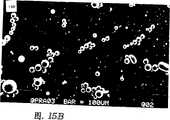

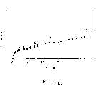


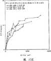


Classifications
- A—HUMAN NECESSITIES
- A61—MEDICAL OR VETERINARY SCIENCE; HYGIENE
- A61K—PREPARATIONS FOR MEDICAL, DENTAL OR TOILETRY PURPOSES
- A61K9/00—Medicinal preparations characterised by special physical form
- A61K9/14—Particulate form, e.g. powders, Processes for size reducing of pure drugs or the resulting products, Pure drug nanoparticles
- A—HUMAN NECESSITIES
- A61—MEDICAL OR VETERINARY SCIENCE; HYGIENE
- A61K—PREPARATIONS FOR MEDICAL, DENTAL OR TOILETRY PURPOSES
- A61K31/00—Medicinal preparations containing organic active ingredients
- A61K31/185—Acids; Anhydrides, halides or salts thereof, e.g. sulfur acids, imidic, hydrazonic or hydroximic acids
- A61K31/19—Carboxylic acids, e.g. valproic acid
- A61K31/20—Carboxylic acids, e.g. valproic acid having a carboxyl group bound to a chain of seven or more carbon atoms, e.g. stearic, palmitic, arachidic acids
- A—HUMAN NECESSITIES
- A61—MEDICAL OR VETERINARY SCIENCE; HYGIENE
- A61K—PREPARATIONS FOR MEDICAL, DENTAL OR TOILETRY PURPOSES
- A61K33/00—Medicinal preparations containing inorganic active ingredients
- A61K33/24—Heavy metals; Compounds thereof
- A—HUMAN NECESSITIES
- A61—MEDICAL OR VETERINARY SCIENCE; HYGIENE
- A61K—PREPARATIONS FOR MEDICAL, DENTAL OR TOILETRY PURPOSES
- A61K31/00—Medicinal preparations containing organic active ingredients
- A61K31/12—Ketones
- A61K31/122—Ketones having the oxygen directly attached to a ring, e.g. quinones, vitamin K1, anthralin
- A—HUMAN NECESSITIES
- A61—MEDICAL OR VETERINARY SCIENCE; HYGIENE
- A61K—PREPARATIONS FOR MEDICAL, DENTAL OR TOILETRY PURPOSES
- A61K31/00—Medicinal preparations containing organic active ingredients
- A61K31/13—Amines
- A61K31/135—Amines having aromatic rings, e.g. ketamine, nortriptyline
- A61K31/136—Amines having aromatic rings, e.g. ketamine, nortriptyline having the amino group directly attached to the aromatic ring, e.g. benzeneamine
- A—HUMAN NECESSITIES
- A61—MEDICAL OR VETERINARY SCIENCE; HYGIENE
- A61K—PREPARATIONS FOR MEDICAL, DENTAL OR TOILETRY PURPOSES
- A61K31/00—Medicinal preparations containing organic active ingredients
- A61K31/16—Amides, e.g. hydroxamic acids
- A61K31/165—Amides, e.g. hydroxamic acids having aromatic rings, e.g. colchicine, atenolol, progabide
- A—HUMAN NECESSITIES
- A61—MEDICAL OR VETERINARY SCIENCE; HYGIENE
- A61K—PREPARATIONS FOR MEDICAL, DENTAL OR TOILETRY PURPOSES
- A61K31/00—Medicinal preparations containing organic active ingredients
- A61K31/185—Acids; Anhydrides, halides or salts thereof, e.g. sulfur acids, imidic, hydrazonic or hydroximic acids
- A61K31/19—Carboxylic acids, e.g. valproic acid
- A61K31/20—Carboxylic acids, e.g. valproic acid having a carboxyl group bound to a chain of seven or more carbon atoms, e.g. stearic, palmitic, arachidic acids
- A61K31/203—Retinoic acids ; Salts thereof
- A—HUMAN NECESSITIES
- A61—MEDICAL OR VETERINARY SCIENCE; HYGIENE
- A61K—PREPARATIONS FOR MEDICAL, DENTAL OR TOILETRY PURPOSES
- A61K31/00—Medicinal preparations containing organic active ingredients
- A61K31/33—Heterocyclic compounds
- A61K31/335—Heterocyclic compounds having oxygen as the only ring hetero atom, e.g. fungichromin
- A61K31/337—Heterocyclic compounds having oxygen as the only ring hetero atom, e.g. fungichromin having four-membered rings, e.g. taxol
- A—HUMAN NECESSITIES
- A61—MEDICAL OR VETERINARY SCIENCE; HYGIENE
- A61K—PREPARATIONS FOR MEDICAL, DENTAL OR TOILETRY PURPOSES
- A61K31/00—Medicinal preparations containing organic active ingredients
- A61K31/33—Heterocyclic compounds
- A61K31/395—Heterocyclic compounds having nitrogen as a ring hetero atom, e.g. guanethidine or rifamycins
- A61K31/41—Heterocyclic compounds having nitrogen as a ring hetero atom, e.g. guanethidine or rifamycins having five-membered rings with two or more ring hetero atoms, at least one of which being nitrogen, e.g. tetrazole
- A61K31/425—Thiazoles
- A61K31/426—1,3-Thiazoles
- A—HUMAN NECESSITIES
- A61—MEDICAL OR VETERINARY SCIENCE; HYGIENE
- A61K—PREPARATIONS FOR MEDICAL, DENTAL OR TOILETRY PURPOSES
- A61K31/00—Medicinal preparations containing organic active ingredients
- A61K31/33—Heterocyclic compounds
- A61K31/395—Heterocyclic compounds having nitrogen as a ring hetero atom, e.g. guanethidine or rifamycins
- A61K31/41—Heterocyclic compounds having nitrogen as a ring hetero atom, e.g. guanethidine or rifamycins having five-membered rings with two or more ring hetero atoms, at least one of which being nitrogen, e.g. tetrazole
- A61K31/425—Thiazoles
- A61K31/427—Thiazoles not condensed and containing further heterocyclic rings
- A—HUMAN NECESSITIES
- A61—MEDICAL OR VETERINARY SCIENCE; HYGIENE
- A61K—PREPARATIONS FOR MEDICAL, DENTAL OR TOILETRY PURPOSES
- A61K31/00—Medicinal preparations containing organic active ingredients
- A61K31/33—Heterocyclic compounds
- A61K31/395—Heterocyclic compounds having nitrogen as a ring hetero atom, e.g. guanethidine or rifamycins
- A61K31/435—Heterocyclic compounds having nitrogen as a ring hetero atom, e.g. guanethidine or rifamycins having six-membered rings with one nitrogen as the only ring hetero atom
- A61K31/47—Quinolines; Isoquinolines
- A61K31/4738—Quinolines; Isoquinolines ortho- or peri-condensed with heterocyclic ring systems
- A61K31/4745—Quinolines; Isoquinolines ortho- or peri-condensed with heterocyclic ring systems condensed with ring systems having nitrogen as a ring hetero atom, e.g. phenantrolines
- A—HUMAN NECESSITIES
- A61—MEDICAL OR VETERINARY SCIENCE; HYGIENE
- A61K—PREPARATIONS FOR MEDICAL, DENTAL OR TOILETRY PURPOSES
- A61K31/00—Medicinal preparations containing organic active ingredients
- A61K31/33—Heterocyclic compounds
- A61K31/395—Heterocyclic compounds having nitrogen as a ring hetero atom, e.g. guanethidine or rifamycins
- A61K31/495—Heterocyclic compounds having nitrogen as a ring hetero atom, e.g. guanethidine or rifamycins having six-membered rings with two or more nitrogen atoms as the only ring heteroatoms, e.g. piperazine or tetrazines
- A61K31/505—Pyrimidines; Hydrogenated pyrimidines, e.g. trimethoprim
- A61K31/519—Pyrimidines; Hydrogenated pyrimidines, e.g. trimethoprim ortho- or peri-condensed with heterocyclic rings
- A61K31/525—Isoalloxazines, e.g. riboflavins, vitamin B2
- A—HUMAN NECESSITIES
- A61—MEDICAL OR VETERINARY SCIENCE; HYGIENE
- A61K—PREPARATIONS FOR MEDICAL, DENTAL OR TOILETRY PURPOSES
- A61K31/00—Medicinal preparations containing organic active ingredients
- A61K31/33—Heterocyclic compounds
- A61K31/555—Heterocyclic compounds containing heavy metals, e.g. hemin, hematin, melarsoprol
- A—HUMAN NECESSITIES
- A61—MEDICAL OR VETERINARY SCIENCE; HYGIENE
- A61K—PREPARATIONS FOR MEDICAL, DENTAL OR TOILETRY PURPOSES
- A61K31/00—Medicinal preparations containing organic active ingredients
- A61K31/56—Compounds containing cyclopenta[a]hydrophenanthrene ring systems; Derivatives thereof, e.g. steroids
- A61K31/565—Compounds containing cyclopenta[a]hydrophenanthrene ring systems; Derivatives thereof, e.g. steroids not substituted in position 17 beta by a carbon atom, e.g. estrane, estradiol
- A—HUMAN NECESSITIES
- A61—MEDICAL OR VETERINARY SCIENCE; HYGIENE
- A61K—PREPARATIONS FOR MEDICAL, DENTAL OR TOILETRY PURPOSES
- A61K31/00—Medicinal preparations containing organic active ingredients
- A61K31/56—Compounds containing cyclopenta[a]hydrophenanthrene ring systems; Derivatives thereof, e.g. steroids
- A61K31/57—Compounds containing cyclopenta[a]hydrophenanthrene ring systems; Derivatives thereof, e.g. steroids substituted in position 17 beta by a chain of two carbon atoms, e.g. pregnane or progesterone
- A—HUMAN NECESSITIES
- A61—MEDICAL OR VETERINARY SCIENCE; HYGIENE
- A61K—PREPARATIONS FOR MEDICAL, DENTAL OR TOILETRY PURPOSES
- A61K31/00—Medicinal preparations containing organic active ingredients
- A61K31/70—Carbohydrates; Sugars; Derivatives thereof
- A61K31/702—Oligosaccharides, i.e. having three to five saccharide radicals attached to each other by glycosidic linkages
- A—HUMAN NECESSITIES
- A61—MEDICAL OR VETERINARY SCIENCE; HYGIENE
- A61K—PREPARATIONS FOR MEDICAL, DENTAL OR TOILETRY PURPOSES
- A61K33/00—Medicinal preparations containing inorganic active ingredients
- A—HUMAN NECESSITIES
- A61—MEDICAL OR VETERINARY SCIENCE; HYGIENE
- A61K—PREPARATIONS FOR MEDICAL, DENTAL OR TOILETRY PURPOSES
- A61K33/00—Medicinal preparations containing inorganic active ingredients
- A61K33/06—Aluminium, calcium or magnesium; Compounds thereof, e.g. clay
- A—HUMAN NECESSITIES
- A61—MEDICAL OR VETERINARY SCIENCE; HYGIENE
- A61K—PREPARATIONS FOR MEDICAL, DENTAL OR TOILETRY PURPOSES
- A61K33/00—Medicinal preparations containing inorganic active ingredients
- A61K33/24—Heavy metals; Compounds thereof
- A61K33/30—Zinc; Compounds thereof
- A—HUMAN NECESSITIES
- A61—MEDICAL OR VETERINARY SCIENCE; HYGIENE
- A61K—PREPARATIONS FOR MEDICAL, DENTAL OR TOILETRY PURPOSES
- A61K38/00—Medicinal preparations containing peptides
- A61K38/16—Peptides having more than 20 amino acids; Gastrins; Somatostatins; Melanotropins; Derivatives thereof
- A61K38/55—Protease inhibitors
- A61K38/57—Protease inhibitors from animals; from humans
- A—HUMAN NECESSITIES
- A61—MEDICAL OR VETERINARY SCIENCE; HYGIENE
- A61K—PREPARATIONS FOR MEDICAL, DENTAL OR TOILETRY PURPOSES
- A61K45/00—Medicinal preparations containing active ingredients not provided for in groups A61K31/00 - A61K41/00
- A61K45/06—Mixtures of active ingredients without chemical characterisation, e.g. antiphlogistics and cardiaca
- A—HUMAN NECESSITIES
- A61—MEDICAL OR VETERINARY SCIENCE; HYGIENE
- A61K—PREPARATIONS FOR MEDICAL, DENTAL OR TOILETRY PURPOSES
- A61K9/00—Medicinal preparations characterised by special physical form
- A61K9/0012—Galenical forms characterised by the site of application
- A61K9/0019—Injectable compositions; Intramuscular, intravenous, arterial, subcutaneous administration; Compositions to be administered through the skin in an invasive manner
- A—HUMAN NECESSITIES
- A61—MEDICAL OR VETERINARY SCIENCE; HYGIENE
- A61K—PREPARATIONS FOR MEDICAL, DENTAL OR TOILETRY PURPOSES
- A61K9/00—Medicinal preparations characterised by special physical form
- A61K9/0012—Galenical forms characterised by the site of application
- A61K9/0048—Eye, e.g. artificial tears
- A61K9/0051—Ocular inserts, ocular implants
- A—HUMAN NECESSITIES
- A61—MEDICAL OR VETERINARY SCIENCE; HYGIENE
- A61K—PREPARATIONS FOR MEDICAL, DENTAL OR TOILETRY PURPOSES
- A61K9/00—Medicinal preparations characterised by special physical form
- A61K9/14—Particulate form, e.g. powders, Processes for size reducing of pure drugs or the resulting products, Pure drug nanoparticles
- A61K9/16—Agglomerates; Granulates; Microbeadlets ; Microspheres; Pellets; Solid products obtained by spray drying, spray freeze drying, spray congealing,(multiple) emulsion solvent evaporation or extraction
- A61K9/1605—Excipients; Inactive ingredients
- A61K9/1629—Organic macromolecular compounds
- A61K9/1635—Organic macromolecular compounds obtained by reactions only involving carbon-to-carbon unsaturated bonds, e.g. polyvinyl pyrrolidone, poly(meth)acrylates
- A—HUMAN NECESSITIES
- A61—MEDICAL OR VETERINARY SCIENCE; HYGIENE
- A61K—PREPARATIONS FOR MEDICAL, DENTAL OR TOILETRY PURPOSES
- A61K9/00—Medicinal preparations characterised by special physical form
- A61K9/14—Particulate form, e.g. powders, Processes for size reducing of pure drugs or the resulting products, Pure drug nanoparticles
- A61K9/16—Agglomerates; Granulates; Microbeadlets ; Microspheres; Pellets; Solid products obtained by spray drying, spray freeze drying, spray congealing,(multiple) emulsion solvent evaporation or extraction
- A61K9/1605—Excipients; Inactive ingredients
- A61K9/1629—Organic macromolecular compounds
- A61K9/1641—Organic macromolecular compounds obtained otherwise than by reactions only involving carbon-to-carbon unsaturated bonds, e.g. polyethylene glycol, poloxamers
- A61K9/1647—Polyesters, e.g. poly(lactide-co-glycolide)
- A—HUMAN NECESSITIES
- A61—MEDICAL OR VETERINARY SCIENCE; HYGIENE
- A61K—PREPARATIONS FOR MEDICAL, DENTAL OR TOILETRY PURPOSES
- A61K9/00—Medicinal preparations characterised by special physical form
- A61K9/14—Particulate form, e.g. powders, Processes for size reducing of pure drugs or the resulting products, Pure drug nanoparticles
- A61K9/16—Agglomerates; Granulates; Microbeadlets ; Microspheres; Pellets; Solid products obtained by spray drying, spray freeze drying, spray congealing,(multiple) emulsion solvent evaporation or extraction
- A61K9/1605—Excipients; Inactive ingredients
- A61K9/1629—Organic macromolecular compounds
- A61K9/1658—Proteins, e.g. albumin, gelatin
- A—HUMAN NECESSITIES
- A61—MEDICAL OR VETERINARY SCIENCE; HYGIENE
- A61K—PREPARATIONS FOR MEDICAL, DENTAL OR TOILETRY PURPOSES
- A61K9/00—Medicinal preparations characterised by special physical form
- A61K9/48—Preparations in capsules, e.g. of gelatin, of chocolate
- A61K9/50—Microcapsules having a gas, liquid or semi-solid filling; Solid microparticles or pellets surrounded by a distinct coating layer, e.g. coated microspheres, coated drug crystals
- A61K9/5005—Wall or coating material
- A61K9/5021—Organic macromolecular compounds
- A61K9/5031—Organic macromolecular compounds obtained otherwise than by reactions only involving carbon-to-carbon unsaturated bonds, e.g. polyethylene glycol, poly(lactide-co-glycolide)
- A—HUMAN NECESSITIES
- A61—MEDICAL OR VETERINARY SCIENCE; HYGIENE
- A61K—PREPARATIONS FOR MEDICAL, DENTAL OR TOILETRY PURPOSES
- A61K9/00—Medicinal preparations characterised by special physical form
- A61K9/48—Preparations in capsules, e.g. of gelatin, of chocolate
- A61K9/50—Microcapsules having a gas, liquid or semi-solid filling; Solid microparticles or pellets surrounded by a distinct coating layer, e.g. coated microspheres, coated drug crystals
- A61K9/51—Nanocapsules; Nanoparticles
- A61K9/5107—Excipients; Inactive ingredients
- A61K9/513—Organic macromolecular compounds; Dendrimers
- A61K9/5146—Organic macromolecular compounds; Dendrimers obtained otherwise than by reactions only involving carbon-to-carbon unsaturated bonds, e.g. polyethylene glycol, polyamines, polyanhydrides
- A61K9/5153—Polyesters, e.g. poly(lactide-co-glycolide)
- A—HUMAN NECESSITIES
- A61—MEDICAL OR VETERINARY SCIENCE; HYGIENE
- A61K—PREPARATIONS FOR MEDICAL, DENTAL OR TOILETRY PURPOSES
- A61K9/00—Medicinal preparations characterised by special physical form
- A61K9/70—Web, sheet or filament bases ; Films; Fibres of the matrix type containing drug
- A61K9/7007—Drug-containing films, membranes or sheets
- A—HUMAN NECESSITIES
- A61—MEDICAL OR VETERINARY SCIENCE; HYGIENE
- A61L—METHODS OR APPARATUS FOR STERILISING MATERIALS OR OBJECTS IN GENERAL; DISINFECTION, STERILISATION OR DEODORISATION OF AIR; CHEMICAL ASPECTS OF BANDAGES, DRESSINGS, ABSORBENT PADS OR SURGICAL ARTICLES; MATERIALS FOR BANDAGES, DRESSINGS, ABSORBENT PADS OR SURGICAL ARTICLES
- A61L31/00—Materials for other surgical articles, e.g. stents, stent-grafts, shunts, surgical drapes, guide wires, materials for adhesion prevention, occluding devices, surgical gloves, tissue fixation devices
- A61L31/08—Materials for coatings
- A61L31/10—Macromolecular materials
- A—HUMAN NECESSITIES
- A61—MEDICAL OR VETERINARY SCIENCE; HYGIENE
- A61L—METHODS OR APPARATUS FOR STERILISING MATERIALS OR OBJECTS IN GENERAL; DISINFECTION, STERILISATION OR DEODORISATION OF AIR; CHEMICAL ASPECTS OF BANDAGES, DRESSINGS, ABSORBENT PADS OR SURGICAL ARTICLES; MATERIALS FOR BANDAGES, DRESSINGS, ABSORBENT PADS OR SURGICAL ARTICLES
- A61L31/00—Materials for other surgical articles, e.g. stents, stent-grafts, shunts, surgical drapes, guide wires, materials for adhesion prevention, occluding devices, surgical gloves, tissue fixation devices
- A61L31/14—Materials characterised by their function or physical properties, e.g. injectable or lubricating compositions, shape-memory materials, surface modified materials
- A61L31/145—Hydrogels or hydrocolloids
- A—HUMAN NECESSITIES
- A61—MEDICAL OR VETERINARY SCIENCE; HYGIENE
- A61L—METHODS OR APPARATUS FOR STERILISING MATERIALS OR OBJECTS IN GENERAL; DISINFECTION, STERILISATION OR DEODORISATION OF AIR; CHEMICAL ASPECTS OF BANDAGES, DRESSINGS, ABSORBENT PADS OR SURGICAL ARTICLES; MATERIALS FOR BANDAGES, DRESSINGS, ABSORBENT PADS OR SURGICAL ARTICLES
- A61L31/00—Materials for other surgical articles, e.g. stents, stent-grafts, shunts, surgical drapes, guide wires, materials for adhesion prevention, occluding devices, surgical gloves, tissue fixation devices
- A61L31/14—Materials characterised by their function or physical properties, e.g. injectable or lubricating compositions, shape-memory materials, surface modified materials
- A61L31/16—Biologically active materials, e.g. therapeutic substances
- A—HUMAN NECESSITIES
- A61—MEDICAL OR VETERINARY SCIENCE; HYGIENE
- A61P—SPECIFIC THERAPEUTIC ACTIVITY OF CHEMICAL COMPOUNDS OR MEDICINAL PREPARATIONS
- A61P1/00—Drugs for disorders of the alimentary tract or the digestive system
- A61P1/04—Drugs for disorders of the alimentary tract or the digestive system for ulcers, gastritis or reflux esophagitis, e.g. antacids, inhibitors of acid secretion, mucosal protectants
- A—HUMAN NECESSITIES
- A61—MEDICAL OR VETERINARY SCIENCE; HYGIENE
- A61P—SPECIFIC THERAPEUTIC ACTIVITY OF CHEMICAL COMPOUNDS OR MEDICINAL PREPARATIONS
- A61P1/00—Drugs for disorders of the alimentary tract or the digestive system
- A61P1/16—Drugs for disorders of the alimentary tract or the digestive system for liver or gallbladder disorders, e.g. hepatoprotective agents, cholagogues, litholytics
- A—HUMAN NECESSITIES
- A61—MEDICAL OR VETERINARY SCIENCE; HYGIENE
- A61P—SPECIFIC THERAPEUTIC ACTIVITY OF CHEMICAL COMPOUNDS OR MEDICINAL PREPARATIONS
- A61P11/00—Drugs for disorders of the respiratory system
- A—HUMAN NECESSITIES
- A61—MEDICAL OR VETERINARY SCIENCE; HYGIENE
- A61P—SPECIFIC THERAPEUTIC ACTIVITY OF CHEMICAL COMPOUNDS OR MEDICINAL PREPARATIONS
- A61P13/00—Drugs for disorders of the urinary system
- A61P13/02—Drugs for disorders of the urinary system of urine or of the urinary tract, e.g. urine acidifiers
- A—HUMAN NECESSITIES
- A61—MEDICAL OR VETERINARY SCIENCE; HYGIENE
- A61P—SPECIFIC THERAPEUTIC ACTIVITY OF CHEMICAL COMPOUNDS OR MEDICINAL PREPARATIONS
- A61P19/00—Drugs for skeletal disorders
- A61P19/02—Drugs for skeletal disorders for joint disorders, e.g. arthritis, arthrosis
- A—HUMAN NECESSITIES
- A61—MEDICAL OR VETERINARY SCIENCE; HYGIENE
- A61P—SPECIFIC THERAPEUTIC ACTIVITY OF CHEMICAL COMPOUNDS OR MEDICINAL PREPARATIONS
- A61P27/00—Drugs for disorders of the senses
- A61P27/02—Ophthalmic agents
- A—HUMAN NECESSITIES
- A61—MEDICAL OR VETERINARY SCIENCE; HYGIENE
- A61P—SPECIFIC THERAPEUTIC ACTIVITY OF CHEMICAL COMPOUNDS OR MEDICINAL PREPARATIONS
- A61P35/00—Antineoplastic agents
- A—HUMAN NECESSITIES
- A61—MEDICAL OR VETERINARY SCIENCE; HYGIENE
- A61P—SPECIFIC THERAPEUTIC ACTIVITY OF CHEMICAL COMPOUNDS OR MEDICINAL PREPARATIONS
- A61P43/00—Drugs for specific purposes, not provided for in groups A61P1/00-A61P41/00
- A—HUMAN NECESSITIES
- A61—MEDICAL OR VETERINARY SCIENCE; HYGIENE
- A61P—SPECIFIC THERAPEUTIC ACTIVITY OF CHEMICAL COMPOUNDS OR MEDICINAL PREPARATIONS
- A61P7/00—Drugs for disorders of the blood or the extracellular fluid
- A61P7/02—Antithrombotic agents; Anticoagulants; Platelet aggregation inhibitors
- A—HUMAN NECESSITIES
- A61—MEDICAL OR VETERINARY SCIENCE; HYGIENE
- A61P—SPECIFIC THERAPEUTIC ACTIVITY OF CHEMICAL COMPOUNDS OR MEDICINAL PREPARATIONS
- A61P7/00—Drugs for disorders of the blood or the extracellular fluid
- A61P7/04—Antihaemorrhagics; Procoagulants; Haemostatic agents; Antifibrinolytic agents
- A—HUMAN NECESSITIES
- A61—MEDICAL OR VETERINARY SCIENCE; HYGIENE
- A61P—SPECIFIC THERAPEUTIC ACTIVITY OF CHEMICAL COMPOUNDS OR MEDICINAL PREPARATIONS
- A61P9/00—Drugs for disorders of the cardiovascular system
- A—HUMAN NECESSITIES
- A61—MEDICAL OR VETERINARY SCIENCE; HYGIENE
- A61P—SPECIFIC THERAPEUTIC ACTIVITY OF CHEMICAL COMPOUNDS OR MEDICINAL PREPARATIONS
- A61P9/00—Drugs for disorders of the cardiovascular system
- A61P9/10—Drugs for disorders of the cardiovascular system for treating ischaemic or atherosclerotic diseases, e.g. antianginal drugs, coronary vasodilators, drugs for myocardial infarction, retinopathy, cerebrovascula insufficiency, renal arteriosclerosis
- A—HUMAN NECESSITIES
- A61—MEDICAL OR VETERINARY SCIENCE; HYGIENE
- A61P—SPECIFIC THERAPEUTIC ACTIVITY OF CHEMICAL COMPOUNDS OR MEDICINAL PREPARATIONS
- A61P9/00—Drugs for disorders of the cardiovascular system
- A61P9/14—Vasoprotectives; Antihaemorrhoidals; Drugs for varicose therapy; Capillary stabilisers
- C—CHEMISTRY; METALLURGY
- C07—ORGANIC CHEMISTRY
- C07C—ACYCLIC OR CARBOCYCLIC COMPOUNDS
- C07C233/00—Carboxylic acid amides
- C07C233/64—Carboxylic acid amides having carbon atoms of carboxamide groups bound to carbon atoms of six-membered aromatic rings
- C—CHEMISTRY; METALLURGY
- C07—ORGANIC CHEMISTRY
- C07K—PEPTIDES
- C07K14/00—Peptides having more than 20 amino acids; Gastrins; Somatostatins; Melanotropins; Derivatives thereof
- C07K14/81—Protease inhibitors
- C07K14/8107—Endopeptidase (E.C. 3.4.21-99) inhibitors
- C07K14/811—Serine protease (E.C. 3.4.21) inhibitors
- C—CHEMISTRY; METALLURGY
- C08—ORGANIC MACROMOLECULAR COMPOUNDS; THEIR PREPARATION OR CHEMICAL WORKING-UP; COMPOSITIONS BASED THEREON
- C08L—COMPOSITIONS OF MACROMOLECULAR COMPOUNDS
- C08L23/00—Compositions of homopolymers or copolymers of unsaturated aliphatic hydrocarbons having only one carbon-to-carbon double bond; Compositions of derivatives of such polymers
- C08L23/02—Compositions of homopolymers or copolymers of unsaturated aliphatic hydrocarbons having only one carbon-to-carbon double bond; Compositions of derivatives of such polymers not modified by chemical after-treatment
- C08L23/04—Homopolymers or copolymers of ethene
- C08L23/08—Copolymers of ethene
- C08L23/0846—Copolymers of ethene with unsaturated hydrocarbons containing atoms other than carbon or hydrogen
- C08L23/0853—Ethene vinyl acetate copolymers
- C—CHEMISTRY; METALLURGY
- C08—ORGANIC MACROMOLECULAR COMPOUNDS; THEIR PREPARATION OR CHEMICAL WORKING-UP; COMPOSITIONS BASED THEREON
- C08L—COMPOSITIONS OF MACROMOLECULAR COMPOUNDS
- C08L67/00—Compositions of polyesters obtained by reactions forming a carboxylic ester link in the main chain; Compositions of derivatives of such polymers
- C08L67/04—Polyesters derived from hydroxycarboxylic acids, e.g. lactones
- A—HUMAN NECESSITIES
- A61—MEDICAL OR VETERINARY SCIENCE; HYGIENE
- A61L—METHODS OR APPARATUS FOR STERILISING MATERIALS OR OBJECTS IN GENERAL; DISINFECTION, STERILISATION OR DEODORISATION OF AIR; CHEMICAL ASPECTS OF BANDAGES, DRESSINGS, ABSORBENT PADS OR SURGICAL ARTICLES; MATERIALS FOR BANDAGES, DRESSINGS, ABSORBENT PADS OR SURGICAL ARTICLES
- A61L2300/00—Biologically active materials used in bandages, wound dressings, absorbent pads or medical devices
- A61L2300/40—Biologically active materials used in bandages, wound dressings, absorbent pads or medical devices characterised by a specific therapeutic activity or mode of action
- A61L2300/416—Anti-neoplastic or anti-proliferative or anti-restenosis or anti-angiogenic agents, e.g. paclitaxel, sirolimus
- A—HUMAN NECESSITIES
- A61—MEDICAL OR VETERINARY SCIENCE; HYGIENE
- A61L—METHODS OR APPARATUS FOR STERILISING MATERIALS OR OBJECTS IN GENERAL; DISINFECTION, STERILISATION OR DEODORISATION OF AIR; CHEMICAL ASPECTS OF BANDAGES, DRESSINGS, ABSORBENT PADS OR SURGICAL ARTICLES; MATERIALS FOR BANDAGES, DRESSINGS, ABSORBENT PADS OR SURGICAL ARTICLES
- A61L2300/00—Biologically active materials used in bandages, wound dressings, absorbent pads or medical devices
- A61L2300/60—Biologically active materials used in bandages, wound dressings, absorbent pads or medical devices characterised by a special physical form
- A61L2300/606—Coatings
- A—HUMAN NECESSITIES
- A61—MEDICAL OR VETERINARY SCIENCE; HYGIENE
- A61L—METHODS OR APPARATUS FOR STERILISING MATERIALS OR OBJECTS IN GENERAL; DISINFECTION, STERILISATION OR DEODORISATION OF AIR; CHEMICAL ASPECTS OF BANDAGES, DRESSINGS, ABSORBENT PADS OR SURGICAL ARTICLES; MATERIALS FOR BANDAGES, DRESSINGS, ABSORBENT PADS OR SURGICAL ARTICLES
- A61L2300/00—Biologically active materials used in bandages, wound dressings, absorbent pads or medical devices
- A61L2300/60—Biologically active materials used in bandages, wound dressings, absorbent pads or medical devices characterised by a special physical form
- A61L2300/62—Encapsulated active agents, e.g. emulsified droplets
- A61L2300/622—Microcapsules
- A—HUMAN NECESSITIES
- A61—MEDICAL OR VETERINARY SCIENCE; HYGIENE
- A61L—METHODS OR APPARATUS FOR STERILISING MATERIALS OR OBJECTS IN GENERAL; DISINFECTION, STERILISATION OR DEODORISATION OF AIR; CHEMICAL ASPECTS OF BANDAGES, DRESSINGS, ABSORBENT PADS OR SURGICAL ARTICLES; MATERIALS FOR BANDAGES, DRESSINGS, ABSORBENT PADS OR SURGICAL ARTICLES
- A61L2430/00—Materials or treatment for tissue regeneration
- A61L2430/36—Materials or treatment for tissue regeneration for embolization or occlusion, e.g. vaso-occlusive compositions or devices
- B—PERFORMING OPERATIONS; TRANSPORTING
- B82—NANOTECHNOLOGY
- B82Y—SPECIFIC USES OR APPLICATIONS OF NANOSTRUCTURES; MEASUREMENT OR ANALYSIS OF NANOSTRUCTURES; MANUFACTURE OR TREATMENT OF NANOSTRUCTURES
- B82Y5/00—Nanobiotechnology or nanomedicine, e.g. protein engineering or drug delivery
- C—CHEMISTRY; METALLURGY
- C08—ORGANIC MACROMOLECULAR COMPOUNDS; THEIR PREPARATION OR CHEMICAL WORKING-UP; COMPOSITIONS BASED THEREON
- C08L—COMPOSITIONS OF MACROMOLECULAR COMPOUNDS
- C08L2203/00—Applications
- C08L2203/02—Applications for biomedical use
- C—CHEMISTRY; METALLURGY
- C08—ORGANIC MACROMOLECULAR COMPOUNDS; THEIR PREPARATION OR CHEMICAL WORKING-UP; COMPOSITIONS BASED THEREON
- C08L—COMPOSITIONS OF MACROMOLECULAR COMPOUNDS
- C08L2312/00—Crosslinking
- C—CHEMISTRY; METALLURGY
- C08—ORGANIC MACROMOLECULAR COMPOUNDS; THEIR PREPARATION OR CHEMICAL WORKING-UP; COMPOSITIONS BASED THEREON
- C08L—COMPOSITIONS OF MACROMOLECULAR COMPOUNDS
- C08L29/00—Compositions of homopolymers or copolymers of compounds having one or more unsaturated aliphatic radicals, each having only one carbon-to-carbon double bond, and at least one being terminated by an alcohol, ether, aldehydo, ketonic, acetal or ketal radical; Compositions of hydrolysed polymers of esters of unsaturated alcohols with saturated carboxylic acids; Compositions of derivatives of such polymers
- C08L29/02—Homopolymers or copolymers of unsaturated alcohols
- C08L29/04—Polyvinyl alcohol; Partially hydrolysed homopolymers or copolymers of esters of unsaturated alcohols with saturated carboxylic acids
- Y—GENERAL TAGGING OF NEW TECHNOLOGICAL DEVELOPMENTS; GENERAL TAGGING OF CROSS-SECTIONAL TECHNOLOGIES SPANNING OVER SEVERAL SECTIONS OF THE IPC; TECHNICAL SUBJECTS COVERED BY FORMER USPC CROSS-REFERENCE ART COLLECTIONS [XRACs] AND DIGESTS
- Y10—TECHNICAL SUBJECTS COVERED BY FORMER USPC
- Y10S—TECHNICAL SUBJECTS COVERED BY FORMER USPC CROSS-REFERENCE ART COLLECTIONS [XRACs] AND DIGESTS
- Y10S977/00—Nanotechnology
- Y10S977/70—Nanostructure
- Y10S977/773—Nanoparticle, i.e. structure having three dimensions of 100 nm or less
- Y—GENERAL TAGGING OF NEW TECHNOLOGICAL DEVELOPMENTS; GENERAL TAGGING OF CROSS-SECTIONAL TECHNOLOGIES SPANNING OVER SEVERAL SECTIONS OF THE IPC; TECHNICAL SUBJECTS COVERED BY FORMER USPC CROSS-REFERENCE ART COLLECTIONS [XRACs] AND DIGESTS
- Y10—TECHNICAL SUBJECTS COVERED BY FORMER USPC
- Y10S—TECHNICAL SUBJECTS COVERED BY FORMER USPC CROSS-REFERENCE ART COLLECTIONS [XRACs] AND DIGESTS
- Y10S977/00—Nanotechnology
- Y10S977/902—Specified use of nanostructure
- Y10S977/904—Specified use of nanostructure for medical, immunological, body treatment, or diagnosis
- Y10S977/915—Therapeutic or pharmaceutical composition
Landscapes
- Health & Medical Sciences (AREA)
- Chemical & Material Sciences (AREA)
- Life Sciences & Earth Sciences (AREA)
- Medicinal Chemistry (AREA)
- General Health & Medical Sciences (AREA)
- Veterinary Medicine (AREA)
- Animal Behavior & Ethology (AREA)
- Public Health (AREA)
- Pharmacology & Pharmacy (AREA)
- Epidemiology (AREA)
- Engineering & Computer Science (AREA)
- Bioinformatics & Cheminformatics (AREA)
- Organic Chemistry (AREA)
- Chemical Kinetics & Catalysis (AREA)
- Nuclear Medicine, Radiotherapy & Molecular Imaging (AREA)
- General Chemical & Material Sciences (AREA)
- Heart & Thoracic Surgery (AREA)
- Inorganic Chemistry (AREA)
- Vascular Medicine (AREA)
- Molecular Biology (AREA)
- Surgery (AREA)
- Biomedical Technology (AREA)
- Ophthalmology & Optometry (AREA)
- Gastroenterology & Hepatology (AREA)
- Physics & Mathematics (AREA)
- Optics & Photonics (AREA)
- Proteomics, Peptides & Aminoacids (AREA)
- Polymers & Plastics (AREA)
- Nanotechnology (AREA)
- Cardiology (AREA)
- Immunology (AREA)
- Biophysics (AREA)
- Biochemistry (AREA)
- Urology & Nephrology (AREA)
- Hematology (AREA)
- Dispersion Chemistry (AREA)
- Zoology (AREA)
- Diabetes (AREA)
- Dermatology (AREA)
- Genetics & Genomics (AREA)
Abstract
Translated fromChineseDescription
Translated fromChinese本发明是申请日为1994年7月19日的中国专利申请200510082207.9的分案申请,原申请的发明名称为“抗血管生长组合物及使用方法”。The present invention is a divisional application of Chinese patent application 200510082207.9 filed on July 19, 1994. The title of the invention of the original application is "anti-angiogenesis composition and method of use".
本发明涉及治疗癌症和其它血管生长依赖疾病的组合物及方法,更具体地,涉及含有抗血管生长因子和聚合载体的组合物、用这些组合物包衣的斯坦特固定模,以及使用这些斯坦特固定模和组合物的方法。The present invention relates to compositions and methods for treating cancer and other angiogenesis-dependent diseases, and more particularly, to compositions containing anti-angiogenic growth factors and polymeric carriers, stents coated with these compositions, and the use of these stents. Methods for specific stents and compositions.
癌症是在美国引发死亡的第二种主要原因,并且占总死亡数的五分之一强。简而言之,癌症以细胞群的分裂不可控制为特征,其一般导致形成一种或几种肿瘤。尽管癌症一般比过去更易于诊断,甚至更早发现,但其仍然不能治愈。Cancer is the second leading cause of death in the United States and accounts for more than one-fifth of all deaths. Briefly, cancer is characterized by the uncontrollable division of cell populations, which generally leads to the formation of one or several tumors. Although cancer is generally easier to diagnose and even found earlier than in the past, it still cannot be cured.
目前有许多方法用于治疗癌症,包括例如各种外科方法。如果单独使用外科,很多患者(尤其是那些患有某些癌症的患者,例如乳腺癌、脑癌、结肠癌和肝癌)会复发。除了外科,许多癌症可以用联合治疗法治疗,包括细胞毒性化疗药物(如长春新碱、长春碱、顺氯氨铂、甲氨喋呤、5-FU等)和/或放射疗法。这种方法的困难在于,放疗和化疗药物对正常组织也有毒性,并且经常产生致命的副作用。此外,这些方法通常具有特别高的失败/缓解率。There are many methods currently used to treat cancer including, for example, various surgical methods. Many patients, especially those with certain cancers such as breast, brain, colon, and liver, will relapse if surgery is used alone. In addition to surgery, many cancers can be treated with combination therapies including cytotoxic chemotherapeutic agents (eg, vincristine, vinblastine, cisplatin, methotrexate, 5-FU, etc.) and/or radiation therapy. The difficulty with this approach is that radiation and chemotherapy drugs are also toxic to normal tissue and often produce fatal side effects. Furthermore, these methods often have exceptionally high failure/remission rates.
除了外科、化学和放射疗法,有人试图用个人的自身免疫系统来消除癌细胞。例如,一些人建议用细菌或病毒成分作为佐剂来刺激免疫系统,从而破坏肿瘤细胞。(见″PirnciplesofCancerBiotherapy,″Oldham(ed.),RavenPress,NewYork,1987。)这些药物通常在动物肿瘤模型中用作佐剂和非特异性刺激剂,但是还没有证明其在人体中普遍有效。In addition to surgery, chemotherapy, and radiation, there are attempts to use an individual's own immune system to eliminate cancer cells. For example, some have suggested using bacterial or viral components as adjuvants to stimulate the immune system to destroy tumor cells. (See "Pirnciples of Cancer Biotherapy," Oldham (ed.), Raven Press, New York, 1987.) These drugs are commonly used as adjuvants and non-specific stimulators in animal tumor models, but have not proven generally effective in humans.
淋巴因子也用来治疗癌症。简而言之,淋巴因子通过各种细胞分泌,并且通常在产生免疫应答中对特定细胞有作用。淋巴因子的例子有白细胞介素(IL)-1,-2,-3和-4,以及白细胞集落刺激因子如G-CSF、GM-CSF和M-CSF。近来,有人用IL-2刺激外周血细胞从而扩大并产生大量对肿瘤细胞有胞毒性的细胞(Rosenbergetal.,N.Engl.J.Med.313:1485-1492,1985)。Lymphokines are also used to treat cancer. Briefly, lymphokines are secreted by various cells and often act on specific cells in generating an immune response. Examples of lymphokines are interleukins (IL)-1, -2, -3 and -4, and leukocyte colony stimulating factors such as G-CSF, GM-CSF and M-CSF. Recently, peripheral blood cells were stimulated with IL-2 to expand and produce a large number of cells cytotoxic to tumor cells (Rosenberg et al., N. Engl. J. Med. 313: 1485-1492, 1985).
另外有人建议用抗体治疗癌症。简而言之,抗体可以识别特定的细胞表面抗原(其可以是独特的,或者与正常细胞相比在癌细胞中占多数)。这些抗体,或者称“魔术子弹”,可以单独或与一种毒素联合使用,从而特异识别并杀死肿瘤细胞(Dillman,″AntibodyTherapy,″PrinciplesofCancerBiotherapy,Oldham(ed.),RavenPress,Ltd.,NewYork,1987)。但是,困难在于大多数单克隆抗体为鼠源,这样对鼠抗体的高度敏感性限制了其的有效性,尤其是在重复治疗后。常见的副作用包括发热、出汗和寒战、皮疹、关节炎和神经麻痹。Others have suggested using antibodies to treat cancer. In short, antibodies recognize specific cell surface antigens (which may be unique or prevalent in cancer cells compared to normal cells). These antibodies, or "magic bullets," can be used alone or in combination with a toxin to specifically recognize and kill tumor cells (Dillman, "Antibody Therapy," Principles of Cancer Biotherapy, Oldham (ed.), RavenPress, Ltd., New York, 1987). However, the difficulty is that most monoclonal antibodies are of murine origin, and thus the high sensitivity to murine antibodies limits their effectiveness, especially after repeated treatments. Common side effects include fever, sweating and chills, rash, arthritis and nerve paralysis.
现有方法的另外一个难点是,局部复发及局部疾病的控制仍然是治疗恶性肿瘤的一个主要挑战。特别地,每年有总数为630,000的患者(美国)在疾病出现时疾病已定位(没有远距离迁移传播的迹象);其代表了64%所有被诊断为恶性肿瘤的患者(不包括非黑瘤皮肤癌或原位癌)。对这些患者的大多数而言,外科切除是治愈的最主要的机会,并且的确有428,000患者在最初治疗后会治愈。不幸的是,202,000位患者(即32%的患者疾病已确定)在最初治疗后会复发。在这些复发者中,每年有133,000人是由于疾病的局部复发而引起的(占所有定位疾病患者的21%)。每年因疾病远距离迁移引发的数目是68,000人(占所有定位疾病的11%)。另外102,139位患者每年将由于不能控制疾患的局部发展而死亡。Another difficulty with existing approaches is that local recurrence and local disease control remain a major challenge in the treatment of malignancies. In particular, a total of 630,000 patients per year (US) have localized disease (no evidence of distant migratory spread) at disease presentation; this represents 64% of all patients diagnosed with malignancy (excluding non-melanoma cutaneous carcinoma or carcinoma in situ). For the majority of these patients, surgical resection is the main chance for cure, and indeed 428,000 patients will be cured after initial treatment. Unfortunately, 202,000 patients (ie, 32% of patients with established disease) will relapse after initial treatment. Of these relapses, 133,000 per year were due to local recurrence of disease (21% of all patients with localized disease). The annual number due to long-distance migration of disease is 68,000 (11% of all localized diseases). Another 102,139 patients will die each year due to uncontrolled local progression of the disease.
这一问题在乳腺癌中更明显,其在美国每年影响186,000个妇女并且其的死亡率经50年仍保持不变。通过乳房彻底切除术、修饰的基本乳房切除术或者肿块切除术进行外科切除疾患是这种疾病的主要治疗方法。不幸的是,单独施用肿块切除术的39%的患者会局部复发疾病,令人吃惊的是,发现25%的患者在切除的边缘清楚地留有原始肿瘤(组织学上)。局部复发的90%发生在先前切除位点2厘米之内。This problem is even more pronounced in breast cancer, which affects 186,000 women in the United States each year and whose mortality rate has remained unchanged for 50 years. Surgical resection of the disease by radical mastectomy, modified primary mastectomy, or lumpectomy is the mainstay of treatment for this disease. Unfortunately, 39% of patients treated with lumpectomy alone had locally recurrent disease, and it was surprising to find that 25% of patients left the original tumor clearly (histologically) at the margins of the resection. Ninety percent of local recurrences occur within 2 cm of the site of previous resection.
类似地,1991年,单单在北美就报道了逾113,000例死亡和238,000例新型肝转移。患有肝转移患者在发生肝损害后的平均生存时间仅为6.6个月。对肝性转移的非外科治疗包括系统化疗、放射化学栓塞、肝动脉化疗及动脉内化疗。但是,尽管这些治疗可以暂时减小肝损害灶的大小(例如,系统化疗和肝动脉化疗最初分别减小了损害的15-20%和80%),损害在不同程度又复发。外科切除肝转移仅代表了治愈的可能性,但是这种方法仅对5%患有癌转移的患者以及15-20%患有原发肝癌的患者是可能的。Similarly, in 1991, more than 113,000 deaths and 238,000 new liver metastases were reported in North America alone. The average survival time of patients with liver metastases after developing liver damage was only 6.6 months. Nonsurgical treatments for liver metastases include systemic chemotherapy, radiochemoembolization, hepatic arterial chemotherapy, and intraarterial chemotherapy. However, although these treatments can temporarily reduce the size of hepatic lesions (eg, systemic chemotherapy and hepatic arterial chemotherapy initially reduce lesions by 15-20% and 80%, respectively), lesions recur to varying degrees. Surgical resection of liver metastases represents only a possibility of cure, but this approach is only possible in 5% of patients with metastases and in 15-20% of patients with primary liver cancer.
一种试图治疗肿瘤的具有有限成功率的方法是治疗性栓塞。简而言之,通过向血管注入一种栓塞物而将滋生肿瘤的血管完全堵塞。已尝试了许多物质,包括自身性物质例如脂肪、血块和切下的肌肉碎片以及人造物质如棉、毛、钢球、塑料或玻璃球、肽粉、硅酮化合物、放射性颗粒、无菌吸收性明胶海绵(Sterispon,Gelforam)、氧化纤维素(oxycel)、钢圈、乙醇、冻干人硬脑脊膜(Lyodura)、微原纤维胶原蛋白(Avitene)、胶原纤维(Tachotop)、聚乙烯醇海绵(PVA;Ivalon)、充盈钡剂的硅球(Biss)和可分离的气囊。通过使用这类物质可暂时减小肝转移的大小,但是肿瘤一般却以引起新的血管生长成肿瘤而作为应答。One approach that has had limited success in attempting to treat tumors is therapeutic embolization. In short, the tumor-bearing blood vessel is completely blocked by injecting an embolus into the blood vessel. Many substances have been tried, including autologous substances such as fat, blood clots, and excised muscle fragments as well as artificial substances such as cotton, wool, steel balls, plastic or glass balls, peptide powders, silicone compounds, radioactive particles, sterile absorbent Gelatin sponge (Sterispon, Gelforam), oxidized cellulose (oxycel), steel ring, ethanol, freeze-dried human dura mater (Lyodura), microfibrillar collagen (Avitene), collagen fibers (Tachotop), polyvinyl alcohol sponge (PVA; Ivalon), barium-filled silicon spheres (Biss), and detachable balloons. The size of liver metastases can be temporarily reduced by the use of such substances, but the tumor generally responds by causing new blood vessels to grow into the tumor.
与肿瘤形成有关的一个问题是形成抑制物质通过体内通道(如胆管、气管、食管、脉管系统和尿道)的癌性阻断。已开发了一种装置-斯坦特固定模以固定已被肿瘤或其它物质阻塞的开放通道。普通斯坦特固定模的代表性的例子包括Wall斯坦特固定模、Streckers斯坦特固定模、Gianturco斯坦特固定模和Palmaz斯坦特固定模。然而斯坦特固定模的主要问题是它们不能阻止肿瘤或炎性物质通过斯坦特固定模的小间隙向内生长。如果这种物质到达斯坦特固定模的内部并且损害斯坦特固定模腔,其可以导致插入斯坦特固定模的体内通道的阻塞。此外,体内存在斯坦特固定模可以诱导反应性或炎性组织(例如血管、成纤维细胞和白细胞)进入斯坦特固定模腔,从而导致斯坦特固定模部分或全部阻塞。One problem associated with tumor formation is the formation of cancerous blockages that inhibit the passage of substances through the body's passages such as the bile duct, trachea, esophagus, vasculature, and urethra. A device, the stent, has been developed to immobilize open channels that have been blocked by tumors or other substances. Representative examples of common stents include Wall stents, Streckers stents, Gianturco stents, and Palmaz stents. The main problem with stents, however, is that they do not prevent the ingrowth of tumor or inflammatory material through the small gaps of the stent. If this substance reaches the interior of the stent and damages the stent cavity, it can lead to blockage of the passage in the body into which the stent is inserted. Additionally, the presence of a stent in vivo can induce reactive or inflammatory tissue (eg, blood vessels, fibroblasts, and leukocytes) into the stent cavity, resulting in partial or total stent occlusion.
本发明提供了适用于治疗癌症及其它血管生长依赖性疾病的组合物和方法,其致力于有关上述讨论的问题,并且进一步提供了其它相关的优点。The present invention provides compositions and methods useful in the treatment of cancer and other angiogenesis-dependent diseases that address the problems discussed above and further provide other related advantages.
简而言之,本发明提供了抗血管生长组合物以及使用该组合物治疗癌症和其它血管生长依赖性疾病的方法和装置。本发明一方面提供了含有(a)一种抗血管生长因子和(b)一种聚合物载体的组合物(此后称为“抗血管生长组合物”)。许许多多的分子可用作本发明范围内的抗血管生长因子,包括例如抗侵袭因子、视黄酸及其衍生物、红豆杉醇、红豆杉醇类似物及红豆杉醇衍生物、包括苏拉明、金属蛋白酶-1的组织抑制剂、金属蛋白酶-2的组织抑制剂、纤溶酶原抑制物激活剂-1和纤溶酶原抑制物激活剂-2。类似地,也可使用许许多多的聚合物载体,代表性的例子有与40%乙烯乙酸酯交联的聚(乙烯乙酸乙烯酯)、聚(乳酸-联-乙醇酸)、聚己内酯聚乳酸、与40%乙烯乙酸酯交联的聚(乙烯乙酸乙烯酯)及聚乳酸的共聚物和聚乳酸及聚己内酯的共聚物。本发明的一个实例是组合物的平均粒度为15至200μm。Briefly, the present invention provides anti-angiogenic compositions and methods and devices for using the compositions to treat cancer and other angiogrowth-dependent diseases. In one aspect, the present invention provides a composition comprising (a) an anti-angiogenic factor and (b) a polymeric carrier (hereinafter referred to as "anti-angiogenic composition"). A wide variety of molecules can be used as anti-angiogenic factors within the scope of the present invention, including, for example, anti-invasion factors, retinoic acid and its derivatives, taxol, taxol analogs and taxol derivatives, including threo Lamin, Tissue Inhibitor of Metalloproteinase-1, Tissue Inhibitor of Metalloproteinase-2, Plasminogen Inhibitor Activator-1, and Plasminogen Inhibitor Activator-2. Similarly, a wide variety of polymeric carriers can be used, representative examples are poly(ethylene vinyl acetate), poly(lactic-linked-glycolic acid), polycaprolactone cross-linked with 40% ethylene acetate, Ester polylactic acid, poly(ethylene vinyl acetate) crosslinked with 40% ethylene acetate and copolymers of polylactic acid and copolymers of polylactic acid and polycaprolactone. An example of the invention is a composition having an average particle size of 15 to 200 μm.
本发明另一方面提供了栓塞血管的方法,其包括将治疗活性量的抗血管生长组合物释入血管中(如上所述),这样有效地堵塞了血管。其中一个实例是,将抗血管组合物释入滋生肿瘤的血管中。Another aspect of the invention provides a method of embolizing a blood vessel comprising releasing a therapeutically active amount of an anti-angiogenic composition into the blood vessel (as described above), such that the blood vessel is effectively occluded. One example is the delivery of an anti-vascular composition into tumor-bearing blood vessels.
本发明另外还提供了包括管状结构、表面用一种或多种抗血管生长组合物的斯坦特固定模。本发明也提供了扩充体内通道腔的方法,其包括将斯坦特固定模插入通道,固定膜一般具有管状结构,该结构的表面包衣有如上所述的抗血管生长组合物,这样通道就被扩充。本发明的各种实例中,提供了包括将胆斯坦特固定模插入胆管而消除胆梗阻的方法;将尿道斯坦特固定模插入尿道而消除尿道梗阻的方法;将食道斯坦特固定模插入食道而消除食道梗阻的方法;将气管/支气管斯坦特固定模插入气管或支气管而消除气管/支气管梗阻的方法。在每一个实例中,斯坦特固定模一般为管状结构,其表面用如上所述的抗血管生长组合物包衣。The present invention additionally provides a stent comprising a tubular structure covered with one or more antiangiogenic compositions. The present invention also provides a method for expanding the lumen of a passageway in the body, which includes inserting a stent into the passageway, the stent generally has a tubular structure, and the surface of the structure is coated with the anti-angiogenic composition as described above, so that the passageway is covered. expansion. In various examples of the present invention, there is provided a method comprising inserting a biliary stent into a bile duct to eliminate biliary obstruction; inserting a urethral stent into the urethra to eliminate urethral obstruction; inserting an esophageal stent into the esophagus to eliminate Method for removing esophageal obstruction; Method for removing tracheal/bronchial obstruction by inserting a tracheal/bronchial stent into the trachea or bronchi. In each instance, the stent is generally a tubular structure, the surface of which is coated with an anti-angiogenic composition as described above.
本发明另一方面还提供了治疗肿瘤切除部位的方法,其包括将上述抗血管生长组合物施于切除肿瘤后的切除边缘,这样就抑制了癌症的局部复发及该部位新血管的形成。本发明还提供了治疗角膜新血管形成的方法,其包括将治疗有效量的抗血管生长组合物施于上述角膜,这样就抑制了血管的形成。其中一个实例中,抗血管组合物进一步包括一种局部皮质甾类化合物。Another aspect of the present invention also provides a method for treating a tumor resection site, which comprises applying the above-mentioned antiangiogenic composition to the resection margin after tumor resection, thus inhibiting the local recurrence of cancer and the formation of new blood vessels at the site. The present invention also provides a method of treating corneal neovascularization comprising administering to said cornea a therapeutically effective amount of an anti-angiogenic composition, thereby inhibiting the formation of blood vessels. In one example, the antivascular composition further includes a topical corticosteroid.
本发明另一方面提供了抑制患有非致瘤性血管生长依赖疾病患者血管生长的方法,其包括对患有非致瘤性血管生长依赖性疾病的患者给予治疗有效量的含有红豆杉醇的组合物,这样就抑制了新的血管的形成。另一方面,本发明提供了在非致瘤性血管生长依赖性疾病中使用栓塞血管的方法,其包括将治疗有效量的含有红豆杉醇的组合物转运至血管中,这样就有效地阻塞了血管。Another aspect of the present invention provides a method for inhibiting the growth of blood vessels in patients suffering from non-tumorigenic angiogenesis-dependent diseases, which comprises administering a therapeutically effective amount of paclitaxel-containing composition, thus inhibiting the formation of new blood vessels. In another aspect, the present invention provides a method of embolizing a blood vessel in a non-tumorigenic angiogenesis-dependent disease, comprising delivering a therapeutically effective amount of a composition comprising taxol into the blood vessel, thereby effectively occluding Blood vessel.
本发明另一方面提供了扩充体内通道腔的方法,其包括将斯坦特固定模插入通道中,该斯坦特固定模一般具有管状结构,并且结构的表面包衣有含红豆杉醇的组合物,这样通道就被扩充。在本发明各种实例中,提供了包括将胆斯坦特固定模插入胆管而消除胆梗阻的方法;将尿道斯坦特固定模插入尿道而消除尿道梗阻的方法;将食道斯坦特固定模插入食道而消除食道梗阻的方法;将气管/支气管斯坦特固定模插入气管或支气管而消除气管/支气管梗阻的方法。在每一个实例中,斯坦特固定模一般为管状结构,其表面用含有红豆杉醇的组合物包衣。Another aspect of the present invention provides a method for expanding the cavity of a passageway in the body, which includes inserting a stent into the passageway, the stent generally has a tubular structure, and the surface of the structure is coated with a taxol-containing composition, Thus the channel is expanded. In various embodiments of the present invention, there are provided methods comprising inserting a cholestate stent into a bile duct to eliminate biliary obstruction; inserting a urethral stent to the urethra to eliminate urethral obstruction; inserting an esophageal stent to the esophagus to eliminate Method for removing esophageal obstruction; Method for removing tracheal/bronchial obstruction by inserting a tracheal/bronchial stent into the trachea or bronchi. In each instance, the stent is generally a tubular structure, the surface of which is coated with a taxol-containing composition.
本发明另一方面还提供了治疗肿瘤切除部位的方法,其包括将含红豆杉醇的组合物施于切除肿瘤后的切除边缘,这样就抑制了癌症的局部复发及该部位新血管的形成。本发明还提供了治疗角膜新血管形成的方法,其包括将治疗有效量的含红豆杉醇的组合物施于上述角膜,这样就抑制了新血管的形成。Another aspect of the present invention also provides a method for treating tumor resection site, which comprises applying taxol-containing composition to the resection margin after tumor resection, thus inhibiting the local recurrence of cancer and the formation of new blood vessels at the site. The present invention also provides a method of treating corneal neovascularization comprising administering to the aforementioned cornea a therapeutically effective amount of a taxol-containing composition, whereby neovascularization is inhibited.
本发明另一方面还提供了药用产品,其包括(a)红豆杉醇(在容器中)及(b)与容器有关的一个由政府机构许可的关于药物的生产、使用和销售的通告,该通告表明有关机构同意将红豆杉醇用于人用或兽用来治疗非致瘤性血管生长依赖性疾病。简而言之,联邦法律要求一种用于治疗人体的药剂必须获得联邦政府机构的许可。实施职责(美国)由FoodandDrugAdministration决定,其颁布了保证这种许可的适当规则,详见21U.S.C.§§301-392。在42U.S.C.§262下也提供了含有来源于动物组织产品的生物材料的规则。大多数国家要求类似的许可,尽管每个国家的规则有所不同。Another aspect of the present invention also provides a pharmaceutical product comprising (a) taxol (in a container) and (b) a notice about the manufacture, use and sale of the drug licensed by a government agency associated with the container, The notice indicates that the relevant agencies agree to use taxol for human or animal use in the treatment of non-tumor-causing angiogenesis-dependent diseases. In short, federal law requires that an agent used to treat the human body must be licensed by a federal government agency. Enforcing responsibility (US) is determined by the Food and Drug Administration, which has promulgated appropriate rules to ensure such authorization, as detailed in 21 U.S.C. §§ 301-392. Regulations for biological materials containing products derived from animal tissue are also provided under 42 U.S.C. §262. Most countries require a similar license, although the rules vary from country to country.
本发明的这些方面在参考了以下细节描述及附图后会更明显。此外,其后列出的各种参考文献在细节上描述了某些方法或组合物,并且其作为参考。These aspects of the invention will become more apparent upon consideration of the following detailed description and accompanying drawings. In addition, various references listed thereafter describe certain methods or compositions in detail and are hereby incorporated by reference.
图1A表示第6天的无壳蛋培养物图。图1B是用立体显微镜对活的未染色毛细管的一个数字转换计算机成像(1040×)。图1C为一个腐蚀模型,其表示由较大的血管提供的CAM微脉管系统(箭头;1300×)。图1D是横向切开CAM的一个0.5mm厚的塑性部分(光学显微镜水平记录)。本图显示了CAM的组成,包括外部的双层外胚层(Ec)、具有毛细管(箭头)和散在外膜细胞的中胚层(M)以及单层内胚层(En)(400×)。图1E是一个电子显微镜水平(3500×)图,其中显示出典型的毛细管结构(薄壁内皮细胞,箭头)和相关的外膜细胞。Figure 1A shows a diagram of a shell-free egg culture at
图2A、2B、2C及2D是4个不同的未染色CAM在接触红豆杉醇48小时后的系列数字化成像。Figures 2A, 2B, 2C and 2D are serial digitized images of 4 different unstained CAMs exposed to taxol for 48 hours.
图3A、3B及3C是在无血管区于3个不同位置通过红豆杉醇处理的CAM横向切开的0.5mm塑性部分系列图。Figures 3A, 3B and 3C are a series of images of 0.5 mm plastic section transversely cut through taxol-treated CAM at 3 different locations in the avascular zone.
图4A。4B及4C是电子显微摄影拍摄到的分别类似于上述图3A、3B及3C部位的系列图。Figure 4A. 4B and 4C are a series of pictures taken by electron micrographs similar to the above-mentioned parts of Figures 3A, 3B and 3C respectively.
图5是一个通过(10mg苏拉明钠结合到5%PVA中的5%ELVAX)个数表达的微球粒度条形分布图。Figure 5 is a bar graph of microsphere size expressed by the number of (10 mg suramin sodium combined with 5% ELVAX in 5% PVA).
图6是一个通过(10mg苏拉明钠结合到5%PVA中的5%ELVAX)重量表达的微球粒度条形分布图。Figure 6 is a bar graph of the particle size distribution of microspheres expressed by weight (10 mg suramin sodium in 5% ELVAX in 5% PVA).
图7表示苏拉明钠在1ml 5%ELVAX中的包囊重量线性图。Fig. 7 represents the encapsulation weight linear diagram of suramin sodium in
图8表示苏拉明钠在ELVAX中的包囊百分比线性图。Figure 8 is a linear graph showing the percent encapsulation of suramin sodium in ELVAX.
图9表示含有10mg苏拉明钠(在含有10%氯化钠的5%PVA中制备)的5%ELVAX微球粒度分布条形图。Figure 9 shows a bar graph showing the particle size distribution of 5% ELVAX microspheres containing 10 mg of suramin sodium (prepared in 5% PVA containing 10% sodium chloride).
图10表示含有10mg苏拉明钠(在含有10%氯化钠的5%PVA中制备)的5%重量的PLL微球粒度分布条形图。Figure 10 shows a bar graph showing the particle size distribution of 5% by weight PLL microspheres containing 10 mg of suramin sodium (prepared in 5% PVA containing 10% sodium chloride).
图11表示含有10mg苏拉明钠(在含有10%氯化钠的5%PVA中制备)的5%个数的PLL微球粒度分布条形图。Figure 11 shows a bar graph showing the particle size distribution of 5% count PLL microspheres containing 10 mg suramin sodium (prepared in 5% PVA containing 10% sodium chloride).
图12表示苏拉明钠释放时间线性图。Figure 12 shows a linear graph of the release time of suramin sodium.
图13表示肝肿瘤栓塞的代表性实例图。Figure 13 shows representative examples of hepatic tumor embolization.
图14表示用本发明抗血管生长组合物包衣的代表性斯坦特固定模插入图。Figure 14 shows a representative stent insert coated with an anti-angiogenic composition of the present invention.
图15A表示在微球聚集时EVA∶PLA混合比例的影响图。图15B是一个扫描电子显微摄影图,其表示出“小”微球的大小。图15C是一个扫描电子显微摄影图,其表示出“大”微球的大小。图15D表示于在体外红豆杉醇从0.6%w/v装载红豆杉醇的50∶50EVA∶PLA聚合物混合物微球中释放入于37℃磷酸盐缓冲盐水(pH7.4)中的时间图。圆圈为“小”颗粒的微球,黑点是“大”颗粒的微球。图15E是一个CAM图,其表示红豆杉醇从微球(“MS”)释放的结果。图15F是15E在放大倍数的类似图。Figure 15A shows a graph showing the effect of EVA:PLA mixing ratio on microsphere aggregation. Figure 15B is a scanning electron micrograph showing the size of the "small" microspheres. Figure 15C is a scanning electron micrograph showing the size of "large" microspheres. Figure 15D represents the time profile of the in vitro release of taxol from 0.6% w/v taxol-loaded 50:50 EVA:PLA polymer mixture microspheres into phosphate buffered saline (pH 7.4) at 37°C. Circles are microspheres of "small" particles, black dots are microspheres of "large" particles. Figure 15E is a CAM map showing the results of taxol release from microspheres ("MS"). Figure 15F is a similar view of 15E at magnification.
图16表示从含有1%、2%、5%或10%红豆杉醇的聚己内酯微球到磷酸盐缓冲盐水中(37℃)的释放速率图。图16B表示用对照组微球处理的CAM图。图16C表示用装载5%红豆杉醇的微球处理的CAM图。Figure 16 shows a graph of the release rate from polycaprolactone microspheres containing 1%, 2%, 5% or 10% taxol into phosphate buffered saline (37°C). Figure 16B shows the CAM map treated with control microspheres. Figure 16C shows a CAM map of microspheres treated with 5% taxol.
图17A及17B分别表示红豆杉醇从EVA薄膜的释放图及残留在同一薄膜中红豆杉醇百分数的时间图。图17C表示不含红豆杉醇时EVA/F127薄膜膨胀的时间图。图17D表示不含红豆杉醇时EVA/Span80薄膜膨胀的时间图。图17E表示不同EVA/F127混合物的应力对张力曲线图。Figures 17A and 17B show the release profile of taxol from the EVA film and the time profile of the percentage of taxol remaining in the same film, respectively. Figure 17C shows the time-lapse of EVA/F127 film swelling without taxol. Figure 17D shows a time graph of swelling of EVA/Span80 films in the absence of taxol. Figure 17E shows a plot of stress versus tension for different EVA/F127 blends.
图18A和18B表示PCL/MePEG聚合物混合物的熔点图(MePEG在制剂中的%)(图18A),及PCL糊剂在60℃开始固化时所需时间的增加百分数(MePEG在制剂中的重量)(18B)。图18C表示不同PCL/MePEG聚合物混合物的脆性图。图18D表示不同MePEG浓度的聚合物混合物百分重量变化时间图。图18E表示红豆杉醇从装载1%红豆杉醇的不同聚合物混合物中的释放速率时间图。图18F和18G表示不同量红豆杉醇对从20%MePEG/PCL聚合物中释放红豆杉醇总量的影响。图18H表示MePEG对MePEG/PCL聚合物抗拉强度的影响。Figures 18A and 18B show the melting point profile of the PCL/MePEG polymer mixture (% of MePEG in the formulation) (Figure 18A), and the percentage increase in the time required for the PCL paste to start solidifying at 60°C (% of MePEG in the formulation). )(18B). Figure 18C shows a graph of friability for different PCL/MePEG polymer blends. Figure 18D is a time graph showing the percent weight change of the polymer mixture for different MePEG concentrations. Figure 18E shows the release rate time graph of taxol from different polymer mixtures loaded with 1% taxol. Figures 18F and 18G show the effect of different amounts of taxol on the total amount of taxol released from 20% MePEG/PCL polymer. Figure 18H shows the effect of MePEG on the tensile strength of MePEG/PCL polymers.
图19A表示CAM上的对照组热糊(未带有)图。图19B表示CAM上的带有20%红豆杉醇的热糊图。Figure 19A shows a control thermal paste (without) image on the CAM. Figure 19B shows thermal paste with 20% Taxol on CAM.
图20A和20B是两个用对照组热糊(未带有)处理CAM肿瘤的图。图20C和20D表示两个用带有红豆杉醇热糊处理CAM肿瘤的图。Figures 20A and 20B are two graphs of CAM tumors treated with a control thermal paste (without). Figures 20C and 20D show two graphs of CAM tumors treated with heat paste with taxol.
图21A表示红豆杉醇/PCL对肿瘤生长的影响。图21B和21C表示对照组、带有10%及20%红豆杉醇热糊对肿瘤生长的影响。Figure 21A shows the effect of taxol/PCL on tumor growth. Figures 21B and 21C show the effect of control, hot paste with 10% and 20% taxol on tumor growth.
图22A表示注射PBS的关节滑膜。图22B表示注射微球的关节滑膜。图22C表示注射PBS的关节软骨,图22D表示注射微球的关节软骨。Figure 22A shows PBS injected joint synovium. Figure 22B shows microsphere injected joint synovium. Figure 22C shows articular cartilage injected with PBS, and Figure 22D shows articular cartilage injected with microspheres.
如上所述,本发明提供了使用抗血管生长因子的方法及组合物。简而言之,在本发明的范围内,抗血管生长因子应理解为包括任何蛋白质、肽、可用于抑制血管生长的化学或其它分子。可以利用各种方法确定给定因子的抗血管生长活性,包括例如鸡绒毛膜尿囊膜(″CAM″)测定。简而言之,如下述实施例2A和2C中所述,除去新鲜鸡受精卵的部分蛋壳,将待检测的含有抗血管生长因子试样的甲基纤维素盘置于膜上。几天后(例如48小时),待测试样的血管生长抑制性即可通过包围甲基纤维素盘区域的鸡绒毛膜尿囊膜的显影来确定。血管生长的抑制性也可以定量确定,例如通过将包围甲基纤维素盘的血管的数量和大小与一个对照甲基纤维素盘比较。适用于本发明范围内的特别优选的抗血管生长因子可完全抑制上述测定中新血管的形成。As noted above, the present invention provides methods and compositions for using anti-angiogenic growth factors. In short, within the scope of the present invention, an anti-angiogenic growth factor is understood to include any protein, peptide, chemical or other molecule that can be used to inhibit the growth of blood vessels. The anti-angiogenic activity of a given factor can be determined using various methods including, for example, the chicken chorioallantoic membrane ("CAM") assay. Briefly, as described in Examples 2A and 2C below, fresh chicken eggs were partially shell removed and the methylcellulose discs to be tested containing the anti-angiogenic growth factor samples were placed on the membrane. After a few days (eg, 48 hours), the angiogrowth inhibitory properties of the test samples can be determined by visualization of the chicken chorioallantoic membrane surrounding the area of the methylcellulose disc. Inhibition of blood vessel growth can also be determined quantitatively, for example, by comparing the number and size of blood vessels surrounding a methylcellulose disc to a control methylcellulose disc. Particularly preferred anti-angiogenic growth factors suitable for use within the scope of the present invention completely inhibit neovascularization in the assays described above.
也可使用各种测定方法来确定体内抗血管生长因子的有效性,包括例如已发展用于此目的的鼠模型(见Roberstonetal.,Cancer.Res.51:1339-1344,1991)。此外,与本发明不同方面有关的各种代表性体内测定详细描述于下述实施例5至7及17至19。Various assays can also be used to determine the effectiveness of anti-angiogenic growth factors in vivo, including, for example, murine models that have been developed for this purpose (see Roberstone et al., Cancer. Res. 51:1339-1344, 1991). In addition, various representative in vivo assays related to different aspects of the invention are described in detail in Examples 5-7 and 17-19 below.
如上所述,本发明提供了含有抗血管生长因子和聚合物载体的组合物。简而言之,在本发明范围内可使用许许多多的抗血管生长因子。代表性的例子包括抗侵袭因子、视黄酸及其衍生物、红豆杉醇、含有苏拉明、金属蛋白酶-1的组织抑制剂、金属蛋白酶-2的组织抑制剂、纤维蛋白溶酶原激活剂抑制剂-1和纤维蛋白溶酶原激活剂抑制剂-2的物质。这些以及其它抗血管生长因子将在下面详细讨论。As noted above, the present invention provides compositions comprising an anti-angiogenic growth factor and a polymeric carrier. In short, a wide variety of anti-angiogenic growth factors can be used within the scope of the present invention. Representative examples include anti-invasion factors, retinoic acid and its derivatives, taxol, containing suramin, tissue inhibitors of metalloproteinase-1, tissue inhibitors of metalloproteinase-2, plasminogen activation Agent inhibitor-1 and plasminogen activator inhibitor-2 substances. These and other anti-angiogenic growth factors are discussed in detail below.
简而言之,已知了从软骨萃取物中制备的抗侵袭因子或者“AIF”含有抗新血管生长的成分。这些成分包括7种低分子量的蛋白质(<50,000)(KuettnerandPauli,“Inhibitionofneovascularizationbyacartilagefactor(通过软骨因子抑制新血管生长)”inDevelopmentoftheVascularSystem,PitmanBooks(CibaFoundationSymposium100),第163-173页,1983),其包括对各种蛋白酶有抑制作用的各种蛋白质(Eisenteinetal,Am.J.Pathol.81:337-346,1975;Langeretal.,Science193:70-72,1976;andHortonetal.,Science199:1342-1345,1978)。适用于本发明范围内的AIF可以通过本领域已知的技术制备(例如Eisenteinetal,supra;KuettnerandPauli,supra;andLangeretal.,supra)。AIF的纯化成分例如软骨来源的抑制剂(“CDI”)(见Mosesetal.,Science248:1408-1414,1990)在本发明范围内也易于制备和使用。Briefly, anti-invasion factor or "AIF" prepared from cartilage extracts is known to contain anti-neovascular components. These components include seven low molecular weight proteins (<50,000) (Kuettner and Pauli, "Inhibition of neovascularization by acartilage factor" in Development of the Vascular System, Pitman Books (Ciba Foundation Symposium 100), pp. 163-173, 1983), which include proteases Various proteins with inhibitory effects (Eisentein et al., Am. J. Pathol. 81: 337-346, 1975; Langer et al., Science 193: 70-72, 1976; and Hortone et al., Science 199: 1342-1345, 1978). AIF suitable for use within the scope of the present invention can be prepared by techniques known in the art (eg Eisenteine et al., supra; Kuettner and Pauli, supra; and Langer et al., supra). Purified fractions of AIF such as cartilage-derived inhibitor ("CDI") (see Moses et al., Science 248:1408-1414, 1990) are also readily prepared and used within the scope of the present invention.
视黄酸改变了细胞外基质组分的代谢,从而抑制血管生长。可以使用脯氨酸类似物的加成物、血管稳定甾类化合物或肝素来协同增加转视黄酸的抗血管生长作用。在本发明范围内也可使用视黄酸及其衍生物,其易于从商业途径获得,包括例如SigmaChemicalCo.(#R2625)。Retinoic acid alters the metabolism of extracellular matrix components, thereby inhibiting blood vessel growth. Adducts of proline analogs, angiostabilizing steroids, or heparin can be used to synergistically increase the antiangiogenic effect of transretinoic acid. Retinoic acid and its derivatives are also useful within the scope of the present invention and are readily available from commercial sources including, for example, Sigma Chemical Co. (#R2625).
红豆杉醇是一种高度衍生化的双萜(Wanietal.,J.Am.Chem.Soc.93:2325,1971),其从成熟干燥的TaxusBrevifolia(PacificYew.)和Taxol is a highly derivatized diterpene (Wanietal., J.Am.Chem.Soc.93: 2325, 1971), which is obtained from mature and dry Taxus Brevifolia (Pacific Yew.) and
TaxomycesAndreanaeandEndophyticFungusofthePacificYew.(Stierleetal.,Science60:214-216,1993)的树皮获得。一般地,红豆杉醇的作用是通过结合微管蛋白形成异常有丝分裂纺锤体来稳定微管结构。“红豆杉醇”(在此应理解为包括红豆杉醇的类似物和衍生物,例如浆果赤霉素和taxotere也可以利用本领域技术人员已知的技术制备(见WO94/07882,WO94/07881,WO94/07880,WO94/07876,WO93/23555,WO93/10076,美国专利The bark of Taxomyces Andreanae and Endophytic Fungus of the Pacific Yew. (Stierle et al., Science 60: 214-216, 1993) was obtained. In general, taxol acts to stabilize microtubule structure by binding tubulin to form abnormal mitotic spindles. "Taxol" (herein understood to include taxol analogues and derivatives, such as baccatin and taxotere can also be prepared by techniques known to those skilled in the art (see WO94/07882, WO94/07881 , WO94/07880, WO94/07876, WO93/23555, WO93/10076, U.S. Patent
5294637,5283253,5279949,5274137,5202448,5200534,5229526和EP590267)或由各种商业途径获得,包括例如SigmaChemicalCo.,St.louis,Missouri(T7402-来自Taxus brevifolia)。5294637, 5283253, 5279949, 5274137, 5202448, 5200534, 5229526 and EP590267) or by various commercial sources including, for example, Sigma Chemical Co., St. louis, Missouri (T7402-from Taxus brevifolia).
苏拉明是一个聚磺化萘基脲化合物,其一般用作杀锥虫剂。简而言之,苏拉明阻断了结合各种生长因子例如血小板来源的生长因子(“PDGF”)、表皮生长因子(“EGF”)、转化生长因子(“TGF-β”)、类胰岛素生长因子(“IGF-1”)、成纤维母细胞生长因子(“βFGF”)的特异细胞表面。苏拉明可以按照已知技术制备,或者由各种商业途径获得,包括例如MobayChemicalCo.,NewYork(见Gagliardietal.,CancerRes.52-5073-5075,1992;andCoffey,Jr.,etal.,J.ofCellPhys.132:143-148,1987)。Suramin is a polysulfonated naphthyl urea compound commonly used as a trypanocide. Briefly, suramin blocks the binding of various growth factors such as platelet-derived growth factor ("PDGF"), epidermal growth factor ("EGF"), transforming growth factor ("TGF-β"), insulin-like Specific cell surface for growth factor ("IGF-1"), fibroblast growth factor ("βFGF"). Suramin can be prepared according to known techniques, or can be obtained from various commercial sources, including, for example, Mobay Chemical Co., New York (see Gagliardi et al., Cancer Res. 52-5073-5075, 1992; and Coffey, Jr., et al., J.ofCellPhys .132:143-148, 1987).
金属蛋白酶-1组织抑制剂(“TIMP-1”)分泌MPT酶的内皮细胞分泌。TIMP为糖基化,且分子量为28.5kDa。TIMP-1通过结合到活化的金属蛋白酶上调节血管生长,因此抑制了血管向细胞外基质侵袭。金属蛋白酶-2组织抑制剂(“TIMP-2”)也可用来抑制血管生长。简而言之,TIMP-2是一个21kDa的非糖基化蛋白质,其可以活化的或潜伏的酶原形式结合到金属蛋白酶上。TIMP-1和TIMP-2都可以从商业途径例如Synergen、Boulder和Colorado获得。Tissue inhibitor of metalloproteinase-1 ("TIMP-1") secretes endothelial cell secretion of MPTase. TIMP is glycosylated and has a molecular weight of 28.5 kDa. TIMP-1 regulates vessel growth by binding to activated metalloproteinases, thereby inhibiting vessel invasion into the extracellular matrix. Tissue inhibitor of metalloproteinase-2 ("TIMP-2") can also be used to inhibit blood vessel growth. Briefly, TIMP-2 is a 21 kDa non-glycosylated protein that binds to metalloproteases in the active or latent zymogen form. Both TIMP-1 and TIMP-2 are available from commercial sources such as Synergen, Boulder and Colorado.
纤溶酶原激活物抑制剂-1(PAI-1)是一个存在于血小板中的50kDa的糖蛋白,其可通过内皮细胞和肌细胞合成。PAI-1在内皮的嗜碱侧抑制t-PA和尿激酶纤溶酶原激活物,并且还调节纤维蛋白溶解过程。纤溶酶原激活物抑制剂-2(PAI-2)一般仅发现于特定情况下的血液中,例如怀孕和肿瘤存在时。简而言之,PAI-2是由单核细胞和巨噬细胞分泌的56kDa的蛋白质。认为其可调节纤维蛋白溶解活性,并且特别抑制尿激酶纤溶酶原激活物和组织纤溶酶原激活物,因此防止了纤维蛋白溶解。Plasminogen activator inhibitor-1 (PAI-1) is a 50 kDa glycoprotein present in platelets and synthesized by endothelial and muscle cells. PAI-1 inhibits t-PA and urokinase plasminogen activator on the basophilic side of the endothelium and also regulates the fibrinolytic process. Plasminogen activator inhibitor-2 (PAI-2) is generally only found in the blood under certain circumstances, such as pregnancy and the presence of tumors. Briefly, PAI-2 is a 56 kDa protein secreted by monocytes and macrophages. It is thought to modulate fibrinolytic activity and specifically inhibits urokinase plasminogen activator and tissue plasminogen activator, thus preventing fibrinolysis.
在本发明的范围内也可使用许许多多其它抗血管生长因子。代表性的例子包括血小板因子4(SigmaChemicalCo.,#F1358);硫酸鱼精蛋白(鲱精蛋白)(SigmaChemicalCo.,#P4504);硫酸壳多糖衍生物(由queencrabshells制备)(SigmaChemicalCo.,#C3641;Murataetal.,CancerRes.51:22-26,1991);硫酸多糖肽聚糖配合物(SP-PG)(此化合物的功能可以通过甾类化合物如雌激素和枸橼酸三苯氧胺的存在而加强);Staurosporine(SigmaChemicalCo.,#S4400);基质代谢调节剂,包括例如脯氨酸类似物{[(L氮杂环丁烷-2-羧酸(LACA)(SigmaChemicalCo.,#A0760)),顺羟基脯氨酸,d,L-3,4-去氢脯氨酸(SigmaChemicalCo.,#D0265),硫脯氨酸(SigmaChemicalCo.,#T0631)],α,α-二吡啶基(SigmaChemicalCo.,#D7505),β-氨基丙腈富马酸(SigmaChemicalCo.,#A3134)]};MDL27032(4-丙基-5-(4-吡啶基)-2(3H)-恶唑酮;MerionMerrelDowResearchInstitute);氨甲喋呤(SigmaChemicalCo.,#A6770;Hirataetal.,ArthritisandRheumatism32:1-65-1073,1989);A wide variety of other anti-angiogenic growth factors may also be used within the scope of the present invention. Representative examples include platelet factor 4 (SigmaChemicalCo., #F1358); protamine sulfate (herringin) (SigmaChemicalCo., #P4504); chitin sulfate derivatives (produced by queencrabshells) (SigmaChemicalCo., #C3641; Murata et al., Cancer Res.51:22-26, 1991); sulfated polysaccharide peptidoglycan complex (SP-PG) (the function of this compound can be enhanced by the presence of steroids such as estrogen and tamoxifen citrate); Staurosporine (SigmaChemicalCo., #S4400); substrate metabolism modulators, including for example proline analogs {[(L-azetidine-2-carboxylic acid (LACA) (SigmaChemicalCo., #A0760)), cis-hydroxyproline amino acid, d, L-3,4-dehydroproline (SigmaChemicalCo., #D0265), sulfurproline (SigmaChemicalCo., #T0631)], α, α-dipyridyl (SigmaChemicalCo., #D7505 ), β-aminopropionitrile fumaric acid (SigmaChemicalCo., #A3134)]}; MDL27032 (4-propyl-5-(4-pyridyl)-2(3H)-oxazolone; MerionMerrelDowResearchInstitute); methotrexate ( Sigma Chemical Co., #A6770; Hirata et al., Arthritis and Rheumatism 32: 1-65-1073, 1989);
Mitoxantrone(PoliveriniandNovak,Biochem.Biophys.Res.Comm.140:901-907);肝素(Folkman,Bio.Phar.34:905-909,1985;SigmaChemicalCo.,#P8754);干扰素(如SigmaChemicalCo.,#13265);2巨球蛋白血清(SigmaChemicalCo.,#M7151);ChIMP-3(Pavloffetal.,J.Bio.Chem.267:17321-17326,1992);抑糜蛋白酶素(SigmaChemicalCo.,#C7268;Tomkinsonetal.,BiochemJ.286:475-480,1992);β-环糊精十四硫酸酯(Tetradecasulfate)(SigmaChemicalCo.,#C4767);Eponemycin;雌氮芥(可从Sigma得到;WangandStearnsCancerRes.48:6262-6271,1988);烟曲霉素(SigmaChemicalCo.,#F6771;加拿大专利2024306号;Ingberetal.,nature348:555-557,1990);硫代苹果酸金钠(“GST”,SigmaG4022;MatsubaraandZiff,J.Clin.Invest.79:1440-1446,1987);(D-青霉胺(“CDPT”;SigmaChemicalCo.,#P4875orP5000(HCl));β-1-抗胶原蛋白酶-血清;α2-抗纤维蛋白溶酶(SigmaChem.Co.:A0914;Holmesetal.,J.Biol.Chem.262(4):1659-1664,1987);蒽双咪腙(NationalCancerInstitute);Lobenzarit二钠(N-(2)-羧苯基-4-氯代氨茴酸二钠或“CCA”;Takeuchietal.,AgentsActions36:312-316,1992);酞胺哌啶酮,Angiostatic甾类化合物,AGM-1470,羧氨基咪唑,金属蛋白酶抑制剂例如BB94和肽CDPGYIGSR-NH2(SEQUENCEIDNO.1)(IwakiGlass,Tokyo,Japan)。Mitoxantrone (PoliveriniandNovak, Biochem.Biophys.Res.Comm.140:901-907); Heparin (Folkman, Bio.Phar.34:905-909, 1985; SigmaChemicalCo., #P8754); Interferon (such as SigmaChemicalCo., #P8754); 13265); 2-macroglobulin serum (SigmaChemicalCo., #M7151); ChIMP-3 (Pavloff et al., J.Bio.Chem.267:17321-17326, 1992); chymotrypsin (SigmaChemicalCo., #C7268; Tomkinson et al. ., BiochemJ.286: 475-480, 1992); β-cyclodextrin Tetradecasulfate (Tetradecasulfate) (Sigma Chemical Co., #C4767); Eponemycin; Estramustine (available from Sigma; Wang and Stearns Cancer Res. 48: 6262- 6271, 1988); fumagillin (Sigma Chemical Co., #F6771; Canadian Patent No. 2024306; Ingber et al., nature 348:555-557, 1990); gold sodium thiomalate ("GST", Sigma G4022; Matsubara and Ziff, J. Clin.Invest.79:1440-1446, 1987); (D-penicillamine ("CDPT"; Sigma Chemical Co., #P4875orP5000(HCl)); β-1-anti-collagenase-serum; α2-anti-fibrinolytic Enzyme (SigmaChem.Co.: A0914; Holmesetal., J.Biol.Chem.262(4):1659-1664, 1987); Anthracenbimidazone (National Cancer Institute); Lobenzarit disodium (N-(2)-carboxybenzene disodium-4-chloroanthranilate or "CCA"; Takeuchie et al., Agents Actions 36:312-316, 1992); thalidomide, Angiostatic steroid, AGM-1470, carboxyaminoimidazole, metalloprotease inhibitor Agents such as BB94 and the peptide CDPGYIGSR-NH2 (SEQUENCE ID NO. 1) (Iwaki Glass, Tokyo, Japan).
本发明的抗血管生长组合物可另外含有除抗血管生长因子和聚合物载体以外的许多化合物。例如,本发明的抗血管组合物也可以(在本发明的某些实例中)含有一种或几种抗生素、抗炎剂、抗病毒剂、抗真菌剂和/或抗原虫剂。在此所述组合物中抗生素的代表性例子包括:青霉素;头孢菌素如头孢羟氨苄、头孢唑啉、头孢氯;氨基糖甙类如庆大霉素和妥布霉素;磺胺类如新诺明;和灭滴灵。抗炎剂的代表性例子包括:甾类化合物如强的松、强的松龙、氢化可的松、促肾上腺皮质激素和柳氮磺胺吡啶;非甾类抗炎药(“NSAID”)如阿司匹林、布洛芬、萘普生、苯氧苯丙酸、消炎痛和保泰松。抗病毒剂的代表性例子包括无环鸟苷、ganciclovir和zidovudine。抗真菌剂的代表性例子包括:致霉菌素、酮哌恶咪唑、氟胞嘧啶、双氯苯咪唑和克霉唑。抗原虫剂的代表性例子包括:羟乙磺酸戊烷脒、奎宁、氯喹和甲氟喹。The anti-angiogenic compositions of the present invention may additionally contain a number of compounds other than the anti-angiogenic factors and the polymeric carrier. For example, antivascular compositions of the invention may also (in certain embodiments of the invention) contain one or more antibiotic, anti-inflammatory, antiviral, antifungal and/or antiprotozoal agents. Representative examples of antibiotics in the compositions described herein include: penicillins; cephalosporins such as cefadroxil, cefazolin, cefaclor; aminoglycosides such as gentamicin and tobramycin; sulfonamides such as Promin; and Metronidazole. Representative examples of anti-inflammatory agents include: steroids such as prednisone, prednisolone, hydrocortisone, corticotropin, and sulfasalazine; non-steroidal anti-inflammatory drugs ("NSAIDs") such as aspirin , ibuprofen, naproxen, phenoxyprofen, indomethacin, and phenylbutazone. Representative examples of antiviral agents include acyclovir, ganciclovir and zidovudine. Representative examples of antifungal agents include: mycostatin, ketoximidazole, flucytosine, diclobendazole, and clotrimazole. Representative examples of antiprotozoal agents include: pentamidine isethionate, quinine, chloroquine, and mefloquine.
本发明抗血管生长组合物也可含有一种或几种激素例如甲状腺激素、雌激素、去氢表雄酮、可的松和/或生长素,其它生物活性分子例如胰岛素及TH2(如白细胞介素-2,-12和-15),γ干扰素或TH2(如白细胞介素-4,和-10)细胞因子。The antiangiogenic composition of the present invention may also contain one or several hormones such as thyroid hormone, estrogen, dehydroepiandrosterone, cortisone and/or auxin, other biologically active molecules such as insulin andTH2 (such as leukocyte interleukin-2, -12 and -15), gamma interferon or TH2 (such as interleukin-4, and -10) cytokines.
本发明的抗血管组合物也可含有其它成分例如表面活性剂(亲水或疏水,见实施例13)、抗肿瘤或化疗药物(例如5-氟尿嘧啶、长春碱、doxyrubicin、阿霉素和三苯氧胺),放射性药物(例如Cu-64,Ga-67,Ga-68,Zr-89,Ru-97,Tc-99m,Rh-105,Pd-109,In-111,I-123,I-125,Re-186,Re-188,Au-198,Au-199,Pb-203,At-211,Pb-212和Bi-212)或者毒素(例如蓖麻毒蛋白、相思豆毒蛋白、白喉毒素、霍乱毒素、gelonin、美洲商陆抗病毒蛋白、tritin、志贺菌属毒素和假单胞菌属外毒素A)。The antivascular composition of the present invention may also contain other ingredients such as surfactants (hydrophilic or hydrophobic, see Example 13), antineoplastic or chemotherapeutic drugs (such as 5-fluorouracil, vinblastine, doxyrubicin, doxorubicin and tamoxifen) , radiopharmaceuticals (such as Cu-64, Ga-67, Ga-68, Zr-89, Ru-97, Tc-99m, Rh-105, Pd-109, In-111, I-123, I-125, Re -186, Re-188, Au-198, Au-199, Pb-203, At-211, Pb-212 and Bi-212) or toxins (such as ricin, abrin, diphtheria toxin, cholera toxin , gelonin, pokeweed antiviral protein, tritin, Shigella toxin and Pseudomonas exotoxin A).
如上所述,本发明的抗血管生长组合物包括一种抗血管生长因子和一种聚合物载体。除了以上讨论的许多抗血管生长因子和其它化合物外,本发明的抗血管生长组合物还包括各种聚合物载体,例如包括生物可降解的和生物不可降解的组合物。生物可降解的组合物的代表性例子包括白蛋白、明胶、淀粉、纤维素、葡聚糖、多糖、血纤维蛋白原、聚(d,l丙交酯)、聚(d,l-丙交酯-co-乙交酯)、聚(乙交酯)、聚(羟丁酸)、聚(碳酸烷基酯)和聚(原酸酯)(见Illum,L.,Davids,S.S.(eds.)″PolymersincontrolledDrugDelivery″Wright,Bristol,1987;Arshady,J.ControlledRelease4:155-0180,1986)。非降解聚合物的代表性例子包括EVA共聚物、硅氧烷橡胶和聚(甲基丙烯酸甲酯)。特别优选的聚合物载体包括EVA共聚物(例如ELVAX40,与40%乙烯基乙酸酯交联的聚(乙烯乙酸乙烯酯);DuPont),聚(乳酸-联-乙醇酸)、聚己内酯、聚乳酸、与40%乙烯基乙酸酯和聚乳酸交联的聚(乙烯乙酸乙烯酯)共聚物以及聚乳酸和聚已内酯的共聚物。As described above, the anti-angiogenic composition of the present invention comprises an anti-angiogenic factor and a polymer carrier. In addition to the many anti-angiogenic factors and other compounds discussed above, the anti-angiogenic compositions of the present invention include various polymeric carriers including, for example, biodegradable and non-biodegradable compositions. Representative examples of biodegradable compositions include albumin, gelatin, starch, cellulose, dextran, polysaccharides, fibrinogen, poly(d,l-lactide), poly(d,l-lactide), ester-co-glycolide), poly(glycolide), poly(hydroxybutyrate), poly(alkyl carbonate) and poly(orthoester) (see Illum, L., Davids, S.S. (eds. ) "Polymers in controlled Drug Delivery" Wright, Bristol, 1987; Arshady, J. Controlled Release 4:155-0180, 1986). Representative examples of non-degradable polymers include EVA copolymers, silicone rubber, and poly(methyl methacrylate). Particularly preferred polymeric carriers include EVA copolymers (e.g. ELVAX40, poly(ethylene vinyl acetate) crosslinked with 40% vinyl acetate; DuPont), poly(lactic-linked-glycolic acid), polycaprolactone , polylactic acid, poly(ethylene vinyl acetate) copolymer crosslinked with 40% vinyl acetate and polylactic acid, and copolymers of polylactic acid and polycaprolactone.
可以使用各种形式的聚合物载体,包括例如毫微球或微球、棒型装置、丸、板或胶囊(见如Goodelletal.,Am.J.Hosp.Pharm.43:1454-1461,1986:Langeretal.,″大分子从聚合物中的控释″,inBiomecicalpolymers,Polymericmaterialsandpharmaceuticalsforbimedicaluse,Goldberg,E.P.,Nakagim,A.(eds.)AcademicPress,PP.11-137,1980;Rhineetal.,J.Pharm.Sci.69:265-270,1980;Brownetal.,J.Pharm.Sci.72:1181-1185,1983;andBawaetal.,J.ControlledReleasel:259-267,1985)。Various forms of polymeric carriers can be used including, for example, nanospheres or microspheres, rod-type devices, pellets, plates or capsules (see, e.g., Goodelle et al., Am. J. Hosp. Pharm. 43:1454-1461, 1986: Langer et al., "Controlled release of macromolecules from polymers", in Biomecicalpolymers, Polymeric materials and pharmaceuticals for bimedical use, Goldberg, E.P., Nakagim, A. (eds.) Academic Press, PP.11-137, 1980; Rhine et al., J. Pharm. Sci. 69: 265-270, 1980; Brown et al., J. Pharm. Sci. 72: 1181-1185, 1983; and Bawa et al., J. Controlled Release 1: 259-267, 1985).
优选地,本发明抗血管生长组合物(包括一种或多种抗血管生长因子和聚合物载体)可以以适用于预想用途的方式使用。在本发明优选的实例中,抗血管生长组合物应是生物相容性的,并且经几周至月余的时期应释放一种或多种的抗血管生长因子。此外,本发明的抗血管生长组合物优选经数月应是稳定的,并且能在无菌条件下生产和保留。Preferably, the anti-angiogenic compositions of the present invention (comprising one or more anti-angiogenic factors and a polymeric carrier) may be used in a manner suitable for the intended use. In preferred embodiments of the invention, the anti-angiogenic composition should be biocompatible and should release the one or more anti-angiogenic factors over a period of several weeks to months. Furthermore, the anti-angiogenic compositions of the present invention should preferably be stable over several months and be able to be produced and maintained under sterile conditions.
在本发明某些方面,抗血管生长组合物的粒度范围可以按照特殊需要从毫微球到微球不等(例如从0.1μm至500μm)。例如,当用于肿瘤栓塞时(如下讨论),一般优选抗血管生长组合物为15至500μm之间的毫微球,优选15至200μm,更优选25至150μm。这种毫微粒易用作“喷雾剂”,其固化为膜或包衣。毫微粒(也称作“毫微球”)可以制备成不同的粒度,例如从0.1μm至3μm,从10μm至30μm,以及从30μm至100μm(见实施例8)。In certain aspects of the invention, the particle size range of the anti-angiogenic composition can vary from nanospheres to microspheres (eg, from 0.1 μm to 500 μm) according to specific needs. For example, when used for tumor embolization (discussed below), it is generally preferred that the anti-angiogenic composition be nanospheres between 15 and 500 μm, preferably 15 to 200 μm, more preferably 25 to 150 μm. Such nanoparticles are readily applied as a "spray", which solidifies into a film or coating. Nanoparticles (also called "nanospheres") can be prepared in different particle sizes, for example from 0.1 μm to 3 μm, from 10 μm to 30 μm, and from 30 μm to 100 μm (see Example 8).
如本文所公开的,抗血管生长组合物也可以制备成其它用途。例如,为了将抗血管生长组合物用于角膜,本发明的组合物可以结合到聚合物中成为毫微粒(见kreuterJ.ControlledRelease16:169-176,1991;CouvreurandVauthier,J.ControlledRelease17:187-198,1991)。这种毫微粒易用作“喷雾剂”,其固化为膜或包衣。毫微粒(也称作“毫微球”)可以制备成不同的粒度,例如从0.1μm至3μm,从10μm至30μm,以及从30μm至100μm(见实施例8)。Anti-angiogenic compositions, as disclosed herein, may also be prepared for other uses. For example, in order to apply anti-angiogenic compositions to the cornea, the compositions of the present invention can be incorporated into polymers as nanoparticles (see kreuter J. Controlled Release 16: 169-176, 1991; Couvreurand Vauthier, J. ). Such nanoparticles are readily applied as a "spray", which solidifies into a film or coating. Nanoparticles (also called "nanospheres") can be prepared in different particle sizes, for example from 0.1 μm to 3 μm, from 10 μm to 30 μm, and from 30 μm to 100 μm (see Example 8).
本发明抗血管生长组合物也可以制备成各种“糊剂”或凝胶型。例如,本发明的一个实例提供了在一定温度(例如高于37℃的温度,如40℃、45℃、50℃、55℃或60℃)液化并且在另一温度(如体温或低于37℃的任意温度)固化或半固化的抗血管生长组合物。这种“热糊剂”可通过本文的公开易于制备(见实施例10和14)。The anti-angiogenic compositions of the present invention can also be prepared in various "paste" or gel forms. For example, one embodiment of the invention provides for liquefaction at a certain temperature (eg, a temperature above 37°C, such as 40°C, 45°C, 50°C, 55°C, or 60°C) and liquefaction at another temperature (such as body temperature or below 37°C). Any temperature of ℃) cured or semi-cured anti-angiogenic composition. Such "hot pastes" can be readily prepared by the disclosure herein (see Examples 10 and 14).
本发明的另一方面,本发明的抗血管生长组合物可以形成膜。优选地,这种膜的厚度一般小于5,4,3,2或1mm,更优选厚度小于0.75mm或0.5mm,尤为优选厚度小于500μm至100μm。这种膜优选具有良好抗拉强度(如大于50,优选大于100,更优选大于150或200N/cm2)的柔韧性,良好的粘合性(即,易于粘合到潮湿或湿表面上),并且可以控制渗透性。这种膜的代表性实例见下面的实施例(如实施例13)。In another aspect of the invention, the anti-angiogenic composition of the invention can form a film. Preferably, the thickness of such a film is generally less than 5, 4, 3, 2 or 1 mm, more preferably less than 0.75 mm or 0.5 mm, particularly preferably less than 500 μm to 100 μm. Such films preferably have good tensile strength (eg, greater than 50, preferably greater than 100, more preferably greater than 150 or 200 N/cm2 ), good adhesion (i.e., adhere easily to wet or wet surfaces) , and the permeability can be controlled. Representative examples of such membranes are given in the Examples below (eg, Example 13).
将抗血管生长组合物结合到聚合物载体上的代表性实例详见下面的实施例3,4和8-15。Representative examples of conjugating anti-angiogenic compositions to polymeric carriers are detailed in Examples 3, 4 and 8-15 below.
动脉栓塞arterial embolism
除了上述的组合物,本发明还提供了使用上述抗血管生长组合物的各种方法。特别地,本发明其中一方面提供了栓塞血管的方法,其包括将治疗有效量的抗血管生长组合物(如上所述)释入血管中,这样就有效地阻塞了血管。适于阻塞血管的治疗有效剂量可以通过如下的公开确定,如实施例6所述。在一个特别优选的实例中,将抗血管生长组合物释入滋养肿瘤的血管中(见图13)。In addition to the compositions described above, the present invention also provides various methods of using the anti-angiogenic compositions described above. In particular, one aspect of the present invention provides a method of embolizing a blood vessel comprising releasing into the blood vessel a therapeutically effective amount of an anti-angiogenic composition (as described above), thereby effectively occluding the blood vessel. A therapeutically effective dose suitable for occlusion of blood vessels can be determined from the following disclosure, as described in Example 6. In a particularly preferred embodiment, the anti-angiogenic composition is delivered into blood vessels feeding the tumor (see Figure 13).
简而言之,有许多需要对器官或区域减少或切断血液供给的临床情况(如出血、肿瘤生长)。正如下面详细描述的那样,其通过一种选择性定位导管将本发明抗血管生长组合物注射到所需血管中而实现(见图13)。组合物通过血流直到其楔入脉管系统,因此物理(或化学)阻塞了血管。对选择区域减少或切断血流导致了梗塞(细胞由于氧和养料供应不足而死亡)或者从受损血管减少血液流失。In short, there are many clinical situations (eg bleeding, tumor growth) that require reducing or cutting off the blood supply to an organ or area. This is accomplished through a selectively positioned catheter to inject the anti-angiogenic composition of the invention into the desired vessel, as described in detail below (see Figure 13). The composition passes through the bloodstream until it wedges into the vasculature, thus physically (or chemically) blocking the blood vessel. Reducing or cutting off blood flow to selected areas results in infarction (death of cells due to insufficient oxygen and nutrient supply) or reduced blood loss from damaged blood vessels.
为了用于栓塞治疗,本发明抗血管生长组合物优选为无毒、可形成血栓并易于沿血管导管注射、不透射线、迅速及持久的作用、无菌并在生产中可以是不同的形状或大小。此外,组合物优选可缓慢(理想为经过几周至月余的时间)释放抗血管生长因子。特别优选的抗血管生长组合物在注入血管后应具有15-200μm的粒度。优选地,其在溶液中或注射时不应聚结成大颗粒。此外,优选的组合物在使用前的贮存过程中不应改变形状或物理性质。For use in embolization therapy, the anti-angiogenic composition of the present invention is preferably non-toxic, thrombogenic and easily injectable along vascular catheters, radiopaque, rapid and long-lasting action, sterile and can be produced in different shapes or size. In addition, the composition preferably releases the anti-angiogenic factor slowly (ideally over a period of several weeks to more than a month). A particularly preferred anti-angiogenic composition should have a particle size of 15-200 [mu]m after injection into a blood vessel. Preferably, they should not agglomerate into large particles in solution or upon injection. Furthermore, preferred compositions should not change shape or physical properties during storage prior to use.
栓塞治疗以至少3种基本方式用于肿瘤的治疗:(1)肿瘤的绝对治疗(通常为良性);(2)术前栓塞;及(3)治标栓塞。简而言之,良性肿瘤有时通过单独栓塞治疗即可成功治愈。这种肿瘤的例子包括血管源的简单肿瘤(如血管瘤),内分泌肿瘤例如甲状旁腺瘤和良性骨瘤。Embolization is used in the treatment of tumors in at least 3 basic ways: (1) absolute treatment of tumors (usually benign); (2) preoperative embolization; and (3) palliative embolization. In short, benign tumors are sometimes successfully cured with embolization alone. Examples of such tumors include simple tumors of vascular origin (such as hemangiomas), endocrine tumors such as parathyroid tumors, and benign osteomas.
对于其它肿瘤而言(例如肾腺癌),在手术切除前数小时或数天应施用术前栓塞以减少手术性失血、缩短手术时间并且减少由于对肿瘤的外科手术而造成的活恶性细胞扩散的危险。许多肿瘤可以成功地进行术前栓塞,例如鼻咽瘤、颈静脉球瘤、脑脊膜瘤、化学感受器瘤和迷走神经瘤。For other tumors (such as renal adenocarcinoma), preoperative embolization should be administered hours or days before surgical resection to reduce surgical blood loss, shorten operative time, and reduce the spread of live malignant cells due to surgery on the tumor danger. Many tumors can be successfully embolized preoperatively, such as nasopharyngeal tumors, glomus jugular tumors, meningiomas, chemoreceptor tumors, and vagus neuromas.
栓塞也可用作治疗不宜手术恶性肿瘤的主要方式来延长晚期患者的生命。栓塞可通过减轻不良症状如出血、静脉阻塞和气管压迫而显著改善恶性肿瘤患者的生活质量。遭受恶性内分泌瘤体液影响(其中来自类癌瘤及其它内分泌瘤如胰岛素瘤和胰高血糖素瘤的转移瘤可缓慢生长,从而导致由于其产生的内分泌综合症的巨大痛苦)的患者最受益于治标肿瘤栓塞。Embolization can also be used as the main modality in the treatment of inoperable malignancies to prolong the lives of patients with advanced disease. Embolization can significantly improve the quality of life of patients with malignancy by alleviating adverse symptoms such as bleeding, venous obstruction, and tracheal compression. Patients suffering from the humoral effects of malignant endocrine tumors in which metastases from carcinoid tumors and other endocrine tumors such as insulinomas and glucagonomas can grow slowly causing great suffering from the resulting endocrine syndromes benefit most from the Palliative tumor embolism.
一般地,使用本发明抗血管生长组合物的栓塞治疗不论其部位通常以相似的方法进行。简而言之,通过沿插入照射X射线的动脉或静脉(依栓塞部位而定)的导管注入不透射线的造影剂首先进行预栓塞区域的血管造影术(血管图)。导管可以经皮或者手术插入。然后通过从导管回流本发明抗血管生长组合物栓塞血管,直至观察到停止流动。通过重复血管造影照片确证闭塞。In general, embolization therapy using the anti-angiogenic composition of the present invention is generally performed in a similar manner regardless of the site. Briefly, angiography (vascular map) of the pre-embolized area is first performed by injecting a radiopaque contrast agent through a catheter inserted into the X-rayed artery or vein (depending on the site of embolization). Catheters can be inserted percutaneously or surgically. The vessel is then embolized by backflowing the anti-angiogenic composition of the invention from the catheter until cessation of flow is observed. Occlusion was confirmed by repeat angiogram.
栓塞治疗通常导致含抗血管生长因子的组合物分布在待治疗肿瘤或血管团块的小间隙。阻塞动脉腔的栓塞颗粒块导致了供血闭塞。除此之外,抗血管生长因子的存在阻止了供给肿瘤或血管团块的新血管的形成,提高了切断血液供应的失活作用。Embolization usually results in the distribution of the anti-angiogenic factor-containing composition within small spaces of the tumor or vascular mass to be treated. Blockage of the blood supply is caused by a mass of embolic particles blocking the lumen of the artery. In addition to this, the presence of anti-angiogenic growth factors prevents the formation of new blood vessels feeding tumors or vascular masses, enhancing the inactivation effect of cutting off the blood supply.
因此,很明显许许多多的肿瘤可用本发明组合物栓塞。简而言之,肿瘤一般可分为两类:良性和恶性。在良性肿瘤中,细胞保持其各自特征,并且不以完全不能控制的方式分化。而且,肿瘤可定位同时不迁徙。在恶性肿瘤中,细胞变得无差异,对机体生长和激素信号不应答,并且以不可控制的方式增殖;肿瘤具有侵害性而且可传播到远部位(迁徙)。Thus, it is clear that a wide variety of tumors can be embolized with the compositions of the present invention. In short, tumors can generally be divided into two categories: benign and malignant. In benign tumors, cells retain their individual characteristics and do not differentiate in a completely uncontrolled manner. Moreover, tumors can localize without migrating. In malignant tumors, cells become indifferent, do not respond to growth and hormonal signals from the body, and proliferate in an uncontrollable manner; tumors are aggressive and can spread to distant sites (migration).
本发明的一个方面,肝转移瘤(继发性肿瘤)可利用栓塞疗法治疗。简而言之,导管经骨肱动脉插入并在荧光屏检查下通过动脉系统深入肝动脉。导管尽可能地深入到肝动脉树中以完全阻断供给肿瘤的血管,同时尽可能多地剩出供给正常结构的动脉分支。理想的是其为肝动脉分支,但是需要阻断的也可以是末端为胃十二指肠动脉开端的整个肝动脉,或者甚至是许多分离的动脉,其依肿瘤的大小及其单独的血液供应而定。当达到了所需的导管位置,经动脉导管注入抗血管生长组合物(如上所述)栓塞动脉直至需阻断动脉的血流停止,优选观察5分钟后。经导管注入不透射线的造影剂确证动脉闭塞,并通过荧光屏检查或X射线胶片证明先前充满造影剂的血管不再如此。In one aspect of the invention, liver metastases (secondary tumors) can be treated using embolization therapy. Briefly, a catheter is inserted through the bony brachial artery and advanced through the arterial system to the hepatic artery under fluoroscopy. The catheter was inserted as far as possible into the hepatic artery tree to completely occlude the vessels supplying the tumor while leaving as many arterial branches supplying normal structures as possible. Ideally it is a branch of the hepatic artery, but what needs to be occluded can also be the entire hepatic artery ending at the beginning of the gastroduodenal artery, or even many separate arteries, depending on the size of the tumor and its separate blood supply depends. When the desired catheter position is achieved, the anti-angiogenic composition (as described above) is injected through the arterial catheter to embolize the artery until blood flow to the artery to be occluded ceases, preferably after 5 minutes of observation. Catheter infusion of radiopaque contrast material confirms arterial occlusion, and fluoroscopy or X-ray film demonstrates that vessels previously filled with contrast material are no longer so.
如上所述,良性及恶性肿瘤都可以用本发明组合物栓塞。良性肝肿瘤的代表性例子包括肝细胞腺瘤、海绵状血管瘤和灶性小结增生(FocalNodularHyperplasia)。其它少见的通常没有临床表现的良性肿瘤也可以治疗。其包括胆管腺瘤、胆管囊腺瘤、纤维瘤、脂肪瘤、平滑肌瘤、间皮瘤、畸胎瘤、粘液瘤和小结退化性增生(NodularRegenerativeHyperplasia)。As mentioned above, both benign and malignant tumors can be embolized with the compositions of the present invention. Representative examples of benign liver tumors include hepatocellular adenoma, cavernous hemangioma, and focal nodular hyperplasia (Focal Nodular Hyperplasia). Other rare, benign tumors that are usually asymptomatic can also be treated. These include bile duct adenoma, bile duct cystadenoma, fibroma, lipoma, leiomyoma, mesothelioma, teratoma, myxoma, and Nodular Regenerative Hyperplasia.
恶性肿瘤一般分支为两类:原发性和继发性。原发性肿瘤直接产生于发现其的组织。因此,原发性肝瘤最初源于组成肝组织的细胞(如肝细胞和胆囊细胞)。可用动脉栓塞治疗的原发性肝癌的代表性例子包括肝细胞癌、胆管癌、血管肉瘤、囊腺癌、鳞状细胞癌和肝胚细胞瘤。Malignant tumors generally fall into two categories: primary and secondary. Primary tumors arise directly from the tissue in which they are found. Thus, primary liver tumors initially arise from the cells that make up liver tissue (such as hepatocytes and gallbladder cells). Representative examples of primary liver cancers amenable to arterial embolization include hepatocellular carcinoma, cholangiocarcinoma, angiosarcoma, cystadenocarcinoma, squamous cell carcinoma, and hepatoblastoma.
继发性肿瘤或转移瘤是一种起源于机体的其它部位但已扩散到远器官的肿瘤。转移瘤的一般路径是直接生长入邻接结构,通过血管或淋巴系统扩散并沿组织平面和体内间隙(腹膜液、脑脊液等)行进。继发性肝癌是癌症患者最常见的死因之一,也是最常见的肝肿瘤形式。尽管实质上任何癌都可能迁徙到肝脏,最可能扩散到肝脏的肿瘤包括:胃癌、结肠癌、胰腺癌、黑素瘤、肺肿瘤、口咽瘤和膀胱瘤、何杰金及非何杰金淋巴瘤、乳腺瘤、卵巢瘤和前列腺瘤。上述每一种原发性肿瘤都具有许多不同的可以通过动脉栓塞治疗的肿瘤类型(例如,有超过32种不同类型的卵巢癌)。A secondary tumor, or metastases, is a tumor that started elsewhere in the body but has spread to distant organs. The general path of metastases is to grow directly into adjacent structures, spread through the vascular or lymphatic system and travel along tissue planes and body spaces (peritoneal fluid, cerebrospinal fluid, etc.). Secondary liver cancer is one of the most common causes of death in cancer patients and is the most common form of liver neoplasm. Although virtually any cancer may migrate to the liver, tumors most likely to spread to the liver include: gastric cancer, colon cancer, pancreatic cancer, melanoma, lung tumors, oropharyngeal and bladder tumors, Hodgkin's and non-Hodgkin's Lymphoma, breast tumor, ovarian tumor and prostate tumor. Each of the above primary tumors has many different tumor types that can be treated by arterial embolization (for example, there are more than 32 different types of ovarian cancer).
如上所述,利用本发明抗血管生长组合物的栓塞疗法也可用于各种其它需要阻塞血管的临床情况。本发明其中一方面,通过给予上述组合物之一可以治疗动静脉畸形。简而言之,动静脉畸形(血管畸形)是指至少一种(最常见的是许多)动脉和静脉之间异常联系的一类疾病,其导致了主要由血管组成的局部肿瘤样团块。这种疾病可以是天生的,也可以是后天的。As noted above, embolization therapy utilizing the anti-angiogenic compositions of the present invention may also be used in a variety of other clinical situations where occlusion of blood vessels is desired. In one aspect of the present invention, arteriovenous malformations are treated by administering one of the compositions described above. Briefly, arteriovenous malformations (vascular malformations) refer to a class of disorders in which at least one (most often many) abnormal connections between arteries and veins result in a localized tumor-like mass composed primarily of blood vessels. This disease can be born or acquired.
本发明的一个实例中,通过将导管经股动脉或肱动脉插入并在荧光指导下深入喂养(feeding)动脉而治疗动静脉畸形。优选导管尽可能地深入以完全阻断血管畸形的供给,同时尽可能多地剩出供给正常结构的动脉分支(理想的是其为单个动脉,但是通常需要阻断的是许多分离的动脉,其依血管畸形的程度及其单独的血液供应而定)。当达到了所需的导管位置,每支动脉可以用本发明抗血管生长组合物栓塞。In one example of the present invention, arteriovenous malformations are treated by inserting a catheter through the femoral or brachial artery and deep into the feeding artery under fluoroscopic guidance. Preferably the catheter goes as far as possible to completely block the supply of the vascular malformation, while leaving as many arterial branches feeding the normal structures as possible (ideally this is a single artery, but usually many separate arteries need to be blocked, which Depending on the extent of the vascular malformation and its separate blood supply). When the desired catheter location has been achieved, each artery can be embolized with an anti-angiogenic composition of the present invention.
本发明另一方面,栓塞可以处理过量出血的情况。例如,月经过多(行经过多出血)可以通过子宫动脉的栓塞治疗。简而言之,子宫动脉是双侧内髂骨动脉的分支。在本发明的一个实例中,将导管经股动脉或肱动脉插入并在荧光指引下通过动脉系统深入每个子宫动脉。导管尽可能地深入以完全阻断至子宫的血管,同时尽可能多地剩出供给正常结构的动脉分支。理想的是将每侧的子宫动脉栓塞,但是偶尔需要阻断的是许多分离的动脉,其依单独的血液供应而定。当达到了所需的导管位置,每支动脉可以给予上述抗血管生长组合物进行栓塞。In another aspect of the present invention, embolization can be used to treat situations of excessive bleeding. For example, menorrhagia (menstrual bleeding) can be treated by embolization of the uterine artery. Briefly, the uterine arteries are branches of the bilateral internal iliac arteries. In one example of the present invention, a catheter is inserted through the femoral or brachial artery and advanced through the arterial system under fluoroscopic guidance into each uterine artery. The catheter goes as far as possible to completely block the blood vessels to the uterus while leaving as many arterial branches feeding normal structures as possible. Ideally, the uterine arteries are embolized on each side, but occasionally it is necessary to occlude many separate arteries, depending on the individual blood supplies. When the desired catheter location is achieved, each artery can be embolized with the anti-angiogenic composition described above.
类似地,动脉栓塞可以在其它许多情况下进行,包括例如急性出血、血管畸形、中枢神经系统障碍和脾机能亢进。Similarly, arterial embolization can be performed in many other conditions including, for example, acute bleeding, vascular malformations, central nervous system disorders, and hypersplenism.
将抗血管生长组合物用于包衣斯坦特固定模的用途Use of an antiangiogenic composition for coating a stent
如上所述,本发明也提供了斯坦特固定模,其包括普通的管状结构(包括例如螺旋型),其表面用上述的组合物包衣。简而言之,斯坦特固定模是一种脚手架,通常为圆筒型,其可插入到由于疾病(如肿瘤向内生长)而变窄的体内通道内(如胆管),从而防止了通道的闭合或再闭合。斯坦特固定模通过物理性维持插入其的体内通道壁的开放而发挥作用。As mentioned above, the present invention also provides a stent comprising a general tubular structure (including, for example, a spiral type) whose surface is coated with the above-mentioned composition. Briefly, a stent is a scaffolding, usually cylindrical, that is inserted into a passageway in the body (such as a bile duct) that is narrowed by disease (such as a tumor ingrowth), thereby preventing the passageway from escaping. Close or reclose. A stent functions by physically maintaining open the walls of the passageway in the body into which it is inserted.
在本发明的范围内可使用各种斯坦特固定模,包括例如食道斯坦特固定模、血管斯坦特固定模、胆管斯坦特固定模、胰管斯坦特固定模、输尿管和尿道斯坦特固定模、泪管斯坦特固定模、咽鼓管斯坦特固定模、输卵管斯坦特固定模和气管/支气管斯坦特固定模。A variety of stents may be used within the scope of the present invention including, for example, esophageal stents, vascular stents, bile duct stents, pancreatic duct stents, ureteral and urethral stents, Lacrimal stents, Eustachian tube stents, fallopian tube stents and tracheal/bronchial stents.
斯坦特固定模易于从市售获得,或者按照熟知的技术制造。斯坦特固定模代表性的例子包括描述于美国专利4776337中,题为“扩张性腔内移植片,移植扩张性腔内移植片的方法和装置”;美国专利5176626,题为“内置斯坦特固定模”;美国专利5147370,题为“用于中空体内导管的镍钛金属互化物斯坦特固定模”;美国专利5064435,题为“具有稳定轴长的自身扩张性修复术”;美国专利5052998,题为“内置斯坦特固定模及使用方法”以及美国专利5041126,题为“血管内斯坦特固定模及转运系统”,以上所有作为本文的参考文献。Stents are readily available commercially, or fabricated according to well-known techniques. Representative examples of stents include those described in U.S. Pat. U.S. Patent 5,147,370, entitled "Nitinol Stent for Hollow Intracorporeal Catheters"; U.S. Patent 5,064,435, entitled "Self-Expanding Prosthesis with Stable Axial Length"; U.S. Patent 5,052,998, Titled "Internal stent and method of use" and US Patent 5,041,126, entitled "Intravascular stent and delivery system", all of which are incorporated herein by reference.
可以用各种方法将斯坦特固定模用本发明抗血管生长组合物或抗血管生长因子包衣,包括例如:(a)将抗血管生长组合物直接粘附到斯坦特固定模上(如用聚合物/药物膜喷雾斯坦特固定模,或者将斯坦特固定模滴入聚合物/药物溶液),(b)用水明胶等可吸收抗血管生长组合物(或上述抗血管生长因子)的物质包衣斯坦特固定模,(c)将抗血管生长组合物包衣的丝(或聚合物本身形成丝)织入斯坦特固定模结构中,(d)将斯坦特固定模插入含有或用抗血管生长组合物包衣的套筒或筛网中,或者(e)用抗血管生长组合物制造斯坦特固定模。本发明优选的实例中,组合物在贮存及插入时应牢固地粘附在斯坦特固定模上,同时在其直径由皱缩扩张至膨胀时不应从斯坦特固定模上掉下。抗血管生长组合物优选在贮存过程中、插入前或者在体内膨胀后升温至体温时不应降解。此外,其应光滑、平整地包衣斯坦特固定模(抗血管生长抑制物均匀分布,同时不改变斯坦特固定模的外形)。本发明优选的实例中,抗血管生长组合物应均匀、可预见的、持续地将抗血管生长组合物释放入放置斯坦特固定模周围的组织中。对于血管斯坦特固定模而言,除了上述性质外,组合物不应使斯坦特固定模形成血栓(造成血栓形成),或者在血流中引起明显的湍流(比未包衣的斯坦特固定模更易于引起这些现象)。Coating a stent with an anti-angiogenic composition or an anti-angiogenic factor of the invention can be done in a variety of ways, including, for example: (a) adhering the anti-angiogenic composition directly to the stent (e.g., with Polymer/drug film spray stent, or the stent is dripped into the polymer/drug solution), (b) water gelatin and other substances that can absorb the anti-angiogenic composition (or the above-mentioned anti-angiogenic growth factors) are wrapped A stent, (c) weaving anti-angiogenic composition-coated filaments (or the polymer itself forming filaments) into the stent structure, (d) inserting the stent containing or using anti-vascular growth composition coated sleeve or mesh, or (e) making a stent with an anti-angiogenic composition. In a preferred embodiment of the invention, the composition should firmly adhere to the stent during storage and insertion, and should not fall off the stent when its diameter expands from shrinkage to expansion. The anti-angiogenic composition preferably should not degrade when warmed to body temperature during storage, prior to insertion, or after expansion in vivo. In addition, it should coat the stent smoothly and evenly (uniform distribution of the anti-angiogenic inhibitor without altering the shape of the stent). In a preferred embodiment of the present invention, the anti-angiogenic composition should uniformly, predictably, and continuously release the anti-angiogenic composition into the tissue surrounding the stent. For vascular stents, in addition to the properties described above, the composition should not cause the stent to thrombus (cause thrombus formation), or cause significant turbulence in the blood stream (compared to that of an uncoated stent). more likely to cause these phenomena).
本发明的另一方面提供了扩张体内通道腔的方法,其包括将斯坦特固定模插入通道,斯坦特固定模一般为管状结构,其表面用抗血管生长组合物(或单独的抗血管生长因子)包衣,这样就扩张了通道。各种实例如下所述,其中体内通道腔被扩张从而消除了胆管、食道、气管/支气管、尿道或血管的阻塞。另外,代表性的实施例详述于实施例7。Another aspect of the present invention provides a method for dilating the lumen of a passageway in the body, which includes inserting a stent into the passageway, the stent is generally a tubular structure, and the surface of the stent is coated with an anti-angiogenic composition (or a separate anti-angiogenic growth factor) ) coating, which expands the channel. Various examples are described below, wherein the lumen of the passageway in the body is dilated to remove obstruction of the bile duct, esophagus, trachea/bronchi, urethra or blood vessel. In addition, a typical example is described in Example 7 in detail.
一般地,斯坦特固定模不论部位或待治疗的疾病都以类似的方式插入。简而言之,一般首先进行插入前的检查,通常为一种诊断性成像-内成像,或者外科手术时直接观察以确定斯坦特固定模插入的准确位置。然后将一导线经损伤或预插入部位深入,其上穿过一个可使斯坦特固定模以皱缩形式插入的转运导管。一般地,斯坦特固定模可以压缩,这样其可经小导管从小腔插入,然后在其预计的位置膨胀至较大的直径。一旦膨胀后,斯坦特固定模物理性迫使通道壁分开,并维持其张开状态。这样,其能经小孔插入,然而仍可以维持在较大直径的腔或通道。斯坦特固定模可以是自身膨胀(例如Wallstent和Gianturcostent)、球膨胀(如Palmaz斯坦特固定模和Strecker斯坦特固定模),或者通过温度的变化移植(如镍钛金属互化物斯坦特固定模)。In general, stents are inserted in a similar manner regardless of the site or disease being treated. Briefly, a pre-insertion inspection is generally performed first, usually a diagnostic imaging-endoimaging, or direct visualization at the time of surgery to determine the exact location of the stent insertion. A lead is then advanced through the lesion or pre-insertion site, over which it is threaded through a delivery catheter that allows the stent to be inserted in a crimped fashion. Typically, a stent is compressible so that it can be inserted through a small catheter from a small lumen, and then expanded to a larger diameter at its intended location. Once inflated, the stent physically forces the channel walls apart and maintains them open. In this way, it can be inserted through a small hole, yet remain within a larger diameter lumen or channel. Stents can be self-expanding (such as Wallstent and Gianturcostent), spherically expanding (such as Palmaz stents and Strecker stents), or grafted by changes in temperature (such as Nitinol stents) .
斯坦特固定模一般在放射性或直接的视觉控制下置入,特别小心地将斯坦特固定模准确穿过待治疗器官的窄口。移去转运导管,使斯坦特固定模作为脚手架。通常使用X射线进行位点插入的检查以确证准确的位置。Stents are generally placed under radiological or direct visual control, with special care to precisely place the stent through the narrow opening of the organ to be treated. The transfer catheter is removed, allowing the stent to act as a scaffold. Inspection of site insertion is usually performed using X-rays to confirm exact placement.
本发明一个优选的实例中,提供了消除胆管阻塞的方法,其包括将胆管斯坦特固定模插入胆管中,斯坦特固定模一般为管状结构,其表面用上述组合物包衣,这样就消除了胆管阻塞。简而言之,肿瘤长满正常的胆管会导致危及生命的进行性胆汁郁积性黄疸。一般地,将胆汁从肝脏引流入十二指肠的胆管体系最易被(1)由胆管细胞组成的肿瘤(胆管癌),(2)侵入胆管的肿瘤(例如胰腺癌)或者(3)对胆管施加外部压力并压缩其的肿瘤(肿大的淋巴结)。In a preferred example of the present invention, a method for eliminating bile duct obstruction is provided, which includes inserting a bile duct stent into the bile duct, the stent is generally a tubular structure, and its surface is coated with the above-mentioned composition, thus eliminating Bile duct blockage. In short, the tumor overgrows the normal bile ducts causing life-threatening progressive cholestatic jaundice. In general, the biliary system, which drains bile from the liver into the duodenum, is most vulnerable to (1) tumors consisting of bile duct cells (cholangiocarcinoma), (2) tumors The bile duct exerts external pressure and compresses its tumor (enlarged lymph node).
原发性胆管肿瘤和其它可引起胆管树压迫的肿瘤可以用本文所述的斯坦特固定模治疗。一个原发性胆管肿瘤的例子是腺癌(当其存在于正常肝管分支处时也称Klatskin肿瘤)。这些肿瘤也称作胆管癌、胆总管腺癌或者胆管系统腺癌。影响胆管的良性肿瘤(如胆管系统的腺瘤)以及少见的胆管鳞状细胞腺癌和胆囊腺癌也可以引起胆管树的压迫,从而导致胆管阻塞。压迫并阻塞胆管的肝和胰肿瘤是压迫胆管树最常见的原因。胰腺的大多数肿瘤由胰管的细胞产生。这是一种致命的癌症(占癌症死亡的5%;在美国每年有26,000例新患者),患者的平均生存时间为6个月,生存率为1年的只有10%。当这些肿瘤位于胰腺的上端时,其经常会引起胆管阻塞,因而显著降低了患者的生活质量。所有的胰肿瘤一般都称作“胰腺癌”,一些组织学亚型包括:腺鳞癌、囊腺癌和acinar细胞癌。上述的肝肿瘤也可以引起胆管树的压迫,从而导致胆管阻塞。Primary bile duct tumors and other tumors that cause compression of the biliary tree can be treated with the stents described herein. An example of a primary cholangiocarcinoma is an adenocarcinoma (also known as a Klatskin tumor when present in a branch of a normal hepatic duct). These tumors are also called cholangiocarcinoma, common bile duct adenocarcinoma, or adenocarcinoma of the biliary system. Benign tumors affecting the bile ducts (eg, adenoma of the biliary system) and, less commonly, squamous cell adenocarcinoma of the cholangiocarcinoma and adenocarcinoma of the gallbladder can also cause compression of the biliary tree, resulting in bile duct obstruction. Tumors of the liver and pancreas that compress and block the bile ducts are the most common cause of compression of the biliary tree. Most tumors of the pancreas arise from cells of the pancreatic ducts. This is a deadly cancer (5% of cancer deaths; 26,000 new cases per year in the US) with an average patient survival of 6 months and only 10% at 1 year. When these tumors are located in the upper part of the pancreas, they often cause obstruction of the bile ducts, which significantly reduces the patient's quality of life. All pancreatic tumors are generally referred to as "pancreatic cancer," and some histologic subtypes include: adenosquamous, cystadenocarcinoma, and acinar cell carcinoma. The liver tumors described above can also cause compression of the biliary tree, resulting in bile duct obstruction.
本发明的一个实例中,首先将胆管斯坦特固定模以几种方式插入胆管中:自顶端穿过腹壁和肝脏将针插入(经皮肝穿刺胆道造影或“PTC”);自低端通过从口腔、胃或十二指肠插入的内窥镜插入胆管套管(内窥镜逆行胆管造影或“ERCP”);或者在外科手术过程中直接切开。一般应进行PTC、ERCP或外科手术时直接观察等插入前的检查以确定斯坦特固定模插入的准确位置。然后将一导线经损伤部位深入,其上穿过一个可使斯坦特固定模以皱缩形式插入的转运导管。如果诊断检查为PTC,导线和转运导管经腹壁插入;如果初始检查为ERCP,则经口腔置入斯坦特固定模。接着在放射、内窥镜或直接观察的控制下放置斯坦特固定模(特别小心地将其穿过导管窄口准确放置)。移去转运导管,使斯坦特固定模作为脚手架维持胆管的张开状态。再进行一次胆道造影以确证斯坦特固定模准确定位。In one example of the present invention, a bile duct stent is first inserted into the bile duct in several ways: from the top with a needle inserted through the abdominal wall and liver (percutaneous transhepatic cholangiography or "PTC"); An endoscope inserted through the mouth, stomach, or duodenum is inserted into the bile duct cannula (endoscopic retrograde cholangiography or "ERCP"); or it is cut directly during a surgical procedure. Generally, PTC, ERCP, or direct observation during surgery should be performed before insertion to determine the exact position of the stent insertion. A lead is then advanced through the lesion over which it is threaded through a delivery catheter that allows the stent to be inserted in a crimped fashion. If the diagnostic test is PTC, the guidewire and transfer catheter are inserted transabdominal; if the initial test is ERCP, a stent is placed transorally. The stent is then placed under radiological, endoscopic or direct observation control (extra care is taken to accurately place it through the narrow opening of the catheter). The transfer catheter is removed, allowing the stent to act as a scaffold to maintain the dilated bile duct. A second cholangiogram was performed to confirm the correct positioning of the stent.
本发明的另一实例中提供了消除食道阻塞的方法,其包括将食道斯坦特固定模插入食道中,斯坦特固定模一般为管状结构,其表面用上述抗血管生长组合物包衣,这样就消除了食道阻塞。简而言之,食道是一个将食物和液体从口腔转运至胃的中空管道。食道癌或者被来自邻接器官的癌(例如胃癌或肺癌)侵入会导致不能吞咽食物或唾液。在这一实例中,应进行插入前的检查,通常是吞咽钡或者内窥镜检查以确定斯坦特固定模插入的准确位置。然后通过口腔置入导管或内窥镜,经阻塞处深入导线。在放射或内窥镜控制下将斯坦特固定模转运导管穿过导线,经食道中的窄口小心放置斯坦特固定模。插入后的检查,通常为钡餐X射线应用于确证准确的定位。Another example of the present invention provides a method for eliminating esophageal obstruction, which includes inserting an esophageal stent into the esophagus, the stent is generally a tubular structure, and its surface is coated with the above-mentioned anti-vascular growth composition, so that Eliminates esophageal obstruction. Simply put, the esophagus is a hollow tube that carries food and liquids from the mouth to the stomach. Cancer of the esophagus or invasion by cancer from an adjacent organ (such as stomach or lung cancer) can lead to inability to swallow food or saliva. In this instance, a pre-insertion examination, usually a barium swallow or endoscopy, should be performed to determine the exact location of the stent insertion. A catheter or endoscope is then inserted through the mouth and over the blockage and into the wire. The stent transfer catheter is passed over the wire under radiological or endoscopic control and the stent is carefully placed through the narrow opening in the esophagus. Post-insertion examination, usually a barium meal x-ray, should be used to confirm accurate positioning.
本发明的另一实例中提供了消除气管/支气管阻塞的方法,其包括将气管/支气管斯坦特固定模插入气管或支气管中,斯坦特固定模一般为管状结构,其表面用上述抗血管生长组合物包衣,这样就消除了气管/支气管阻塞。简而言之,气管和支气管是将空气从口鼻载入肺的通道。由于癌、被来自邻接器官的癌(例如肺癌)侵入或者由于软骨软化(软骨环削弱)导致的气管或支气管塌陷而引起的气管阻塞会导致不能呼吸。本发明的这一实例中,应进行插入前的检查,通常是内窥镜检查以确定斯坦特固定模插入的准确位置。然后通过口腔置入导管或内窥镜,经阻塞处深入导线。然后将转运导管穿过导线以插入皱缩的斯坦特固定模。在放射或内窥镜控制下放置斯坦特固定模以使其穿过窄口准确放置。接着移去转运导管,使斯坦特固定模作为脚手架。插入后的检查,通常为支气管镜检查应用于确证准确的定位。Another example of the present invention provides a method for eliminating tracheal/bronchial obstruction, which includes inserting a tracheal/bronchial stent into the trachea or bronchi. material coating, which eliminates tracheal/bronchial obstruction. Simply put, the trachea and bronchi are the passages that carry air from the mouth and nose into the lungs. Obstruction of the trachea due to cancer, invasion by cancer from an adjacent organ (such as lung cancer), or collapse of the trachea or bronchi due to chondromalacia (weakening of the rings of cartilage) can lead to inability to breathe. In this embodiment of the invention, a pre-insertion inspection, usually endoscopy, should be performed to determine the exact location of the stent insertion. A catheter or endoscope is then inserted through the mouth and over the blockage and into the wire. A transfer catheter is then threaded over the wire for insertion into the crimped stent. The stent is placed under radiological or endoscopic control for precise placement through the narrow opening. The transfer catheter is then removed, allowing the stent to act as a scaffold. Post-insertion examination, usually bronchoscopy, should be used to confirm accurate positioning.
本发明的另一实例中提供了消除尿道阻塞的方法,其包括将尿道斯坦特固定模插入尿道中,斯坦特固定模一般为管状结构,其表面用上述抗血管生长组合物包衣,这样就消除了尿道阻塞。简而言之,尿道是将通过阴茎排空膀胱的通道。几乎每个年过60的男人都会有由于前列腺肥大而导致的前列腺尿道外部狭窄,其可引起进行性排尿困难。在这一实例中,首先应进行插入前的检查,通常是内窥镜检查或尿道内径扫描以确定斯坦特固定模插入的准确位置(其在下端的外尿道括约肌上,在上端接近充盈的膀胱颈)。然后通过阴茎口置入内窥镜或导管,将导线深入膀胱。然后将转运导管穿过导线以插入斯坦特固定模。接着移去转运导管,斯坦特固定模膨胀定位。插入后的检查,通常为内窥镜或逆行尿道内径扫描检查应用于确证准确的定位。Another example of the present invention provides a method for eliminating urethral obstruction, which includes inserting a urethral stent into the urethra, the stent is generally a tubular structure, and its surface is coated with the above-mentioned antiangiogenic composition, so that Eliminates urethral obstruction. Simply put, the urethra is the passage that will empty the bladder through the penis. Nearly every man over the age of 60 will have an external prostatic urethral stricture due to an enlarged prostate, which can cause progressive dysuria. In this instance, a pre-insertion examination, usually an endoscopy or a scan of the inner diameter of the urethra, should be performed first to determine the exact location of the stent insertion (over the lower external urethral sphincter, upper near the full bladder neck). An endoscope or catheter is then inserted through the opening of the penis and the wires are passed deep into the bladder. The transfer catheter is then threaded over the wire for insertion into the stent. The transfer catheter is then removed and the stent is expanded in place. Post-insertion examination, usually endoscopy or retrograde urethral scan, should be used to confirm accurate positioning.
本发明另一实例提供了消除血管阻塞的方法,其包括将血管斯坦特固定模插入血管中,斯坦特固定模一般为管状结构,其表面用上述抗血管生长组合物包衣,这样就消除了血管阻塞。简而言之,斯坦特固定模可置于各种血管(动脉和静脉)中用以防止在失败的血管成形术处复发狭窄,治疗用血管成形术处理可能失败的狭窄以及治疗术后狭窄(如透析移植狭窄)。适当部位的代表性例子包括髂骨、肾和冠状动脉、上腔静脉以及透析移植物中。其中一个实例,首先进行血管造影术以确定斯坦特固定模插入的位置。这通常通过经插入动脉或静脉(照射)的导管注射不透射线的造影剂而完成。然后经皮或经外科手术将导管插入股动脉、肱动脉、股静脉或肱静脉,并在荧光屏检查下通过动脉系统深入确定的血管中。然后穿过血管狭窄处定位斯坦特固定模。插入后的血管造影术也可以用以确证准确的定位。Another example of the present invention provides a method for eliminating vascular blockage, which includes inserting a vascular stent into a blood vessel, the stent is generally a tubular structure, and its surface is coated with the above-mentioned anti-vascular growth composition, thus eliminating Vascular blockage. Briefly, stents can be placed in a variety of vessels (arteries and veins) to prevent recurrent stenosis at the site of failed angioplasty, to treat possible failed stenosis with therapeutic angioplasty, and to treat postoperative stenosis ( Such as dialysis graft stricture). Representative examples of suitable sites include the iliac crest, renal and coronary arteries, the superior vena cava, and in dialysis grafts. In one instance, an angiography was performed first to determine where the stent was inserted. This is usually done by injecting a radiopaque contrast agent through a catheter inserted into an artery or vein (irradiated). The catheter is then inserted percutaneously or surgically into the femoral artery, brachial artery, femoral vein, or brachial vein and deep into the identified vessel through the arterial system under fluoroscopy. The stent is then positioned across the vessel stenosis. Post-insertion angiography can also be used to confirm accurate positioning.
抗血管生长组合物在外科手术过程中的用途Use of anti-angiogenic compositions during surgical procedures
如上所述,抗血管生长组合物可用于各种外科手术过程中。例如,本发明的抗血管生长组合物(如喷雾剂或膜的形式)可在去除肿瘤前用以包衣或喷雾特定区域,从而将正常的周围组织与恶性组织隔离开来,并且/或者防止疾病向周围组织扩散。本发明的另一方面,抗血管生长组合物(如喷雾剂形式)可以通过内窥镜检查方式转运以包衣肿瘤或者在预定部位抑制血管生长。本发明的另一方面,用本发明组合物包衣的外科手术用筛网可用于其可使用的任何过程。例如,本发明的一个实例中,装满抗血管生长组合物的外科手术用筛网可用于腹部癌切除手术(如结肠切除术后)以支持结构,同时释放一些抗血管生长因子。As noted above, anti-angiogenic compositions are useful in a variety of surgical procedures. For example, an anti-angiogenic composition of the invention (e.g., in the form of a spray or film) can be used to coat or spray a specific area prior to removal of a tumor, thereby isolating normal surrounding tissue from malignant tissue, and/or preventing The disease spreads to surrounding tissues. In another aspect of the invention, the anti-angiogenic composition (eg, in the form of a spray) can be delivered endoscopically to coat a tumor or inhibit angiogenesis at a desired site. In another aspect of the invention, a surgical mesh coated with a composition of the invention can be used in any procedure in which it can be used. For example, in one embodiment of the invention, a surgical mesh filled with an anti-angiogenic composition can be used in abdominal cancer resection surgery (eg, following a colectomy) to support the structure while releasing some anti-angiogenic factors.
本发明另一方面提供了处理肿瘤切除部位的方法,其包括将上述抗血管生长组合物施于切除后的肿瘤切除边缘,从而抑制了癌症复发以及该部位新血管的形成。本发明的一个实例中,将抗血管生长组合物(或单独的抗血管生长因子)直接施于肿瘤切除部位(例如用拭抹、刷擦或其它方法用抗血管生长组合物或因子包衣肿瘤的切除边缘)。同样,在给药前也可将抗血管生长组合物或因子制成已知的外科手术用糊剂。在本发明特别优选的实例中,在切除肝恶性肿瘤后及神经外科术后施用抗血管生长组合物。Another aspect of the present invention provides a method for treating a tumor resection site, which comprises applying the above-mentioned anti-angiogenic composition to the tumor resection margin after resection, thereby inhibiting cancer recurrence and the formation of new blood vessels at the site. In one example of the present invention, the anti-angiogenic composition (or a separate anti-angiogenic growth factor) is directly applied to the tumor resection site (for example, by swabbing, brushing or other methods to coat the tumor with the anti-angiogenic composition or factor) cutting edge). Likewise, anti-angiogenic compositions or factors may also be formulated into known surgical pastes prior to administration. In a particularly preferred embodiment of the invention, the anti-angiogenic composition is administered after resection of the hepatic malignancy and after neurosurgery.
本发明的另一本方面,抗血管生长组合物(如上所述)可用于各种肿瘤的切除边缘,包括例如乳腺瘤、结肠肿瘤、脑和肝肿瘤。例如,本发明的一个实例中,抗血管生长组合物可在切除后的神经瘤部位施用,从而抑制了该部位新血管的形成。简而言之,大脑是高度功能定位的,即每一特定的解剖区域执行特定的功能。因此,大脑病理的部位通常比其类型更为重要。在关键区域中一个相对小的损伤比不太重要区域中较大的损伤更具有破坏性。类似地,大脑表面的损伤或许易于手术切除,但是位于大脑深层的相同肿瘤可能不能(预切除需通过太多的重要结构才能到达)。同样,甚至良性肿瘤由于几种原因也可以有危险:其可能长在关键区域而引起显著损伤;尽管其通过手术切除可能治愈这也是不可能的;最后,如果未控制,其可能引起颅内压的升高。头颅骨是一个不能扩充的封闭空间。因此,如果在某一部位有东西生长,在另外的部位就会受压-结果头颅骨内的压力就升高或者是颅内压升高。如果不处理这种状况,就会压迫重要结构而导致死亡。CNS(中枢神经系统)恶性肿瘤的发生率为每100,000人8-16。大脑早期恶性肿瘤的预后是致命的,存活中数少于一年,甚至在手术切除后。这些肿瘤,特别是神经胶质瘤是主要的一种手术切除后在原始病灶2公分内复发的局部疾病。In another aspect of the invention, the anti-angiogenic composition (as described above) can be applied to the resection margins of various tumors including, for example, breast, colon, brain and liver tumors. For example, in one embodiment of the present invention, the anti-angiogenic composition can be administered at the excised neuroma site, thereby inhibiting the formation of new blood vessels at the site. In short, the brain is highly functionally localized, meaning that each specific anatomical region performs a specific function. Therefore, the site of brain pathology is often more important than its type. A relatively small lesion in a critical area is more damaging than a larger lesion in a less critical area. Similarly, a lesion on the surface of the brain may be easily resected surgically, but the same tumor located deep in the brain may not (pre-resection would require traversing too many vital structures to reach). Likewise, even benign tumors can be dangerous for several reasons: they may grow in critical areas causing significant damage; although their surgical removal may be curable this is unlikely; and finally, if left uncontrolled, they may cause intracranial pressure increase. The skull is a closed space that cannot be expanded. So if something grows in one place, it puts pressure on another place - and as a result the pressure inside the skull increases or the intracranial pressure increases. If this condition is not addressed, it can compress vital structures and cause death. The incidence of CNS (central nervous system) malignancies is 8-16 per 100,000 people. The prognosis for early malignancies of the brain is fatal, with a median survival of less than one year, even after surgical resection. These tumors, especially gliomas, are predominantly a localized disease that recurs within 2 cm of the original lesion after surgical resection.
可以用此所述组合物及方法治疗的脑肿瘤的代表性例子包括神经胶质瘤(如多形性成胶质细胞瘤、多态恶性胶质瘤、纤维性星性细胞瘤、少突神经胶质瘤、室管膜瘤、粘液乳头室管膜瘤、亚室管膜瘤、脉络丛乳头瘤);神经瘤(例如成神经细胞瘤、成神经节细胞瘤、神经节瘤和成神经管细胞瘤)松果体腺瘤(如成松果体细胞瘤和松果体瘤);脑脊膜肿瘤(如脑脊膜瘤、脑脊膜血管外皮细胞瘤、脑脊膜肉瘤);神经鞘细胞瘤(如神经鞘瘤和神经纤维瘤);淋巴瘤(如何杰金及非何杰金淋巴瘤(包括各种亚型,原发性和继发性))、变态瘤(如颅咽管瘤、表皮样囊肿、皮样囊肿和胶态囊肿)以及转移瘤(其可来源于任何肿瘤,最常见的来自肺、乳腺、黑素瘤、肾及胃肠道肿瘤)。Representative examples of brain tumors that can be treated with the compositions and methods described herein include gliomas (e.g., glioblastoma multiforme, malignant glioma, fibrous astrocytoma, oligodendroglioma, Glioma, ependymoma, myxapillary ependymoma, subependymoma, choroid plexus papilloma); neuromas (eg, neuroblastoma, ganglioneuroma, ganglioma, and medullary tube cell tumors) pineal adenomas (eg, pinealoblastoma and pinealoma); meningeal tumors (eg, meningioma, meningoangiopericytoma, meningiosarcoma); nerve sheath cells Lymphomas (such as schwannomas and neurofibromas); lymphomas (Hodgkin's and non-Hodgkin's lymphomas (including various subtypes, primary and secondary)), abnormal tumors (such as craniopharyngiomas , epidermoid, dermoid, and colloid cysts) and metastases (which can arise from any tumor, most commonly from lung, breast, melanoma, kidney, and gastrointestinal tumors).
抗血管生长组合物的其它治疗性用途Other therapeutic uses of anti-angiogenic compositions
除了肿瘤,许多其它非致瘤性血管生长依赖疾病(其的特征为血管的异常生长)也可以用本发明的抗血管生长组合物或抗血管生长因子治疗。这种非致瘤性血管生长依赖疾病的代表性例子包括角膜新生血管形成、肥大瘢痕和瘢痕瘤、增生性糖尿病视网膜病、类风湿性关节炎、动静脉畸形(上面已讨论)、动脉粥样硬化斑、伤口愈合延迟、血友病性关节、骨不连合骨折、Osler-Weber综合症、牛皮癣、生脓肉芽肿、硬皮病、沙眼、痛经(上面已讨论)以及血管粘连。In addition to tumors, many other non-neoplastic angiogenesis-dependent diseases, which are characterized by abnormal growth of blood vessels, can also be treated with the anti-angiogenic compositions or anti-angiogenic growth factors of the present invention. Representative examples of such non-neoplastic vascular growth-dependent diseases include corneal neovascularization, hypertrophic scars and keloids, proliferative diabetic retinopathy, rheumatoid arthritis, arteriovenous malformations (discussed above), atherosclerosis Plaque sclerosis, delayed wound healing, hemophilic joints, nonunion fractures, Osler-Weber syndrome, psoriasis, pyogenic granuloma, scleroderma, trachoma, dysmenorrhea (discussed above), and vascular adhesions.
特别地,本发明的其中一方面提供了治疗角膜新生血管形成(包括角膜移植物新生血管形成)的方法,其包括将治疗有效量的抗血管生长组合物(如上所述)给予角膜,从而抑制了血管的形成。简而言之,角膜是一种一般缺乏血管的组织。在特定的病理状态下,毛细管可以从角膜缘的角膜周血管丛深入到角膜。当角膜形成血管时,其也变得模糊不清,结果患者的视力敏锐性下降。如果角膜完全混浊,则视力丧失。In particular, one aspect of the present invention provides a method of treating corneal neovascularization, including corneal graft neovascularization, comprising administering to the cornea a therapeutically effective amount of an anti-angiogenic composition (as described above), thereby inhibiting the formation of blood vessels. In short, the cornea is a tissue that generally lacks blood vessels. Under certain pathological conditions, capillaries can penetrate deep into the cornea from the pericorneal vascular plexus at the limbus. As the cornea forms blood vessels, it also becomes blurred, with the result that the patient's visual acuity decreases. If the cornea is completely clouded, vision is lost.
血管可以以各种形式和深度进入角膜,其取决于促成新生血管形成的过程。眼科学家将这些形式传统地定义为以下几类:角膜翳沙眼、角膜翳leprosus、角膜翳phylctenulosus、角膜翳变性和青光眼角膜翳。角膜基质也可以通过睫前动脉分支而被侵袭(称为间质血管化),其可引起几种独特的临床损伤:末端襻、刷子样图案、伞型、格子型、间质连拱(从巩膜外层血管)和迷行不规则血管。Blood vessels can enter the cornea in various forms and depths, depending on the processes that drive neovascularization. Ophthalmologists have traditionally defined these forms into the following categories: pannus trachoma, pannus leprosus, pannus phylctenulosus, pannus degeneration, and glaucomatous pannus. The corneal stroma can also be invaded by branches of the anterior ciliary artery (termed interstitial vascularization), which can cause several distinct clinical lesions: terminal loops, brush-like pattern, umbrella, lattice, interstitial arches (from episcleral vessels) and labyrinthine irregular vessels.
许许多多的疾病都可以导致角膜新生血管形成,包括例如角膜感染(如沙眼、单纯疱疹性角膜炎、利什曼病和盘尾丝虫病)、免疫过程(如移植物排斥和Stevens-Johnxon′s综合症)、碱灼伤、创伤、发炎(任何起因)、毒性和营养不足状态以及配戴隐形眼镜引起的并发症。A wide variety of diseases can lead to corneal neovascularization, including, for example, corneal infections (eg, trachoma, herpes simplex keratitis, leishmaniasis, and onchocerciasis), immune processes (eg, graft rejection and Stevens-Johnxon 's syndrome), alkali burns, trauma, inflammation (of any origin), toxicity and nutritional deficiencies, and complications from contact lens wear.
尽管角膜新生血管形成的原因有很多,但是角膜对损伤的反应以及随后的血管向内生长不论什么原因都是类似的。简而言之,只有那些位于缘重要距离内的受伤的部位似乎很重要,其会促发血管生长应答。这可能是由于激发血管侵袭的抗血管生长因子是在损伤部位产生的,其必然会扩散到最邻近的血管(缘)以发挥其的作用。从缘越过一定距离,这就不可能发生,并且不会诱导缘内皮长入角膜。一些血管生长因子可能与此过程有关,它们许多是炎性反应的产物。的确,角膜新生血管的形成似乎仅发生在炎性细胞浸润过程中,并且血管生长的程度与炎性反应的程度成正比。角膜水肿通过松弛角膜基质韧带及提供“最小阻力”的毛细管生长通道而进一步助长了血管的向内生长。Although corneal neovascularization has many causes, the corneal response to injury and subsequent vascular ingrowth are similar regardless of the cause. In short, only those sites of injury that lie within a significant distance from the border appear to be important in triggering an angiogenic response. This may be due to the fact that anti-angiogenic growth factors that stimulate vascular invasion are produced at the site of injury and must diffuse to the nearest adjacent vessels (margin) to exert their effects. Beyond a certain distance from the limbus, this is unlikely to occur and does not induce limbal endothelial growth into the cornea. Several vascular growth factors may be involved in this process, many of which are products of inflammatory responses. Indeed, corneal neovascularization appears to occur only during inflammatory cell infiltration, and the extent of vascular growth is directly proportional to the extent of the inflammatory response. Corneal edema further promotes vascular ingrowth by loosening the corneal stromal ligaments and providing a "least resistance" capillary growth channel.
初期炎性反应后,毛细管以其在其它组织中相同的方式长入角膜。缘毛细管和小静脉的通常静止性内皮细胞受到刺激而分化并迁移。内皮细胞从其原来的血管突出,消化周围的基膜及其将通过的组织,并且向血管生长刺激源迁移。盲端的新芽具有了腔,然后吻合在一起形成毛细管襻。最后的结果是在角膜内建立了血管丛。Following the initial inflammatory response, capillaries grow into the cornea in the same manner as they do in other tissues. Normally quiescent endothelial cells of marginal capillaries and venules are stimulated to differentiate and migrate. Endothelial cells protrude from their native blood vessels, digest the surrounding basement membrane and the tissue through which they will pass, and migrate toward the source of vascular growth stimuli. The blind-ended sprouts acquire lumens and then anastomose together to form capillary loops. The end result is the establishment of a vascular plexus within the cornea.
本发明的抗血管生长组合物通过阻断血管生长助催化剂的刺激作用、减少内皮细胞分化、减少内皮细胞迁移并且破坏内皮分泌的蛋白水解酶的活性而发挥作用。The anti-angiogenic composition of the present invention acts by blocking the stimulatory effect of angiogenesis promoters, reducing endothelial cell differentiation, reducing endothelial cell migration, and disrupting the activity of endothelial-secreted proteolytic enzymes.
本发明特别优选的实例中,抗血管生长因子可以在盐水中制备成局部用药的形式(含有通常用于眼用制剂的任何防腐剂和抗菌剂),并且以滴眼剂的形成给药。抗血管生长因子溶液可以其纯净形式制备,每天给药数次。同样,抗血管生长组合物(如上制备)也可直接用于角膜。在优选的实例中,抗血管生长组合物用可结合到角膜的粘液附着性聚合物制备。在另外的实例中,抗血管生长因子或抗血管生长组合物可以用作常规甾类化合物治疗的佐剂。In a particularly preferred embodiment of the present invention, anti-angiogenic growth factors can be prepared in saline for topical application (containing any preservatives and antibacterial agents usually used in ophthalmic preparations), and administered in the form of eye drops. The anti-angiogenic growth factor solution can be prepared in its pure form and administered several times a day. Likewise, the anti-angiogenic composition (prepared above) can also be applied directly to the cornea. In a preferred embodiment, the anti-angiogenic composition is prepared with a mucoadhesive polymer that binds to the cornea. In additional examples, anti-angiogenic factors or anti-angiogenic compositions can be used as adjuvants to conventional steroid therapy.
局部治疗也可用于角膜损伤,角膜损伤极有可能诱导血管生长应答(如化学灼烧)。在这些情况下,立即进行治疗(可能与类固醇一起)可有助于防止随后的并发症。Topical treatments may also be used for corneal injury, which most likely induces an angiogenic response (eg, chemical cauterization). In these cases, immediate treatment (possibly with steroids) can help prevent subsequent complications.
在其它实例中,上述抗血管生长组合物可以在显微镜指引下由眼科医生直接注射入角膜基质。注射优选部位可随患者个人损伤的形态学的不同而不同,但是用药的目的是将组合物置于脉管系统的前方(即,散布在血管和正常角膜间)。在多数情况下,进行角膜缘注射以“防止”角膜生长血管。这一方法也可在角膜损伤后立刻施用以预防性防止角膜新生血管形成。在这种情况下,药物应注射入散布在角膜损伤及其不希望的?角膜缘中。在防止毛细管侵袭移植角膜中也可类似使用这种方法。如果使用持续释放的制剂,每年仅需注射2-3次。也可在注射液中加入类固醇以减少由注射剂本身导致的发炎。In other examples, the above-mentioned anti-angiogenic composition can be directly injected into the corneal stroma by an ophthalmologist under the guidance of a microscope. The preferred site of injection will vary depending on the morphology of the individual patient's injury, but the goal of administration is to place the composition anteriorly to the vasculature (ie, spread between blood vessels and the normal cornea). In most cases, limbal injections are given to "prevent" the corneal from growing blood vessels. This method can also be administered immediately after corneal injury to prevent corneal neovascularization prophylactically. In this case, the drug should be injected into the cornea to spread its undesired damage? in the limbus. This approach can be similarly used in preventing capillary invasion of transplanted corneas. If a sustained-release formulation is used, only 2-3 injections per year are required. Steroids may also be added to the injection to reduce inflammation caused by the injection itself.
本发明另一方面提供了治疗肥大瘢痕和瘢痕瘤的方法,其包括将上述一种抗血管生长组合物用于肥大瘢痕或瘢痕瘤。Another aspect of the present invention provides a method for treating hypertrophic scars and keloids, which comprises using the above-mentioned anti-angiogenic composition for hypertrophic scars or keloids.
简而言之,伤口的愈合及瘢痕的形成发生在三个阶段中:发炎,增殖和成熟。第一阶段-发炎出现在对严重到?的应答中。在这一阶段(持续3到4天),血液和组织液形成粘性凝块和纤维网,其可将伤口表面连接到一起。然后是增殖阶段,其中伤口边缘的毛细管和结缔组织向内生长,并且闭合皮肤的缺陷。最后,一旦毛细管和成纤维细胞的增殖停止,成熟阶段就开始,其中瘢痕收缩并且变得少细胞、少血管,并且看起来平坦和白净。这一最后的阶段要花费6至12个月。Briefly, wound healing and scar formation occur in three phases: inflammation, proliferation and maturation. Stage 1 - Inflammation present to severe? in response. During this phase (which lasts 3 to 4 days), blood and interstitial fluid form a viscous clot and fibrous network that can hold wound surfaces together. This is followed by a proliferative phase in which capillaries and connective tissue ingrowth at the wound edges and closes the skin defect. Finally, once capillary and fibroblast proliferation ceases, the maturation phase begins in which the scar shrinks and becomes less cellular, less vascular, and appears flat and pale. This final stage takes 6 to 12 months.
如果产生了太多的结缔组织并且伤口仍不断地细胞化,瘢痕会变红并且高出来。如果瘢痕保持在最初伤口的边界内,其是指肥大瘢痕,但是如果其延伸出最初的瘢痕并进入周围的组织,则损伤是指瘢痕瘤。肥大瘢痕和瘢痕瘤产生于瘢痕形成的第二和第三阶段。一些伤口特别容易过度的内皮和成纤维细胞增殖,包括灼烧、开放伤口和感染伤口。随肥大瘢痕一起,产生不同程度的成熟并且逐渐改善。在瘢痕瘤中,则产生一种能变得很大的实实在在的肿瘤。这种情况下少见自发性改善。If too much connective tissue is produced and the wound continues to become cellular, the scar will become red and raised. If the scar remains within the boundaries of the original wound, it is referred to as a hypertrophic scar, but if it extends beyond the original scar and into the surrounding tissue, the injury is referred to as a keloid. Hypertrophic scars and keloids arise from the second and third stages of scarring. Some wounds are particularly prone to excessive endothelial and fibroblast proliferation, including burns, open wounds, and infected wounds. Along with hypertrophic scars, varying degrees of maturation occur and gradually improve. In keloids, a solid tumor develops that can grow very large. Spontaneous improvement is rare in this setting.
因此,在本发明一个实例中,如上所述的单独的抗血管生长因子或者抗血管生长组合物直接注射入肥大瘢痕或瘢痕瘤中以防止损伤的发展。注射的频率取决于所用聚合物(如果有的话)的释放动力学及临床应答。这一治疗在预防性治疗(一些已知可导致肥大瘢痕和瘢痕瘤生长的情况(如灼烧))中尤其有用,并且优选在增殖阶段有时间进展后开始(在最初损伤后约14天)。Thus, in one example of the present invention, an anti-angiogenic factor alone or an anti-angiogenic composition as described above is injected directly into a hypertrophic scar or keloid to prevent the progression of lesions. The frequency of injections will depend on the release kinetics of the polymer used (if any) and the clinical response. This treatment is especially useful in the prophylaxis of conditions known to cause hypertrophic scars and keloid growth (eg, burns) and is preferably initiated after the proliferative phase has time-progressed (approximately 14 days after the initial injury) .
本发明另一方面提供了治疗新生血管性青光眼的方法,其包括将治疗有效量的抗血管生长组合物用于眼,这样就抑制了血管的形成。Another aspect of the invention provides a method of treating neovascular glaucoma comprising administering to the eye a therapeutically effective amount of an anti-angiogenic composition, whereby blood vessel formation is inhibited.
简而言之,新生血管性青光眼是一种病理状态,其中在眼虹膜中生长新的毛细管。血管生长一般源于瞳孔边缘的血管,沿虹膜根生长并进入角膜滤帘。成纤维细胞及其它结缔组织成分与毛细管的生长有关,纤维血管膜沿虹膜的前表面生长。最后,该组织到达前房角,并在此形成角膜粘连。角膜粘连依次连接、成瘢痕及收缩,直至最后闭合前房角。瘢痕的形成阻止了水样液从前房角的充分排出及进入角膜滤帘,从而导致眼内压升高而可能失明。In short, neovascular glaucoma is a pathological condition in which new capillaries grow in the iris of the eye. Vascular growth generally originates from blood vessels at the edge of the pupil, grows along the root of the iris and enters the corneal filter curtain. Fibroblasts and other connective tissue components are involved in the growth of capillary, fibrovascular membranes along the anterior surface of the iris. Eventually, this tissue reaches the anterior chamber angle, where it forms corneal synechiae. Corneal adhesions successively join, scar, and shrink until finally closing the anterior chamber angle. Scar formation prevents adequate drainage of aqueous fluid from the anterior chamber angle and into the corneal filter curtain, resulting in increased intraocular pressure and possible blindness.
新生血管性青光眼一般作为视网膜局部缺血为主的疾病的一种并发症。特别地,患有这种疾病的约三分之一患者具有糖尿病视网膜疾病,28%的患者具有中心视网膜静脉阻塞。其它的还有慢性视网膜脱落、晚期青光眼、颈动脉阻塞性疾病、晶状体后纤维组织化、镰刀性细胞贫血、眼内瘤和颈动脉多孔瘘。在早期阶段,新生血管性青光眼可以通过高倍缝灯生物显微镜诊断,即在虹膜表面出现了小的、膨胀的混乱毛细管(漏荧光)。后来的前房角镜通过纤维血管带证实了前房角的进行性闭塞。如果前房角仍是开放状态,保留性治疗会有帮助。但是,一旦前房角关闭,就需要进行手术以减轻眼内压。Neovascular glaucoma generally occurs as a complication of diseases in which retinal ischemia predominates. In particular, about one-third of patients with this disease have diabetic retinopathy, and 28 percent have central retinal vein occlusion. Others are chronic retinal detachment, advanced glaucoma, carotid artery occlusive disease, retrolentic fibrosis, sickle cell anemia, intraocular tumors, and carotid multihole fistula. In the early stages, neovascular glaucoma can be diagnosed by high-power slit-lamp biomicroscopy with the appearance of small, dilated, disorganized capillaries (leaky fluorescence) on the surface of the iris. Later gonioscopy demonstrated progressive occlusion of the anterior chamber angle through the fibrovascular band. If the anterior chamber angle remains open, sparing therapy can be helpful. However, once the angle of the anterior chamber is closed, surgery is required to relieve intraocular pressure.
因此,本发明的一个实例中,抗血管生长因子(单独或者在抗血管生长组合物中,如上所述)可局部用于眼以治疗早期的新生血管性青光眼。Thus, in one embodiment of the present invention, anti-angiogenic growth factors (alone or in anti-angiogenic compositions, as described above) may be topically applied to the eye to treat early neovascular glaucoma.
本发明的另一实例中,抗血管生长组合物可以通过将该组合物注入前房角部位而移植。其可提供一种持续集中增加的抗血管生长因子,并防止血管长入该区域。移植或注射的抗血管生长组合物(置于虹膜的前毛细管和前房角之间)可以“保护”开放的前房角不进行新生血管化。当毛细管不在抗血管生长组合物的明显半径内生长时,即可维持前房角的开放性。另一个实例中,抗血管生长组合物也可置于任何地方,这样抗血管生长因子就连续地释放入水样液中。这可以增加水样液中抗血管生长因子的浓度,其反过来可浸泡虹膜表面及其异常的毛细管,从而提供了另一种转运药物的机制。这些治疗形式也可用于预防,并且可与现有的治疗方法结合。In another example of the present invention, the anti-angiogenic composition can be implanted by injecting the composition into the anterior chamber angle. It provides a sustained concentration of increased anti-angiogenic growth factors and prevents blood vessels from growing into the area. Transplanted or injected anti-angiogenic compositions (placed between the anterior capillary of the iris and the angle) can "protect" the open angle from neovascularization. Patency of the anterior chamber angle is maintained when capillaries do not grow within the apparent radius of the anti-angiogenic composition. In another example, the anti-angiogenic composition can also be placed anywhere so that the anti-angiogenic factors are continuously released into the aqueous fluid. This increases the concentration of anti-angiogenic factors in the aqueous fluid, which in turn soaks the surface of the iris and its abnormal capillaries, providing another mechanism for drug delivery. These forms of treatment can also be used for prevention and in combination with existing treatments.
本发明的另一方面提供了治疗增生性糖尿病视网膜疾病的方法,其包括将治疗有效量的抗血管生长组合物用于眼,从而抑制了血管的形成。Another aspect of the invention provides a method of treating proliferative diabetic retinopathy comprising administering to the eye a therapeutically effective amount of an anti-angiogenic composition, whereby the formation of blood vessels is inhibited.
简而言之,糖尿病视网膜疾病的病理学被认为与上述新生血管性青光眼类似。特别地,认为糖尿病视网膜疾病的背景是在视网膜缺氧的影响下转化为增生性糖尿病视网膜疾病。一般地,新生血管组织从视神经(通常在边缘的10mm内)长出,以及从组织灌注缺乏部位的视网膜表面长出。最初,毛细管在视网膜内部有限的膜及玻璃体的后表面之间生长。最后,血管长入玻璃体并穿过内部有限的膜。当玻璃体收缩时,牵引力施加在血管上,通常会导致血管断裂并遮蔽玻璃体(由于出血)。视网膜中瘢痕的纤维牵引力也可产生视网膜脱落。In short, the pathology of diabetic retinopathy is thought to be similar to that of neovascular glaucoma described above. In particular, the background of diabetic retinopathy is considered to be transformation into proliferative diabetic retinopathy under the influence of retinal hypoxia. Typically, neovascular tissue grows from the optic nerve (usually within 10 mm of the edge) and from the retinal surface at sites of tissue perfusion deficit. Initially, capillaries grow between a limited membrane inside the retina and the posterior surface of the vitreous. Eventually, blood vessels grow into the vitreous and pass through the inner limiting membrane. When the vitreous shrinks, traction is exerted on the blood vessels, often causing the blood vessels to rupture and obscure the vitreous (due to bleeding). Fiber traction from scarring in the retina can also produce a retinal detachment.
选择的常规治疗是全视网膜光凝固以减少视网膜组织,从而降低视网膜的氧需求量。尽管其最初是有效的,但是会有高的复发率(新的损伤在视网膜其它部位形成)。这一治疗的并发症包括周围视觉的下降(50%的患者)、角膜的机械性磨损、激光诱导的白内障形成、急性青光眼及亚视网膜新生血管生长的刺激(其会导致视力的丧失)。因此,这一方法只是在存在一些危险因素并且危险-有益的比例明显有利于干涉治疗时才进行。The conventional treatment of choice is panretinal photocoagulation to reduce retinal tissue and thereby reduce the oxygen demand of the retina. Although initially effective, there is a high rate of relapse (new lesions form elsewhere in the retina). Complications of this treatment include a decrease in peripheral vision (50% of patients), mechanical abrasion of the cornea, laser-induced cataract formation, acute glaucoma, and stimulation of subretinal neovascularization which leads to loss of vision. Therefore, this approach should only be undertaken when some risk factors are present and the risk-benefit ratio clearly favors intervention.
因此,本发明特别优选的实例中,增生性糖尿病视网膜疾病可以通过将抗血管生长因子(或抗血管生长组合物)注射入水样液或玻璃体中以增加视网膜内抗血管生长因子的浓度而进行治疗。优选地,这种治疗应在患上严重疾病(需要光凝固)之前开始。本发明的另一个实例中,可以栓塞供给新生血管损伤的动脉(使用如上所述的抗血管生长组合物)。Therefore, in a particularly preferred embodiment of the present invention, proliferative diabetic retinopathy can be treated by injecting anti-angiogenic growth factors (or anti-angiogenic compositions) into aqueous fluid or vitreous to increase the concentration of anti-angiogenic growth factors in the retina. treat. Preferably, this treatment should be started before serious disease (requiring photocoagulation) develops. In another embodiment of the present invention, arteries feeding neovascular injury can be embolized (using an anti-angiogenic composition as described above).
本发明的另一方面提供了治疗晶状体后纤维组织化的方法,其包括将治疗有效量的抗血管生长因子(或抗血管生长组合物)用于眼,从而抑制血管的形成。Another aspect of the invention provides a method of treating retrolens fibrosis comprising administering to the eye a therapeutically effective amount of an anti-angiogenic growth factor (or an anti-angiogenic composition), thereby inhibiting the formation of blood vessels.
简而言之,晶状体后纤维组织化是一种发生在接受氧治疗的早产婴儿中的疾病。周围视网膜脉管系统(特别是太阳穴边)直到胎儿生命末才完全形成。过量的氧气(甚至在分娩期为生理性的水平)及氧游离基的形成被认为是引起未成熟视网膜血管损伤的重要原因。这些血管收缩,然后由于暴露在氧气中其变得结构性阻塞。因此,周围视网膜不能形成血管,结果产生视网膜局部缺血。由于局部缺血,在正常的及局部缺血的视网膜连接处产生新生血管形成。Briefly, retrolentic fibrosis is a condition that occurs in premature infants receiving oxygen therapy. Peripheral retinal vasculature (particularly at the temples) is not fully developed until late in fetal life. Excess oxygen (even at physiological levels during childbirth) and the formation of oxygen free radicals are considered to be important causes of damage to immature retinal vessels. These blood vessels constrict and then become structurally obstructed by exposure to oxygen. Consequently, the peripheral retina fails to form blood vessels, resulting in retinal ischemia. As a result of ischemia, neovascularization occurs at the junction of the normal and ischemic retina.
在75%的病例中,这些血管自动退化。但是,剩下的25%仍继续毛细管生长、纤维血管成分的收缩及血管和视网膜的牵引。这会导致玻璃体出血和/或视网膜脱落而引起失明。新生血管角闭合性青光眼也是这一疾病的并发症。In 75% of cases, these blood vessels degenerate spontaneously. However, the remaining 25% continued with capillary growth, constriction of fibrovascular components, and traction of blood vessels and retina. This can lead to vitreous hemorrhage and/or retinal detachment leading to blindness. Neovascular angle-closure glaucoma is also a complication of this disease.
确定哪种疾病会自愈及哪种将更为严重通常是可能的,一般仅在患有确定疾病及具有充分发展的病理学的患者中进行治疗。这种“等候查看”的方法阻止了早期治疗干涉,使得疾病进一步发展(25%的患者病情复杂化)。因此,本发明的一个实例中,可在婴儿(其中该疾病的进展具有高度的危险性)中局部施用抗血管生长因子(或抗血管生长组合物,如上所述)以切断晶状体后纤维组织化进展的影响。另一实例中,可以进行玻璃体内注射和/或眼内移植抗血管生长组合物。在已确定的疾病中这些方法尤为优选,从而减少手术的需求。Determining which disease will resolve itself and which will be more severe is usually possible and treatment is generally only offered in patients with established disease and well-developed pathology. This "wait and see" approach prevents early therapeutic intervention, allowing further disease progression (complications in 25% of patients). Thus, in one example of the present invention, anti-angiogenic growth factors (or anti-angiogenic compositions, as described above) may be administered topically in infants (where the disease progression is at high risk) to induce post-severing lens fibrosis impact on progress. In another example, intravitreal injection and/or intraocular implantation of the anti-angiogenic composition may be performed. These methods are especially preferred in established disease, thereby reducing the need for surgery.
本发明的另一方面提供了治疗类风湿性关节炎的方法,其包括将治疗有效量的抗血管生长组合物用于关节,从而抑制了血管的形成。Another aspect of the invention provides a method of treating rheumatoid arthritis comprising administering to a joint a therapeutically effective amount of an anti-angiogenic composition, thereby inhibiting the formation of blood vessels.
简而言之,在类风湿性关节炎中,关节的损伤是由于炎症(包括白细胞和白细胞产物)和关节翳组织(一种由新生血管组织、结缔组织和炎症细胞组成的组织)的发展。一般地,慢性炎症其本身不足以导致关节表面的损伤,但是一旦纤维血管组织消化了软骨组织就会产生永久的缺陷。Briefly, in rheumatoid arthritis, joint damage is due to inflammation (including leukocytes and leukocyte products) and the development of pannus tissue (a tissue consisting of new vascular tissue, connective tissue, and inflammatory cells). Typically, chronic inflammation by itself is not sufficient to cause damage to the articular surface, but once the fibrovascular tissue digests the cartilage tissue a permanent defect can result.
本发明的一个优选的实例中,抗血管生长因子(包括抗血管生长组合物,如上所述)可以通过关节内注射、作为外科手术用糊剂或口服制剂(例如,含有抗血管生长因子的反应停)而应用,从而抑制了关节内血管的形成。这一方法代表性例子详见下面的实施例9。In a preferred embodiment of the present invention, anti-angiogenic growth factors (including anti-angiogenic growth composition, as described above) can be given by intra-articular injection, as a surgical paste or oral formulations (for example, reaction containing anti-angiogenic growth factors Stop) and application, thereby inhibiting the formation of blood vessels in the joint. A representative example of this approach is detailed in Example 9 below.
如上所述,本发明另一方面提供了含有一个合成管(其表面包衣有如上所述的抗血管生长组合物)的血管移植物。简而言之,血管移植物是一种合成管(通常由涤纶或Gortex制得),经手术插入而绕过动脉阻塞,最常见的是从主动脉至股动脉,或者从股动脉至腿弯部动脉。使股动脉-腿弯部动脉旁道移植物复杂化的一个主要问题是血管壁中亚内皮类瘢痕反应的形成(称为新生内膜增生),其可使邻接移植物任一末端的管腔变窄,并且其可能为进行性的。包衣有或含有抗血管生长因子(或抗血管生长组合物,如上所述)的移植物可用于在移植物任一末端限制新生内膜增生的形成。然后可以通过常规的旁道技术将移植物手术置入。As noted above, another aspect of the present invention provides a vascular graft comprising a synthetic tube coated with an anti-angiogenic composition as described above. Briefly, a vascular graft is a synthetic tube (usually made of Dacron or Gortex) that is surgically inserted around a blocked artery, most commonly from the aorta to the femoral artery, or from the femoral artery tothe crook of the leg internal artery . A major problem complicating femoral-thigh arterial bypass grafts is the formation of a subendothelial scarring reaction in the vessel wall (termed neointimal hyperplasia), which can devastate the lumen adjacent to either end of the graft. narrowing, and it may be progressive. Grafts coated with or containing anti-angiogenic growth factors (or anti-angiogenic compositions, as described above) can be used to limit the formation of neointimal hyperplasia at either end of the graft. The graft can then be placed surgically through conventional bypass techniques.
本发明的抗血管生长组合物也可以各种其它方式应用。例如,其可以结合到手术缝线中以防止缝线肉芽瘤;移植到子宫内(像宫内节育器那样)来治疗痛经或者作为一种控制女性生育的方式;作为腹膜灌注液或在治疗子宫内膜异位时用于腹膜移植;作为一种系统化疗形式附着在对抗活化内皮细胞的单克隆抗体上;或者附着在放射性标记的识别活化内皮细胞的单克隆抗体上而用于诊断性成像。The anti-angiogenic compositions of the present invention can also be used in various other ways. For example, it can be incorporated into surgical sutures to prevent suture granulomas; implanted in the uterus (like an IUD) to treat dysmenorrhea or as a form of fertility control in women; as a peritoneal infusion or in the treatment of uterine For peritoneal transplantation in the setting of endometriosis; as a form of systemic chemotherapy attached to monoclonal antibodies against activated endothelial cells; or attached to radiolabeled monoclonal antibodies that recognize activated endothelial cells for diagnostic imaging.
下述实施例用于说明,而非限定。The following examples are illustrative, not limiting.
实施例Example
实施例1:抗侵袭性因子的制备Example 1: Preparation of Anti-Invasion Factors
切开角鲨的肩胛带和颅骨,然后用手术刀刮以除去软骨上所有的肌肉及相关的结缔组织。然后用组织研磨机使软骨均匀,在含有2.0M盐酸胍和0.02MMES的溶液(pH6.0)中于室温连续搅拌2至5天来萃取。The dogfish's shoulder girdle and skull are cut open, then scraped with a scalpel to remove all muscle and associated connective tissue over the cartilage. The cartilage was then homogenized using a tissue grinder and extracted in a solution (pH 6.0) containing 2.0 M guanidine hydrochloride and 0.02 MMES at room temperature for 2 to 5 days with continuous stirring.
2至5天后,将软骨萃取物通过网状丝布除去大的成分。然后将滤液通过使用螺旋形筒的Amicon超滤器(其可切去分子量100,000)。滤液(含有分子量小于100,000道尔顿的蛋白质)在0.02MMES缓冲液(pH6)中渗析,其用Amicon超滤器保留住分子量大于3,000道尔顿的蛋白质。通过使用这一方法可除去低分子量的蛋白质和成分,以及过量的盐酸胍。将渗析液浓缩至终浓度9mg/ml。After 2 to 5 days, the cartilage extract is passed through a mesh silk cloth to remove large components. The filtrate was then passed through an Amicon ultrafilter (which cuts molecular weight 100,000) using a spiral cartridge. The filtrate (containing proteins with a molecular weight less than 100,000 Daltons) was dialyzed against 0.02 MMES buffer (pH 6), which retained proteins with a molecular weight greater than 3,000 Daltons using an Amicon ultrafilter. Low molecular weight proteins and components, as well as excess guanidine hydrochloride, can be removed by using this method. The dialyzate was concentrated to a final concentration of 9 mg/ml.
实施例2:各种药物的抗血管生长活性分析Embodiment 2: Analysis of antiangiogenic activity of various drugs
A:鸡绒毛膜尿囊膜(“Cam”)试验A: Chicken Chorioallantoic Membrane (“Cam”) Test
将受精的家养鸡胚在无壳培养前孵育3天。在此过程中,通过除去位于空气中的蛋壳而倒空蛋的内容物。然后分离里面的蛋壳膜,将蛋壳的另一末端穿孔以使蛋内容物缓慢滑出钝端。将蛋内容物倒入圆底无菌玻璃碗中,用陪替氏培养皿盖上。然后将其置于孵育箱(90%的相对湿度,3%CO2)中孵育3天。Fertilized domestic chicken embryos were incubated for 3 days before shell-free culture. During this process, the contents of the egg are emptied by removing the shell that is in the air. The inner eggshell membrane is then separated, and the other end of the shell is perforated so that the egg contents slowly slide out of the blunt end. Pour the egg contents into a round-bottomed sterile glass bowl and cover with a Petri dish. They were then placed in an incubator (90% relative humidity, 3% CO2 ) for 3 days.
将红豆杉醇(Sigma,St.Louis,MI)在浓度为1,5,10,30mg每10ml等分试样的0.5%含水甲基纤维素中混合。由于红豆杉醇不溶于水,所以使用玻璃珠来产生细颗粒。将该溶液的10微升等分试样在间隔膜上干燥1小时形成直径2mm的圆盘。然后将含有红豆杉醇的干燥圆盘小心置于孵育6天的每一CAM的生长边缘。通过将不含红豆杉醇的甲基纤维素圆盘放置在经过相同时间的CAM上而获得对照。放置2天后(孵育的第8天),用立体显微镜检查脉管系统。将LiposynII-一种白色不透明溶液注入CAM中以增加血管细节的可见度。用一个与摄像机接口的Zeiss立体显微镜(Dage-MTIInc.,MichiganCity,IN)将未染色的活鸡胚的脉管系统成像。然后将这些视频信号放大160倍并用影像分析系统(Vidas,Kontron;Etching,Germany)捕捉。然后在图形记录仪(Model3000;MatrixInstruments,Orangeburg,NY)上制作影像底片。Taxol (Sigma, St. Louis, MI) was mixed in 0.5% aqueous methylcellulose at concentrations of 1, 5, 10, 30 mg per 10 ml aliquot. Since taxol is insoluble in water, glass beads were used to generate fine particles. A 10 microliter aliquot of this solution was dried on a septum for 1 hour to form a 2 mm diameter disc. Dry discs containing taxol were then placed carefully on the growth edge of each CAM incubated for 6 days. Controls were obtained by placing a methylcellulose disc without taxol on the CAM over the same period of time. After 2 days of standing (
用2%戊二烯的0.1M二甲胂酸钠缓冲液灌注8天的无壳鸡胚膜;在CAM下注射另外的固定剂。10分钟后,除去CAM并置于室温下新鲜的固定剂中2小时。将该组织在含有6%蔗糖的二甲胂酸盐缓冲液中洗涤过夜。将待用区域在1%的四氧化饿中于4℃再固定1.5小时。然后将该组织于分级乙醇中脱水,用氧化丙烯交换溶剂并包埋在Spurr树脂中。用金刚刀切下薄的部分,置于铜栅上,染色,用Joel1200EX电子显微镜检查。类似地,将0.5mm部分切下,用甲苯兰染色以用于光学显微镜检查。Eight-day-old shellless chicken embryo membranes were perfused with 2% pentadiene in 0.1 M sodium cacodylate buffer; additional fixative was injected under CAM. After 10 min, the CAM was removed and placed in fresh fixative for 2 h at room temperature. The tissue was washed overnight in cacodylate buffer containing 6% sucrose. The area to be used was refixed for 1.5 hours at 4° C. in 1% hydrogen tetroxide. The tissue was then dehydrated in graded ethanol, solvent exchanged with propylene oxide and embedded in Spurr resin. Thin parts were cut off with a diamond knife, placed on a copper grid, stained, and examined with a Joel1200EX electron microscope. Similarly, 0.5 mm sections were excised and stained with toluene blue for light microscopy.
进行11天后,将鸡胚用于腐蚀浇铸技术。用30规皮下针将Mercox树脂(TedPella,Inc.,Redding,CA)注入CAM脉管系统中。浇铸材料由2.5克MercoxCL-2B聚合物及0.05克具有5分钟聚合时间的催化剂(55%的过氧化苯甲酰)组成。注射后,将该塑料在室温下放置1小时,然后在65℃的烘箱中过夜。接着将CAM置于50%氢氧化钠水溶液中以消化所有的有机成分。在蒸馏水中彻底洗涤塑料模子,空气中干燥,以金/钯包衣,用Philips501B扫描电子显微镜观察。After 11 days, the chick embryos were used for the corrosion casting technique. Mercox resin (TedPella, Inc., Redding, CA) was injected into the CAM vasculature with a 30 gauge hypodermic needle. The casting material consisted of 2.5 grams of Mercox CL-2B polymer and 0.05 grams of catalyst (55% benzoyl peroxide) with a polymerization time of 5 minutes. After injection, the plastic was left at room temperature for 1 hour and then in a 65°C oven overnight. The CAM was then placed in 50% aqueous sodium hydroxide to digest all organic components. The plastic molds were thoroughly washed in distilled water, dried in the air, coated with gold/palladium, and observed with a Philips 501B scanning electron microscope.
上述实验的结果如图1-4所示。简而言之,正常鸡无壳蛋培养物的一般特征如图1A所示。在孵育的第6天,在鸡胚中心有一个径向扩展的血管网;CAM在鸡胚旁发育。这些生长的血管紧靠表面,并且用该系统易于观察,其是一个研究血管生长的理想模型。活的未染色的CAM毛细管网可以用立体显微镜非侵袭性成像。图1B表明了一种血管区,其中毛细管内的细胞性血液元素用视频/计算机接口记录。这种CAM毛细管网的三维构造可通过腐蚀浇铸法显示,并且可用扫描电子显微镜观察(图1C)。这些浇铸显示了伸向CAM表面的下层血管,在CAM表面其可形成单层吻合毛细管。The results of the above experiments are shown in Figures 1-4. Briefly, the general characteristics of normal chicken shellless egg cultures are shown in Figure 1A. On
CAM的横向部分显示出由双细胞层组成的外胚层、含有毛细管(位于外胚层下)的较宽的中胚层、外膜细胞及内部的单内胚层细胞层(图1D)。在电子显微镜水平显示CAM毛细管的典型结构细节。一般地,这些血管与外胚层的内部细胞层紧密相连(图1E)。The transverse section of the CAM shows an ectoderm consisting of a double-cell layer, a wider mesoderm containing capillaries (underlying the ectoderm), adventitial cells, and an inner single endoderm cell layer (Fig. 1D). Typical structural details of a CAM capillary shown at the electron microscope level. Typically, these blood vessels were tightly associated with the inner cell layer of the ectoderm (Fig. 1E).
置于浓度为1,5,10或30mg的红豆杉醇48小时后,在生存条件下用配有视频/计算机接口的立体显微镜检查每一CA以评估血管生长的效应。在放大160倍下(目的是直接观察毛细管内的血细胞)使用这种成像设备;这样待查区域的血流可易于评价和记录。对于这一研究,将血管生长的抑制定义为在直径2-6mm的范围内缺少血管网的CAM区域。抑制区缺乏血管血流,因此只有在含有红豆杉醇的甲基纤维素的实验条件下才能观察到;在不含红豆杉醇圆盘的对照条件下,对生长的毛细管系统没有作用。剂量依赖的、在不同浓度红豆杉醇作用的实验数据如表II所示。After 48 hours of exposure to taxol at concentrations of 1, 5, 10 or 30 mg, each CA was examined under living conditions with a stereomicroscope equipped with a video/computer interface to assess the effect of vessel growth. This imaging device was used at 160X magnification (in order to directly observe blood cells inside the capillaries); thus the blood flow in the area of interest could be easily evaluated and recorded. For this study, inhibition of vessel growth was defined as areas of the CAM lacking the vascular network in the range of 2-6 mm in diameter. The zone of inhibition lacks vascular blood flow and is therefore only observed under the experimental conditions of methylcellulose containing taxol; in the control condition of taxol-free discs, there is no effect on the growing capillary system. The experimental data of the dose-dependent effect of taxol at different concentrations are shown in Table II.
表IITable II
红豆杉醇的血管生长抑制作用Inhibitory effect of taxol on angiogenesis
用中心放置有直径为6mm的无血管区的透明的甲基纤维素圆盘中显示典型的红豆杉醇处理的CAM(图2A和2B)。在稍高的倍数,这种无血管区的边界非常明显(图2C);周围的功能性血管通常从红豆杉醇源改向(图2C和2D)。在正常条件下从未观察到这种血流的多角改向。红豆杉醇作用的另一特征是在无血管区形成血液岛,其表示血细胞的聚集。Typical taxol-treated CAMs were shown in clear methylcellulose discs with a centrally placed avascular zone of 6 mm in diameter (Figures 2A and 2B). At slightly higher magnifications, the borders of this avascular zone were very distinct (Fig. 2C); surrounding functional vessels were often redirected from the taxol source (Fig. 2C and 2D). This polygonal redirection of blood flow has never been observed under normal conditions. Another feature of the action of taxol is the formation of blood islands in avascular areas, which represent aggregations of blood cells.
用红豆杉醇处理CAM的相关形态学改变在光学及电子显微镜下很明显。为方便起见,显示出从正常转变到无血管状态的3个不同阶段。在靠近无血管区的边缘,通过所有三个胚层内的有丝分裂细胞将CAM标记(图3A和4A)。这种增加的有丝分裂也可连续观察毛细管内皮细胞。但是,内皮细胞仍保持连接完整,没有血细胞的外渗。进一步降解后,CAM便以毛细管的破裂和溶解为特征(图3B和4B)。假想的内皮细胞(一般在有丝分裂中抑制)仍然保持与血细胞的紧密空间联系,并且位于外胚层下;但是,这些细胞没有连接。无血管区的最中心处为加厚的外胚层和内胚层(图3C和4C)。尽管这些层加厚了,细胞连接处仍保持完整,同时各层仍保持其结构特征。在中胚层内充满散在的有丝分裂抑制的细胞,这些细胞没有显示出前一阶段观察到的内皮细胞极化作用。同时,在整个无血管区常见降解的细胞,其为电子密集的液泡和细胞碎片(图4C)。Morphological changes associated with taxol treatment of CAMs were evident under light and electron microscopy. For convenience, 3 distinct stages of transition from normal to avascular state are shown. Near the edge of the avascular zone, CAMs were marked by mitotic cells in all three germ layers (Figures 3A and 4A). This increased mitosis was also observed serially in capillary endothelial cells. However, the endothelial cells remained connected and there was no extravasation of blood cells. After further degradation, CAM was characterized by capillary rupture and dissolution (Figures 3B and 4B). Putative endothelial cells (generally arrested in mitosis) remain in close spatial association with blood cells and are located below the ectoderm; however, these cells are not connected. The most central part of the avascular zone was thickened ectoderm and endoderm (Figures 3C and 4C). Despite the thickening of the layers, cell junctions remained intact while the layers retained their structural characteristics. The mesoderm is filled with scattered mitotically inhibited cells that do not show the endothelial polarization observed in the previous stage. Meanwhile, degraded cells were common throughout the avascular zone as electron-dense vacuoles and cellular debris (Fig. 4C).
概括而言,本项研究证实了对CAM使用红豆杉醇48小时后抑制了血管生长。血管抑制作用形成了一个无血管区,其由红豆杉醇作用的3个转变阶段代表。无血管区中心最受影响的区域含有外渗红细胞的破裂的毛细管;其表明在内皮细胞间没有了细胞内的连接。内胚层细胞和外胚层细胞保留了其的细胞内连接,因此这些胚层是完整的;但是其稍微加厚了。接近正常血管区,血管保持了其连接复杂性,因此也是完整的。在红豆杉醇处理区的边缘,进一步的血管生长受到抑制,其特征是血管典型的改向或者是“弯头”作用(图24D)。In summary, this study demonstrates that taxol administration to the CAM inhibits vessel growth after 48 hours. Vasoinhibition creates an avascular zone represented by 3 transitional phases of taxol action. The most affected area in the center of the avascular zone contains ruptured capillaries with extravasated erythrocytes; this indicates the absence of intracellular connections between endothelial cells. Endoderm and ectoderm cells retain their intracellular junctions, so these germ layers are intact; however, they are slightly thickened. Close to normal vascular areas, vessels retain their connectivity complexity and are therefore intact. At the edge of the taxol-treated zone, further vessel growth was inhibited, characterized by the typical redirection or "bend" action of vessels (Fig. 24D).
红豆杉醇处理的无血管区也呈现出CAM所有3个胚层的大量细胞在有丝分裂中的抑制;这是红豆杉醇所特有的,因为以前的研究没有说明这一现象。由于在有丝分裂中受到抑制,内皮细胞不能进行其正常的血管生长代谢功能。相比而言,由苏拉明和醋酸可的松形成的无血管区在CAM中不产生有丝分裂抑制的细胞;其只是阻止了血管进一步长入处理过的区域。因此,尽管都是抗血管生长的药物,血管生长过程的靶位有许多位点。Taxol-treated avascular zones also exhibited mitotic inhibition of a large number of cells from all 3 germ layers of the CAM; this was unique to taxol, as previous studies had not accounted for this phenomenon. Due to the arrest in mitosis, endothelial cells are unable to carry out their normal angiogenic metabolic functions. In contrast, the avascular zone formed by suramin and cortisone acetate did not produce mitotically inhibited cells in the CAM; it simply prevented further growth of blood vessels into the treated zone. Therefore, although they are all anti-angiogenic drugs, there are many targets for the angiogenesis process.
同时也花费了48小时的时间观察红豆杉醇的作用,并且注意到血管生长的抑制发生在用药后9小时。组织学部分表明了如48小时无血管区第一个转变阶段出现的类似的形态学(如图3a和4a所示)。同样,观察到了先前观察的无血管区重新血管化过程。已发现由肝素和血管稳定性类固醇形成的无血管区在用药后60小时重新血管化。本项研究中,用红豆杉醇处理的无血管区在用药后至少7天未发生重新血管化,其表明了一种更有力的长期作用。It also took 48 hours to observe the effects of taxol, and noted that the inhibition of blood vessel growth occurred 9 hours after administration. The histological section showed a similar morphology as the first transition phase of the avascular zone at 48 hours (shown in Figures 3a and 4a). Likewise, the previously observed process of revascularization of the avascular zone was observed. The avascular zone formed by heparin and vasostabilizing steroids has been found to
实施例3:苏拉明的包囊Embodiment 3: Encapsulation of suramin
将1毫升5%ELVAX(与5%乙烯基乙酸酯交联的聚(乙烯乙酸乙烯酯))的二氯甲烷(“DCM”)与固定重量的苏拉明钠亚微细粒粉末混合。将该混合物注入30ml平底试管中的5ml5%聚乙烯醇(“PVP”)水溶液中。然后将含有不同重量药物的试管悬浮在40℃自动搅拌的多试样水浴中90分钟。移去混合物,将微球试样用于粒度分析。将试管在1000g离心5分钟。除去PVA上清液,留作分析(未包囊药物)。然后在5ml水中洗涤(漩涡)微球并重新离心。5ml洗液留作分析(表面结合的药物)。然后在50ul甲醇中润湿微球,在1mlDCM中涡旋以溶解ELVAX。接着将微球升温至40℃,搅拌下缓缓加入5ml50℃的水。此过程导致DCM迅速蒸发,因此使苏拉明钠释放入5ml水中。然后将所有5ml试样用于测定药物含量。1 mL of 5% ELVAX (poly(ethylene vinyl acetate) crosslinked with 5% vinyl acetate) in dichloromethane ("DCM") was mixed with a fixed weight of submicron sodium suramin powder. This mixture was poured into 5 ml of 5% polyvinyl alcohol ("PVP") in water in a 30 ml flat bottom test tube. The test tubes containing different weights of drug were then suspended in an automatically stirred multi-sample water bath at 40°C for 90 minutes. The mixture was removed and microsphere samples were used for particle size analysis. The tubes were centrifuged at 1000g for 5 minutes. The PVA supernatant was removed and set aside for analysis (unencapsulated drug). The microspheres were then washed (vortexed) in 5 ml of water and centrifuged again. 5 ml of wash was retained for analysis (surface bound drug). The microspheres were then wetted in 50ul of methanol and vortexed in 1ml of DCM to dissolve ELVAX. Then the temperature of the microspheres was raised to 40°C, and 5ml of water at 50°C was slowly added under stirring. This process resulted in a rapid evaporation of the DCM, thus releasing the suramin sodium into 5 ml of water. All 5ml samples were then used to determine the drug content.
苏拉明钠在λmax312nm吸收紫外/可见光。该吸收在0至100ug/ml的范围内(水及5%PVA中)成线性。药物在312nm的最大激发波长处及400nm的最大发射波长处有强烈的荧光。这种荧光在0至25ug/ml的范围内可定量。Sodium suramin absorbs UV/Vis light at λmax 312nm. The absorption is linear in the range of 0 to 100 ug/ml (in water and 5% PVA). The drug has strong fluorescence at the maximum excitation wavelength of 312nm and the maximum emission wavelength of 400nm. This fluorescence is quantifiable in the range of 0 to 25 ug/ml.
结果如图5-10所示。简而言之,微球的粒度分布似乎不受DCM中药物内含物的影响(见图5和6)。在20至60um范围内可获得很好的微球产率。The results are shown in Figure 5-10. In short, the particle size distribution of the microspheres appeared to be unaffected by the drug inclusion in DCM (see Figures 5 and 6). Good yields of microspheres can be obtained in the range of 20 to 60um.
苏拉明的包囊非常低(<1%)(见图8)。但是,随着DCM中药物重量的增加,包囊药物的总量也增加,尽管包囊的百分数下降。如图7所示,在50mgELVAX中可包囊50ug药物。苏拉明钠在含有10%NaCl的5%PVA中的包囊如图9-10所示。Cysts with suramin were very low (<1%) (see Figure 8). However, as the weight of drug in DCM increased, the total amount of encapsulated drug also increased, although the percentage of encapsulated decreased. As shown in Figure 7, 50ug of drug can be encapsulated in 50mg of ELVAX. The encapsulation of suramin sodium in 5% PVA containing 10% NaCl is shown in Figures 9-10.
实施例4:红豆杉醇的包囊Embodiment 4: the encapsulation of taxol
将500毫克的红豆杉醇或浆果赤霉素(一种红豆杉醇类似物,可从InflazymePharmaceuticalsInc.,Vancouver,BritishColumbia,Canada获得)溶于1m150∶50的ELVAX∶聚乳酸混合物(dcm)中。然后在溶解器(六锭子溶解测试仪,VanKillIndustriesInc.,U.S.A.)中以200转/分的速率于42℃制备3份微球(3小时)。所制得的微球在水中洗涤两次,于显微镜上测定大小。500 mg of taxol or baccatin (a taxol analog, available from Inflazyme Pharmaceuticals Inc., Vancouver, British Columbia, Canada) was dissolved in 1 ml of a 50:50 ELVAX:polylactic acid mixture (dcm). Three microspheres were then prepared in a dissolver (six-spindle dissolution tester, Van Kill Industries Inc., U.S.A.) at 200 rpm at 42°C (3 hours). The prepared microspheres were washed twice in water, and their sizes were measured on a microscope.
在紫外/可见试验(紫外/可见λmax237nm,在激发波长237及发射波长325nm处进行荧光试验;荧光结果用方括号[]表示)下确定红豆杉醇的包囊。使用上述的方法,58ug(+/-12ug)[75ug(+/-25ug)]的红豆杉醇可从500ug起始材料中包囊。其表示12%(+/-2.4%)[15%(+/-5%)]的原始重量,或者1.2%(+/-0.25%)[1.5%(+/-0.5%)]重量的聚合物。在烘箱中于37℃转动18小时后,10.3%(+/-10%)[6%(+/-5.6%)]总量的红豆杉醇从微球中释放出。Encapsulation of Taxol was determined under UV/Vis test (UV/Vis λmax 237 nm, fluorescence test at excitation wavelength 237 and emission wavelength 325 nm; fluorescence results are indicated by square brackets [ ]). Using the method described above, 58 ug (+/- 12 ug) [75 ug (+/- 25 ug)] of taxol could be encapsulated from 500 ug of starting material. It represents 12% (+/-2.4%) [15% (+/-5%)] original weight, or 1.2% (+/-0.25%) [1.5% (+/-0.5%)] weight aggregate things. After 18 hours of rotation in an oven at 37°C, 10.3% (+/-10%) [6% (+/-5.6%)] of the total amount of taxol was released from the microspheres.
对于浆果赤霉素而言,100+/-15ug[83+/-23ug]的浆果赤霉素可从500ug总量的起始材料中包囊。其表示20%(+/-3%)[17%(+/-5%)]的浆果原始重量,及2%(+/-0.3%)[1.7%(+/-0.5%)]重量的聚合物。在烘箱中于37℃转动18小时后,55%(+/-13%)[60%(+/-23%)]的浆果赤霉素从微球中释放出。For baccatin, 100+/-15ug [83+/-23ug] of baccatin could be encapsulated from 500ug total starting material. It represents 20% (+/-3%) [17% (+/-5%)] of the original weight of the berries, and 2% (+/-0.3%) [1.7% (+/-0.5%)] of the weight of the polymer. After 18 hours of rotation in an oven at 37°C, 55% (+/- 13%) [60% (+/- 23%)] of the baccatin was released from the microspheres.
实施例5:含有抗血管生长组合物的手术用糊剂的分析Example 5: Analysis of Surgical Pastes Containing Anti-Angiogenic Compositions
将重量约为300克的Fisher大鼠麻醉,横向切开1cm的上腹部。将十分之二毫升含有1×10⁶活的9L神经胶质肉瘤(在用于组织培养前立即洗脱)的盐水注入2个肝叶中(共5个,通过将一个27规针穿入肝囊1cm处)。用6.0吸收缝线和皮肤钳闭合腹部伤口,并用全身麻醉剂终止。Fisher rats weighing approximately 300 g were anesthetized, and a 1 cm upper abdomen was incised transversely. Two tenths of a milliliter of saline containing 1 x10⁶ viable 9L gliosarcoma (eluted immediately before use in tissue culture) was injected into 2 liver lobes (5 in total, by inserting a 27-gauge needle into 1cm from the liver capsule). The abdominal wound was closed with 6.0 absorbable sutures and skin forceps, and terminated with general anesthesia.
2周后,肿瘤沉积处约有1cm。这时,切除肝肿瘤,并且将止血药施于肝暴露的边缘。大鼠分为两组:一半单独给予聚合物载体,另一半接受抗血管生长组合物。After 2 weeks, the tumor deposition was about 1 cm. At this point, the liver tumor is resected, and a hemostatic drug is applied to the exposed margins of the liver. The rats were divided into two groups: half were given the polymeric vehicle alone and the other half received the antiangiogenic composition.
肝切除后2,7,14,21及84天处死大鼠。特别地,将Euthanyl注入尾背静脉而使大鼠安乐死去。除去肝、脾及肺,进行组织学分析以研究肿瘤作为抗血管生长活性的证据。Rats were sacrificed 2, 7, 14, 21 and 84 days after hepatectomy. Specifically, rats were euthanized by infusing Euthanyl into the dorsal tail vein. Liver, spleen and lungs were removed and histological analyzes were performed to study tumors as evidence of anti-angiogenic activity.
实施例6:大鼠动脉的栓塞Example 6: Embolization of rat arteries
将重量约为300克的Fisher大鼠麻醉。在无菌条件下,横向切开1cm的上腹部。将十分之二毫升含有一百万活的9L神经胶质肉瘤(在用于组织培养前立即洗脱)的盐水分别注入5个肝叶中(通过将一个27规针穿入肝囊1cm处)。在退出针时,将十分之一毫升普通盐水注入针中以确保没有溢出的细胞流入腹膜腔中。把一个明胶海绵小拭子置于每个穿针部位以止血。用6.0吸收缝线和皮肤钳闭合腹部伤口,并用麻醉剂终止。使大鼠返回到动物房,进行14天标准饮食,此时测得肿瘤沉积处的直径为1cm。用Wester大鼠及一种结肠癌细胞系(RadiologicOncologyLab,M.D.Anderson,Houston,Texas)重复同样的步骤。在这种情况下,注射后需3周的时间测得每个肿瘤沉积处的直径为1cm。Fisher rats weighing approximately 300 g were anesthetized. Under aseptic conditions, a 1 cm upper abdomen was incised transversely. Two-tenths of a milliliter of saline containing one million live 9L gliosarcoma (eluted immediately before use in tissue culture) was injected into each of the five liver lobes (by penetrating a 27-
2或3周后,进行相同的全身麻醉并沿腹中线切开(由大鼠的种类而定)。翻出十二指肠,识别胃十二指肠动脉并使之活动。在胃十二指肠动脉(GDA)切口的上方和下方结扎,利用手术显微镜将0.038英寸的聚乙烯管逆行引入该动脉。插入点下方的线会结扎动脉,而上方的线可将导管固定住。经导管注入0.5ml60%的不透射线的造影剂(X射线照射)施行血管造影术。然后通过从胃十二指肠动脉导管回流15-200um的颗粒而栓塞肝动脉,直到观察(通过手术显微镜)到停止流动至少30秒为止。通过GDA导管重复血管造影照片以确证肝动脉的阻塞。使用这一方法,一半的大鼠接受单一的15-200um的聚合物颗粒,另一半接受15-200um的聚合物-抗血管生长因子组合物颗粒。撤出导管时,系紧GDA上部的结扎线以阻塞GDA并止血,肝动脉(尽管栓塞)仍是完整的。用6.0吸收缝线及手术钳闭合腹部。After 2 or 3 weeks, the same general anesthesia was performed and an incision was made along the midline of the abdomen (depending on the species of rat). The duodenum is turned out, and the gastroduodenal artery is identified and mobilized. The gastroduodenal artery (GDA) was ligated above and below the incision, and a 0.038 inch polyethylene tube was retrogradely introduced into the artery using an operating microscope. The thread below the insertion point ligates the artery, while the thread above holds the catheter in place. Angiography was performed by infusing 0.5 ml of 60% radiopaque contrast medium (X-ray irradiation) through the catheter. The hepatic artery was then embolized by refluxing 15-200um particles from the gastroduodenal artery catheter until flow ceased for at least 30 seconds under observation (through an operating microscope). Angiograms were repeated through the GDA catheter to confirm hepatic artery occlusion. Using this method, half of the rats received single 15-200um polymer particles and the other half received 15-200um polymer-anti-angiogenic growth factor composition particles. When the catheter is withdrawn, the ligature above the GDA is tightened to occlude the GDA and stop bleeding, and the hepatic artery (despite the embolism) remains intact. The abdomen was closed with 6.0 absorbable sutures and surgical forceps.
栓塞后2,7,14,21及84天时处死大鼠以确定抗血管生长因子的效应。简而言之,进行全身麻醉,使用无菌预防措施,施行中线切开。再一次活动GDA,在接近GDA与肝动脉连接处(即,先前切口的正上方)放置一结扎线后,经血管切口插入0.038英寸的聚乙烯管,施行血管造影术。然后通过将Euthanyl注入尾背静脉使大鼠安乐死去。确证安乐死后,除去整个肝脏,连同胃、脾和两肺。Rats were sacrificed at 2, 7, 14, 21 and 84 days after embolization to determine the effect of anti-angiogenic growth factors. Briefly, general anesthesia was performed, a midline incision was performed using sterile precautions. The GDA was mobilized again, and angiography was performed by inserting a 0.038 inch polyethylene tube through the vascular incision after placing a ligature close to the junction of the GDA and the hepatic artery (ie, just above the previous incision). Rats were then euthanized by infusion of Euthanyl into the dorsal tail vein. After confirmatory euthanasia, the entire liver was removed, along with the stomach, spleen and both lungs.
在一个用苏木紫和曙红(“H和E”)染色的制备幻灯上进行组织学分析。简而言之,将肺在1cm间隔处切开以评价栓塞物质通过肝静脉进入右侧循环的通道。同样也切开胃和脾以评价从颗粒回流入副循环的腹腔入口的非特意固定术。Histological analysis was performed on a preparatory slide stained with hematoxylin and eosin ("H and E"). Briefly, lungs were dissected at 1 cm intervals to evaluate passage of embolic material through the hepatic veins into the right circulation. The stomach and spleen were also dissected to evaluate involuntary fixation of the peritoneal inlet from the reflux of particles into the collateral circulation.
实施例7:大鼠胆管斯坦特固定模移植术Example 7: Rat bile duct stent transplantation
将300克Fisher大鼠进行全身麻醉。在上腹部切开一个横向切口,识别肝脏。在其最上面的一叶,将0.2ml含有一百万活的9L神经胶质肉瘤(在使用前立即从组织培养物中洗脱)的盐水用一个27规针注入1cm深的肝囊处。在退出针后,将一个明胶海绵小拭子置于穿针部位以止血。在退出针时,注射盐水以确保没有溢出的细胞流入腹膜腔中或者沿针眼溢出。用全身麻醉终止,将动物返回到动物房,进行正常饮食。300 g Fisher rats were general anesthetized. A transverse incision is made in the upper abdomen to identify the liver. In its uppermost lobe, 0.2 ml of saline containing one million live 9 L gliosarcoma (eluted from tissue culture immediately before use) was injected with a 27 gauge needle into the hepatic capsule at a depth of 1 cm. After the needle is withdrawn, a gelfoam swab is placed at the needle insertion site to stop bleeding. As the needle is withdrawn, saline is injected to ensure that no spilled cells flow into the peritoneal cavity or along the eye of the needle. Terminate with general anesthesia and return the animal to the animal room for a normal diet.
2周后,施用全身麻醉,采用无菌预防措施,通过中线切口识别含有肿瘤的肝叶。然后将一个16规血管造影术用针经肝囊插入肿瘤,把一个0.038英寸的导线穿过针,在导线上方退出针。将一5号French扩张器穿过导线进入肿瘤并退出。然后再将一5号French转运导管穿过含有自身膨胀性不锈钢Wall斯坦特固定模(直径5mm,长1cm)的导线。将斯坦特固定模深入肿瘤,除去导线转运导管。1/3的大鼠是将常规用不锈钢斯坦特固定模插入肿瘤,1/3的大鼠是一包衣有聚合物的斯坦特固定模,另外1/3的为一种用聚合物-抗血管生长因子化合物包衣的斯坦特固定模。用全身麻醉终止,将大鼠返回到动物房中。After 2 weeks, general anesthesia was administered and, using aseptic precautions, the tumor-containing lobe of the liver was identified through a midline incision. A 16-gauge angiographic needle was then inserted into the tumor through the hepatic capsule, a 0.038-inch guidewire was threaded through the needle, and the needle was withdrawn over the guidewire. A No. 5 French dilator is passed over the wire into the tumor and out. A No. 5 French transfer catheter was then threaded through the wire containing a self-expandable stainless steel Wall stent (5 mm in diameter and 1 cm long). The stent was advanced into the tumor and the wire delivery catheter was removed. One-third of the rats had a conventional stainless steel stent inserted into the tumor, one-third of the rats had a polymer-coated stent, and the other one-third had a polymer-resistant stent. Stents coated with vascular growth factor compounds. Termination with general anesthesia and return the rat to the animal room.
第2天用普通腹部X射线照射以评价斯坦特固定模张开的程度。插入斯坦特固定模后第2,7,14,28及第56天时注射Euthanyl处死大鼠,一旦确证安乐死后,将肝脏整个移去。于福尔马林中固定48小时后,在每0.5mm间隔处切开肝脏(包括用一个新刀片横向切开斯坦特固定模)。然后分析H和E染色的组织学部分,评价肿瘤长入斯坦特固定模腔的程度。Plain abdominal X-rays were performed on
实施例8:微球的制备Embodiment 8: the preparation of microsphere
生产微球的优选设备如下所述:200ml水夹套烧杯(Kimax或Pyrex),Haake循环水浴,直径为2英寸的架空搅拌器和控制器(4叶,螺旋桨型不锈钢搅拌器-Fisher牌),500ml玻璃烧杯,热板/搅拌器(Corning牌),4×50ml聚丙烯离心管(Nalgene),塑料插入盖的玻璃闪烁瓶,台式离心机(GPRBeckman),高速离心机-落地式(JS21Beckman),Mettler分析天平(AJ100,0.1mg),Mettler数字顶加载天平(AE163,0.01mg),自动吸量管(Gilson)。试剂包括聚己内酯(“PCL”-分子量10,000到20,000,Polyscience,WarringtonPennsylvania,USA),“洗涤后”的乙烯乙酸乙烯(“EVA”,洗涤是为了除去抗氧剂BHT),聚(DL)乳酸(“PLA”-分子量15,000到25,000,Polyscience),聚乙烯醇(“PVA”-分子量124,000到186,000,99%水解;AldrichChemicalCo.,MilwaukeeWI,USA),二氯甲烷(“DCM”;HPLCgradeFisherscentific)及蒸馏水。The preferred equipment for producing microspheres is as follows: 200 ml water jacketed beaker (Kimax or Pyrex), Haake circulating water bath, 2 inch diameter overhead stirrer and controller (4 blade, propeller type stainless steel stirrer - Fisher brand), 500ml glass beaker, hot plate/stirrer (Corning brand), 4 x 50ml polypropylene centrifuge tubes (Nalgene), glass scintillation vials with plastic insert caps, bench top centrifuge (GPRBeckman), high speed centrifuge - floor standing (JS21Beckman), Mettler analytical balance (AJ100, 0.1 mg), Mettler digital top loading balance (AE163, 0.01 mg), automatic pipette (Gilson). Reagents included polycaprolactone ("PCL" - molecular weight 10,000 to 20,000, Polyscience, Warrington Pennsylvania, USA), "washed" ethylene vinyl acetate ("EVA", washed to remove antioxidant BHT), poly(DL) Lactic acid ("PLA" - molecular weight 15,000 to 25,000, Polyscience), polyvinyl alcohol ("PVA" - molecular weight 124,000 to 186,000, 99% hydrolyzed; Aldrich Chemical Co., Milwaukee WI, USA), dichloromethane ("DCM"; HPLCgrade Fishercentific) and distilled water.
A:5%(重量/体积,w/v)聚合物溶液的制备A: Preparation of 5% (weight/volume, w/v) polymer solution
依赖于预制备的聚合物溶液,称重1.00g的PCL或PLA,或者0.50g的PLA及洗涤后的EVA,将其直接倒入20ml玻璃闪烁瓶中。然后加入20毫升DCM,盖紧瓶。将此瓶于室温下(25℃)贮存1小时(可以偶尔振摇),或者直至所有的聚合物都溶解了(溶液应澄清)。将溶液于室温贮存至少两周。Depending on the pre-prepared polymer solution, 1.00 g of PCL or PLA, or 0.50 g of PLA and washed EVA were weighed and poured directly into 20 ml glass scintillation vials. Then 20 ml of DCM was added and the bottle was tightly capped. Store the vial at room temperature (25°C) for 1 hour (with occasional shaking) or until all polymer is dissolved (solution should be clear). The solution was stored at room temperature for at least two weeks.
B:5%(重量/体积)储备液的制备B: Preparation of 5% (w/v) stock solution
称重25克PVA,直接倒入600ml玻璃烧杯中。加入500毫升蒸馏水及涂有3英寸特氟隆的搅拌条。用玻璃罩盖上烧杯以减少蒸发,并且放入盛有300ml水的2000ml玻璃烧杯中(作为水浴)。将PVA在300转/分下于85℃搅拌(Corning热板/搅拌器)2小时或者至完全溶解。PVA的溶解可以观察确定,溶液应澄清。然后将溶液转入一个玻璃螺旋塞的容器中,在4℃最长可贮存2个月。但是该溶液在使用或稀释前应升至室温。Weigh 25 grams of PVA and pour directly into a 600ml glass beaker. Add 500 mL of distilled water and a 3-inch Teflon-coated stir bar. The beaker was covered with a glass cover to reduce evaporation and placed in a 2000ml glass beaker with 300ml of water (as a water bath). The PVA was stirred (Corning hot plate/stirrer) at 300 rpm at 85°C for 2 hours or until completely dissolved. Dissolution of PVA can be observed and the solution should be clear. The solution is then transferred to a glass screw-top container and stored at 4°C for up to 2 months. However, the solution should be warmed to room temperature before use or dilution.
C:生产微球的步骤C: Steps for producing microspheres
基于预制备微球的粒度(见表1),将100mlPVA溶液(浓度如表III所示)放入200ml的水夹套烧杯中。Haake循环水浴与该烧杯连接,将内容物在27℃(+/-10℃)平衡10分钟。基于预制备微球的粒度(见表III),调节架空搅拌器的初始速率,将架空搅拌器的桨叶置于PVA溶液的中间。然后开启搅拌器,接着用5ml自动吸量管经2分钟将10ml聚合物溶液(使用的聚合物溶液基于预制备微球的类型)滴加到搅拌着的PVA中。3分钟后,调整搅拌速率(见图III),继续搅拌溶液2.5小时。然后从微球制剂溶液中移去搅拌桨叶,用10ml蒸馏水漂洗以使漂洗液并入微球制剂溶液中。然后将微球制剂液倒入500ml烧杯中,用70ml蒸馏水洗涤夹套水浴,将其也并入微球制剂溶液中。然后用玻璃棒搅拌180ml的微球制剂溶液,分别等量倒入四个50ml聚丙烯离心管中。然后盖好离心管,离心10分钟(离心力见表1)。用5ml的自动吸量管或者真空吸管将微球丸中45ml的PVA溶液吸掉。Based on the particle size of the pre-prepared microspheres (see Table 1), 100 ml of the PVA solution (concentrations shown in Table III) were placed in a 200 ml water-jacketed beaker. A Haake circulating water bath was attached to the beaker and the contents were equilibrated at 27°C (+/- 10°C) for 10 minutes. Based on the particle size of the pre-prepared microspheres (see Table III), the initial speed of the overhead stirrer was adjusted, placing the blades of the overhead stirrer in the middle of the PVA solution. The stirrer was then turned on and 10 ml of the polymer solution (polymer solution used based on the type of pre-prepared microspheres) was added dropwise to the stirring PVA using a 5 ml automatic pipette over 2 minutes. After 3 minutes, the stirring rate was adjusted (see Figure III) and the solution was continued to stir for 2.5 hours. The stirring blade was then removed from the microsphere formulation solution and rinsed with 10 ml of distilled water to incorporate the rinse into the microsphere formulation solution. The microsphere preparation solution was then poured into a 500ml beaker, and the jacketed water bath was washed with 70ml of distilled water, which was also incorporated into the microsphere preparation solution. Then 180ml of the microsphere preparation solution was stirred with a glass rod, and poured into four 50ml polypropylene centrifuge tubes in equal amounts respectively. Then cover the centrifuge tube and centrifuge for 10 minutes (centrifugal force is shown in Table 1). Use a 5ml automatic pipette or a vacuum pipette to suck up 45ml of the PVA solution in the microspheres.
表IIITable III
PVA浓度,搅拌速率及用于微球不同直径范围的离心力PVA concentration, stirring rate and centrifugal force for different diameter ranges of microspheres
制备阶段微球直径范围Microsphere diameter range during preparation
30um至 10um至 0.1um至
100um 30um 3um
PVA浓度 2.5%(w/v)( 5%(w/v)(即 3.5%(w/v)(PVA concentration 2.5% (w/v) ( 5% (w/v) (ie 3.5% (w/v) (
即,用蒸馏水稀释,未稀释的储备即,用蒸馏水稀释 ie, diluted with distilled water, undiluted stock ie, diluted with distilled water
的5%储备液) 液) 的5%储备液)5% stock solution of ) solution) 5% stock solution of )
初始搅拌速 500转/分 500转/分 3000转/分率 +/-50转/分 +/-50转/分 +/-50转/分Initial Stirring Speed 500 RPM 500 RPM 3000 RPM Rate +/-50 RPM +/-50 RPM +/-50 RPM
调节后的搅 500转/分 500转/分 2500转/分拌速率 +/-50转/分 +/-50转/分 +/-50转/分Adjusted Stirring 500 RPM 500 RPM 2500 RPM Stirring Rate +/-50 RPM +/-50 RPM +/-50 RPM
离心力 1000g+/- 1000g+/- 10000g+/-Centrifugal force 1000g+/- 1000g+/- 10000g+/-
100g(台式) 100g(台式) 1000g(高速式)
将5毫升蒸馏水分别加入每个离心管中,然后将其涡旋以再悬浮微球。将四种微球悬浮液连同20ml蒸馏水一起倒入一个离心管中,再离心10分钟(离心力见表1)。另外重复两次这一过程,总共洗涤3次。然后最后一次离心微球,于10ml蒸馏水中再悬浮。最后洗涤后,将微球制剂转入一个事先称重的玻璃闪烁瓶中。塞好瓶,于室温(25℃)放置过夜以使微球在重力作用下沉降。粒度范围是0.1um至3um的微球不会在重力作用下沉降,因此其留在10ml的悬浮液中。Add 5 mL of distilled water to each centrifuge tube separately, which is then vortexed to resuspend the microspheres. Pour the four microsphere suspensions together with 20ml of distilled water into a centrifuge tube, and then centrifuge for 10 minutes (see Table 1 for centrifugal force). Repeat this process two more times for a total of 3 washes. The microspheres were then centrifuged a final time and resuspended in 10 ml of distilled water. After the final wash, the microsphere preparation was transferred to a pre-weighed glass scintillation vial. The vials were stoppered and left overnight at room temperature (25°C) to allow the microspheres to settle by gravity. Microspheres with a particle size range of 0.1um to 3um do not settle under the action of gravity, so they remain in 10ml of suspension.
D:直径为10um至30um或者30um至100um微球的干燥D: Drying of microspheres with a diameter of 10um to 30um or 30um to 100um
微球在室温下放置过夜后,用5ml的自动吸量管或真空吸管吸去沉降后微球的上清液。在抽屉中的一个敞口瓶中将微球干燥1星期或者直至其完全干燥(瓶恒重)。可以通过将敞口瓶放置在通风橱中缓慢流动的氮气流(流速约为10ml/分钟)下以加速干燥。当完全干燥后(瓶恒重),称重瓶,盖上塞。将标记好的加塞瓶于室温下存放在抽屉中。微球一般保存期不超过3个月。After the microspheres were left overnight at room temperature, use a 5ml automatic pipette or a vacuum pipette to remove the supernatant from the settled microspheres. The microspheres were dried in an open bottle in a drawer for 1 week or until completely dry (constant bottle weight). Drying can be accelerated by placing the open bottle under a slow flow of nitrogen (approximately 10 ml/min) in a fume hood. When completely dry (constant bottle weight), the bottle was weighed and stoppered. Store labeled stoppered bottles in a drawer at room temperature. Microspheres are generally stored for no more than 3 months.
E:直径为0.1um至3um微球的干燥E: Drying of microspheres with a diameter of 0.1um to 3um
这一范围内的微球不会沉降下来,因此其可以在4℃悬浮4周。为确定10ml悬浮液中微球的浓度,将200ul悬浮液试样吸入1.5ml事先称重的微型离心管中。然后在10,000g离心(Eppendorf台式微型离心管),除去上清液,将离心管在50℃放置过夜。然后再称重离心管以确定管中干燥微球的重量。Microspheres in this range do not settle, so they can be suspended at 4°C for 4 weeks. To determine the concentration of microspheres in 10 ml of the suspension, a 200 ul sample of the suspension was pipetted into a 1.5 ml pre-weighed microcentrifuge tube. Then centrifuge at 10,000 g (Eppendorf benchtop microcentrifuge tubes), remove the supernatant, and place the centrifuge tubes at 50°C overnight. The centrifuge tube is then reweighed to determine the weight of dry microspheres in the tube.
F:装有红豆杉醇微球的制备F: Preparation of taxol-loaded microspheres
为制备含有微球的红豆杉醇,将适量的称重红豆杉醇(基于预包囊红豆杉醇的百分比)直接放入20ml玻璃闪烁瓶中。然后向含有红豆杉醇的瓶中加入10毫升适当的聚合物溶液,使之涡旋直至红豆杉醇溶解。To prepare taxol containing microspheres, an appropriate amount of weighed taxol (percentage based on pre-encapsulated taxol) was placed directly into a 20 ml glass scintillation vial. 10 ml of the appropriate polymer solution was then added to the vial containing taxol and vortexed until the taxol was dissolved.
然后如上述步骤(C)至(E)制备含有红豆杉醇的微球。Taxol-containing microspheres were then prepared as described above in steps (C) to (E).
实施例9:斯坦特固定模包衣的制备Example 9: Preparation of Stent Coating
用于下述实验的试剂和设备包括(从许多生产商获得的医用级斯坦特固定模,如“Stecker”斯坦特固定模)固定装置,20ml带塞玻璃闪烁瓶(塑料插入型),TLC喷雾器,氮气缸,玻璃试管(1ml以上的不同刻度),玻璃烧杯(各种型号),Pasteur吸量管,镊子,聚己内酯(“PCL”-分子量10,000到20,000,Polyscience),红豆杉醇(SigmaChemicalCo.,St.,Louis,Mo.,95%纯度),乙烯乙酸乙烯(“EVA”,洗涤,见前),聚(DL)乳酸(“PLA”-分子量15,000到25,000,Polyscience),二氯甲烷(“DCM”;HPLCgradeFisherscentific)。Reagents and equipment used for the experiments described below included (medical grade stents available from many manufacturers, such as "Stecker" stents), fixtures, 20 ml stoppered glass scintillation vials (plastic insert type), TLC nebulizer , nitrogen cylinder, glass test tubes (different scales from 1 ml), glass beakers (various models), Pasteur pipettes, tweezers, polycaprolactone ("PCL" - molecular weight 10,000 to 20,000, Polyscience), taxol ( Sigma Chemical Co., St., Louis, Mo., 95% purity), ethylene vinyl acetate ("EVA", washed, see above), poly(DL) lactic acid ("PLA" - molecular weight 15,000 to 25,000, Polyscience), dichloro Methane ("DCM"; HPLC grade Fishercentific).
A:喷雾斯坦特固定模的制备A: Preparation of Spray Stent
下面描述了使用长约3cm的3mm卷曲直径隔离金属线斯坦特固定模的典型方法。如果是较大直径的斯坦特固定模,则用大容积的聚合物/药物溶液。A typical method of using a 3 mm crimped diameter isolated wire stent about 3 cm in length is described below. For larger diameter stents, use a larger volume of polymer/drug solution.
直接称重足量聚合物到20ml玻璃闪烁瓶中,加入足量DCM而得到2%w/v的溶液。盖好瓶,混合溶液以溶解聚合物(用手振摇)。用一块尼龙并把斯坦特固定模放在曲颈瓶中而将其置于垂直方向。将此斯坦特固定模固定装置放在距通风橱底6至12英寸的一个适当的支持物上(例如,倒扣的2000ml玻璃烧杯)以进行水平喷雾。用一自动吸量管将适量(最少5ml)2%聚合物溶液转入另外一个20ml玻璃闪烁瓶中。将适量红豆杉醇加入溶液中,手摇带塞瓶以溶解之。Sufficient polymer was weighed directly into a 20ml glass scintillation vial and sufficient DCM was added to obtain a 2% w/v solution. Cap the bottle and mix the solution to dissolve the polymer (shake by hand). Use a piece of nylon and place the stent in the retort so that it is in a vertical orientation. Place the stent fixture on a suitable support (eg, an inverted 2000 ml glass beaker) 6 to 12 inches from the bottom of the fume hood for horizontal spraying. Use an automatic pipette to transfer an appropriate amount (minimum 5ml) of the 2% polymer solution into another 20ml glass scintillation vial. Add an appropriate amount of taxol to the solution, and shake the bottle with a stopper by hand to dissolve it.
开始喷雾时,拿下瓶盖,将TLC喷雾器的吸管(仅此)浸入聚合物溶液中。注意在本过程中不需使用喷雾器的储蓄器:20ml的玻璃瓶作为储蓄器。将氮气缸连接到喷雾器的气体入口。逐渐增加压力直至开始喷雾。记录这一压力,并且在此整个过程中都使用该压力。用5秒种振动喷雾(每次喷雾间有15秒种的干燥时间)来喷雾斯坦特固定模。喷雾5次后,将斯坦特固定模旋转90°后喷雾该部分。重复直至已喷雾斯坦特固定模所有的面。干燥期间,用手指卷曲气体线以避免浪费喷雾。继续喷雾直至在斯坦特固定模上沉积了适量(基于用于体内的特定斯坦特固定模)的聚合物。为确定该数量,完成喷雾及干燥斯坦特固定模后称重斯坦特固定模。从终重减去斯坦特固定模的初始重量即为斯坦特固定模上聚合物的量(加红豆杉醇)。在密封容器中贮存包衣好的斯坦特固定模。To start spraying, remove the bottle cap and dip the straw of the TLC nebulizer (that's all) into the polymer solution. Note that the nebulizer's reservoir is not used in this procedure: the 20ml glass bottle serves as the reservoir. Connect the nitrogen cylinder to the gas inlet of the nebulizer. Gradually increase pressure until spray begins. Record this pressure and use it throughout the procedure. The stents were sprayed with 5 second vibrating sprays (with 15 second drying time between each spray). After spraying 5 times, rotate the
B:浸渍斯坦特固定模的制备B: Preparation of dipped stent
下面描述了使用长约3cm的3mm卷曲直径隔离金属线斯坦特固定模的典型方法。如果是较大直径的斯坦特固定模,则用大号试管的聚合物/药物溶液。A typical method of using a 3 mm crimped diameter isolated wire stent about 3 cm in length is described below. For larger diameter stents, use the polymer/drug solution in the larger tube.
直接称重2gEVA到20ml玻璃闪烁瓶中,加入足量20mlDCM。盖好瓶,放置2小时以溶解(不断用手振摇瓶以帮助溶解)。直接称重已知重量的红豆杉醇到1ml玻璃试管中,加入0.5ml聚合物溶液。用一个玻璃Pasteur吸量管轻轻抽吸聚合物溶液以溶解红豆杉醇。红豆杉醇溶解后,将试管放在近乎水平的地方(粘稠的聚合物溶液不会流出)。用镊子将斯坦特固定模插入试管的底部。倾斜试管口于水平面下以使聚合物溶液几乎流至管口,然后再将试管稍稍倾斜到水平面上。一边缓缓转动试管中的斯坦特固定模,一边缓缓移去斯坦特固定模(约30秒种)。Weigh 2g of EVA directly into a 20ml glass scintillation vial, and add a sufficient amount of 20ml of DCM. Cap the bottle and let it sit for 2 hours to dissolve (shake the bottle by hand constantly to aid dissolution). Weigh taxol of known weight directly into 1ml glass test tube, add 0.5ml polymer solution. The taxol was dissolved by gently aspirating the polymer solution with a glass Pasteur pipette. After the taxol is dissolved, place the tube in a nearly horizontal position (the viscous polymer solution will not flow out). Insert the stent into the bottom of the test tube with forceps. Tilt the mouth of the test tube below the horizontal plane so that the polymer solution flows almost to the mouth of the tube, then tilt the test tube slightly to the horizontal plane. Slowly remove the stent while slowly rotating the stent in the test tube (about 30 seconds).
在垂直位置干燥斯坦特固定模。一些封闭的孔可能会爆裂,因此在聚合物连续层中会有一个洞。其可以通过重复前面的浸渍步骤而修复,但是重复步骤也可能导致进一步爆裂及产生不平整的聚合物。一般地,最好就浸渍斯坦特固定模一次,切下没有爆裂孔的斯坦特固定模。在密闭容器中贮存浸渍斯坦特固定模。Dry the stent in a vertical position. Some closed pores may burst, so there will be a hole in the continuous layer of polymer. It can be repaired by repeating the previous dipping steps, but repeating steps may also lead to further cracking and uneven polymer. Generally, it is best to dip the stent once and cut the stent without the blowout hole. Store dipped stents in an airtight container.
实施例10:手术用“糊”的生产Example 10: Production of "paste" for surgery
如上所述,本发明提供了各种含有聚合物的药物组合物,其可用于各种临床情况。例如,可以制备如下的组合物:(1)作为“热糊”以液态形式用于所需部位,在特定温度下(如体温)硬化成所需形状的固态;(2)作为喷雾剂(即“毫微喷雾剂”)直接或通过一个特殊装置(如内窥镜)啧在所需部位,随后硬化成固体,其可以粘附在施用其的组织上;(3)作为一种粘附、柔韧、弹性的血管生长抑制剂聚合物膜直接或通过一个特殊装置施用于所需部位,并且其优选粘附在施用其的部位上;及(4)作为一种在适当载体介质中由微球悬浮液组成液体的直接或通过一个特殊装置施用于所需部位,并且其可以在施用部位留下一微球层。上述实例的代表性例子如下详述。As mentioned above, the present invention provides various polymer-containing pharmaceutical compositions, which can be used in various clinical situations. For example, compositions can be prepared: (1) as a "hot paste" applied in liquid form to the desired area, which hardens to a solid state of the desired shape at a specific temperature (e.g., body temperature); (2) as a spray (i.e. "nanosprays") are applied directly or through a special device (such as an endoscope) to the desired site and subsequently harden into a solid that can adhere to the tissue to which it is applied; (3) as an adhesive, The flexible, elastic angiogenesis inhibitory polymer film is applied directly or through a special device to the desired site, and it preferably adheres to the site where it is applied; and (4) as a microsphere in a suitable carrier medium Suspensions consist of liquids that are applied directly or through a special device to the desired site, and which leave a layer of microspheres at the site of application. Representative examples of the above examples are detailed below.
A:生产热糊的方法A: Method of producing hot paste
用于下述实验的试剂和装置包括无菌玻璃注射器(1ml),Corning热板/搅拌器,20ml玻璃闪烁瓶,塑模(如50ulDSC盘或50ml离心管的塞内部分),手术刀和镊子,聚己内酯(“PCL”-分子量10,000至20,000;Polysciences,Warrington,PennsylvaniaUSA),及红豆杉醇(Sigma级,至少95%的纯度)。Reagents and equipment used for the following experiments include sterile glass syringes (1ml), Corning hot plate/stirrer, 20ml glass scintillation vials, molds (such as 50ul DSC discs or plugged parts of 50ml centrifuge tubes), scalpels and forceps , polycaprolactone ("PCL" - molecular weight 10,000 to 20,000; Polysciences, Warrington, Pennsylvania USA), and taxol (Sigma grade, at least 95% pure).
直接称重5.00g聚己内酯到20ml玻璃闪烁瓶中。将瓶放在一个含有50ml水的600ml烧杯中。缓缓加热烧杯至65℃,在此温度下放置20分钟。其可使聚合物熔化。于65℃彻底将已知重量的红豆杉醇或其它血管生长抑制剂混合到熔化的聚合物中。在倾倒过程中可借助一刮铲。冷却塑模以使聚合物固化。将聚合物切成小块(大小约为2mm)。这些小块必须和1ml玻璃注射器匹配。从1ml玻璃注射器外抽柱塞(不要抽去塞头),使之平衡。调零。Weigh 5.00 g of polycaprolactone directly into a 20 ml glass scintillation vial. Place the bottle in a 600ml beaker containing 50ml of water. Slowly heat the beaker to 65°C and leave at this temperature for 20 minutes. It melts the polymer. A known weight of taxol or other angiogrowth inhibitor is thoroughly mixed into the molten polymer at 65°C. A spatula can be used during the pouring process. Cool the mold to solidify the polymer. The polymer was cut into small pieces (about 2 mm in size). These nubs must fit in 1ml glass syringes. Withdraw the plunger (do not remove the stopper) from the 1ml glass syringe to equilibrate. zeroing.
直接称重0.5g小块到注射器的开口端。在65℃(corning热板)将玻璃注射器朝上放置(塞端朝下)在含有蒸馏水的500ml玻璃烧杯中,这样水不会加入注射器。10分钟内聚合物在该装置中完全熔化。当聚合物小块熔化后,将注射器移出水浴,水平放置,拔去塞。把柱塞插入注射器中,在注射器末端将熔化的聚合物压成粘稠团。塞好注射器,于室温冷却。Weigh a 0.5 g pellet directly into the open end of the syringe. Place the glass syringe upside down (plug end down) in a 500ml glass beaker containing distilled water at 65°C (corning hot plate) so that water does not enter the syringe. The polymer melted completely in the apparatus within 10 minutes. When the polymer nub has melted, remove the syringe from the water bath, place it horizontally, and unplug it. Insert the plunger into the syringe and press the molten polymer into a viscous mass at the end of the syringe. Stopper the syringe and cool at room temperature.
当使用时,需再将注射器加热至60℃,以液体形式用药,其可在冷至体温时固化。When in use, the syringe needs to be reheated to 60°C to administer the drug in liquid form, which solidifies when cooled to body temperature.
B:生产毫微喷雾剂的方法B: Methods of Producing Nanosprays
毫微喷雾剂是已知盐水中的小微球。如果微球非常小(即直径在1um以下),其形成胶体,一次悬浮液在重力作用下不会沉降。正如下面的详述,可以制备适用于通过指泵气溶胶沉积在组织上的0.1um至1um微粒的悬浮液。用于生产毫微喷雾剂的设备及材料包括200ml水夹套烧杯(Kimax或Pyrex),Haake循环水浴,直径为2英寸的架空搅拌器和控制器(4叶,螺旋桨型不锈钢搅拌器-Fisher牌),500ml玻璃烧杯,热板/搅拌器(Corning牌),4×50ml聚丙烯离心管(Nalgene),塑料插入盖的玻璃闪烁瓶,台式离心机(GPRBeckman),高速离心机-落地式(JS21Beckman),Mettler分析天平(AJ100,0.1mg),Mettler数字顶加载天平(AE163,0.01mg),自动吸量管(Gilson)、无菌吸量管头、泵气溶胶(Pfeifferpharmaceuticals)20ml、层流通风橱、聚己内酯(“PCL”-分子量10,000到20,000,Polyscience,WarringtonPennsylvania,USA),“洗涤后”(见前)的乙烯乙酸乙烯(“EVA”),聚(DL)乳酸(“PLA”-分子量15,000到25,000,Polyscience),聚乙烯醇(“PVA”-分子量124,000到186,000,99%水解;AldrichChemicalCo.,MilwaukeeWI,USA),二氯甲烷(“DCM”;HPLCgradeFisherscentific)、蒸馏水及无菌盐水(BectonandDickensonorequivalent)。Nanosprays are known to be small microspheres in saline. If the microspheres are very small (ie, below 1 um in diameter), they form colloids and the primary suspension will not settle under gravity. As detailed below, suspensions of 0.1 um to 1 um microparticles suitable for deposition on tissue by finger pump aerosol can be prepared. The equipment and materials used to produce the nanospray consisted of a 200 ml water jacketed beaker (Kimax or Pyrex), a Haake circulating water bath, a 2 inch diameter overhead stirrer and controller (4 blade, propeller type stainless steel stirrer - Fisher brand ), 500ml glass beaker, hot plate/stirrer (Corning brand), 4 × 50ml polypropylene centrifuge tubes (Nalgene), glass scintillation vials with plastic insert caps, bench top centrifuge (GPRBeckman), high speed centrifuge - floor standing (JS21Beckman ), Mettler analytical balance (AJ100, 0.1 mg), Mettler digital top-loading balance (AE163, 0.01 mg), automatic pipette (Gilson), sterile pipette tip, pump aerosol (Pfeiffer pharmaceuticals) 20 ml, laminar flow ventilation Chlorine, polycaprolactone ("PCL" - molecular weight 10,000 to 20,000, Polyscience, Warrington Pennsylvania, USA), ethylene vinyl acetate ("EVA") "after washing" (see above), poly(DL) lactic acid ("PLA") - molecular weight 15,000 to 25,000, Polyscience), polyvinyl alcohol ("PVA" - molecular weight 124,000 to 186,000, 99% hydrolyzed; Aldrich Chemical Co., Milwaukee WI, USA), dichloromethane ("DCM"; HPLC grade Fishercentific), distilled water and sterile saline (Becton and Dickenson equivalent).
1.5%(w/v)聚合物溶液的制备Preparation of 1.5% (w/v) polymer solution
依赖于预制备的聚合物溶液,称重1.00g的PCL或PLA,或者0.50g的PLA及洗涤后的EVA,将其直接倒入20ml玻璃闪烁瓶中。然后用量筒加入20毫升DCM,盖紧瓶。将此瓶于室温下(25℃)放置1小时或者直至所有的聚合物都溶解(可以偶尔振摇)。聚合物的溶解可以观察确定,溶液应澄清。标记溶液的名称和生产日期。将溶液于室温贮存,于两周内使用。Depending on the pre-prepared polymer solution, 1.00 g of PCL or PLA, or 0.50 g of PLA and washed EVA were weighed and poured directly into 20 ml glass scintillation vials. Then 20 ml of DCM was added with a graduated cylinder, and the bottle was tightly capped. The bottle was left at room temperature (25°C) for 1 hour or until all the polymer was dissolved (occasional shaking may be used). Dissolution of the polymer can be observed and the solution should be clear. Label the name and date of manufacture of the solution. The solution was stored at room temperature and used within two weeks.
2.3.5%(w/v)储备液的制备2.3.5% (w/v) stock solution preparation
可以通过下述的步骤制备溶液,或者通过稀释用于制备微球的5%(w/v)PVA储备液(见实施例8)。简而言之,直接称重17.5克PVA到600ml玻璃烧杯中,加入500毫升蒸馏水。在烧杯中放置一个涂有3英寸特氟隆的搅拌条。用玻璃罩盖上烧杯以减少蒸发。将烧杯放入盛有300ml水的2000ml玻璃烧杯中(作为水浴)。将PVA在300转/分下于85℃搅拌(Corning热板/搅拌器)2小时或者至完全溶解。PVA的溶解可以观察确定,溶液应澄清。然后用吸量管将溶液转入一个玻璃螺旋塞的容器中,在4℃最长可贮存2个月。该溶液在使用或稀释前应升至室温。Solutions can be prepared by the procedure described below, or by diluting the 5% (w/v) PVA stock solution used to make the microspheres (see Example 8). Briefly, 17.5 g of PVA was directly weighed into a 600 ml glass beaker and 500 ml of distilled water was added. Place a 3-inch Teflon-coated stir bar in the beaker. Cover the beaker with a glass cover to reduce evaporation. Place the beaker in a 2000ml glass beaker with 300ml of water (as a water bath). The PVA was stirred (Corning hot plate/stirrer) at 300 rpm at 85°C for 2 hours or until completely dissolved. Dissolution of PVA can be observed and the solution should be clear. The solution is then pipetted into a glass screw-top container and stored at 4°C for a maximum of 2 months. The solution should be warmed to room temperature before use or dilution.
3.毫微球的生产方法3. Production method of nanospheres
将搅拌装置置于通风橱中。将100mlPVA溶液(浓度如表III所示)放入200ml的水夹套烧杯中。Haake循环水浴与该烧杯连接,将内容物在27℃(+/-10℃)平衡10分钟。将架空搅拌器的初始速率调节到3000转/分(+/-200转/分)。把架空搅拌器的桨叶置于PVA溶液的中间,开启搅拌器。用5ml自动吸量管经2分钟将10ml聚合物溶液(使用的聚合物溶液基于预制备毫微喷雾剂的类型)滴加到搅拌着的PVA中。3分钟后,调整搅拌速率至2500转/分(+/-200转/分),继续搅拌2.5小时。然后从毫微喷雾剂制剂溶液中移去搅拌桨叶,用10ml蒸馏水漂洗。将漂洗液并入毫微喷雾剂制剂溶液中。Place the stirring apparatus in a fume hood. 100ml of PVA solution (concentration shown in Table III) was put into a 200ml water-jacketed beaker. A Haake circulating water bath was attached to the beaker and the contents were equilibrated at 27°C (+/- 10°C) for 10 minutes. Adjust the initial speed of the overhead stirrer to 3000 rpm (+/- 200 rpm). Place the paddle of an overhead stirrer in the middle of the PVA solution and turn on the stirrer. 10 ml of the polymer solution (polymer solution used based on the type of pre-prepared nanospray) was added dropwise to the stirring PVA with a 5 ml automatic pipette over 2 minutes. After 3 minutes, adjust the stirring rate to 2500 rpm (+/- 200 rpm) and continue stirring for 2.5 hours. The stirring paddle was then removed from the nanospray formulation solution and rinsed with 10 ml of distilled water. The rinse solution is incorporated into the nanospray formulation solution.
将毫微喷雾剂制剂液倒入500ml烧杯中。用70ml蒸馏水洗涤夹套水浴,将其也并入毫微喷雾剂制剂溶液中。然后用玻璃棒搅拌180ml的毫微喷雾剂制剂溶液,分别等量倒入四个50ml聚丙烯离心管中。然后盖好离心管,在10000g(+/-1000g)离心10分钟。用5ml的自动吸量管或者真空吸管将毫微喷雾剂丸中45ml的PVA溶液吸出弃去。将5毫升蒸馏水分别加入每个离心管中,然后将其涡旋以再悬浮微球。用20ml蒸馏水将四种微球悬浮液冲入一个离心管中。为了洗涤微球,于10000g(+/-1000g)将毫微喷雾剂离心10分钟。除去微球丸的上清液。加入40ml蒸馏水,用涡旋再悬浮微球。另外重复两次这一过程,总共洗涤3次。进行第四次洗涤,但是只用10ml(不是40m1)蒸馏水再悬浮微球。第四次洗涤后,将微球制剂转入一个事先称重的玻璃闪烁瓶中。The nanospray formulation was poured into a 500ml beaker. The jacketed water bath was washed with 70 ml of distilled water, which was also incorporated into the nanospray formulation solution. Then 180 ml of the nanospray preparation solution was stirred with a glass rod, and poured into four 50 ml polypropylene centrifuge tubes in equal amounts respectively. Then cap the centrifuge tube and centrifuge at 10000g (+/-1000g) for 10 minutes. Use a 5ml automatic pipette or a vacuum pipette to suck out 45ml of the PVA solution in the nanospray pellet and discard it. Add 5 mL of distilled water to each centrifuge tube separately, which is then vortexed to resuspend the microspheres. Flush the four microsphere suspensions into one centrifuge tube with 20 ml of distilled water. To wash the microspheres, the nanosprays were centrifuged at 10000 g (+/- 1000 g) for 10 minutes. Remove the supernatant from the pellets. Add 40 ml of distilled water and resuspend the microspheres by vortexing. Repeat this process two more times for a total of 3 washes. A fourth wash was performed, but only 10ml (not 40ml) of distilled water was used to resuspend the microspheres. After the fourth wash, the microsphere preparation was transferred to a pre-weighed glass scintillation vial.
塞好瓶,于室温(25℃)放置1小时以使直径为2um及3um的微球在重力作用下沉降。1小时后,用一5ml自动吸量管吸去上部9ml的悬浮液。将此9ml放入无菌带塞的50ml离心管中。在10000g(+/-1000g)将悬浮液离心10分钟。弃去上清液,在无菌盐水中再悬浮球丸。所用盐水的量取决于所需悬浮液的终浓度(通常为10%w/v)。在无菌盐水中彻底漂洗气溶胶装置,将毫微喷雾剂悬浮液加入到气溶胶中。The bottle was corked and placed at room temperature (25° C.) for 1 hour to allow the microspheres with diameters of 2 um and 3 um to settle under the action of gravity. After 1 hour, the upper 9 ml of the suspension was aspirated with a 5 ml automatic pipette. Put this 9ml into a sterile stoppered 50ml centrifuge tube. The suspension was centrifuged at 10000 g (+/- 1000 g) for 10 minutes. Discard the supernatant and resuspend the pellet in sterile saline. The amount of saline used depends on the desired final concentration of the suspension (typically 10% w/v). Rinse the aerosol device thoroughly in sterile saline and add the nanospray suspension to the aerosol.
C:装有红豆杉醇微球的制备C: Preparation of taxol-loaded microspheres
为制备含有红豆杉醇的毫微喷雾剂,使用红豆杉醇(Sigma级95%纯度)。为制备聚合物药物储备液,直接称取适量的红豆杉醇到20ml玻璃闪烁瓶中。直接放入20ml玻璃闪烁瓶中。适量的确定基于毫微喷雾剂中红豆杉醇的百分比。例如,如果需要含有5%红豆杉醇的毫微喷雾剂,则需称取的红豆杉醇量为25mg,因为所加入的聚合物的量为10ml5%的聚合物DCM溶液(见下一步骤)。For the preparation of taxol-containing nanosprays, taxol (95% purity Sigma grade) was used. To prepare the polymer drug stock solution, directly weigh an appropriate amount of taxol into a 20ml glass scintillation vial. Place directly into 20ml glass scintillation vials. The appropriate amount was determined based on the percentage of taxol in the nanospray. For example, if a nanospray containing 5% taxol is desired, the amount of taxol to be weighed is 25 mg, since the amount of polymer added is 10 ml of a 5% polymer solution in DCM (see next step) .
向含有红豆杉醇的瓶中加入10毫升适当的5%聚合物溶液。盖好塞并涡旋或者手振荡以溶解红豆杉醇(观察确定红豆杉醇的溶解)。标记生产日期。当天使用。Add 10 mL of the appropriate 5% polymer solution to the bottle containing taxol. Cap and vortex or shake by hand to dissolve the taxol (observe to confirm dissolution of the taxol). Mark the date of manufacture. Use same day.
按上述步骤,除了用聚合物/药物(如红豆杉醇)储备液代替聚合物溶液。Follow the above procedure, except that the polymer solution is replaced by a polymer/drug (eg taxol) stock solution.
D:生产薄膜的方法D: Method of producing film
术语“薄膜”是指可形成各种几何形状其中一种的聚合物。薄膜可以是薄的聚合弹性层或者2mm厚的聚合物圆盘。将薄膜设计为可以放在暴露的组织上,因此任何包囊的药物在该组织部位可以经过很长的时间从聚合物释放出。可以通过几种方法制备薄膜,包括例如浇铸和喷雾。The term "film" refers to a polymer that can be formed into one of various geometric shapes. The membrane can be a thin polymeric elastic layer or a 2mm thick polymer disc. The film is designed to be placed on exposed tissue so that any encapsulated drug at that tissue site can be released from the polymer over an extended period of time. Films can be prepared by several methods including, for example, casting and spraying.
浇铸技术中,将聚合物熔化并倒入塑模中,或者将其溶于二氯甲烷并倒入塑模中。然后将聚合物冷却固化或者将溶剂蒸发而固化。喷雾技术中,将聚合物溶于溶剂中,喷雾到玻璃上,当溶剂蒸发后,聚合物就固化在玻璃上。重复喷雾能使聚合物形成可从玻璃上剥离下来的薄膜。In the casting technique, the polymer is melted and poured into a mold, or it is dissolved in methylene chloride and poured into a mold. The polymer is then cooled to solidify or the solvent is evaporated to solidify. In the spray technique, the polymer is dissolved in a solvent and sprayed onto the glass. When the solvent evaporates, the polymer solidifies on the glass. Repeated spraying causes the polymer to form a film that can be peeled off the glass.
用于下述实验的试剂和设备包括小烧杯,corning热板搅拌器,浇铸塑模(如50ml离心管塞)及塑模支持装置,20ml带塞玻璃闪烁瓶(塑料插入型),TLC喷雾器,氮气缸,聚己内酯(“PCL”-分子量10,000到20,000,Polyscience),红豆杉醇(Sigma95%纯度),乙醇,“洗涤后”(见前)乙烯乙酸乙烯(“EVA”),聚(DL)乳酸(“PLA”-分子量15,000到25,000,Polyscience),二氯甲烷(HPLCgradeFisherscentific)。Reagents and equipment used for the following experiments include small beakers, corning hot plate stirrers, casting molds (such as 50ml centrifuge tube stoppers) and mold support devices, 20ml stoppered glass scintillation vials (plastic insert type), TLC sprayer, Nitrogen cylinder, polycaprolactone ("PCL" - molecular weight 10,000 to 20,000, Polyscience), taxol (Sigma 95% purity), ethanol, "washed" (see above) ethylene vinyl acetate ("EVA"), poly( DL) Lactic acid ("PLA" - molecular weight 15,000 to 25,000, Polyscience), dichloromethane (HPLC grade Fishercentific).
1.生产薄膜的方法-熔融浇铸1. Method of producing film - melt casting
直接称取已知重量的PCL到一个小烧杯中。将该烧杯放在含有水的较大的烧杯中(作为水浴),置于70℃的热板上15分钟或者直至聚合物完全熔化。将已知重量的药物加入熔化的聚合物中,充分搅拌混合物。为了协助药物在熔化PCL中的分散,可以将药物悬浮/溶解在少量(<10%体积的熔化PVL)的100%乙醇中。然后将乙醇悬浮液混合到熔化的聚合物中。将熔化的聚合物倒入塑模中,使其冷却。冷却后,在一个容器中贮存薄膜。Weigh a known weight of PCL directly into a small beaker. Place the beaker in a larger beaker containing water (as a water bath) on a hot plate at 70°C for 15 minutes or until the polymer is completely melted. A known weight of drug is added to the molten polymer and the mixture is stirred thoroughly. To assist drug dispersion in molten PCL, drug can be suspended/dissolved in a small amount (<10% volume of molten PVL) of 100% ethanol. The ethanol suspension was then mixed into the molten polymer. The molten polymer is poured into the mold and allowed to cool. After cooling, store the film in a container.
2.生产薄膜的方法-溶剂浇铸2. Method of producing thin film - solvent casting
直接称取已知重量的PCL到20ml玻璃闪烁瓶,加入足量的DCM得到10%w/v的溶液。盖好瓶塞,混合溶液。将足量的红豆杉醇加入溶液中以获得所需的红豆杉醇终浓度。用手振摇或者涡旋以溶解溶液中的红豆杉醇。将溶液放置1小时(直至没有气泡),然后缓缓倒入塑模中。所用的塑模基于所需的形状。在通风橱中将塑模放置过夜以蒸发DCM。将薄膜留在塑模中贮存或者刮下贮存在密闭的容器中。Weigh a known weight of PCL directly into a 20ml glass scintillation vial, and add enough DCM to obtain a 10% w/v solution. Cap the bottle and mix the solution. Sufficient taxol was added to the solution to obtain the desired final concentration of taxol. Shake or vortex by hand to dissolve the taxol in solution. The solution was left to stand for 1 hour (until there were no air bubbles) and then slowly poured into molds. The mold used is based on the desired shape. The mold was left overnight in a fume hood to evaporate the DCM. Store the film in the mold or scrape it off and store in an airtight container.
3.生产薄膜的方法-喷雾3. The method of producing film - spraying
直接称取足量的聚合物到20ml玻璃闪烁瓶中,加入足量的DCM,得到2%w/v的溶液。盖好瓶塞,混合溶液以溶解聚合物(手振Directly weigh enough polymer into a 20ml glass scintillation vial, add enough DCM to obtain a 2% w/v solution. Cap the vial and mix the solution to dissolve the polymer (shaking by hand
摇)。在通风橱中于适当的塑模支持装置中垂直方向放置塑模。将此塑模固定装置放在距通风橱底6至12英寸的一个适当的支持物上(例如,倒扣的2000ml玻璃烧杯)以进行水平喷雾。用一自动吸量shake). Place the mold vertically in a suitable mold support in a fume hood. Place the mold fixture on a suitable support (eg, an inverted 2000 ml glass beaker) 6 to 12 inches from the bottom of the fume hood for horizontal spraying. with an automatic suction
管将适量(最少5ml)2%聚合物溶液转入另外一个20ml玻璃闪烁瓶中。将足量红豆杉醇加入溶液中,手摇带塞瓶以溶解之。开始喷雾时,Tube Transfer an appropriate amount (minimum 5ml) of the 2% polymer solution to another 20ml glass scintillation vial. Add sufficient amount of taxol to the solution, and shake the corked bottle by hand to dissolve it. When starting to spray,
拿下瓶盖,将TLC喷雾器的吸管(仅此)浸入聚合物溶液中。注意:在本过程中不需使用喷雾器的储蓄器-20ml的玻璃瓶作为储蓄器。Remove the cap and dip the straw of the TLC nebulizer (and that alone) into the polymer solution. NOTE: The nebulizer reservoir is not required for this procedure - the 20ml glass bottle serves as the reservoir.
将氮气缸连接到喷雾器的气体入口。逐渐增加压力直至开始喷雾。记录这一压力,并且在此整个过程中都使用该压力。用5秒种振动喷雾(每次喷雾间有15秒种的干燥时间)来喷雾塑模。继续喷雾直至适当厚度的聚合物沉积在塑模上。厚度基于所需量。使喷雾薄膜留在塑模上,将其贮存在密闭容器中。Connect the nitrogen cylinder to the gas inlet of the nebulizer. Gradually increase pressure until spray begins. Record this pressure and use it throughout the procedure. The molds were sprayed with 5 second vibrating sprays (with 15 second drying time between each spray). Continue spraying until a suitable thickness of polymer is deposited on the mold. Thickness is based on desired amount. Leave the spray film on the mold and store it in an airtight container.
E:毫微糊的制备过程E: Preparation process of nanopaste
毫微糊是一种悬浮在亲水胶中的微球悬浮液。本发明的一方面,作为将装有药物的微球紧置于靶组织上的一种方法,可以将胶或糊涂敷在组织上。由于糊是水性的,其可以很快被体液稀释,因此导致糊稠度的下降,使微球易于沉积在邻近的组织上。因此微球包囊的药物池就定位在靶组织周围。Nanopaste is a suspension of microspheres suspended in a hydrophilic gel. In one aspect of the invention, as a means of immobilizing the drug-loaded microspheres onto the target tissue, a glue or paste may be applied to the tissue. Since the paste is aqueous, it can be diluted quickly by body fluids, thus resulting in a decrease in the consistency of the paste, allowing easy deposition of the microspheres on adjacent tissues. The microsphere-encapsulated drug pool is thus localized around the target tissue.
用于本实验的试剂和烧杯包括玻璃烧杯、聚羧乙烯925(药用级,GoodyerChemicalCo.)、蒸馏水、氢氧化钠(1M)水溶液、氢氧化钠(5M)水溶液、悬浮在水中的20%w/v,0.1um至3um粒度范围的微球(见前)。The reagents and beakers used in this experiment include glass beakers, carbopol 925 (pharmaceutical grade, Goodyer Chemical Co.), distilled water, sodium hydroxide (1M) aqueous solution, sodium hydroxide (5M) aqueous solution, 20% w /v, microspheres in the size range from 0.1um to 3um (see above).
1.5%聚羧乙烯胶的制备Preparation of 1.5% carboxyvinyl glue
将足量的聚羧乙烯加入1M的氢氧化钠,得到5%w/v的溶液。为了溶解1M氢氧化钠中的聚羧乙烯,放置混合液约1小时,在此期间,用玻璃棒搅拌混合物。1小时后,测定混合液的pH值。低pH值表示聚羧乙烯尚未完全溶解。所需达到的pH值应为7.4。用5M的氢氧化钠调节pH值(将5M氢氧化钠缓缓滴加到混合液中,不断搅拌混合液并测定混合液的pH值)。将pH值调至7.4通常要花约1个小时的时间。一旦pH值成为7.4,盖好凝胶并放置2至3小时。然后,检测pH值以确保其仍为7.4。重复该过程,直至达到了所需的稳定的pH值。在容器上标记凝胶名称及日期。在下一周用此凝胶制备毫微糊。Sufficient carbopol was added to 1M NaOH to give a 5% w/v solution. To dissolve the carbopol in 1M sodium hydroxide, the mixture was left for about 1 hour, during which time the mixture was stirred with a glass rod. After 1 hour, the pH of the mixture was measured. A low pH indicates that the carbopol has not fully dissolved. The desired pH to be achieved should be 7.4. Use 5M sodium hydroxide to adjust the pH value (slowly add 5M sodium hydroxide dropwise to the mixed solution, keep stirring the mixed solution and measure the pH value of the mixed solution). It usually takes about 1 hour to adjust the pH to 7.4. Once the pH is 7.4, cover the gel and let it sit for 2 to 3 hours. Then, check the pH to make sure it's still 7.4. This process is repeated until the desired stable pH is reached. Label the container with the name and date of the gel. A nanopaste was prepared from this gel during the following week.
2.生产毫微糊的方法2. Method for Producing Nanopaste
将足量0.1um至3um的微球加入水中,得到20%微球悬浮液。将8ml5%w/v聚羧乙烯胶放在一个玻璃烧杯中。加入2ml20%的微球悬浮液。用玻璃棒或混合铲搅拌混合液以充分分散凝胶中的微球。这需30分钟。一旦微球分散在凝胶中,将混合液放在一个储存罐中。于4℃贮存。其必须在一个月内使用。A sufficient amount of 0.1um to 3um microspheres was added to water to obtain a 20% microsphere suspension. Put 8ml of 5% w/v carbopol glue in a glass beaker. Add 2 ml of 20% microsphere suspension. Stir the mixture with a glass rod or a mixing spatula to fully disperse the microspheres in the gel. This takes 30 minutes. Once the microspheres are dispersed in the gel, place the mixture in a storage tank. Store at 4°C. It must be used within one month.
实施例11:红豆杉醇从微球中(由乙烯乙酸乙烯酯共聚物及聚(D,L乳酸)混合组成)的控制性释放。微球在CAM试验中的体内实验Example 11: Controlled release of taxol from microspheres (composed of a mixture of ethylene vinyl acetate copolymer and poly(D,L lactic acid)). In Vivo Experiment of Microspheres in CAM Test
本实施例描述了制备装有红豆杉醇的微球(由生物降解性聚(d,l-乳酸)(PLA)聚合物及非生物降解性乙烯乙酸乙烯酯(EVA)共聚物组成)。此外,表明了红豆杉醇从CAM上的微球中的体外释放率及抗血管生长活性。This example describes the preparation of taxol-loaded microspheres (composed of a biodegradable poly(d,l-lactic acid) (PLA) polymer and a non-biodegradable ethylene vinyl acetate (EVA) copolymer). Furthermore, the in vitro release rate and anti-angiogenic activity of taxol from microspheres on CAM are shown.
用于本实验的试剂包括红豆杉醇(从SigmaChemicalCo.(St.Louis,MO)购买);PLA(分子量15,000-25,000)及EVA(60%乙酸乙烯酯)(从Polyscience(Warrington,PA)购买);聚乙烯醇(PVA)(分子量124,000-186,000,99%水解掉,从AkdrichChemicalCo.(Milwaukee,WI)购买)以及二氯甲烷(DCM)(HPLC级,来自FisherScientificCo.)。蒸馏水用于整个过程。Reagents used in this experiment included Taxol (purchased from Sigma Chemical Co. (St. Louis, MO); PLA (molecular weight 15,000-25,000) and EVA (60% vinyl acetate) (purchased from Polyscience (Warrington, PA)) Polyvinyl alcohol (PVA) (molecular weight 124,000-186,000, 99% hydrolyzed, purchased from Akdrich Chemical Co. (Milwaukee, WI)) and dichloromethane (DCM) (HPLC grade, from Fisher Scientific Co.). Distilled water was used throughout the process.
A:微球的制备A: Preparation of Microspheres
如实施例8用溶剂蒸发法制备微球。简而言之,用EVA∶PLA为35∶65至90∶10之间的混合物制备5%w/v聚合物的20mlDCM溶液。向20ml玻璃瓶中5ml2.5%w/v的PVA水溶液中边搅拌边滴加1ml的聚合物溶液。将6个类似的瓶放在架空搅拌器、溶解测试仪(Vanderkamp)的6个位置,在200转/分搅拌。将瓶温经15分钟从室温升至40℃,并在40℃保持2小时。于500×g离心瓶,在水中洗涤微球三次。在一些EVA∶PLA聚合物的混合物中,由于除去了分散剂或乳化剂-PVA,微球试样在洗涤过程中会聚集。可以半定量分析聚集作用,因为聚集的微球会熔融并且熔融后的聚合物团块漂浮在洗液的表面。在洗涤过程中弃去这层表面聚合物层,将剩下的丸状微球称重。聚集百分数(%)由下式得到:Microspheres were prepared by solvent evaporation as in Example 8. Briefly, 5% w/v polymer solutions in 20 ml DCM were prepared with EVA:PLA mixtures between 35:65 and 90:10. To 5 ml of 2.5% w/v PVA aqueous solution in a 20 ml glass bottle, 1 ml of the polymer solution was added dropwise while stirring. Six similar bottles were placed on an overhead stirrer, dissolution tester (Vanderkamp) at 6 positions and stirred at 200 rpm. The temperature of the bottle was raised from room temperature to 40°C over 15 minutes and held at 40°C for 2 hours. Centrifuge the vial at 500 xg and wash the microspheres three times in water. In some EVA:PLA polymer blends, the microsphere samples aggregated during washing due to the removal of the dispersant or emulsifier-PVA. Aggregation can be analyzed semiquantitatively because aggregated microspheres melt and fused polymer clumps float on the surface of the wash solution. This surface polymer layer was discarded during washing and the remaining pelleted microspheres were weighed. The percent aggregation (%) is obtained from the following formula:
聚集百分数(%)=1-(丸状微球的重量)×100Aggregation percentage (%)=1-(the weight of pellet microsphere)×100
初始聚合物重量initial polymer weight
将红豆杉醇溶解在5%w/v聚合物的DCM溶液中制备装有红豆杉醇的微球(0.6%w/w红豆杉醇)。所用的聚合物混合物为50∶50的EVA∶PLA。通过将红豆杉醇/聚合物溶液分别滴加到2.5%w/vPVA及5%w/vPVA中而制备微球的“大”颗粒部分及“小”颗粒部分。在200转/分于40℃搅拌分散液2小时,离心并在水中洗涤3次(如前所述)。空气中干燥微球,用带有分级测微计的光学显微镜测量试样的粒度。每个试样计数逾300个微球。制备对照组微球(不含红豆杉醇),如前测定大小。Taxol-loaded microspheres were prepared by dissolving taxol in a 5% w/v polymer solution in DCM (0.6% w/w taxol). The polymer mixture used was 50:50 EVA:PLA. The "large" particle fraction and the "small" particle fraction of the microspheres were prepared by adding the taxol/polymer solution dropwise to 2.5% w/v PVA and 5% w/v PVA, respectively. The dispersion was stirred at 200 rpm for 2 hours at 40°C, centrifuged and washed 3 times in water (as previously described). The microspheres were dried in air, and the particle size of the samples was measured with an optical microscope equipped with a graded micrometer. Over 300 microspheres were counted per sample. Control microspheres (without taxol) were prepared and sized as before.
B:包囊效率B: Encapsulation efficiency
将已知重量的装有红豆杉醇的微球溶于1mlDCM中,于50℃加入20ml40%的乙腈水溶液,涡旋直至DCM蒸发掉。用HPLC(流动相为水∶甲醇∶乙腈(37∶5∶58),流速1ml/分钟,(Beckmanisocratic泵),C8反相柱(Beckman),232nmUV检测)测定40%乙腈中红豆杉醇的浓度。为确定提取过程的回收率,将已知重量(100-1000ug)的红豆杉醇溶于1mlDCM中,如前再进行相同的提取过程。回收率总大于85%,适当校正包囊率的数值。A known weight of taxol-loaded microspheres was dissolved in 1 ml of DCM, and 20 ml of 40% aqueous acetonitrile was added at 50° C., and vortexed until the DCM evaporated. Measure the concentration of taxol in 40% acetonitrile by HPLC (mobile phase is water: methanol: acetonitrile (37: 5: 58), flow rate 1ml/min, (Beckmanisocratic pump), C8 reverse phase column (Beckman), 232nmUV detection) . To determine the recovery of the extraction process, a known weight (100-1000 ug) of taxol was dissolved in 1 ml of DCM, and the same extraction process was performed as before. The recovery rate was always greater than 85%, and the value of the encapsulation rate was properly corrected.
C:药物释放研究C: Drug release studies
在15ml螺旋塞玻璃试管中放入10ml10mM磷酸盐缓冲盐水(PBS)(pH7.4)及35mg装有红豆杉醇的微球。在37℃转动试管,于给定间隔在1500×g离心5分钟,留下上清液以作分析。于37℃在新鲜的PBS(10ml)中再悬浮微球丸并保温。将红豆杉醇吸入1mlDCM中来确定其的浓度,随后在氮气流下蒸发干燥,在1ml40%乙腈水溶液中再生,用如前所述的HPLC分析。10 ml of 10 mM phosphate-buffered saline (PBS) (pH 7.4) and 35 mg of taxol-containing microspheres were placed in a 15 ml screw-top glass test tube. The tubes were rotated at 37°C, centrifuged at 1500 xg for 5 minutes at given intervals, and the supernatant was retained for analysis. The pellets were resuspended in fresh PBS (10 ml) and incubated at 37°C. Concentrations of taxol were determined by pipetting into 1 ml of DCM, followed by evaporation to dryness under nitrogen flow, reconstitution in 1 ml of 40% acetonitrile in water, and analysis by HPLC as previously described.
D:扫描电子显微镜(SEM)D: Scanning Electron Microscope (SEM)
将微球放在试样架上,用金喷镀包衣,用Philips501 BSEM(在15kV操作)进行显微摄影。The microspheres were placed on a sample holder, spray-coated with gold, and photomicrographed with a Philips 501 BSEM (operated at 15kV).
E:CAM研究E: CAM research
将受精的家养鸡胚在无壳培养前孵育4天。在90%相对湿度及3%CO2中孵育蛋内容物。孵育的第6天,将1mg等分试样的装有0.6%红豆杉醇或对照组微球(不含红豆杉醇)直接置于CAM表面。暴露2天后,用一个与摄像机接口的立体显微镜检查脉管系统;然后将视频信号显示在计算机上并打印。Fertilized domestic chicken embryos were incubated for 4 days before shell-free culture. Egg contents were incubated at 90% relative humidity and 3% CO2. On
F:结果F: result
由100%EVA制得的微球可自由悬浮在PVA溶液中,但是在随后于水中洗涤以除去PVA的过程中则发生凝聚及结合或者彻底的熔融。与不断增加比例的PVA混合的EVA所制得的微球,当在水中洗涤时,则表现出下降的凝聚力及结合力(如图15A所示)。由50∶50混合的EVA∶PLA形成的微球具有良好的物理稳定性,即,微球可保持分离状态,并且可以很好的悬浮而不发生聚集及结合(可忽略)。Microspheres made from 100% EVA were freely suspended in the PVA solution, but agglomerated and coalesced or completely melted during subsequent washing in water to remove the PVA. Microspheres prepared from EVA mixed with increasing proportions of PVA showed decreased cohesion and binding when washed in water (as shown in FIG. 15A ). The microspheres formed by the 50:50 mixture of EVA:PLA have good physical stability, that is, the microspheres can remain separated and can be well suspended without aggregation and binding (negligible).
“小”颗粒部分微球的粒度范围确定为>95%的微球试样(重量)在10-30mm之间,而“大”颗粒部分的微球则为>95%的微球试样(重量)在30-100mm之间。装有红豆杉醇的50∶50EVA∶PLA微球的“小”和“大”粒度范围的代表性扫描电子显微摄影分别如图15B及15C所示。微球为表面光滑的球体,并且在微球的表面没有固体药物。将红豆杉醇装入50∶50EVA∶PLA微球的效率在95-100%之间(红豆杉醇的初始浓度为每50mg聚合物100-1000mg的红豆杉醇)。在“小”或“大”微球的包囊效率之间没有显著的差异(Studentt-检验,p<0.05)。The particle size range for the microspheres of the "small" particle fraction was determined to be >95% of the microsphere sample (by weight) between 10-30 mm, and the microspheres of the "large" particle fraction were >95% of the microsphere sample ( Weight) between 30-100mm. Representative scanning electron micrographs of the "small" and "large" particle size ranges of 50:50 EVA:PLA microspheres loaded with taxol are shown in Figures 15B and 15C, respectively. The microspheres are smooth-surfaced spheres, and there is no solid drug on the surface of the microspheres. The efficiency of loading taxol into 50:50 EVA:PLA microspheres was between 95-100% (initial concentration of taxol was 100-1000 mg taxol per 50 mg polymer). There was no significant difference between the encapsulation efficiency of "small" or "large" microspheres (Student t-test, p<0.05).
红豆杉醇从0.6%w/v装载的50∶50EVA∶PLA微球中释放的时间如图15D所示(“小”颗粒微球是圆圈,“大”颗粒微球是黑点)。在三个试管中用3项不同的实验进行释放速率的研究。释放图为双相,即红豆杉醇最初的快速释放或“突发”相(发生在两种粒度范围微球的最初4天)。随后是一个非常慢的释放相。在“小”或“大”微球的释放速率之间没有显著的差异。在含有10-13%红豆杉醇的微球中,红豆杉醇在50天释放完。The release time of taxol from 0.6% w/v loaded 50:50 EVA:PLA microspheres is shown in Figure 15D ("small" particle microspheres are circles, "large" particle microspheres are black dots). The release rate study was performed in 3 different experiments in 3 tubes. The release profile was biphasic, with an initial fast release or "burst" phase of taxol (occurring in the first 4 days of microspheres in both size ranges). A very slow release phase follows. There was no significant difference in release rate between "small" or "large" microspheres. In the microspheres containing 10-13% taxol, the release of taxol was complete within 50 days.
用CAM试验测试装有红豆杉醇的微球(0.6%w/v的装载量),结果示于图15E中。红豆杉醇微球将足量的药物释放出,从而在周围组织中产生一个无血管区(图15F)。注意紧接微球(图15E及15F中的“MS”)的是一个完全没有血管的区域(区1);离微球再远一些的地方是一个萎缩的无功能毛细管区(区2);只有在离微球约6mm远的地方毛细管才恢复正常。在用对照组微球(不含红豆杉醇)处理的CAM中,血管网为正常。Taxol-loaded microspheres (0.6% w/v loading) were tested in the CAM assay and the results are shown in Figure 15E. Taxol microspheres released sufficient drug to create an avascular zone in the surrounding tissue (Fig. 15F). Note that immediately adjacent to the microspheres ("MS" in Figures 15E and 15F) is a region completely devoid of blood vessels (zone 1); further away from the microspheres is a region of shrunken non-functional capillaries (zone 2); The capillary returns to normal only at a distance of about 6 mm from the microsphere. In CAMs treated with control microspheres (without taxol), the vascular network was normal.
讨论discuss
动脉化学性栓塞是一种侵袭性外科技术。因此,理想地,抗血管生长药物及抗癌药物(如红豆杉醇)化学性栓塞的形成可以在肿瘤部位以长时间(几个月)发挥活性的足够浓度释放药物。EVA是一种组织相容性非降解聚合物,其已用于长期(>100天)控制释放大分子。Arterial chemoembolization is an invasive surgical technique. Therefore, ideally, chemoembolization of anti-angiogenic drugs and anti-cancer drugs (such as taxol) can release the drug at the tumor site at a concentration sufficient to be active for a long time (several months). EVA is a histocompatible non-degradable polymer that has been used for long-term (>100 days) controlled release of macromolecules.
最初选择EVA作为制备微球的聚合物生物材料,其中红豆杉醇分散在聚合物基质中。但是,用100%EVA制备的微球在洗涤过程中几乎会全部聚集及结合。EVA was initially chosen as the polymer biomaterial for the preparation of microspheres in which taxol was dispersed in the polymer matrix. However, microspheres prepared with 100% EVA were almost completely aggregated and bound during washing.
以乳酸和羟基乙酸为基础的聚合物及共聚物是生理惰性和生物相容性的,并且可以通过水解而降解为毒理学上可接受的产物。乳酸及羟基乙酸的共聚物比PLA具有更快的降解率,用这些共聚物制备的药物装载微球不适于几个月的长期控制性释放。Dollinger和Sawan将PLA与EVA混合发现PLA的降解周期随EVA在混合物中比例的增加而增加。他们设想EVA与PLA的混合可以提供给聚合物基质比PLA更好的机械稳定性及更好的药物释放速率的控制。Lactic acid and glycolic acid based polymers and copolymers are physiologically inert and biocompatible and can degrade by hydrolysis to toxicologically acceptable products. Copolymers of lactic acid and glycolic acid have faster degradation rates than PLA, and drug-loaded microspheres prepared with these copolymers are not suitable for long-term controlled release over several months. Dollinger and Sawan mixed PLA with EVA and found that the degradation cycle of PLA increased with the proportion of EVA in the mixture. They envisioned that the blending of EVA with PLA could provide the polymer matrix with better mechanical stability than PLA and better control of the drug release rate.
图15A表EVA∶PLA基质中混合50%或更少的EVA可生产出在水或PBS中物理稳定的微球悬浮液。选择50∶50EVA∶PLA的混合用于随后所有的研究。Figure 15A shows that mixing 50% or less EVA in an EVA:PLA matrix produced physically stable suspensions of microspheres in water or PBS. A mixture of 50:50 EVA:PLA was chosen for all subsequent studies.
不同粒度范围的微球可以通过改变乳化剂-PVA在水相中的浓度而制备。“小”微球在5%w/v的高PLA浓度制备,而“大”微球在2.5%w/v的PVA中制备。所有其它生产变量对于两种粒度微球都相同。高浓度的乳化剂得到稍粘稠的水性分散介质,并且制得乳化在水相中的聚合物/红豆杉醇/DCM小滴,这样即为小微球。将含有95-100%初始红豆杉醇的微球加入有机相中在固态微球中包囊。红豆杉醇低的水溶性有利于其分散到含有聚合物的有机相中。Microspheres with different size ranges can be prepared by changing the concentration of emulsifier-PVA in the aqueous phase. "Small" microspheres were prepared at a high PLA concentration of 5% w/v, while "large" microspheres were prepared in 2.5% w/v PVA. All other production variables were the same for both particle size microspheres. A high concentration of emulsifier results in a slightly viscous aqueous dispersion medium and produces polymer/taxol/DCM droplets emulsified in the water phase, which are small microspheres. Microspheres containing 95-100% of the original taxol were added to the organic phase to encapsulate in solid microspheres. The low water solubility of taxol facilitates its dispersion into the polymer-containing organic phase.
红豆杉醇从50∶50EVA∶PLA微球中的释放速率非常慢,在50天中释放出少于15%的装载红豆杉醇。药物释放的最初突发相是由于微球表层(靠近微球的表面)药物的分散。The release rate of taxol from the 50:50 EVA:PLA microspheres was very slow, releasing less than 15% of the loaded taxol in 50 days. The initial burst phase of drug release is due to drug dispersion in the microsphere skin (near the surface of the microsphere).
药物从非降解性聚合物基质(如EVA)的释放机制认为是水通过聚合物中分散的药物相而扩散,药物的溶解及溶质通过一系列中间连接(充满液体的孔)而扩散。EVA和PLA的混合物在PLA中30-70%EVA的范围内是不混溶的或者是双连续的。37℃在PBS缓冲液中进行的降解研究中(在诱导或滞后期后),PLA进行水解性降解并且从EVA∶PLA聚合物混合基质中浸蚀下来,残留下一个惰性海绵状空架。尽管诱导期及PLA降解的速率以及从混合基质的浸蚀取决于PLA在基质中的比例及所经历的过程,但是直至40-50天后几乎没有或没有PLA的损失。The drug release mechanism from non-degradable polymer matrices (such as EVA) is considered to be the diffusion of water through the dispersed drug phase in the polymer, the dissolution of the drug and the diffusion of the solute through a series of intermediate connections (pores filled with liquid). Mixtures of EVA and PLA are either immiscible or bicontinuous in the range of 30-70% EVA in PLA. In degradation studies performed in PBS buffer at 37°C (after induction or lag phases), PLA underwent hydrolytic degradation and eroded out of the EVA:PLA polymer mixed matrix, leaving an inert spongy void. Although the induction period and the rate of PLA degradation and erosion from the mixed matrix depended on the proportion of PLA in the matrix and the process experienced, there was little or no loss of PLA up to 40-50 days later.
尽管在50天的体外释放速率研究中可能发生PLA从50∶50EVA∶PLA微球的某些浸蚀(图15C),但是很可能药物从聚合物混合体释放的原始机制是溶质通过聚合物基质中多孔网的扩散。Although some erosion of PLA from 50:50 EVA:PLA microspheres may have occurred in the 50-day in vitro release rate study (Fig. 15C), it is likely that the primary mechanism of drug release from the polymer hybrid is the passage of the solute through the polymer matrix. Diffusion in Mesoporous Networks.
在总结释放速率的研究时,分析微球中残留的药物量。保留在50天孵育微球试样中的红豆杉醇的百分数值分别为94%+/-9%(“大”微球)和89%+/-12%(“小”微球)。When summarizing the release rate studies, analyze the amount of drug remaining in the microspheres. The percentage values of taxol retained in the 50 day incubated microsphere samples were 94% +/- 9% ("large" microspheres) and 89% +/- 12% ("small" microspheres), respectively.
装载6mg每mg聚合物(0.6%)的微球当其置于鸡胚CAM上时显示出强烈的血管生长抑制作用(图15E和15F)。Microspheres loaded with 6 mg per mg of polymer (0.6%) showed strong angiogenesis inhibition when placed on chick embryo CAM (Fig. 15E and 15F).
实施例12:红豆杉醇在聚(E-己内酯)微球中的包囊。装载红豆杉醇微球在CAM试验中的血管生长抑制作用Example 12: Encapsulation of taxol in poly(E-caprolactone) microspheres. Inhibitory effect of taxol-loaded microspheres on angiogenesis in CAM experiment
本实施例阐述了聚(e-己内酯)的生物可降解微球中红豆杉醇的体外释放速率,并且表明了在CAM上从这些微球中释放出的红豆杉醇的抗血管生长活性。用于本实验的试剂包括:聚(e-己内酯)(“PCL”)(分子量35,000-45,000;从Polysciences(Warrington,PA)购买);二氯甲烷(“DCM”)(购自FisherScientificCo.,加拿大;聚乙烯醇(PVA)(分子量12,000-18,000,99%水解掉,从AkdrichChemicalCo.(Milwaukee,Wis.)购买)以及来自SigmaChemicalCo.(St.Louis,MO)的红豆杉醇。除非另有说明,所用的化学试剂都使用所提供的。蒸馏水用于整个过程。This example illustrates the in vitro release rate of taxol from biodegradable microspheres of poly(e-caprolactone) and demonstrates the antiangiogenic activity of taxol released from these microspheres on the CAM . Reagents used in this experiment included: poly(e-caprolactone) ("PCL") (molecular weight 35,000-45,000; purchased from Polysciences (Warrington, PA)); dichloromethane ("DCM") (available from Fisher Scientific Co. , Canada; polyvinyl alcohol (PVA) (molecular weight 12,000-18,000, 99% hydrolyzed, purchased from Akdrich Chemical Co. (Milwaukee, Wis.)) and taxol from Sigma Chemical Co. (St. Louis, MO). Unless otherwise Instructions, all chemicals used were provided. Distilled water was used throughout the procedure.
A:微球的制备A: Preparation of Microspheres
如实施例8用溶剂蒸发法制备微球。简而言之,通过将10mg红豆杉醇及190mgPCL溶于2mlDCM中、加入100ml1%PVP水溶液并在1000转/分于25℃搅拌2小时而制备装载5%w/w红豆杉醇的微球。将微球悬浮液在1000×g离心10分钟(BeckmanGPR),除去上清液,用水洗涤微球3次。洗涤后的微球空气干燥过夜,在室温下储存。对照组微球(不含红豆杉醇)如上制备。用一个带有分级测微计的光学显微镜测定微球的粒度。Microspheres were prepared by solvent evaporation as in Example 8. Briefly, microspheres loaded with 5% w/w taxol were prepared by dissolving 10 mg taxol and 190 mg PCL in 2 ml DCM, adding 100
B:包囊效率B: Encapsulation efficiency
将已知重量的装载药物微球(约5mg)溶于8ml乙腈中,加入2ml蒸馏水沉淀聚合物。混合物在1000g离心10分钟,包囊的红豆杉醇量从上清液中的吸收计算(用UV分光光度计(Hewlett-Packard8452ADiodeArraySpectrophotometer)在232nm测定)。A known weight of drug-loaded microspheres (about 5 mg) was dissolved in 8 ml of acetonitrile, and 2 ml of distilled water was added to precipitate the polymer. The mixture was centrifuged at 1000 g for 10 minutes and the amount of encapsulated taxol was calculated from the absorbance in the supernatant (measured at 232 nm with a UV spectrophotometer (Hewlett-Packard 8452 ADiode Array Spectrophotometer)).
C:药物释放研究C: Drug release studies
将约10ml装载红豆杉醇的微球悬浮在一个螺旋塞玻璃试管中10ml10mM磷酸盐缓冲盐水中(PBS)(pH7.4)。在37℃转动(开口及管底方向)试管,于给定间隔除去19.5ml上清液(微球沉积在管底后),通过0.45mm的膜滤器过滤后留作红豆杉醇分析。在每个试管中放入等体积的PBS以在整个研究中保持下降的状态。用3×1mlDCM萃取滤液,将DCM萃取物于氮气流下蒸发干燥,再溶于1ml乙腈中,用HPLC(流动相为水∶甲醇∶乙腈(37∶5∶58),流速1ml/分钟,(Beckmanisocratic泵),C8反相柱(Beckman),232nmUV检测(ShimadzuSPDA))分析。About 10 ml of taxol-loaded microspheres were suspended in 10 ml of 10 mM phosphate-buffered saline (PBS) (pH 7.4) in a screw-top glass test tube. The test tube was rotated (opening and bottom direction) at 37°C, and 19.5 ml of supernatant (after the microspheres were deposited at the bottom of the tube) were removed at given intervals, filtered through a 0.45 mm membrane filter and retained for taxol analysis. An equal volume of PBS was placed in each tube to maintain the decline throughout the study. The filtrate was extracted with 3×1ml DCM, and the DCM extract was evaporated to dryness under a nitrogen stream, and then dissolved in 1ml acetonitrile, and the HPLC (mobile phase was water: methanol: acetonitrile (37:5:58), flow rate 1ml/min, (Beckmanisocratic pump), C8 reverse phase column (Beckman), 232nm UV detection (Shimadzu SPDA)) analysis.
D:CAM研究D: CAM research
将受精的家养鸡胚在无壳培养前孵育4天。孵育的第6天,将1mg等分试样的装载5%红豆杉醇或对照组微球(不含红豆杉醇)直接置于CAM表面。暴露2天后,用一个与摄像机接口的立体显微镜检查脉管系统;然后将视频信号显示在计算机上并打印。Fertilized domestic chicken embryos were incubated for 4 days before shell-free culture. On
E:扫描电子显微镜(SEM)E: Scanning Electron Microscope (SEM)
将微球放在试样架上,用金喷镀包衣,用Philips50IB扫描电子显微镜(在15kV操作)进行显微摄影。The microspheres were placed on sample holders, spray coated with gold, and photomicrographed with a Philips 50IB scanning electron microscope (operated at 15 kV).
F:结果F: result
微球试样的粒度范围在30-100mm之间,尽管在所有装载红豆杉醇或者对照组微球中发现一些微球落在此范围外。PCL微球装载红豆杉醇的效率在所有装载药物的微球中通常大于95%。扫描电子显微镜检查表明所有的微球都是球体,形态上许多为粗糙或纹孔表面。在微球表面没有发现固体药物。The size range of the microsphere samples was between 30-100 mm, although some microspheres were found to fall outside this range in all taxol-loaded or control microspheres. The taxol loading efficiency of PCL microspheres was generally greater than 95% among all drug-loaded microspheres. Scanning electron microscopy revealed that all microspheres were spherical, many with rough or pitted surfaces in morphology. No solid drug was found on the surface of the microspheres.
红豆杉醇从装载1%、2%及5%红豆杉醇PCL微球中的释放时间如图16A所示。释放图为双相。在所有装载的药物中有一个红豆杉醇最初的快速释放或“突发”相。突发相在1%和2%红豆杉醇的装载中发生1-2天,在5%装载的微球中发生3-4天。继最初的快速释放相后是一个非常慢的药物释放相。含有1%或2%红豆杉醇的微球在21天后就没有更多的药物释放了。在装载5%红豆杉醇的微球中,21天后微球已释放出全部药量的20%。The release time of taxol from PCL microspheres loaded with 1%, 2% and 5% taxol is shown in Fig. 16A. The release profile was biphasic. There is an initial rapid release or "burst" phase of taxol in all drug loads. The burst phase occurred for 1-2 days in the 1% and 2% taxol loadings and 3-4 days in the 5% loaded microspheres. An initial fast release phase is followed by a very slow drug release phase. Microspheres containing 1% or 2% taxol released no more drug after 21 days. In the microspheres loaded with 5% taxol, 20% of the total drug dose had been released from the microspheres after 21 days.
图16B表明用对照组PCL微球处理的CAM,图16C表明用装载5%红豆杉醇微球处理的CAM。用对照组微球处理的CAM具有正常的毛细管网。用红豆杉醇-PCL微球处理的CAM显示出明显的血管退化及缺乏血管网的区域。Figure 16B shows CAMs treated with control PCL microspheres and Figure 16C shows CAMs treated with 5% Taxol loaded microspheres. CAMs treated with control microspheres had a normal capillary network. CAMs treated with taxol-PCL microspheres showed marked vascular degeneration and areas lacking vascular network.
G:讨论G: Discuss
制备装载红豆杉醇微球的溶剂蒸发法具有很高的红豆杉醇包囊效率-95-100%。这是由于红豆杉醇难溶于水,其的憎水特性有利于其在含有聚合物的有机溶剂相中分配。The solvent evaporation method for preparing taxol-loaded microspheres has a high encapsulation efficiency of taxol-95-100%. This is because taxol is poorly soluble in water, and its hydrophobic properties are conducive to its distribution in the organic solvent phase containing polymers.
红豆杉醇的双相释放图是许多药物从生物可降解聚合物基质中释放的典型方式。聚(e-己内酯)是一个酯族聚酯,其在生理条件下通过水解而降解,无毒,同时其是组织相容性的。PCL的降解比已做过详细研究的乳酸和羟基乙酸的聚合物及共聚物的降解要慢的多,因此其适用于长期药物释放体系。红豆杉醇释放的最初快速或突发相认为是由于微球表层(靠近微球的表面)药物的分散。红豆杉醇在第二相(慢)的释放方式不可能归因于PCL的降解或浸蚀,因为研究表明在体外的条件下,经过7.5星期的时间,PCL在水中没有明显的重量损失或表面浸蚀。红豆杉醇释放的慢相可能是由于药物溶解在聚合物基质充满液体的孔隙中以及在孔隙中的扩散。在装载红豆杉醇较多的微球中较高的释放速率可能是聚合物基质中更多孔隙网的结果。The biphasic release profile of taxol is typical for the release of many drugs from biodegradable polymer matrices. Poly(e-caprolactone) is an aliphatic polyester that degrades by hydrolysis under physiological conditions, is nontoxic, and at the same time it is tissue compatible. PCL degrades much more slowly than well-studied polymers and copolymers of lactic acid and glycolic acid, making it suitable for long-term drug delivery systems. The initial rapid or burst phase of taxol release is believed to be due to drug dispersion in the microsphere skin (near the surface of the microsphere). The second-phase (slow) release pattern of taxol is unlikely to be attributable to degradation or erosion of PCL, as studies have shown no appreciable weight loss or surface erosion of PCL in water over a period of 7.5 weeks under in vitro conditions erosion. The slow phase of taxol release may be due to drug dissolution in the liquid-filled pores of the polymer matrix and diffusion within the pores. The higher release rate in microspheres loaded with more taxol may be the result of a more porous network in the polymer matrix.
已表明含有5%红豆杉醇的微球当置于CAM上时能释放出足量的可产生血管生长强烈抑制作用的药物。对血管生长的抑制导致了图16C中所示的无血管区。It has been shown that microspheres containing 5% taxol, when placed on the CAM, release sufficient drug to produce a strong inhibition of angiogenesis. Inhibition of vessel growth resulted in the avascular zone shown in Figure 16C.
实施例13:由乙烯乙酸乙烯酯及表面活性剂组成的装载红豆杉醇的聚合物薄膜Example 13: Taxol-loaded polymer film composed of ethylene vinyl acetate and surfactant
如实施例10制备两种类型的薄膜:装载红豆杉醇的纯EVA薄膜及装载红豆杉醇的EVA/表面活性剂混合薄膜(即,PluronicF127,Span80和PluronicL101)。Two types of films were prepared as in Example 10: taxol-loaded pure EVA films and taxol-loaded EVA/surfactant hybrid films (ie, PluronicF127, Span80 and PluronicL101).
检测的表面活性剂是两种憎水表面活性剂(Span80和PluronicL101)和一种亲水表面活性剂(PluronicF127)。Pluronic表面活性剂本身是聚合物,这一点很吸引人,因为其可与EVA混合而使各种因为释放性质最佳化。Span80的分子稍小,其可以某种方式分散在聚合物基质中,但是不形成混合物。The surfactants tested were two hydrophobic surfactants (Span80 and PluronicL101) and one hydrophilic surfactant (PluronicF127). The fact that Pluronic surfactants are themselves polymers is attractive because they can be mixed with EVA to optimize various release properties. The slightly smaller molecule of Span80 can somehow be dispersed in the polymer matrix, but does not form a mixture.
表面活性剂可以用于调节红豆杉醇从薄膜的释放速率,并且可以优化薄膜的某些物理参数。表面活性剂混合的薄膜(其表明药物释放速率可以控制)具有改变速率及化合物在水中膨胀程度的能力。水在聚合物-药物基质中的扩散对药物从载体中的释放很重要。图17C和17D表明了当表面活性剂水平在混合物中改变时薄膜膨胀的程度。纯EVA薄膜在2个月内没有显著程度的膨胀。但是,当增加加入EVA中表面活性剂的水平时,可以增加混合物的膨胀程度,增加亲水性也可以增加膨胀。Surfactants can be used to adjust the release rate of taxol from the film and can optimize certain physical parameters of the film. Surfactant-mixed films, which demonstrate that the rate of drug release can be controlled, have the ability to alter the rate and extent of compound swelling in water. Diffusion of water in the polymer-drug matrix is important for drug release from the carrier. Figures 17C and 17D demonstrate the extent to which films swell when the surfactant level is varied in the mixture. Pure EVA films did not swell to a significant degree within 2 months. However, when increasing the level of surfactant added to EVA, the degree of swelling of the mixture can be increased, and increasing hydrophilicity can also increase swelling.
这些薄膜的实验结果如图17A-E所示。简而言之,图17A表明红豆杉醇从纯EVA薄膜中的释放量(mg)与时间。图17B表明留在同一薄膜中的药物百分数。从这两个图中可看出,当装载红豆杉醇的量增加时(即红豆杉醇的重量百分数增加),药物的释放速率也增加,其表明了预计的浓度依赖性。当装载红豆杉醇的量增加时,保留在薄膜中的红豆杉醇百分数也增加,这表明高装载量更适用于长期释放的制剂。Experimental results for these films are shown in Figures 17A-E. Briefly, Figure 17A shows the amount (mg) of taxol released from pure EVA films versus time. Figure 17B shows the percentage of drug remaining in the same film. From these two figures, it can be seen that when the amount of taxol loaded increases (ie, the weight percent of taxol increases), the release rate of the drug also increases, which shows the expected concentration dependence. When the amount of loaded taxol was increased, the percentage of taxol retained in the film also increased, which indicated that high loading was more suitable for long-term release formulations.
薄膜物理强度和弹性的评价如图17E所示。简而言之,图17E表示纯EVA和EVA-表面活性剂混合膜的应力/张力曲线。这一应力的粗测量表明,表明的弹性随PluronicF127的加入而增加,抗拉强度(断裂时的应力)随PluronicF127的加入而增加(浓度依赖性)。在设计薄膜时弹性和强度是重要的考虑因素,薄膜可设计为特殊的临床应用形式而不引起化合物的形变。Evaluation of film physical strength and elasticity is shown in Fig. 17E. Briefly, Figure 17E shows the stress/strain curves for pure EVA and EVA-surfactant hybrid films. A crude measurement of this stress shows that the apparent elasticity increases with the addition of Pluronic F127 and the tensile strength (stress at break) increases with the addition of Pluronic F127 (concentration dependent). Elasticity and strength are important considerations when designing films that can be designed for specific clinical applications without causing deformation of the compound.
上述数据表明了一些表面活性剂添加剂控制药物释放速率的能力,以及改变赋形剂物理性质的能力。The above data demonstrate the ability of some surfactant additives to control the rate of drug release, as well as modify the physical properties of the excipient.
实施例14:结合甲氧基聚乙二醇350(MEPEG)到聚(E-己内酯)中以制备红豆杉醇糊剂的控制释放制剂Example 14: Incorporation of methoxypolyethylene glycol 350 (MEPEG) into poly(E-caprolactone) to prepare a controlled release formulation of taxol paste
用于本实验的试剂和设备包括甲氧基聚乙二醇350(“MePEG”-UnionCarbide,Danbury,CT)。MePEG在室温下呈液态,凝固点为10到-5℃。Reagents and equipment used in this experiment included methoxypolyethylene glycol 350 ("MePEG"-UnionCarbide, Danbury, CT). MePEG is liquid at room temperature with a freezing point of 10 to -5°C.
A:含有红豆杉醇的MePEG/PCL糊剂的制备A: Preparation of MePEG/PCL paste containing taxol
MePEG/PCL糊剂的制备首先将定量的红豆杉醇溶于MePEG,然后将其结合到熔化的PCL中。这一方法的优点在于不需要DCM。MePEG/PCL paste was prepared by first dissolving a quantitative amount of taxol in MePEG, which was then incorporated into molten PCL. The advantage of this approach is that no DCM is required.
B:熔点分析B: Melting point analysis
PCL/MePEG聚合物混合物的熔点通过差示扫描量热法从30℃到70℃以每分钟2.5℃的加热速率来确定。实验结果如图18A和18B所示。简而言之,图18A中所示的聚合物混合物熔点(热分析确定)以一种MePEG浓度依赖的方式而降低。MePEG浓度对聚合物混合物的熔点的作用如图18A所示。这种低熔点也可平移到聚合物混合物由熔化到固化的增加时间中(如图18B所示)。30∶70混合的MePEG∶PCL从液态熔化物到固化所需的时间比单独的PCL从液态熔化物到固化所需的时间长两倍多。The melting point of the PCL/MePEG polymer mixture was determined by differential scanning calorimetry from 30°C to 70°C at a heating rate of 2.5°C per minute. The experimental results are shown in Figures 18A and 18B. Briefly, the melting point (determined by thermal analysis) of the polymer mixture shown in Figure 18A decreased in a MePEG concentration-dependent manner. The effect of MePEG concentration on the melting point of the polymer mixture is shown in Figure 18A. This low melting point also translates into the increased time for the polymer mixture to solidify from melting (as shown in Figure 18B). The time required for the 30:70 blend of MePEG:PCL to solidify from a liquid melt was more than twice that of PCL alone.
C:脆性测定C: Determination of brittleness
将MePEG结合到PCL中与单独的PCL相比似乎可产生脆性低的固体。作为一种“粗”定量方法,将已称重的针从相同的高度落入含有0%至30%MePEG的PCL聚合物混合物中,然后测量针穿入固体的距离。实验结果图如图18C所示。实验点是4次测量的平均值+/-1标准偏差(S.D.)。Incorporation of MePEG into PCL appears to produce less brittle solids than PCL alone. As a "coarse" quantification method, a weighed needle was dropped from the same height into a PCL polymer mixture containing 0% to 30% MePEG, and the distance the needle penetrated into the solid was measured. The experimental result graph is shown in Fig. 18C. Experimental points are mean +/- 1 standard deviation (S.D.) of 4 measurements.
D.红豆杉醇释放的测试D. Taxol release test
将聚合物丸(含有0%,5%,10%或20%MePEG的PCL)在磷酸盐缓冲盐水中(PBS,pH7.4)保温,随时间测量聚合物重量的百分数(%)变化。从图18D中可看出,当MePEG开始出现在混合物中时,重量损失的量即增加。这种重量的损失可能是由于MePEG从聚合物基质释放入保温液。这表明红豆杉醇易于从MePEG/PCL混合物中释放,因为红豆杉醇在结合到PCL前首先溶于MePEG中。Polymer pellets (PCL with 0%, 5%, 10% or 20% MePEG) were incubated in phosphate buffered saline (PBS, pH 7.4) and the percent (%) change in polymer weight was measured over time. As can be seen in Figure 18D, the amount of weight loss increases when MePEG begins to appear in the mixture. This weight loss may be due to the release of MePEG from the polymer matrix into the incubation solution. This indicates that taxol is easily released from the MePEG/PCL mixture because taxol is first dissolved in MePEG before being incorporated into PCL.
E:不同量MePEG对红豆杉醇释放的影响E: Effects of different amounts of MePEG on the release of taxol
制备在PCL中含有0.8%-20%MePEG的热糊。其用1%的红豆杉醇装载。用HPLC监测37℃在PBS缓冲液中红豆杉醇从10mg丸的释放。如图18E所示,制剂中MePEG的量不影响释放出的红豆杉醇量。Prepare hot pastes containing 0.8%-20% MePEG in PCL. It was loaded with 1% taxol. The release of taxol from 10 mg pellets in PBS buffer at 37°C was monitored by HPLC. As shown in Figure 18E, the amount of MePEG in the formulation did not affect the amount of taxol released.
F:不同量红豆杉醇对红豆杉醇从20%MePEG/PCL混合物中释放总量的影响F: Effects of different amounts of taxol on the total amount of taxol released from 20% MePEG/PCL mixture
制备在PCL中含有0.8%-20%MePEG的热糊。红豆杉醇的释放量如上测量。如图18F所示,红豆杉醇的释放量随装载红豆杉醇量的增加而增加。但是当绘制为总红豆杉醇释放量的百分数时,顺序则反过来(图18G)。这表明在糊剂中仍残留有红豆杉醇(如果假设外推这些数据是有效的),其也可延长一段时间,期间红豆杉醇将从20%MePEG热糊中释放出。Prepare hot pastes containing 0.8%-20% MePEG in PCL. The released amount of taxol was measured as above. As shown in Figure 18F, the release of taxol increased with the loading of taxol. But when plotted as a percentage of total taxol release, the order was reversed (FIG. 18G). This suggests that there is still taxol remaining in the paste (if it is assumed to be valid to extrapolate these data), which may also be extended over a period of time during which taxol will be released from the 20% MePEG hot paste.
G:不同MePEG/PCL混合物的强度分析G: Intensity analysis of different MePEG/PCL mixtures
用CT-40机械强度检测仪测量直径为0.88cm、平均厚度为0.560cm的固态聚合物“片”的强度。聚合物片是浓度为0%,5%,10%或20%的MePEG在PCL中的混合物。The strength of a solid polymer "sheet" with a diameter of 0.88 cm and an average thickness of 0.560 cm was measured with a CT-40 Mechanical Strength Tester. The polymer sheets were mixtures of MePEG in PCL at concentrations of 0%, 5%, 10% or 20%.
试验结果如图18H所示,其中抗拉强度和断裂时间绘制为%MePEG在混合物中的功能。单一变量ANOVA表明每组中的片厚是相同的。从图18H可看出,PCL中MePEG的加入降低了所得固体的硬度。The test results are shown in Figure 18H, where tensile strength and time to break are plotted as a function of %MePEG in the mixture. A univariate ANOVA indicated that slice thicknesses were the same in each group. As can be seen from Figure 18H, the addition of MePEG to PCL decreased the hardness of the resulting solid.
实施例15:装载红豆杉醇的热糊对体内血管生长的作用Example 15: Effect of taxol-loaded hot paste on angiogenesis in vivo
将受精的家养鸡胚在无壳培养前孵育4天(如实施例2所述)。将蛋内容物从壳中倒入圆底无菌玻璃碗中,用陪替氏培养皿盖好。Fertilized domestic chicken embryos were incubated for 4 days prior to shell-free culture (as described in Example 2). Pour the egg contents from the shell into a round-bottomed sterile glass bowl and cover with a Petri dish.
将如上所述(见实施例10)的5%、10%及20%(w/v)浓度的红豆杉醇结合到热糊中用于下述实验。然后将干燥后切下的热糊加热至60℃,并且在两层parafilm中挤压成扁平状,冷却。6个胚胎接受20%的装载红豆杉醇热糊,6个胚胎接受用此方法制备的无装载热糊。每组中都有一个胚胎死去,因此在每个对照组及处理组中留下5个胚胎。Taxol was incorporated into the hot paste at concentrations of 5%, 10% and 20% (w/v) as described above (see Example 10) for the experiments described below. The hot paste cut after drying is then heated to 60° C. and extruded into a flat shape in two layers of parafilm and cooled. Six embryos received 20% taxol-loaded thermal paste and 6 embryos received unloaded thermal paste prepared in this way. One embryo died in each group, thus leaving 5 embryos in each control and treatment group.
将无装载热糊及含有20%红豆杉醇的热糊加热至60℃,直接放在孵育6天的CAM生长边缘;以此方式每次处理两个胚胎。Unloaded hot paste and hot paste containing 20% taxol were heated to 60°C and placed directly on the growth edge of CAMs incubated for 6 days; two embryos were treated in this way at a time.
采用不同方法所得到的结果中没有明显的不同,这表明糊剂在施用时的温度对所得结果不是一个原因。There were no significant differences in the results obtained with the different methods, suggesting that the temperature at which the paste was applied was not a factor in the results obtained.
将含有10%红豆杉醇的热糊置于11个CAM上,无装载热糊置于另外11个CAM上,同时将装载5%红豆杉醇的热糊置于10个CAM上,无装载热糊置于另外10个CAM上。暴露2天后(孵育的第8天),在立体显微镜的协助下检查脉管系统。将LiposynII(一种白色不透明溶液)注入CAM中以增加血管细节的可见度。A thermal paste containing 10% taxol was placed on 11 CAMs and an unloaded thermal paste was placed on another 11 CAMs, while a thermal paste loaded with 5% taxol was placed on 10 CAMs without a loaded thermal paste. The paste was placed on another 10 CAMs. After 2 days of exposure (
装载20%红豆杉醇的热糊在所有接受此处理的5个CAM中显示出广泛的无血管区(见图19B)。抑制作用的最高程度定义为6mm×6mm的无血管区域。用装载20%红豆杉醇热糊处理的所有CAM都显示出这一程度的血管生长抑制作用。Hot paste loaded with 20% taxol showed extensive avascular areas in all 5 CAMs that received this treatment (see Figure 19B). The highest degree of inhibition was defined as an avascular area of 6 mm x 6 mm. All CAMs treated with 20% taxol-loaded hot paste showed this degree of angiogenesis inhibition.
相比而言,对照组(无装载)热糊不抑制CAM的血管生长(见图19A);这一高倍影像(注意在影像的顶部可看到热糊的边缘)说明邻接糊剂的血管不受热糊的影响。其表明所观察到的作用归因于红豆杉醇的持续释放,而不是由于聚合物本身或者是糊剂对生长中的脉管系统的辅助压力。In contrast, the control (no loading) thermal paste did not inhibit vessel growth in the CAM (see Figure 19A); this high magnification image (note that the edge of the thermal paste can be seen at the top of the image) demonstrates that the blood vessels adjacent to the paste did not Affected by heat paste. It suggests that the observed effects are due to the sustained release of taxol and not to secondary pressure of the polymer itself or the paste on the growing vasculature.
本项研究证实了热糊能释放出可以抑制CAM脉管系统正常生长的足量血管生长抑制剂(本实验中为红豆杉醇)。This study demonstrates that the hot paste releases sufficient amounts of an angiogenic inhibitor (taxol in this case) to inhibit the normal growth of CAM vasculature.
实施例16:装载红豆杉醇的热糊对体内肿瘤生长及肿瘤血管生长的作用Example 16: Effects of hot paste loaded with taxol on tumor growth and tumor angiogenesis in vivo
将受精的家养鸡胚在除去期蛋壳前孵育3天。通过除去位于空气中的蛋壳而倒空蛋的内容物,分离里面的蛋壳膜,将蛋壳的另一末端穿孔以使蛋内容物缓慢滑出钝端。将蛋内容物倒入圆底无菌玻璃碗中,用陪替氏培养皿盖上。于90%的相对湿度及3%CO2的条件下孵育(见实施例2)。Fertilized domestic chicken embryos were incubated for 3 days before shell removal. The egg contents are emptied by removing the airborne eggshell, separating the inner eggshell membrane, and piercing the other end of the eggshell to allow the egg contents to slide slowly out of the blunt end. Pour the egg contents into a round-bottomed sterile glass bowl and cover with a Petri dish. Incubate under conditions of 90% relative humidity and 3% CO2 (see Example 2).
将MDAY-D2细胞(一种鼠淋巴瘤)注入鼠中使其长成重量为0.5-1.0g的肿瘤。处死鼠,用酒精擦拭肿瘤部位,切开,置于无菌组织培养基中,在层流通风橱中切成1mm见方的小块。在将切好的肿瘤放到9天大的鸡胚之前,用30规的针轻刮CAM表面以保证肿瘤的移植。然后将肿瘤置于孵育8天(脱壳后4天)的CAM上,使其在CAM上生长4天用以建立血管供给。用此方法制备4个胚胎,每个胚胎接种3个肿瘤。在这些胚胎中,一个肿瘤接受装载20%红豆杉醇的热糊,一个接受无装载的热糊,另一个没有任何处理。处理持续2天后,记录结果。MDAY-D2 cells (a type of murine lymphoma) were injected into mice to grow tumors weighing 0.5-1.0 g. The mice were killed, the tumor site was wiped with alcohol, cut open, placed in sterile tissue culture medium, and cut into small pieces of 1 mm square in a laminar flow fume hood. Before placing the dissected tumors into 9-day-old chick embryos, lightly scrape the surface of the CAM with a 30-gauge needle to ensure tumor engraftment. Tumors were then placed on CAMs incubated for 8 days (4 days after decapsulation) and allowed to grow on CAMs for 4 days to establish vascular supply. Four embryos were prepared by this method, and each embryo was inoculated with three tumors. Among these embryos, one tumor received a thermal paste loaded with 20% taxol, one received a non-loaded thermal paste, and the other received no treatment. After treatment continued for 2 days, the results were recorded.
移植的MDAY-D2肿瘤分泌血管生长因子,其诱导毛细管(来自CAM)向肿瘤块内的生长,并且使其的大小继续长大。由于肿瘤所有的血管都来自CAM,同时所有的肿瘤细胞都来自移植物,因此可以独立评价两个过程中的治疗效果。本试验已用于确定装载红豆杉醇的热糊在(a)抑制肿瘤血管化及(b)抑制肿瘤本身生长的效果。Transplanted MDAY-D2 tumors secrete angiogenic growth factors that induce the growth of capillaries (from the CAM) into the tumor mass and continue to grow in size. Since all tumor vessels are derived from the CAM and all tumor cells are derived from the graft, treatment effects can be assessed independently during the two procedures. This assay has been used to determine the effect of taxol-loaded hot paste on (a) inhibiting tumor vascularization and (b) inhibiting the growth of the tumor itself.
本实验中直接进行的体内立体显微镜检查及组织固定后的组织学检查如下阐述。在用装载20%红豆杉醇的热糊处理的肿瘤中,与对照组肿瘤相比,供给肿瘤的血管数量有所下降(见图20C和20D),肿瘤内血管的数量也有所下降,同时肿瘤周围的血管(该区域在实心瘤中具有最多的血管)数量有所下降。进行研究两天中肿瘤开始减小其的大小及质量。另外,观察到许多内皮细胞在细胞分裂中受到抑制,其表明内皮细胞的增殖受到影响。也不断发现肿瘤细胞在有丝分裂中的抑制。所有4个胚胎都表明装载红豆杉醇的热糊可以抑制肿瘤血管生长,而无装载的热糊没有作用。The in vivo stereomicroscopic examination performed directly in this experiment and the histological examination after tissue fixation are described below. In tumors treated with hot paste loaded with 20% taxol, compared with control tumors, the number of blood vessels supplying tumors decreased (see Figure 20C and 20D), and the number of intratumoral blood vessels also decreased, while tumor The number of peripheral blood vessels (the region with the most blood vessels in solid tumors) decreased. Tumors began to decrease in size and mass within two days of the study. In addition, many endothelial cells were observed to be inhibited in cell division, which indicated that the proliferation of endothelial cells was affected. Inhibition of tumor cells in mitosis has also been consistently found. All 4 embryos showed that taxol-loaded heat paste could inhibit tumor vessel growth, while unloaded heat paste had no effect.
相比而言,在用无装载热糊处理的CAM中,肿瘤高度血管化,即与正常的周围组织相比,在数量及密度上增加,同时与用装载红豆杉醇的糊剂处理的肿瘤相比,可观察到明显增多的血管。新形成的血管从各种角度进入肿瘤,就象辐条连接到轮子的中心(见图20A和20B)。在研究过程中,对照组肿瘤继续在大小和质量上增加。从组织学而言,在质量周围观察到许多膨胀的薄壁毛细管,并且在细胞分裂中几乎没有发现内皮细胞。肿瘤组织在整个过程中高度血管化并且是活动的。In contrast, in CAMs treated with non-loaded thermal paste, tumors were highly vascularized, i.e., increased in number and density compared with normal surrounding tissue, and compared with tumors treated with paclitaxel-loaded paste Compared with , significantly more blood vessels can be observed. Newly formed vessels entered the tumor from various angles, like spokes connecting to the center of a wheel (see Figures 20A and 20B). Over the course of the study, tumors in the control group continued to increase in size and mass. Histologically, many dilated, thin-walled capillaries were observed around the mass, and few endothelial cells were found during cell division. Tumor tissue is highly vascularized and mobile throughout.
作为一个例子,在置于相同CAM上的两个近似大小(最初移植时)的肿瘤中,得到下述数据。对于用装载20%红豆杉醇的热糊处理的肿瘤而言,测得肿瘤为330mm×597mm;肿瘤最邻近的四周有14条血管,肿瘤块中只有3-4条小毛细管。对于用无装载热糊处理的肿瘤而言,肿瘤大小为623mm×678mm;肿瘤最邻近的四周有54条血管,而肿瘤块中有12-14条小毛细管。此外,与红豆杉醇处理的肿瘤四周区域相比,CAM的四周含有更多的血管。As an example, in two tumors of similar size (at initial implantation) placed on the same CAM, the following data were obtained. For the tumor treated with the hot paste loaded with 20% taxol, the tumor was measured to be 330 mm x 597 mm; there were 14 blood vessels in the nearest periphery of the tumor, and only 3-4 small capillaries in the tumor mass. For tumors treated with unloaded thermal paste, the tumor size was 623 mm x 678 mm; there were 54 blood vessels in the immediate periphery of the tumor, and 12-14 small capillaries in the tumor mass. In addition, the periphery of the CAM contained more blood vessels compared to the taxol-treated peritumor region.
本项研究证实了热糊能释放出可以抑制伴有肿瘤生长及进展的病理性血管生长的足量血管生长抑制剂(本实验中为红豆杉醇)。在这种情况下,血管生长通过肿瘤细胞被最大程度地激化,该肿瘤细胞可以产生诱导毛细管从四周组织向肿瘤块内生长的血管生长因子。装载红豆杉醇的热糊可以阻滞这一过程,并且限制了肿瘤组织维持足量血液供给的能力。其导致了通过药物对肿瘤细胞的细胞毒作用以及通过剥夺组织生长和扩大的营养物质而使肿瘤块减小。This study demonstrates that the hot paste releases sufficient amounts of an angiogenesis inhibitor (taxol in this case) to inhibit pathological angiogenesis associated with tumor growth and progression. In this case, vascular growth is maximally stimulated by tumor cells that produce angiogenic growth factors that induce capillary growth from surrounding tissue into the tumor mass. Hot paste loaded with taxol blocked this process and limited the tumor tissue's ability to maintain an adequate blood supply. This results in a reduction in tumor mass through the cytotoxic effect of the drug on the tumor cells and by depriving the tissue of nutrients for growth and expansion.
实施例17:血管生长抑制物对鼠肿瘤模型体内肿瘤生长的作用Example 17: Effects of Angiogenesis Inhibitors on Tumor Growth in Mouse Tumor Models in Vivo
用鼠MDAY-D2肿瘤模型检查化疗用以及抗血管生长化合物(如红豆杉醇)对肿瘤生长、肿瘤迁徙及动物存活率的局部慢释放作用。MDAY-D2细胞系在含有5%小牛血清的αmem培养基的细胞悬浮液中生长。细胞在37℃于含有5%二氧化碳的潮湿气中孵育,每隔3天用因子15稀释直至得到足够数目的细胞,孵育期后,用光学显微镜检查细胞的生活性,然后在1500转/分离心5分钟。将PBS加入到细胞中,得到每毫升1,000,000个细胞的稀释液。The local slow release effects of chemotherapeutic and anti-angiogenic compounds such as taxol on tumor growth, tumor migration and animal survival were examined using the murine MDAY-D2 tumor model. The MDAY-D2 cell line was grown in cell suspension in αmem medium containing 5% calf serum. Cells were incubated at 37°C in a humid atmosphere containing 5% carbon dioxide, and diluted with
使新到的10周大的DBA/2j雌性鼠适应环境3-4天。然后在每只鼠的后侧肋腹皮下注射100,000MDAY-D2细胞的100mlPBS溶液。先前的研究已表明这一过程可在3-4天内于注射部位产生可见的肿瘤,14天后肿瘤大小达1.0-1.7g;并且在注射后19到25天可在肝脏产生可见的迁移瘤。基于本研究的目的,可以在疾病进展中的任何时候进行治疗性干预。Newly arrived 10-week-old DBA/2j female mice were acclimatized for 3-4 days. Then, a solution of 100,000 MDAY-D2 cells in 100 ml of PBS was subcutaneously injected into the rear flank of each mouse. Previous studies have shown that this process produces visible tumors at the injection site within 3-4 days, reaching a tumor size of 1.0-1.7 g after 14 days; and visible metastases in the liver 19 to 25 days after injection. For the purposes of this study, therapeutic intervention can be performed at any point during disease progression.
使用上述动物模型,将140,000相同的MDAY-D2细胞注射到20只鼠中,使肿瘤生长。在第5天将鼠分成5组。麻醉下手术切开肿瘤,用装载药物的热糊或者对照组热糊处理局部区域而不干扰现存的肿瘤组织,缝合伤口。5个组或者不接受任何处理(只是缝合了伤口),或者只接受聚合物(PCL),或者是装载10%的热糊,亦或是植入到肿瘤附近的装载20%红豆杉醇的热糊(只注射4只)。在第16天处死鼠,切开肿瘤并检查(总体及组织学)肿瘤的生长、迁徙、由于该处理所导致的局部及全身毒性、伤口的愈合作用、肿瘤血管化作用以及残留在切开处的糊剂的状况。Using the animal model described above, 140,000 of the same MDAY-D2 cells were injected into 20 mice to allow tumor growth. On
每只动物中肿瘤的重量如下表所示:Tumor weights in each animal are shown in the table below:
表IVTable IV
肿瘤重量(gm)Tumor weight (gm)
装载20%红豆杉醇的热糊与对照组动物(平均重量为0.618)相比降低了肿瘤生长的85%(平均重量为0.105)。单独用热糊或者含有10%红豆杉醇的热糊处理的动物对肿瘤生长只有中度的作用;肿瘤的重量分别仅减少了10%和35%(图21A)。因此,含有20%红豆杉醇的热糊比含有10%红豆杉醇的热糊在减少肿瘤生长中更有效(见图21C及图21B)。Hot paste loaded with 20% taxol reduced tumor growth by 85% (average weight 0.105) compared to control animals (average weight 0.618). Animals treated with heat paste alone or with 10% taxol had only a moderate effect on tumor growth; tumor weights were only reduced by 10% and 35%, respectively (Fig. 21A). Thus, the hot paste containing 20% taxol was more effective in reducing tumor growth than the hot paste containing 10% taxol (see Figure 21C and Figure 21B).
在一些动物的用药部位检测到了热糊。15至鼠中有8只检测到了重量在0.026g至0.078g不等的聚合物。实验组中每只含有装载20%红豆杉醇热糊的动物都含有残留的聚合物,表明其不易于溶解。在组织学上,用装载红豆杉醇热糊处理的肿瘤比对照组肿瘤含有更少的细胞构成及更多的组织坏死。脉管系统减少并且不断观察到细胞分裂中内皮细胞的抑制。装载红豆杉醇的热糊没有表现出影响肿瘤四周皮肤或组织的完整性或细胞构成。总体上看,伤口的愈合未受到影响。Heat paste was detected on the application site in some animals. Polymers ranging in weight from 0.026 g to 0.078 g were detected in 8 out of 15 mice. Every animal in the experimental group containing the 20% taxol-loaded hot paste contained residual polymer, indicating that it was not readily soluble. Histologically, tumors treated with taxol-loaded hot paste contained less cellularity and more tissue necrosis than control tumors. Vasculature was reduced and inhibition of endothelial cells in cell division was continuously observed. Taxol-loaded heat paste did not appear to affect the integrity or cellularity of the skin or tissue surrounding the tumor. Overall, wound healing was not affected.
实施例18:装载血管生长抑制物的手术用薄膜在癌切除手术中用于防止肿瘤细胞的医源性迁移Example 18: Surgical films loaded with angiogenesis inhibitors are used to prevent iatrogenic migration of tumor cells during cancer resection
作为一种无菌、柔韧、可伸展的药物-聚合物化合物,其可用于癌切除过程。在切除手术中通常希望将正常的周围组织与恶性组织隔离以防止疾病向邻近器官的医源性扩展(通过癌细胞的不经意污染)。装载药物的parafilm可以在肿瘤控制前伸展到正常组织。如果在肠癌切除术中将其置于肝脏及其它腹部内脏周围会特别有用,因为其可防止疾病经腹膜扩散到肝脏。将生物可降解性薄膜置于该部位可以提供持续的防护。As a sterile, flexible, stretchable drug-polymer compound, it can be used in cancer resection procedures. It is often desirable to isolate normal surrounding tissue from malignant tissue during resection surgery to prevent iatrogenic extension of disease to adjacent organs (by inadvertent contamination by cancer cells). Drug-loaded parafilm can be extended to normal tissues before tumor control. It is especially useful if placed around the liver and other abdominal viscera during bowel cancer resection because it prevents the disease from spreading through the peritoneum to the liver. Placing a biodegradable film over the site provides continued protection.
切口处也是一种常见的术后恶性肿瘤复发部位。认为其是由于手术过程中肿瘤细胞对伤口的污染。为阐明这些问题,进行实验来确定装载血管生长抑制物薄膜阻止这一现象发生的能力。The incision site is also a common site of postoperative malignancy recurrence. It is believed to be due to contamination of the wound by tumor cells during surgery. To address these questions, experiments were performed to determine the ability of films loaded with angiogenesis inhibitors to prevent this phenomenon from occurring.
A:材料和方法A: Materials and methods
手术用薄膜的制备。如实施例10制备手术用薄膜。制备测得约1cm×1cm的薄膜,其含有单独的聚合物(PCL)或用5%红豆杉醇装载的PCL。Preparation of thin films for surgery. Surgical films were prepared as in Example 10. Films measuring approximately 1 cm x 1 cm were prepared containing the polymer alone (PCL) or PCL loaded with 5% taxol.
大鼠肝肿瘤模型。在初始研究中,将体重约为300g的Wistar大鼠进行全身麻醉,沿腹中线切开一个3-5cm的切口。在最大的肝叶的肝实质中切开一个1cm的切口,切下肝脏边缘部分。将悬浮在100ml磷酸盐缓冲盐水中的一百万活的9L神经胶质肉瘤(在使用前立即从组织培养物中洗脱)用一个30规针沉积在切下的肝脏边缘。然后在含有肿瘤细胞的切下的肝脏边缘施行手术,用明胶海绵固定。两只动物接受含有5%红豆杉醇PCL薄膜,另外两只动物接受仅含有PVL的薄膜。用3.0Dexon及皮肤钳闭合腹壁。用全身麻醉终止,使动物恢复。10天后,处死动物,组织学检查肝脏。Rat liver tumor model. In the initial study, Wistar rats weighing approximately 300 g were under general anesthesia, and a 3-5 cm incision was made along the abdominal midline. A 1 cm incision was made in the liver parenchyma of the largest liver lobe, and a marginal part of the liver was excised. One million live 9 L gliosarcoma (eluted from tissue culture immediately before use) suspended in 100 ml phosphate-buffered saline was deposited on the edge of the excised liver with a 30 gauge needle. Surgery is then performed on the margins of the excised liver containing the tumor cells, which are held in place with gelatin sponge. Two animals received PCL films containing 5% taxol and two other animals received films containing PVL only. The abdominal wall was closed with 3.0 Dexon and skin forceps. Terminate with general anesthesia and allow the animal to recover. After 10 days, the animals were sacrificed and the livers were examined histologically.
B:结果B: result
在用单独聚合物处理的2个肝脏中观察到了局部肿瘤的生长。用聚合物加红豆杉醇处理的2个肝脏当进行组织学检查时完全没有肿瘤。同样重要的是,肝囊已退化并且完全生长在聚合物薄膜及肝脏的切口表面上,这表明其对伤口的愈合没有明显的作用。在任何手术薄膜(装载药物的及不含药物的)周围都没有发现局部肝毒性。Localized tumor growth was observed in 2 livers treated with polymer alone. The 2 livers treated with the polymer plus taxol were completely tumor free when examined histologically. Equally important, the hepatic capsule had degenerated and grown entirely on the polymer film and the incision surface of the liver, suggesting that it had no apparent role in wound healing. No local hepatotoxicity was found around any of the surgical membranes (drug-loaded and drug-free).
C:讨论C: discuss
本项研究表明,手术中置于正常组织及切口部位的手术用薄膜可以降低恶性肿瘤切除术中肿瘤细胞意外移植入正常周围组织的发生率。这有助于降低术后疾病局部复发的重要问题。This study shows that surgical thin films placed on normal tissues and incision sites during surgery can reduce the incidence of accidental transplantation of tumor cells into normal surrounding tissues during malignant tumor resection. This helps reduce the important problem of local recurrence of disease after surgery.
实施例19:在治疗关节炎时向关节内注射装载血管生长抑制物的生物可降解性的微球Example 19: Intra-articular injection of biodegradable microspheres loaded with angiogenesis inhibitors in the treatment of arthritis
关节在关节炎中的损伤是由于炎症(包括白细胞及白细胞产物)和关节翳组织的进展(一种由新生血管组织、结缔组织和炎性细胞组成的组织)。已选择红豆杉醇用于初始研究,因为其是一种新生血管形成的强烈抑制剂。就此而言,红豆杉醇的局部高浓度将是一种关节炎中的病症减缓剂。Damage to the joints in arthritis is due to inflammation (including leukocytes and leukocyte products) and the progression of pannus tissue (a tissue consisting of neovascular tissue, connective tissue, and inflammatory cells). Taxol has been chosen for initial studies because it is a strong inhibitor of neovascularization. In this regard, high local concentrations of taxol would be a symptom-modifying agent in arthritis.
为了确定是否微球对关节具有有害作用,进行下述实验。简而言之,如前述实施例8制备普通的PCL和装载红豆杉醇的微球。In order to determine whether the microspheres have a deleterious effect on joints, the following experiments were performed. Briefly, normal PCL and taxol-loaded microspheres were prepared as in Example 8 above.
将3只兔子关节内注射总量为0.2ml的0.5-5.0um,10-30um或30-80um的微球(含有0.5mg微球)。每日观察(临床)评价关节。两周后,处死动物,组织学检查关节的发炎迹象及蛋白多糖的亏空。A total of 0.2 ml of 0.5-5.0 um, 10-30 um or 30-80 um microspheres (containing 0.5 mg microspheres) were injected intra-articularly in 3 rabbits. The joints were assessed daily (clinically). Two weeks later, the animals were sacrificed and the joints were examined histologically for signs of inflammation and proteoglycan depletion.
使用兔子炎性关节炎及骨关节炎模型来评价微球在减轻滑膜炎及软骨降解中的作用。退化性关节炎是由于十字性韧带及膝关节半月板的部分裂缝引起的。4到6周后,兔子发生了与在人骨关节中观察到的类似的软骨浸蚀。炎性关节炎是通过弗氏完全佐剂中用小牛血清白蛋白(BSA)进行免疫而引起的。3周后,将含有高滴度抗-BSA抗体的兔子进行关节内注射BSA(5mg)。7天后,发生明显的关节肿胀及滑膜炎,7-14天可观察到蛋白多糖的亏空,4-6周可观察到软骨浸蚀。Rabbit inflammatory arthritis and osteoarthritis models were used to evaluate the effect of microspheres in reducing synovitis and cartilage degradation. Osteoarthritis is caused by cracks in the cruciate ligaments and parts of the meniscus of the knee. After 4 to 6 weeks, rabbits developed cartilage erosion similar to that observed in human bone joints. Inflammatory arthritis is induced by immunization with bovine serum albumin (BSA) in Freund's complete adjuvant. Three weeks later, rabbits with high titers of anti-BSA antibodies received an intra-articular injection of BSA (5 mg). After 7 days, obvious joint swelling and synovitis occurred, proteoglycan deficiency could be observed in 7-14 days, and cartilage erosion could be observed in 4-6 weeks.
如上所述诱导炎性关节炎。4天后,用含有5%红豆杉醇或赋形剂的微球注射关节。在14天时处死一组动物,第28天时处死另一组。进行组织学检查关节的炎症及软骨降解。该实验被设计为确定红豆杉醇微球是否能影响关节的发炎及软骨基质的降解。Inflammatory arthritis was induced as described above. Four days later, the joints were injected with microspheres containing 5% taxol or vehicle. One group of animals was sacrificed on
在骨关节炎模型中进一步检查含血管生长抑制剂的微球。简而言之,如上所述诱导兔子的降解性关节炎,第4天时,关节内注射微球(含有5%红豆杉醇或只有赋形剂)。在21天及42天时处死动物,进行组织学检查软骨降解的迹象。The angiogenic inhibitor-containing microspheres were further examined in an osteoarthritis model. Briefly, osteoarthritis in rabbits was induced as described above, and microspheres (with 5% taxol or vehicle alone) were injected intra-articularly on
进行研究以评价作为软骨保护剂的经关节内微球释放的血管生长抑制剂。A study was performed to evaluate angiogenesis inhibitors delivered via intra-articular microspheres as chondroprotective agents.
结果result
将不同粒度的无装载PCL微球(0.5-5.0um,10-30um或30-80um)关节内注射到兔子的膝关节中。实验结果如图22A至D所示。简而言之,图22A是注射PBS的关节滑膜图。图22B是注射微球的关节图。图22C是注射PBS的关节软骨图。图22D是注射微球的关节软骨图。Unloaded PCL microspheres of different particle sizes (0.5-5.0um, 10-30um or 30-80um) were intra-articularly injected into the knee joints of rabbits. The experimental results are shown in Figures 22A to D. Briefly, Figure 22A is an image of a PBS-injected joint synovium. Figure 22B is an articulation of injected microspheres. Figure 22C is a picture of articular cartilage injected with PBS. Figure 22D is a diagram of articular cartilage injected with microspheres.
从这些图中可看出,在组织学上,注射微球的关节与没有注射其的关节无差异。在临床上,在进行实验的14天中,没有发现关节发炎的迹象。总体上,与未处理的正常关节相比,在注射微球的关节中没有关节发炎或软骨损伤的迹象。As can be seen from these figures, histologically, the joints injected with the microspheres were not different from the joints not injected with them. Clinically, no signs of joint inflammation were found during the 14 days the experiment was conducted. Overall, there was no evidence of joint inflammation or cartilage damage in the microsphere-injected joints compared to untreated normal joints.
结论in conclusion
微球可以关节内注射而不在关节表面引起任何可见的改变。这表明该方法是一种将靶向的、持续释放的病症减缓剂转运到患病关节的有效的有效方式,同时使与这种生物活性化合物全身给药有关的毒性降为最低。Microspheres can be injected intra-articularly without causing any visible changes on the articular surface. This suggests that this approach is an efficient and efficient way to deliver targeted, sustained-release disease-modifying agents to diseased joints while minimizing the toxicity associated with systemic administration of this bioactive compound.
如上所述,可以将微球制成具有给定药物释放动力学的特殊粒度。同时也证实了红豆杉醇是一种强烈的血管生长抑制剂,其以足以阻滞CAM试验中新生血管形成的量从微球中释放出。因此,关节内给予装载血管生长抑制剂(如装载红豆杉醇)的微球可以阻滞出现在诸如类风湿性关节炎等疾病中的新生血管形成及其导致的关节内软骨的毁坏。就此而言,装载药物的微球可以作为一种“软骨保护”剂而防止软骨的不可逆损坏(由于侵入的新生血管关节翳组织)。As noted above, microspheres can be made to a specific particle size with a given drug release kinetics. It was also demonstrated that taxol is a potent angiogenesis inhibitor, released from the microspheres in an amount sufficient to block neovascularization in the CAM assay. Therefore, intra-articular administration of microspheres loaded with angiogenesis inhibitors (eg, taxol-loaded) can block neovascularization and its resulting destruction of intra-articular cartilage that occurs in diseases such as rheumatoid arthritis. In this regard, drug-loaded microspheres can act as a "chondroprotective" agent preventing irreversible damage to cartilage due to invading neovascular pannus tissue.
由上可得,尽管本文为了阐明已描述了特殊的实例,在不偏离本发明的宗旨及范围下可以进行各种修饰。因此,本发明除了权利要求书以外并无限定。From the above it will be appreciated that, although specific examples have been described herein for purposes of illustration, various modifications may be made without departing from the spirit and scope of the invention. Accordingly, the invention is not to be limited except by the appended claims.
序列表sequence listing
(1)总的信息:(1) General information:
(i)申请人:(i) Applicant:
Hunter,william L.Hunter, william L.
Machan,Lindsay S.Machan, Lindsay S.
Arsenault,A.LarryArsenault, A. Larry
Burt,Helen M.Burt, Helen M.
(ii)发明题目:抗血管生长组合物及使用方法(ii) Title of invention: anti-angiogenic composition and method of use
(iii)序列数:1(iii) Number of sequences: 1
(iv)有关地址:(iv) Relevant address:
(A)收信人:SEED and BERRY(A) Recipient: SEED and BERRY
(B)街道:701 Fifth Avenue,6300 Columbia Center(B) Street: 701 Fifth Avenue, 6300 Columbia Center
(C)城市:Seattle(C) City: Seattle
(D)州:Washington(D) State: Washington
(E)国家:USA(E) Country: USA
(F)ZIP:98104(F)ZIP: 98104
(v)计算机类型:(v) Computer type:
(A)介质类型:软盘(A) Media type: floppy disk
(B)计算机:IBM PC兼容机(B) Computer: IBM PC compatible
(C)操作系统:PC-DOS/MS-DOS(C) Operating system: PC-DOS/MS-DOS
(D)软件:Patent In Release#1.0,Version#1.25(D) Software: Patent In Release#1.0, Version#1.25
(vi)目前的申请数据:(vi) Current application data:
(A)申请号:(A) Application number:
(B)申请目:(B) Application purpose:
(C)分类:(C) Classification:
(viii)律师/代理人信息:(viii) Lawyer/Representative Information:
(A)姓名:McMasters,David D(A) Name: McMasters, David D
(B)登记号:33,963(B) Registration number: 33,963
(C)对照/存档号:110129,401(C) Control/Archive No.: 110129,401
(ix)通讯信息:(ix) Communication information:
(A)电话:(206)622-4900(A) Phone: (206) 622-4900
(B)传真:(206)682-6031(B) Fax: (206) 682-6031
(C)电传:3723836(C) Telex: 3723836
(2)SEQ ID NO:1的信息:(2) Information on SEQ ID NO: 1:
(i)序列特征:(i) Sequential features:
(A)长度:9个氨基酸(A) Length: 9 amino acids
(B)类型:氨基酸(B) Type: amino acid
(C)股:单股(C) shares: single share
(D)拓扑:线性(D) Topology: Linear
(ii)分子类型:肽(ii) Molecule type: peptide
(v)片段类型:N-末端(v) Fragment type: N-terminal
(xi)序列描述:SEQ ID NO:1:(xi) Sequence description: SEQ ID NO: 1:
Cys Asp Pro Gly Tyr Ile Gly Ser ArgCys Asp Pro Gly Tyr Ile Gly Ser Arg
1 51 5
Claims (34)
Translated fromChineseApplications Claiming Priority (2)
| Application Number | Priority Date | Filing Date | Title |
|---|---|---|---|
| US9453693A | 1993-07-19 | 1993-07-19 | |
| US08/094,536 | 1993-07-19 |
Related Parent Applications (1)
| Application Number | Title | Priority Date | Filing Date |
|---|---|---|---|
| CN2005100822079ADivisionCN1704121B (en) | 1993-07-19 | 1994-07-19 | Antiangiogenic compositions and methods of use |
Publications (1)
| Publication Number | Publication Date |
|---|---|
| CN100998565Atrue CN100998565A (en) | 2007-07-18 |
Family
ID=22245764
Family Applications (7)
| Application Number | Title | Priority Date | Filing Date |
|---|---|---|---|
| CN2005100822079AExpired - LifetimeCN1704121B (en) | 1993-07-19 | 1994-07-19 | Antiangiogenic compositions and methods of use |
| CNA2006100998899APendingCN100998869A (en) | 1993-07-19 | 1994-07-19 | Anti-angiogene compositions and methods of use |
| CNB941933792AExpired - LifetimeCN1138505C (en) | 1993-07-19 | 1994-07-19 | Antiangiogenic compositions and methods of use |
| CN2006100998884AExpired - LifetimeCN101185759B (en) | 1993-07-19 | 1994-07-19 | Antiangiogenic compositions and methods of use |
| CNA2006100998901APendingCN101007173A (en) | 1993-07-19 | 1994-07-19 | Anti-angiogene compositions and methods of use |
| CNA2003101198825APendingCN1502331A (en) | 1993-07-19 | 1994-07-19 | Antiangiogenic compositions and methods of use |
| CNA200610099887XAPendingCN100998565A (en) | 1993-07-19 | 1994-07-19 | Anti-angiogene compositions and methods of use |
Family Applications Before (6)
| Application Number | Title | Priority Date | Filing Date |
|---|---|---|---|
| CN2005100822079AExpired - LifetimeCN1704121B (en) | 1993-07-19 | 1994-07-19 | Antiangiogenic compositions and methods of use |
| CNA2006100998899APendingCN100998869A (en) | 1993-07-19 | 1994-07-19 | Anti-angiogene compositions and methods of use |
| CNB941933792AExpired - LifetimeCN1138505C (en) | 1993-07-19 | 1994-07-19 | Antiangiogenic compositions and methods of use |
| CN2006100998884AExpired - LifetimeCN101185759B (en) | 1993-07-19 | 1994-07-19 | Antiangiogenic compositions and methods of use |
| CNA2006100998901APendingCN101007173A (en) | 1993-07-19 | 1994-07-19 | Anti-angiogene compositions and methods of use |
| CNA2003101198825APendingCN1502331A (en) | 1993-07-19 | 1994-07-19 | Antiangiogenic compositions and methods of use |
Country Status (19)
| Country | Link |
|---|---|
| US (23) | US6506411B2 (en) |
| EP (13) | EP1155689B1 (en) |
| JP (4) | JP3423317B2 (en) |
| KR (5) | KR20090090403A (en) |
| CN (7) | CN1704121B (en) |
| AT (10) | ATE420628T1 (en) |
| AU (1) | AU693797B2 (en) |
| CA (4) | CA2167268C (en) |
| DE (10) | DE69435185D1 (en) |
| DK (5) | DK1159974T3 (en) |
| ES (5) | ES2267638T3 (en) |
| GR (1) | GR3024833T3 (en) |
| HK (1) | HK1042054B (en) |
| LU (2) | LU92422I2 (en) |
| NO (2) | NO324275B1 (en) |
| NZ (5) | NZ533467A (en) |
| PT (4) | PT1155689E (en) |
| RU (3) | RU2180844C2 (en) |
| WO (1) | WO1995003036A1 (en) |
Families Citing this family (452)
| Publication number | Priority date | Publication date | Assignee | Title |
|---|---|---|---|---|
| US5853746A (en)* | 1991-01-31 | 1998-12-29 | Robert Francis Shaw | Methods and compositions for the treatment and repair of defects or lesions in cartilage or bone using functional barrier |
| US6515009B1 (en)* | 1991-09-27 | 2003-02-04 | Neorx Corporation | Therapeutic inhibitor of vascular smooth muscle cells |
| US5811447A (en) | 1993-01-28 | 1998-09-22 | Neorx Corporation | Therapeutic inhibitor of vascular smooth muscle cells |
| US5981568A (en) | 1993-01-28 | 1999-11-09 | Neorx Corporation | Therapeutic inhibitor of vascular smooth muscle cells |
| US5886026A (en)* | 1993-07-19 | 1999-03-23 | Angiotech Pharmaceuticals Inc. | Anti-angiogenic compositions and methods of use |
| ATE420628T1 (en)* | 1993-07-19 | 2009-01-15 | Angiotech Pharm Inc | ANTI-ANGIogenic AGENTS AND METHODS OF USE THEREOF |
| US20030203976A1 (en)* | 1993-07-19 | 2003-10-30 | William L. Hunter | Anti-angiogenic compositions and methods of use |
| ES2210258T5 (en)* | 1993-07-29 | 2009-01-16 | The Government Of The Usa, As Represented By The Secretary, Department Of Health And Human Services | PROCEDURE OF TREATMENT OF ATEROSCLEROSIS OR RESTENOSIS USING A STABILIZING AGENT OF MICROTUBLES. |
| US5618925A (en)* | 1994-04-28 | 1997-04-08 | Les Laboratories Aeterna Inc. | Extracts of shark cartilage having an anti-angiogenic activity and an effect on tumor regression; process of making thereof |
| US6028118A (en)* | 1996-08-08 | 2000-02-22 | Les Laboratoires Aeterna Inc. | Methods of using extracts of shark cartilage |
| US6380366B1 (en) | 1994-04-28 | 2002-04-30 | Les Laboratoires Aeterna Inc. | Shark cartilage extract:process of making, methods of using and compositions thereof |
| US6025334A (en)* | 1994-04-28 | 2000-02-15 | Les Laboratoires Aeterna Inc. | Extracts of shark cartilage having anti-collagenolytic, anti-inflammatory, anti-angiogenic and anti-tumoral activities; process of making, methods of using and compositions thereof |
| US6558798B2 (en) | 1995-02-22 | 2003-05-06 | Scimed Life Systems, Inc. | Hydrophilic coating and substrates coated therewith having enhanced durability and lubricity |
| US5605696A (en)* | 1995-03-30 | 1997-02-25 | Advanced Cardiovascular Systems, Inc. | Drug loaded polymeric material and method of manufacture |
| US6774278B1 (en)* | 1995-06-07 | 2004-08-10 | Cook Incorporated | Coated implantable medical device |
| US7846202B2 (en)* | 1995-06-07 | 2010-12-07 | Cook Incorporated | Coated implantable medical device |
| US7550005B2 (en)* | 1995-06-07 | 2009-06-23 | Cook Incorporated | Coated implantable medical device |
| US7867275B2 (en)* | 1995-06-07 | 2011-01-11 | Cook Incorporated | Coated implantable medical device method |
| US7896914B2 (en)* | 1995-06-07 | 2011-03-01 | Cook Incorporated | Coated implantable medical device |
| US7611533B2 (en)* | 1995-06-07 | 2009-11-03 | Cook Incorporated | Coated implantable medical device |
| US20070203520A1 (en)* | 1995-06-07 | 2007-08-30 | Dennis Griffin | Endovascular filter |
| WO1996041616A1 (en) | 1995-06-09 | 1996-12-27 | Euro-Celtique, S.A. | Formulations and methods for providing prolonged local anesthesia |
| CA2250295C (en)* | 1996-03-12 | 2008-12-30 | Pg-Txl Company L.P. | Water soluble paclitaxel prodrugs |
| US6441025B2 (en) | 1996-03-12 | 2002-08-27 | Pg-Txl Company, L.P. | Water soluble paclitaxel derivatives |
| CN1219872A (en)* | 1996-05-24 | 1999-06-16 | 血管技术药物公司 | Compositions and methods for treating or preventing diseases of the body passage |
| AU775787B2 (en)* | 1996-05-24 | 2004-08-12 | Angiotech Pharmaceuticals, Inc. | Compositions and methods for treating or preventing diseases of body passageways |
| US6143037A (en)* | 1996-06-12 | 2000-11-07 | The Regents Of The University Of Michigan | Compositions and methods for coating medical devices |
| AU733867B2 (en) | 1996-06-24 | 2001-05-31 | Euro-Celtique S.A. | Methods for providing safe local anesthesia |
| US7351421B2 (en)* | 1996-11-05 | 2008-04-01 | Hsing-Wen Sung | Drug-eluting stent having collagen drug carrier chemically treated with genipin |
| US6515016B2 (en) | 1996-12-02 | 2003-02-04 | Angiotech Pharmaceuticals, Inc. | Composition and methods of paclitaxel for treating psoriasis |
| US6495579B1 (en) | 1996-12-02 | 2002-12-17 | Angiotech Pharmaceuticals, Inc. | Method for treating multiple sclerosis |
| US6867305B2 (en)* | 1996-12-03 | 2005-03-15 | Sloan-Kettering Institute For Cancer Research | Synthesis of epothilones, intermediates thereto and analogues thereof |
| JP4579351B2 (en)* | 1996-12-03 | 2010-11-10 | スローン−ケッタリング インスティトュート フォア キャンサー リサーチ | Synthesis of epothilone and its intermediates and analogs and uses thereof |
| US20050043376A1 (en)* | 1996-12-03 | 2005-02-24 | Danishefsky Samuel J. | Synthesis of epothilones, intermediates thereto, analogues and uses thereof |
| US6204388B1 (en)* | 1996-12-03 | 2001-03-20 | Sloan-Kettering Institute For Cancer Research | Synthesis of epothilones, intermediates thereto and analogues thereof |
| ATE241993T1 (en) | 1997-03-11 | 2003-06-15 | Aeterna Lab Inc | PREPARATIONS FOR TUMOR TREATMENT CONTAINING SHARK CARTIL EXTRACTS AND ANTINEOPLASTIC AGENTS |
| US6511477B2 (en) | 1997-03-13 | 2003-01-28 | Biocardia, Inc. | Method of drug delivery to interstitial regions of the myocardium |
| US6273913B1 (en)* | 1997-04-18 | 2001-08-14 | Cordis Corporation | Modified stent useful for delivery of drugs along stent strut |
| US20030199425A1 (en)* | 1997-06-27 | 2003-10-23 | Desai Neil P. | Compositions and methods for treatment of hyperplasia |
| US8853260B2 (en) | 1997-06-27 | 2014-10-07 | Abraxis Bioscience, Llc | Formulations of pharmacological agents, methods for the preparation thereof and methods for the use thereof |
| US6306166B1 (en) | 1997-08-13 | 2001-10-23 | Scimed Life Systems, Inc. | Loading and release of water-insoluble drugs |
| CA2298543A1 (en)* | 1997-08-13 | 1999-02-25 | James Barry | Loading and release of water-insoluble drugs |
| CA2302569A1 (en)* | 1997-09-05 | 1999-03-18 | Maruho Kabushikikaisha | Nanocapsule preparation for treatment of intra-articular diseases |
| DE19744135C1 (en)* | 1997-09-29 | 1999-03-25 | Schering Ag | Medical implants coated with epothilone |
| US6485514B1 (en) | 1997-12-12 | 2002-11-26 | Supergen, Inc. | Local delivery of therapeutic agents |
| ES2214013T3 (en)* | 1998-03-04 | 2004-09-01 | Takeda Chemical Industries Ltd | PREPARATION OF SUSTAINED RELEASE FOR AN ANTAGONIST OF AII, ITS PRODUCTION AND USE. |
| US7208011B2 (en)* | 2001-08-20 | 2007-04-24 | Conor Medsystems, Inc. | Implantable medical device with drug filled holes |
| US20040254635A1 (en) | 1998-03-30 | 2004-12-16 | Shanley John F. | Expandable medical device for delivery of beneficial agent |
| US7208010B2 (en) | 2000-10-16 | 2007-04-24 | Conor Medsystems, Inc. | Expandable medical device for delivery of beneficial agent |
| US6241762B1 (en)* | 1998-03-30 | 2001-06-05 | Conor Medsystems, Inc. | Expandable medical device with ductile hinges |
| EP1174157B1 (en) | 1998-04-27 | 2005-06-29 | Surmodics Inc. | Bioactive agent release coating |
| US6168807B1 (en) | 1998-07-23 | 2001-01-02 | Les Laboratoires Aeterna Inc. | Low molecular weight components of shark cartilage, processes for their preparation and therapeutic uses thereof |
| EP1105169A1 (en) | 1998-08-20 | 2001-06-13 | Cook Incorporated | Coated implantable medical device |
| AU5687199A (en)* | 1998-08-24 | 2000-03-14 | Global Vascular Concepts, Inc. | Use of anti-angiogenic agents for inhibiting vessel wall injury |
| US6293967B1 (en) | 1998-10-29 | 2001-09-25 | Conor Medsystems, Inc. | Expandable medical device with ductile hinges |
| WO2000032238A1 (en)* | 1998-12-03 | 2000-06-08 | Scimed Life Systems, Inc. | Stent having drug crystals thereon |
| US6120847A (en)* | 1999-01-08 | 2000-09-19 | Scimed Life Systems, Inc. | Surface treatment method for stent coating |
| US6419692B1 (en) | 1999-02-03 | 2002-07-16 | Scimed Life Systems, Inc. | Surface protection method for stents and balloon catheters for drug delivery |
| CA2369739C (en)* | 1999-02-23 | 2008-12-16 | Angiotech Pharmaceuticals, Inc. | Compositions and methods for improving integrity of compromised body passageways and cavities |
| US20020058286A1 (en)* | 1999-02-24 | 2002-05-16 | Danishefsky Samuel J. | Synthesis of epothilones, intermediates thereto and analogues thereof |
| WO2000056283A1 (en)* | 1999-03-24 | 2000-09-28 | The B.F.Goodrich Company | Inhibition of matrix metalloproteinases with polymers and pharmaceutical applications thereof |
| US6156373A (en)* | 1999-05-03 | 2000-12-05 | Scimed Life Systems, Inc. | Medical device coating methods and devices |
| US6290673B1 (en) | 1999-05-20 | 2001-09-18 | Conor Medsystems, Inc. | Expandable medical device delivery system and method |
| JP2003500174A (en)* | 1999-05-27 | 2003-01-07 | フアルマシア・コーポレーシヨン | Biomaterials modified with superoxide dismutase mimics |
| US20040110722A1 (en)* | 1999-05-27 | 2004-06-10 | Ornberg Richard L. | Modified hyaluronic acid polymers |
| US6258121B1 (en)* | 1999-07-02 | 2001-07-10 | Scimed Life Systems, Inc. | Stent coating |
| DE29911689U1 (en)* | 1999-07-06 | 2000-04-06 | Sterk, Peter, Dr., 88212 Ravensburg | Agents for occluding organic tissue |
| US7687462B2 (en) | 1999-10-05 | 2010-03-30 | The Regents Of The University Of California | Composition for promoting cartilage formation or repair comprising a nell gene product and method of treating cartilage-related conditions using such composition |
| WO2001036007A2 (en)* | 1999-11-12 | 2001-05-25 | Angiotech Pharmaceuticals, Inc. | Compositions of a combination of radioactive therapy and cell-cycle inhibitors |
| WO2001047451A1 (en)* | 1999-12-29 | 2001-07-05 | Gishel New | Apparatus and method for delivering compounds to a living organism |
| JP2003525243A (en)* | 2000-02-28 | 2003-08-26 | ザ ユニバーシティ オブ ブリティッシュ コロンビア | Compositions and methods for the treatment of inflammatory diseases |
| CA2401879A1 (en)* | 2000-03-06 | 2001-09-13 | Stephan Mangin | Embolic agents visible under ultrasound |
| US20020077290A1 (en) | 2000-03-17 | 2002-06-20 | Rama Bhatt | Polyglutamic acid-camptothecin conjugates and methods of preparation |
| CN1211130C (en)* | 2000-03-18 | 2005-07-20 | 保利詹尼克斯有限公司 | Antibacterial polyphosphazene derivatives and their applications |
| WO2001072280A2 (en)* | 2000-03-24 | 2001-10-04 | Biosphere Medical Inc. | Microspheres for gene therapy |
| EP1820495B1 (en)* | 2000-03-24 | 2017-03-01 | Biosphere Medical, Inc. | Microspheres for active embolization |
| US20030212022A1 (en)* | 2001-03-23 | 2003-11-13 | Jean-Marie Vogel | Compositions and methods for gene therapy |
| ES2278747T3 (en) | 2000-04-11 | 2007-08-16 | Polyzenix Gmbh | POLY-TRI-FLUORO-ETOXIPOLIFOSFACENO FILMS TO COVER MEDICAL DEVICES. |
| US20090004240A1 (en)* | 2000-08-11 | 2009-01-01 | Celonova Biosciences, Inc. | Implants with a phosphazene-containing coating |
| EP1179353A1 (en)* | 2000-08-11 | 2002-02-13 | B. Braun Melsungen Ag | Antithrombogenic implants with coating of polyphosphazenes and a pharmacologically active agent |
| DE60135352D1 (en) | 2000-08-30 | 2008-09-25 | Univ Johns Hopkins | DEVICE FOR INTRA-OCCULAR ACTIVE AGGREGATION |
| US7402173B2 (en)* | 2000-09-18 | 2008-07-22 | Boston Scientific Scimed, Inc. | Metal stent with surface layer of noble metal oxide and method of fabrication |
| US7101391B2 (en)* | 2000-09-18 | 2006-09-05 | Inflow Dynamics Inc. | Primarily niobium stent |
| WO2002026139A1 (en)† | 2000-09-29 | 2002-04-04 | Cordis Corporation | Coated medical devices |
| US6764507B2 (en) | 2000-10-16 | 2004-07-20 | Conor Medsystems, Inc. | Expandable medical device with improved spatial distribution |
| EP1498084B1 (en) | 2000-10-16 | 2014-06-18 | Innovational Holdings, LLC | Expandable medical device for delivery of beneficial agent |
| EP1372742A2 (en)* | 2000-11-16 | 2004-01-02 | Microspherix LLC | Polymeric imagable brachytherapy seed |
| US7425217B2 (en) | 2000-11-16 | 2008-09-16 | Maier Nathan C | System and method for inhibiting cellular proliferation with tachykinins |
| US7776310B2 (en) | 2000-11-16 | 2010-08-17 | Microspherix Llc | Flexible and/or elastic brachytherapy seed or strand |
| US6514193B2 (en)* | 2000-11-16 | 2003-02-04 | Microspherix Llc | Method of administering a therapeutically active substance |
| WO2002041929A1 (en) | 2000-11-21 | 2002-05-30 | Schering Ag | Tubular vascular implants (stents) and methods for producing the same |
| GB0100761D0 (en) | 2001-01-11 | 2001-02-21 | Biocompatibles Ltd | Drug delivery from stents |
| US9080146B2 (en) | 2001-01-11 | 2015-07-14 | Celonova Biosciences, Inc. | Substrates containing polyphosphazene as matrices and substrates containing polyphosphazene with a micro-structured surface |
| GB0100760D0 (en) | 2001-01-11 | 2001-02-21 | Biocompatibles Ltd | Drug delivery from stents |
| DE10100961B4 (en)* | 2001-01-11 | 2005-08-04 | Polyzenix Gmbh | Body-compatible material and substrate coated with this material for the cultivation of cells and artificial organic implants constructed or grown from cells |
| US6685626B2 (en) | 2001-02-02 | 2004-02-03 | Regeneration Technologies, Inc. | Compositions, devices, methods, and kits for induction of adhesions |
| US20020176893A1 (en)* | 2001-02-02 | 2002-11-28 | Wironen John F. | Compositions, implants, methods, and kits for closure of lumen openings, repair of ruptured tissue, and for bulking of tissue |
| US20040073294A1 (en) | 2002-09-20 | 2004-04-15 | Conor Medsystems, Inc. | Method and apparatus for loading a beneficial agent into an expandable medical device |
| US6964680B2 (en) | 2001-02-05 | 2005-11-15 | Conor Medsystems, Inc. | Expandable medical device with tapered hinge |
| US20040204756A1 (en)* | 2004-02-11 | 2004-10-14 | Diaz Stephen Hunter | Absorbent article with improved liquid acquisition capacity |
| GB0104383D0 (en) | 2001-02-22 | 2001-04-11 | Psimedica Ltd | Cancer Treatment |
| WO2002072150A2 (en) | 2001-03-13 | 2002-09-19 | Angiotech Pharmaceuticals Inc. | Micellar drug delivery vehicles and uses thereof |
| US7771468B2 (en) | 2001-03-16 | 2010-08-10 | Angiotech Biocoatings Corp. | Medicated stent having multi-layer polymer coating |
| DE10115740A1 (en) | 2001-03-26 | 2002-10-02 | Ulrich Speck | Preparation for restenosis prophylaxis |
| US20040185101A1 (en)* | 2001-03-27 | 2004-09-23 | Macromed, Incorporated. | Biodegradable triblock copolymers as solubilizing agents for drugs and method of use thereof |
| DK1392254T3 (en) | 2001-04-20 | 2007-07-02 | Univ British Columbia | Micelle drug delivery system for hydrophobic drugs |
| US20030157161A1 (en)* | 2001-05-01 | 2003-08-21 | Angiotech Pharmaceuticals, Inc. | Compositions and methods for treating inflammatory conditions utilizing protein or polysaccharide containing anti-microtubule agents |
| US6613083B2 (en) | 2001-05-02 | 2003-09-02 | Eckhard Alt | Stent device and method |
| US7651695B2 (en) | 2001-05-18 | 2010-01-26 | Advanced Cardiovascular Systems, Inc. | Medicated stents for the treatment of vascular disease |
| US20030032935A1 (en)* | 2001-08-10 | 2003-02-13 | Scimed Life Systems, Inc. | Packages facilitating convenient mixing and delivery of liquids |
| PT1432380E (en)* | 2001-08-17 | 2007-01-31 | Polyzenix Gmbh | Device based on nitinol with a polyphosphazene coating |
| US7056338B2 (en)* | 2003-03-28 | 2006-06-06 | Conor Medsystems, Inc. | Therapeutic agent delivery device with controlled therapeutic agent release rates |
| US7842083B2 (en) | 2001-08-20 | 2010-11-30 | Innovational Holdings, Llc. | Expandable medical device with improved spatial distribution |
| US20040249443A1 (en)* | 2001-08-20 | 2004-12-09 | Shanley John F. | Expandable medical device for treating cardiac arrhythmias |
| US20040121981A1 (en)* | 2001-11-21 | 2004-06-24 | Glycogenesys, Inc. | Method for controlling angiogenesis in animals |
| US20030181810A1 (en) | 2002-03-25 | 2003-09-25 | Murphy Kieran P. | Kit for image guided surgical procedures |
| US7927368B2 (en) | 2002-03-25 | 2011-04-19 | Kieran Murphy Llc | Device viewable under an imaging beam |
| US9375203B2 (en) | 2002-03-25 | 2016-06-28 | Kieran Murphy Llc | Biopsy needle |
| US7094369B2 (en) | 2002-03-29 | 2006-08-22 | Scimed Life Systems, Inc. | Processes for manufacturing polymeric microspheres |
| US7131997B2 (en) | 2002-03-29 | 2006-11-07 | Scimed Life Systems, Inc. | Tissue treatment |
| US7462366B2 (en) | 2002-03-29 | 2008-12-09 | Boston Scientific Scimed, Inc. | Drug delivery particle |
| US7053134B2 (en) | 2002-04-04 | 2006-05-30 | Scimed Life Systems, Inc. | Forming a chemically cross-linked particle of a desired shape and diameter |
| US20040038303A1 (en)* | 2002-04-08 | 2004-02-26 | Unger Gretchen M. | Biologic modulations with nanoparticles |
| US20050234130A1 (en)* | 2002-04-22 | 2005-10-20 | Ryozo Nagai | Medicament for therapeutic treatment of vascular disease |
| US8313760B2 (en) | 2002-05-24 | 2012-11-20 | Angiotech International Ag | Compositions and methods for coating medical implants |
| EP2075014B9 (en) | 2002-05-24 | 2012-02-01 | Angiotech International Ag | Compositions and methods for coating medical implants |
| CZ294371B6 (en)* | 2002-06-10 | 2004-12-15 | Pliva - Lachema, A. S. | Stabilized pharmaceutical composition based on polyoxyethylated castor oil and process for preparing thereof |
| US7649023B2 (en)* | 2002-06-11 | 2010-01-19 | Novartis Ag | Biodegradable block copolymeric compositions for drug delivery |
| AU2003240000A1 (en) | 2002-06-12 | 2003-12-31 | Boston Scientific Limited | Bulking agents |
| US20030232087A1 (en)* | 2002-06-18 | 2003-12-18 | Lawin Laurie R. | Bioactive agent release coating with aromatic poly(meth)acrylates |
| US7097850B2 (en) | 2002-06-18 | 2006-08-29 | Surmodics, Inc. | Bioactive agent release coating and controlled humidity method |
| CN100346850C (en)* | 2003-05-28 | 2007-11-07 | 微创医疗器械(上海)有限公司 | Medicine coating rack |
| US20080138433A1 (en)* | 2002-07-05 | 2008-06-12 | Celonova Biosciences, Inc. | Vasodilator eluting blood storage and administration devices with a specific polyphosphazene coating and methods for their manufacture and use |
| US20080138377A1 (en)* | 2002-07-05 | 2008-06-12 | Celonova Biosciences, Inc. | Vasodilator Eluting Luminal Stent Devices With A Specific Polyphosphazene Coating and Methods for Their Manufacture and Use |
| DK1521603T3 (en) | 2002-07-12 | 2011-04-18 | Cook Inc | Coated medical device |
| US20050163821A1 (en)* | 2002-08-02 | 2005-07-28 | Hsing-Wen Sung | Drug-eluting Biodegradable Stent and Delivery Means |
| US20040076582A1 (en)* | 2002-08-30 | 2004-04-22 | Dimatteo Kristian | Agent delivery particle |
| US7842377B2 (en)* | 2003-08-08 | 2010-11-30 | Boston Scientific Scimed, Inc. | Porous polymeric particle comprising polyvinyl alcohol and having interior to surface porosity-gradient |
| US7449236B2 (en) | 2002-08-09 | 2008-11-11 | Boston Scientific Scimed, Inc. | Porous polymeric particle comprising polyvinyl alcohol and having interior to surface porosity-gradient |
| DK1506203T3 (en)* | 2002-08-23 | 2007-05-14 | Sloan Kettering Inst Cancer | Synthesis of epothilones, intermediates thereof, analogs thereof and uses thereof |
| US6921769B2 (en) | 2002-08-23 | 2005-07-26 | Sloan-Kettering Institute For Cancer Research | Synthesis of epothilones, intermediates thereto and analogues thereof |
| US7649006B2 (en) | 2002-08-23 | 2010-01-19 | Sloan-Kettering Institute For Cancer Research | Synthesis of epothilones, intermediates thereto and analogues thereof |
| US8012454B2 (en) | 2002-08-30 | 2011-09-06 | Boston Scientific Scimed, Inc. | Embolization |
| JP3887588B2 (en)* | 2002-08-30 | 2007-02-28 | 株式会社リガク | Stress measurement method by X-ray diffraction |
| US8163726B2 (en)* | 2002-09-18 | 2012-04-24 | University Of Pennsylvania | Method of inhibiting choroidal neovascularization |
| AU2003276920A1 (en)* | 2002-09-20 | 2004-04-08 | Innovational Holdings, Llc | Expandable medical device with openings for delivery of multiple beneficial agents |
| US20040127976A1 (en)* | 2002-09-20 | 2004-07-01 | Conor Medsystems, Inc. | Method and apparatus for loading a beneficial agent into an expandable medical device |
| DE10244847A1 (en) | 2002-09-20 | 2004-04-01 | Ulrich Prof. Dr. Speck | Medical device for drug delivery |
| EP1542739B1 (en)* | 2002-09-26 | 2008-12-10 | Angiotech International Ag | Perivascular wraps |
| CA2501717C (en)* | 2002-10-15 | 2012-09-18 | Board Of Supervisors Of Louisiana State University And Agricultural And Mechanical College | Use of epothilone derivatives for the treatment of hyperparathyroidism |
| US7544932B2 (en)* | 2002-10-21 | 2009-06-09 | The United States Of America, As Represented By The Secretary, Of The Department Of Health And Human Services | Contiguous capillary electrospray sources and analytical devices |
| US7588825B2 (en) | 2002-10-23 | 2009-09-15 | Boston Scientific Scimed, Inc. | Embolic compositions |
| US7883490B2 (en) | 2002-10-23 | 2011-02-08 | Boston Scientific Scimed, Inc. | Mixing and delivery of therapeutic compositions |
| AU2003285195A1 (en)* | 2002-11-08 | 2004-06-03 | Innovational Holdings, Llc | Method and apparatus for treating vulnerable artherosclerotic plaque |
| AU2003291470A1 (en)* | 2002-11-08 | 2004-06-03 | Innovational Holdings, Llc | Expandable medical device and method for treating chronic total occlusions with local delivery of an angiogenic factor |
| US20040142014A1 (en)* | 2002-11-08 | 2004-07-22 | Conor Medsystems, Inc. | Method and apparatus for reducing tissue damage after ischemic injury |
| ATE402675T1 (en) | 2002-11-13 | 2008-08-15 | Setagon Inc | MEDICAL DEVICES WITH POROUS LAYERS AND PRODUCTION PROCESSES THEREOF |
| US20060121080A1 (en) | 2002-11-13 | 2006-06-08 | Lye Whye K | Medical devices having nanoporous layers and methods for making the same |
| US9770349B2 (en) | 2002-11-13 | 2017-09-26 | University Of Virginia Patent Foundation | Nanoporous stents with enhanced cellular adhesion and reduced neointimal formation |
| EP1585548B1 (en) | 2002-12-09 | 2018-06-27 | Abraxis BioScience, LLC | Compositions and methods of delivery of pharmacological agents |
| US7338557B1 (en)* | 2002-12-17 | 2008-03-04 | Advanced Cardiovascular Systems, Inc. | Nozzle for use in coating a stent |
| US7842791B2 (en) | 2002-12-19 | 2010-11-30 | Nancy Jean Britten | Dispersible pharmaceutical compositions |
| WO2004084966A1 (en)* | 2003-03-26 | 2004-10-07 | Polyzenix Gmbh | Coated dental implants |
| BRPI0408891A (en)* | 2003-03-28 | 2006-04-11 | Kosan Biosciences Inc | devices, methods, and compositions for preventing restenosis |
| AU2004226327A1 (en) | 2003-03-28 | 2004-10-14 | Innovational Holdings, Llc | Implantable medical device with beneficial agent concentration gradient |
| US20050010170A1 (en)* | 2004-02-11 | 2005-01-13 | Shanley John F | Implantable medical device with beneficial agent concentration gradient |
| US20040202692A1 (en)* | 2003-03-28 | 2004-10-14 | Conor Medsystems, Inc. | Implantable medical device and method for in situ selective modulation of agent delivery |
| US7306580B2 (en) | 2003-04-16 | 2007-12-11 | Cook Incorporated | Medical device with therapeutic agents |
| US20040220534A1 (en)* | 2003-04-29 | 2004-11-04 | Martens Paul W. | Medical device with antimicrobial layer |
| AU2004237774B2 (en)* | 2003-05-02 | 2009-09-10 | Surmodics, Inc. | Implantable controlled release bioactive agent delivery device |
| US8246974B2 (en)* | 2003-05-02 | 2012-08-21 | Surmodics, Inc. | Medical devices and methods for producing the same |
| US7169179B2 (en) | 2003-06-05 | 2007-01-30 | Conor Medsystems, Inc. | Drug delivery device and method for bi-directional drug delivery |
| WO2005018702A2 (en)* | 2003-08-13 | 2005-03-03 | Medtronic, Inc. | Active agent delivery systems including a miscible polymer blend, medical devices, and methods |
| WO2005018600A2 (en)* | 2003-08-22 | 2005-03-03 | Cube Medical A/S | Method of treating a patient suffering from a solid tumour |
| US7976823B2 (en) | 2003-08-29 | 2011-07-12 | Boston Scientific Scimed, Inc. | Ferromagnetic particles and methods |
| AU2004274026A1 (en)* | 2003-09-18 | 2005-03-31 | Macusight, Inc. | Transscleral delivery |
| US7785653B2 (en) | 2003-09-22 | 2010-08-31 | Innovational Holdings Llc | Method and apparatus for loading a beneficial agent into an expandable medical device |
| US7056337B2 (en)* | 2003-10-21 | 2006-06-06 | Cook Incorporated | Natural tissue stent |
| US7901770B2 (en) | 2003-11-04 | 2011-03-08 | Boston Scientific Scimed, Inc. | Embolic compositions |
| FR2861993B1 (en)* | 2003-11-06 | 2006-01-21 | Jean Claude Rigaud | ANTI RESTENOSE MOLECULAR COMPLEX FOR INTRACORONARY ENDOPROTHESIS |
| US20050100529A1 (en)* | 2003-11-06 | 2005-05-12 | Zeldis Jerome B. | Methods of using and compositions comprising immunomodulatory compounds for the treatment and management of asbestos-related diseases and disorders |
| WO2005046516A2 (en)* | 2003-11-10 | 2005-05-26 | Angiotech International Ag | Medical implants and anti-scarring agents |
| US20050100577A1 (en)* | 2003-11-10 | 2005-05-12 | Parker Theodore L. | Expandable medical device with beneficial agent matrix formed by a multi solvent system |
| US20050208095A1 (en)* | 2003-11-20 | 2005-09-22 | Angiotech International Ag | Polymer compositions and methods for their use |
| AU2004293075A1 (en)* | 2003-11-20 | 2005-06-09 | Angiotech International Ag | Soft tissue implants and anti-scarring agents |
| US20050209664A1 (en)* | 2003-11-20 | 2005-09-22 | Angiotech International Ag | Electrical devices and anti-scarring agents |
| RU2363497C2 (en)* | 2003-12-24 | 2009-08-10 | Новартис Аг | Pharmaceutical compositions |
| WO2005074913A2 (en)* | 2004-01-30 | 2005-08-18 | Angiotech International Ag | Compositions and methods for treating contracture |
| US7349971B2 (en)* | 2004-02-05 | 2008-03-25 | Scenera Technologies, Llc | System for transmitting data utilizing multiple communication applications simultaneously in response to user request without specifying recipient's communication information |
| US7294145B2 (en)* | 2004-02-26 | 2007-11-13 | Boston Scientific Scimed, Inc. | Stent with differently coated inside and outside surfaces |
| US7736671B2 (en) | 2004-03-02 | 2010-06-15 | Boston Scientific Scimed, Inc. | Embolization |
| US8173176B2 (en) | 2004-03-30 | 2012-05-08 | Boston Scientific Scimed, Inc. | Embolization |
| US8003122B2 (en)* | 2004-03-31 | 2011-08-23 | Cordis Corporation | Device for local and/or regional delivery employing liquid formulations of therapeutic agents |
| US20060083772A1 (en)* | 2004-04-06 | 2006-04-20 | Dewitt David M | Coating compositions for bioactive agents |
| WO2005097228A2 (en) | 2004-04-06 | 2005-10-20 | Surmodics, Inc. | Coating compositions for bioactive agents |
| US20050238870A1 (en)* | 2004-04-22 | 2005-10-27 | Marcia Buiser | Embolization |
| ATE367132T1 (en)* | 2004-05-25 | 2007-08-15 | Cook William Europ | STENT AND STENT REMOVING DEVICE |
| US7311861B2 (en) | 2004-06-01 | 2007-12-25 | Boston Scientific Scimed, Inc. | Embolization |
| US20050287287A1 (en)* | 2004-06-24 | 2005-12-29 | Parker Theodore L | Methods and systems for loading an implantable medical device with beneficial agent |
| US20080280973A1 (en)* | 2004-06-28 | 2008-11-13 | Wender Paul A | Laulimalide Analogues as Therapeutic Agents |
| USD516723S1 (en) | 2004-07-06 | 2006-03-07 | Conor Medsystems, Inc. | Stent wall structure |
| US8236338B2 (en) | 2004-07-13 | 2012-08-07 | The University Of Tennessee Research Foundation | Adhesive composition for carrying therapeutic agents as delivery vehicle on coatings applied to vascular grafts |
| US8119153B2 (en)* | 2004-08-26 | 2012-02-21 | Boston Scientific Scimed, Inc. | Stents with drug eluting coatings |
| US20060079956A1 (en)* | 2004-09-15 | 2006-04-13 | Conor Medsystems, Inc. | Bifurcation stent with crushable end and method for delivery of a stent to a bifurcation |
| WO2006047279A2 (en)* | 2004-10-21 | 2006-05-04 | University Of Iowa Research Foundation | In situ controlled release drug delivery system |
| US20210299056A9 (en) | 2004-10-25 | 2021-09-30 | Varian Medical Systems, Inc. | Color-Coded Polymeric Particles of Predetermined Size for Therapeutic and/or Diagnostic Applications and Related Methods |
| US9114162B2 (en) | 2004-10-25 | 2015-08-25 | Celonova Biosciences, Inc. | Loadable polymeric particles for enhanced imaging in clinical applications and methods of preparing and using the same |
| US9107850B2 (en) | 2004-10-25 | 2015-08-18 | Celonova Biosciences, Inc. | Color-coded and sized loadable polymeric particles for therapeutic and/or diagnostic applications and methods of preparing and using the same |
| US20060093647A1 (en)* | 2004-10-29 | 2006-05-04 | Villafana Manuel A | Multiple layer coating composition |
| EP1809279B1 (en)* | 2004-11-08 | 2013-05-29 | Baxter International Inc. | Particulate compositions of tubulin inhibitor |
| US8425550B2 (en) | 2004-12-01 | 2013-04-23 | Boston Scientific Scimed, Inc. | Embolic coils |
| CA2596283C (en)* | 2005-01-28 | 2011-11-01 | Tepha, Inc. | Embolization using poly-4-hydroxybutyrate particles |
| CN101111225A (en)* | 2005-02-03 | 2008-01-23 | 金文申有限公司 | Drug delivery materials made by sol/gel technology |
| US9050393B2 (en) | 2005-02-08 | 2015-06-09 | Bruce N. Saffran | Medical devices and methods for modulation of physiology using device-based surface chemistry |
| US8637070B2 (en)* | 2005-02-09 | 2014-01-28 | Santen Pharmaceutical Co., Ltd. | Rapamycin formulations and methods of their use |
| US8663639B2 (en) | 2005-02-09 | 2014-03-04 | Santen Pharmaceutical Co., Ltd. | Formulations for treating ocular diseases and conditions |
| US8735394B2 (en) | 2005-02-18 | 2014-05-27 | Abraxis Bioscience, Llc | Combinations and modes of administration of therapeutic agents and combination therapy |
| CA2598239C (en)* | 2005-02-18 | 2019-10-29 | Abraxis Bioscience, Inc. | Nanoparticulate formulations of taxanes and carrier proteins for use in combination chemotherapy |
| AU2005100176A4 (en)* | 2005-03-01 | 2005-04-07 | Gym Tv Pty Ltd | Garbage bin clip |
| US7858183B2 (en) | 2005-03-02 | 2010-12-28 | Boston Scientific Scimed, Inc. | Particles |
| US7727555B2 (en) | 2005-03-02 | 2010-06-01 | Boston Scientific Scimed, Inc. | Particles |
| US20060224170A1 (en)* | 2005-03-30 | 2006-10-05 | Michael Duff | Surgical marker clip and method for cholangiography |
| AU2006264574B2 (en) | 2005-04-15 | 2012-09-27 | Interface Biologics Inc. | Methods and compositions for the delivery of biologically active agents |
| US7963287B2 (en) | 2005-04-28 | 2011-06-21 | Boston Scientific Scimed, Inc. | Tissue-treatment methods |
| JO3058B1 (en) | 2005-04-29 | 2017-03-15 | Applied Molecular Evolution Inc | Anti-IL-6 Antibodies,Compositions,Methods and uses |
| EP3085362B1 (en) | 2005-05-09 | 2021-01-13 | Biosphere Medical, S.A. | Compositions and methods using microspheres and non-ionic contrast agents |
| US20060280787A1 (en)* | 2005-06-14 | 2006-12-14 | Baxter International Inc. | Pharmaceutical formulation of the tubulin inhibitor indibulin for oral administration with improved pharmacokinetic properties, and process for the manufacture thereof |
| US20070004973A1 (en)* | 2005-06-15 | 2007-01-04 | Tan Sharon M L | Tissue treatment methods |
| US20060286071A1 (en)* | 2005-06-21 | 2006-12-21 | Epstein Samuel J | Therapeutic pastes for medical device coating |
| US9463426B2 (en) | 2005-06-24 | 2016-10-11 | Boston Scientific Scimed, Inc. | Methods and systems for coating particles |
| US8187159B2 (en) | 2005-07-22 | 2012-05-29 | Biocompatibles, UK | Therapeutic member including a rail used in brachytherapy and other radiation therapy |
| US7736293B2 (en) | 2005-07-22 | 2010-06-15 | Biocompatibles Uk Limited | Implants for use in brachytherapy and other radiation therapy that resist migration and rotation |
| US20080247987A1 (en)* | 2005-08-04 | 2008-10-09 | Angiotech International Ag | Block Copolymer Compositions and Uses Thereof |
| EP1928435B8 (en)* | 2005-08-31 | 2019-03-20 | Abraxis BioScience, LLC | Compositions of poorly water soluble drugs with increased stability and methods for preparation thereof |
| ES2718323T3 (en) | 2005-08-31 | 2019-07-01 | Abraxis Bioscience Llc | Compositions comprising low water soluble pharmaceutical agents |
| US8192483B2 (en) | 2005-10-06 | 2012-06-05 | Kaneka Corporation | Stent to be placed in the living body |
| US8007509B2 (en) | 2005-10-12 | 2011-08-30 | Boston Scientific Scimed, Inc. | Coil assemblies, components and methods |
| RU2316290C2 (en)* | 2005-11-21 | 2008-02-10 | Институт биохимии им. А.Н. Баха РАН | Gauze endoprosthesis usable in plastic surgery |
| US8152839B2 (en) | 2005-12-19 | 2012-04-10 | Boston Scientific Scimed, Inc. | Embolic coils |
| US8101197B2 (en) | 2005-12-19 | 2012-01-24 | Stryker Corporation | Forming coils |
| US7501179B2 (en) | 2005-12-21 | 2009-03-10 | Boston Scientific Scimed, Inc. | Block copolymer particles |
| US7947368B2 (en) | 2005-12-21 | 2011-05-24 | Boston Scientific Scimed, Inc. | Block copolymer particles |
| EP2001438A2 (en) | 2006-02-09 | 2008-12-17 | Macusight, Inc. | Stable formulations, and methods of their preparation and use |
| CN100486560C (en)* | 2006-03-06 | 2009-05-13 | 济南康泉医药科技有限公司 | Compound anticancer sustained-release injection containing blood vessel inhibitor |
| CN100464736C (en)* | 2006-03-17 | 2009-03-04 | 山东蓝金生物工程有限公司 | Anticancer sustained-release injection loaded with antimetabolite and synergist thereof |
| BRPI0709016A2 (en) | 2006-03-23 | 2011-06-21 | Macusight Inc | formulations and methods for diseases or conditions related to vascular permeability |
| US7993675B2 (en) | 2006-05-10 | 2011-08-09 | Medtronic Xomed, Inc. | Solvating system and sealant for medical use in the sinuses and nasal passages |
| KR100722607B1 (en)* | 2006-05-11 | 2007-05-28 | 주식회사 펩트론 | Method for producing sustained-release microspheres with improved dispersibility and injection dose |
| US20080045589A1 (en)* | 2006-05-26 | 2008-02-21 | Susan Kelley | Drug Combinations with Substituted Diaryl Ureas for the Treatment of Cancer |
| US20070298069A1 (en)* | 2006-06-26 | 2007-12-27 | Boston Scientific Scimed, Inc. | Medical devices for release of low solubility therapeutic agents |
| US9023075B2 (en)* | 2006-06-30 | 2015-05-05 | Cvdevices, Llc | Devices, systems, and methods for lead delivery |
| NZ599528A (en)* | 2006-06-30 | 2013-11-29 | Cvdevices Llc | Percutaneous intravascular access to cardiac tissue |
| US8328752B2 (en)* | 2006-06-30 | 2012-12-11 | Cvdevices, Llc | Devices, systems, and methods for promotion of infarct healing and reinforcement of border zone |
| WO2008134104A2 (en)* | 2007-04-27 | 2008-11-06 | Cvdevices, Llc | Devices, systems, and methods for accessing the epicardial surface of the heart |
| US20080051702A1 (en)* | 2006-08-24 | 2008-02-28 | Herrmann Robert A | Therapeutic agent delivery for the treatment of asthma via implantable and insertable medical devices |
| BRPI0717739A2 (en)* | 2006-10-10 | 2014-07-29 | Celonova Biosciences Inc | MEDICAL DEVICE, METHOD FOR MANUFACTURING THIS AND METHOD FOR INCREASING THE BIOCOMPATIBILITY OF A MEDICAL DEVICE |
| US7922764B2 (en) | 2006-10-10 | 2011-04-12 | Celonova Bioscience, Inc. | Bioprosthetic heart valve with polyphosphazene |
| US20080167592A1 (en)* | 2006-10-26 | 2008-07-10 | Greer Steven E | Preventing or treating wounds with a collodion barrier incorporating active agents |
| US8414927B2 (en) | 2006-11-03 | 2013-04-09 | Boston Scientific Scimed, Inc. | Cross-linked polymer particles |
| PL2091514T3 (en) | 2006-11-09 | 2014-03-31 | Alcon Res Ltd | Water insoluble polymer matrix for drug delivery |
| AU2007319437B2 (en)* | 2006-11-09 | 2013-01-10 | Alcon Research, Ltd. | Punctal plug comprising a water-insoluble polymeric matrix |
| US20080175887A1 (en) | 2006-11-20 | 2008-07-24 | Lixiao Wang | Treatment of Asthma and Chronic Obstructive Pulmonary Disease With Anti-proliferate and Anti-inflammatory Drugs |
| US8414910B2 (en) | 2006-11-20 | 2013-04-09 | Lutonix, Inc. | Drug releasing coatings for medical devices |
| US9700704B2 (en) | 2006-11-20 | 2017-07-11 | Lutonix, Inc. | Drug releasing coatings for balloon catheters |
| US20080276935A1 (en) | 2006-11-20 | 2008-11-13 | Lixiao Wang | Treatment of asthma and chronic obstructive pulmonary disease with anti-proliferate and anti-inflammatory drugs |
| US9737640B2 (en) | 2006-11-20 | 2017-08-22 | Lutonix, Inc. | Drug releasing coatings for medical devices |
| US8998846B2 (en) | 2006-11-20 | 2015-04-07 | Lutonix, Inc. | Drug releasing coatings for balloon catheters |
| US8414526B2 (en) | 2006-11-20 | 2013-04-09 | Lutonix, Inc. | Medical device rapid drug releasing coatings comprising oils, fatty acids, and/or lipids |
| US8414525B2 (en) | 2006-11-20 | 2013-04-09 | Lutonix, Inc. | Drug releasing coatings for medical devices |
| US8425459B2 (en) | 2006-11-20 | 2013-04-23 | Lutonix, Inc. | Medical device rapid drug releasing coatings comprising a therapeutic agent and a contrast agent |
| US20080171068A1 (en)* | 2007-01-17 | 2008-07-17 | Etcetera Llc | Antimicrobial, infection-control and odor-control film and film composite |
| DE102007003184A1 (en) | 2007-01-22 | 2008-07-24 | Orlowski, Michael, Dr. | Method for loading structured surfaces |
| US8932345B2 (en)* | 2007-02-07 | 2015-01-13 | Cook Medical Technologies Llc | Medical device coatings for releasing a therapeutic agent at multiple rates |
| US8088095B2 (en) | 2007-02-08 | 2012-01-03 | Medtronic Xomed, Inc. | Polymeric sealant for medical use |
| US9888924B2 (en)* | 2007-03-06 | 2018-02-13 | Covidien Lp | Wound closure material |
| US20100076489A1 (en)* | 2007-03-06 | 2010-03-25 | Joshua Stopek | Wound closure material |
| CA2678889A1 (en)* | 2007-03-06 | 2008-09-12 | Tyco Healthcare Group Lp | Wound closure material |
| US9305334B2 (en) | 2007-03-08 | 2016-04-05 | Sync-Rx, Ltd. | Luminal background cleaning |
| US10716528B2 (en) | 2007-03-08 | 2020-07-21 | Sync-Rx, Ltd. | Automatic display of previously-acquired endoluminal images |
| US9968256B2 (en) | 2007-03-08 | 2018-05-15 | Sync-Rx Ltd. | Automatic identification of a tool |
| US11064964B2 (en) | 2007-03-08 | 2021-07-20 | Sync-Rx, Ltd | Determining a characteristic of a lumen by measuring velocity of a contrast agent |
| US9629571B2 (en) | 2007-03-08 | 2017-04-25 | Sync-Rx, Ltd. | Co-use of endoluminal data and extraluminal imaging |
| WO2008107905A2 (en) | 2007-03-08 | 2008-09-12 | Sync-Rx, Ltd. | Imaging and tools for use with moving organs |
| US8781193B2 (en) | 2007-03-08 | 2014-07-15 | Sync-Rx, Ltd. | Automatic quantitative vessel analysis |
| US11197651B2 (en) | 2007-03-08 | 2021-12-14 | Sync-Rx, Ltd. | Identification and presentation of device-to-vessel relative motion |
| US9375164B2 (en) | 2007-03-08 | 2016-06-28 | Sync-Rx, Ltd. | Co-use of endoluminal data and extraluminal imaging |
| US20080265343A1 (en)* | 2007-04-26 | 2008-10-30 | International Business Machines Corporation | Field effect transistor with inverted t shaped gate electrode and methods for fabrication thereof |
| US9050064B2 (en)* | 2007-04-27 | 2015-06-09 | Cvdevices, Llc | Systems for engaging a bodily tissue and methods of using the same |
| US8540674B2 (en) | 2007-04-27 | 2013-09-24 | Cvdevices, Llc | Devices, systems, and methods for transeptal atrial puncture using an engagement catheter platform |
| JP5174891B2 (en)* | 2007-04-27 | 2013-04-03 | シーヴィ デヴァイシズ,エルエルシー | Devices, systems, and methods for accessing the epicardial surface of the heart |
| CA2684150C (en) | 2007-05-14 | 2016-10-04 | Research Foundation Of State University Of New York | Decenoic acid dispersion inducers in the treatment of biofilms |
| US8147769B1 (en) | 2007-05-16 | 2012-04-03 | Abbott Cardiovascular Systems Inc. | Stent and delivery system with reduced chemical degradation |
| JP2008305262A (en)* | 2007-06-08 | 2008-12-18 | Konica Minolta Business Technologies Inc | Printer introduction method in server and thin client environment |
| US9192697B2 (en) | 2007-07-03 | 2015-11-24 | Hemoteq Ag | Balloon catheter for treating stenosis of body passages and for preventing threatening restenosis |
| AU2008298592A1 (en)* | 2007-09-14 | 2009-03-19 | Nitto Denko Corporation | Drug carriers |
| FR2922452B1 (en)* | 2007-10-19 | 2010-01-22 | Coatex Sas | FORMULATIONS OF ORGANOPLATINIC COMPOUNDS IN THE PRESENCE OF ASSOCIATIVE POLYMERS, PRODUCTS OBTAINED AND USES THEREOF |
| EP2050441A1 (en)* | 2007-10-19 | 2009-04-22 | Université Victor Segalen Bordeaux 2 | Use of beta blocker for the manufacture of a medicament for the treatment of hemangiomas |
| US20090110730A1 (en)* | 2007-10-30 | 2009-04-30 | Celonova Biosciences, Inc. | Loadable Polymeric Particles for Marking or Masking Individuals and Methods of Preparing and Using the Same |
| CA2713341C (en)* | 2008-02-05 | 2016-09-27 | Cvdevices, Llc | Steering engagement catheter devices, systems, and methods |
| DE102008008263A1 (en) | 2008-02-08 | 2009-08-13 | Thomas Kuczera | Percutaneous transluminal coronary angioplasty catheter with a coating, useful e.g. to treat restenosis, comprises amino-/carboxyl-functional group, oligonucleotides, microbodies, C-type natriuretic peptide, proteins and/or oligopeptide |
| WO2009116145A1 (en)* | 2008-03-19 | 2009-09-24 | 株式会社モリモト医薬 | Freeze-drying method and freeze-drying apparatus |
| KR101925436B1 (en) | 2008-04-10 | 2018-12-05 | 버지니아 커먼웰스 유니버시티 | Induction of tumor hypoxia for cancer therapy |
| DE202008006190U1 (en) | 2008-05-06 | 2008-07-17 | Sellin, Lothar | restenosis prophylaxis |
| DE202008007347U1 (en) | 2008-05-31 | 2008-10-16 | Orlowski, Benjamin Daniel | Copper stent |
| CA2727432C (en) | 2008-06-12 | 2016-10-11 | Medtronic Xomed, Inc. | Method for treating chronic wounds with an extracellular polymeric substance solvating system |
| US8188235B2 (en) | 2008-06-18 | 2012-05-29 | Pfizer Inc. | Antibodies to IL-6 and their uses |
| US20090319031A1 (en)* | 2008-06-19 | 2009-12-24 | Yunbing Wang | Bioabsorbable Polymeric Stent With Improved Structural And Molecular Weight Integrity |
| ES2770273T3 (en) | 2008-06-27 | 2020-07-01 | Tepha Inc | Injectable administration of microparticles and compositions therefor |
| US20100036476A1 (en)* | 2008-07-03 | 2010-02-11 | Vesseltek Biomedical Llc | Controlled and Localized Release of Retinoids to Improve Neointimal Hyperplasia |
| WO2010024898A2 (en) | 2008-08-29 | 2010-03-04 | Lutonix, Inc. | Methods and apparatuses for coating balloon catheters |
| US8257722B2 (en) | 2008-09-15 | 2012-09-04 | Cv Ingenuity Corp. | Local delivery of water-soluble or water-insoluble therapeutic agents to the surface of body lumens |
| US8114429B2 (en) | 2008-09-15 | 2012-02-14 | Cv Ingenuity Corp. | Local delivery of water-soluble or water-insoluble therapeutic agents to the surface of body lumens |
| US8128951B2 (en) | 2008-09-15 | 2012-03-06 | Cv Ingenuity Corp. | Local delivery of water-soluble or water-insoluble therapeutic agents to the surface of body lumens |
| US9198968B2 (en) | 2008-09-15 | 2015-12-01 | The Spectranetics Corporation | Local delivery of water-soluble or water-insoluble therapeutic agents to the surface of body lumens |
| US20100070013A1 (en)* | 2008-09-18 | 2010-03-18 | Medtronic Vascular, Inc. | Medical Device With Microsphere Drug Delivery System |
| US8049061B2 (en) | 2008-09-25 | 2011-11-01 | Abbott Cardiovascular Systems, Inc. | Expandable member formed of a fibrous matrix having hydrogel polymer for intraluminal drug delivery |
| US8226603B2 (en) | 2008-09-25 | 2012-07-24 | Abbott Cardiovascular Systems Inc. | Expandable member having a covering formed of a fibrous matrix for intraluminal drug delivery |
| US8076529B2 (en) | 2008-09-26 | 2011-12-13 | Abbott Cardiovascular Systems, Inc. | Expandable member formed of a fibrous matrix for intraluminal drug delivery |
| US11298113B2 (en) | 2008-10-01 | 2022-04-12 | Covidien Lp | Device for needle biopsy with integrated needle protection |
| US9782565B2 (en) | 2008-10-01 | 2017-10-10 | Covidien Lp | Endoscopic ultrasound-guided biliary access system |
| US8968210B2 (en) | 2008-10-01 | 2015-03-03 | Covidien LLP | Device for needle biopsy with integrated needle protection |
| US9332973B2 (en) | 2008-10-01 | 2016-05-10 | Covidien Lp | Needle biopsy device with exchangeable needle and integrated needle protection |
| US9186128B2 (en) | 2008-10-01 | 2015-11-17 | Covidien Lp | Needle biopsy device |
| US10362962B2 (en) | 2008-11-18 | 2019-07-30 | Synx-Rx, Ltd. | Accounting for skipped imaging locations during movement of an endoluminal imaging probe |
| US11064903B2 (en) | 2008-11-18 | 2021-07-20 | Sync-Rx, Ltd | Apparatus and methods for mapping a sequence of images to a roadmap image |
| US8855744B2 (en) | 2008-11-18 | 2014-10-07 | Sync-Rx, Ltd. | Displaying a device within an endoluminal image stack |
| US9974509B2 (en) | 2008-11-18 | 2018-05-22 | Sync-Rx Ltd. | Image super enhancement |
| US9144394B2 (en) | 2008-11-18 | 2015-09-29 | Sync-Rx, Ltd. | Apparatus and methods for determining a plurality of local calibration factors for an image |
| US9095313B2 (en) | 2008-11-18 | 2015-08-04 | Sync-Rx, Ltd. | Accounting for non-uniform longitudinal motion during movement of an endoluminal imaging probe |
| US9101286B2 (en) | 2008-11-18 | 2015-08-11 | Sync-Rx, Ltd. | Apparatus and methods for determining a dimension of a portion of a stack of endoluminal data points |
| DE102009059070A1 (en) | 2008-12-19 | 2010-07-01 | Lothar Sellin | Medical device e.g. percutaneous transluminal coronary angioplasty catheter, for direct application of C-type natriuretic peptide on blood vessel wall, has base body with coating, where device consists of C-type natriuretic peptide |
| EP2210584A1 (en)* | 2009-01-27 | 2010-07-28 | Bayer Schering Pharma Aktiengesellschaft | Stable polymeric composition comprising an epothilone and an amphiphilic block copolymer |
| WO2010093945A2 (en) | 2009-02-13 | 2010-08-19 | Glaukos Corporation | Uveoscleral drug delivery implant and methods for implanting the same |
| JP4782850B2 (en)* | 2009-02-24 | 2011-09-28 | シャープ株式会社 | Image data processing device |
| US7828996B1 (en)* | 2009-03-27 | 2010-11-09 | Abbott Cardiovascular Systems Inc. | Method for the manufacture of stable, nano-sized particles |
| WO2010114770A1 (en)* | 2009-03-30 | 2010-10-07 | Cerulean Pharma Inc. | Polymer-agent conjugates, particles, compositions, and related methods of use |
| CA2756072A1 (en)* | 2009-03-30 | 2010-10-14 | Cerulean Pharma Inc. | Polymer-agent conjugates, particles, compositions, and related methods of use |
| WO2010114768A1 (en)* | 2009-03-30 | 2010-10-07 | Cerulean Pharma Inc. | Polymer-epothilone conjugates, particles, compositions, and related methods of use |
| EP2243501A1 (en) | 2009-04-24 | 2010-10-27 | Eurocor Gmbh | Shellac and paclitaxel coated catheter balloons |
| RU2404243C1 (en)* | 2009-04-27 | 2010-11-20 | Учреждение Российской академии медицинских наук Российский онкологический научный центр имени Н.Н.Блохина РАМН | APPLICATION OF HUMAN SKIN MELANOMA CELL LINE mel Cher AS POSITIVE MODEL OF VASCULOGENIC MIMICRY |
| CN105214143A (en) | 2009-04-28 | 2016-01-06 | 苏尔莫迪克斯公司 | For sending the apparatus and method of bioactivator |
| DE202009006632U1 (en) | 2009-05-07 | 2009-07-16 | Sellin, Lothar | Coating of medical surfaces |
| US20100285085A1 (en)* | 2009-05-07 | 2010-11-11 | Abbott Cardiovascular Systems Inc. | Balloon coating with drug transfer control via coating thickness |
| WO2012071476A2 (en) | 2010-11-24 | 2012-05-31 | David Haffner | Drug eluting ocular implant |
| WO2010135369A1 (en) | 2009-05-18 | 2010-11-25 | Dose Medical Corporation | Drug eluting ocular implant |
| US10206813B2 (en) | 2009-05-18 | 2019-02-19 | Dose Medical Corporation | Implants with controlled drug delivery features and methods of using same |
| US8992601B2 (en) | 2009-05-20 | 2015-03-31 | 480 Biomedical, Inc. | Medical implants |
| US20110319987A1 (en) | 2009-05-20 | 2011-12-29 | Arsenal Medical | Medical implant |
| CA2762811C (en)* | 2009-05-20 | 2023-03-21 | Arsenal Medical, Inc. | Self-expandable medical device comprising polymeric strands and coatings thereon |
| US9265633B2 (en) | 2009-05-20 | 2016-02-23 | 480 Biomedical, Inc. | Drug-eluting medical implants |
| US8888840B2 (en)* | 2009-05-20 | 2014-11-18 | Boston Scientific Scimed, Inc. | Drug eluting medical implant |
| US9309347B2 (en) | 2009-05-20 | 2016-04-12 | Biomedical, Inc. | Bioresorbable thermoset polyester/urethane elastomers |
| US10369256B2 (en) | 2009-07-10 | 2019-08-06 | Boston Scientific Scimed, Inc. | Use of nanocrystals for drug delivery from a balloon |
| EP2453938B1 (en) | 2009-07-17 | 2015-08-19 | Boston Scientific Scimed, Inc. | Nucleation of drug delivery balloons to provide improved crystal size and density |
| US8372133B2 (en)* | 2009-10-05 | 2013-02-12 | 480 Biomedical, Inc. | Polymeric implant delivery system |
| EP2488172A4 (en) | 2009-10-13 | 2014-08-13 | Univ Michigan | DENDRIMER COMPOSITIONS AND METHODS OF SYNTHESIS |
| DE202009014776U1 (en) | 2009-11-02 | 2010-03-11 | Sellin, Lothar | coating |
| CN107033368A (en) | 2009-11-09 | 2017-08-11 | 聚光灯技术合伙有限责任公司 | fragmentation hydrogel |
| AU2010314992B2 (en) | 2009-11-09 | 2016-09-15 | Spotlight Technology Partners Llc | Polysaccharide based hydrogels |
| WO2011072398A1 (en) | 2009-12-18 | 2011-06-23 | Interface Biologics, Inc. | Local delivery of drugs from self assembled coatings |
| DE202009017490U1 (en) | 2009-12-22 | 2010-04-08 | Sellin, Lothar | Frankincense and / or boswellic acid coating |
| RU2431481C1 (en)* | 2010-01-11 | 2011-10-20 | Федеральное Государственное Образовательное Учреждение Высшего Профессионального Образования "Саратовский Государственный Аграрный Университет Имени Н.И. Вавилова" | Drug preparation for wound repair in animals |
| US20110218606A1 (en)* | 2010-03-02 | 2011-09-08 | Medtronic Vascular, Inc. | Methods for Stabilizing Femoral Vessels |
| US20110224720A1 (en)* | 2010-03-11 | 2011-09-15 | Cvdevices, Llc | Devices, systems, and methods for closing a hole in cardiac tissue |
| WO2011119536A1 (en) | 2010-03-22 | 2011-09-29 | Abbott Cardiovascular Systems Inc. | Stent delivery system having a fibrous matrix covering with improved stent retention |
| BR112012024590A2 (en) | 2010-03-29 | 2016-05-31 | Abraxis Bioscience Inc | methods of enhancing drug release and efficacy of therapeutic agents |
| CA3087813A1 (en) | 2010-03-29 | 2011-10-06 | Abraxis Bioscience, Llc | Methods of treating cancer |
| US10413506B2 (en) | 2010-04-03 | 2019-09-17 | Praful Doshi | Medical devices including medicaments and methods of making and using same including enhancing comfort, enhancing drug penetration, and treatment of myopia |
| US9554888B2 (en)* | 2010-04-20 | 2017-01-31 | University Of Utah Research Foundation | Phase separation sprayed scaffold |
| CA2798084A1 (en) | 2010-05-17 | 2011-11-24 | Aerie Pharmaceuticals, Inc. | Drug delivery devices for delivery of ocular therapeutic agents |
| US8945156B2 (en) | 2010-05-19 | 2015-02-03 | University Of Utah Research Foundation | Tissue fixation |
| US8858577B2 (en) | 2010-05-19 | 2014-10-14 | University Of Utah Research Foundation | Tissue stabilization system |
| AU2011261684B2 (en)* | 2010-06-02 | 2016-09-15 | Abraxis Bioscience, Llc | Methods of treating bladder cancer |
| WO2011153010A1 (en) | 2010-06-04 | 2011-12-08 | Abraxis Biosciences, Llc | Methods of treatment of pancreatic cancer |
| US8889211B2 (en) | 2010-09-02 | 2014-11-18 | Boston Scientific Scimed, Inc. | Coating process for drug delivery balloons using heat-induced rewrap memory |
| US8884027B2 (en) | 2010-10-22 | 2014-11-11 | University Of Rochester | Melampomagnolide B derivatives as antileukemic and cytotoxic agents |
| WO2012073627A1 (en) | 2010-12-02 | 2012-06-07 | 丸善製薬株式会社 | Tie2 ACTIVATOR, VASCULAR ENDOTHELIAL GROWTH FACTOR (VEGF) INHIBITOR, ANTI-ANGIOGENIC AGENT, AGENT FOR MATURING BLOOD VESSELS, AGENT FOR NORMALIZING BLOOD VESSELS, AGENT FOR STABILIZING BLOOD VESSELS, AND PHARMACEUTICAL COMPOSITION |
| US9757480B2 (en)* | 2011-01-28 | 2017-09-12 | Abk Biomedical Incorporated | Radiopaque embolic particles |
| US8852214B2 (en) | 2011-02-04 | 2014-10-07 | University Of Utah Research Foundation | System for tissue fixation to bone |
| DE202011002713U1 (en) | 2011-02-14 | 2011-04-14 | Sellin, Lothar | Biodegradable medical coating and its use |
| US9861727B2 (en) | 2011-05-20 | 2018-01-09 | Surmodics, Inc. | Delivery of hydrophobic active agent particles |
| US10213529B2 (en) | 2011-05-20 | 2019-02-26 | Surmodics, Inc. | Delivery of coated hydrophobic active agent particles |
| US9757497B2 (en) | 2011-05-20 | 2017-09-12 | Surmodics, Inc. | Delivery of coated hydrophobic active agent particles |
| US10245178B1 (en) | 2011-06-07 | 2019-04-02 | Glaukos Corporation | Anterior chamber drug-eluting ocular implant |
| RU2467705C1 (en)* | 2011-06-30 | 2012-11-27 | ФЕДЕРАЛЬНОЕ ГОСУДАРСТВЕННОЕ УЧРЕЖДЕНИЕ "РОССИЙСКИЙ НАУЧНЫЙ ЦЕНТР РАДИОЛОГИИ И ХИРУРГИЧЕСКИХ ТЕХНОЛОГИЙ" МИНИСТЕРСТВА ЗДРАВООХРАНЕНИЯ И СОЦИАЛЬНОГО РАЗВИТИЯ РОССИЙСКОЙ ФЕДЕРАЦИИ (ФГУ "РНЦРХТ" Минздравсоцразвития России) | Method of treating obliterating arterial sclerosis of lower extremities |
| US9056152B2 (en) | 2011-08-25 | 2015-06-16 | Boston Scientific Scimed, Inc. | Medical device with crystalline drug coating |
| EP2574918B1 (en) | 2011-09-28 | 2014-12-10 | Mems Ag | Microthermal method and sensor for determining physical gas properties |
| DE202011106744U1 (en) | 2011-10-15 | 2011-11-11 | Lothar Sellin | Better bioavailability of shellac / paclitaxel coatings |
| US20130197657A1 (en)* | 2011-12-08 | 2013-08-01 | Diana Anca | Central airway stent |
| US20130245759A1 (en)* | 2012-03-09 | 2013-09-19 | The Florida International University Board Of Trustees | Medical devices incorporating silicone nanoparticles, and uses thereof |
| CA2874415A1 (en)* | 2012-05-21 | 2013-11-28 | Sync-Rx, Ltd. | Co-use of endoluminal data and extraluminal imaging |
| US20130317622A1 (en)* | 2012-05-25 | 2013-11-28 | Antonio Sambusseti | Surgical method for grafting an artificial implant in a bladder and/or in a urethral or ureteral segment |
| JP6549482B2 (en) | 2012-06-01 | 2019-07-24 | サーモディクス,インコーポレイテッド | Device and method for coating a balloon catheter |
| US9827401B2 (en) | 2012-06-01 | 2017-11-28 | Surmodics, Inc. | Apparatus and methods for coating medical devices |
| EP2863802B1 (en) | 2012-06-26 | 2020-11-04 | Sync-RX, Ltd. | Flow-related image processing in luminal organs |
| US9956385B2 (en) | 2012-06-28 | 2018-05-01 | The Spectranetics Corporation | Post-processing of a medical device to control morphology and mechanical properties |
| WO2014008875A1 (en) | 2012-07-12 | 2014-01-16 | Cardionovum Gmbh | Catheter balloon, method for producing a coated catheter balloon and use of the pharmacological active ingredient |
| US11944531B2 (en) | 2012-07-30 | 2024-04-02 | Conextions, Inc. | Devices, systems, and methods for repairing soft tissue and attaching soft tissue to bone |
| US11957334B2 (en) | 2012-07-30 | 2024-04-16 | Conextions, Inc. | Devices, systems, and methods for repairing soft tissue and attaching soft tissue to bone |
| US10835241B2 (en) | 2012-07-30 | 2020-11-17 | Conextions, Inc. | Devices, systems, and methods for repairing soft tissue and attaching soft tissue to bone |
| US10219804B2 (en) | 2012-07-30 | 2019-03-05 | Conextions, Inc. | Devices, systems, and methods for repairing soft tissue and attaching soft tissue to bone |
| US11253252B2 (en) | 2012-07-30 | 2022-02-22 | Conextions, Inc. | Devices, systems, and methods for repairing soft tissue and attaching soft tissue to bone |
| US10390935B2 (en) | 2012-07-30 | 2019-08-27 | Conextions, Inc. | Soft tissue to bone repair devices, systems, and methods |
| US9427309B2 (en) | 2012-07-30 | 2016-08-30 | Conextions, Inc. | Soft tissue repair devices, systems, and methods |
| US9605175B2 (en) | 2012-10-29 | 2017-03-28 | Ariste Medical, Llc | Polymer coating compositions and coated products |
| WO2014071387A1 (en) | 2012-11-05 | 2014-05-08 | Surmodics, Inc. | Composition and method for delivery of hydrophobic active agents |
| US11246963B2 (en) | 2012-11-05 | 2022-02-15 | Surmodics, Inc. | Compositions and methods for delivery of hydrophobic active agents |
| RU2528249C2 (en)* | 2012-11-23 | 2014-09-10 | Федеральное государственное бюджетное образовательное учреждение высшего профессионального образования "Московский государственный университет имени М.В. Ломоносова" (МГУ) | Method for angiogenesis inhibition with recombinant forms of urokinase |
| US20140212355A1 (en)* | 2013-01-28 | 2014-07-31 | Abbott Cardiovascular Systems Inc. | Trans-arterial drug delivery |
| EP2967328A1 (en)* | 2013-03-15 | 2016-01-20 | Microtech Medical Technologies Ltd. | Implantable anchor |
| DE202013002567U1 (en) | 2013-03-18 | 2013-05-07 | Lothar Sellin | NABP coating |
| ES2834964T3 (en) | 2013-04-01 | 2021-06-21 | Allergan Inc | Microsphere Drug Delivery System for Sustained Intraocular Release |
| CN105873529A (en)* | 2013-10-25 | 2016-08-17 | 墨卡托医疗系统公司 | Maintenance of bronchial patency by local delivery of cytotoxic, cytostatic, or anti-neoplastic agent |
| US10842969B2 (en) | 2013-10-25 | 2020-11-24 | Mercator Medsystems, Inc. | Systems and methods of treating malacia by local delivery of hydrogel to augment tissue |
| US10525171B2 (en) | 2014-01-24 | 2020-01-07 | The Spectranetics Corporation | Coatings for medical devices |
| WO2015138760A1 (en) | 2014-03-12 | 2015-09-17 | Conextions, Inc. | Soft tissue repair devices, systems, and methods |
| US11583384B2 (en) | 2014-03-12 | 2023-02-21 | Conextions, Inc. | Devices, systems, and methods for repairing soft tissue and attaching soft tissue to bone |
| US10213526B2 (en) | 2014-03-21 | 2019-02-26 | University Of Pittsburgh-Of The Commonwealth System Of Higher Education | Methods for preparation of a terminally sterilized hydrogel derived from extracellular matrix |
| GB2528421B (en) | 2014-04-22 | 2016-12-14 | Ariste Medical Inc | Methods and processes for application of drug delivery polymeric coatings |
| EP3677229A1 (en) | 2014-05-29 | 2020-07-08 | Glaukos Corporation | Implants with controlled drug delivery features |
| RU2555378C1 (en)* | 2014-07-10 | 2015-07-10 | Вадим Викторович Евдокимов | Method of endoscopic arrest and prevention of ulcerous bleedings from duodenum defects |
| CN107072963B (en) | 2014-09-03 | 2020-07-07 | 吉倪塞思公司 | Therapeutic nanoparticles and related compositions, methods and systems |
| US11083806B2 (en) | 2014-11-26 | 2021-08-10 | Abk Biomedical Incorporated | Radioembolic particles |
| WO2016093412A1 (en)* | 2014-12-10 | 2016-06-16 | 주식회사 에이치제이메디칼 | Gelatin sponge containing phosphate buffer solution (pbs) or phosphate buffer and applied to transcatheter arterial chemoembolization, and method for preparing same |
| DE202015002048U1 (en) | 2015-03-16 | 2015-09-24 | Han Bock-Sun | implant |
| KR101620090B1 (en)* | 2015-04-20 | 2016-05-12 | 주식회사 티젤바이오 | Kit for drug delivery, Apparatus for preparing drug delivery system, and A preparation method of drug delivery system |
| RU2739033C2 (en)* | 2015-06-18 | 2020-12-21 | Акьюитибайо Корпорейшн | Implantable drug delivery compositions and methods for using them |
| KR101748120B1 (en)* | 2015-07-13 | 2017-06-16 | 서울대학교산학협력단 | Compositions comprising nanoparticle-protein complex based on vitreous as an active ingredient for inhibiting angiogenesis, and uses thereof |
| US11925578B2 (en) | 2015-09-02 | 2024-03-12 | Glaukos Corporation | Drug delivery implants with bi-directional delivery capacity |
| CN108778244B (en) | 2015-09-16 | 2022-04-01 | Dfb索里亚有限责任公司 | Delivery of drug nanoparticles and methods of use thereof |
| US11564833B2 (en) | 2015-09-25 | 2023-01-31 | Glaukos Corporation | Punctal implants with controlled drug delivery features and methods of using same |
| WO2017066121A1 (en) | 2015-10-12 | 2017-04-20 | Toth, Landy | Controlled and precise treatment of cardiac tissues |
| WO2017123883A1 (en) | 2016-01-13 | 2017-07-20 | University Of Pittsburgh-Of The Commonwealth System Of Higher Education | Vascular extracellular matrix hydrogel |
| CN109937025B (en) | 2016-04-20 | 2022-07-29 | 多斯医学公司 | Delivery device for bioabsorbable ocular drugs |
| US11696822B2 (en) | 2016-09-28 | 2023-07-11 | Conextions, Inc. | Devices, systems, and methods for repairing soft tissue and attaching soft tissue to bone |
| US10898446B2 (en) | 2016-12-20 | 2021-01-26 | Surmodics, Inc. | Delivery of hydrophobic active agents from hydrophilic polyether block amide copolymer surfaces |
| WO2018170196A1 (en) | 2017-03-15 | 2018-09-20 | Dfb Soria, Llc | Topical therapy for the treatment of skin malignancies using nanoparticles of taxanes |
| WO2019040335A1 (en) | 2017-08-19 | 2019-02-28 | University Of Rochester | Micheliolide derivatives, methods for their preparation and their use as anticancer and antiinflammatory agents |
| US10973509B2 (en) | 2017-12-20 | 2021-04-13 | Conextions, Inc. | Devices, systems, and methods for repairing soft tissue and attaching soft tissue to bone |
| US12102317B2 (en) | 2017-12-20 | 2024-10-01 | Conextions, Inc. | Devices, systems, and methods for repairing soft tissue and attaching soft tissue to bone |
| US11547397B2 (en) | 2017-12-20 | 2023-01-10 | Conextions, Inc. | Devices, systems, and methods for repairing soft tissue and attaching soft tissue to bone |
| US20190247638A1 (en) | 2018-02-13 | 2019-08-15 | Kieran P. Murphy | Delivery system for delivering a drug depot to a target site under image guidance and methods and uses of same |
| US11497726B2 (en) | 2018-03-16 | 2022-11-15 | Dfb Soria, Ll. | Topical therapy for the treatment of cervical intraepithelial neoplasia (CIN) and cervical cancer using nanoparticles of taxanes |
| WO2019222843A1 (en) | 2018-05-22 | 2019-11-28 | Interface Biologics, Inc. | Compositions and methods for delivering drugs to a vessel wall |
| CN109224119B (en)* | 2018-10-30 | 2021-02-23 | 北京大学深圳医院 | A π-conjugated nanoparticle self-assembled particle for intratumoral injection of an anticancer agent for embolizing tumor blood vessels |
| US11628466B2 (en) | 2018-11-29 | 2023-04-18 | Surmodics, Inc. | Apparatus and methods for coating medical devices |
| US11819590B2 (en) | 2019-05-13 | 2023-11-21 | Surmodics, Inc. | Apparatus and methods for coating medical devices |
| US12226552B2 (en) | 2019-09-30 | 2025-02-18 | Surmodics, Inc. | Active agent depots formed in situ |
| CN111388757B (en)* | 2020-03-21 | 2022-07-15 | 哈尔滨工程大学 | A kind of degradable magnesium matrix composite material prepared by using spiral magnesium wire |
| AU2021400746A1 (en) | 2020-12-17 | 2023-07-06 | Mobius Therapeutics, Llc | Injection apparatus and method of use |
| WO2025050106A1 (en)* | 2023-08-31 | 2025-03-06 | University Of Tennessee Research Foundation | Sustained release ocular implants |
| WO2025101648A1 (en)* | 2023-11-07 | 2025-05-15 | Khan Sciences, Inc. | Colchicine for treating connective tissue disorders |
Family Cites Families (253)
| Publication number | Priority date | Publication date | Assignee | Title |
|---|---|---|---|---|
| US3016308A (en)* | 1957-08-06 | 1962-01-09 | Moore Business Forms Inc | Recording paper coated with microscopic capsules of coloring material, capsules and method of making |
| US3029188A (en) | 1958-02-20 | 1962-04-10 | Olin Mathieson | Gelatin adhesive pharmaceutical preparations |
| US3991527A (en) | 1975-07-10 | 1976-11-16 | Bates Abrasive Products, Inc. | Coated abrasive disc |
| US4042457A (en) | 1975-11-10 | 1977-08-16 | Rush-Presbyterian-St. Luke's Medical Center | Preparation of tissue invasion inhibitor and method of treatment utilizing the inhibitor |
| USRE30239E (en) | 1975-11-10 | 1980-03-25 | Cell proliferation and tissue invasion inhibitor | |
| US4391797A (en) | 1977-01-05 | 1983-07-05 | The Children's Hospital Medical Center | Systems for the controlled release of macromolecules |
| US4176177A (en) | 1978-11-16 | 1979-11-27 | Rush-Presbyterian-St. Luke's Medical Center | Inhibition of bone resorption |
| US4416877A (en) | 1979-02-13 | 1983-11-22 | Symphar S.A. | Anti-atherosclerotic pharmaceutical compositions containing diphosphonate compounds |
| US4300244A (en) | 1979-09-19 | 1981-11-17 | Carbomedics, Inc. | Cardiovascular grafts |
| US4356261A (en) | 1980-04-22 | 1982-10-26 | Rush-Presbyterian-St. Luke's Medical Center | Anti-invasion factor containing cultures |
| US4531936A (en)* | 1981-01-29 | 1985-07-30 | Gordon Robert T | Device and method for the selective delivery of drugs to the myocardium |
| US4768523A (en) | 1981-04-29 | 1988-09-06 | Lifecore Biomedical, Inc. | Hydrogel adhesive |
| US4357312A (en) | 1981-07-16 | 1982-11-02 | The Children's Hospital Medical Center | Method of making prolonged release body |
| US4534899A (en)* | 1981-07-20 | 1985-08-13 | Lipid Specialties, Inc. | Synthetic phospholipid compounds |
| US5441745A (en) | 1982-03-30 | 1995-08-15 | Vestar, Inc. | Method of delivering micellular particles encapsulating chemotherapeutic agents to tumors in a body |
| US4542025A (en) | 1982-07-29 | 1985-09-17 | The Stolle Research And Development Corporation | Injectable, long-acting microparticle formulation for the delivery of anti-inflammatory agents |
| US4994443A (en) | 1982-12-20 | 1991-02-19 | The Children's Medical Center Corporation | Inhibition of angiogenesis |
| US5001116A (en) | 1982-12-20 | 1991-03-19 | The Children's Medical Center Corporation | Inhibition of angiogenesis |
| US4906474A (en) | 1983-03-22 | 1990-03-06 | Massachusetts Institute Of Technology | Bioerodible polyanhydrides for controlled drug delivery |
| US5286763A (en) | 1983-03-22 | 1994-02-15 | Massachusetts Institute Of Technology | Bioerodible polymers for drug delivery in bone |
| US4834755A (en)* | 1983-04-04 | 1989-05-30 | Pfizer Hospital Products Group, Inc. | Triaxially-braided fabric prosthesis |
| US4959358A (en) | 1983-06-06 | 1990-09-25 | Beth Israel Hospital Assn. | Drug administration |
| US4500676A (en) | 1983-12-15 | 1985-02-19 | Biomatrix, Inc. | Hyaluronate modified polymeric articles |
| GB8334484D0 (en) | 1983-12-24 | 1984-02-01 | Smith & Nephew Ass | Surgical dressing |
| US4591496A (en) | 1984-01-16 | 1986-05-27 | Massachusetts Institute Of Technology | Process for making systems for the controlled release of macromolecules |
| MX163953B (en) | 1984-03-27 | 1992-07-03 | Univ New Jersey Med | PROCEDURE FOR PREPARING A BIODEGRADABLE COLLAGEN MATRIX |
| US4779806A (en) | 1984-07-23 | 1988-10-25 | Massachusetts Institute Of Technology | Ultrasonically modulated polymeric devices for delivering compositions |
| US4580568A (en)* | 1984-10-01 | 1986-04-08 | Cook, Incorporated | Percutaneous endovascular stent and method for insertion thereof |
| US4789501A (en) | 1984-11-19 | 1988-12-06 | The Curators Of The University Of Missouri | Glass microspheres |
| US5128326A (en) | 1984-12-06 | 1992-07-07 | Biomatrix, Inc. | Drug delivery systems based on hyaluronans derivatives thereof and their salts and methods of producing same |
| US4916219A (en) | 1985-03-28 | 1990-04-10 | University Of Iowa Research Foundation | Oligosaccharide heparin fragments as inhibitors of complement cascade |
| US4824436A (en)* | 1985-04-09 | 1989-04-25 | Harvey Wolinsky | Method for the prevention of restenosis |
| US4733665C2 (en) | 1985-11-07 | 2002-01-29 | Expandable Grafts Partnership | Expandable intraluminal graft and method and apparatus for implanting an expandable intraluminal graft |
| US5102417A (en) | 1985-11-07 | 1992-04-07 | Expandable Grafts Partnership | Expandable intraluminal graft, and method and apparatus for implanting an expandable intraluminal graft |
| US4771042A (en) | 1985-11-25 | 1988-09-13 | The Upjohn Company | Inhibition of angiogenesis involving the coadministration of steroids with heparin or heparin fragments |
| GB8601100D0 (en) | 1986-01-17 | 1986-02-19 | Cosmas Damian Ltd | Drug delivery system |
| US4806621A (en) | 1986-01-21 | 1989-02-21 | Massachusetts Institute Of Technology | Biocompatible, bioerodible, hydrophobic, implantable polyimino carbonate article |
| WO1987004935A1 (en)* | 1986-02-24 | 1987-08-27 | Fischell Robert | An intravascular stent and percutaneous insertion system |
| US4955878A (en)* | 1986-04-04 | 1990-09-11 | Biotechnology, Inc. | Kit for preventing or treating arterial dysfunction resulting from angioplasty procedures |
| US4966890A (en) | 1986-04-04 | 1990-10-30 | Angiogenics, Ltd. | Method and composition for arresting angiogenesis and capillary, cell or membrane leakage |
| US4746729A (en) | 1986-07-30 | 1988-05-24 | Kuettner Klaus E | Cartilage-derived leukocyte elastase-inhibitor |
| US4882168A (en) | 1986-09-05 | 1989-11-21 | American Cyanamid Company | Polyesters containing alkylene oxide blocks as drug delivery systems |
| GB2194885A (en) | 1986-09-10 | 1988-03-23 | Ephraim Kaplan | Pharmaceutical compositions containing vanadium |
| US4893623A (en) | 1986-12-09 | 1990-01-16 | Advanced Surgical Intervention, Inc. | Method and apparatus for treating hypertrophy of the prostate gland |
| CH673117A5 (en) | 1986-12-10 | 1990-02-15 | Ajinomoto Kk | |
| US5092885A (en) | 1987-02-12 | 1992-03-03 | The Government Of The United States Of America As Represented By The Secretary Of The Department Of Health And Human Services | Peptides with laminin activity |
| US5084441A (en) | 1987-03-04 | 1992-01-28 | Shaw Jack M | Acetylated low density lipoproteins: a delivery system to phagocytic cells for stimulating immunologic response and host resistance |
| US5041126A (en) | 1987-03-13 | 1991-08-20 | Cook Incorporated | Endovascular stent and delivery system |
| US4800882A (en)* | 1987-03-13 | 1989-01-31 | Cook Incorporated | Endovascular stent and delivery system |
| US4861627A (en) | 1987-05-01 | 1989-08-29 | Massachusetts Institute Of Technology | Preparation of multiwall polymeric microcapsules |
| US4808402A (en) | 1987-05-29 | 1989-02-28 | Northwestern University | Method and compositions for modulating neovascularization |
| NL8701337A (en) | 1987-06-09 | 1989-01-02 | Sentron V O F | SUBSTRATE PROVIDED WITH A BLOOD COMPATIBLE SURFACE OBTAINED BY COUPLING WITH THE SURFACE OF A PHYSIOLOGICALLY ACTIVE SUBSTANCE WITH AN INHIBITORY INFLUENCE ON THE FORMATION OF BLOOD CLOTS AND / OR CONTAINED FROM HARMFOLIC CIRCULARS. |
| US5059211A (en)* | 1987-06-25 | 1991-10-22 | Duke University | Absorbable vascular stent |
| US4829099A (en) | 1987-07-17 | 1989-05-09 | Bioresearch, Inc. | Metabolically acceptable polyisocyanate adhesives |
| US5019379A (en) | 1987-07-31 | 1991-05-28 | Massachusetts Institute Of Technology | Unsaturated polyanhydrides |
| GB8720440D0 (en) | 1987-08-28 | 1987-10-07 | Smith & Nephew Ass | Curable compositions |
| US5001009A (en) | 1987-09-02 | 1991-03-19 | Sterilization Technical Services, Inc. | Lubricious hydrophilic composite coated on substrates |
| US5059189A (en) | 1987-09-08 | 1991-10-22 | E. R. Squibb & Sons, Inc. | Method of preparing adhesive dressings containing a pharmaceutically active ingredient |
| US4983395A (en) | 1987-11-12 | 1991-01-08 | Theratech Inc. | Device for administering an active agent to the skin or mucosa |
| US5135919A (en)* | 1988-01-19 | 1992-08-04 | Children's Medical Center Corporation | Method and a pharmaceutical composition for the inhibition of angiogenesis |
| US5460817A (en) | 1988-01-19 | 1995-10-24 | Allied Colloids Ltd. | Particulate composition comprising a core of matrix polymer with active ingredient distributed therein |
| CN1038304C (en)* | 1988-01-19 | 1998-05-13 | 摩塞斯·朱旦·福克迈 | Growth inhibiting compositions |
| US5446070A (en) | 1991-02-27 | 1995-08-29 | Nover Pharmaceuticals, Inc. | Compositions and methods for topical administration of pharmaceutically active agents |
| US5157049A (en)* | 1988-03-07 | 1992-10-20 | The United States Of America As Represented By The Department Of Health & Human Services | Method of treating cancers sensitive to treatment with water soluble derivatives of taxol |
| US4942184A (en) | 1988-03-07 | 1990-07-17 | The United States Of America As Represented By The Department Of Health And Human Services | Water soluble, antineoplastic derivatives of taxol |
| JPH0722576B2 (en) | 1988-04-11 | 1995-03-15 | 三菱電機株式会社 | Magnetic resonance device |
| US4989601A (en)* | 1988-05-02 | 1991-02-05 | Medical Engineering & Development Institute, Inc. | Method, apparatus, and substance for treating tissue having neoplastic cells |
| WO1989010758A1 (en) | 1988-05-02 | 1989-11-16 | Zynaxis Technologies, Inc. | Compounds, compositions and method for binding bio-affecting substances to surface membranes of bio-particles |
| US5096892A (en) | 1988-05-27 | 1992-03-17 | The Children's Medical Center Corporation | Arylsulfatase inhibition and potentiation of angiostatic steroids and heparin |
| US5100668A (en) | 1988-06-14 | 1992-03-31 | Massachusetts Institute Of Technology | Controlled release systems containing heparin and growth factors |
| JPH0643336B2 (en) | 1988-06-30 | 1994-06-08 | 呉羽化学工業株式会社 | Vascular growth inhibitor |
| DE3825374A1 (en)* | 1988-07-26 | 1990-02-01 | Schwendener Reto Dipl Apotheke | Complex of at least one lipophilic acid and mitoxantrone and/or bisantrene |
| US4966605A (en) | 1988-07-28 | 1990-10-30 | Thieler William R | Method for opening and closing surgical wounds |
| JPH0255064A (en)* | 1988-08-03 | 1990-02-23 | Toa O | Skin removal for throm bus in blood vessel using catheter and throm bus removing system in blood vessel using catheter |
| US5213580A (en)* | 1988-08-24 | 1993-05-25 | Endoluminal Therapeutics, Inc. | Biodegradable polymeric endoluminal sealing process |
| US5019090A (en)* | 1988-09-01 | 1991-05-28 | Corvita Corporation | Radially expandable endoprosthesis and the like |
| US5053048A (en)* | 1988-09-22 | 1991-10-01 | Cordis Corporation | Thromboresistant coating |
| US5252713A (en)* | 1988-09-23 | 1993-10-12 | Neorx Corporation | Polymeric carriers for non-covalent drug conjugation |
| US4938763B1 (en) | 1988-10-03 | 1995-07-04 | Atrix Lab Inc | Biodegradable in-situ forming implants and method of producing the same |
| US5091176A (en) | 1988-11-02 | 1992-02-25 | W. R. Grace & Co.-Conn. | Polymer-modified peptide drugs having enhanced biological and pharmacological activities |
| US5211657A (en)* | 1988-11-07 | 1993-05-18 | The United States Government As Represented By The Secretary Of The Department Of Health And Human Services | Laminin a chain deduced amino acid sequence, expression vectors and active synthetic peptides |
| LU87410A1 (en)* | 1988-12-20 | 1990-07-10 | Cird | COSMETIC OR PHARMACEUTICAL COMPOSITION CONTAINING POLYMERIC OR FATTY BODY MICROSPHERES CHARGED WITH AT LEAST ONE ACTIVE PRODUCT |
| US5086164A (en) | 1989-01-10 | 1992-02-04 | Repligen Corporation | Novel methods and compositions for treatment of angiogenic diseases |
| US5356630A (en) | 1989-02-22 | 1994-10-18 | Massachusetts Institute Of Technology | Delivery system for controlled release of bioactive factors |
| US4960790A (en) | 1989-03-09 | 1990-10-02 | University Of Kansas | Derivatives of taxol, pharmaceutical compositions thereof and methods for the preparation thereof |
| US4905694A (en) | 1989-04-04 | 1990-03-06 | Ethicon, Inc. | Intracorporeal temporary wound closure |
| WO1990013332A1 (en)* | 1989-05-11 | 1990-11-15 | Cedars-Sinai Medical Center | Stent with sustained drug delivery |
| US5132315A (en) | 1989-05-19 | 1992-07-21 | The United States Of America As Represented By The Department Of Health And Human Services | Therapeutic application of an anti-invasive compound |
| US4990155A (en)* | 1989-05-19 | 1991-02-05 | Wilkoff Howard M | Surgical stent method and apparatus |
| WO1990014363A1 (en)* | 1989-05-19 | 1990-11-29 | Amgen Inc. | Metalloproteinase inhibitor |
| US5171262A (en) | 1989-06-15 | 1992-12-15 | Cordis Corporation | Non-woven endoprosthesis |
| CA2015946A1 (en)* | 1989-06-27 | 1990-12-27 | Lawrence P. Klemann | Diol lipid analogues as edible fat replacements |
| US5290807A (en) | 1989-08-10 | 1994-03-01 | Children's Medical Center Corporation | Method for regressing angiogenesis using o-substituted fumagillol derivatives |
| EP0415294A3 (en) | 1989-08-31 | 1991-06-12 | Takeda Chemical Industries, Ltd. | Cyclohexanol derivatives, production and use thereof |
| CA1340994C (en)* | 1989-09-21 | 2000-05-16 | Rudolf Edgar Dr. Falk | Treatment of conditions and disease |
| JPH03109324A (en) | 1989-09-22 | 1991-05-09 | Microbial Chem Res Found | Vascularization inhibitor |
| US5461081A (en) | 1989-09-28 | 1995-10-24 | Alcon Laboratories, Inc. | Topical ophthalmic pharmaceutical vehicles |
| US5525348A (en) | 1989-11-02 | 1996-06-11 | Sts Biopolymers, Inc. | Coating compositions comprising pharmaceutical agents |
| US5120548A (en) | 1989-11-07 | 1992-06-09 | Merck & Co., Inc. | Swelling modulated polymeric drug delivery device |
| US5527532A (en) | 1989-11-13 | 1996-06-18 | President And Fellows Of Harvard College | Extraluminal regulation of the growth and repair of tubular structures in vivo |
| AU6747790A (en) | 1989-11-13 | 1991-06-13 | President And Fellows Of Harvard College | Extraluminal regulation of the growth and repair of tubular structures in vivo |
| US5059166A (en)* | 1989-12-11 | 1991-10-22 | Medical Innovative Technologies R & D Limited Partnership | Intra-arterial stent with the capability to inhibit intimal hyperplasia |
| US5176617A (en)* | 1989-12-11 | 1993-01-05 | Medical Innovative Technologies R & D Limited Partnership | Use of a stent with the capability to inhibit malignant growth in a vessel such as a biliary duct |
| US5439446A (en)* | 1994-06-30 | 1995-08-08 | Boston Scientific Corporation | Stent and therapeutic delivery system |
| US5304121A (en)* | 1990-12-28 | 1994-04-19 | Boston Scientific Corporation | Drug delivery system making use of a hydrogel polymer coating |
| US5049132A (en)* | 1990-01-08 | 1991-09-17 | Cordis Corporation | Balloon catheter for delivering therapeutic agents |
| US5192744A (en)* | 1990-01-12 | 1993-03-09 | Northwestern University | Method of inhibiting angiogenesis of tumors |
| ATE144710T1 (en)* | 1990-01-25 | 1996-11-15 | Childrens Hosp Medical Center | METHODS AND COMPOSITIONS FOR INHIBITING ANGIOGENESIS |
| DK0441516T3 (en) | 1990-02-08 | 1995-06-12 | Howmedica | Inflatable catheter |
| US5075112A (en) | 1990-02-12 | 1991-12-24 | Cartilage Technologies Inc. | Method of and dosage unit for inhibiting angiogenesis or vascularization in an animal using shark cartilage |
| US5200397A (en) | 1990-02-22 | 1993-04-06 | W. R. Grace & Co.-Conn. | Use of peptide analogs of thrombospondin for the inhibition of angiogenic activity |
| US5545208A (en)* | 1990-02-28 | 1996-08-13 | Medtronic, Inc. | Intralumenal drug eluting prosthesis |
| CA2049973C (en) | 1990-02-28 | 2002-12-24 | Rodney G. Wolff | Intralumenal drug eluting prosthesis |
| DE4006609A1 (en) | 1990-03-02 | 1991-09-26 | Max Planck Gesellschaft | INHIBITOR OF PROLIFERATION OF ENDOTHEL CELLS |
| US5052998A (en) | 1990-04-04 | 1991-10-01 | Zimmon David S | Indwelling stent and method of use |
| DE69131574T2 (en) | 1990-05-14 | 2000-05-04 | Gary R. Jernberg | SURGICAL IMPLANT AND METHOD FOR EMBEDDING CHEMOTHERAPEUTICS |
| US5219564A (en) | 1990-07-06 | 1993-06-15 | Enzon, Inc. | Poly(alkylene oxide) amino acid copolymers and drug carriers and charged copolymers based thereon |
| US5290271A (en)* | 1990-05-14 | 1994-03-01 | Jernberg Gary R | Surgical implant and method for controlled release of chemotherapeutic agents |
| US5092841A (en)* | 1990-05-17 | 1992-03-03 | Wayne State University | Method for treating an arterial wall injured during angioplasty |
| AU7998091A (en)* | 1990-05-17 | 1991-12-10 | Harbor Medical Devices, Inc. | Medical device polymer |
| JPH05509008A (en) | 1990-05-18 | 1993-12-16 | スタック、リチャード・エス | bioabsorbable stent |
| US5407683A (en) | 1990-06-01 | 1995-04-18 | Research Corporation Technologies, Inc. | Pharmaceutical solutions and emulsions containing taxol |
| AU8074591A (en)* | 1990-06-15 | 1992-01-07 | Cortrak Medical, Inc. | Drug delivery apparatus and method |
| US5462751A (en) | 1990-06-22 | 1995-10-31 | The Regeants Of The University Of California | Biological and pharmaceutical agents having a nanomeric biodegradable core |
| US5064435A (en) | 1990-06-28 | 1991-11-12 | Schneider (Usa) Inc. | Self-expanding prosthesis having stable axial length |
| JP3120187B2 (en)* | 1990-08-08 | 2000-12-25 | 武田薬品工業株式会社 | Endovascular embolic agent containing angiogenesis inhibitor |
| US5202352A (en)* | 1990-08-08 | 1993-04-13 | Takeda Chemical Industries, Ltd. | Intravascular embolizing agent containing angiogenesis-inhibiting substance |
| US5210155A (en)* | 1990-08-24 | 1993-05-11 | Exxon Chemical Patents Inc. | Phenol terminated diester compositions derived from dicarboxylic acids, polyester polymers or alkyd polymers, and curable compositions containing same |
| US5278324A (en) | 1990-08-28 | 1994-01-11 | Virginia Tech Intellectual Properties, Inc. | Water soluble derivatives of taxol |
| ZA918168B (en) | 1990-10-16 | 1993-04-14 | Takeda Chemical Industries Ltd | Prolonged release preparation and polymers thereof. |
| US5330500A (en)* | 1990-10-18 | 1994-07-19 | Song Ho Y | Self-expanding endovascular stent with silicone coating |
| WO1992006701A1 (en) | 1990-10-18 | 1992-04-30 | Huffstutler, M., Conrad, Jr. | Preparation of concentrated fluid symphytum extracts, therapeutic forms and methods of use |
| WO1992008475A1 (en) | 1990-11-20 | 1992-05-29 | Kowa Co., Ltd. | Therapeutic agent for skin or corneal disease |
| US5268384A (en) | 1990-11-21 | 1993-12-07 | Galardy Richard E | Inhibition of angiogenesis by synthetic matrix metalloprotease inhibitors |
| US5114953A (en) | 1990-11-21 | 1992-05-19 | University Of Florida | Treatment for tissue ulceration |
| JP2928892B2 (en) | 1990-11-27 | 1999-08-03 | 三洋化成工業株式会社 | Surgical adhesive |
| US5149368A (en) | 1991-01-10 | 1992-09-22 | Liu Sung Tsuen | Resorbable bioactive calcium phosphate cement |
| AU1411992A (en)* | 1991-01-15 | 1992-08-27 | Robert A Bok | A composition containing a tetracycline and use for inhibiting angiogenesis |
| US5540928A (en) | 1991-02-27 | 1996-07-30 | President And Fellows Of Harvard College | Extraluminal regulation of the growth and repair of tubular structures in vivo |
| AU1579092A (en)* | 1991-02-27 | 1992-10-06 | Nova Pharmaceutical Corporation | Anti-infective and anti-inflammatory releasing systems for medical devices |
| US5171217A (en) | 1991-02-28 | 1992-12-15 | Indiana University Foundation | Method for delivery of smooth muscle cell inhibitors |
| DK0528039T3 (en) | 1991-03-08 | 1999-12-13 | Keiji Igaki | Stent for vessels, structure for holding the stent and device for mounting the stent |
| KR0177492B1 (en)* | 1991-03-08 | 1999-05-01 | 케이지 이가키 | Vascular Stent, Vascular Stent Retention Structure and Vascular Stent Insertion Fixture |
| IT1250421B (en) | 1991-05-30 | 1995-04-07 | Recordati Chem Pharm | CONTROLLED RELEASE PHARMACEUTICAL COMPOSITION WITH BIO-ADHESIVE PROPERTIES. |
| US5147370A (en) | 1991-06-12 | 1992-09-15 | Mcnamara Thomas O | Nitinol stent for hollow body conduits |
| US5330768A (en) | 1991-07-05 | 1994-07-19 | Massachusetts Institute Of Technology | Controlled drug delivery using polymer/pluronic blends |
| FR2678833B1 (en) | 1991-07-08 | 1995-04-07 | Rhone Poulenc Rorer Sa | NEW PHARMACEUTICAL COMPOSITIONS BASED ON DERIVATIVES OF THE TAXANE CLASS. |
| US5344644A (en) | 1991-08-01 | 1994-09-06 | Takeda Chemical Industries, Ltd. | Water-soluble composition for sustained-release |
| JPH0558882A (en)* | 1991-09-04 | 1993-03-09 | Yoshiaki Kawashima | Production of nanocapsule |
| AU663732B2 (en) | 1991-09-23 | 1995-10-19 | Bristol-Myers Squibb Company | 10-desacetoxytaxol derivatives |
| US5283253A (en) | 1991-09-23 | 1994-02-01 | Florida State University | Furyl or thienyl carbonyl substituted taxanes and pharmaceutical compositions containing them |
| US5229526A (en) | 1991-09-23 | 1993-07-20 | Florida State University | Metal alkoxides |
| US5811447A (en) | 1993-01-28 | 1998-09-22 | Neorx Corporation | Therapeutic inhibitor of vascular smooth muscle cells |
| US6515009B1 (en)* | 1991-09-27 | 2003-02-04 | Neorx Corporation | Therapeutic inhibitor of vascular smooth muscle cells |
| US5464450A (en) | 1991-10-04 | 1995-11-07 | Scimed Lifesystems Inc. | Biodegradable drug delivery vascular stent |
| WO1993006792A1 (en)* | 1991-10-04 | 1993-04-15 | Scimed Life Systems, Inc. | Biodegradable drug delivery vascular stent |
| CA2079417C (en)* | 1991-10-28 | 2003-01-07 | Lilip Lau | Expandable stents and method of making same |
| US5318780A (en) | 1991-10-30 | 1994-06-07 | Mediventures Inc. | Medical uses of in situ formed gels |
| JPH05131753A (en)* | 1991-11-15 | 1993-05-28 | Fujicopian Co Ltd | Thermal transfer ink sheet that can be used many times |
| US5302397A (en) | 1991-11-19 | 1994-04-12 | Amsden Brian G | Polymer-based drug delivery system |
| US5270047A (en)* | 1991-11-21 | 1993-12-14 | Kauffman Raymond F | Local delivery of dipyridamole for the treatment of proliferative diseases |
| WO1993010076A1 (en) | 1991-11-22 | 1993-05-27 | The University Of Mississippi | Synthesis and optical resolution of the taxol side chain and related compounds |
| AU3221993A (en)* | 1991-11-27 | 1993-06-28 | Phanos Technologies, Inc. | Compounds, compositions and methods for binding bio-affecting substances to surface membranes of bio-particles |
| US5260002A (en) | 1991-12-23 | 1993-11-09 | Vanderbilt University | Method and apparatus for producing uniform polymeric spheres |
| US5516781A (en) | 1992-01-09 | 1996-05-14 | American Home Products Corporation | Method of treating restenosis with rapamycin |
| CA2086642C (en) | 1992-01-09 | 2004-06-15 | Randall E. Morris | Method of treating hyperproliferative vascular disease |
| US5176626A (en) | 1992-01-15 | 1993-01-05 | Wilson-Cook Medical, Inc. | Indwelling stent |
| US5260066A (en) | 1992-01-16 | 1993-11-09 | Srchem Incorporated | Cryogel bandage containing therapeutic agent |
| US6080777A (en) | 1992-01-31 | 2000-06-27 | The Trustees Of Columbia University In The City Of New York | Taxol as a radiation sensitizer |
| US5301664A (en) | 1992-03-06 | 1994-04-12 | Sievers Robert E | Methods and apparatus for drug delivery using supercritical solutions |
| US5200534A (en) | 1992-03-13 | 1993-04-06 | University Of Florida | Process for the preparation of taxol and 10-deacetyltaxol |
| DE69326631T2 (en) | 1992-03-19 | 2000-06-08 | Medtronic, Inc. | Intraluminal expansion device |
| US5474765A (en)* | 1992-03-23 | 1995-12-12 | Ut Sw Medical Ctr At Dallas | Preparation and use of steroid-polyanionic polymer-based conjugates targeted to vascular endothelial cells |
| CA2132711C (en) | 1992-03-23 | 2005-02-08 | Aquilar Rahman | Liposome encapsulated paclitaxel and a method of using the same |
| KR100284210B1 (en) | 1992-04-28 | 2001-03-02 | 이건 이. 버그 | Formulations for the treatment of hyperproliferative vascular diseases |
| US5461140A (en) | 1992-04-30 | 1995-10-24 | Pharmaceutical Delivery Systems | Bioerodible polymers for solid controlled release pharmaceutical compositions |
| DE4214215A1 (en)* | 1992-04-30 | 1993-11-04 | Behringwerke Ag | USE OF INHIBITORS OF PLASMINOGEN ACTIVATORS FOR TREATING INFLAMMATION |
| AU4373993A (en)* | 1992-05-14 | 1993-12-13 | Ohio State University Research Foundation, The | Treatment of vascular leakage syndrome and collagenase induced disease by administration of matrix metalloproteinase inhibitors |
| CA2136213A1 (en) | 1992-05-21 | 1993-11-25 | Richard N. Arteca | Cultured taxu tissues as a source of taxol, related taxanes and other novel anti-tumor/anti-viral compounds |
| AU4406793A (en) | 1992-06-04 | 1993-12-30 | Clover Consolidated, Limited | Water-soluble polymeric carriers for drug delivery |
| US5383928A (en)* | 1992-06-10 | 1995-01-24 | Emory University | Stent sheath for local drug delivery |
| GB9213077D0 (en) | 1992-06-19 | 1992-08-05 | Erba Carlo Spa | Polymerbound taxol derivatives |
| US5274137A (en) | 1992-06-23 | 1993-12-28 | Nicolaou K C | Intermediates for preparation of taxols |
| US5294637A (en) | 1992-07-01 | 1994-03-15 | Bristol-Myers Squibb Company | Fluoro taxols |
| US5273965A (en) | 1992-07-02 | 1993-12-28 | Cambridge Biotech Corporation | Methods for enhancing drug delivery with modified saponins |
| FR2693193B1 (en)* | 1992-07-03 | 1994-09-02 | Rhone Poulenc Rorer Sa | New derivatives of 10-deacetyl baccatin III, their preparation and the pharmaceutical compositions containing them. |
| US5472954A (en) | 1992-07-14 | 1995-12-05 | Cyclops H.F. | Cyclodextrin complexation |
| US5202448A (en) | 1992-08-14 | 1993-04-13 | Napro Biotherapeutics, Inc. | Processes of converting taxanes into baccatin III |
| KR940003548U (en) | 1992-08-14 | 1994-02-21 | 김형술 | Laundry dryer |
| WO1994005282A1 (en) | 1992-09-04 | 1994-03-17 | The Scripps Research Institute | Water soluble taxol derivatives |
| US5342621A (en)* | 1992-09-15 | 1994-08-30 | Advanced Cardiovascular Systems, Inc. | Antithrombogenic surface |
| US6306421B1 (en)* | 1992-09-25 | 2001-10-23 | Neorx Corporation | Therapeutic inhibitor of vascular smooth muscle cells |
| US5770609A (en) | 1993-01-28 | 1998-06-23 | Neorx Corporation | Prevention and treatment of cardiovascular pathologies |
| EP0752885B1 (en) | 1992-09-25 | 2003-07-09 | Neorx Corporation | Therapeutic inhibitor of vascular smooth muscle cells |
| CA2100808A1 (en) | 1992-10-01 | 1994-04-02 | Vittorio Farina | Deoxy paclitaxels |
| FR2696461B1 (en) | 1992-10-05 | 1994-11-10 | Rhone Poulenc Rorer Sa | New derivatives of taxol analogs, their preparation and compositions containing them. |
| FR2696464B1 (en) | 1992-10-05 | 1994-11-10 | Rhone Poulenc Rorer Sa | New esterification process for baccatin III and 10-deacetyl baccatin III. |
| FR2696462B1 (en) | 1992-10-05 | 1994-11-25 | Rhone Poulenc Rorer Sa | Process for obtaining 10-deacetyl baccatin III. |
| FR2696463B1 (en) | 1992-10-05 | 1994-11-25 | Rhone Poulenc Rorer Sa | Process for obtaining 10-deacetyl baccatin III. |
| FR2698543B1 (en) | 1992-12-02 | 1994-12-30 | Rhone Poulenc Rorer Sa | New taxoid-based compositions. |
| US5342348A (en)* | 1992-12-04 | 1994-08-30 | Kaplan Aaron V | Method and device for treating and enlarging body lumens |
| US5380751A (en) | 1992-12-04 | 1995-01-10 | Bristol-Myers Squibb Company | 6,7-modified paclitaxels |
| US5279949A (en) | 1992-12-07 | 1994-01-18 | Board Of Trustees Operating Michigan State University | Process for the isolation and purification of taxol and taxanes from Taxus spp |
| EP0604022A1 (en) | 1992-12-22 | 1994-06-29 | Advanced Cardiovascular Systems, Inc. | Multilayered biodegradable stent and method for its manufacture |
| US5419760A (en)* | 1993-01-08 | 1995-05-30 | Pdt Systems, Inc. | Medicament dispensing stent for prevention of restenosis of a blood vessel |
| US6491938B2 (en)* | 1993-05-13 | 2002-12-10 | Neorx Corporation | Therapeutic inhibitor of vascular smooth muscle cells |
| US6663881B2 (en) | 1993-01-28 | 2003-12-16 | Neorx Corporation | Therapeutic inhibitor of vascular smooth muscle cells |
| US5981568A (en)* | 1993-01-28 | 1999-11-09 | Neorx Corporation | Therapeutic inhibitor of vascular smooth muscle cells |
| US5439686A (en) | 1993-02-22 | 1995-08-08 | Vivorx Pharmaceuticals, Inc. | Methods for in vivo delivery of substantially water insoluble pharmacologically active agents and compositions useful therefor |
| US5378456A (en)* | 1993-03-25 | 1995-01-03 | American Cyanamid Company | Antitumor mitoxantrone polymeric compositions |
| CA2158757C (en) | 1993-04-23 | 2000-01-04 | Joseph E. Laptewicz Jr. | Covered stent and stent delivery device |
| US5464650A (en) | 1993-04-26 | 1995-11-07 | Medtronic, Inc. | Intravascular stent and method |
| CA2162587C (en)* | 1993-05-13 | 2008-07-08 | Kunz, Lawrence Leroy | Therapeutic inhibitor of vascular smooth muscle cells |
| IT1276342B1 (en) | 1993-06-04 | 1997-10-30 | Ist Naz Stud Cura Dei Tumori | METAL STENT COVERED WITH BIOCOMPATIBLE POLYMERIC MATERIAL |
| US5468769A (en) | 1993-07-15 | 1995-11-21 | Abbott Laboratories | Paclitaxel derivatives |
| AU728873B2 (en) | 1993-07-19 | 2001-01-18 | Angiotech Pharmaceuticals, Inc. | Anti-angiogenic compositions and methods of use |
| ATE420628T1 (en)* | 1993-07-19 | 2009-01-15 | Angiotech Pharm Inc | ANTI-ANGIogenic AGENTS AND METHODS OF USE THEREOF |
| AU771815B2 (en) | 1993-07-19 | 2004-04-01 | Angiotech Pharmaceuticals, Inc. | Anti-angiogenic compositions and methods of use |
| US5886026A (en)* | 1993-07-19 | 1999-03-23 | Angiotech Pharmaceuticals Inc. | Anti-angiogenic compositions and methods of use |
| US20030203976A1 (en)* | 1993-07-19 | 2003-10-30 | William L. Hunter | Anti-angiogenic compositions and methods of use |
| ES2210258T5 (en)† | 1993-07-29 | 2009-01-16 | The Government Of The Usa, As Represented By The Secretary, Department Of Health And Human Services | PROCEDURE OF TREATMENT OF ATEROSCLEROSIS OR RESTENOSIS USING A STABILIZING AGENT OF MICROTUBLES. |
| US5380299A (en)* | 1993-08-30 | 1995-01-10 | Med Institute, Inc. | Thrombolytic treated intravascular medical device |
| US5455046A (en) | 1993-09-09 | 1995-10-03 | Edward Mendell Co., Inc. | Sustained release heterodisperse hydrogel systems for insoluble drugs |
| US5457113A (en)* | 1993-10-15 | 1995-10-10 | Eli Lilly And Company | Methods for inhibiting vascular smooth muscle cell proliferation and restinosis |
| US5643575A (en) | 1993-10-27 | 1997-07-01 | Enzon, Inc. | Non-antigenic branched polymer conjugates |
| US5415869A (en) | 1993-11-12 | 1995-05-16 | The Research Foundation Of State University Of New York | Taxol formulation |
| US5443505A (en) | 1993-11-15 | 1995-08-22 | Oculex Pharmaceuticals, Inc. | Biocompatible ocular implants |
| JPH09508892A (en)* | 1993-11-17 | 1997-09-09 | マサチューセッツ インスティテュート オブ テクノロジー | Method for inhibiting angiogenesis using heparinase |
| US5462726A (en) | 1993-12-17 | 1995-10-31 | Bristol-Myers Squibb Company | Method of inhibiting side effects of solvents containing ricinoleic acid or castor oil or derivatives thereof employing a thromboxane A2 receptor antagonist and pharmaceutical compositions containing such solvents |
| CA2147813A1 (en) | 1994-04-28 | 1995-10-29 | Richard Dixon | Intravascular prosthesis with anti-thrombogenic coating |
| US5626862A (en) | 1994-08-02 | 1997-05-06 | Massachusetts Institute Of Technology | Controlled local delivery of chemotherapeutic agents for treating solid tumors |
| US5489589A (en) | 1994-12-07 | 1996-02-06 | Bristol-Myers Squibb Company | Amino acid derivatives of paclitaxel |
| US6231600B1 (en) | 1995-02-22 | 2001-05-15 | Scimed Life Systems, Inc. | Stents with hybrid coating for medical devices |
| JP3224121B2 (en) | 1995-04-24 | 2001-10-29 | ヤマハ発動機株式会社 | Steering head members for motorcycles |
| US5801191A (en) | 1995-06-01 | 1998-09-01 | Biophysica Foundation | Taxoids |
| US5766584A (en) | 1995-06-02 | 1998-06-16 | Massachusetts Institute Of Technology | Inhibition of vascular smooth muscle cell proliferation with implanted matrix containing vascular endothelial cells |
| US6783543B2 (en)* | 2000-06-05 | 2004-08-31 | Scimed Life Systems, Inc. | Intravascular stent with increasing coating retaining capacity |
| CN1219872A (en)* | 1996-05-24 | 1999-06-16 | 血管技术药物公司 | Compositions and methods for treating or preventing diseases of the body passage |
| CA2288198A1 (en) | 1997-03-25 | 1998-10-01 | Daniel I. Simon | Modulation of vascular healing by inhibition of leukocyte adhesion and function |
| EP0975340B2 (en) | 1997-03-31 | 2009-10-28 | Boston Scientific Limited | Therapeutic inhibitor of vascular smooth muscle cells |
| US6273908B1 (en)* | 1997-10-24 | 2001-08-14 | Robert Ndondo-Lay | Stents |
| IL133053A0 (en) | 1998-03-23 | 2001-03-19 | Conjuchem Inc | Local delivery of long lasting therapeutic agents |
| US6241762B1 (en)* | 1998-03-30 | 2001-06-05 | Conor Medsystems, Inc. | Expandable medical device with ductile hinges |
| US7208010B2 (en) | 2000-10-16 | 2007-04-24 | Conor Medsystems, Inc. | Expandable medical device for delivery of beneficial agent |
| EP1132058A1 (en) | 2000-03-06 | 2001-09-12 | Advanced Laser Applications Holding S.A. | Intravascular prothesis |
| US6395326B1 (en)* | 2000-05-31 | 2002-05-28 | Advanced Cardiovascular Systems, Inc. | Apparatus and method for depositing a coating onto a surface of a prosthesis |
| WO2002045717A1 (en) | 2000-12-06 | 2002-06-13 | Tularik Inc. | Lometrexol combination therapy |
| JP3502053B2 (en) | 2001-03-15 | 2004-03-02 | パナソニック コミュニケーションズ株式会社 | MFP |
| WO2004001780A2 (en)* | 2002-06-20 | 2003-12-31 | Federal-Mogul Powertrain, Inc. | Multiple layer insulating sleeve |
| JP3990961B2 (en)* | 2002-09-09 | 2007-10-17 | 株式会社コナミデジタルエンタテインメント | Game machines and bingo game machines |
| US20040127976A1 (en)* | 2002-09-20 | 2004-07-01 | Conor Medsystems, Inc. | Method and apparatus for loading a beneficial agent into an expandable medical device |
| US20070003636A1 (en)* | 2003-01-22 | 2007-01-04 | Francois Mach | Statins (HMG-COA reductase inhibitors) as a novel type of immunomodulator, immunosuppressor and anti-inflammatory agent |
| US20050100577A1 (en) | 2003-11-10 | 2005-05-12 | Parker Theodore L. | Expandable medical device with beneficial agent matrix formed by a multi solvent system |
- 1994
- 1994-07-19ATAT96119361Tpatent/ATE420628T1/enactive
- 1994-07-19PTPT01117863Tpatent/PT1155689E/enunknown
- 1994-07-19ESES01117863Tpatent/ES2267638T3/ennot_activeExpired - Lifetime
- 1994-07-19DKDK01117873Tpatent/DK1159974T3/enactive
- 1994-07-19CNCN2005100822079Apatent/CN1704121B/ennot_activeExpired - Lifetime
- 1994-07-19ATAT01117882Tpatent/ATE407712T1/ennot_activeIP Right Cessation
- 1994-07-19EPEP01117863Apatent/EP1155689B1/ennot_activeRevoked
- 1994-07-19EPEP05020792Apatent/EP1695698B1/ennot_activeExpired - Lifetime
- 1994-07-19AUAU71192/94Apatent/AU693797B2/ennot_activeExpired
- 1994-07-19DEDE69435185Tpatent/DE69435185D1/ennot_activeExpired - Lifetime
- 1994-07-19EPEP94920360Apatent/EP0706376B2/ennot_activeExpired - Lifetime
- 1994-07-19CNCNA2006100998899Apatent/CN100998869A/enactivePending
- 1994-07-19EPEP05020791Apatent/EP1632259B1/ennot_activeExpired - Lifetime
- 1994-07-19EPEP01117872Apatent/EP1155690B1/ennot_activeExpired - Lifetime
- 1994-07-19CACA002167268Apatent/CA2167268C/ennot_activeExpired - Lifetime
- 1994-07-19KRKR1020097015869Apatent/KR20090090403A/ennot_activeCeased
- 1994-07-19DEDE69435141Tpatent/DE69435141D1/ennot_activeExpired - Lifetime
- 1994-07-19EPEP96119361Apatent/EP0797988B1/ennot_activeExpired - Lifetime
- 1994-07-19DKDK10153077.2Tpatent/DK2226085T3/enactive
- 1994-07-19ESES01117873Tpatent/ES2290074T3/ennot_activeExpired - Lifetime
- 1994-07-19DKDK01117863Tpatent/DK1155689T3/enactive
- 1994-07-19DEDE69403966Tpatent/DE69403966T3/ennot_activeExpired - Lifetime
- 1994-07-19DKDK94920360Tpatent/DK0706376T4/enactive
- 1994-07-19EPEP05020783Apatent/EP1695697A3/ennot_activeWithdrawn
- 1994-07-19PTPT01117873Tpatent/PT1159974E/enunknown
- 1994-07-19KRKR1019960700266Apatent/KR100389223B1/ennot_activeExpired - Lifetime
- 1994-07-19JPJP50482395Apatent/JP3423317B2/ennot_activeExpired - Fee Related
- 1994-07-19DEDE69435002Tpatent/DE69435002T2/ennot_activeExpired - Lifetime
- 1994-07-19DEDE69435342Tpatent/DE69435342D1/ennot_activeExpired - Lifetime
- 1994-07-19ESES96119361Tpatent/ES2321241T3/ennot_activeExpired - Lifetime
- 1994-07-19NZNZ533467Apatent/NZ533467A/ennot_activeIP Right Cessation
- 1994-07-19KRKR1020117007294Apatent/KR101222904B1/ennot_activeExpired - Lifetime
- 1994-07-19EPEP01117876Apatent/EP1155691B1/ennot_activeExpired - Lifetime
- 1994-07-19ESES10153077.2Tpatent/ES2449311T3/ennot_activeExpired - Lifetime
- 1994-07-19CNCNB941933792Apatent/CN1138505C/ennot_activeExpired - Lifetime
- 1994-07-19PTPT96119361Tpatent/PT797988E/enunknown
- 1994-07-19CNCN2006100998884Apatent/CN101185759B/ennot_activeExpired - Lifetime
- 1994-07-19ESES94920360Tpatent/ES2106553T5/ennot_activeExpired - Lifetime
- 1994-07-19CACA002472404Apatent/CA2472404A1/ennot_activeAbandoned
- 1994-07-19ATAT01117876Tpatent/ATE408429T1/ennot_activeIP Right Cessation
- 1994-07-19EPEP01117873Apatent/EP1159974B1/ennot_activeRevoked
- 1994-07-19CNCNA2006100998901Apatent/CN101007173A/enactivePending
- 1994-07-19CACA002468375Apatent/CA2468375A1/ennot_activeAbandoned
- 1994-07-19NZNZ523799Apatent/NZ523799A/ennot_activeIP Right Cessation
- 1994-07-19ATAT01117872Tpatent/ATE277649T1/ennot_activeIP Right Cessation
- 1994-07-19KRKR1020087007363Apatent/KR20080043375A/ennot_activeCeased
- 1994-07-19ATAT94920360Tpatent/ATE154757T1/enactive
- 1994-07-19EPEP10153077.2Apatent/EP2226085B1/ennot_activeExpired - Lifetime
- 1994-07-19ATAT05020791Tpatent/ATE537858T1/enactive
- 1994-07-19CNCNA2003101198825Apatent/CN1502331A/enactivePending
- 1994-07-19WOPCT/CA1994/000373patent/WO1995003036A1/enactiveApplication Filing
- 1994-07-19EPEP08006468Apatent/EP1946749A1/ennot_activeWithdrawn
- 1994-07-19NZNZ268326Apatent/NZ268326A/ennot_activeIP Right Cessation
- 1994-07-19DEDE69434048Tpatent/DE69434048T2/ennot_activeExpired - Lifetime
- 1994-07-19DKDK96119361Tpatent/DK0797988T3/enactive
- 1994-07-19ATAT05020792Tpatent/ATE502625T1/ennot_activeIP Right Cessation
- 1994-07-19PTPT101530772Tpatent/PT2226085E/enunknown
- 1994-07-19DEDE69434856Apatent/DE69434856D1/ennot_activeExpired - Lifetime
- 1994-07-19DEDE69435139Tpatent/DE69435139D1/ennot_activeExpired - Lifetime
- 1994-07-19CNCNA200610099887XApatent/CN100998565A/enactivePending
- 1994-07-19DEDE69434856Tpatent/DE69434856T4/ennot_activeExpired - Lifetime
- 1994-07-19EPEP05020782Apatent/EP1652539B8/ennot_activeExpired - Lifetime
- 1994-07-19CACA002472373Apatent/CA2472373C/ennot_activeExpired - Lifetime
- 1994-07-19ATAT05020782Tpatent/ATE502664T1/ennot_activeIP Right Cessation
- 1994-07-19RURU96105391/14Apatent/RU2180844C2/enactive
- 1994-07-19NZNZ511762Apatent/NZ511762A/ennot_activeIP Right Cessation
- 1994-07-19DEDE69435341Tpatent/DE69435341D1/ennot_activeExpired - Lifetime
- 1994-07-19ATAT01117873Tpatent/ATE367173T1/ennot_activeIP Right Cessation
- 1994-07-19EPEP01117882Apatent/EP1159975B1/ennot_activeRevoked
- 1994-07-19ATAT01117863Tpatent/ATE339975T1/enactive
- 1996
- 1996-01-18NONO19960226Apatent/NO324275B1/ennot_activeIP Right Cessation
- 1997
- 1997-09-24GRGR970402471Tpatent/GR3024833T3/enunknown
- 1997-11-17NZNZ329193Apatent/NZ329193A/ennot_activeIP Right Cessation
- 1999
- 1999-04-19USUS09/294,458patent/US6506411B2/ennot_activeExpired - Fee Related
- 2001
- 2001-08-08USUS09/925,220patent/US6544544B2/ennot_activeExpired - Fee Related
- 2001-08-09USUS09/927,882patent/US20020119202A1/ennot_activeAbandoned
- 2001-11-28RURU2001132111/15Apatent/RU2304433C2/ennot_activeApplication Discontinuation
- 2002
- 2002-03-11JPJP2002066179Apatent/JP4476536B2/ennot_activeExpired - Lifetime
- 2002-05-29HKHK02103990.2Apatent/HK1042054B/ennot_activeIP Right Cessation
- 2002-11-29KRKR1020027016338Apatent/KR100934111B1/ennot_activeExpired - Lifetime
- 2003
- 2003-03-13USUS10/389,262patent/US20040076672A1/ennot_activeAbandoned
- 2003-03-14USUS10/390,534patent/US20040062810A1/ennot_activeAbandoned
- 2004
- 2004-10-07USUS10/959,349patent/US7820193B2/ennot_activeExpired - Fee Related
- 2004-10-07USUS10/959,398patent/US20050208137A1/ennot_activeAbandoned
- 2005
- 2005-06-14USUS11/151,399patent/US20060127445A1/ennot_activeAbandoned
- 2006
- 2006-01-17USUS11/332,170patent/US20060121117A1/ennot_activeAbandoned
- 2006-05-18USUS11/435,854patent/US20070003630A1/ennot_activeAbandoned
- 2006-05-18USUS11/435,742patent/US20060240113A1/ennot_activeAbandoned
- 2006-05-18USUS11/435,780patent/US20070003629A1/ennot_activeAbandoned
- 2006-09-04JPJP2006239650Apatent/JP4920353B2/ennot_activeExpired - Lifetime
- 2006-12-07JPJP2006331088Apatent/JP4597115B2/ennot_activeExpired - Lifetime
- 2007
- 2007-03-29RURU2007111679/15Apatent/RU2007111679A/enunknown
- 2007-06-15NONO20073066Apatent/NO20073066L/ennot_activeApplication Discontinuation
- 2007-07-30USUS11/830,208patent/US20090074830A1/ennot_activeAbandoned
- 2007-07-30USUS11/830,186patent/US20080020063A1/ennot_activeAbandoned
- 2007-07-30USUS11/830,080patent/US20070298123A1/ennot_activeAbandoned
- 2007-07-30USUS11/830,240patent/US20080166387A1/ennot_activeAbandoned
- 2008
- 2008-04-04USUS12/098,173patent/US20090036517A1/ennot_activeAbandoned
- 2010
- 2010-03-03USUS12/716,854patent/US8221794B2/ennot_activeExpired - Fee Related
- 2010-06-17USUS12/817,682patent/US20110071612A1/ennot_activeAbandoned
- 2010-06-22USUS12/820,572patent/US20100292314A1/ennot_activeAbandoned
- 2010-06-22USUS12/820,523patent/US20110066251A1/ennot_activeAbandoned
- 2010-06-22USUS12/820,614patent/US20110118825A1/ennot_activeAbandoned
- 2012
- 2012-07-13USUS13/549,282patent/US20130102657A1/ennot_activeAbandoned
- 2014
- 2014-04-03LULU92422Cpatent/LU92422I2/enunknown
- 2014-04-03LULU92423Cpatent/LU92423I2/enunknown
Also Published As
Similar Documents
| Publication | Publication Date | Title |
|---|---|---|
| CN101185759B (en) | Antiangiogenic compositions and methods of use | |
| AU728873B2 (en) | Anti-angiogenic compositions and methods of use | |
| HK1105885A (en) | Anti-angiogenic compositions and methods of use | |
| HK1105883A (en) | Anti-angiogenic compositions and methods of use | |
| HK1105884A (en) | Anti-angiogenic compositions and methods of use | |
| HK1079715B (en) | Anti-angiogenic compositions and methods of use | |
| AU2004200646A1 (en) | Anti-angiogenic compositions and methods of use | |
| AU2011218716A1 (en) | Anti-angiogenic compositions and methods of use |
Legal Events
| Date | Code | Title | Description |
|---|---|---|---|
| C06 | Publication | ||
| PB01 | Publication | ||
| C10 | Entry into substantive examination | ||
| SE01 | Entry into force of request for substantive examination | ||
| REG | Reference to a national code | Ref country code:HK Ref legal event code:DE Ref document number:1105885 Country of ref document:HK | |
| AD01 | Patent right deemed abandoned | Effective date of abandoning:20070718 | |
| C20 | Patent right or utility model deemed to be abandoned or is abandoned | ||
| REG | Reference to a national code | Ref country code:HK Ref legal event code:WD Ref document number:1105885 Country of ref document:HK |