CN100553554C - System for identifying and classifying dynamic thermodynamic processes of mammals and differentiating such processes - Google Patents
System for identifying and classifying dynamic thermodynamic processes of mammals and differentiating such processesDownload PDFInfo
- Publication number
- CN100553554C CN100553554CCNB2004800158185ACN200480015818ACN100553554CCN 100553554 CCN100553554 CCN 100553554CCN B2004800158185 ACNB2004800158185 ACN B2004800158185ACN 200480015818 ACN200480015818 ACN 200480015818ACN 100553554 CCN100553554 CCN 100553554C
- Authority
- CN
- China
- Prior art keywords
- patient
- optical element
- infrared
- frame
- frames
- Prior art date
- Legal status (The legal status is an assumption and is not a legal conclusion. Google has not performed a legal analysis and makes no representation as to the accuracy of the status listed.)
- Expired - Fee Related
Links
Images


Classifications
- A—HUMAN NECESSITIES
- A61—MEDICAL OR VETERINARY SCIENCE; HYGIENE
- A61B—DIAGNOSIS; SURGERY; IDENTIFICATION
- A61B5/00—Measuring for diagnostic purposes; Identification of persons
- A61B5/01—Measuring temperature of body parts ; Diagnostic temperature sensing, e.g. for malignant or inflamed tissue
- A61B5/015—By temperature mapping of body part
- G—PHYSICS
- G01—MEASURING; TESTING
- G01J—MEASUREMENT OF INTENSITY, VELOCITY, SPECTRAL CONTENT, POLARISATION, PHASE OR PULSE CHARACTERISTICS OF INFRARED, VISIBLE OR ULTRAVIOLET LIGHT; COLORIMETRY; RADIATION PYROMETRY
- G01J5/00—Radiation pyrometry, e.g. infrared or optical thermometry
- G01J5/02—Constructional details
- G01J5/06—Arrangements for eliminating effects of disturbing radiation; Arrangements for compensating changes in sensitivity
- G01J5/061—Arrangements for eliminating effects of disturbing radiation; Arrangements for compensating changes in sensitivity by controlling the temperature of the apparatus or parts thereof, e.g. using cooling means or thermostats
- G—PHYSICS
- G01—MEASURING; TESTING
- G01J—MEASUREMENT OF INTENSITY, VELOCITY, SPECTRAL CONTENT, POLARISATION, PHASE OR PULSE CHARACTERISTICS OF INFRARED, VISIBLE OR ULTRAVIOLET LIGHT; COLORIMETRY; RADIATION PYROMETRY
- G01J5/00—Radiation pyrometry, e.g. infrared or optical thermometry
- G01J5/52—Radiation pyrometry, e.g. infrared or optical thermometry using comparison with reference sources, e.g. disappearing-filament pyrometer
- G01J5/53—Reference sources, e.g. standard lamps; Black bodies
- G01J5/532—Reference sources, e.g. standard lamps; Black bodies using a reference heater of the emissive surface type, e.g. for selectively absorbing materials
- A—HUMAN NECESSITIES
- A61—MEDICAL OR VETERINARY SCIENCE; HYGIENE
- A61B—DIAGNOSIS; SURGERY; IDENTIFICATION
- A61B5/00—Measuring for diagnostic purposes; Identification of persons
- A61B5/72—Signal processing specially adapted for physiological signals or for diagnostic purposes
- A61B5/7235—Details of waveform analysis
- A61B5/7264—Classification of physiological signals or data, e.g. using neural networks, statistical classifiers, expert systems or fuzzy systems
Landscapes
- Physics & Mathematics (AREA)
- Health & Medical Sciences (AREA)
- Life Sciences & Earth Sciences (AREA)
- General Physics & Mathematics (AREA)
- Spectroscopy & Molecular Physics (AREA)
- Medical Informatics (AREA)
- Surgery (AREA)
- Engineering & Computer Science (AREA)
- Biomedical Technology (AREA)
- Heart & Thoracic Surgery (AREA)
- Biophysics (AREA)
- Molecular Biology (AREA)
- Pathology (AREA)
- Animal Behavior & Ethology (AREA)
- General Health & Medical Sciences (AREA)
- Public Health (AREA)
- Veterinary Medicine (AREA)
- Measuring And Recording Apparatus For Diagnosis (AREA)
- Radiation Pyrometers (AREA)
- Investigating, Analyzing Materials By Fluorescence Or Luminescence (AREA)
- Investigating Or Analysing Materials By Optical Means (AREA)
Abstract
Translated fromChinese
Description
Translated fromChinese相关申请的交叉参考Cross References to Related Applications
本申请是2002年5月6日申请的题为“高清晰度动态红外成像的方法和设备”的美国专利申请No.10/019,904的部分延续,并且要求2003年5月6日申请的题为“识别和分类哺乳动物的动态热力学过程并区分此类过程的系统和方法”的美国临时专利申请No.60/468,321的优先权,在此援引参考上述两份申请。This application is a continuation-in-part of U.S. Patent Application No. 10/019,904, filed May 6, 2002, entitled "Method and Apparatus for High Definition Dynamic Infrared Imaging," and claims the May 6, 2003 application, entitled Priority to US Provisional Patent Application No. 60/468,321 for "System and Method for Identifying and Classifying Dynamic Thermodynamic Processes in Mammals and Distinguishing Such Processes," both of which are incorporated herein by reference.
技术领域technical field
本发明涉及红外成像,更具体涉及用于诊断的红外成像。The present invention relates to infrared imaging, and more particularly to infrared imaging for diagnostic purposes.
背景技术Background technique
迄今为止,有时称作热成像的红外成像已被应用于诸如印刷电路板和涡轮叶片等材料的无损检测。而红外成像在医疗诊断上的应用一直受到很大程度的限制,这是由于设备的不足以及缺乏关于生物的红外能量辐射和造成此种辐射的潜在生理过程的普遍性理论。To date, infrared imaging, sometimes called thermography, has been applied to the non-destructive inspection of materials such as printed circuit boards and turbine blades. The application of infrared imaging to medical diagnosis has been largely limited due to insufficient equipment and the lack of a general theory about the infrared energy radiation of living things and the underlying physiological processes that cause such radiation.
早期使用的红外成像依赖于缺乏足够分辨率的检测器,对于医疗诊断不具有足够且可靠的价值。虽然红外检测器技术已有所提高,但利用红外造影机(imaging cameras)来检测人体表面温度的细微变化仍无法为有效的医疗诊断提供包含足够信息的数据。Early use of infrared imaging relied on detectors that lacked sufficient resolution to be of sufficient and reliable value for medical diagnosis. Although infrared detector technology has improved, the use of imaging cameras to detect subtle changes in body surface temperature still does not provide sufficiently informative data for effective medical diagnosis.
因此,需要提供一种红外成像系统和使用这种红外成像系统的方法,以检测从受到热应力的人体发出的红外辐射特性的变化,并从人体对热应力的反应获得关于人体生理机能的诊断信息。还需要提供一种红外成像系统,其能够立体地观察和分析人体发出的红外辐射。Therefore, there is a need to provide an infrared imaging system and a method of using the infrared imaging system to detect changes in the characteristics of infrared radiation emitted from a human body subjected to thermal stress, and to obtain a diagnosis about the physiological functions of the human body from the response of the human body to the thermal stress information. It is also necessary to provide an infrared imaging system capable of stereoscopically observing and analyzing the infrared radiation emitted by the human body.
发明内容Contents of the invention
本发明提供一种生成患者的红外成像的方法。该方法包括:提供一种红外造影机,用以从该红外造影机能观看到的视野(field-of-view)内的光学元件阵列接收红外辐射。获取来自位于视野内的患者的红外辐射的多个帧。各个帧是在相应的帧取样间隔期间获取的,且各个帧对应在其帧取样间隔期间从该光学元件阵列接收的红外辐射。从光学元件阵列接收的红外辐射的多个积分(integral)可以被确定,各个积分是由同一光学元件在至少两个帧中接收的红外辐射。各个积分被映射(map)为一种色彩或灰度(shade of gray),The present invention provides a method of generating an infrared image of a patient. The method includes providing an infrared imaging machine to receive infrared radiation from an array of optical elements within a field-of-view of the infrared imaging machine. Multiple frames of infrared radiation from a patient within the field of view are acquired. Each frame is acquired during a corresponding frame sampling interval, and each frame corresponds to infrared radiation received from the array of optical elements during its frame sampling interval. Multiple integrals of infrared radiation received from the array of optical elements may be determined, each integral being infrared radiation received by the same optical element in at least two frames. Each integral is mapped (map) to a color or shade of gray,
而各个积分的色彩或灰度可映射至图像中与该视野中的相应光学元件的位置相对应的位置。Instead, each integrated color or grayscale can be mapped to a position in the image corresponding to the position of the corresponding optical element in the field of view.
所述多个帧是在成像间隔期间获取的,且每个帧的获取次数可为固定或可变的。在每个光学元件上接收到的红外辐射可对于绝对温度进行调整。在帧取样间隔期间从视野内的各个光学元件顺序地获取红外辐射。或者,在基本相同的时间从视野内所有的光学元件获取红外辐射。The plurality of frames are acquired during the imaging interval, and the number of acquisitions of each frame may be fixed or variable. The infrared radiation received on each optical element can be adjusted for absolute temperature. Infrared radiation is acquired sequentially from each optical element within the field of view during the frame sampling interval. Alternatively, infrared radiation is acquired from all optical elements within the field of view at substantially the same time.
在开始获取帧之前,患者可暴露于环境温度的空气。然后,开始获取帧,且患者可暴露于调节空气流,该调节空气流的温度不同于环境温度。在成像间隔之后,停止获取帧。在患者身上并在视野中,设置至少一个标记(marker),所述至少一个标记的辐射率不同于患者的辐射率。优选的,所述至少一个标记位于患者的固定解剖结构的位置上。Before starting to acquire frames, the patient may be exposed to ambient temperature air. Then, frame acquisition begins and the patient may be exposed to a conditioned air flow that is at a different temperature than the ambient temperature. After the imaging interval, stop acquiring frames. On the patient and in the field of view, at least one marker is positioned, the at least one marker having an emissivity different from the emissivity of the patient. Preferably, said at least one marker is located at a fixed anatomical location of the patient.
在邻近患者的视野内放置一个或多个反射镜。所述一个或多个反射镜被定向,以反射来自患者身体某一部位的红外辐射,患者身体的该部位处于该视野内,但从该红外造影机的角度,患者身体的该部位被另一部位遮挡。在红外造影机与患者之间放置栅格。通过栅格将热能传递至患者,而红外辐射的帧可直接从患者以及从一个或多个反射镜获取。患者的三维图像可根据直接从患者以及从一个或多个反射镜获取的红外辐射来构建。One or more mirrors are placed adjacent to the patient's field of view. The one or more mirrors are oriented to reflect infrared radiation from a part of the patient's body which is within the field of view but which, from the perspective of the infrared imaging machine, is viewed by another Part occlusion. Place the grid between the infrared contrast machine and the patient. Thermal energy is delivered to the patient through the grid, while frames of infrared radiation are taken directly from the patient and from one or more mirrors. A three-dimensional image of the patient can be constructed from infrared radiation taken directly from the patient and from one or more mirrors.
本发明还提供一种红外成像设备,其包括检测装置,用于检测光学元件阵列的每个光学元件接收的红外辐射,该光学元件阵列位于成像设备的视野中,其中每个光学元件对应于该视野中的一个单元,该单元能够被该检测装置处理。控制器被连接,以控制该检测装置,以在多个相同取样间隔中有选择地检测光学元件阵列接收的红外辐射的多个帧。每个帧对应于在一个取样间隔期间从光学元件阵列的所有或部分光学元件接收的红外辐射。该红外成像设备还提供确定装置,用于确定检测到的红外辐射的多个积分。每个积分是由在至少两个帧中从同一光学元件接收的红外辐射的变化而确定的。该确定装置将每个积分映射为一种色彩或灰度,并可将每个积分的色彩或灰度映射至图像中与视野中的相应光学元件的位置相对应的位置。The present invention also provides an infrared imaging device, which includes detection means for detecting infrared radiation received by each optical element of an optical element array located in the field of view of the imaging device, wherein each optical element corresponds to the A unit in the field of view that can be processed by the detection device. A controller is connected to control the detection device to selectively detect a plurality of frames of infrared radiation received by the array of optical elements in a plurality of identical sampling intervals. Each frame corresponds to infrared radiation received from all or some of the optical elements of the array of optical elements during a sampling interval. The infrared imaging device also provides determining means for determining integrals of the detected infrared radiation. Each integral is determined from a change in infrared radiation received from the same optical element in at least two frames. The determining means maps each integral to a color or grayscale and may map the color or grayscale of each integral to a location in the image corresponding to the location of the corresponding optical element in the field of view.
优选的,每个光学元件对应于可由检测装置处理的视野中的最小单元。Preferably, each optical element corresponds to the smallest unit in the field of view that can be processed by the detection means.
该红外成像设备包括转换装置,用于将从每个光学元件接收的红外辐射转换为对应的数据。确定装置根据对应于在每个帧中从每个光学元件接收的红外辐射的数据,确定相同光学元件在至少两个帧中的积分。The infrared imaging device includes converting means for converting infrared radiation received from each optical element into corresponding data. The determining means determines the integral of the same optical element in at least two frames from the data corresponding to the infrared radiation received from each optical element in each frame.
优选的,在成像间隔期间每个帧的获取次数以对数的方式出现,且获取次数在成像间隔后期增大。Preferably, the number of acquisitions per frame occurs logarithmically during the imaging interval, and the number of acquisitions increases later in the imaging interval.
帧的获取可与处于视野中的患者的心跳周期同步。至少两个帧可在不同心跳周期的相同部分期间获取。Acquisition of frames may be synchronized with the heartbeat cycle of the patient in view. At least two frames may be acquired during the same portion of different heartbeat cycles.
附图说明Description of drawings
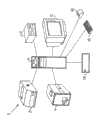
图1为包括红外造影机的红外成像系统的框图;Fig. 1 is a block diagram of an infrared imaging system including an infrared imaging machine;
图2为图1所示的红外造影机的框图,其中包括单一的可选择定位的红外检测器;Fig. 2 is a block diagram of the infrared imaging machine shown in Fig. 1, which includes a single selectable positionable infrared detector;
图3为表示图2所示的红外造影机可观看到的总视野(tFOV)的帧的示意图,该红外造影机包括形成该帧的光学元件阵列;3 is a schematic diagram showing a frame of the total field of view (tFOV) viewable by the infrared imaging machine shown in FIG. 2, the infrared imaging machine including an array of optical elements forming the frame;
图4为利用图2所示的红外造影机获得的多个帧的温度与时间示意图;Fig. 4 is a schematic diagram of temperature and time of multiple frames obtained by using the infrared imaging machine shown in Fig. 2;
图5为根据从图4所示的每个帧由四个相同的光学元件接收的红外辐射确定的温度与时间曲线图;Figure 5 is a graph of temperature versus time determined from infrared radiation received by the four identical optical elements from each frame shown in Figure 4;
图6为图2所示的红外造影机的示意图,该红外造影机被定位以便从暴露于加热/冷却泵产生的热应力的患者获取红外图像的帧;6 is a schematic diagram of the infrared contrast machine shown in FIG. 2 positioned to acquire frames of infrared images from a patient exposed to thermal stress generated by a heating/cooling pump;
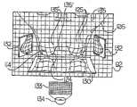
图7为图6中沿VII-VII线观看患者的视图,包括视野中的四个光学元件在患者身上的位置,从所述光学元件获取红外辐射以生成图5所示的温度与时间曲线;Figure 7 is a view of the patient viewed along line VII-VII in Figure 6, including the positions on the patient of the four optical elements in the field of view from which infrared radiation is obtained to generate the temperature versus time curve shown in Figure 5;
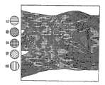
图8a为黑白绘图,包括色彩符号以表示患者乳房的色彩斜率映射图像的色彩,其中与多个帧中位于总视野中的每个位置处的光学元件相关的每条温度与时间曲线被映射成与该温度与时间曲线的积分相关的色彩;Figure 8a is a black and white plot including color symbols to represent the color of a color slope mapped image of a patient's breast where each temperature versus time curve associated with an optical element at each position in the total field of view over multiple frames is mapped as the color associated with the integral of the temperature versus time curve;
图8b为图8a所示的患者乳房的灰度映射图像,其中具有小于与红色相关的温度与时间曲线的积分的温度与时间曲线,将根据其积分被映射成灰度,而红色部分是以红色的色彩符号来表示;Figure 8b is a grayscale mapped image of the patient's breast shown in Figure 8a, wherein temperature versus time curves with integrals less than those associated with red are mapped to grayscale according to their integrals, while the red portion is represented by indicated by a red color symbol;
图9为具有锯齿状脉管系统的另一个患者的乳房的灰度映射图像;Figure 9 is a grayscale mapped image of another patient's breast with jagged vasculature;
图10为图7所示的患者的示意图,其包括位于患者乳房下方的胸骨反射镜以及位于患者乳房的相对两侧上的侧边反射镜;10 is a schematic illustration of the patient shown in FIG. 7, including a sternal mirror positioned below the patient's breast and side mirrors positioned on opposite sides of the patient's breast;
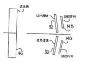
图11为用于获取立体图像的一对检测器和一对红外透镜的分体示意图;Fig. 11 is a split schematic diagram of a pair of detectors and a pair of infrared lenses for obtaining stereoscopic images;
图12为图1所示的红外造影机的框图,该红外造影机包括红外检测器的凝视(staring)阵列;以及Fig. 12 is a block diagram of the infrared contrast machine shown in Fig. 1, the infrared contrast machine including a staring (staring) array of infrared detectors; and
图13为用于获取立体图像的一对红外透镜和一对凝视阵列的分体示意图。Fig. 13 is a split schematic diagram of a pair of infrared lenses and a pair of staring arrays for acquiring stereoscopic images.
具体实施方式Detailed ways
请参阅图1,红外(IR)成像系统2包括连接至工作站6的红外造影机4。红外造影机4从工作站6接收指令信号,并向工作站6提供关于红外造影机4接收的红外辐射的定量数据及信息。工作站6还连接有打印机8、储存装置10、显示器12、定点(pointing)装置14、键盘16以及电源调节器18,上述装置供红外成像系统2的使用者以常规的方式使用。Referring to FIG. 1 , an infrared (IR) imaging system 2 includes an
请参阅图2并继续参阅图1,红外造影机4包括数据接收器22及数据传输器24,用于与工作站6通信。在软件程序的控制下运行的控制器26被连接以接收来自数据接收器22的数据。红外造影机4包括与冷却系统30连接的检测器28,用于以常规的方式将检测器28冷却到可接受的工作温度。检测器28接收来自红外透镜32的红外辐射,红外透镜32将其接收的红外辐射聚焦到检测器28上。红外辐射在到达红外透镜32之前通过前端面板视窗44,且在特定的情况下会通过滤光器46。Please refer to FIG. 2 and continue to refer to FIG. 1 , the
用于红外透镜32聚焦的聚焦系统62被连接以接收来自控制器26的控制信号。在控制器26的控制下,红外透镜32的光学聚焦和/或缩放可通过聚焦系统62以现代数字摄影系统的常规方式进行调整。A focusing
机动的X-Y定位台64连接至红外透镜32及检测器28,用以控制方向,红外透镜32及检测器28沿该方向检测从滤光器46传播的红外辐射。定位控制器66被连接以接收来自控制器26的控制信号。在控制器26的控制下,定位控制器66控制X-Y定位台64的位置,以使红外透镜32及检测器28能被选择性定位,以检测从滤光器46的选定部分传播的红外辐射光束。A motorized
前置放大器76被连接以从检测器28接收对应于接收到的红外辐射的强度的信号。前置放大器76放大并过滤检测器28输出的每个信号,并将每个经过放大与过滤的信号提供给图像模数转换器(ADC)78,该模数转换器78会将每个来自前置放大器76且经过放大与过滤的信号转换为对应的数字信号,此数字信号将被提供给图像处理系统80,如栅极阵列。在控制器26的控制下工作的图像处理系统80将红外辐射的数据及信息提供给数据传输器24,以传输至工作站6。A
优选的,前置放大器76被连接以接收来自温度校准系统82的关于绝对温度的数据与信息。在控制器26的控制下,温度校准系统82将绝对温度校准数据提供给前置放大器76。前置放大器76将来自温度校准系统82的绝对温度校准数据与检测器28输出的每个信号结合,以对应检测器28接收的红外辐射的绝对温度调整前置放大器76输出的经过放大与过滤的信号。
控制模数转换器84被连接以接收聚焦系统62、X-Y定位台64、温度校准系统82、前置放大器76以及环境温度传感器86输出的模拟信号。在控制器26的控制下,控制模数转换器84有选择地将与控制模数转换器84接收的模拟信号相对应的数字信号提供给控制器26。控制器26从控制模数转换器84接收的数字信号被控制器26使用,以控制红外造影机4的工作。A control analog-to-
请参阅图3,并继续参阅图1与图2,控制器26根据需要控制通过检测器28进行的红外辐射的取样、红外透镜32的聚焦以及X-Y定位台64的位置,以接收并记录来自光学元件90阵列中的每个光学元件90的红外辐射,该光学元件90阵列处于红外成像机4可观看到的总视野(tFOV)92之内。在此,术语“光学元件”(optel)是指在总视野92中的最小单元,其在检测器28的瞬时视野(iFOV)中可被单独处理。Please refer to Fig. 3, and continue to refer to Fig. 1 and Fig. 2,
红外造影机4在工作中从总视野92中的每个光学元件90获得红外辐射。例如,从位于图3所示的总视野92中的位置(X1,Y1)的光学元件90开始,控制器26控制X-Y定位台64、检测器28以及红外透镜32,以便对于固定的垂直Y轴位移、沿水平X轴获取来自每个光学元件90的红外辐射。具体而言,红外造影机4从总视野92内包括的介于位置(X1,Y1)与位置(X640,Y1)之间的每个光学元件90获取红外辐射。然后,红外成像系统2调整X-Y定位台64的位置,以使检测器28从总视野92内包括的介于位置(X1,Y2)与位置(X640,Y2)之间的每个光学元件90获取红外辐射。红外造影机4利用上述方式持续扫描总视野92中的光学元件90,直到获取了构成总视野92的全部光学元件90为止。In operation, the
由于图2所示的红外造影机4具有单一的检测器28,所以红外造影机4必须在不连续的时间段内从总视野92中每个位置上的光学元件90获取信息。在总视野92中的位置(X1,Y1)进行光学元件90取样与在位置(X640,Y480)进行光学元件90取样之间的间隔(即帧取样间隔)是由从被成像的物体接收的红外辐射量来决定。在此方面,帧取样间隔可作为从被成像的物体接收的红外辐射量的函数而被调整。优选的,红外造影机4在每个帧取样间隔中对总视野92中的每个位置的光学元件90进行多次取样,并对总视野92中各个位置的每个光学元件90进行取样平均,以便获得从每个光学元件90所接收的红外辐射的平均值,而此平均值可依照以下所述的方式加以运用。在光导检测器的情况下,来自上述取样的信息(电压输出)被求和;而在光电检测器的情况下,信息(电流输出)被积分。Since the
图像模数转换器78为总视野92中每个位置的光学元件90确定检测器28检测到的瞬时或平均红外辐射的绝对温度的数字值。接着,图像处理系统80在帧扫描间隔期间将为总视野92中每个位置的光学元件90获得的数字值排列在帧94中。优选的,帧94中与每个光学元件90相关的数字值代表红外辐射的绝对温度,该红外辐射是在相应的帧取样间隔期间由检测器28从被成像物体的特定位置接收到的。Image analog-to-
请参阅图4,并继续参阅所有上述的图式,红外造影机4在成像间隔(如五分钟)中获得被成像物体的多个帧,例如F1-F200。当在图像处理系统80中接收并汇集每个帧94时,控制器26将每个帧94从图像处理系统80经由数据传输器24传送至工作站6。在医疗应用中,优选的,每个帧94的获取是以对数的方式在成像间隔期间出现,并在成像间隔后期增加相邻帧94的获取次数。然而,相邻帧94的获取可以是固定的或以任何所需的方式变化。Referring to FIG. 4 and continuing to refer to all the above-mentioned drawings, the
请参阅图5,并继续参阅所有上述的图式,为了进行说明,对于每个帧94的相同光学元件90获取的温度可以表示为温度-时间曲线,如曲线100-106。例如,对于图4所示的帧F1-F200,温度-时间曲线100显示位置(X3, Y3)上的光学元件90的温度与时间的关系。同样地,对于图4所示的帧F1-F200,温度-时间曲线102、104及106分别显示位置(X3,Y7)、(X10,Y3)及(X10,Y7)上的光学元件90的温度与时间的关系。Referring to FIG. 5 , and continuing to all of the above figures, for illustration, the temperature acquired for the same
当在成像间隔期间已获取多个帧94时,工作站6为每个帧94中的相同光学元件90确定每条温度-时间曲线的积分(或积分值)。每个积分可关于特定时间间隔的时间(例如取样帧F1与帧F200之间的时间)确定。或者,每个积分可关于特定的多个子集帧94确定,例如帧F85-F150、帧F85-F150之间的每个其它帧,等等。例如,从每个帧94中相同位置的光学元件90,或者从选定的多个帧94、例如帧F85-F150,工作站6可确定关于每条温度-时间曲线(如曲线100、102、104和106)的时间的积分。例如,当帧F200被采样时,工作站6可在帧F1被采样至时刻t1时从时刻t0起确定每条温度-时间曲线的积分。在另一实例中,当帧F150被采样时,工作站6可在帧F85被采样至时刻t1时从时刻t0起确定每条温度-时间曲线的积分。When
出于显示的目的,工作站6可将每条温度-时间曲线(如曲线100-106)的积分值映射为单个色彩。优选的,在用于早期鉴定乳房肿瘤的医疗应用中,具有最小积分值的曲线映射为蓝色,而具有最大积分值的曲线则映射为红色。此外,具有介于最大与最小积分值之间的积分值的曲线映射为红色与蓝色之间的色彩。例如,如图5所示,在帧F85与帧F150之间,工作站6将蓝色映射于具有最小积分值的温度-时间曲线100;将绿色映射于温度-时间曲线102;将黄色映射于温度-时间曲线104;而将红色映射于具有最大积分值的温度-时间曲线106。For display purposes, workstation 6 may map the integral value of each temperature-time curve (eg, curves 100-106) as a single color. Preferably, in a medical application for early identification of breast tumors, the curve with the smallest integral value is mapped in blue, and the curve with the largest integral value is mapped in red. Also, a curve with an integral value between the maximum and minimum integral value is mapped to a color between red and blue. For example, as shown in FIG. 5, between frame F85 and frame F150, workstation 6 maps blue to temperature-
接着,工作站6将总视野92中的每个光学元件90或一组光学元件90的位置映射在显示器12上对应的像点或一组像点。当一种色彩被映射至部分或所有温度-时间曲线(如曲线100-106)的积分值时,工作站6会将温度-时间曲线的积分值映射的色彩显示在显示器12上,并显示于具有与总视野92内相应光学元件90的位置相对应的位置的像点或像点组上。如此映射于显示器12的色彩将在其上形成积分值的色彩斜率映射图像。The workstation 6 then maps the position of each
或者,工作站6可将选定的多个帧94上的每条温度-时间曲线的积分值映射成灰度,从而形成灰度斜率映射图像。优选的,灰度在最大与最小积分值之间延展,最大或最小积分值为白色,而最大或最小积分值中的另一个为黑色。工作站6也可将选定的多个帧94上具有小于或大于预定积分值的积分值的每条温度-时间曲线映射成灰度。此外,每个帧94或选定的多个帧94中的数据可利用常规的方式加以过滤或放大,以增强灰度和/或色彩斜率映射图像的细节。Alternatively, the workstation 6 may map the integral value of each temperature-time curve over the selected plurality of
本发明可应用于乳房癌的早期检测,特别是与乳房肿瘤生长过程有关的近期血管生成(angiogenesis)检测。具体而言,众所周知,随着乳房癌的生长,会出现血管生成过程,且肿瘤病变(lesion)或初期的肿瘤会形成独立的血液供给。据观察,通过近期血管生成所形成的血管并不会对身体的交感神经系统或自主神经系统有所回应。因此,就对于外因性热应力的反应而言,体内由于近期血管生成而被供血的部位对于外因性热应力的反应与身体同一器官内其供血与近期血管生成无关的邻近部位的反应不一致。从皮肤区随时间发出的红外辐射可被映射成不同结构与系统组织层面(例如细胞、组织、器宫和/或系统)的潜在生理、生化及神经过程。若将体内和近期血管生成活动区有关的皮肤区发出的红外辐射与来自其供血和近期血管生成无关的身体皮肤区的红外辐射相比较,可发现其间存在显著的不同。The invention can be applied to the early detection of breast cancer, especially the detection of short-term angiogenesis related to the growth process of breast tumors. In particular, it is well known that as breast cancer grows, an angiogenic process occurs and the neoplastic lesion or incipient tumor develops an independent blood supply. It has been observed that blood vessels formed by recent angiogenesis do not respond to the body's sympathetic or autonomic nervous system. Thus, in terms of response to extrinsic heat stress, parts of the body that are supplied with blood due to recent angiogenesis do not respond to extrinsic heat stress in the same way as adjacent parts of the body whose blood supply has not been associated with recent angiogenesis in the same organ. Infrared radiation emanating from a skin region over time can be mapped to underlying physiological, biochemical, and neural processes at different structural and systemic tissue levels (eg, cells, tissues, organs, and/or systems). Significant differences can be found when infrared radiation emanating from skin regions in the body associated with areas of recent angiogenesis activity is compared with infrared radiation from skin regions of the body whose blood supply has not been associated with recent angiogenesis.
利用红外成像系统2,获取温度-时间曲线及关于每条温度-时间曲线积分的信息的上述方法,本发明能够识别出是否存在近期血管生成,并提供乳房内这种血管生成所在位置的有意义的数据,从而提供表明患者体内活跃的肿瘤过程可能在发展之中或患者已罹患癌症的早期指示。Utilizing the above-described method of obtaining temperature-time curves and information about the integration of each temperature-time curve using the infrared imaging system 2, the present invention is able to identify the presence or absence of recent angiogenesis and provide meaningful information on the location of such angiogenesis within the breast. data, thereby providing early indications that an active tumor process in the patient may be developing or that the patient has cancer.
本发明的另一应用是应用数学与统计方法来检测从皮肤发出的红外辐射,以识别血管体(angiosome)或热节(thermatome)以及红外辐射统计上过高或过低的异常区域,而此红外辐射可映射成不同组织层面(例如细胞、组织、器官和/或系统)的潜在生理、生化及神经过程。在通过如针刺疗法及针压疗法等替代疗法治疗慢性疼痛、特别是肌筋膜(myofascial)疼痛中,或在如治疗性红外线或射频能量等外生电磁辐射的应用中,这类信息具有特殊的价值。Another application of the present invention is to apply mathematical and statistical methods to detect infrared radiation emitted from the skin to identify angiosomes or thermal knots (thermatome) and abnormal areas where infrared radiation is statistically too high or too low. Infrared radiation can be mapped to underlying physiological, biochemical, and neural processes at different tissue levels (eg, cells, tissues, organs, and/or systems). Such information is useful in the treatment of chronic pain, particularly myofascial pain, through alternative therapies such as acupuncture and acupressure, or in the application of exogenous electromagnetic radiation such as therapeutic infrared or radiofrequency energy. special value.
本发明在识别血管体及热节上的应用可让“西医”能够有效地利用“中医”疗法,如针刺疗法及针压疗法,并让“中医”能够将其观点与技术传达给“西方”的医疗业者。The application of the present invention in identifying vascular bodies and heat joints can enable "Western medicine" to effectively use "Chinese medicine" therapy, such as acupuncture and acupressure therapy, and allow "Chinese medicine" to convey its views and techniques to "Western medicine". " medical practitioners.
请参阅图6,并继续参阅所有上述的图式,下面参照在检测乳房114的过程中根据从患者P接收的红外辐射获取信息说明本发明。优选的,患者P坐在具有椅背112的凳子或座椅110上,且椅背112相对于垂直轴113以角度θ倾斜;或者,患者P站立并靠在倾斜相同角度的斜板上(未示出)。选择角度θ以使得每个乳房114的下侧理想地处于红外造影机4的总视野92之内。为了便于进行乳房114侧部及腋下区的红外成像,患者P需将其手臂向侧面及头部移动而离开身体,并将其手肘及前臂搁在支持器115上。红外造影机4与患者P之间具有一定的间隔,使得患者P的整个前胸区C、特别是患者P的乳房114处于红外造影机4的总视野92之内。优选的,红外造影机4被定位以使得患者P的乳房114及邻近的身体躯干116充满总视野92的大部分区域。然而,红外透镜32可被调整,以在患者P的特定区域进行光学缩放,从而能够获取来自此选定区域的红外辐射。Referring to FIG. 6 , and with continued reference to all of the above figures, the present invention will now be described with reference to acquiring information from infrared radiation received from a patient P during examination of the
在本发明中,利用红外透镜32进行光学缩放可隔离来自患者P选定区域的红外辐射的获取,但是并不能增加每个光学元件90观看到的患者P的表面区。通过光学缩放而在较小的光学元件90阵列获取红外图像,被认为能够提高患者P身体被成像部位的子集合中的图像分辨率。In the present invention, optical zoom using
根据红外透镜32的焦距与患者P必须在总视野92之内的面积之间的权衡(tradeoff),来选择患者P与红外造影机4之间的距离,以便获得有意义的生理信息。为达此目的,红外透镜32优选设定为使得患者P在总视野92中被成像的所有部位处于对焦点。优选的,红外造影机4的红外透镜32被设定成具有长焦距,且其模糊圆环与衍射界限小于由检测器28可观看到的每个光学元件90的尺寸。以上述的组合,红外造影机4具有可使位于总视野92内的患者P身体的每个部位都处于对焦点的景深,而不论患者P身体的每个子部位与红外造影机4相隔距离的大小。The distance between the patient P and the
患者P及座椅110处于当患者P脱去衣物时其环境温度使患者P感觉舒适的房间118内。在患者P坐上座椅110后的适当时间,红外造影机4开始获取对应于绝对温度数值的帧94,该绝对温度由未对患者P施加热应力时从患者P接收到的红外辐射表示。为了在施以外因热应力之前获取足够的“基准”信息而需要获取的帧个数与时间长度是由操作者确定,或通过计算机分析获取的数据来自动确定。在没有热应力时取得所需个数的帧94之后,患者P暴露在来自位于患者P前方的一个或多个加热/冷却泵122的调节空气流120。一个或多个加热/冷却泵122被定位以提供基本均匀的调节空气流120,从而以基本恒定的速率对患者P身体位于总视野92内的部位依照特定的检查规则加以冷却或加热。优选的,加热/冷却泵122提供至患者P的冷调节空气流120的温度不同于房间118的环境温度,但对于患者P而言仍然是舒适的。据观察,仅低于环境室温10°F的调节空气流120的温度就可使患者P产生所需的交感神经反应。The patient P and the
然而,在特定的临床条件下,在进行冷却之前优选使每个乳房114暖化,以便促使更多血液流至皮肤表面,并保证患者P不会因为周围环境条件而产生血管收缩。相应于热应力变化,例如从加热至冷却或从冷却至加热,从患者P接收到的红外辐射中能够取得其它有意义的信息。这些热应力的变化会在特定皮肤区的红外辐射放射中诱发一种生理上的磁滞现象(hysteresis),该特定皮肤区与下层组织和器官的某些生理过程相关。However, under certain clinical conditions, it may be preferable to warm each
例如,加热/冷却泵122首先为患者P提供调节空气流120,同时红外造影机4获取患者P的乳房114的多个帧94。在加热/冷却的循环期间,某些潜在的生理过程会使来自与此生理过程有关的皮肤区的红外辐射出现变化,导致从与这些特定区域相关的光学元件90接收的红外辐射的积分值和从与较小反应的组织相关的光学元件90接收的红外辐射的积分值在统计上存在显著的差异。此积分值之间的差异可在显示器12上显示为灰度和/或色彩斜率映射图像中的差异。下层结构与系统的三维图像还可从获取的数据产生。For example, heating/
由于与某些磁性材料相关的磁滞现象相似的生理组织特性,由加热而后冷却(即“热-冷”循环)和/或冷却而后加热(即“冷-热”循环)构成的热应力增强了红外成像系统2的效能,以识别与近期血管生成活动相关的生理组织区,例如患者P的乳房114。Increased thermal stress consisting of heating followed by cooling (i.e., "hot-cold" cycling) and/or cooling followed by heating (i.e., "cold-hot" cycling) due to physiological tissue properties similar to hysteresis associated with certain magnetic materials The effectiveness of the infrared imaging system 2 to identify regions of physiological tissue associated with recent angiogenic activity, such as patient P's
响应从加热/冷却泵122接收到冷调节空气流120,患者P的交感神经系统会限制血液流至患者P被冷却的皮肤表面。然而,据观察,患者P的交感神经系统不会限制血液流至患者P已遭受近期血管生成或正遭受血管生成发作的生理组织。因此,患者P的一些生理组织或器官的供血与近期血管生成或正在进行的血管生成活动相关,来自与这些生理组织或器官相关的患者P的皮肤区的红外辐射不会以与患者P的其它生理组织相同的方式对冷调节空气流120进行反应。例如,如图5与图7所示,在总视野92的位置(X3,Y3)上的光学元件90观看到的患者P的皮肤表面区,将响应冷调节空气流120对患者P的冷却,而在帧F4与帧F200之间产生温度-时间曲线100。同样地,冷调节空气流120对患者P的冷却将导致在位置(X3,Y7)、(X10,Y3)及(X10,Y7)上的光学元件90观看到的患者P的皮肤表面区在帧F4与帧F200之间分别产生温度-时间曲线102、104及106。In response to receiving the flow of cool conditioned
如图5所示,在帧F85与帧F150之间,曲线104及曲线106比曲线100及曲线102呈现出较小的变化率。此变化率的差异表明:在位置(X10,Y3)及位置(X10,Y7)上的光学元件90观看到的患者P的皮肤表面区,没有象在位置(X3,Y3)及位置(X3,Y7)上的光学元件90观看到的患者P的皮肤表面区一样,响应冷调节空气流120的冷却。对帧F85与帧F150之间的每条曲线102-106进行积分将生成积分值,由此,曲线106的积分值大于曲线104的积分值,曲线104的积分值大于曲线102的积分值,而曲线102的积分值大于曲线100的积分值。这表明患者P处于总视野92内的生理组织可能正在遭受血管生成的发作或者可能已由于近期血管生成而被供血。As shown in FIG. 5 , between the frame F85 and the frame F150 , the
除了提供关于血管生成及血管生成活动的信息之外,本发明还能提供关于其血液流动受交感神经系统调节的任何组织、器官或生理系统的活动的有意义信息。In addition to providing information on angiogenesis and angiogenic activity, the present invention can also provide meaningful information on the activity of any tissue, organ or physiological system whose blood flow is regulated by the sympathetic nervous system.
优选的,在人体乳房肿瘤疾病的早期检测的特例中,用于检测总视野92内每个位置的光学元件90的温度-时间曲线的积分的起始帧94(如帧F85)及帧的个数可根据需要改变,以便在显示器12上显现图像。例如,图5所示的每条温度-时间曲线100、102、104及106的积分是在帧F85与帧F150之间被确定。然而,每条温度-时间曲线100、102、104及106的积分也可根据需要在帧F100与帧F125之间、帧F20与帧F85之间,帧F75与帧F175之间等确定。Preferably, in the special case of the early detection of human breast tumor disease, the starting frame 94 (such as frame F85) and the individual starting frame 94 (such as frame F85) of the integral of the temperature-time curve of the
此外,用于将灰度和/或色彩映射到图5所示的每条温度-时间曲线的积分的数字位数也可以改变。例如,若图像模数转换器78为12位模数转换器,则灰度和/或色彩斜率映射图像可被映射至小于图像模数转换器78的完整12位范围。例如,工作站6可将每条温度-时间曲线的积分映射至与以十为基底的数字800至1600对应的数值范围,从而从即时显示器12中消除含有较少或没有即时诊断价值的信息,但却不会使保存于患者数据库内的患者纪录丢失此类信息。Additionally, the number of digits of integration used to map grayscale and/or color to each temperature-time curve shown in FIG. 5 may also vary. For example, if
图8a及图8b分别显示以黑白色绘制的患者P的乳房114的色彩斜率映射图像和结合灰度与黑白色绘制的患者P的乳房114的色彩斜率映射图像。由于在成像间隔期间获取帧94,因此温度-时间曲线表示随着时间的过去,来自患者P的乳房114的整个组织肿块(tissue mass)的红外辐射,而并非只是来自于皮肤表面的温度。能够看到,图8a所示的色彩斜率映射图像提供关于患者P的乳房114对来自加热/冷却泵122的调节空气流120的交感神经反应的详细信息。在此色彩斜率映射图像中,靠近光谱蓝色端的色彩是与具有较小积分值的光学元件90相关,如图5中的曲线100;而以红色显示的区域则是与具有较大积分值的光学元件90相关,如图5中的曲线106。图8a所显示的色彩斜率映射图像中的绿色、黄色及橙色是与其积分值介于图5中的温度-时间曲线100与106的积分值之间的温度-时间曲线相关。8a and 8b show a color slope mapped image of patient P's
在图8b中,对于小于与红色相关的积分值的积分值,总视野92中具有与该积分值相关的温度-时间曲线的光学元件90的区域被映射成灰度,而具有对应于红色的积分值的温度-时间曲线则以红色符号表示。更具体而言,图8b中结合灰度与黑白色绘制的色彩斜率映射图像是通过结合最后32个帧94的灰度斜率映射图像以及相同的最后32个帧94的色彩斜率映射图像而形成。灰度图像是通过以下方式形成的:将最后32个帧94中的相同光学元件90的积分值映射成灰度以形成图8b的灰度映射图像。与红色相关的色彩斜率映射图像的光学元件90取代灰度映射图像中的相同光学元件90,如此生成的灰度/色彩斜率映射图像显示在显示器12上,以生成图8b的灰度/色彩斜率图像。在图8b中,以红色符号所显示的区域与近期血管生成活动或血管生成已出现或可能正在出现的生理组织相关,因而此生理组织可能需要更进一步检查。In Fig. 8b, for an integral value smaller than the integral value associated with red, the area of the
图8b中的灰度与黑白色结合绘制的色彩斜率映射图像还清楚地显示了患者P的乳房114内的血管结构,以为医师提供更多具有诊断价值的信息。根据经验已经注意到,乳房114若如图9所示呈现复杂或极不规则的血管结构,则被视为可疑的,并表示相较于血管结构更规则的情况该患者P具有更高的危险罹患乳癌。通过能够详细地检测并显示乳房114的血管结构,本发明可进行早期的检测,如与乳癌等肿瘤疾病相关的血管生成过程的检测,以及代表与乳癌等肿瘤疾病相关的致病因素的解剖结构或生理特征的检测,从而让患者P能够早在肿瘤可被触知之前,并使患者P在其个人生活方式中采取的改变可防止乳癌发展时就采取行动,如消除“生活方式”的致病因素。The grayscale and black-and-white color slope mapping image in FIG. 8 b also clearly shows the blood vessel structure in the
本发明也可应用于通过比较在多个“心跳”周期的相同时间获取的帧94而确定患者P的血液流动。具体而言,由红外造影机4获取的每个帧94可与患者P的心跳周期的特定部分同步。例如,帧F1、F6及F11等可在每个心跳周期的P波期间获取;帧F2、F7及F12等可在每个相同心跳周期的Q波期间获取;帧F3、F8及F13等可在每个相同心跳周期的R波期间获取;帧F4、F9及F14等可在每个相同心跳周期的S波期间获取;而帧F5、F10及F15等则可在每个相同心跳周期的T波期间获取。在整个图像获取序列中利用患者P自己的心跳周期要素作为“时间码”(“time code”),从而允许以更高的精确度更有效率地应用图像处理及分析的数学方法。此外,利用明确定义的电生理学过程(如心跳)以及适当设立的图形显示表示法(如心电图),可增强对在不同检测期间获得的图像序列的比较,并且可在对相同或不同患者进行多次检测时提高获取数据的统计分析效率。The invention is also applicable to determining the blood flow of a patient P by comparing
当在所需的多个心跳周期已获取所需的多个帧94时,工作站6确定与每个心跳周期中特定时刻相关的帧94中相同光学元件90的积分值。例如,工作站6确定在帧F1、F6及F11等(即在多个心跳周期的P波期间获取的帧)中相同光学元件90的积分值;确定帧F2、F7及F12等(即在多个心跳周期的Q波期间获取的帧)中相同光学元件90的积分值;以及确定在多个心跳周期的R波、S波及T波期间获取的帧中相同光学元件90的积分值。When the desired number of
工作站6以上述方式将在多个心跳周期的相同时刻获取的帧94中相同光学元件90的积分值映射成灰度和/或色彩。工作站6会将如此映射的灰度和/或色彩显示于显示器12的像点或像点组上,所述像点或像点组的位置对应于总视野92内相应的一个或多个光学元件90的位置,以形成在多个心跳周期的相同时刻积分值的灰度和/或色彩斜率映射图像。例如,工作站6将灰度和/或色彩映射到帧F1、F6及F11等中相同光学元件90的积分值,并在显示器12上显示对应于多个心跳周期的P波期间患者P的血液流动的灰度和/或色彩斜率映射图像。此外,工作站6将灰度和/或色彩映射到与患者P的多个心跳周期的Q波、R波、S波或T波相关的帧94中相同光学元件90的积分值,并选择性地在显示器12上显示其灰度和/或色彩斜率映射图像。The workstation 6 maps the integrated values of the same
隔离与多个心跳周期的相同时刻相关的帧94,能够使来自患者P的皮肤表面的红外辐射变化与遍布被关注的生理组织的多层及多处的血液流动之间产生关联,从而允许对位于红外造影机4总视野92中的患者P体内的血液流动进行量化评估,进而为医师提供更多有价值的诊断信息。利用可靠而一致的时间码标记、如患者P自己的心跳,允许应用增强图像的数学处理,并提高整体成像系统的总分辨率,从而改善系统的灵敏度与选择性,以作为识别、定性及评估活的人体及其它动物体内以交感神经为媒介的复杂生理过程的手段。Isolating
本发明也可用于通过以相减的方式结合包含于两个帧94中的数字信息而获得诊断信息。例如,从帧F2的每个相同位置的光学元件90获得的相应数字信息减去从帧F4的每个位置的光学元件90获得的数字信息。工作站6可将在相同位置的光学元件90获得的这些差异映射成灰度和/或色度,以在显示器12上生成这些差异的灰度和/或色彩斜率映射图像。The invention can also be used to obtain diagnostic information by combining the digital information contained in the two
请参阅图10并参阅图2至图7,乳房114的红外成像存在一个问题,即不论患者P的倾斜角度θ为何,都难以获得患者P的乳房114的下部124的红外图像。同样,当红外造影机4以图6所示的方式置于患者P前方时,红外造影机4若没有重新定位,则不易获得与患者P的手臂126相邻的每个乳房114的侧部125的红外图像。为了使红外造影机4能够观看到每个乳房114的下部124及其侧部125以及相关的腋下区,胸骨反射镜130可被放置在患者P的乳房114下方,且侧边反射镜132可被放置在患者P的乳房114的相对两侧125。为胸骨反射镜130及侧边反射镜132定位以使得每个乳房114的下部124及其侧部125以及腋下区都处于总视野92之内,并为其定向以使来自每个乳房114下部124及其侧部125的红外辐射能够被反射到红外造影机4。Referring to FIG. 10 and FIGS. 2 to 7 , there is a problem in the infrared imaging of the
利用图像处理技术,工作站6由直接从乳房114接收的红外辐射以及由胸骨反射镜130和/或侧边反射镜132反射的红外辐射来构建乳房114的灰度和/或色彩斜率映射图像。为了增强红外造影机4的功能以检测胸骨反射镜130、侧边反射镜132、患者P以及其间空间的过渡,每个胸骨反射镜130及侧边反射镜132可在红外造影机4可看到的其一个或多个边上包括一个或多个条板,所述条板材料的辐射率基本不同于人体或其它活体动物或生理组织的辐射率。这种特殊应用在评估皮肤对于外部材料(如化妆品)的敏感度中特别有用,并提供一种确定皮肤对材料(如局部性施用的药物)的吸收率的途径。这种特殊应用可为人类及其它动物对于化合物和混合物(如化妆品)的皮肤敏感度提供精确定量且可再现的确定,从而消除了对某些具有争议性的动物试验程序(如“Draiz”试验)的需要。本发明还允许对于局部性施用的药物的吸收率或吸收进行精确定量且可再现的确定,进一步消除了对具有争议性的动物试验程序的需要。Utilizing image processing techniques, workstation 6 constructs a gray scale and/or color slope mapped image of
请参阅图7并继续参阅图10,为了在患者P的整个寿命期间延续的时间上进行帧94的连续记录,并提供来自胸骨反射镜130及侧边反射镜132的帧94的精确叠加及合并,以及为了调和由于红外造影机4的离轴定位而造成的透视变化,可将标记158放置于某个固定的解剖结构的界标,如前肋骨凹陷160、剑胸骨接合点162、下锁骨骨沟164、前腋窝线166以及肩峰突起168。标记158是由具有基本上不同于患者P的辐射率的材料制成。此外,某些有用的解剖界标的特征可从通过图像处理技术获得的信息识别出来。特别参照人体乳房,乳房血管树的现存分支可用作时间上的图像记录的方法。Referring to FIG. 7 and with continued reference to FIG. 10 , for continuous recording of
有许多种方法能够根据直接来自患者P以及从反射镜130、132反射的红外辐射,构建前面、侧面和/或自下而上的图像或三维图像。例如,栅格133可放置于照射器(lamp)134或其它热源与患者P身体位于红外造影机4的总视野92内的部位之间。在获取帧94之后的适当时间,照射器134被供给能量,从而经过栅格133而将热能传递至患者P身体位于总视野92内的部位。来自照射器134的一部分热能会被栅格133吸收,从而使得患者P以栅格状的图样接收来自照射器134的热能。直接从照射器134接收到的热能会在患者P上形成红外辐射图样135,当直接观看患者P时该红外辐射图样135为栅格状,而当经由反射镜130及132观看时该红外辐射图样135沿着患者P的轮廓。同样地,经由反射镜130及132从照射器134接收的热能会在患者P上形成被反射的红外辐射图样135’,且当经由反射镜130及132通过红外造影机4观看时该红外辐射图样135’是栅格状,但直接从患者P观看时则沿着患者P的轮廓。利用图像重建技术,工作站6能够根据直接可观看到的与经由反射镜130及132可观看得到的红外辐射图样135及135’,构建乳房114的前面、侧面和/或自下而上的灰度和/或色彩斜率映射图像、或三维灰度和/或色彩斜率映射图像。There are many ways to construct frontal, lateral and/or bottom-up or three-dimensional images from infrared radiation directly from the patient P and reflected from the mirrors 130,132. For example, a
在另一实施例中,患者P穿着尼龙胸罩(未示出),而标记158设置在其上选定的位置,且被选定的位置通过红外造影机4可直接观看到,或经由反射镜130及132可观看到。由于尼龙对于红外辐射而言是透明的,因此工作站6可利用标记158在胸罩上的位置,根据直接可观看到的以及经由反射镜130及132可观看到的红外辐射图样来构建乳房114的前面、侧面和/或自下而上的灰度和/或色彩斜率映射图像、或三维灰度和/或色彩斜率映射图像。In another embodiment, the patient P wears a nylon bra (not shown), and the
请参阅图11并同时参阅图2,红外造影机4可配置为利用设置在X-Y定位台64上的一对检测器28以及一对红外透镜32以进行立体成像。该对检测器28及该对红外透镜32被设置在X-Y定位台64上,以同时观看并获取来自总视野92内的共用光学元件90的红外辐射。X-Y定位台64可用于调整该对检测器28及该对红外透镜32的位置,以便观看并获取来自总视野92内的每个光学元件90的红外辐射。在此实施例中,前置放大器76、图像模数转换器78以及图像处理系统80配置为处理由每个检测器28响应同时接收来自同一光学元件90的红外辐射而输出的信号。为总视野92内每个位置上的光学元件90获得的两个图像可被工作站6组合,以生成受到热应力的患者P的生理组织的立体灰度和/或色彩斜率映射图像。Referring to FIG. 11 and FIG. 2 at the same time, the
请参阅图12并继续参阅图2,检测器28可被检测器28的阵列148取代,该阵列148被定位以接收通过前端面板视窗44、滤光器46以及红外透镜32的红外辐射。在此实施例中,红外透镜32已被“加大尺寸”(“up-sized”),以将接收到的红外辐射聚焦在检测器28的阵列148上,而该阵列148即为常规的“凝视阵列”148,以下将使用此名称。在操作中,控制器26基本上同时(即帧取样间隔)对凝视阵列148中每个检测器28的输出进行取样,以形成一个图3所示型态的帧94。控制器26可在预设的间隔内从凝视阵列148获得多个帧94。利用以上结合图4与图5描述的方法,在成像间隔期间,对于每个帧94中相同位置的光学元件90,工作站6获得由光学元件90表示的患者P身体某一部位的热反应。Referring to FIG. 12 with continued reference to FIG. 2 ,
请参阅图13并同时参阅图11与图12,以如图11所示的一对检测器28及一对红外透镜32的相同方式,利用一对凝视阵列148及一对红外透镜32来生成患者P的立体图像。在此实施例中,该对凝视阵列148接收来自总视野92内每个光学元件90的红外信息,且前置放大器76、图像模数转换器78以及图像处理系统80被配置为处理从每个凝视阵列148接收的图像数据。从该对凝视阵列148为总视野92内每个位置上的光学元件90获得的两个图像可被工作站6组合,以生成受到热应力的患者P的生理组织的立体灰度和/或色彩斜率映射图像。Please refer to FIG. 13 and FIG. 11 and FIG. 12 at the same time, in the same manner as a pair of
在图11所示的实施例中,每个检测器28被配置为检测不同波长的红外辐射。例如,一个检测器28可被配置为检测波长介于1微米与2微米之间的红外辐射,而另一检测器28则可被配置为检测波长介于8微米与12微米之间的红外辐射。此外,配置为检测特定波长的红外辐射的每个检测器28,可以如图11所示的方式而与另一个相同的检测器28配对,以生成不同波长的红外辐射的立体图像。优选的,每个检测器28与另一检测器28之间的距离大于每个检测器28可观看到的光学元件90的尺寸。In the embodiment shown in Figure 11, each
优选的,将在成像间隔期间获得的患者P的热图像的多个帧94储存在工作站6的非易失性存储器(例如磁性或光学数据储存装置)中的患者数据档案中,以用于后续的本地查找及分析。也可以将患者数据档案传输至储存多个患者数据档案的分布数据系统,以用于后续的查找及分析。优选的,分布数据系统具有多个位于不同地理位置并以常规的方式如互联网而相互连接的计算机。每个相互连接的计算机包括非易失性存储器、适当的操作系统软体以及适当的用户图形界面,其中非易失性存储器用以接收及储存来自本地地理位置的多个患者的数据档案,而用户图形界面则有助于用户与计算机之间的互动。优选的,每个患者的数据档案包括在成像间隔期间获得的红外图像数据的帧94,以及其它患者数据,例如生活习惯、病史及其它致病因素等,其与分析患者是否有罹患乳癌的危险相关。优选的,每个患者数据档案作为单一对象存放在分布数据系统的计算机中分布的相关数据库中。Preferably, a plurality of
优选的,分布数据系统的操作系统软体支持能够对多个患者数据档案或多个患者数据档案的子集进行分析的专家系统。具体而言,专家系统利用公知的分析技术,如数值统计分析、差异分析或因子分析等,来分析多个患者P的数据档案,或将单个患者P的数据档案与多个患者P的数据档案进行比较,以找出统计上的不一致性,例如与乳房中正在成长的肿瘤过程相关的血管生成症状。优选的,专家系统定期将接收到的患者P的数据与多个患者P的数据档案中的所有其他患者P的数据进行比较,以便识别在统计上显著的关联性,如与血管生成活动的产生有关的致病因素,或与肿瘤疾病有关的临床症状。Preferably, the operating system software of the distributed data system supports an expert system capable of analyzing multiple patient data files or subsets of multiple patient data files. Specifically, the expert system uses well-known analysis techniques, such as numerical statistical analysis, difference analysis, or factor analysis, to analyze the data files of multiple patients P, or combine the data files of a single patient P with the data files of multiple patients P Comparisons were made to identify statistical inconsistencies, such as angiogenic symptoms associated with growing tumor processes in the breast. Preferably, the expert system periodically compares the received data of a patient P with the data of all other patients P in the plurality of patient P data files in order to identify statistically significant associations, such as with the generation of angiogenic activity Related pathogenic factors, or clinical symptoms related to tumor diseases.
在此说明的红外成像方法与设备可用作进行监测以及客观地将传统与“非传统”疼痛处理疗法的有效性进行量化的手段,其中传统疗法例如为物理治疗及脊椎按摩疗法等,而“非传统”疗法例如为针压疗法及针刺疗法等。此外,本发明的方法与设备可用于确定适当点,以施用特定的疼痛处理疗法,如针压疗法、针刺疗法、推拿治疗、刺激点注射、增生疗法,以及施用聚焦电磁能,如红外辐射。The infrared imaging methods and devices described herein can be used as a means of monitoring and objectively quantifying the effectiveness of traditional and "non-traditional" pain management therapies, such as physical therapy and chiropractic, and " Examples of non-traditional treatments include acupressure and acupuncture. In addition, the method and apparatus of the present invention can be used to determine appropriate points for administering specific pain management therapies, such as acupressure, acupuncture, massage therapy, stimulating point injections, proliferative therapy, and for the administration of focused electromagnetic energy, such as infrared radiation .
在评估软组织损伤时,利用在此说明的红外成像方法与设备可通过各种装置随时间获得来自整个身体上或较少的关注区域上的皮肤的红外辐射的图像序列,其中所述装置包括通过软体创建合成图像的主要表面反射镜。In assessing soft tissue damage, using the infrared imaging methods and apparatus described herein, image sequences of infrared radiation from the skin over the entire body or on lesser regions of interest can be obtained over time by various means, including through The soft body creates a composite image of the main surface mirror.
从检测器28和/或凝视阵列148获得的数据可用于确定对侧肌肉群、皮区及热节的对称轴。可采用统计方法确定来自可比较的对侧区域的红外辐射是否具有统计上的显著差异。若存在显著的差异,则继续进行分析以确定在每一个对侧群中的对称区域之间是否存在统计上的显著差异。Data obtained from
例如,从右手臂的皮肤表面发出的红外能量与左手比较时,在统计上的显著差异性可能仅仅表明患者P为惯用右手的人。然而,进一步独立进行的每个手臂的统计分析,例如,考虑二头肌群及三头肌群的内部对称性,然后对每个可比较的肌肉群中的内部对称性关系进行统计分析及比较,可消除“偏手性”效应。这种分析方法在处理与宽广平坦的肌肉区(例如斜方肌、背阔肌群以及其它的背部区域)有关的疼痛疾病时将显得格外重要。For example, a statistically significant difference in infrared energy emanating from the skin surface of the right arm compared to the left hand may simply indicate that patient P is right-handed. However, a further statistical analysis of each arm independently, for example, considers the internal symmetry of the biceps and triceps groups, and then performs a statistical analysis and comparison of the internal symmetry relationships in each comparable muscle group , which can eliminate the "handedness" effect. This type of analysis will be especially important when dealing with pain disorders associated with broad, flat muscle areas such as the trapezius, latissimus dorsi, and other back regions.
对侧肌群、皮区及热节的多个对称轴的准确位置允许对连续的图像时间序列进行准确记录以及对治疗的有效性进行定量评估。将红外辐射数据映射至表面解剖结构的界标,并为医疗人员实时显示信息,能够提高治疗的效率及有效性。Accurate positioning of multiple axes of symmetry for contralateral muscle groups, dermatomes, and heat knots allows accurate recording of continuous image time-series and quantitative assessment of therapeutic effectiveness. Mapping infrared radiation data to landmarks of surface anatomy and displaying the information in real time for medical personnel can improve the efficiency and effectiveness of treatment.
参照优选实施例说明了本发明。其他人在阅读并理解上述说明时,可进行明显的更改及替换。本发明包含落入所附权利要求书或其等效范围内的所有这种更改及替换。The invention has been described with reference to preferred embodiments. Obvious changes and substitutions may be made by others upon reading and understanding the above description. The present invention embraces all such modifications and substitutions as come within the scope of the appended claims or equivalents thereof.
Claims (8)
Applications Claiming Priority (2)
| Application Number | Priority Date | Filing Date | Title |
|---|---|---|---|
| US46832103P | 2003-05-06 | 2003-05-06 | |
| US60/468,321 | 2003-05-06 |
Publications (2)
| Publication Number | Publication Date |
|---|---|
| CN1802121A CN1802121A (en) | 2006-07-12 |
| CN100553554Ctrue CN100553554C (en) | 2009-10-28 |
Family
ID=35529844
Family Applications (1)
| Application Number | Title | Priority Date | Filing Date |
|---|---|---|---|
| CNB2004800158185AExpired - Fee RelatedCN100553554C (en) | 2003-05-06 | 2004-05-06 | System for identifying and classifying dynamic thermodynamic processes of mammals and differentiating such processes |
Country Status (7)
| Country | Link |
|---|---|
| EP (1) | EP1620003B1 (en) |
| JP (1) | JP4495154B2 (en) |
| CN (1) | CN100553554C (en) |
| CA (1) | CA2524807C (en) |
| MX (1) | MXPA05011994A (en) |
| TW (1) | TWI286927B (en) |
| WO (1) | WO2004098392A2 (en) |
Cited By (1)
| Publication number | Priority date | Publication date | Assignee | Title |
|---|---|---|---|---|
| CN106108854A (en)* | 2016-07-18 | 2016-11-16 | 深圳市衣信互联网科技有限公司 | Underwear infrared light heat radiation detection system and the method for detection radiation field status information |
Families Citing this family (22)
| Publication number | Priority date | Publication date | Assignee | Title |
|---|---|---|---|---|
| WO2007095330A2 (en) | 2006-02-15 | 2007-08-23 | Hologic Inc | Breast biopsy and needle localization using tomosynthesis systems |
| CN101959456A (en) | 2007-12-31 | 2011-01-26 | 真实成像有限公司 | System and method for registration of imaging data |
| US8620041B2 (en)* | 2007-12-31 | 2013-12-31 | Real Imaging Ltd. | Method apparatus and system for analyzing thermal images |
| US10299686B2 (en) | 2008-03-28 | 2019-05-28 | Real Imaging Ltd. | Method apparatus and system for analyzing images |
| ES2862525T3 (en) | 2009-10-08 | 2021-10-07 | Hologic Inc | Needle Breast Biopsy System and Method of Use |
| JP6057922B2 (en) | 2011-03-08 | 2017-01-11 | ホロジック, インコーポレイテッドHologic, Inc. | System and method for dual energy and / or contrast enhanced breast imaging for screening, diagnosis and biopsy |
| US9007466B2 (en)* | 2011-04-27 | 2015-04-14 | General Electric Company | System and method for thermographic inspection |
| EP2782505B1 (en) | 2011-11-27 | 2020-04-22 | Hologic, Inc. | System and method for generating a 2d image using mammography and/or tomosynthesis image data |
| JP6240097B2 (en) | 2012-02-13 | 2017-11-29 | ホロジック インコーポレイティッド | How to navigate a tomosynthesis stack using composite image data |
| CN105451657A (en) | 2013-03-15 | 2016-03-30 | 霍罗吉克公司 | System and method for navigating tomosynthesis stack including automatic focusing |
| US10092358B2 (en) | 2013-03-15 | 2018-10-09 | Hologic, Inc. | Tomosynthesis-guided biopsy apparatus and method |
| CN104217421A (en)* | 2013-05-29 | 2014-12-17 | 杭州美盛红外光电技术有限公司 | Device and method for thermal image analysis |
| KR101550408B1 (en) | 2014-05-09 | 2015-09-08 | (주)메디텍 | Breast Diagnostic Apparatus |
| US10068330B2 (en)* | 2016-02-26 | 2018-09-04 | Niramai Health Analytix Pvt Ltd | Automatic segmentation of breast tissue in a thermographic image |
| US9865052B2 (en)* | 2016-03-18 | 2018-01-09 | Niramai Health Analytix Pvt Ltd | Contour-based determination of malignant tissue in a thermal image |
| CN110621233B (en) | 2017-03-30 | 2023-12-12 | 豪洛捷公司 | Method for processing breast tissue image data |
| WO2020068851A1 (en)* | 2018-09-24 | 2020-04-02 | Hologic, Inc. | Breast mapping and abnormality localization |
| TWI689895B (en)* | 2018-10-19 | 2020-04-01 | 國立臺灣大學 | System and method for monitoring color change of skin under unstable light source |
| TWI723877B (en)* | 2020-05-13 | 2021-04-01 | 國家中山科學研究院 | Plug-in coaxial thermal radiation image measurement system |
| US12254586B2 (en) | 2021-10-25 | 2025-03-18 | Hologic, Inc. | Auto-focus tool for multimodality image review |
| WO2023097279A1 (en) | 2021-11-29 | 2023-06-01 | Hologic, Inc. | Systems and methods for correlating objects of interest |
| CN115005782A (en)* | 2022-06-06 | 2022-09-06 | 杭州新瀚光电科技有限公司 | A human health assessment method, system, terminal device and storage medium |
Citations (8)
| Publication number | Priority date | Publication date | Assignee | Title |
|---|---|---|---|---|
| DE19544187A1 (en)* | 1995-11-28 | 1997-06-05 | Telefunken Microelectron | Thermal imaging system is triggered by, e.g. heart beat |
| US5803082A (en)* | 1993-11-09 | 1998-09-08 | Staplevision Inc. | Omnispectramammography |
| WO1998046976A2 (en)* | 1997-03-31 | 1998-10-22 | Zhong Qi Liu | Method and apparatus for thermal radiation imaging |
| EP0885587A1 (en)* | 1997-06-17 | 1998-12-23 | David Keith Harrison | Thermal imaging method and apparatus |
| US6241672B1 (en)* | 1990-08-10 | 2001-06-05 | University Of Washington | Method and apparatus for optically imaging solid tumor tissue |
| CN2474084Y (en)* | 2000-09-30 | 2002-01-30 | 吴士明 | Controlling device for infra red camera standardized imaging environment |
| CN1399527A (en)* | 1999-06-30 | 2003-02-26 | 里奥格兰德医学技术有限公司 | Method and apparatus for non-invasive blood analyte measurement |
| CN1399526A (en)* | 1999-06-21 | 2003-02-26 | 威克特约翰雅那柯尼二世 | Method and equipment for high-definition dynamic digital infrared imaging |
Family Cites Families (7)
| Publication number | Priority date | Publication date | Assignee | Title |
|---|---|---|---|---|
| JPS61252782A (en)* | 1985-05-01 | 1986-11-10 | Fujitsu Ltd | infrared imaging device |
| US6671540B1 (en)* | 1990-08-10 | 2003-12-30 | Daryl W. Hochman | Methods and systems for detecting abnormal tissue using spectroscopic techniques |
| IL104423A (en)* | 1993-01-18 | 1998-09-24 | Opgal Optronic Ind Ltd | Infra-red vascular angiography system |
| JPH09281065A (en)* | 1996-04-18 | 1997-10-31 | Nitto Chem Ind Co Ltd | Method of detecting defects in structures |
| JPH09189610A (en)* | 1996-01-11 | 1997-07-22 | Nippon Avionics Co Ltd | Two-dimensional time delay integration type thermal imager |
| JPH09247847A (en)* | 1996-03-11 | 1997-09-19 | Toshiba Fa Syst Eng Kk | Temperature monitoring device |
| US6276798B1 (en)* | 1998-09-29 | 2001-08-21 | Applied Spectral Imaging, Ltd. | Spectral bio-imaging of the eye |
- 2004
- 2004-05-06EPEP04751477.3Apatent/EP1620003B1/ennot_activeExpired - Lifetime
- 2004-05-06MXMXPA05011994Apatent/MXPA05011994A/enactiveIP Right Grant
- 2004-05-06WOPCT/US2004/014110patent/WO2004098392A2/enactiveApplication Filing
- 2004-05-06CACA2524807Apatent/CA2524807C/ennot_activeExpired - Fee Related
- 2004-05-06CNCNB2004800158185Apatent/CN100553554C/ennot_activeExpired - Fee Related
- 2004-05-06TWTW093112800Apatent/TWI286927B/ennot_activeIP Right Cessation
- 2004-05-06JPJP2006514304Apatent/JP4495154B2/ennot_activeExpired - Fee Related
Patent Citations (9)
| Publication number | Priority date | Publication date | Assignee | Title |
|---|---|---|---|---|
| US6241672B1 (en)* | 1990-08-10 | 2001-06-05 | University Of Washington | Method and apparatus for optically imaging solid tumor tissue |
| US5803082A (en)* | 1993-11-09 | 1998-09-08 | Staplevision Inc. | Omnispectramammography |
| DE19544187A1 (en)* | 1995-11-28 | 1997-06-05 | Telefunken Microelectron | Thermal imaging system is triggered by, e.g. heart beat |
| WO1998046976A2 (en)* | 1997-03-31 | 1998-10-22 | Zhong Qi Liu | Method and apparatus for thermal radiation imaging |
| US6023637A (en)* | 1997-03-31 | 2000-02-08 | Liu; Zhong Qi | Method and apparatus for thermal radiation imaging |
| EP0885587A1 (en)* | 1997-06-17 | 1998-12-23 | David Keith Harrison | Thermal imaging method and apparatus |
| CN1399526A (en)* | 1999-06-21 | 2003-02-26 | 威克特约翰雅那柯尼二世 | Method and equipment for high-definition dynamic digital infrared imaging |
| CN1399527A (en)* | 1999-06-30 | 2003-02-26 | 里奥格兰德医学技术有限公司 | Method and apparatus for non-invasive blood analyte measurement |
| CN2474084Y (en)* | 2000-09-30 | 2002-01-30 | 吴士明 | Controlling device for infra red camera standardized imaging environment |
Cited By (2)
| Publication number | Priority date | Publication date | Assignee | Title |
|---|---|---|---|---|
| CN106108854A (en)* | 2016-07-18 | 2016-11-16 | 深圳市衣信互联网科技有限公司 | Underwear infrared light heat radiation detection system and the method for detection radiation field status information |
| CN106108854B (en)* | 2016-07-18 | 2019-09-17 | 深圳市衣信互联网科技有限公司 | The infrared optical heat radiation detection system of underwear and the method for detecting radiation field status information |
Also Published As
| Publication number | Publication date |
|---|---|
| EP1620003A4 (en) | 2009-05-13 |
| MXPA05011994A (en) | 2006-02-02 |
| CA2524807C (en) | 2010-01-26 |
| JP2007525244A (en) | 2007-09-06 |
| EP1620003A2 (en) | 2006-02-01 |
| EP1620003B1 (en) | 2015-04-15 |
| TWI286927B (en) | 2007-09-21 |
| CN1802121A (en) | 2006-07-12 |
| WO2004098392A2 (en) | 2004-11-18 |
| JP4495154B2 (en) | 2010-06-30 |
| TW200505389A (en) | 2005-02-16 |
| CA2524807A1 (en) | 2004-11-18 |
| HK1095075A1 (en) | 2007-04-27 |
| WO2004098392A3 (en) | 2005-03-24 |
Similar Documents
| Publication | Publication Date | Title |
|---|---|---|
| CN100553554C (en) | System for identifying and classifying dynamic thermodynamic processes of mammals and differentiating such processes | |
| US7996066B2 (en) | Topographic optical infrared tomography system for biophysical imaging with infrared diagnostic exploratory algorithm sequencing (IDEAS) scripting language | |
| US6023637A (en) | Method and apparatus for thermal radiation imaging | |
| JP4733265B2 (en) | Brain tissue imaging and feature display | |
| CN112218576A (en) | Device and method for acquiring and analyzing images of the skin | |
| JPH07271963A (en) | Forming method of medical picture | |
| WO2013163211A1 (en) | Method and system for non-invasive quantification of biological sample physiology using a series of images | |
| Mazdeyasna et al. | Noncontact speckle contrast diffuse correlation tomography of blood flow distributions in tissues with arbitrary geometries | |
| JP4649965B2 (en) | Health degree determination device and program | |
| Rajmanova et al. | Application and technology of thermal imagine camera in medicine | |
| Mo et al. | The importance of illumination in a non-contact photoplethysmography imaging system for burn wound assessment | |
| JP4652643B2 (en) | Method and apparatus for high resolution dynamic digital infrared imaging | |
| US7408156B2 (en) | System and method for identifying and classifying dynamic thermodynamic processes in mammals and discriminating between and among such processes | |
| US20120078114A1 (en) | System and method for real-time perfusion imaging | |
| Anbar | Computerized Thermography: The Emergence of a New Diagnostic Imaging Modality | |
| Schwartz et al. | The American academy of thermology guidelines for breast thermology 2021 | |
| US8210682B2 (en) | Method and apparatus for early diagnosis of Alzheimer's using non-invasive eye tomography by Teraherts | |
| RU2302194C1 (en) | Method and device for computerized-thermal vision diagnostics in dentistry | |
| CN113303787A (en) | Pressure sore wound detection and calculation system based on infrared imaging technology | |
| HK1095075B (en) | System for identifying and classifying dynamic thermodynamic processes in mammals and discriminating between and among such processes | |
| HK1053597B (en) | Method and apparatus for high resolution dynamic digital infrared imaging | |
| Maehara et al. | Changes in hemoglobin concentration in the lateral occipital regions during shape recognition:<? xpp qa?> a near-infrared spectroscopy study | |
| KR20200142807A (en) | Apparatus for training eye movement and method for operating the same | |
| CN103431844A (en) | Human body biological remote sensing diagnostic technique | |
| Nahm et al. | Optical imaging methods in medicine: how can we escape the plausibility trap? |
Legal Events
| Date | Code | Title | Description |
|---|---|---|---|
| C06 | Publication | ||
| PB01 | Publication | ||
| C10 | Entry into substantive examination | ||
| SE01 | Entry into force of request for substantive examination | ||
| REG | Reference to a national code | Ref country code:HK Ref legal event code:DE Ref document number:1095075 Country of ref document:HK | |
| C14 | Grant of patent or utility model | ||
| GR01 | Patent grant | ||
| REG | Reference to a national code | Ref country code:HK Ref legal event code:GR Ref document number:1095075 Country of ref document:HK | |
| CF01 | Termination of patent right due to non-payment of annual fee | ||
| CF01 | Termination of patent right due to non-payment of annual fee | Granted publication date:20091028 Termination date:20180506 |