CN100463664C - Device for cutting the connection between cells and basement membrane or lens capsule - Google Patents
Device for cutting the connection between cells and basement membrane or lens capsuleDownload PDFInfo
- Publication number
- CN100463664C CN100463664CCNB2004800442779ACN200480044277ACN100463664CCN 100463664 CCN100463664 CCN 100463664CCN B2004800442779 ACNB2004800442779 ACN B2004800442779ACN 200480044277 ACN200480044277 ACN 200480044277ACN 100463664 CCN100463664 CCN 100463664C
- Authority
- CN
- China
- Prior art keywords
- basement membrane
- cell
- light source
- light
- lens capsule
- Prior art date
- Legal status (The legal status is an assumption and is not a legal conclusion. Google has not performed a legal analysis and makes no representation as to the accuracy of the status listed.)
- Expired - Fee Related
Links
Images
Classifications
- A—HUMAN NECESSITIES
- A61—MEDICAL OR VETERINARY SCIENCE; HYGIENE
- A61F—FILTERS IMPLANTABLE INTO BLOOD VESSELS; PROSTHESES; DEVICES PROVIDING PATENCY TO, OR PREVENTING COLLAPSING OF, TUBULAR STRUCTURES OF THE BODY, e.g. STENTS; ORTHOPAEDIC, NURSING OR CONTRACEPTIVE DEVICES; FOMENTATION; TREATMENT OR PROTECTION OF EYES OR EARS; BANDAGES, DRESSINGS OR ABSORBENT PADS; FIRST-AID KITS
- A61F9/00—Methods or devices for treatment of the eyes; Devices for putting in contact-lenses; Devices to correct squinting; Apparatus to guide the blind; Protective devices for the eyes, carried on the body or in the hand
- A61F9/007—Methods or devices for eye surgery
- A61F9/00736—Instruments for removal of intra-ocular material or intra-ocular injection, e.g. cataract instruments
- A—HUMAN NECESSITIES
- A61—MEDICAL OR VETERINARY SCIENCE; HYGIENE
- A61F—FILTERS IMPLANTABLE INTO BLOOD VESSELS; PROSTHESES; DEVICES PROVIDING PATENCY TO, OR PREVENTING COLLAPSING OF, TUBULAR STRUCTURES OF THE BODY, e.g. STENTS; ORTHOPAEDIC, NURSING OR CONTRACEPTIVE DEVICES; FOMENTATION; TREATMENT OR PROTECTION OF EYES OR EARS; BANDAGES, DRESSINGS OR ABSORBENT PADS; FIRST-AID KITS
- A61F9/00—Methods or devices for treatment of the eyes; Devices for putting in contact-lenses; Devices to correct squinting; Apparatus to guide the blind; Protective devices for the eyes, carried on the body or in the hand
- A61F9/007—Methods or devices for eye surgery
- A61F9/008—Methods or devices for eye surgery using laser
- A61F2009/00861—Methods or devices for eye surgery using laser adapted for treatment at a particular location
- A61F2009/00868—Ciliary muscles or trabecular meshwork
- A—HUMAN NECESSITIES
- A61—MEDICAL OR VETERINARY SCIENCE; HYGIENE
- A61F—FILTERS IMPLANTABLE INTO BLOOD VESSELS; PROSTHESES; DEVICES PROVIDING PATENCY TO, OR PREVENTING COLLAPSING OF, TUBULAR STRUCTURES OF THE BODY, e.g. STENTS; ORTHOPAEDIC, NURSING OR CONTRACEPTIVE DEVICES; FOMENTATION; TREATMENT OR PROTECTION OF EYES OR EARS; BANDAGES, DRESSINGS OR ABSORBENT PADS; FIRST-AID KITS
- A61F9/00—Methods or devices for treatment of the eyes; Devices for putting in contact-lenses; Devices to correct squinting; Apparatus to guide the blind; Protective devices for the eyes, carried on the body or in the hand
- A61F9/007—Methods or devices for eye surgery
- A61F9/008—Methods or devices for eye surgery using laser
- A61F2009/00885—Methods or devices for eye surgery using laser for treating a particular disease
- A61F2009/00887—Cataract
- A61F2009/00889—Capsulotomy
Landscapes
- Health & Medical Sciences (AREA)
- Ophthalmology & Optometry (AREA)
- Life Sciences & Earth Sciences (AREA)
- Public Health (AREA)
- Engineering & Computer Science (AREA)
- Biomedical Technology (AREA)
- Heart & Thoracic Surgery (AREA)
- Vascular Medicine (AREA)
- Nuclear Medicine, Radiotherapy & Molecular Imaging (AREA)
- Animal Behavior & Ethology (AREA)
- General Health & Medical Sciences (AREA)
- Surgery (AREA)
- Veterinary Medicine (AREA)
- Physics & Mathematics (AREA)
- Optics & Photonics (AREA)
- Prostheses (AREA)
- Laser Surgery Devices (AREA)
- Micro-Organisms Or Cultivation Processes Thereof (AREA)
- Radiation-Therapy Devices (AREA)
Abstract
Description
Translated fromChinese发明领域field of invention
本发明公开了一种用于切割细胞与基膜之间的连接而不损伤所述细胞或所述基膜的装置。此装置能够从两个方向将细胞基膜复合体暴露于特定强度的光能,入射至细胞侧的光能具有非常低的强度,而入射至基膜侧的光能具有较高强度,从而实现它们之间的连接的切割。The present invention discloses a device for cutting the connection between cells and basement membrane without damaging the cells or the basement membrane. This device is capable of exposing the cell basement membrane complex to light energy of a specific intensity from two directions, the light energy incident on the cell side has a very low intensity, and the light energy incident on the basement membrane side has a higher intensity, thereby achieving The cut of the connection between them.
光对细胞基膜复合体的效应取决于波长、强度、照射持续时间、在照射时间所述组织的内在构成以及所述照射被影响的方向。本申请涉及通过使用特定波长的光能从细胞侧使用非常低强度光的照射,同时从基膜侧使用较高强度的照射,实现细胞与基膜的连接的特定切割。The effect of light on the cell basement membrane complex depends on the wavelength, intensity, duration of irradiation, the intrinsic composition of the tissue at the time of irradiation and the direction in which the irradiation is affected. The present application relates to the specific cleavage of the cell's junction with the basement membrane by irradiation with very low intensity light from the cell side using specific wavelengths of light energy, while using higher intensity illumination from the basement membrane side.
发明背景Background of the invention
在实验室操作和各种手术操作中,有必要有效分离由于各种原因附着于基膜或晶状体囊的细胞。这样的分离必将有效预防膜或组织的进一步的并发症或衰退,促进细胞或基膜后的结构或组织更好的显像,并获得光学的优点,例如更好的染色以拍摄细胞,和更好的操作以研究它们的性质。In laboratory operations and various surgical procedures, it is necessary to efficiently separate cells that are attached to the basement membrane or lens capsule for various reasons. Such separation will be effective in preventing further complications or deterioration of the membrane or tissue, facilitating better visualization of cells or structures or tissue behind the basement membrane, and obtaining optical advantages such as better staining for photographing cells, and Better manipulation to study their properties.
现有技术中公开有一些旨在分离附着于细胞膜的细胞的装置和方法。Several devices and methods aimed at isolating cells attached to cell membranes are disclosed in the prior art.
本发明涉及一种装置和一种方法,其克服了现有技术有关的各种问题。本发明包括发出所选波长的低强度光用于分离上皮细胞的装置。该装置使得操作者能够从两个方向使用光能照射细胞基膜复合体,以获得从附着的基膜分离上皮细胞的期望效应。此效应通过切割细胞和基膜间的连接而实现。The present invention relates to an apparatus and a method which overcome various problems associated with the prior art. The present invention includes devices that emit low intensity light of selected wavelengths for isolating epithelial cells. This device enables the operator to irradiate the cell-basement membrane complex with light energy from two directions to achieve the desired effect of detaching epithelial cells from the attached basement membrane. This effect is achieved by cleaving the junction between the cell and the basement membrane.
此装置可用于许多治疗学、实验室和科学操作中。This device can be used in many therapeutic, laboratory and scientific operations.
在人体内和实验室中,人们可遇到许多情况,其中细胞以单层或以许多层覆面于基膜上。例如,在人眼中,在角膜表面,上皮按四至六层整齐的层被布置在称为鲍曼氏膜的基膜上。在细胞和基膜间的附着是非常强的。这些上皮细胞对从外界照到它们上的光具有非常强的抵抗力。然而,我们已从我们的研究发现,如果光从内部以低强度照向这些附着,而同时,强光从外部落到细胞上,那么这些细胞与基膜的附着对光能是非常脆弱和易损的。In the human body and in the laboratory, one encounters many situations in which cells are coated on a basement membrane in a single layer or in many layers. For example, in the human eye, on the surface of the cornea, the epithelium is arranged in four to six regular layers over a basement membrane called Bowman's membrane. The attachment between cells and basement membrane is very strong. These epithelial cells are very resistant to light that shines on them from the outside world. However, we have found from our studies that the attachment of these cells to the basement membrane is very fragile and vulnerable to light energy if light is shone on these attachments from the inside at low intensity, while at the same time, strong light falls on the cells from the outside. damaged.
在哺乳动物中,在白内障手术过程中去除其他晶状体物质后,眼的晶状体上皮细胞增生。它们可能变得不透明,且引起“后发性白内障”,其影响视力。这些细胞的一些在手术后改变了它们的特征,变为成纤维细胞,并可在晶状体囊中引起纤维性的瘢痕形成,引起晶状体囊收缩综合征。即使细胞不产生任何这些问题,它们也引起晶状体囊的浑浊化,并妨碍对其后面的结构的显像。这使得因为视光学原因,视网膜的治疗和检查非常困难。In mammals, the lens epithelium of the eye proliferates following removal of other lens material during cataract surgery. They may become opaque and cause "post-cataracts," which affect vision. Some of these cells change their characteristics after surgery, becoming fibroblasts and can cause fibrous scarring in the lens capsule, causing capsular contraction syndrome. Even if the cells do not create any of these problems, they cause opacification of the lens capsule and prevent visualization of structures behind it. This makes the treatment and examination of the retina very difficult for optical reasons.
期望在内障手术期间除去这些细胞以在术后期中避免全部这些问题。It is desirable to remove these cells during cataract surgery to avoid all of these problems in the postoperative period.
细胞膜例如眼晶状体囊是非常薄和脆弱的。外科医生必须在其中进行操作的空间是非常有限的,且在任何情况下,此晶状体囊必须沿着外围组织而被空出。眼的所述内部结构不容许任何高能的损伤,如化学试剂、热、电、激光、机械性磨损等。Cell membranes such as the lens capsule of the eye are very thin and fragile. The space in which the surgeon has to operate is very limited, and in any case the lens capsule must be emptied along the surrounding tissue. The internal structure of the eye is intolerant of any high-energy damage, such as chemical agents, heat, electricity, lasers, mechanical abrasion, and the like.
晶状体上皮细胞从内部附着于晶状体囊上。它们不能通过简单的洗涤而出来,因为细胞与晶状体囊间的附着是非常强的。如果这种附着被松弛或分离,所述细胞可容易地被洗掉,或者通过附着于注射器的简单的管状冲洗套管被吸出。The lens epithelium attaches to the lens capsule from the inside. They cannot be removed by simple washing because the attachment of the cells to the lens capsule is very strong. If this attachment is loosened or detached, the cells can be easily washed off, or aspirated through a simple tubular irrigation cannula attached to the syringe.
不能通过激光装置烧蚀这些细胞,因为该细胞接着将死亡,且死细胞将粘在晶状体囊上,引起术后期的视光学问题。These cells cannot be ablated by a laser device because the cells would then die and the dead cells would stick to the lens capsule causing optometry problems in the post-operative period.
本领域的现有技术公开了用于克服除去上皮细胞的难题的各种方法。The prior art in the art discloses various methods for overcoming the difficulty of removing epithelial cells.
有些现有技术公开了机械方法用于除去不需要的细胞的用途。这些方法的主要局限是可能损伤外围组织。Some prior art discloses the use of mechanical methods for removing unwanted cells. The main limitation of these methods is the possibility of damage to surrounding tissues.
国际专利公布WO 00/49976,PCT/US00/04339描述了一种Nicapsulorhexis瓣膜。这是一种硅橡胶瓣膜,其将以水封形式附于撕囊术开口。这排除了眼睛内表面的其余部分与一些被引入囊袋来破坏上皮细胞的细胞毒物质接触。International Patent Publication WO 00/49976, PCT/US00/04339 describes a Nicapsulorhexis valve. This is a silicone rubber valve that will attach to the capsulorhexis opening with a water seal. This rules out that the rest of the inner surface of the eye comes into contact with some cytotoxic substances that are introduced into the capsular bag to damage the epithelium.
国际专利公布WO 99/04729涉及一种眼内环的装置。其公开内容涉及一种称为眼内环(intra ocular ring)的物理装置,其通过由其与细胞接触引起的压力效应杀灭细胞或预防它们增殖。International Patent Publication WO 99/04729 relates to a device for an intraocular ring. Its disclosure relates to a physical device called an intraocular ring, which kills cells or prevents them from proliferating through the pressure effect caused by its contact with cells.
国际专利公布WO 2004/039295描述了一种在晶状体囊中进行撕囊术的方法。从眼的晶状体囊移除晶状体,且该撕囊术用密封方法/装置密封,以提供防漏气的密封。使用气体膨胀晶状体囊,且在所述膨胀的晶状体囊内部进行期望的操作。International Patent Publication WO 2004/039295 describes a method of performing capsulorhexis in the lens capsule. The lens is removed from the lens capsule of the eye and the capsulorhexis is sealed with a sealing method/device to provide an airtight seal. The lens capsule is inflated with gas, and the desired manipulation is performed inside the inflated lens capsule.
在此,本发明人公开了一种气密密封装置,它将囊袋与眼睛的其余部分密封隔离,以便有毒气体或液体可被引入袋中以杀灭细胞。Here, the inventors disclose an airtight seal that seals the capsular bag from the rest of the eye so that noxious gases or liquids can be introduced into the bag to kill cells.
美国专利第6,432,078号描述了使用喷水器和吸水管除去眼中白内障或其他细胞的系统和方法。其公开了一种使用喷水器、机械刷等从眼中擦去并接着吸出细胞的机械装置。US Patent No. 6,432,078 describes a system and method for removing cataracts or other cells in the eye using a water jet and suction tube. It discloses a mechanical device for wiping and then aspirating cells from the eye using a water jet, mechanical brush, etc.
国际专利公布WO 98/25610/PCT/CA97/00949公开了用于治疗继发性白内障的药剂的生产的绿卟啉的用途。在该文件中,哥伦比亚大学的研究员公开了称为绿卟啉的一些化学物质。这些化学物质被应用于上皮细胞,并接着被光照射,以使他们破坏被所述物质应用到的细胞。这被称为晶状体囊的光动力学疗法。International Patent Publication WO 98/25610/PCT/CA97/00949 discloses the use of green porphyrins for the production of medicaments for the treatment of secondary cataracts. In that document, researchers at Columbia University disclosed some chemicals called green porphyrins. These chemicals are applied to epithelial cells and then illuminated with light so that they destroy the cells to which the substances are applied. This is called photodynamic therapy of the lens capsule.
卟啉是眼睛中必须引入的化学物质。因此,该方法是不理想的。Porphyrins are chemicals that must be introduced into the eye. Therefore, this method is not ideal.
国际专利公布WO 99/39722,PCT/IB99/00905公开了用于分离晶状体上皮细胞和预防后囊混浊的组合物和方法。这通过使用含有调节灶性接触的物质或酶原如被引入眼睛的Lys-纤溶酶原的治疗溶液调节灶性接触(介导晶状体上皮细胞和晶状体囊之间的附着)而实现。International Patent Publication WO 99/39722, PCT/IB99/00905 discloses compositions and methods for isolating lens epithelial cells and preventing posterior capsule opacities. This is achieved by modulating focal contacts (mediating attachment between lens epithelial cells and lens capsule) using therapeutic solutions containing substances or zymogens that modulate focal contacts, such as Lys-plasminogen, which is introduced into the eye.
国际专利公布WO 02/047728,PCT/GB01/05465公开了后囊混浊的治疗。其公开内容涉及杀灭具有化学配体的细胞。所述配体优选是Fas配体。间隔臂优选聚乙二醇。该聚合物优选地组成人工晶状体。International Patent Publication WO 02/047728, PCT/GB01/05465 discloses the treatment of posterior capsule opacities. Its disclosure relates to killing cells with chemical ligands. The ligand is preferably a Fas ligand. The spacer arm is preferably polyethylene glycol. The polymer preferably constitutes an intraocular lens.
国际专利公布WO 02/43632,PCT/AU01/01554公开了一种用于密封眼的囊袋的装置,和一种用于向眼的晶状体递送流体或治疗物质的方法。公开了一种将囊袋从眼的其余部分密封隔离,同时允许递送强化学物质进入所述囊袋,以杀灭细胞的方法。International Patent Publication WO 02/43632, PCT/AU01/01554 discloses a device for sealing the capsular bag of the eye, and a method for delivering a fluid or therapeutic substance to the lens of the eye. A method of sealing the capsular bag from the rest of the eye while allowing the delivery of a potent chemical into the capsular bag to kill cells is disclosed.
美国专利4,966,577公开了一种用于预防眼内在除去晶状体后继发性白内障形成的组合物,包括继发性白内障形成相关的特定晶状体细胞的特异性抗体,所述抗体与抗增殖剂结合。特别优选的抗增殖剂需要在所述抗体结合靶细胞后活化,且活化可通过加入第二种组合物或通过使用电磁能照射眼而实现。还公开了一种使用所述组合物的方法,通过直接将其向除去晶状体的部位给药以杀灭或预防晶状体细胞的增殖。US Patent No. 4,966,577 discloses a composition for preventing secondary cataract formation in the eye following removal of the lens, comprising an antibody specific for a specific lens cell involved in secondary cataract formation, said antibody being bound to an antiproliferative agent. Particularly preferred antiproliferative agents require activation following binding of the antibody to target cells, and activation can be achieved by adding a second composition or by irradiating the eye with electromagnetic energy. Also disclosed is a method of using the composition to kill or prevent proliferation of lens cells by administering it directly to the site of removal of the lens.
其公开内容(美国专利4,966,577)还详细说明了下述:首先,引入一种化学物质;接着,引入另一种化学物质;并接着通过使用电磁能活化这种结合,以破坏晶状体囊细胞。Its disclosure (US Patent 4,966,577) also specifies the following: first, one chemical is introduced; second, another chemical is introduced; and then the combination is activated by the use of electromagnetic energy to destroy the lens capsule cells.
美国专利US 5,620,013、US 5,843,893、US 5,627,162公开了破坏晶状体囊的细胞的化学剂。US Patents US 5,620,013, US 5,843,893, US 5,627,162 disclose chemical agents that destroy cells of the lens capsule.
上述公开的化学方法的主要局限是所述化学剂对外围组织的毒性和不良作用。A major limitation of the chemical methods disclosed above is the toxicity and adverse effects of the chemical agents on peripheral tissues.
国际专利公布WO 01/54603,PCT/US01/03052公开了用于治疗在体内的位点例如在眼的晶状体囊的细胞的系统和方法。所述系统和方法应用能量发射装置和定位装置,所述定位装置适于在与体内位点的细胞(如:晶状体囊的细胞)有关的位置定位能量发射装置,以使从能量发射装置发出的能量加热细胞至高于体温且低于细胞内发生蛋白质变性的温度的温度,以杀灭细胞或阻止细胞增殖。所述能量发射装置可包括一个含有加热流体的容器,其加热细胞至期望的温度。International Patent Publication WO 01/54603, PCT/US01/03052 discloses systems and methods for treating cells at a site in vivo, such as in the lens capsule of the eye. The systems and methods employ an energy emitting device and a positioning device adapted to position the energy emitting device at a location in relation to cells of a site in the body, such as cells of the lens capsule, such that energy emitted from the energy emitting device The energy heats cells to a temperature above body temperature and below the temperature at which protein denaturation occurs within the cell to kill the cell or prevent cell proliferation. The energy emitting device may include a container containing a heating fluid that heats the cells to a desired temperature.
本发明的公开内容涉及一种加热细胞至变性或凝固藉此破坏它们的方法。The present disclosure relates to a method of heating cells to denature or coagulate thereby destroying them.
国际专利公布WO 98/18392,PCT/US96/17322公开了一种在眼的晶状体囊中破坏残留的晶状体上皮细胞的仪器。所述仪器包括:电能源;被电耦合至所述电能源的包括电极的探针,且所述探针具有被配制用于在所述眼的虹膜和所述晶状体囊之间插入所述眼中的远端部分;以及绝缘外壳。在其公开内容中,发明人公开了一种电烙晶状体囊细胞以杀灭它们的方法。International Patent Publication WO 98/18392, PCT/US96/17322 discloses a device for destroying residual lens epithelial cells in the lens capsule of the eye. The instrument comprises: an electrical source; a probe comprising electrodes electrically coupled to the electrical source, and having a probe configured for insertion into the eye between the iris of the eye and the lens capsule. the distal portion; and the insulating housing. In their disclosure, the inventors disclose a method of electrocauterying lens capsule cells to kill them.
电方法的主要局限是细胞周围的精细组织也可能被烧灼。The main limitation of the electrical method is that the delicate tissue surrounding the cells may also be cauterized.
美国专利第6,669,694号公开了用于高度集中的热介导治疗的医疗器械和技术。其描述了向组织递送高热能以获得细胞上的烧蚀效应。US Patent No. 6,669,694 discloses medical devices and techniques for highly concentrated heat-mediated therapy. It describes the delivery of high thermal energy to tissue to obtain an ablative effect on cells.
美国专利第4,963,142号公开了一种用于眼内激光显微外科手术的装置。公开了进行眼内激光显微外科手术的方法和装置,所述装置包括激光递送系统,所述激光递送系统与能够通过合适的介质如蓝宝石传送激光能的探针相耦合。所述探针包括用于烧蚀的组织和/或流体的抽吸的同轴管。所述方法包括通过激光和抽吸所述烧蚀的组织和/或流体进行烧蚀组织的步骤,所述方法可用于巩膜造口术、玻璃体切割术和作为其他晶状体超声乳化术的替代方法。描述了用于进行眼内激光显微外科手术和除去烧蚀的组织的探针。此处公开的装置意在释放激光能,并烧蚀所述组织,继之除去所述烧蚀的组织。US Patent No. 4,963,142 discloses a device for intraocular laser microsurgery. Methods and apparatus for performing intraocular laser microsurgery are disclosed, the apparatus including a laser delivery system coupled to a probe capable of delivering laser energy through a suitable medium, such as sapphire. The probe includes a coaxial tube for aspiration of ablated tissue and/or fluid. The method includes the step of ablating tissue by laser and aspirating the ablated tissue and/or fluid, and may be used in sclerostomy, vitrectomy and as an alternative to other phacoemulsification procedures. A probe for performing intraocular laser microsurgery and removing ablated tissue is described. The devices disclosed herein are intended to deliver laser energy and ablate the tissue, followed by removal of the ablated tissue.
术语“烧蚀”,是一个地质学术语。从定义上来讲,其意思是“融掉”或通过熔化或蒸发除去。此处描述的激光能意在达到高能水平,足够高以熔化组织,并接着除去烧蚀的或熔化的产物。高能的获得通过使用激光完成,激光允许在小面积较短时间具有非常高能量的浓度,并实现熔化而不损伤外围组织。The term "ablation", is a geological term. By definition, it means "to melt away" or to remove by melting or evaporating. The laser energy described here is intended to be at a high energy level, high enough to melt tissue and subsequently remove ablated or melted products. Acquisition of high energy is accomplished using a laser, which allows very high energy concentrations in a small area for a short period of time and achieves melting without damaging surrounding tissue.
美国专利第6,238,386、6,554,824、6,582,421、6,712,808、6,726,680号公开了一种将激光能量应用于人类组织的仪器。US Patent Nos. 6,238,386, 6,554,824, 6,582,421, 6,712,808, 6,726,680 disclose an apparatus for applying laser energy to human tissue.
美国专利第6,454,762号公开了一种将光特别是激光应用于人类或者动物体的仪器。其描述了一种由可移动的端头组成的仪器,其使来自外源的光能量或激光能量能够指向人体的所需部分。US Patent No. 6,454,762 discloses an apparatus for applying light, particularly laser light, to the human or animal body. It describes an instrument consisting of a movable tip that enables light or laser energy from an external source to be directed at a desired part of the body.
美国专利第6,238,386号公开了通过内窥镜将声能量和激光能量应用于内部体腔。在人体内所述能量的应用经由光导纤维递送系统。所使用的激光是治疗激光并以至少5瓦的光能在所述远端或以至少1kW cm.sup.-2的强度在所述远端提供激光辐射。公开的所述功率为例如凝固组织所需的。US Patent No. 6,238,386 discloses the application of acoustic energy and laser energy to internal body cavities via an endoscope. The application of said energy in the human body is via a fiber optic delivery system. The laser used is a therapeutic laser and provides laser radiation at the distal end with a light energy of at least 5 watts or at the distal end with an intensity of at least 1 kW cm.sup.-2. Such power is disclosed as required for example to coagulate tissue.
Muller公开了一种使用激光能量和声能量以内窥镜治疗内部身体部分的装置,但如上所述,所述装置使用能量以凝固组织。在所述发明中公开的最低能量是5瓦。因为1瓦=408勒克司,所以使用的能量的强度将为2040勒克司/cm.sup-2或20400000勒克司/metersq。Muller discloses a device that uses laser energy and acoustic energy to endoscopically treat internal body parts, but as noted above, the device uses energy to coagulate tissue. The minimum power disclosed in said invention is 5 watts. Since 1 watt = 408 lux, the intensity of the energy used will be 2040 lux/cm.sup-2 or 20400000 lux/metersq.
本申请中公开的装置从细胞侧使用非常低的最大值达1000勒克司/sq mtr的能量,同时从基膜侧使用较高的能量。在该能级没有凝固。此处公开的装置同时以两个特定的方向,从细胞侧和从基膜侧将能量指向细胞基膜复合体,以获得期望的效应。The devices disclosed in this application use very low energies up to a maximum of 1000 lux/sq mtr from the cell side, while using higher energies from the basement membrane side. There is no solidification at this level. The device disclosed herein directs energy to the cell-basement membrane complex simultaneously in two specific directions, from the cell side and from the basement membrane side, to achieve the desired effect.
激光包含高能量,并可通过在转瞬间提高组织温度到高水平来引起热损伤或组织的热凝固。然而,当高能系统如激光被使用时,外围组织也可能被烧蚀。如果上皮细胞将被凝固的话,这样的能量将必定损伤下面的晶状体囊。众所周知的是,晶状体囊在1.2毫焦耳的能级被打破,因此,在所述发明中公开的装置不能用于眼科学从晶状体囊分离上皮细胞。这破坏了角膜和晶状体囊自身。Lasers contain high energy and can cause thermal damage or thermal coagulation of tissue by raising tissue temperature to high levels in a fraction of a second. However, surrounding tissue may also be ablated when high-energy systems such as lasers are used. Such energy would necessarily damage the underlying lens capsule if the epithelial cells were to coagulate. It is well known that the lens capsule breaks down at an energy level of 1.2 mJ, therefore, the device disclosed in said invention cannot be used in ophthalmology to isolate epithelial cells from the lens capsule. This destroys the cornea and lens capsule itself.
现有技术的局限Limitations of Existing Technology
上文引用的现有技术试图通过下列的一般方法破坏晶状体囊上皮细胞并接着移除细胞而避免与晶状体囊上皮细胞有关的问题:The prior art cited above attempts to avoid the problems associated with the lens capsule epithelium by disrupting the lens capsule epithelium and then removing the cells in the following general manner:
A.机械方法,这些方法公开了用于移除不需要的细胞的机械装置。这些方法的主要局限是可能损伤外围组织。A. Mechanical methods, which disclose mechanical means for removing unwanted cells. The main limitation of these methods is the possibility of damage to surrounding tissues.
B.化学方法,这些方法使用用于移除细胞的化学试剂。此方法的主要局限是这些化学试剂对外围组织的毒性。B. Chemical methods, which use chemical reagents to remove cells. The main limitation of this method is the toxicity of these chemicals to peripheral tissues.
C.电方法,主要局限是细胞周围的精细组织也可能再次被烧灼。C. The electrical method, the main limitation is that the delicate tissues surrounding the cells may also be cauterized again.
D.激光或声波的方法/亮光源,所述激光包含高能量,并可通过在转瞬间提高组织温度到高水平来实现热损伤或组织的热凝固。然而,当高能系统如激光被使用时,外围组织也可能被烧蚀。这破坏了角膜和晶状体囊自身。D. Laser or sonic methods/bright light sources that contain high energy and can achieve thermal injury or thermal coagulation of tissue by raising tissue temperature to high levels in a fraction of a second. However, surrounding tissue may also be ablated when high-energy systems such as lasers are used. This destroys the cornea and lens capsule itself.
从基膜轻轻分离细胞的目的不能通过使用激光实现,这是因为光凝固的细胞附着于基膜上,并引起甚至比激光照射前更强的附着。The purpose of gently detaching cells from the basement membrane cannot be achieved by using laser light, since photocoagulated cells attach to the basement membrane and cause even stronger attachment than before laser irradiation.
发明概述Summary of the invention
本发明包括一种装置,其影响特定的低强度的光能量从一个方向即从细胞侧和较高的光能量从基膜侧对细胞基膜复合体的同时照射,以影响这些细胞从基膜的分离。所述装置可包括光导纤维端头或传输镜,其能够使同时来自两个特定方向的能量照射细胞。本发明通过提供一种包括低强度光源的装置和以使上皮细胞和晶状体囊间的附着变松的方式照射所述上皮细胞的方法,克服了现有技术的各种缺陷。如果需要,可通过简单的洗涤实现从基膜除去细胞。The present invention comprises a device that affects the simultaneous irradiation of specific low-intensity light energy from one direction, namely from the cell side and higher light energy from the basement membrane separation. The device may include a fiber optic tip or a delivery mirror that enables energy from two specific directions to illuminate the cell at the same time. The present invention overcomes the various deficiencies of the prior art by providing a device comprising a low intensity light source and a method of illuminating epithelial cells in such a manner as to loosen the attachment between the epithelial cells and the lens capsule. Removal of cells from the basement membrane can be achieved by simple washing, if desired.
这经由一种装置和一种方法,通过在细胞侧用预选的非常低强度的波长在194至850纳米间的光直接照射靶细胞,同时用较高强度的光能照射基膜侧而实现。所述低强度光从细胞侧而非从基膜侧指向细胞。通过使光源载体的端头几乎实际接触细胞-晶状体囊复合体,所述光被从内部传输至细胞上,且上皮细胞和光源间的距离几乎为零。照射时间少于60秒。所选的特定波长在194纳米和850纳米间的光能照射细胞基膜复合体的基膜侧。所述光可为相干的或非相干的。落到特定此处的细胞基膜复合体上的光为从194至850纳米,特定此处的光照度为0.002至500000勒克司。This is achieved via a device and a method by directly illuminating the target cell with preselected very low intensity light having a wavelength between 194 and 850 nanometers on the cell side, while illuminating the basement membrane side with higher intensity light energy. The low intensity light is directed at the cells from the cell side and not from the basement membrane side. By having the tip of the light source carrier nearly physically in contact with the cell-lens capsule complex, the light is transmitted onto the cell from the inside with almost zero distance between the epithelial cell and the light source. The irradiation time is less than 60 seconds. Light energy selected to have a specific wavelength between 194 nanometers and 850 nanometers illuminates the basement membrane side of the cell basement membrane complex. The light can be coherent or incoherent. The light falling on the cell basement membrane complex here is from 194 to 850 nanometers, and the illuminance here is 0.002 to 500000 lux.
如果光来自正常外侧,那么细胞对这种光具有非常强的抵抗力,但如果光是从内部以本发明提供的方式指向它们,那么所述细胞对这种光是非常敏感的。所述细胞基膜复合体的基膜侧必须被光照度从0.002勒克司至500000勒克司的较高强度的光能照射。Cells are very resistant to light if it comes from the normal outside, but very sensitive to it if light is directed at them from the inside in the manner provided by the present invention. The basement membrane side of the cell basement membrane complex must be irradiated with light energy of relatively high intensity from 0.002 lux to 500,000 lux.
附图简述Brief description of the drawings
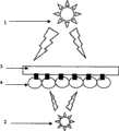
图1是用于分离上皮细胞的低(光)强度装置的图示。Figure 1 is a schematic representation of a low (light) intensity setup used to isolate epithelial cells.
1 用于照射细胞基膜复合体的基膜侧的光源;1 A light source for illuminating the basement membrane side of the cell basement membrane complex;
2 用于照射细胞基膜复合体的细胞侧的光源2;2
3 基膜;3 basement membrane;
4 细胞。4 cells.
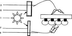
图2显示了使用单外部光的装置,其中滤光器和衰减器调节从基膜侧和从细胞侧落到细胞基膜复合体上的光的强度和波长,使得从细胞侧的照射与从基膜侧的照射相比,具有非常低的(光)强度。所述光通过光导纤维电缆传送。Figure 2 shows the setup using a single external light, where filters and attenuators adjust the intensity and wavelength of light falling onto the cell-basement membrane complex from the basement membrane side and from the cell side such that illumination from the cell side is identical to that from Compared to the irradiation on the basement film side, the (light) intensity is very low. The light is transmitted through a fiber optic cable.
1 单光源;1 single light source;
5 光导纤维电缆;5 fiber optic cables;
6 向所述复合体的所述基膜侧传送光能的滤光器与衰减器及起偏振镜;6. an optical filter, an attenuator, and a polarizer for transmitting light energy to the base film side of the complex;
7 向所述细胞基膜复合体的所述细胞侧传送不同特定光谱组成的较暗光的滤光器与衰减器及起偏振镜。7. Filters and attenuators and polarizers for delivering dimmer light of different specific spectral composition to the cell side of the cell basement membrane complex.
图3显示了一种外部光源,其直接落在基膜上,但经由反射镜指向细胞侧。所述衰减器、滤光器和起偏振镜以示意图的方式描述,且对本领域技术人员是显而易见的。所述照射应为:落在所述细胞基膜复合体的基膜侧的能量高于落在细胞基膜复合体的细胞侧的能量。Figure 3 shows an external light source falling directly on the basement membrane but directed towards the cell side via a mirror. The attenuators, filters and polarizers are described schematically and are obvious to those skilled in the art. The irradiation should be such that the energy falling on the basement membrane side of the cell basement membrane complex is higher than the energy falling on the cell side of the cell basement membrane complex.
1 波长194至1600纳米的光源;1 light sources with a wavelength of 194 to 1600 nanometers;
9 滤光器/起偏振镜/衰减器;9 filter/polarizer/attenuator;
3 基膜;3 basement membrane;
4 细胞;4 cells;
10 多个滤光器多个起偏振镜/多个衰减器;More than 10 filters, multiple polarizers/multiple attenuators;
8 镜。8 mirrors.
图4显示了由来自外部的光源对基膜的照射,所述光源穿过透明角膜,并用光能照射晶状体囊的外侧,而光导纤维从另一光源或相同的但通过滤光器和衰减器调节的光源传送光,且用光能照射所述复合体的细胞侧。Figure 4 shows the illumination of the basement membrane by a light source from the outside passing through the clear cornea and illuminating the outside of the lens capsule with light energy, while the light guides from another light source or the same but through filters and attenuators A modulated light source delivers light and illuminates the cellular side of the complex with light energy.
11 外部光源例如手术显微镜光源;11 External light sources such as operating microscope light sources;
12 从外部光源传送至组织的基膜侧的光;12 Light delivered from an external light source to the basement membrane side of the tissue;
13 从另一光源或从相同但被衰减和滤过的光源传送光能至细胞基膜复合体的另一侧(即:从细胞侧)的光导纤维;13 an optical fiber that transmits light energy from another light source or from the same but attenuated and filtered light source to the other side of the cell basement membrane complex (i.e. from the cell side);
14 细胞基膜复合体的内侧或细胞侧被通过带有平滑非创伤性端头的光导纤维传送的光照射;14 The medial or cellular side of the cell basement membrane complex is illuminated by light delivered through an optical fiber with a smooth non-traumatic tip;
15 角膜,其为透明的;15 cornea, which is transparent;
16 囊袋的切口部分,被称为撕囊术开口;16 The cut portion of the capsular bag, called the capsulorhexis opening;
17 囊袋的外侧。17 Outside of the pouch.
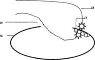
图5显示了从内部接触或接近晶状体囊的平滑端头,并显示了弯曲的端头,双重来源装置,带有所述装置端头的照射和放置的正确方法。Figure 5 shows the smooth tip touching or approaching the lens capsule from the inside and shows the curved tip, dual source device, with the correct method of irradiation and placement of the device tip.
19、18 光导纤维光源;19, 18 fiber optic light source;
16 晶状体囊或基膜;16 Lens capsule or basement membrane;
17 从内部覆面于晶状体囊的细胞。17 Cells lining the lens capsule from the inside.
图6显示了两种由光导纤维束或包住的光导纤维束制得的平滑的弯曲钩。所述平滑的钩是非创伤性,且这样做是为避免对可能靠近的其他生物结构的损伤。所述光载体与所述细胞基膜复合体的距离是非常靠近细胞的。Figure 6 shows two smooth curved hooks made from fiber optic bundles or sheathed fiber optic bundles. The smooth hooks are non-invasive, and this is done to avoid damage to other biological structures that may be in close proximity. The distance between the photocarrier and the cell basement membrane complex is very close to the cell.
20 由非创伤性设计的平滑弯曲的光传送系统照射基膜细胞复合体的基膜侧;20 The basement membrane side of the basement membrane cell complex is illuminated by an atraumatically designed smoothly curved light delivery system;
21 被照射的基膜方位;21 The orientation of the irradiated basement membrane;
22 第二光源,其通过另一平滑表面的非创伤性套管照射细胞基膜复合体的细胞侧;22 a second light source that illuminates the cell side of the cell basement membrane complex through another smooth-surfaced atraumatic cannula;
23 细胞基膜复合体的细胞侧。23 Cellular side of the basement membrane complex.
现将参考上述图1至6描述本发明。The present invention will now be described with reference to FIGS. 1 to 6 described above.
用于切割细胞基膜连接的装置由光源(1)和传送系统(5、13、17、18)组成,所述传送系统传送此光至特定部位,且如果基膜或晶状体囊成形为象卷曲的袋子或包封(envelop),所述传送系统传送能量至囊袋中(穿过开口进入袋中)。仪器与晶状体囊接触处的传送系统(14、20、22)的端头是平滑且非创伤性。The device for cutting the cell-basement membrane junction consists of a light source (1) and a delivery system (5, 13, 17, 18) that transmits this light to a specific site, and if the basement membrane or lens capsule is shaped like a curl The delivery system transmits energy into the pouch (through the opening into the pouch) of the pouch or envelope. The tip of the delivery system (14, 20, 22) where the instrument meets the lens capsule is smooth and atraumatic.
在另一实施方案中,两个光管传送光进入眼中,一个进入囊袋内部,另一个从外部照射囊袋,如图5所示,标记为19、18。In another embodiment, two light pipes transmit light into the eye, one into the interior of the capsular bag and the other illuminates the capsular bag from the outside, as shown in Figure 5, labeled 19,18.
光源light source
所述光源可为相干的或不相干的,单色的或多色的。其可为LED,或可为激光源、弧光灯源,钨丝光源或任何其他光源。日光可被使用并调节为一种光源。The light source can be coherent or incoherent, monochromatic or polychromatic. It can be an LED, or it can be a laser source, an arc lamp source, a tungsten light source or any other light source. Daylight can be used and adjusted as a light source.
所述光源可为白色的或可为彩色的。通过使用滤光器,白色光源可转化为某些纯色的光源。带有滤光器的单光源可用于产生纯色波长且所述囊袋的内部可用纯色照射。白光的混合光源也可使用。所选的波长在194至850纳米之间。The light source may be white or may be colored. By using filters, white light sources can be converted to light sources of certain pure colors. A single light source with filters can be used to generate a pure color wavelength and the interior of the pouch can be illuminated with a pure color. Mixed light sources of white light may also be used. The selected wavelengths are between 194 and 850 nm.
强度是所述装置的关键要素。用于本发明的所述光源的强度必须为这样的,其使落在细胞上的最终入射光具有非常低的强度以产生0.001勒克司至约1000勒克司的照度。应注意的是,如果非常接近于灯泡表面测量,一个40瓦的家用灯泡产生数千勒克司的照度。Strength is a key element of the device. The intensity of the light source used in the present invention must be such that the final incident light falling on the cells has a very low intensity to produce an illumination of 0.001 lux to about 1000 lux. It should be noted that a 40 watt household light bulb produces an illuminance of several thousand lux if measured very close to the surface of the bulb.
在一个优选的实施方案中,所述光源可被一秒数次地转换或脉冲开关。In a preferred embodiment, the light source can be toggled or pulsed on and off several times a second.
所述光源也可多于一个,以使细胞可交替地被不同波长的光照射。The light source can also be more than one, so that the cells can be illuminated alternately with light of different wavelengths.
与第一光源结合,必须使用第二光源。这可以是手术显微镜的外部光源,或完全不同的光源,藉此,光可被传送至所述基膜。这样的光源可为微小的LED、通过滤光器、光学聚焦透镜或起偏振镜或衰减器调节的日光、激光源、外部灯泡光源。In combination with the first light source, a second light source must be used. This can be an external light source of the surgical microscope, or a different light source entirely, whereby light can be transmitted to the basement membrane. Such light sources can be tiny LEDs, daylight conditioned by filters, optical focusing lenses or polarizers or attenuators, laser sources, external bulb sources.
在一优选的实施方案中,以0.002勒克司至500000勒克司的照度使用这样的光源。In a preferred embodiment, such light sources are used with an illumination of 0.002 lux to 500,000 lux.
此第二光源是必须设定的,且其必须以照度高于第一光源从细胞基膜复合体的内部或细胞侧起作用的照度照射细胞基膜复合体的基底侧。这样的照射应为同时的,以得到最好的效果。第二光源可为白色的,但也可具有不同的颜色。This second light source must be set and it must illuminate the basal side of the cell basement membrane complex with an illuminance higher than that of the first light source acting from the inside or the cell side of the cell basement membrane complex. Such exposures should be simultaneous for best results. The second light source can be white, but can also be of a different color.
为符合入射至基膜侧的能量高于入射至细胞侧的能量的条件,可使用多于一个的光源,以照射细胞基膜复合体的基膜侧。To comply with the condition that the energy incident on the basement membrane side is higher than the energy incident on the cell side, more than one light source may be used to illuminate the basement membrane side of the cell basement membrane complex.
如果第一光源是白光,且如果使用滤光镜以产生待被传送至晶状体囊内部的纯波长,通过绕过所述滤光器和添加如图2中所示的新的滤光器和衰减器,第一光源也可用作第二光源。If the first light source is white light, and if a filter is used to generate pure wavelengths to be delivered to the interior of the lens capsule, by bypassing the filter and adding a new filter and attenuation as shown in Figure 2 device, the first light source can also be used as the second light source.
传送系统delivery system
光导纤维电缆(图2中的5,图4中的13,图5中的18、19)或反射镜(图3中的8)用于直接向细胞传送光能。此光导纤维电缆也可被包在透明水封管状套管中以避免其与眼组织接触。Fiber optic cables (5 in Figure 2, 13 in Figure 4, 18, 19 in Figure 5) or mirrors (8 in Figure 3) are used to deliver light energy directly to the cells. The fiber optic cable may also be enclosed in a transparent water-sealed tubular sleeve to prevent it from coming into contact with ocular tissue.
套管的端头(图4中的14和图6中的20、22)是平滑、圆形的,以使当其与所述晶状体囊的下表面接触时,不会撕裂或损伤下表面。The tip of the sleeve (14 in FIG. 4 and 20, 22 in FIG. 6) is smooth and rounded so that when it contacts the lower surface of the lens capsule, it does not tear or damage the lower surface .
方法method
在实际操作期间,在第一次操作以后,所有可粘在细胞基膜复合体上的碎屑和污物可通过温和的抽吸和洗涤而被清理。如果此操作在实验室(在皿或容器中)进行,储存细胞基膜复合体的液体要免受污物或不溶的浮游颗粒污染。当此操作用在人体内时,如在白内障外科手术期间,则要除去白内障的内核。清洁皮质。通过所述装置传送低强度的光至囊袋内,且用所述光从内部照射所述细胞。显微镜用灯也可用作从基膜侧照射的第二光源。在实验室中,可将平滑细胞基膜复合体置于载玻片上并从双侧使用光能照射,来自低强度光源的能量直接落在细胞侧。在实验室中,当操作在显微镜下进行时,所述显微镜用灯也可用作第二光源,其将同时以较高能量照射基膜侧。通过用低强度光照射细胞表面和来自所述装置的高强度光照射基膜表面来游离/分离所述细胞。如果需要,通过已知的方法如简单的洗涤和抽吸可除去所述被分离的上皮细胞。During the actual operation, after the first operation, all debris and dirt that can stick to the cell basement membrane complex can be removed by gentle suction and washing. If this is done in the laboratory (in a dish or container), the liquid in which the cell-based membrane complexes are stored should be protected from contamination by dirt or insoluble suspended particles. When this procedure is used in the human body, as during cataract surgery, the inner core of the cataract is removed. Clean leather. Low intensity light is delivered through the device into the capsular bag, and the cells are illuminated with the light from the inside. A microscope lamp can also be used as a second light source for illuminating from the basement film side. In the laboratory, smooth cell basement membrane complexes can be placed on a glass slide and illuminated with light energy from both sides, with energy from a low-intensity light source falling directly on the cell side. In the laboratory, when working under a microscope, the microscope lamp can also be used as a second light source, which will simultaneously illuminate the basement membrane side with higher energy. The cells were dissociated/isolated by illuminating the cell surface with low intensity light and the basement membrane surface with high intensity light from the device. If desired, the isolated epithelial cells can be removed by known methods such as simple washing and aspiration.
此装置可有效地在相同时间从两侧用光照射所述晶状体囊细胞。一束光从外部落在晶状体前囊上。此光束来自被外科医生用作手术显微镜的光源,或来自位于外部的光源,且被由光导纤维制得的光管带至所述晶状体囊的前表面上。从外部落在前晶状体囊上的光可具有从0.002勒克司至500000勒克司的照度。This device effectively illuminates the lens capsule cells with light from both sides at the same time. A beam of light falls on the anterior capsule of the lens from the outside. This light beam comes from a light source used by the surgeon as an operating microscope, or from a light source located externally, and is brought onto the anterior surface of the lens capsule by a light pipe made of an optical fiber. Light falling on the anterior lens capsule from the outside may have an illuminance from 0.002 lux to 500,000 lux.
然而,此外部光束单独不能形成所述装置,所述装置必须基本上含有具有特定的低照度从内部同时落在细胞上的内部光束。However, this external light beam alone cannot form the device, which must essentially contain an internal light beam with a certain low illumination falling on the cells from the inside while falling on it.
用于从晶状体囊内部治疗细胞的来源的光可每秒开关一至十五次。The light from the source used to treat the cells from inside the lens capsule can be switched on and off one to fifteen times per second.
在本发明的另一实施方案中,所述光能通过一组置于弯管中的镜(以便代替光导纤维载体)被传送至晶状体前囊内部,所述光经由所述空管传送并通过这些反射镜和棱镜转入需要的路径。In another embodiment of the present invention, the light energy is delivered to the interior of the anterior capsule of the lens through a set of mirrors (in place of the fiber optic carrier) placed in the curved tube, the light is delivered through the empty tube and through the These mirrors and prisms turn into the desired path.
在本发明的另一实施方案中,使用图3所示的反射镜,将光源直接传送至可能的晶状体囊细胞照射位点,而不通过光导纤维电缆传送所述光。In another embodiment of the present invention, the light source is delivered directly to the potential lens capsule cell irradiation site using the mirror shown in FIG. 3 without delivering the light through a fiber optic cable.
然而,本发明不限于任何特定的应用或环境。相反,本领域技术人员将发现本发明可有益地应用于使用不同的低强度光源或其多重组合的任何应用或环境,以及通过任何其他直接的或间接的方法或方式应用这样的低强度光源的方法,以及镜或其他反射装置的用途。因此,接着的典型实施方案的描述是为了说明而非限制的目的。However, the invention is not limited to any particular application or environment. Rather, those skilled in the art will find that the present invention is beneficially applicable to any application or environment in which different low-intensity light sources or multiple combinations thereof are used, and to any application of such low-intensity light sources by any other direct or indirect method or means. methods, and the use of mirrors or other reflective devices. Accordingly, the ensuing description of typical embodiments is for purposes of illustration and not limitation.
最优选的实施方案most preferred embodiment
A.装置A. Device
两个光源,一个由蓝和红LED组成,其中蓝光为360至420纳米,且红LED为从700至850纳米。从每秒零次至每秒十五次脉冲所述LED。此光源用于从内部或细胞表面照射所述细胞基膜复合体。此强度是非常低的,以至在细胞表面的照度为0.001至1000勒克司。Two light sources, one consisting of blue and red LEDs, where the blue light is from 360 to 420 nm and the red LED is from 700 to 850 nm. The LED is pulsed from zero to fifteen times per second. This light source is used to illuminate the cell basement membrane complex from inside or from the cell surface. The intensity is so low that the illumination on the cell surface is 0.001 to 1000 lux.
第二光源为直接从手术显微镜使用的光。此光用于直接经由角膜照射所述细胞基膜复合体的基膜侧。为促进照射,通过滴眼剂或通过外科医生机械操作扩大瞳孔,以使虹膜移出第二光源的路径。使用的强度是使得基膜的照度为0.002至500000勒克司。The second light source is the light used directly from the surgical microscope. This light is used to illuminate the basement membrane side of the cell-basement membrane complex directly through the cornea. To facilitate irradiation, the pupil is dilated by eye drops or mechanically manipulated by the surgeon to move the iris out of the path of the second light source. The intensity used was such that the illumination of the basement film was between 0.002 and 500,000 lux.
从第一光源出来的光经由光导纤维光管采集,所述光导纤维光管传送光至眼内部。Light from the first light source is collected via a fiber optic light pipe that transmits the light to the interior of the eye.
所述光导纤维的末段为一套管(图6中的20、22),其端头是透明的,并允许此光被传送至晶状体囊。The end section of the optical fiber is a sleeve (20, 22 in Figure 6), the tip of which is transparent and allows this light to be transmitted to the lens capsule.
B.优选的实施方案——方法B. Preferred Embodiment - Method
对于用于分离上皮细胞的低强度装置的应用,所述套管应用于被清空内核与皮质的囊袋内部,且允许来自手术显微镜的第二光源经由外科医生通过手术前药物扩大瞳孔或机械地拉出虹膜来落在基膜上。在多处位置使用第一套管从内部接触晶状体囊,允许来自所述装置的光暂时地落在晶状体囊的不同区域。细胞被松弛并甚至已可在前房的流体中开始浮游。这些可通过已知的方法例如使用手持注射器和套管,或使用可与大多数超声乳化机器一起使用的自动化系统来温和冲洗和抽吸而被除去。For the application of low-intensity devices for separating epithelial cells, the cannula is applied inside the capsular bag that is emptied of the inner nucleus and cortex, and allows a second light source from the surgical microscope to dilate the pupil via preoperative medications by the surgeon or mechanically Pull out the iris to land on the basilar membrane. Contacting the lens capsule from the inside using the first sleeve at multiple locations allows light from the device to temporarily fall on different areas of the lens capsule. The cells are relaxed and can even begin to float in the fluid of the anterior chamber. These can be removed by known methods such as gentle irrigation and suction using a hand-held syringe and cannula, or using an automated system that can be used with most phacoemulsification machines.
在其最优选的实施方案中,所述装置不同于现有技术中公开的机械装置。本发明的装置不包含任何可移动的部分,不向细胞传送任何高强度的光,且特别是对于细胞基膜复合体的良定的部分,以良定的低强度和良定的时间周期,传送仅某些良定的波长的光。In its most preferred embodiment, said device is different from the mechanical devices disclosed in the prior art. The device of the present invention does not contain any movable parts, does not deliver any high-intensity light to the cells, and in particular delivers light at a well-defined low intensity and for a well-defined period of time to a well-defined portion of the cell-basement membrane complex. Only certain well-defined wavelengths of light.
在本申请中描述的装置在细胞侧使用具有特定能级的光能,所述光能级比现有技术所使用的低几千倍。本发明中的能量传送不意在“凝固”组织。在本申请中公开的装置在细胞侧使用非常低的光能并在细胞基膜复合体的基膜侧使用较高的光能,以通过切割细胞和基膜间的连接来温和地分离或松开所述细胞,以便所述细胞能被分离。The device described in this application uses light energy on the cell side with a specific energy level that is thousands of times lower than that used by the prior art. Energy delivery in the present invention is not intended to "coagulate" tissue. The device disclosed in this application uses very low light energy on the cell side and higher light energy on the basement membrane side of the cell basement membrane complex to gently detach or loosen the cell by cutting the junction between the cell and the basement membrane. The cells are opened so that the cells can be isolated.
用于现有技术中的典型的激光能显示的能量高于本申请中详细说明的被传送的能量几千倍。本申请中公开的装置使用从细胞侧0.001勒克司至最大1000勒克司的照度水平,并同时从晶状体囊侧或外测使用0.002勒克司至500000勒克司的较高的照度水平。此处公开的装置所需的能量为从内部0.0000024瓦的照度。Typical laser energies used in the prior art exhibit energies thousands of times higher than the delivered energies specified in this application. The device disclosed in this application uses illumination levels from 0.001 lux to a maximum of 1000 lux from the cell side, while simultaneously using higher illumination levels from 0.002 lux to 500,000 lux from the lens capsule side or lateral. The power required for the device disclosed here is 0.0000024 watts of illumination from the inside.
Claims (15)
Applications Claiming Priority (2)
| Application Number | Priority Date | Filing Date | Title |
|---|---|---|---|
| IN905MU2004 | 2004-08-23 | ||
| IN905/MUM/2004 | 2004-08-23 |
Publications (2)
| Publication Number | Publication Date |
|---|---|
| CN101048119A CN101048119A (en) | 2007-10-03 |
| CN100463664Ctrue CN100463664C (en) | 2009-02-25 |
Family
ID=34965238
Family Applications (1)
| Application Number | Title | Priority Date | Filing Date |
|---|---|---|---|
| CNB2004800442779AExpired - Fee RelatedCN100463664C (en) | 2004-08-23 | 2004-12-24 | Device for cutting the connection between cells and basement membrane or lens capsule |
Country Status (9)
| Country | Link |
|---|---|
| US (1) | US20070270923A1 (en) |
| EP (1) | EP1788993A1 (en) |
| JP (1) | JP2008510561A (en) |
| CN (1) | CN100463664C (en) |
| AU (1) | AU2004322538A1 (en) |
| CA (1) | CA2577889A1 (en) |
| IL (1) | IL181430A0 (en) |
| WO (1) | WO2006021970A1 (en) |
| ZA (1) | ZA200702305B (en) |
Citations (5)
| Publication number | Priority date | Publication date | Assignee | Title |
|---|---|---|---|---|
| CN1160530A (en)* | 1995-10-27 | 1997-10-01 | Ir视力公司 | Method and apparatus for removing corneal tissue with infrared laser radiation |
| US20020123744A1 (en)* | 2000-12-28 | 2002-09-05 | Michael Reynard | Phacophotolysis method and apparatus |
| CN1419432A (en)* | 2000-04-13 | 2003-05-21 | 株式会社尼康 | laser therapy equipment |
| US6673067B1 (en)* | 2000-01-31 | 2004-01-06 | Gholam A. Peyman | System and method for thermally and chemically treating cells at sites of interest in the body to impede cell proliferation |
| CN1148154C (en)* | 1997-03-14 | 2004-05-05 | 艾维希恩公司 | Short pulse mid-infrared parameter generator for surgery |
Family Cites Families (15)
| Publication number | Priority date | Publication date | Assignee | Title |
|---|---|---|---|---|
| US4966577A (en)* | 1988-03-16 | 1990-10-30 | Allergan, Inc. | Prevention of lens-related tissue growth in the eye |
| US4963142A (en)* | 1988-10-28 | 1990-10-16 | Hanspeter Loertscher | Apparatus for endolaser microsurgery |
| US6099522A (en)* | 1989-02-06 | 2000-08-08 | Visx Inc. | Automated laser workstation for high precision surgical and industrial interventions |
| US5627162A (en)* | 1990-01-11 | 1997-05-06 | Gwon; Arlene E. | Methods and means for control of proliferation of remnant cells following surgery |
| GB9203533D0 (en)* | 1992-02-19 | 1992-04-08 | Erba Carlo Spa | Use of the conjugate between a fibroblast growth factor and a saporin in treating ocular pathologies |
| DE4322955B4 (en)* | 1992-07-20 | 2007-12-20 | Aesculap Ag & Co. Kg | Invasive surgical instrument |
| US5445637A (en)* | 1993-12-06 | 1995-08-29 | American Cyanamid Company | Method and apparatus for preventing posterior capsular opacification |
| US5491343A (en)* | 1994-03-25 | 1996-02-13 | Brooker; Gary | High-speed multiple wavelength illumination source, apparatus containing the same, and applications thereof to methods of irradiating luminescent samples and of quantitative luminescence ratio microscopy |
| US5620013A (en)* | 1994-10-21 | 1997-04-15 | American Cyanamid Company | Method for destroying residual lens epithelial cells |
| DE29801223U1 (en)* | 1998-01-27 | 1998-05-14 | Rösler, Peter, 81377 München | Fiber optic application set |
| US6669694B2 (en)* | 2000-09-05 | 2003-12-30 | John H. Shadduck | Medical instruments and techniques for highly-localized thermally-mediated therapies |
| FR2796295B1 (en)* | 1999-07-13 | 2001-10-05 | Inst Nat Sante Rech Med | LASER PHOTOCOAGULATOR WITH FLUENCE ADAPTATION |
| US6432078B1 (en)* | 2000-06-19 | 2002-08-13 | Gholam A. Peyman | System and method for removing cataract or other cells in an eye using water jet and suction |
| JP4046937B2 (en)* | 2000-10-02 | 2008-02-13 | 株式会社ニデック | Laser surgical device |
| US6554824B2 (en)* | 2000-12-15 | 2003-04-29 | Laserscope | Methods for laser treatment of soft tissue |
- 2004
- 2004-12-24CNCNB2004800442779Apatent/CN100463664C/ennot_activeExpired - Fee Related
- 2004-12-24WOPCT/IN2004/000410patent/WO2006021970A1/enactiveApplication Filing
- 2004-12-24JPJP2007529141Apatent/JP2008510561A/enactivePending
- 2004-12-24AUAU2004322538Apatent/AU2004322538A1/ennot_activeAbandoned
- 2004-12-24USUS11/574,111patent/US20070270923A1/ennot_activeAbandoned
- 2004-12-24CACA002577889Apatent/CA2577889A1/ennot_activeAbandoned
- 2004-12-24EPEP04821395Apatent/EP1788993A1/ennot_activeWithdrawn
- 2007
- 2007-02-19ILIL181430Apatent/IL181430A0/enunknown
- 2007-03-20ZAZA200702305Apatent/ZA200702305B/enunknown
Patent Citations (5)
| Publication number | Priority date | Publication date | Assignee | Title |
|---|---|---|---|---|
| CN1160530A (en)* | 1995-10-27 | 1997-10-01 | Ir视力公司 | Method and apparatus for removing corneal tissue with infrared laser radiation |
| CN1148154C (en)* | 1997-03-14 | 2004-05-05 | 艾维希恩公司 | Short pulse mid-infrared parameter generator for surgery |
| US6673067B1 (en)* | 2000-01-31 | 2004-01-06 | Gholam A. Peyman | System and method for thermally and chemically treating cells at sites of interest in the body to impede cell proliferation |
| CN1419432A (en)* | 2000-04-13 | 2003-05-21 | 株式会社尼康 | laser therapy equipment |
| US20020123744A1 (en)* | 2000-12-28 | 2002-09-05 | Michael Reynard | Phacophotolysis method and apparatus |
Also Published As
| Publication number | Publication date |
|---|---|
| IL181430A0 (en) | 2007-07-04 |
| AU2004322538A1 (en) | 2006-03-02 |
| JP2008510561A (en) | 2008-04-10 |
| US20070270923A1 (en) | 2007-11-22 |
| WO2006021970A1 (en) | 2006-03-02 |
| CN101048119A (en) | 2007-10-03 |
| ZA200702305B (en) | 2008-08-27 |
| CA2577889A1 (en) | 2006-03-02 |
| EP1788993A1 (en) | 2007-05-30 |
Similar Documents
| Publication | Publication Date | Title |
|---|---|---|
| AU2011289406B2 (en) | Dual-mode illumination for surgical instrument | |
| JP4532051B2 (en) | Irradiation device for treating eye diseases | |
| US9510847B2 (en) | Targeted illumination for surgical instrument | |
| JP2983561B2 (en) | Medical surgical device for living tissue using laser beam | |
| US5885279A (en) | Method and apparatus for preventing posterior capsular opacification | |
| JP2002515774A (en) | Fiber optic sleeve for surgical instruments | |
| JP2021509841A (en) | Ultraviolet laser vitrectomy probe | |
| MX2013008284A (en) | WHITE COHERENT LASER LIGHT LAUNCHED TO NANOFIBERS FOR SURGICAL LIGHTING. | |
| US20100318074A1 (en) | Ophthalmic endoillumination using low-power laser light | |
| MX2012005502A (en) | Single-fiber multi-spot laser probe for ophthalmic endoillumination. | |
| EP0293458A1 (en) | METHOD AND DEVICE FOR REMOVING AND REMOVING AN EYE LENS INFECTED BY GRAY STAR. | |
| US5445636A (en) | Method and apparatus for preventing posterior capsular opacification | |
| CN100463664C (en) | Device for cutting the connection between cells and basement membrane or lens capsule | |
| Hemo et al. | Vitreoretinal surgery assisted by the 193-nm excimer laser. | |
| KR20070091101A (en) | Incision of cell binding to the basement membrane |
Legal Events
| Date | Code | Title | Description |
|---|---|---|---|
| C06 | Publication | ||
| PB01 | Publication | ||
| C10 | Entry into substantive examination | ||
| SE01 | Entry into force of request for substantive examination | ||
| C14 | Grant of patent or utility model | ||
| GR01 | Patent grant | ||
| C17 | Cessation of patent right | ||
| CF01 | Termination of patent right due to non-payment of annual fee | Granted publication date:20090225 Termination date:20101224 |


