CN100453026C - Device to be introduced into subject - Google Patents
Device to be introduced into subjectDownload PDFInfo
- Publication number
- CN100453026C CN100453026CCNB2005800079385ACN200580007938ACN100453026CCN 100453026 CCN100453026 CCN 100453026CCN B2005800079385 ACNB2005800079385 ACN B2005800079385ACN 200580007938 ACN200580007938 ACN 200580007938ACN 100453026 CCN100453026 CCN 100453026C
- Authority
- CN
- China
- Prior art keywords
- subject
- magnetic field
- unit
- capsule
- permanent magnet
- Prior art date
- Legal status (The legal status is an assumption and is not a legal conclusion. Google has not performed a legal analysis and makes no representation as to the accuracy of the status listed.)
- Expired - Fee Related
Links
Images

Classifications
- A—HUMAN NECESSITIES
- A61—MEDICAL OR VETERINARY SCIENCE; HYGIENE
- A61B—DIAGNOSIS; SURGERY; IDENTIFICATION
- A61B5/00—Measuring for diagnostic purposes; Identification of persons
- A61B5/06—Devices, other than using radiation, for detecting or locating foreign bodies ; Determining position of diagnostic devices within or on the body of the patient
- A—HUMAN NECESSITIES
- A61—MEDICAL OR VETERINARY SCIENCE; HYGIENE
- A61B—DIAGNOSIS; SURGERY; IDENTIFICATION
- A61B1/00—Instruments for performing medical examinations of the interior of cavities or tubes of the body by visual or photographical inspection, e.g. endoscopes; Illuminating arrangements therefor
- A61B1/04—Instruments for performing medical examinations of the interior of cavities or tubes of the body by visual or photographical inspection, e.g. endoscopes; Illuminating arrangements therefor combined with photographic or television appliances
- A61B1/041—Capsule endoscopes for imaging
- A—HUMAN NECESSITIES
- A61—MEDICAL OR VETERINARY SCIENCE; HYGIENE
- A61B—DIAGNOSIS; SURGERY; IDENTIFICATION
- A61B5/00—Measuring for diagnostic purposes; Identification of persons
- A61B5/06—Devices, other than using radiation, for detecting or locating foreign bodies ; Determining position of diagnostic devices within or on the body of the patient
- A61B5/061—Determining position of a probe within the body employing means separate from the probe, e.g. sensing internal probe position employing impedance electrodes on the surface of the body
- A61B5/062—Determining position of a probe within the body employing means separate from the probe, e.g. sensing internal probe position employing impedance electrodes on the surface of the body using magnetic field
- A—HUMAN NECESSITIES
- A61—MEDICAL OR VETERINARY SCIENCE; HYGIENE
- A61B—DIAGNOSIS; SURGERY; IDENTIFICATION
- A61B5/00—Measuring for diagnostic purposes; Identification of persons
- A61B5/06—Devices, other than using radiation, for detecting or locating foreign bodies ; Determining position of diagnostic devices within or on the body of the patient
- A61B5/061—Determining position of a probe within the body employing means separate from the probe, e.g. sensing internal probe position employing impedance electrodes on the surface of the body
- A61B5/064—Determining position of a probe within the body employing means separate from the probe, e.g. sensing internal probe position employing impedance electrodes on the surface of the body using markers
- A—HUMAN NECESSITIES
- A61—MEDICAL OR VETERINARY SCIENCE; HYGIENE
- A61B—DIAGNOSIS; SURGERY; IDENTIFICATION
- A61B5/00—Measuring for diagnostic purposes; Identification of persons
- A61B5/68—Arrangements of detecting, measuring or recording means, e.g. sensors, in relation to patient
- A61B5/6846—Arrangements of detecting, measuring or recording means, e.g. sensors, in relation to patient specially adapted to be brought in contact with an internal body part, i.e. invasive
- A61B5/6847—Arrangements of detecting, measuring or recording means, e.g. sensors, in relation to patient specially adapted to be brought in contact with an internal body part, i.e. invasive mounted on an invasive device
- A61B5/6861—Capsules, e.g. for swallowing or implanting
Landscapes
- Health & Medical Sciences (AREA)
- Life Sciences & Earth Sciences (AREA)
- Engineering & Computer Science (AREA)
- Surgery (AREA)
- General Health & Medical Sciences (AREA)
- Veterinary Medicine (AREA)
- Biomedical Technology (AREA)
- Heart & Thoracic Surgery (AREA)
- Medical Informatics (AREA)
- Molecular Biology (AREA)
- Physics & Mathematics (AREA)
- Animal Behavior & Ethology (AREA)
- Biophysics (AREA)
- Pathology (AREA)
- Public Health (AREA)
- Human Computer Interaction (AREA)
- Nuclear Medicine, Radiotherapy & Molecular Imaging (AREA)
- Optics & Photonics (AREA)
- Radiology & Medical Imaging (AREA)
- Endoscopes (AREA)
- Measurement Of The Respiration, Hearing Ability, Form, And Blood Characteristics Of Living Organisms (AREA)
- Infusion, Injection, And Reservoir Apparatuses (AREA)
Abstract
Description
Translated fromChinese技术领域technical field
本发明涉及为了从外部获取被检体内部的位置信息而被导入所述被检体内、并在该被检体内移动的被检体内导入装置。The present invention relates to a subject-introducing device that is introduced into a subject and moves within the subject in order to acquire positional information inside the subject from the outside.
背景技术Background technique
近年来,在内窥镜领域中,装备了摄像功能和无线功能的胶囊型内窥镜已经登场。该胶囊型内窥镜构成为,为了进行观察(检查),在由被检体即被检查者吞入后,到从被检查者的生体(人体)自然排出之前的观察期间,在胃、小肠等脏器内部(体腔内)伴随其蠕动运动而移动,使用摄像功能依次摄像。In recent years, in the field of endoscopes, capsule endoscopes equipped with imaging functions and wireless functions have appeared. This capsule endoscope is configured such that, for observation (inspection), after being swallowed by the subject, that is, the subject, and before being naturally excreted from the subject's living body (human body), The inside of the organ (inside the body cavity) moves along with its peristaltic movement, and the camera function is used to take pictures in sequence.
并且,在这些脏器内移动的该观察期间,通过胶囊型内窥镜在体腔内拍摄到的图像数据,借助于无线通信等无线功能,被依次发送到设于被检体外部的外部装置,存储在设于外部装置内的存储器中。被检查者携带具有该无线功能和存储器功能的外部装置,使得被检查者在吞入胶囊型内窥镜后到该胶囊型内窥镜被排出之前的观察期间,可以自由行动。在观察后,医生或护士根据存储在外部装置的存储器中的图像数据,可以使显示器等显示单元显示体腔内的图像并进行诊断。And, during the observation period when these organs move, the image data captured by the capsule endoscope in the body cavity is sequentially transmitted to an external device installed outside the subject by means of a wireless function such as wireless communication, Stored in the memory installed in the external device. The subject carries the external device having the wireless function and the memory function, so that the subject can move freely during the observation period after swallowing the capsule endoscope and before the capsule endoscope is discharged. After observation, the doctor or nurse can make a display unit such as a monitor display images in the body cavity and make a diagnosis based on the image data stored in the memory of the external device.
可是,该体腔有时由于对病情的移位等,产生管腔狭窄、狭小或堵塞等情况。因此,需要在上述观察步骤中确认体腔内的胶囊型内窥镜的通过是否在顺畅进行。在这种情况下,如果能够预先获得与体腔内的检查装置(被检体内导入装置)的通过相关的信息(位置信息),则对判断是否可以安全地对患者使用胶囊型内窥镜方面是有益的。However, the body cavity may be narrowed, narrowed, or clogged due to displacement of the disease or the like. Therefore, it is necessary to confirm whether or not the passage of the capsule endoscope in the body cavity is being carried out smoothly in the above observation step. In this case, if the information (position information) related to the passage of the inspection device (introduction device in the subject) in the body cavity can be obtained in advance, it is useful for judging whether the capsule endoscope can be safely used on the patient. benefit.
因此,以往在被检体内导入装置中使用电子ID标签,把该被检体内导入装置在所导入的被检体内的位置信息电发送到外部,验证被检体内导入装置是否在体腔内顺畅通过(参照专利文献1)。在该现有例中,在被检体内导入装置中设置电力源,该电力源用于驱动为了获取位置信息而设置的电子ID,向外部发送标签信息。Therefore, in the past, an electronic ID tag was used in the introduction device in the subject to electronically send the position information of the introduction device in the subject to the outside to verify whether the introduction device in the subject passed smoothly in the body cavity ( Refer to Patent Document 1). In this conventional example, a power source is provided in the subject's body-introduction device, and the power source is used to drive an electronic ID provided for acquiring positional information and to transmit tag information to the outside.
专利文献1 日本国际公开第03/005877号小册子Patent Document 1 Japanese International Publication No. 03/005877 Pamphlet
但是,被检体内导入装置的封装部(外壳)与胶囊型内窥镜相同,具有过了一定时间则分解的结构,例如由于体腔的管道狭窄等原因,内部具有电力源的被检体内导入装置在被检体内滞留该一定时间以上,则外壳分解,存在有可能对被检体带来不良影响的问题。However, the sealing part (housing) of the device introduced into the subject has a structure that disintegrates after a certain period of time, like the capsule endoscope. If the case stays in the subject for a certain period of time or longer, the case may disintegrate, which may have a negative effect on the subject.
并且,在被检体内导入装置内部,除了上述电力源外,用于发送位置信息的无线装置等与其他装置一起例如被设计成芯片,因此存在结构变复杂,而且制造成本升高的问题。Furthermore, in the body-introduced device, in addition to the above-mentioned power source, a wireless device for transmitting position information is designed as a chip, for example, along with other devices. Therefore, there is a problem that the structure becomes complicated and the manufacturing cost increases.
发明内容Contents of the invention
本发明就是鉴于上述问题而提出的,其目的在于,提供一种被检体内导入装置,即使封装部分解也不会给被检体造成影响,结构简单,而且能够安全地获得位置信息。The present invention was made in view of the above problems, and an object of the present invention is to provide a body-introduced device that does not affect the subject even if the package is disassembled, has a simple structure, and can securely obtain position information.
为了解决上述问题并达到目的,本发明的被检体内导入装置被导入被检体内并在该被检体内移动,其特征在于,具备:磁场产生单元,其产生恒定磁场;覆盖单元,其覆盖所述磁场产生单元,由生物体适合性材料形成;以及封装单元,其在内部收纳被所述覆盖单元覆盖的所述磁场产生单元并密封。In order to solve the above problems and achieve the purpose, the device for introducing into the subject of the present invention is introduced into the subject and moves in the subject, and is characterized in that it includes: a magnetic field generating unit that generates a constant magnetic field; a covering unit that covers the The magnetic field generating unit is formed of a biocompatible material; and a packaging unit accommodates and seals the magnetic field generating unit covered by the covering unit inside.
并且,本发明之二的被检体内导入装置的特征在于,在上述发明中,所述封装单元构成为在被导入所述被检体内后经过规定时间后分解,可以释放收纳于内部的所述磁场产生单元。In addition, the device for introducing into a subject according to the second aspect of the present invention is characterized in that, in the above invention, the packaging unit is configured to be disassembled after a predetermined time has elapsed after being introduced into the subject, thereby releasing the Magnetic field generating unit.
并且,本发明之三的被检体内导入装置的特征在于,在上述发明中,还具备填充单元,其被填充于所述封装单元的内部。In addition, the device for introducing into a subject according to the third aspect of the present invention is characterized in that, in the above invention, it further includes a filling unit that fills the inside of the sealing unit.
并且,本发明之四的被检体内导入装置的特征在于,在上述发明中,所述生物体适合性材料由陶瓷或钛构成。Furthermore, the fourth aspect of the present invention is the device for introducing into a subject in the above-mentioned invention, wherein the biocompatible material is made of ceramics or titanium.
并且,本发明之五的被检体内导入装置的特征在于,在上述发明中,所述填充单元由生理盐水或钡构成。Furthermore, according to the fifth aspect of the present invention, the device for introducing into a subject is characterized in that, in the above invention, the filling unit is made of physiological saline or barium.
本发明的被检体内导入装置具备向被检体外部输出恒定磁场的磁场产生单元,并且利用生物体适合性材料覆盖该磁场产生单元,所以即使例如封装部分解,磁场产生单元被生物体适合性材料覆盖,所以不会给被检体造成影响,由此具有结构简单、且能够安全地获得位置信息的效果。The device for introducing into the subject of the present invention has a magnetic field generating unit that outputs a constant magnetic field to the outside of the subject, and the magnetic field generating unit is covered with a biocompatible material, so even if the package is disassembled, the magnetic field generating unit is biocompatible. Since it is covered with a material, it does not affect the subject, thereby having an effect that the structure is simple and positional information can be obtained safely.
附图说明Description of drawings
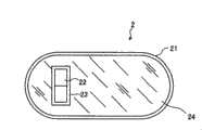
图1是表示本发明的被检体内导入装置的概要结构的一例的结构图。FIG. 1 is a configuration diagram showing an example of a schematic configuration of the device introduced into a subject of the present invention.
图2是表示本发明的被检体内导入装置的概要结构的另一例子的结构图。Fig. 2 is a configuration diagram showing another example of the schematic configuration of the device introduced into the subject of the present invention.
图3是表示实施例的被检体内移动状态检测系统的整体结构的示意图。Fig. 3 is a schematic diagram showing the overall configuration of a system for detecting a moving state in a subject according to the embodiment.
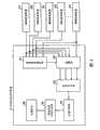
图4是表示图3所示移动状态导出装置的结构的示意图。FIG. 4 is a schematic diagram showing the configuration of the movement state deriving device shown in FIG. 3 .
符号说明Symbol Description
1被检体;2通过确认用胶囊;3移动状态检测装置;4显示装置;5便携式记录介质;6a~6h磁场检测装置;7a、7b固定部件;8移动状态导出装置;9接收用天线;10发送用天线;21外壳;22永磁;23生物体适合性材料;24填充部件;81基准装置选择部;82选择器;83距离导出部;84位置计算部;85移动状态信息生成部;86存储单元。1 Subject; 2 Pass confirmation capsule; 3 Movement state detection device; 4 Display device; 5 Portable recording medium; 6a~6h Magnetic field detection device; 7a, 7b Fixed parts; 10 antenna for transmission; 21 casing; 22 permanent magnet; 23 biocompatible material; 24 filling part; 81 reference device selection unit; 82 selector; 83 distance derivation unit; 86 storage units.
具体实施方式Detailed ways
以下,根据图1~图4详细说明本发明的被检体内导入装置的实施例。另外,本发明不限于这些实施例,可以在不脱离本发明宗旨的范围内实现各种变更的实施方式。Hereinafter, an embodiment of the device introduced into the subject of the present invention will be described in detail with reference to FIGS. 1 to 4 . In addition, this invention is not limited to these Examples, Various changes can be implemented in the range which does not deviate from the summary of this invention.
实施例1Example 1
图1是表示本发明的被检体内导入装置的概要结构的一例的结构图,图2是表示本发明的被检体内导入装置的概要结构的另一例子的结构图。该被检体内导入装置涉及通过确认用胶囊,该通过确认用胶囊在进行被检体内是否存在胶囊型内窥镜很难通过的狭窄部等的事前检查时使用。FIG. 1 is a configuration diagram showing an example of a schematic configuration of the device introduced into a subject according to the present invention, and FIG. 2 is a configuration diagram showing another example of a schematic configuration of the device introduced into a subject according to the present invention. The device introduced into the subject relates to a passage confirmation capsule used for pre-examination of whether there is a narrow portion in the subject through which a capsule endoscope is difficult to pass.
在图1中,作为被检体内导入装置的通过确认用胶囊2具备:作为封装单元的外壳21,其具有与胶囊型内窥镜的封装部相同的胶囊形状;作为磁场产生单元的永磁22,其配置在外壳21的内部,而且被生物体适合性材料23所涂覆;以及作为填充单元的填充部件24,其被填充于外壳21和永磁22之间的间隙中。In FIG. 1 , the
外壳21由大致半球状的圆顶形状的两个前端部和形成于这两个前端部之间的圆筒状的躯干部构成为一体。这些前端部和躯干部由可以根据来自外部的规定量以上的加压而变形的柔软性材料形成,例如利用柔软明胶或生物分解性聚合物等形成,而且具有当在被检体1内滞留了一定期间时,与在被检体1内部分泌的体液(特别是消化道的消化液)反应而分解的特性。这样,外壳21形成为在被检体1内分解的结构,具有如下优点:在万一被导入被检体1内的通过确认用胶囊2未能排出到被检体1外部时,不需要对被检体1进行开腹手术等。此处,关于该外壳21分解所需要的时间,通过调整其厚度、或层叠外壳21的材料来确定。The
永磁22例如构成为圆板型,在外部形成恒定磁场。该永磁22例如由钐钴磁铁或钕磁铁等形成,外表面被利用不会给被检体1造成坏影响的生物体适合性材料、例如由陶瓷或钛等构成的生物体适合性材料23实施了涂覆。另外,该生物体适合性材料23形成为完全不会给永磁22输出的恒定磁场的磁场强度和行进方向带来影响。该永磁22的外径形成为比外壳21的躯干部分的内径小,而且长度也小于外壳21的内部长度,与填充部件24一起被收纳在外壳21内。另外,也可以不进行对永磁22的外表面的涂覆,而利用涂覆了生物体适合性材料的热缩管覆盖在外表面。另外,也可以通过这种覆盖,形成内部具有永磁22的大致球状的结构体,将这种结构体与填充部件24一起收纳在外壳21内。The
填充部件24填充外壳21和永磁22之间的空间,用于抑制配置在外壳21内部的永磁22的移动,例如使用生理盐水或硫酸钡等作为材料,即使外壳21分解也不必担心给被检体1带来不良影响。特别是在使用硫酸钡形成填充部件24时,可以把填充部件24用作造影剂,具有例如可以通过X线检查来检测永磁22的位置的优点。The filling
这样,本实施例的被检体内导入装置在外壳内具有利用生物体适合性材料覆盖的永磁,所以例如在被导入被检体内的被检体内导入装置滞留在体腔内部而难以排出到外部的情况下,在经过一定时间后,外壳分解,只剩下小径的圆板型外形的、且外表面被实施了生物体适合性材料的涂覆或覆盖的永磁,所以不会给被检体造成不良影响,容易在体腔内部通过排出到外部,结构简单,且能够安全地获得位置信息。In this way, the device introduced into the subject of this embodiment has a permanent magnet covered with a biocompatible material in the housing, so for example, the device introduced into the subject stays inside the body cavity and is difficult to discharge to the outside. In some cases, after a certain period of time, the shell disintegrates, leaving only a small-diameter disk-shaped permanent magnet whose outer surface is coated or covered with a biocompatible material, so it will not cause damage to the subject. It causes adverse effects, and is easy to be discharged to the outside through the inside of the body cavity. The structure is simple, and position information can be obtained safely.
另外,外壳21和永磁22的形状不限于上述方式,如图2所示,也可以使外壳21和永磁22都形成为大致球状。此处,该外壳21的外径设定为与胶囊型内窥镜的外径相同的大小,由此发挥与图1所述相同的效果。In addition, the shapes of the
下面,使用图3的示意图说明该实施例的被检体移动状态检测系统。在图3中,该实施例的被检体移动状态检测系统具有:上述通过确认用胶囊2,其被导入被检体1的内部,作为被检体内导入装置的一例发挥作用;和移动状态检测装置3,其进行通过确认用胶囊2在被检体1内部的移动状态的检测等。另外,在图3中除此之外还具有:显示利用胶囊内窥镜拍摄到的被检体1内的图像等的显示装置4;和用于进行移动状态检测装置3和显示装置4之间的信息交换的便携式记录介质5。即,该实施例的被检体移动状态检测系统用于调查通过确认用胶囊在被检体内如何移动。Next, the subject movement state detection system of this embodiment will be described using the schematic diagram of FIG. 3 . In FIG. 3 , the subject movement state detection system of this embodiment includes: the
移动状态检测装置3根据从通过确认用胶囊2输出的恒定磁场,检测通过确认用胶囊2在被检体1内部的移动状态。具体讲,移动状态检测装置3如图3所示,具有:检测从通过确认用胶囊2输出的恒定磁场的磁场检测装置6a~6h;把磁场检测装置6a~6d相对被检体1固定的固定部件7a;把磁场检测装置6e~6h相对被检体1固定的固定部件7b;和根据磁场检测装置6a~6h检测出的磁场强度,导出通过确认用胶囊2的位置的移动状态导出装置8。并且,移动状态检测装置3除此之外还具有:接收从胶囊型内窥镜发送的无线信号的接收用天线9;和向胶囊型内窥镜发送无线信号的发送用天线10。磁场检测装置6a~6h、接收用天线9以及发送用天线10具有与移动状态导出装置8电连接,向移动状态导出装置8进行信息的输入或输出的结构。另外,磁场检测装置6a~6h例如使用MI(magneto impedance,磁阻抗)传感器形成,分别检测所配置场所的磁场强度和磁场方向,在该实施例中,8个传感器分别配置在形成立方体的顶点的位置上。The moving
显示装置4用于显示通过未图示的胶囊型内窥镜拍摄到的体腔内图像等,具有根据通过便携式记录介质5获得的数据进行图像显示的工作站等的结构。具体讲,显示装置4可以是利用CRT显示器、液晶显示器等直接显示图像的结构,也可以是向打印机等那样的其它介质输出图像的结构。The display device 4 is used to display images in a body cavity captured by a capsule endoscope (not shown), and has a configuration such as a workstation for displaying images based on data obtained from the
便携式记录介质5也可以连接到移动状态导出装置8和显示装置4,具有被插装连接在两者上时可以进行信息的输出或记录的结构。在该实施例中,在通过确认用胶囊2在被检体1的体腔内移动的期间,便携式记录介质5被插装在移动状态导出装置8上,记录与通过确认用胶囊2的位置相关的信息。然后,在通过确认用胶囊2从被检体1被排出后,便携式记录介质5从移动状态导出装置8被取出,插装到显示装置4上,通过该显示装置4读出记录在便携式记录介质5中的数据。例如,该便携式记录介质5由紧凑式闪存(CF)(注册商标)存储器等构成,可以通过便携式记录介质5间接进行移动状态导出装置8和显示装置4之间的数据的输入输出,与移动状态导出装置8和显示装置4之间通过有线直接连接时不同,即使在通过确认用胶囊2在被检体1内部移动时,被检体1也能够自由行动。The
如图4所示,移动状态导出装置8具有:基准装置选择部81,其从磁场检测装置6a~6h中选择基准装置;选择器82,其根据所选择的基准装置,选择被选择装置,输出通过基准装置和被选择装置获得的磁场强度;距离导出部83,其根据从选择器82输出的磁场强度,导出与通过确认用胶囊2之间的距离;位置计算部84,其根据所导出的距离,导出通过确认用胶囊2的位置;移动状态信息生成部85,其根据所导出的位置,生成移动状态信息。另外,移动状态导出装置8具有存储单元86,其存储由移动状态信息生成部85生成的移动状态信息。并且,形成为通过该存储单元86和便携式记录介质5向显示装置4输出移动状态信息的结构,从而使医生等能够把握通过确认用胶囊2的移动状态。As shown in FIG. 4, the moving state deriving device 8 has: a reference
另外,在该实施例中,作为要导出的移动状态信息,只把通过确认用胶囊2的位置变动作为对象,但不限于此,也可以构成为利用指向方向即永磁22输出的恒定磁场的行进方向的场所依赖性,导出通过确认用胶囊2的长轴所朝向的方向的变动。并且,通过确认用胶囊2和永磁22的形状例如也可以形成为球状,该情况下,更容易进行向被检体1内部的导入和向被检体1外部的排出。In addition, in this embodiment, as the moving state information to be derived, only the position change of the
并且,在该实施例中,针对被检体内导入装置说明了通过确认用胶囊的情况,但本发明不限于此,例如胶囊型内窥镜也可以采用本发明涉及的被检体内导入装置的结构。该情况时,可以获得被检体内的图像信息,并且可同时获得其位置信息,例如可以容易地设想摄像位置。In addition, in this embodiment, the case of passing through the capsule for confirmation is described for the device introduced into the subject, but the present invention is not limited thereto. For example, a capsule endoscope may adopt the structure of the device introduced into the subject according to the present invention. . In this case, image information inside the subject can be obtained, and position information thereof can be obtained at the same time, for example, imaging positions can be easily imagined.
如上所述,本发明的被检体内导入装置被导入人体内部,对观察被检查部位的医疗用观察装置比较有用,特别适合于即使外壳分解也不会给被检体带来影响,结构简单,且安全地获取被检体内导入装置的位置信息的应用。As mentioned above, the device for introducing into the subject of the present invention is introduced into the human body, and is useful as a medical observation device for observing the part to be inspected, and is particularly suitable for not affecting the subject even if the casing is disassembled, and has a simple structure. And securely obtain the application of the position information of the device introduced into the subject.
Claims (5)
Translated fromChineseApplications Claiming Priority (2)
| Application Number | Priority Date | Filing Date | Title |
|---|---|---|---|
| JP071582/2004 | 2004-03-12 | ||
| JP2004071582AJP2005253798A (en) | 2004-03-12 | 2004-03-12 | Internally introduced device in subject |
Publications (2)
| Publication Number | Publication Date |
|---|---|
| CN1929775A CN1929775A (en) | 2007-03-14 |
| CN100453026Ctrue CN100453026C (en) | 2009-01-21 |
Family
ID=34975265
Family Applications (1)
| Application Number | Title | Priority Date | Filing Date |
|---|---|---|---|
| CNB2005800079385AExpired - Fee RelatedCN100453026C (en) | 2004-03-12 | 2005-02-02 | Device to be introduced into subject |
Country Status (5)
| Country | Link |
|---|---|
| US (2) | US20070049818A1 (en) |
| EP (1) | EP1723896A4 (en) |
| JP (1) | JP2005253798A (en) |
| CN (1) | CN100453026C (en) |
| WO (1) | WO2005087079A1 (en) |
Families Citing this family (11)
| Publication number | Priority date | Publication date | Assignee | Title |
|---|---|---|---|---|
| EP1965698B1 (en)* | 2005-12-29 | 2014-02-19 | Given Imaging Ltd. | System and method of in-vivo magnetic position determination |
| JP2008012094A (en)* | 2006-07-06 | 2008-01-24 | Kenichi Katsu | Capsule endoscope |
| WO2008093578A1 (en)* | 2007-01-30 | 2008-08-07 | Olympus Medical Systems Corp. | Device for checking for lumen passage, method of melting the same and method of producing the same |
| EP2134403B1 (en)* | 2007-04-11 | 2012-12-12 | Elcam Medical Agricultural Cooperative Association Ltd. | System for accurate placement of a catheter tip in a patient |
| CN101711673B (en)* | 2009-10-16 | 2012-11-21 | 重庆金山科技(集团)有限公司 | System, device and method for wireless monitoring and positioning of pH value of esophagus |
| CN102670158A (en)* | 2012-05-10 | 2012-09-19 | 无锡市华焯光电科技有限公司 | Locatable capsule endoscope system |
| US10143364B2 (en)* | 2015-07-23 | 2018-12-04 | Ankon Technologies Co., Ltd | Controlled image capturing method including position tracking and system used therein |
| US10679018B1 (en) | 2019-02-05 | 2020-06-09 | International Business Machines Corporation | Magnetic tracking for medicine management |
| US10824822B2 (en)* | 2019-02-05 | 2020-11-03 | International Business Machines Corporation | Magnetic tracking for medicine management |
| CN110464289A (en)* | 2019-07-12 | 2019-11-19 | 重庆金山医疗器械有限公司 | A kind of patency capsule and patency capsule detection system |
| CN111887848A (en)* | 2020-08-04 | 2020-11-06 | 重庆金山医疗器械有限公司 | Device for detecting patency degree of cavity or channel |
Citations (4)
| Publication number | Priority date | Publication date | Assignee | Title |
|---|---|---|---|---|
| JPH048341A (en)* | 1990-04-25 | 1992-01-13 | Olympus Optical Co Ltd | Inserting device into testee body |
| US20030040685A1 (en)* | 2001-07-12 | 2003-02-27 | Shlomo Lewkowicz | Device and method for examining a body lumen |
| JP2003093332A (en)* | 2001-09-21 | 2003-04-02 | Olympus Optical Co Ltd | Capsulated medical apparatus |
| CN1441647A (en)* | 2000-06-30 | 2003-09-10 | 内窥成像有限责任公司 | Endoscope |
Family Cites Families (8)
| Publication number | Priority date | Publication date | Assignee | Title |
|---|---|---|---|---|
| JP2768029B2 (en)* | 1991-02-19 | 1998-06-25 | 日新電機株式会社 | Digestive system diagnostic device |
| US6690963B2 (en)* | 1995-01-24 | 2004-02-10 | Biosense, Inc. | System for determining the location and orientation of an invasive medical instrument |
| IL134017A (en)* | 2000-01-13 | 2008-04-13 | Capsule View Inc | Camera for viewing inside intestines |
| DE10121191C1 (en)* | 2000-04-13 | 2002-09-12 | Guenter Duerschinger | Capsule used for magnetic field therapy comprises a housing made from a material resistant to all digestion secretions of human or animal organisms completely surrounded by a permanent magnet |
| DE10018341C1 (en)* | 2001-04-30 | 2001-08-30 | Guenter Duerschinger | Capsule for magnetic field therapy |
| US7160258B2 (en)* | 2001-06-26 | 2007-01-09 | Entrack, Inc. | Capsule and method for treating or diagnosing the intestinal tract |
| US20030007515A1 (en)* | 2001-07-03 | 2003-01-09 | Apostolopoulos John G. | System and method for receiving mutiple description media streams in fixed and mobile streaming media systems |
| US6776165B2 (en)* | 2002-09-12 | 2004-08-17 | The Regents Of The University Of California | Magnetic navigation system for diagnosis, biopsy and drug delivery vehicles |
- 2004
- 2004-03-12JPJP2004071582Apatent/JP2005253798A/enactivePending
- 2005
- 2005-02-02CNCNB2005800079385Apatent/CN100453026C/ennot_activeExpired - Fee Related
- 2005-02-02WOPCT/JP2005/001505patent/WO2005087079A1/ennot_activeApplication Discontinuation
- 2005-02-02EPEP05709625Apatent/EP1723896A4/ennot_activeWithdrawn
- 2006
- 2006-09-12USUS11/519,403patent/US20070049818A1/ennot_activeAbandoned
- 2010
- 2010-04-23USUS12/766,045patent/US20100224201A1/ennot_activeAbandoned
Patent Citations (4)
| Publication number | Priority date | Publication date | Assignee | Title |
|---|---|---|---|---|
| JPH048341A (en)* | 1990-04-25 | 1992-01-13 | Olympus Optical Co Ltd | Inserting device into testee body |
| CN1441647A (en)* | 2000-06-30 | 2003-09-10 | 内窥成像有限责任公司 | Endoscope |
| US20030040685A1 (en)* | 2001-07-12 | 2003-02-27 | Shlomo Lewkowicz | Device and method for examining a body lumen |
| JP2003093332A (en)* | 2001-09-21 | 2003-04-02 | Olympus Optical Co Ltd | Capsulated medical apparatus |
Also Published As
| Publication number | Publication date |
|---|---|
| JP2005253798A (en) | 2005-09-22 |
| EP1723896A1 (en) | 2006-11-22 |
| WO2005087079A1 (en) | 2005-09-22 |
| US20070049818A1 (en) | 2007-03-01 |
| US20100224201A1 (en) | 2010-09-09 |
| EP1723896A4 (en) | 2010-07-21 |
| CN1929775A (en) | 2007-03-14 |
Similar Documents
| Publication | Publication Date | Title |
|---|---|---|
| US20100224201A1 (en) | Body-insertable apparatus | |
| JP4578740B2 (en) | Capsule medical device | |
| US8021356B2 (en) | Capsule medication administration system, medication administration method using capsule medication administration system, control method for capsule medication administration system | |
| JP4643089B2 (en) | Capsule medical device | |
| JP5203962B2 (en) | Capsule endoscope | |
| JP3756797B2 (en) | Capsule type medical equipment | |
| US20070249900A1 (en) | In vivo device with balloon stabilizer and valve | |
| CN105813536A (en) | Systems and methods for capsule devices having multiple density phases | |
| US10674899B2 (en) | Capsule enteric coating for controlling balloon expansion start time | |
| WO2011093791A1 (en) | Medical diagnostic apparatus and method of marking and/or treating an area of interest in the body of a human or an animal | |
| KR20220048991A (en) | Dosage Device with Propulsion and Imaging Capabilities | |
| JP4811405B2 (en) | Capsule type medical device and diagnostic system | |
| CN102008319B (en) | Bidirectional color Doppler ultrasound capsule enteroscopy system | |
| US11998175B2 (en) | Modular capsule endoscope reconfigurable in digestive organ | |
| CN102028504B (en) | Color Doppler ultrasound capsule enteroscopy system with CCD function | |
| CN215191392U (en) | Capsule endoscope capable of inhibiting autorotation | |
| CN102008325B (en) | Color Doppler ultrasound capsule enteroscopy system | |
| JP2005131012A (en) | Capsule medication system | |
| JP2008183451A (en) | Introduction-into-subject device | |
| Filip | Self–stabilizing capsule endoscope for early detection and biopsy of colonic polyps | |
| JP6510591B2 (en) | System and method for use in capsule devices having multiple density phases | |
| CN217186056U (en) | Capsule type medical instrument | |
| JP3884454B2 (en) | Capsule medical device | |
| Sakpal et al. | International Journal of PharmaO2 | |
| WO2008091260A2 (en) | In vivo device with balloon stabilizer and valve |
Legal Events
| Date | Code | Title | Description |
|---|---|---|---|
| C06 | Publication | ||
| PB01 | Publication | ||
| C10 | Entry into substantive examination | ||
| SE01 | Entry into force of request for substantive examination | ||
| C14 | Grant of patent or utility model | ||
| GR01 | Patent grant | ||
| CF01 | Termination of patent right due to non-payment of annual fee | ||
| CF01 | Termination of patent right due to non-payment of annual fee | Granted publication date:20090121 Termination date:20180202 |