CN100434042C - Method and device for optical and ultrasonic acquisition and tomographic imaging of biological tissue - Google Patents
Method and device for optical and ultrasonic acquisition and tomographic imaging of biological tissueDownload PDFInfo
- Publication number
- CN100434042C CN100434042CCNB2004100150170ACN200410015017ACN100434042CCN 100434042 CCN100434042 CCN 100434042CCN B2004100150170 ACNB2004100150170 ACN B2004100150170ACN 200410015017 ACN200410015017 ACN 200410015017ACN 100434042 CCN100434042 CCN 100434042C
- Authority
- CN
- China
- Prior art keywords
- trigger
- data acquisition
- ultrasonic
- electrically connected
- acquisition card
- Prior art date
- Legal status (The legal status is an assumption and is not a legal conclusion. Google has not performed a legal analysis and makes no representation as to the accuracy of the status listed.)
- Expired - Fee Related
Links
- 238000003384imaging methodMethods0.000titleclaimsabstractdescription30
- 230000003287optical effectEffects0.000titleclaimsabstractdescription7
- 238000000034methodMethods0.000titleabstractdescription22
- 239000000919ceramicSubstances0.000claimsdescription4
- 238000002604ultrasonographyMethods0.000abstractdescription17
- 230000003902lesionEffects0.000abstractdescription7
- 206010028980NeoplasmDiseases0.000abstractdescription2
- 201000011510cancerDiseases0.000abstractdescription2
- 238000001514detection methodMethods0.000abstractdescription2
- 238000002592echocardiographyMethods0.000abstract1
- 239000000523sampleSubstances0.000description15
- 206010006187Breast cancerDiseases0.000description8
- 208000026310Breast neoplasmDiseases0.000description8
- 238000003745diagnosisMethods0.000description6
- 230000031700light absorptionEffects0.000description6
- 239000006096absorbing agentSubstances0.000description4
- 230000001960triggered effectEffects0.000description3
- 229920001817AgarPolymers0.000description2
- 238000010521absorption reactionMethods0.000description2
- 239000008272agarSubstances0.000description2
- 230000001427coherent effectEffects0.000description2
- 230000003111delayed effectEffects0.000description2
- 230000005284excitationEffects0.000description2
- 238000002474experimental methodMethods0.000description2
- QVGXLLKOCUKJST-UHFFFAOYSA-Natomic oxygenChemical compound[O]QVGXLLKOCUKJST-UHFFFAOYSA-N0.000description1
- 239000008280bloodSubstances0.000description1
- 210000004369bloodAnatomy0.000description1
- 210000004204blood vesselAnatomy0.000description1
- 210000004556brainAnatomy0.000description1
- 230000001112coagulating effectEffects0.000description1
- 238000001816coolingMethods0.000description1
- 230000007812deficiencyEffects0.000description1
- 238000002405diagnostic procedureMethods0.000description1
- 239000003814drugSubstances0.000description1
- 238000001914filtrationMethods0.000description1
- 238000010438heat treatmentMethods0.000description1
- 230000001678irradiating effectEffects0.000description1
- 235000020121low-fat milkNutrition0.000description1
- 230000003211malignant effectEffects0.000description1
- 238000013421nuclear magnetic resonance imagingMethods0.000description1
- 239000013307optical fiberSubstances0.000description1
- 229910052760oxygenInorganic materials0.000description1
- 239000001301oxygenSubstances0.000description1
- 239000000843powderSubstances0.000description1
- 230000005855radiationEffects0.000description1
- 238000001028reflection methodMethods0.000description1
- 238000005070samplingMethods0.000description1
- 238000001228spectrumMethods0.000description1
- 239000013589supplementSubstances0.000description1
- 238000003325tomographyMethods0.000description1
- 238000012285ultrasound imagingMethods0.000description1
- XLYOFNOQVPJJNP-UHFFFAOYSA-NwaterSubstancesOXLYOFNOQVPJJNP-UHFFFAOYSA-N0.000description1
Images


Landscapes
- Investigating Or Analyzing Materials By The Use Of Ultrasonic Waves (AREA)
- Ultra Sonic Daignosis Equipment (AREA)
Abstract
Translated fromChineseDescription
Translated fromChinese技术领域technical field
本发明属于成像技术,更具体地是涉及一种生物组织光学和超声的采集和层析成像的方法及其装置。The invention belongs to imaging technology, and more specifically relates to a method and device for optical and ultrasonic acquisition and tomographic imaging of biological tissue.
技术背景technical background
光声方法在医学上的应用,特别是对乳腺癌的诊断成为目前研究的热点,大多数的研究者认为这是一种很有前途的诊断方法,特别是针对早期乳腺癌的诊断,因为乳房组织的主要成分是脂肪,早期癌变组织周围的毛细血管非常丰富,而且血管中血氧含量高,这就导致它与周围脂肪组织的光学吸收特性有较大差异,实验证明乳腺癌组织的光吸收比正常组织高出5倍以上,通过光声方法能实现光吸收体的层析成像,该方法相对于其它成像方法有很多的优点:与核磁共振成像相比,它成像成本低很多;与X射线方法相比它没有辐射;与OCT相比它的成像深度要深得多,达到10mm~20mm左右;通过特定波长的激光辐照生物组织,光声方法还能实现对组织功能成像,例如脑功能成像。The application of photoacoustic methods in medicine, especially the diagnosis of breast cancer, has become a hot spot in current research. Most researchers believe that this is a promising diagnostic method, especially for the diagnosis of early breast cancer, because breast cancer The main component of the tissue is fat. The capillaries around the early cancerous tissue are very rich, and the blood oxygen content in the blood vessels is high, which leads to a large difference in the optical absorption characteristics between it and the surrounding fat tissue. Experiments have shown that the light absorption of breast cancer tissue It is more than 5 times higher than that of normal tissue, and the tomographic imaging of light absorbers can be realized by photoacoustic method. Compared with other imaging methods, this method has many advantages: compared with nuclear magnetic resonance imaging, its imaging cost is much lower; compared with X Compared with the ray method, it has no radiation; compared with OCT, its imaging depth is much deeper, reaching about 10mm~20mm; by irradiating biological tissues with laser light of a specific wavelength, the photoacoustic method can also achieve functional imaging of tissues, such as brain functional imaging.
但是光声方法对乳腺癌的诊断和成像形成一种实用的方法和仪器还存在很多困难:考虑到生物组织的损伤阈值,入射的激光不能太强,又由于组织表层对光的强散射和吸收,到达深层组织的光强减弱,因此实践应用中,必须快速找到癌变组织的可疑部位,使激发光源尽可能的接近癌变组织附近;作为一种临床的成像方法,还要求成像速度尽可能的快,因此要求多探头采集;其次作为一种癌变的诊断,希望得到的信息越多越好,而光声方法也只能提供光吸收分布的信息,还需要其它的方法来补充佐证。However, there are still many difficulties in forming a practical method and instrument for the diagnosis and imaging of breast cancer by photoacoustic methods: considering the damage threshold of biological tissues, the incident laser light cannot be too strong, and due to the strong scattering and absorption of light by the surface of the tissue , the light intensity reaching the deep tissue is weakened, so in practical application, it is necessary to quickly find the suspicious part of the cancerous tissue, so that the excitation light source is as close as possible to the vicinity of the cancerous tissue; as a clinical imaging method, the imaging speed is also required to be as fast as possible , so multi-probe acquisition is required; secondly, as a diagnosis of cancer, the more information the better, and the photoacoustic method can only provide information on the light absorption distribution, and other methods are needed to supplement evidence.
由于癌变组织的声阻抗也会发生变化,因此对乳腺癌的检测来说,超声反射方法也能对癌变组织检测和成像,B超仪成像的原理是通过测量反射超声强度,实现对组织的声阻抗成像。B超仪的优点是多探头成像(线阵探头包含了几百个探头单元),成像速度快,成像深度深,但成像对比度比较差,不能做出良性或者恶性的判断。对乳腺癌而言,癌变组织的声阻抗和光吸收都有变化,因此应用B超仪接收组织中的超声并结合光声方法层析成像,能够实现快速有效的乳腺癌的诊断。Since the acoustic impedance of cancerous tissue will also change, for the detection of breast cancer, the ultrasonic reflection method can also detect and image cancerous tissue. Impedance Imaging. The advantage of B-ultrasound instrument is multi-probe imaging (the linear array probe contains hundreds of probe units), fast imaging speed and deep imaging depth, but the imaging contrast is relatively poor, and benign or malignant judgment cannot be made. For breast cancer, the acoustic impedance and light absorption of cancerous tissue change. Therefore, the application of B-ultrasound equipment to receive ultrasound in the tissue combined with photoacoustic tomography imaging can achieve rapid and effective diagnosis of breast cancer.
发明内容Contents of the invention
本发明的目的在于克服现有技术的不足,而提供一种对生物组织无损伤、成像快速、成像信息全面的生物组织光学和超声的采集和层析成像的方法。The object of the present invention is to overcome the deficiencies of the prior art and provide a method for optical and ultrasonic acquisition and tomographic imaging of biological tissue without damage to biological tissue, fast in imaging and comprehensive in imaging information.
本发明的另一个目的在于提供一种实现上述方法的装置。Another object of the present invention is to provide a device for implementing the above method.
为了实现上述的发明目的,本发明采用如下的技术方案:In order to realize above-mentioned purpose of the invention, the present invention adopts following technical scheme:
本发明生物组织光学和超声的采集和层析成像的方法及其装置包括:The method and device for optical and ultrasonic acquisition and tomographic imaging of biological tissue of the present invention include:
(1)、超声扫描生物组织,查找可能的病变部位;(1) Ultrasonic scanning of biological tissues to find possible lesion sites;
(2)、脉冲激光和超声先后入射到第1步找出的可疑病变部位的组织中,产生光声和超声回波信号;(2) The pulsed laser and ultrasound are successively injected into the tissue of the suspicious lesion found in the first step, generating photoacoustic and ultrasound echo signals;
(3)、接收到的超声回波和光声信号通过相控聚焦电路;采集到的信号记录在计算机里;(3) The received ultrasonic echo and photoacoustic signal pass through the phase-controlled focusing circuit; the collected signal is recorded in the computer;
(4)、计算机对信号进行数据处理后,通过直线投影成像。(4) After the computer performs data processing on the signal, it is imaged by linear projection.
所述步骤(2)中脉冲激光优选波长为500nm~1064nm,超声的脉宽70ns;In the step (2), the preferred wavelength of the pulsed laser is 500nm to 1064nm, and the pulse width of the ultrasound is 70ns;
所述步骤(4)计算机在接收到超声回波和光声信号后,在成像之前可对信号进行滤波处理。In the step (4), after the computer receives the ultrasonic echo and the photoacoustic signal, it can filter the signal before imaging.
实现上述方法的装置由激光器、多元超声阵列、外触发信号源、数据采集卡、延迟电路、计算机构成;多元超声阵列为320个压电陶瓷探测器和相控聚焦电路组成;多元超声阵列、数据采集卡、计算机依次电气连接;外触发信号源包括4KHz的高频触发源和30Hz的低频触发源,30Hz的触发源和数据采集卡、多元超声阵列电气连接,并通过触发延迟电路与激光器连接;4KHz的触发源与多元超声阵列连接;多元超声阵列前的压电陶瓷探测器前上方配有聚焦超声透镜。The device for realizing the above method is composed of a laser, a multi-element ultrasonic array, an external trigger signal source, a data acquisition card, a delay circuit, and a computer; the multi-element ultrasonic array is composed of 320 piezoelectric ceramic detectors and a phase-controlled focusing circuit; the multi-element ultrasonic array, data The acquisition card and the computer are electrically connected in turn; the external trigger signal source includes a 4KHz high-frequency trigger source and a 30Hz low-frequency trigger source, and the 30Hz trigger source is electrically connected to the data acquisition card and the multi-element ultrasonic array, and is connected to the laser through a trigger delay circuit; The 4KHz trigger source is connected with the multi-element ultrasonic array; the piezoelectric ceramic detector in front of the multi-element ultrasonic array is equipped with a focusing ultrasonic lens.
本发明装置的工作过程是:4KHz的触发源触发多元超声阵列,这个触发频率能在本装置的屏幕上直接看到超声成的像,找出可能的病变部位后,30Hz的外触发信号触发超声阵列的某一群元M,使之相干发出一束超声,超声的脉宽70ns,同时高速数据采集卡被触发并开始接收信号,群元M同时也是接收信号的探测器,群元M的各个探测器接收到的信号通过相控聚焦电路获得群元M中心轴线上的超声的回波信号,并被数据采集卡采集;同一触发信号经触发延迟电路,延迟时间为165微秒,再触发脉冲激光器,脉冲激光照射样品,产生的光声信号被激发的群元M接收,群元M的各个探测器接收到的信号通过相控聚焦电路获得群元M中心轴线上的超声的光声信号,并被数据采集卡采集。数据采集卡同时也采集被触发群元的地址信息。通过电子开关扫描所有的群元,得到了二维平面的超声回波信号和光声信号,计算机采集完数据后,对光声、超声信号进行滤波等信号处理,再将处理后的信号按直线投影的方法并根据采集到的群元地址的信号直线投影到每次采集的群元的聚焦的轴线上,分别获得光声和超声的像。The working process of the device of the present invention is: the trigger source of 4KHz triggers the multi-element ultrasonic array, and this trigger frequency can directly see the image formed by the ultrasonic on the screen of the device. After finding out the possible lesion, the external trigger signal of 30Hz triggers the ultrasonic A certain group element M of the array makes it coherently emit a beam of ultrasound. The pulse width of the ultrasound is 70ns. At the same time, the high-speed data acquisition card is triggered and starts to receive signals. The group element M is also a detector for receiving signals. The signal received by the detector obtains the ultrasonic echo signal on the central axis of the group element M through the phase-controlled focusing circuit, and is collected by the data acquisition card; the same trigger signal passes through the trigger delay circuit with a delay time of 165 microseconds, and then triggers the pulse laser , the pulsed laser irradiates the sample, and the generated photoacoustic signal is received by the excited group element M, and the signals received by each detector of the group element M are obtained by the phase-controlled focusing circuit to obtain the ultrasonic photoacoustic signal on the central axis of the group element M, and Collected by the data acquisition card. The data acquisition card also collects the address information of the triggered group element at the same time. Scan all the group elements through the electronic switch, and obtain the two-dimensional plane ultrasonic echo signal and photoacoustic signal. After the computer collects the data, it performs signal processing such as filtering on the photoacoustic and ultrasonic signals, and then projects the processed signal in a straight line. According to the method, the signal of the collected group element address is linearly projected onto the focusing axis of the group element collected each time, and the photoacoustic and ultrasonic images are respectively obtained.
本发明装置中,44个动态探测器组成一个群元接收信号,通过相控延迟电路,对不同探测器进行时间延迟,然后将各个延迟了的信号合成,这个群元实际上等效为一个相干探测器,在它的相干聚焦方向上,44个探测器能接收到信号由于相位被补偿相同,合成的信号最大;在探测器的非聚焦方向上,44个探测器的信号相位补偿不同,彼此抵消,因此成像时只要按直线投影的方法将信号投影到聚焦方向上则可。In the device of the present invention, 44 dynamic detectors form a group unit to receive signals, through the phase control delay circuit, time delay is performed on different detectors, and then the delayed signals are synthesized. This group unit is actually equivalent to a coherent Detectors, in its coherent focusing direction, 44 detectors can receive signals because the phases are compensated the same, and the synthesized signal is the largest; in the non-focusing direction of the detectors, the signal phase compensation of 44 detectors is different, and each other Offset, so when imaging, it is only necessary to project the signal to the focus direction according to the method of linear projection.
本发明装置的探测器由320个矩形压电陶瓷的换能器组成,尺寸为0.1mm*10mm,压电陶瓷探测器前上方配有聚焦超声透镜,这种结构能使群元只接收来自一个层面上的光声、超声信号,从而获得二维层析图像。每个群元由44个超声探测器动态组成,也就是320个探测器按照一定的规则每次选择44个探测器组合成一个群元,例如第一次为第1-44个探测器组合,第二次为第2-45个探测器组合,脉冲激光优选波长为500nm~1064nm,最常用波长为532nm和1064nm的调Q激光脉冲。The detector of the device of the present invention is composed of 320 rectangular piezoelectric ceramic transducers, the size of which is 0.1mm*10mm. The photoacoustic and ultrasonic signals on the layer can be used to obtain two-dimensional tomographic images. Each group is composed of 44 ultrasonic detectors dynamically, that is, 320 detectors are selected according to certain rules to form a group of 44 detectors each time. The second time is the combination of the 2nd to 45th detectors. The preferred wavelength of the pulsed laser is 500nm-1064nm, and the most commonly used Q-switched laser pulses are the wavelengths of 532nm and 1064nm.
本发明与现有技术相比具有如下优点:Compared with the prior art, the present invention has the following advantages:
1、能够利用超声的快速成像的优点迅速找到可疑部位,使激发光源尽可能的接近可疑区域,从而提高成像的效率;1. It can use the advantages of fast ultrasound imaging to quickly find suspicious parts, so that the excitation light source can be as close as possible to the suspicious area, thereby improving the imaging efficiency;
2、能够在一次采集中同时得到声阻抗和光吸收特性的断层图像,为诊断提供了两方面的信息;2. The tomographic image of acoustic impedance and light absorption characteristics can be obtained at the same time in one acquisition, which provides two aspects of information for diagnosis;
3、通过电路的相控聚焦的方法成像,不需要复杂的算法;3. Imaging through the method of phase control focusing of the circuit, no complicated algorithm is required;
4、由于本发明装置的结构简单,所以组装容易,操作方便。4. Due to the simple structure of the device of the present invention, it is easy to assemble and easy to operate.
附图说明Description of drawings
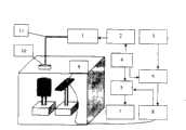
图1是本发明的装置结构示意图;Fig. 1 is a schematic view of the device structure of the present invention;
图2为本发明的一个实施例的样品和吸收体;Fig. 2 is a sample and an absorber of an embodiment of the present invention;
图3是采集到的超声反射的回波的图像;Fig. 3 is the image of the echo of the ultrasonic reflection collected;
图4是采集到的光声信号的二维层析图。Fig. 4 is a two-dimensional tomogram of the collected photoacoustic signal.
具体实施方式Detailed ways
本发明的装置结构如附图1所示,本发明装置主要由激光器1、触发延迟电路2、100KHz外触发源电路3、30Hz的外触发电路4、高速数据采集卡5、外触发电路选择开关6、计算机7、相控聚焦电路8、多元超声阵列探测器9、准直透镜10、光纤11组成;其中多元超声阵列探测器9和相控聚焦电路8电气连接,组成多元超声阵列;30Hz的外触发电路4、触发延迟电路2、激光器1电气连接;多元超声阵列、数据采集卡5、计算机7依次电连接;30Hz的外触发电路4同时和高速数据采集卡5、多元超声阵列连接。The device structure of the present invention is as shown in accompanying
选用各构件组成本装置,其中:激光器1选用美国光谱物理公司生产的MOPO,可以发出波长为500nm-2000nm的脉冲激光,本实施例选用波长为532nm的绿色激光;高速数据采集卡5选用Gage Applied公司的Compuscope 12100型高速数据采集卡(采样速率100MHz);计算机可以选用P4微机。Select various components to form the device, wherein: the
本发明方法的具体实施步骤为:The specific implementation steps of the inventive method are:
1、30Hz的外触发电路触发多元超声探测器的某一群元M,该群元相干发出一束超声,同时高速数据采集卡被触发开始接收信号,数据采集卡接收到超声回波信号并传输到计算机存储;1. The 30Hz external trigger circuit triggers a group M of the multi-element ultrasonic detector, and the group coherently emits a beam of ultrasound, and at the same time the high-speed data acquisition card is triggered to start receiving signals, and the data acquisition card receives the ultrasonic echo signal and transmits it to computer storage;
2、30Hz的外触发电路经过延迟电路被延迟165微秒,再触发脉冲激光器;2. The 30Hz external trigger circuit is delayed by 165 microseconds through the delay circuit, and then triggers the pulse laser;
3、脉冲激光照射样品产生光声信号被群元M接收,通过相控聚焦电路后被高速数据采集卡采集并传输到计算机存储;3. The pulsed laser irradiates the sample to generate a photoacoustic signal, which is received by the group unit M. After passing through the phase control focusing circuit, it is collected by the high-speed data acquisition card and transmitted to the computer for storage;
4、计算机图像重建时对信号进行滤波处理后再根据采集的群元地址将处理后的超声和光声信号按直线投影的方法投影到群元的焦区。4. When the computer image is reconstructed, the signal is filtered and processed, and then the processed ultrasonic and photoacoustic signals are projected to the focal area of the group according to the linear projection method according to the collected group unit address.
在实验中,模拟生物组织样品实验样品是用1克琼脂粉、100克水和2ml浓度为20%的低脂牛奶溶液加热到70摄氏度后在圆形烧杯中冷却凝结而成,样品的有效光散射系数约是μs=10mm-1,样品的吸收系数很小,因此样品没有产生光声信号,样品中埋藏的吸收体是同神配比的琼脂再加上0.008克的泰扑兰染色,其光吸收系数约为0.3mm-1,实验样品的直径为35mm,吸收体的直径为3mm,实际样品如附图2所示,光声和超声分别成的图像如图3、图4所示,图3和4中,纵坐标表示距离探测器的距离,O表示探测器的位置。In the experiment, the experimental sample of the simulated biological tissue sample is formed by
在实际使用当中,30Hz的外触发电路触发多元超声阵列发射超声和激光器之前,需要通过4KHz的触发信号触发多元超声阵列发射超声到生物组织,查找可能的病变部位,30Hz触发电路触发的超声和激光都是针对病变部位的扫描。In actual use, before the 30Hz external trigger circuit triggers the multi-element ultrasound array to emit ultrasound and lasers, it is necessary to trigger the multi-element ultrasound array to emit ultrasound to biological tissues through a 4KHz trigger signal to find possible lesions. The 30Hz trigger circuit triggers the ultrasound and laser All are scans of the lesion.
Claims (1)
Translated fromChinesePriority Applications (1)
| Application Number | Priority Date | Filing Date | Title |
|---|---|---|---|
| CNB2004100150170ACN100434042C (en) | 2004-01-06 | 2004-01-06 | Method and device for optical and ultrasonic acquisition and tomographic imaging of biological tissue |
Applications Claiming Priority (1)
| Application Number | Priority Date | Filing Date | Title |
|---|---|---|---|
| CNB2004100150170ACN100434042C (en) | 2004-01-06 | 2004-01-06 | Method and device for optical and ultrasonic acquisition and tomographic imaging of biological tissue |
Publications (2)
| Publication Number | Publication Date |
|---|---|
| CN1555764A CN1555764A (en) | 2004-12-22 |
| CN100434042Ctrue CN100434042C (en) | 2008-11-19 |
Family
ID=34351279
Family Applications (1)
| Application Number | Title | Priority Date | Filing Date |
|---|---|---|---|
| CNB2004100150170AExpired - Fee RelatedCN100434042C (en) | 2004-01-06 | 2004-01-06 | Method and device for optical and ultrasonic acquisition and tomographic imaging of biological tissue |
Country Status (1)
| Country | Link |
|---|---|
| CN (1) | CN100434042C (en) |
Families Citing this family (18)
| Publication number | Priority date | Publication date | Assignee | Title |
|---|---|---|---|---|
| JP5087007B2 (en)* | 2005-11-23 | 2012-11-28 | インサイテック・リミテッド | Hierarchical switching ultra high density ultrasonic array |
| CN101770650B (en)* | 2009-01-07 | 2013-04-24 | 深圳迈瑞生物医疗电子股份有限公司 | Three-dimensional ultrasound real-time imaging method and device and imaging system |
| AU2010239360A1 (en)* | 2009-04-20 | 2011-11-24 | The Curators Of The University Of Missouri | Photoacoustic detection of analytes in solid tissue and detection system |
| CN101828902B (en)* | 2010-04-01 | 2011-09-21 | 江西科技师范学院 | A photoacoustic sensor for 3D medical diagnosis of breast or brain |
| CN101828928B (en)* | 2010-04-01 | 2012-06-20 | 江西科技师范学院 | Three-dimensional optoacoustic mammary gland or brain non-destructive imaging system |
| JP5881582B2 (en)* | 2012-02-07 | 2016-03-09 | 富士フイルム株式会社 | Manufacturing method of ultrasonic probe |
| WO2014056134A1 (en)* | 2012-10-08 | 2014-04-17 | 财团法人工业技术研究院 | Imaging method combining ultrasound with photoacoustic image, and imaging device |
| CN103211620B (en)* | 2013-04-26 | 2015-05-20 | 杨迪武 | Breast carcinoma early-stage detecting instrument based on annular array opto-acoustic sensing technology |
| CN105030273A (en)* | 2014-04-28 | 2015-11-11 | 株式会社东芝 | Ultrasonic diagnosis apparatus and biomedical light measuring apparatus |
| CN104990993A (en)* | 2015-04-17 | 2015-10-21 | 北京理工大学 | Ultrasound slowness difference tomography algorithm for weak scattering mediums |
| CN105068030A (en)* | 2015-09-08 | 2015-11-18 | 中国石油大学(北京) | Nuclear magnetic resonance spectrometer |
| CN108603784B (en)* | 2015-11-02 | 2021-07-30 | 普渡研究基金会 | Method and device for detection of cancer margins |
| CN105232004A (en)* | 2015-11-16 | 2016-01-13 | 华南师范大学 | Opto-acoustic-ultrasonic united imaging device and imaging method for precisely measuring thickness of melanoma |
| CN105395170B (en)* | 2015-12-15 | 2018-07-27 | 同济大学 | A kind of photoacoustic ultrasound bimodal synchronous imaging system |
| CN105631879B (en)* | 2015-12-30 | 2018-10-12 | 哈尔滨工业大学 | A kind of ultrasound tomography system and method based on linear array |
| CN107589425B (en)* | 2017-10-17 | 2019-11-26 | 广州极飞科技有限公司 | Ultrasonic distance measurement equipment and its detection of the backscatter signal method, apparatus and aircraft |
| JP7158796B2 (en)* | 2017-12-07 | 2022-10-24 | 株式会社アドバンテスト | Optical ultrasonic measuring device, method, program, recording medium |
| CN108594714B (en)* | 2018-05-18 | 2021-11-16 | 南京大学 | Acquisition and preprocessing system for reconfigurable echo pulse and photoacoustic signal |
Citations (5)
| Publication number | Priority date | Publication date | Assignee | Title |
|---|---|---|---|---|
| CN1128493A (en)* | 1994-05-12 | 1996-08-07 | 通用电器横河医疗系统株式会社 | Method and device for multichannel digital reception and ultrasonic diagnostic device |
| CN1249162A (en)* | 1998-09-25 | 2000-04-05 | 中国科学院西安光学精密机械研究所 | Safety method for identifying calculus and human tissue |
| CN1279054A (en)* | 2000-06-27 | 2001-01-10 | 华南师范大学 | Optically Chronatographic imaging method and equipment with focusing, ultraconic wave and modulation |
| CN1422597A (en)* | 2002-12-31 | 2003-06-11 | 华南师范大学 | Focusing supersonic modulation reflection type optical chromatography imaging method and its apparatus |
| CN1433739A (en)* | 2003-02-26 | 2003-08-06 | 华南师范大学 | Method and device for biological tissue photoacoustic tomography |
- 2004
- 2004-01-06CNCNB2004100150170Apatent/CN100434042C/ennot_activeExpired - Fee Related
Patent Citations (5)
| Publication number | Priority date | Publication date | Assignee | Title |
|---|---|---|---|---|
| CN1128493A (en)* | 1994-05-12 | 1996-08-07 | 通用电器横河医疗系统株式会社 | Method and device for multichannel digital reception and ultrasonic diagnostic device |
| CN1249162A (en)* | 1998-09-25 | 2000-04-05 | 中国科学院西安光学精密机械研究所 | Safety method for identifying calculus and human tissue |
| CN1279054A (en)* | 2000-06-27 | 2001-01-10 | 华南师范大学 | Optically Chronatographic imaging method and equipment with focusing, ultraconic wave and modulation |
| CN1422597A (en)* | 2002-12-31 | 2003-06-11 | 华南师范大学 | Focusing supersonic modulation reflection type optical chromatography imaging method and its apparatus |
| CN1433739A (en)* | 2003-02-26 | 2003-08-06 | 华南师范大学 | Method and device for biological tissue photoacoustic tomography |
Also Published As
| Publication number | Publication date |
|---|---|
| CN1555764A (en) | 2004-12-22 |
Similar Documents
| Publication | Publication Date | Title |
|---|---|---|
| CN100434042C (en) | Method and device for optical and ultrasonic acquisition and tomographic imaging of biological tissue | |
| US9528966B2 (en) | Reflection-mode photoacoustic tomography using a flexibly-supported cantilever beam | |
| US20070287912A1 (en) | Functional imaging using capacitive micromachined ultrasonic transducers | |
| US10241199B2 (en) | Ultrasonic/photoacoustic imaging devices and methods | |
| JP5284129B2 (en) | Imaging apparatus and analysis method | |
| Andreev et al. | Optoacoustic tomography of breast cancer with arc-array transducer | |
| US6567688B1 (en) | Methods and apparatus for scanning electromagnetically-induced thermoacoustic tomography | |
| Lashkari et al. | Comparison between pulsed laser and frequency-domain photoacoustic modalities: signal-to-noise ratio, contrast, resolution, and maximum depth detectivity | |
| CN101813672B (en) | Rapid three-dimensional photoacoustic imaging system based on ultrasonic plane array detector and method thereof | |
| CN104706323B (en) | High-speed large-view-field multi-spectral photoacoustic imaging method and device | |
| CN102944521B (en) | Non-contact photoacoustic and optical coherence tomography dual-imaging device and detection method thereof | |
| Paltauf et al. | Progress in biomedical photoacoustic imaging instrumentation toward clinical application | |
| WO2007088709A1 (en) | 3d acoustic imaging device and 3d acoustic imaging method | |
| CN102621115A (en) | Confocal simultaneous opto-acoustic imaging and fluorescence imaging method and device | |
| CN100446730C (en) | Photoacoustic imaging and tomographic imaging method and device based on acoustic lens | |
| CN104146685B (en) | A kind of cutaneous pigmentation imaging device based on photoacoustic principle | |
| Zhang et al. | Three-dimensional photoacoustic imaging of vascular anatomy in small animals using an optical detection system | |
| Valluru et al. | Photoacoustic imaging: opening new frontiers in medical imaging | |
| JP4422626B2 (en) | Biological imaging device | |
| Sharma et al. | A comparative study of continuous versus stop-and-go scanning in circular scanning photoacoustic tomography | |
| CN114176554B (en) | Multi-pulse-width microwave excitation multi-scale thermo-acoustic imaging method and system | |
| CN107174208A (en) | A kind of photoacoustic imaging system and method suitable for peripheral vascular imaging | |
| CN101336832A (en) | Pulsed photoacoustic scanning soft tissue imaging method and device | |
| JP2011045514A (en) | Photoacoustic tomography apparatus | |
| Beard et al. | 2D line-scan photoacoustic imaging of absorbers in a scattering tissue phantom |
Legal Events
| Date | Code | Title | Description |
|---|---|---|---|
| C06 | Publication | ||
| PB01 | Publication | ||
| C10 | Entry into substantive examination | ||
| SE01 | Entry into force of request for substantive examination | ||
| C14 | Grant of patent or utility model | ||
| GR01 | Patent grant | ||
| EE01 | Entry into force of recordation of patent licensing contract | Application publication date:20041222 Assignee:Guangzhou Baiaoting Electronic Technology Co., Ltd. Assignor:South China Normal University Contract record no.:2013440000032 Denomination of invention:Method of biological tissue optical and ultrasonic collection and tomographic imaging and its device Granted publication date:20081119 License type:Exclusive License Record date:20130121 | |
| LICC | Enforcement, change and cancellation of record of contracts on the licence for exploitation of a patent or utility model | ||
| CF01 | Termination of patent right due to non-payment of annual fee | Granted publication date:20081119 Termination date:20180106 | |
| CF01 | Termination of patent right due to non-payment of annual fee |