CN100405051C - Biosensor and method for measuring the same - Google Patents
Biosensor and method for measuring the sameDownload PDFInfo
- Publication number
- CN100405051C CN100405051CCNB028109554ACN02810955ACN100405051CCN 100405051 CCN100405051 CCN 100405051CCN B028109554 ACNB028109554 ACN B028109554ACN 02810955 ACN02810955 ACN 02810955ACN 100405051 CCN100405051 CCN 100405051C
- Authority
- CN
- China
- Prior art keywords
- electrode
- biology sensor
- illustrating
- enzyme
- biosensor
- Prior art date
- Legal status (The legal status is an assumption and is not a legal conclusion. Google has not performed a legal analysis and makes no representation as to the accuracy of the status listed.)
- Expired - Fee Related
Links
Images


Classifications
- B—PERFORMING OPERATIONS; TRANSPORTING
- B82—NANOTECHNOLOGY
- B82Y—SPECIFIC USES OR APPLICATIONS OF NANOSTRUCTURES; MEASUREMENT OR ANALYSIS OF NANOSTRUCTURES; MANUFACTURE OR TREATMENT OF NANOSTRUCTURES
- B82Y30/00—Nanotechnology for materials or surface science, e.g. nanocomposites
- G—PHYSICS
- G01—MEASURING; TESTING
- G01N—INVESTIGATING OR ANALYSING MATERIALS BY DETERMINING THEIR CHEMICAL OR PHYSICAL PROPERTIES
- G01N27/00—Investigating or analysing materials by the use of electric, electrochemical, or magnetic means
- G01N27/26—Investigating or analysing materials by the use of electric, electrochemical, or magnetic means by investigating electrochemical variables; by using electrolysis or electrophoresis
- G01N27/28—Electrolytic cell components
- G01N27/30—Electrodes, e.g. test electrodes; Half-cells
- G01N27/327—Biochemical electrodes, e.g. electrical or mechanical details for in vitro measurements
- G01N27/3271—Amperometric enzyme electrodes for analytes in body fluids, e.g. glucose in blood
- G01N27/3272—Test elements therefor, i.e. disposable laminated substrates with electrodes, reagent and channels
- G—PHYSICS
- G01—MEASURING; TESTING
- G01N—INVESTIGATING OR ANALYSING MATERIALS BY DETERMINING THEIR CHEMICAL OR PHYSICAL PROPERTIES
- G01N33/00—Investigating or analysing materials by specific methods not covered by groups G01N1/00 - G01N31/00
- G01N33/48—Biological material, e.g. blood, urine; Haemocytometers
- G01N33/50—Chemical analysis of biological material, e.g. blood, urine; Testing involving biospecific ligand binding methods; Immunological testing
- G01N33/53—Immunoassay; Biospecific binding assay; Materials therefor
- G01N33/543—Immunoassay; Biospecific binding assay; Materials therefor with an insoluble carrier for immobilising immunochemicals
- G01N33/54366—Apparatus specially adapted for solid-phase testing
- G01N33/54373—Apparatus specially adapted for solid-phase testing involving physiochemical end-point determination, e.g. wave-guides, FETS, gratings
- G01N33/5438—Electrodes
Landscapes
- Health & Medical Sciences (AREA)
- Chemical & Material Sciences (AREA)
- Life Sciences & Earth Sciences (AREA)
- Engineering & Computer Science (AREA)
- Immunology (AREA)
- Physics & Mathematics (AREA)
- Molecular Biology (AREA)
- General Physics & Mathematics (AREA)
- Hematology (AREA)
- Pathology (AREA)
- Nanotechnology (AREA)
- General Health & Medical Sciences (AREA)
- Biomedical Technology (AREA)
- Biochemistry (AREA)
- Analytical Chemistry (AREA)
- Urology & Nephrology (AREA)
- Microbiology (AREA)
- Biophysics (AREA)
- Materials Engineering (AREA)
- Food Science & Technology (AREA)
- Medicinal Chemistry (AREA)
- Cell Biology (AREA)
- Biotechnology (AREA)
- Crystallography & Structural Chemistry (AREA)
- Composite Materials (AREA)
- Condensed Matter Physics & Semiconductors (AREA)
- Chemical Kinetics & Catalysis (AREA)
- Electrochemistry (AREA)
- Investigating Or Analysing Biological Materials (AREA)
- Apparatus Associated With Microorganisms And Enzymes (AREA)
- Measuring Or Testing Involving Enzymes Or Micro-Organisms (AREA)
- Photoreceptors In Electrophotography (AREA)
- Holo Graphy (AREA)
- Measurement Of The Respiration, Hearing Ability, Form, And Blood Characteristics Of Living Organisms (AREA)
Abstract
Translated fromChineseDescription
Translated fromChinese技术领域technical field
本发明涉及一种生物传感器,更具体地,涉及一种包含微孔膜载体,第一电极系统,第二电极系统,和一对绝缘膜的生物传感器,其中第一电极系统在微孔膜载体的表面形成并且第二电极系统在微孔膜载体的反面形成。The present invention relates to a biosensor, more specifically, to a biosensor comprising a microporous membrane carrier, a first electrode system, a second electrode system, and a pair of insulating membranes, wherein the first electrode system is on a microporous membrane carrier and a second electrode system is formed on the reverse side of the microporous membrane support.
发明技术invention technology
生物传感器是指一种设备,探针,或电极,其当与适当的样品接触时,在存在目的分析物的条件下产生电信号。通常通过固定与合适的转换系统紧密接触的生物敏感物质以将分析物的浓度转化成可定量的信号来构建生物传感器。A biosensor refers to a device, probe, or electrode that, when contacted with an appropriate sample, generates an electrical signal in the presence of an analyte of interest. Biosensors are usually constructed by immobilizing a biologically sensitive substance in close contact with a suitable conversion system to convert the concentration of an analyte into a quantifiable signal.
尽管有几个优点,但当前可用的生物传感器大都有些有待解决的问题:化学干扰,环境影响,长期稳定性,信噪比和传感器包装系统的设计。Despite several advantages, currently available biosensors have several open issues: chemical interference, environmental influence, long-term stability, signal-to-noise ratio, and design of sensor packaging systems.
在生物传感器的发展中可看出下列趋势:The following trends can be seen in the development of biosensors:
a)小型化a) Miniaturization
b)用组合多于一种敏感元件的阵列传感器系统测定几种试剂b) Determination of several reagents with an array sensor system combining more than one sensitive element
c)开发可大规模生产的任意使用的(disposable)传感器c) Development of disposable sensors that can be mass-produced
d)使用可植入生物芯片体内分析d) In vivo analysis using implantable biochips
e)处理来自具有人工智能系统的阵列传感器系统的输出e) Processing output from array sensor systems with artificial intelligence systems
同时,一个长期目标是开发一种迅速,简单,免分离的检测蛋白的方法。已经使用显色和发荧光的半乳糖苷-葡聚糖底物来设计针对C-活性蛋白(C-reactive protein),铁蛋白和免疫球蛋白的均相酶免疫测定(EIAs)(Gibbons等,“Homogeneous Enzyme Immunoassay for Proteins Employingβ-Galactosidase,”Analytical Biochemistry 102/167-170,1980;和Armenta等,“Improved Sensitivity in Homogeneous Enzyme Immunoassays Using aFluorogenic Macromolecular Substrate:An Assay tor Serum Ferritin,”Analytical Biochemistry 146/211-219,1985)。然而,在该均相方案中酶活性的低程度调节已使该方法对于现实应用而言不切实际。Meanwhile, a long-term goal is to develop a rapid, simple, and isolation-free method for protein detection. Chromogenic and fluorescent galactoside-dextran substrates have been used to design homogeneous enzyme immunoassays (EIAs) for C-reactive protein, ferritin, and immunoglobulins (Gibbons et al. “Homogeneous Enzyme Immunoassay for Proteins Employingβ-Galactosidase,”Analytical Biochemistry 102/167-170,1980;和Armenta等,“Improved Sensitivity in Homogeneous Enzyme Immunoassays Using aFluorogenic Macromolecular Substrate:An Assay tor Serum Ferritin,”Analytical Biochemistry 146/211- 219, 1985). However, the low degree of modulation of enzyme activity in this homogeneous protocol has made this method impractical for real-world applications.
此外,已报道一种针对大分子的免分离的双固相EIA,其依赖于一种酶偶联物(生物素-葡萄糖-6-磷酸脱氢酶-抗体)在结合了聚苯乙烯胶乳的抗生物素蛋白和结合了聚苯乙烯胶乳的分析物的两种固体相之间的分配(Schray等,“Separation-Free Dual Solid Phase Enzyme Immunoassay forMacromolecules,”Analytical Chemistry,60/353-56,1988)。然而,该测定方案需要24小时来酶促产生可检测的产物。In addition, a separation-free dual solid-phase EIA for macromolecules has been reported, which relies on an enzyme conjugate (biotin-glucose-6-phosphate dehydrogenase-antibody) in a polystyrene latex-bound Partition between two solid phases of avidin and analyte bound to polystyrene latex (Schray et al., "Separation-Free Dual Solid Phase Enzyme Immunoassay for Macromolecules," Analytical Chemistry, 60/353-56, 1988) . However, this assay protocol requires 24 hours to enzymatically generate a detectable product.
早就认识到将电化学检测与EIAs结合将是有利的。电极对检测样品的颜色或浊度不敏感,因此可用来开发可直接应用于全血样的方法。然而,许多关于使用电化学检测来设计EIAs或“免疫传感器”的报道的大多数已依赖于使用这样的传感器如多相测定设备中的固相,其中将抗体固定在给定电极的表面。在样品与其它试剂温育后,在加入测量束缚酶(bound enzyme)活性所需的底物之前必须洗涤电极表面。It has long been recognized that combining electrochemical detection with EIAs would be advantageous. The electrodes are not sensitive to the color or turbidity of the detected samples and thus can be used to develop methods that can be applied directly to whole blood samples. However, the majority of the many reports on the use of electrochemical detection to design EIAs or "immunosensors" have relied on the use of such sensors as solid phases in heterogeneous assay devices, where antibodies are immobilized on the surface of a given electrode. After incubation of the sample with other reagents, the electrode surface must be washed before adding the substrate required to measure bound enzyme activity.
作为一个具体实例,在1991年11月5日颁发的美国专利No.5,063,081中,Cozzette等公开了一种用于检测特定分析物种类如抗原的基于配体/配体受体的生物传感器。这里,一种基底传感器(base sensor),其包含一种使用贵后过渡金属(noble late transition metal)如铱,金,铂或银的催化指示电极,并被一种由例如银和氯化银制成的组合参比和对电极(柱25-26)包围。一种抗体固定在基底传感器上。然后将得到的生物传感器与含有样品和标记的第二种分析物特异性抗体的混合物接触(柱45-46)。还可将选择透性硅烷层用作针对干扰物的筛子。然而,优选通过使用洗涤溶液或通过使用含有酶底物作为洗涤物的溶液从传感器去除未结合物质和干扰电活化粒种(柱47-49)。As a specific example, in US Patent No. 5,063,081 issued November 5, 1991, Cozzette et al. disclose a ligand/ligand receptor based biosensor for the detection of specific analyte species such as antigens. Here, a base sensor comprising a catalytic indicator electrode using a noble late transition metal such as iridium, gold, platinum or silver, and surrounded by a material such as silver and silver chloride The fabricated combined reference and counter electrodes (columns 25-26) surround. An antibody is immobilized on the substrate sensor. The resulting biosensor is then contacted with a mixture containing the sample and a labeled second analyte-specific antibody (columns 45-46). The selectively permeable silane layer can also be used as a screen for interfering substances. However, it is preferred to remove unbound material and interfering electroactive species from the sensor by using a washing solution or by using a solution containing the enzyme substrate as wash (columns 47-49).
作为另一个具体实例,在1998年11月3日颁发的美国专利No.5,830,680中,Meyerhoff等公开了一种适合在任何本底信号上检测分析信号的酶夹层免疫测定盒,所述本底信号是源自与所述盒接触的本体溶液(bulksolution)。这里,所述盒包含一种微孔膜载体,其一侧已经用一种导电金属层涂布,和至少一个第一捕捉抗体层,其被固定在微孔膜载体的至少一个第一空间独特区域中的导电金属层上。参考图1,其是扩散电池设备的图解,可以看到该盒需要另外的辅助电极和/或参比电极,因此未实现小型化和现场检测(point-of-care testing)。As another specific example, in U.S. Patent No. 5,830,680 issued November 3, 1998, Meyerhoff et al. disclose an enzyme-sandwich immunoassay cartridge suitable for detecting an assay signal over any background signal that is derived from the bulk solution in contact with the cartridge. Here, the cartridge comprises a microporous membrane carrier, one side of which has been coated with a conductive metal layer, and at least one first capture antibody layer, which is immobilized on at least one first spatially distinct portion of the microporous membrane carrier. on the conductive metal layer in the area. Referring to Figure 1, which is a diagram of a diffusion cell device, it can be seen that the cartridge requires additional auxiliary and/or reference electrodes, thus not enabling miniaturization and point-of-care testing.
因此,尽管所有该领域中过去和当前的研究活动,长期以来期望一种可以避免上述缺点的新型生物传感器。Therefore, despite all past and current research activities in this field, a new type of biosensor that can avoid the above-mentioned disadvantages has long been desired.
发明概述Summary of the invention
本发明的一个目的是提供一种生物传感器,其包含微孔膜载体;第一电极系统,其由其上固定生物活性物质的工作电极,第一电极连接器和第一引线出口组成;第二电极系统,其由对电极,第二电极连接器和第二引线出口组成;一对绝缘膜,其覆盖除了工作电极,第一电极连接器,对电极和第二电极连接器区域以外的第一和第二电极系统的表面,其中第一电极系统在微孔膜载体的表面形成并且第二电极系统在微孔膜载体的反面形成,其中工作电极和对电极在微孔膜载体上直接形成,由此电极是多孔的并且允许样品经过工作电极表面到达对电极的表面。An object of the present invention is to provide a biosensor comprising a microporous membrane carrier; a first electrode system consisting of a working electrode on which a biologically active substance is immobilized, a first electrode connector and a first lead wire outlet; An electrode system consisting of a counter electrode, a second electrode connector and a second lead wire outlet; a pair of insulating films covering the first electrode except for the working electrode, first electrode connector, counter electrode and second electrode connector area. and the surface of the second electrode system, wherein the first electrode system is formed on the surface of the microporous membrane carrier and the second electrode system is formed on the opposite side of the microporous membrane carrier, wherein the working electrode and the counter electrode are directly formed on the microporous membrane carrier, The electrode is thus porous and allows the sample to pass through the surface of the working electrode to the surface of the counter electrode.
本发明的另一个目的是提供一种生物传感器,其包含微孔膜载体,其上固定生物活性物质的第一电极系统,第二电极系统和一对绝缘膜,一个垫板和一对带孔的盖子,其中第一电极系统在微孔膜载体的表面形成并且第二电极系统在微孔膜载体的反面形成,并且将一种酶-分析物偶联物或一种酶-抗体偶联体固定在垫板上。Another object of the present invention is to provide a biosensor comprising a microporous membrane carrier, a first electrode system immobilizing a biologically active substance on it, a second electrode system and a pair of insulating membranes, a backing plate and a pair of holes. wherein the first electrode system is formed on the surface of the microporous membrane carrier and the second electrode system is formed on the opposite side of the microporous membrane carrier, and an enzyme-analyte conjugate or an enzyme-antibody conjugate fixed on the backing plate.
附图简述Brief description of the drawings
参考附图可最好的理解本发明最优实施方案的应用,附图中相同的参考号用于相同和对应的部分,其中:The application of the preferred embodiment of the present invention is best understood by reference to the accompanying drawings, in which like reference numerals are used for like and corresponding parts, in which:
图1是现有技术扩散电池设备的示意图,其中在微孔膜载体上只形成一个电极系统;Fig. 1 is the schematic diagram of prior art diffusion battery equipment, wherein only one electrode system is formed on the microporous membrane carrier;
a:分析物溶液b:底物溶液a: Analyte solution b: Substrate solution
图2a为依照本发明实施方案1在整个条带上形成的基于对称微孔电极的生物传感器的分解透视图;Figure 2a is an exploded perspective view of a biosensor based on symmetrical microporous electrodes formed on an entire strip according to Embodiment 1 of the present invention;
图2b是本发明实施方案1的生物传感器的透视图;Figure 2b is a perspective view of a biosensor according to Embodiment 1 of the present invention;
图2c是基于对称微孔电极的生物传感器的分解透视图,其中依照本发明实施方案1使用分别在两个绝缘基片上形成的电连接器改装生物传感器;Fig. 2c is an exploded perspective view of a biosensor based on symmetrical microporous electrodes, wherein the biosensor is modified according to Embodiment 1 of the present invention using electrical connectors respectively formed on two insulating substrates;
图2d是2c生物传感器的透视图,其中依照本发明实施方案1改装生物传感器;Figure 2d is a perspective view of 2c biosensor, wherein the biosensor is modified according to Embodiment 1 of the present invention;
图2e是基于不对称微孔电极的生物传感器的分解透视图,其中依照本发明实施方案1使用分别在两个绝缘基片上形成的电连接器改装生物传感器;Fig. 2e is an exploded perspective view of a biosensor based on asymmetric microporous electrodes, wherein the biosensor is modified according to Embodiment 1 of the present invention using electrical connectors respectively formed on two insulating substrates;
图2f是2e生物传感器的透视图,其中依照本发明实施方案1改装生物传感器;Figure 2f is a perspective view of a biosensor 2e, wherein the biosensor is modified according to Embodiment 1 of the present invention;
图3是依照本发明实施方案1说明生物传感器应用实例的图解;3 is a diagram illustrating an application example of a biosensor according to Embodiment 1 of the present invention;
图4a是依照本发明实施方案2的生物传感器的分解透视图;4a is an exploded perspective view of a biosensor according to
图4b是依照本发明实施方案2的生物传感器的透视图;Figure 4b is a perspective view of a biosensor according to
图5是说明依照本发明实施方案2将生物传感器应用于样品溶液的实例的图解;5 is a diagram illustrating an example in which a biosensor is applied to a sample solution according to
图6a和图6b是依照本发明实施方案2装备了改良进样毛细管的生物传感器的截面图;6a and 6b are cross-sectional views of a biosensor equipped with an improved sampling capillary according to
图7a是依照本发明实施方案3的生物传感器的分解透视图;Figure 7a is an exploded perspective view of a biosensor according to
(关于图7b-7e我推荐使用单独的图号)(I recommend separate figure numbers for Figures 7b-7e)
图7b-7e显示依照本发明实施方案3生物传感器的各种改装;Figures 7b-7e show various modifications of the biosensor according to
图8显示提及的免分离免疫测定步骤的原理和顺序的图解;Figure 8 shows a schematic diagram of the principle and sequence of the mentioned separation-free immunoassay steps;
图9是本发明扩散电池设备的示意图;Fig. 9 is a schematic diagram of a diffusion cell device of the present invention;
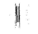
图10是生物传感器的环状伏安图,该生物传感器是实施例1中基于不对称微孔电极的生物传感器,其中将铁氰化钾用作电极的氧化还原剂;Fig. 10 is the ring voltammogram of biosensor, and this biosensor is the biosensor based on asymmetric microporous electrode in embodiment 1, wherein potassium ferricyanide is used as the redox agent of electrode;
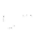
图11是实施例1生物传感器的典型校正曲线,其中将葡糖氧化酶物理法固定在用于葡萄糖分析的工作电极上;Fig. 11 is a typical calibration curve of the biosensor of Example 1, wherein the glucose oxidase is physically immobilized on the working electrode for glucose analysis;
图12是实施例2生物传感器的动态响应曲线,其中将葡糖氧化酶和铁氰化钾物理法固定在工作电极上;Figure 12 is the dynamic response curve of the biosensor in Example 2, wherein glucose oxidase and potassium ferricyanide are physically immobilized on the working electrode;
图13是实施例3生物传感器的校正曲线,其中将葡糖氧化酶-生物素偶联物用作酶-分析物偶联物并且将抗生物素蛋白用作抗体;Figure 13 is a calibration curve for the biosensor of Example 3, wherein a glucose oxidase-biotin conjugate was used as the enzyme-analyte conjugate and avidin was used as the antibody;
图14是实施例3生物传感器的动态响应曲线和Fig. 14 is the dynamic response curve and the dynamic response curve of
图15是实施例4CRP生物传感器的校正曲线;使用固定在前面微孔电极的CRP抗体,在上垫板中干燥的碱性磷酸酶-CRP偶联物和通过底板浸泡的对-氨基苯磷酸盐来获取信号。Fig. 15 is the calibration curve of
发明详述Detailed description of the invention
本发明涉及一种生物传感器,其包含微孔膜载体,第一电极系统,第二电极系统和一对绝缘膜(或一对带有电连接器的绝缘基片),其中第一电极系统在微孔膜载体的表面形成并且第二电极系统在微孔膜载体的反面形成。依照本发明的生物传感器不需要任何附加电极。The present invention relates to a biosensor comprising a microporous membrane carrier, a first electrode system, a second electrode system and a pair of insulating films (or a pair of insulating substrates with electrical connectors), wherein the first electrode system is A surface of the microporous membrane support is formed and a second electrode system is formed on the opposite side of the microporous membrane support. The biosensor according to the invention does not require any additional electrodes.
参考图2a和2b,依照本发明的生物传感器使用的电化学电池(100)包含一个微孔膜载体(101),第一电极系统(102),第二电极系统(103)。第一电极系统(102)由其上固定生物活性物质的工作电极(104),第一电极连接器(105)和第一引线出口(106)组成。第二电极系统由一个对电极(107),第二电极连接器(108)和第二引线出口组成(109)。第一电极系统和第二电极系统(102,103)通过电极连接器(105,108)经由例如鳄鱼夹或焊剂的连接被电连接。Referring to Figures 2a and 2b, the electrochemical cell (100) used in the biosensor according to the present invention comprises a microporous membrane support (101), a first electrode system (102), and a second electrode system (103). The first electrode system (102) is composed of a working electrode (104) on which a bioactive substance is immobilized, a first electrode connector (105) and a first lead outlet (106). The second electrode system consists of a counter electrode (107), a second electrode connector (108) and a second lead wire outlet (109). The first electrode system and the second electrode system (102, 103) are electrically connected by electrode connectors (105, 108) via connections such as alligator clips or solder.
参考图2b至2f,其中改装2a的生物传感器,按照本发明的生物传感器中使用的电化学电池(100)包含微孔膜载体(101),第一电极系统(102),和第二对称(2b)或不对称(2c)电极系统(103)。第一电极系统(102)由其上固定生物活性物质的工作电极(104),在顶部绝缘膜的下侧形成的第一电极连接器(105)和第一引线出口(106)组成。第二电极系统由一个对电极(107),底部绝缘膜的上表面形成的第二电极连接器(108)和第二引线出口(109)组成。将第一、第二引线出口(106和109)紧压与第一、第二电极连接器(105和108)连接,并且还将在顶部绝缘膜下侧形成的分段的第一连接器(105’)和在底部绝缘膜的表面形成的105紧压连接以在底部基片上以面朝上的方向安装两个连接器(105’和105)。通过将它们插入插座中连接电极连接器(105,108)电连接第一和第二电极系统(102,103)。2b to 2f, wherein the biosensor of modification 2a, according to the electrochemical cell (100) used in the biosensor of the present invention comprises a microporous membrane support (101), a first electrode system (102), and a second symmetrical ( 2b) or asymmetrical (2c) electrode system (103). The first electrode system (102) consists of a working electrode (104) on which a bioactive substance is immobilized, a first electrode connector (105) and a first lead outlet (106) formed on the underside of the top insulating film. The second electrode system consists of a counter electrode (107), a second electrode connector (108) formed on the upper surface of the bottom insulating film and a second lead outlet (109). Connect the first and second lead wire outlets (106 and 109) tightly to the first and second electrode connectors (105 and 108), and also form the segmented first connector ( 105') and 105 formed on the surface of the bottom insulating film are press-connected to mount the two connectors (105' and 105) on the bottom substrate in a face-up direction. The first and second electrode systems (102, 103) are electrically connected by connecting the electrode connectors (105, 108) by plugging them into sockets.
通常通过化学气相沉积或物理蒸气沉积法镀上导电材料或通过丝网印刷导电材料形成第一和第二电极系统(102,103)。能够用来形成第一和第二电极系统(102,103)的导电材料的实例包括但不限于导电聚合物,碳,和导电金属如金,铂,银,铑,铱,钌,钯,锇或铜。导电材料通常形成厚度为大约150


可用来形成微孔膜载体(101)的材料应与经常用于将生物活性物质如酶,电子传递介质和抗体固定在电极系统上的有机溶剂相容,并且应显示足够高的拉伸强度以抵抗在操作过程中多孔结构的撕裂和/或其它扭变。可用来形成微孔膜载体(101)的材料的实例包括有机聚合物(例如尼龙,硝化纤维,聚偏二氟乙烯,聚砜,聚酯或聚碳酸酯)和无机材料如多孔陶瓷或吸附陶瓷。优选微孔尼龙网,因为它是天然亲水的而且可以以基本上不含湿润剂的形式商购。Materials that can be used to form the microporous membrane support (101) should be compatible with organic solvents that are often used to immobilize biologically active substances such as enzymes, electron transfer mediators, and antibodies on electrode systems, and should exhibit high enough tensile strength to Resists tearing and/or other distortion of the porous structure during handling. Examples of materials that can be used to form the microporous membrane support (101) include organic polymers (such as nylon, nitrocellulose, polyvinylidene fluoride, polysulfone, polyester or polycarbonate) and inorganic materials such as porous ceramics or adsorbent ceramics . A microporous nylon mesh is preferred because it is naturally hydrophilic and is commercially available in a substantially humectant-free form.
即使在用导电层涂布膜之后,它也仍可以是微孔性的,具有约0.01微米至约10微米的通常范围内的孔径。甚至理论上更大的孔径也是可能的,其只被用于部分封闭(occlude)孔的附加导电材料的费用所限制。优选的平均孔径为约0.2μm至约0.45μm。该孔径允许具有不超过5000Da分子量的分析物以及底物通过孔。Even after the membrane is coated with the conductive layer, it can still be microporous, with pore sizes typically in the range of about 0.01 microns to about 10 microns. Even theoretically larger pore sizes are possible, limited only by the cost of additional conductive material to partially occlude the pores. A preferred average pore size is from about 0.2 μm to about 0.45 μm. The pore size allows analytes with molecular weights not exceeding 5000 Da as well as substrates to pass through the pores.
电化学电池(100)的表面,除了工作电极(104),第一电极连接器(105),对电极(107)和第二电极连接器(108)的区域以外,都用绝缘膜(110,111)覆盖。可以用来形成绝缘膜(110,111)的材料包括但不限于聚氯乙烯(PVC)或它的共聚物如聚氯乙烯-二(2-乙基已基)癸二酸酯,聚乙烯,聚氨基甲酸酯,聚碳酸酯,聚酯等。还可通过将绝缘聚合物糊丝网印刷在尼龙膜上,接着热处理来形成绝缘膜(110,111)。绝缘膜(110,111)应该仅限制底物通过微孔膜区域的扩散并且应该最小化生物传感器的扭变。如果微孔电极和尼龙膜的尺寸与图2c-2f中所示相同,可以将电连接分别印刷在绝缘膜上,并且可以紧压以连接电极和组装生物传感器系统。The surface of the electrochemical cell (100), except the working electrode (104), the first electrode connector (105), the area of the opposite electrode (107) and the second electrode connector (108), is all covered with an insulating film (110, 111) Coverage. Materials that can be used to form the insulating film (110, 111) include but are not limited to polyvinyl chloride (PVC) or its copolymers such as polyvinyl chloride-bis(2-ethylhexyl) sebacate, polyethylene, Polyurethane, polycarbonate, polyester, etc. The insulating film (110, 111) can also be formed by screen printing insulating polymer paste on the nylon film, followed by heat treatment. The insulating membrane (110, 111) should only limit the diffusion of the substrate through the microporous membrane region and should minimize twisting of the biosensor. If the dimensions of the microporous electrodes and the nylon membrane are the same as shown in Fig. 2c–2f, the electrical connections can be printed on the insulating membrane, respectively, and can be pressed tightly to connect the electrodes and assemble the biosensor system.
该形式的生物传感器(2c-2f)在大规模制造方面有优势;可以分别制备印在绝缘盖上沉积金的微孔电极和连接电极并且在将生物材料固定在工作电极上后一步组装为一个整体。如果采用等离子沉积的方法,非对称的双面电极(2e和2f)可能比对称的双面电极(2c和2d)更容易制备;非对称双面电极,需要在丝网印刷的对电极反面电极沉积一次即可,而对称的双面电极需要在破坏真空后电极沉积两次。This form of biosensor (2c-2f) has advantages in terms of large-scale fabrication; the gold-deposited microporous electrode and the connecting electrode printed on the insulating cover can be prepared separately and assembled in one step after immobilizing the biomaterial on the working electrode. overall. If the plasma deposition method is used, asymmetric double-sided electrodes (2e and 2f) may be easier to prepare than symmetrical double-sided electrodes (2c and 2d); asymmetric double-sided electrodes need to be screen-printed on the opposite electrode. One deposition is sufficient, while symmetrical double-sided electrodes require two electrode depositions after breaking the vacuum.
按照本发明的生物传感器可以迅速,简单,免分离的电化学免疫测定和选择性检测分析物。另外,该生物传感器不需要任何附加电极因此可以实现小型化,现场检测和任意使用。The biosensor according to the present invention allows rapid, simple, separation-free electrochemical immunoassay and selective detection of analytes. In addition, this biosensor does not require any additional electrodes and thus enables miniaturization, on-site detection, and arbitrary use.
如图3所示,按照本发明的生物传感器可直接应用于人体来检测各种样品,包括生物材料。待测的生物材料包括代谢物,例如葡萄糖,胆固醇,乳酸,肌酸酐,蛋白质,过氧化氢,醇,氨基酸,谷氨酸丙酮酸酯和谷氨酸草酰乙酯。例如,使用胆固醇氧化酶,乳酸氧化酶,谷氨酸氧化酶,辣根过氧化物酶或醇氧化酶可分别定量分析胆固醇,乳酸,谷氨酸,过氧化氢和乙醇。为工作电极提供的电子传送介质可使用二茂铁或它的衍生物,醌或它的衍生物,有机导电盐类或viologen。电子传送介质可固定在与酶结合的工作电极上,并且优选是一种能形成氧化还原对的混合价化合物。优选的化合物包括三氯化六胺合钌(III),铁氰化钾,亚铁氰化钾,二甲基二茂铁,二茂铁,一元羧酸二茂铁,7,7,8,8-四氰基对醌二甲烷,四硫富瓦烯,二茂镍,N-甲基吖啶鎓,四硫并四苯(tetrathiatetracene),N-甲基吩嗪鎓(N-methylphenazinium),氢醌,3-二甲氨基安息香酸,3-甲基-2-苯并噻唑啉酮,2-甲氧基-4-烯丙基苯酚,4-氨基安替比林(4-aminoantipyrin),二甲替苯胺,4-氨基安替比林(4-aminoantipyrene),4-甲氧基萘酚,3,3,5,5-四甲基联苯胺,2,2-连氮基-二-[3-乙基苯并噻唑啉磺酸盐(酯)](2,2-azino-d-[3-ethylbenzthiazoline sulfonate],邻联(二)茴香胺,邻甲苯胺,2,4-二氯苯酚,4-氨基非那宗,联苯胺和普鲁士蓝。As shown in FIG. 3, the biosensor according to the present invention can be directly applied to the human body to detect various samples, including biological materials. Biological materials to be tested include metabolites such as glucose, cholesterol, lactic acid, creatinine, proteins, hydrogen peroxide, alcohols, amino acids, glutamate pyruvate, and glutamate oxalyl ethyl. For example, cholesterol, lactic acid, glutamic acid, hydrogen peroxide, and ethanol can be quantitatively analyzed using cholesterol oxidase, lactate oxidase, glutamate oxidase, horseradish peroxidase, or alcohol oxidase, respectively. The electron transport medium provided for the working electrode may use ferrocene or its derivatives, quinone or its derivatives, organic conductive salts or viologen. The electron transport medium can be immobilized on the working electrode bound to the enzyme and is preferably a compound of mixed valence capable of forming redox pairs. Preferred compounds include hexaamine ruthenium(III) chloride, potassium ferricyanide, potassium ferrocyanide, dimethylferrocene, ferrocene, ferrocene monocarboxylate, 7, 7, 8, 8-tetracyanoquinodimethane, tetrathiafulvalene, nickelocene, N-methylacridinium, tetrathiatetracene (tetrathiatetracene), N-methylphenazinium (N-methylphenazinium), Hydroquinone, 3-dimethylaminobenzoic acid, 3-methyl-2-benzothiazolinone, 2-methoxy-4-allylphenol, 4-aminoantipyrin, Dimethylaniline, 4-aminoantipyrene (4-aminoantipyrene), 4-methoxynaphthol, 3,3,5,5-tetramethylbenzidine, 2,2-azino-di- [3-Ethylbenzothiazoline sulfonate (ester)] (2,2-azino-d-[3-ethylbenzthiazoline sulfonate], o-(di)anisidine, o-toluidine, 2,4-dichloro Phenol, 4-aminophenazone, benzidine and Prussian blue.
按照本发明的生物传感器的另一个优选实施方案如图4a和4b所示,其中生物传感器包含:一个微孔膜载体(101);第一电极系统(102),其由一个其上固定生物活性物质的工作电极(104)组成,第一电极连接器(105)和第一引线出口组成(106);第二电极系统(103),其由一个对电极(107)组成,第二电极连接器(108)和第二引线出口组成(109);一对绝缘膜(110,111);一个吸收垫板(112);和一个多孔材料(113)制成的薄膜,通过毛细管作用分析物透过其导入。Another preferred embodiment of the biosensor according to the present invention is shown in Figures 4a and 4b, wherein the biosensor comprises: a microporous membrane carrier (101); The working electrode (104) of the substance is composed of a first electrode connector (105) and a first lead outlet (106); the second electrode system (103) is composed of a counter electrode (107), and the second electrode connector (108) and the second lead wire outlet composition (109); a pair of insulating films (110, 111); an absorption backing plate (112); and a thin film made of a porous material (113), through which the analyte permeates through capillary action its import.
参照图4a,4b,和5,更详细地描述生物传感器的工作原理。透过与微孔电极后的吸收垫板(112)结合的薄多孔材料(113),通过毛细管作用导入含有分析物的样品溶液(2)。在该过程中,通过尺寸和电荷排阻,极性磷脂和混合控制,可以消除干扰如人血中含有的固体粒子(血细胞比容)使得只有血浆接触到固定在工作电极(104)上的生物活性物质。在生物活性物质的催化作用下,分析物是产生容易氧化或还原种类的电化学活性种类。该氧化还原种类在电极传递或接受电子而产生与分析物浓度成比例的电信号,因此可以实现定量测量。另外,在由微孔膜载体(101),吸收垫板(112)和薄多孔材料(113)作用产生的化学势的调节下,可将样品溶液的流量调整在合适的范围。Referring to Figures 4a, 4b, and 5, the working principle of the biosensor is described in more detail. Through the thin porous material (113) combined with the absorption backing plate (112) behind the microporous electrode, the sample solution (2) containing the analyte is introduced by capillary action. In this process, through size and charge exclusion, polar phospholipids and mixing control, interference such as solid particles (hematocrit) contained in human blood can be eliminated so that only plasma contacts the biological substance immobilized on the working electrode (104). active substance. Under the catalysis of biologically active species, the analyte is an electrochemically active species that yields readily oxidized or reduced species. The redox species transfer or accept electrons at the electrode to generate an electrical signal proportional to the analyte concentration, thus enabling quantitative measurement. In addition, under the adjustment of the chemical potential generated by the action of the microporous membrane carrier (101), the absorption pad (112) and the thin porous material (113), the flow rate of the sample solution can be adjusted in an appropriate range.
同时,导入生物传感器内部的样品溶液以及通过酶反应产生的种类被吸收到吸收垫板(112)中,这样可以保证大约30分钟至大约20小时的连续测量。该类型的生物传感器可称为自动进样和流动生物传感器。维持样品的连续流动不需要机械组件如蠕动泵和复杂的流体通道。此外,由于可以连续测量,如图3所示,该生物传感器除了血液外还可以适用于体液。At the same time, the sample solution introduced into the inside of the biosensor and the species produced by the enzyme reaction are absorbed into the absorption pad (112), which can ensure continuous measurement for about 30 minutes to about 20 hours. Biosensors of this type may be referred to as autosampling and flow biosensors. Maintaining a continuous flow of sample does not require mechanical components such as peristaltic pumps and complex fluidic pathways. Furthermore, since continuous measurement is possible, as shown in Fig. 3, this biosensor can be applied to body fluids in addition to blood.
图6a和6b显示本发明另外的实施方案。在图6a中,生物传感器另外包括第一和第二微孔膜载体(101a,101b)和粘合层(130);在图6b中,生物传感器另外包括第二微孔膜载体(101b),粘附层(130)和塑料基片(140)。Figures 6a and 6b show further embodiments of the invention. In Figure 6a, the biosensor additionally includes first and second microporous membrane carriers (101a, 101b) and an adhesive layer (130); in Figure 6b, the biosensor additionally includes a second microporous membrane carrier (101b), Adhesive layer (130) and plastic substrate (140).
按照本发明的生物传感器还可用作通过EIA检测分析物蛋白质的免分离固相免疫传感器。The biosensor according to the present invention can also be used as a separation-free solid-phase immunosensor for the detection of analyte proteins by EIA.
图7a显示了按照本发明生物传感器的一个优选实施方案。参照图7a,用于EIA的生物传感器包含:一个微孔膜载体(101);第一电极系统(102),其由一个其上固定生物活性物质(抗体)的工作电极组成,第一电极连接器(105)和第一引线出口(106)组成;第二电极系统(103),其由一个对电极(107)组成,第二电极连接器(108)和第二引线出口(109)组成;一对绝缘膜(110,111);一个其上吸收和干燥酶-分析物偶联物的垫板(114a);和一对分别带孔(117,118)的盖子(115,116)。Figure 7a shows a preferred embodiment of a biosensor according to the invention. With reference to Fig. 7 a, the biosensor that is used for EIA comprises: a microporous membrane carrier (101); The first electrode system (102), it is made up of the working electrode that immobilizes bioactive substance (antibody) on it, and the first electrode connects device (105) and the first lead wire outlet (106); the second electrode system (103), which consists of a counter electrode (107), the second electrode connector (108) and the second lead wire outlet (109); A pair of insulating films (110, 111); a backing plate (114a) on which the enzyme-analyte conjugate is absorbed and dried; and a pair of lids (115, 116) respectively provided with holes (117, 118).
参照图7a和图8,更充分描述提及的免分离免疫测定步骤的原理和顺序。通过顶盖(115)上形成的孔(117)将含有分析物的样品溶液(例如血液)导入生物传感器。导入的溶液与吸收了预定量的酶-分析物偶联物的垫板(114a)(例如硝化纤维膜,纸和玻璃纤维)接触。然后,分析物蛋白质和溶解在溶液中的酶-分析物偶联物形成与固定在工作电极(104)上的生物活性物质(抗体)的竞争性结合。在已经形成充分结合后,通过在底盖(116)上形成的孔(118)导入对缀合酶特异的底物。该底物穿过孔,与结合的酶-分析物偶联物接触,并引发酶反应。通过该反应,将底物转化为产物,并且传递到电极的电子产生电信号。该电信号传至电极系统(102)表面并通过适当的设备检测。通过底物与未结合的酶-分析物偶联物的反应也可产生电信号。然而,未结合的酶-分析物偶联物离电极表面足够远结果酶-底物反应产生的电子不能到达电极。由此,可以省略洗涤步骤和分离步骤。Referring to Figure 7a and Figure 8, the principle and sequence of the mentioned separation-free immunoassay steps are more fully described. A sample solution (eg, blood) containing an analyte is introduced into the biosensor through the hole (117) formed in the top cover (115). The introduced solution contacts a backing (114a) (eg, nitrocellulose membrane, paper, and glass fiber) to which a predetermined amount of enzyme-analyte conjugate has been absorbed. The analyte protein and enzyme-analyte conjugate dissolved in solution then form a competitive binding to the bioactive species (antibody) immobilized on the working electrode (104). After sufficient binding has been established, the substrate specific for the conjugating enzyme is introduced through the hole (118) formed in the bottom cover (116). The substrate passes through the pore, contacts the bound enzyme-analyte conjugate, and initiates the enzymatic reaction. Through this reaction, the substrate is converted into a product, and the electrons delivered to the electrodes generate an electrical signal. This electrical signal is passed to the surface of the electrode system (102) and detected by suitable equipment. An electrical signal can also be generated by the reaction of the substrate with the unbound enzyme-analyte conjugate. However, the unbound enzyme-analyte conjugate is far enough from the electrode surface that electrons generated by the enzyme-substrate reaction cannot reach the electrode. Thus, washing steps and separation steps can be omitted.
电信号与捕获的酶-分析物偶联体的量成比例,因此从校正曲线评估可以定量分析物蛋白质的量。此外,由于对电极系统(103)是在微孔膜载体(101)的反面形成,不需要另外的电极系统。因此可以实现一种小型化,免分离的固相免疫传感器。The electrical signal is proportional to the amount of captured enzyme-analyte conjugate, so the amount of analyte protein can be quantified from the calibration curve evaluation. Furthermore, since the counter electrode system (103) is formed on the opposite side of the microporous membrane support (101), no additional electrode system is required. Therefore, a miniaturized, separation-free solid-phase immunosensor can be realized.
图7b,7c,7d,和7e各自显示其它使用改进的按照本发明生物传感器的优选实施方案。如图7b和7d所示,可以加入吸附有底物的垫板(114b),其使得分析物蛋白质的检测更简单。如图7c和7d所示,可以加入过滤垫(114c),这样通过例如尺寸排阻,电荷排阻,极性磷脂和混合控制,可以预先消除干扰如人血中含有的固体粒子(血细胞比容)。此外,过滤垫(114c)上可吸收蛋白质稳定剂和/或缓冲溶液以实现改善的分析物蛋白质检测。如图7e所示,通过其加入样品溶液的孔和经由毛细管与底物槽(reservoir)连接、通过其加入底物溶液的孔可以在相同一侧形成,这样可以实现更简单的样品和底物溶液供应。通过在底部基片(116)上冲孔或模压图案可以形成连接毛细管(119)与底物槽(118’)的孔(118)。如果将底部基片(116)模压以形成连接毛细管(119)与槽(118’)的孔(118),第二底板是不必要的。Figures 7b, 7c, 7d, and 7e each show other preferred embodiments using modified biosensors according to the invention. As shown in Figures 7b and 7d, a substrate-adsorbed backing plate (114b) can be added, which makes detection of the analyte protein easier. As shown in Figures 7c and 7d, filter pads (114c) can be added so that, by, for example, size exclusion, charge exclusion, polar phospholipids and mixing control, interference such as solid particles contained in human blood (hematocrit ). Additionally, protein stabilizers and/or buffer solutions can be absorbed onto the filter pad (114c) to enable improved analyte protein detection. As shown in Figure 7e, the well through which the sample solution is added and the well through which the substrate solution is added via capillary connection to the substrate reservoir can be formed on the same side, which allows for simpler sample and substrate Solution supply. The holes (118) connecting the capillaries (119) to the substrate wells (118') can be formed by punching or embossing patterns in the base substrate (116). If the base substrate (116) is embossed to form the holes (118) connecting the capillaries (119) to the grooves (118'), the second base plate is not necessary.
如本领域的技术人员所熟知,可以将酶-抗体偶联物而不是酶-分析物偶联物吸收于垫板中(114a)。此外,选择合适的酶-底物对也是众所周知的。例如,尿酸酶-尿酸对,肌氨酸氧化酶-肌氨酸对,胆固醇氧化酶-胆固醇对,3-磷酸甘油氧化酶-3-磷酸甘油对,丙酮酸氧化酶-丙酮酸对,硫辛酰胺脱氢酶-NADH对,过氧化氢酶-H2O2对,L-谷氨酸氧化酶-L-谷氨酸对,胆红素氧化酶-胆红素对,碱性磷酸化酶-对-氨基苯酚磷酸盐对,和葡糖氧化酶-葡萄糖对都是合适的酶-底物对(见美国专利No.5,830,680)。Enzyme-antibody conjugates rather than enzyme-analyte conjugates can be absorbed into the backing plate (114a), as is well known to those skilled in the art. Furthermore, the selection of suitable enzyme-substrate pairs is also well known. For example, uricase-uric acid pair, sarcosine oxidase-sarcosine pair, cholesterol oxidase-cholesterol pair, 3-phosphoglycerol oxidase-3-phosphate glycerol pair, pyruvate oxidase-pyruvate pair, lipoic acid Amide Dehydrogenase-NADH Pair, Catalase-H2O2 Pair, L-Glutamate Oxidase-L-Glutamate Pair, Bilirubin Oxidase-Bilirubin Pair,Alkaline Phosphorylase The -p-aminophenol phosphate pair, and the glucose oxidase-glucose pair are suitable enzyme-substrate pairs (see US Patent No. 5,830,680).
也可以非干燥的形式使用依照本发明的生物传感器。The biosensors according to the invention can also be used in non-dried form.
如图9所示,非干燥的生物传感器包含:一个微孔膜载体(101);其上固定抗体的第一电极系统(102);第二电极系统(103);和一对绝缘膜,其中第一电极系统(102)在微孔膜载体(101)的表面形成而第二电极系统(103)在微孔膜载体(101)的反面形成。将分析物和酶-偶联物(酶-分析物偶联物或酶-抗体偶联物)放在第一个隔室(200)中,将酶的底物放在第二个隔室(300)中。As shown in Figure 9, the non-drying biosensor comprises: a microporous membrane carrier (101); a first electrode system (102) on which antibody is immobilized; a second electrode system (103); and a pair of insulating membranes, wherein The first electrode system (102) is formed on the surface of the microporous membrane carrier (101) and the second electrode system (103) is formed on the opposite side of the microporous membrane carrier (101). The analyte and enzyme-conjugate (enzyme-analyte conjugate or enzyme-antibody conjugate) are placed in the first compartment (200), and the substrate of the enzyme is placed in the second compartment ( 300).
如图9所示,因为第一电极系统(102)和第二电极系统(103)一起在微孔膜载体(101)的两个表面形成,不需要另外的电极。As shown in Figure 9, since the first electrode system (102) and the second electrode system (103) are formed together on both surfaces of the microporous membrane support (101), no additional electrodes are required.
本发明的生物传感器可以适用于任何能够导致抗体产生的分析物,例如,C-活性蛋白,hCG,PSA,肌氨酸磷酸激酶(同工酶MB,BB和MM),肌钙蛋白,肌红蛋白,肌球蛋白轻链,血纤蛋白原,促甲状腺激素,FSH,肝炎抗原,糖基化蛋白(glycated proteins)(如Hb A1c和果糖胺)和与各种各样的特异性病毒如马铃薯病毒相关的各种蛋白质。The biosensor of the present invention can be applied to any analyte that can lead to antibody production, for example, C-active protein, hCG, PSA, sarcosine phosphokinase (isoenzyme MB, BB and MM), troponin, myoglobin protein, myosin light chain, fibrinogen, thyrotropin, FSH, hepatitis antigen, glycosylated proteins (such as Hb A1c and fructosamine) and with various specific viruses such as Various proteins associated with potato virus.
所用的底物取决于使用的酶。技术人员将容易实现酶-底物对的合适选择。例如,当将葡糖氧化酶用作酶时,优选底物为葡萄糖。通过葡糖氧化酶的催化作用葡萄糖氧化为葡萄糖酸并产生H2O2。H2O2在大约+700mV的电压下(相对于Ag/AgCl)产生电信号。The substrate used depends on the enzyme used. Suitable selection of enzyme-substrate pairs will be readily accomplished by the skilled artisan. For example, when glucose oxidase is used as the enzyme, the preferred substrate is glucose. Glucose is oxidized to gluconic acid and produces H2 O2 by the catalysis of glucose oxidase. H2 O2 produces an electrical signal at a voltage of approximately +700 mV (vs. Ag/AgCl).
另一方面,当使用碱性磷酸化酶时,优选对-氨基苯磷酸盐。对-氨基苯基磷酸盐通过碱性磷酸酶的作用产生电活性物质对-氨基苯酚,其进一步被氧化而在+190mV的外加电压下(相对于Ag/AgCl)产生最优电信号。On the other hand, when alkaline phosphorylase is used, p-aminophenylphosphate is preferred. p-Aminophenylphosphate generates the electroactive species p-aminophenol by the action of alkaline phosphatase, which is further oxidized to produce an optimal electrical signal at an applied voltage of +190 mV (vs. Ag/AgCl).
同时,辣根过氧化物酶利用H2O2作为底物,其中用作电子传移介质的Fe(II)氧化为Fe(III)。通过施加-100mV的还原电压可以检测由Fe(III)还原所产生的电流。Meanwhile, horseradish peroxidase utilizesH2O2 as a substrate, in which Fe(II), which serves as an electron transfer medium, is oxidized to Fe(III). The current generated by Fe(III) reduction can be detected by applying a reduction voltage of -100 mV.
物理吸附或化学成键可以实现将生物活性物质固定在工作电极上,如酶,电极传递介质或抗体。物理吸附是通过将生物活性物质溶液滴在工作电极上,然后培养而实现的。该方法利用生物活性物质和电极形成材料之间的亲和力。化学成键利用在工作电极表面形成的活化自组装单层。通过各种方法使用活性化合物如烷基硫醇,胺和羧酸实现工作电极表面的修饰。形成生物活性物质与自组装单层的共价结合(Meyerhoff等,Mikrochim.Acta.117/195-206,1995)。Physical adsorption or chemical bonding can be used to immobilize biologically active substances on the working electrode, such as enzymes, electrode transfer media or antibodies. Physisorption is achieved by dropping a solution of bioactive substances on the working electrode followed by incubation. This method exploits the affinity between bioactive substances and electrode-forming materials. The chemical bonding utilizes an activated self-assembled monolayer formed on the surface of the working electrode. Modification of the working electrode surface is achieved by various methods using active compounds such as alkylthiols, amines and carboxylic acids. Formation of covalent association of biologically active substances with self-assembled monolayers (Meyerhoff et al., Mikrochim. Acta. 117/195-206, 1995).
按照下列实施例可以获得本发明的更好理解,所述实施例是提出来说明本发明而不可解释为来限制本发明。A better understanding of the invention may be obtained in light of the following examples, which are set forth to illustrate the invention and are not to be construed as limiting the invention.
试剂Reagent
使用材料和试剂的来源如下:The sources of materials and reagents used are as follows:
葡糖氧化酶(GOx;EC 1.1.3.4,VII-S类型,245-900单位/g,来源于Aspergillus Niger;2-[N-吗啉代]乙磺酸(MES),1-乙基-3,3-二甲氨基丙基碳二亚胺(EDAC),N-羟磺基琥珀酰亚胺(NHS),铁氰化钾(K3Fe(CN)6),β-D(+)-葡萄糖,2-巯基乙胺和DL-6,8-硫辛酰胺,来源于Sigma(St.Louis,Missouri,美国);1,2-二硫戊环-3-戊酸,3-巯基丙酸,11-巯基十一酸,16-巯基十六酸(MHDA/C16),和乙酸二茂铁(Fc-COOH),来源于Aldrich(Milwaukee,Wisconsin,美国);磷酸氢二钠和磷酸二氢钠,来源于Kanto Chemical(东京,日本);Nytran中性微孔尼龙膜(孔径0.2μm),来源于Schleicher和Schuell of Keene,New Hampshire;硝化纤维素(NC)膜来源于Whatman International(Maidstone,英国)。所有其它使用的化学药品为分析级。Glucose oxidase (GOx; EC 1.1.3.4, type VII-S, 245-900 units/g, from Aspergillus Niger; 2-[N-morpholino]ethanesulfonic acid (MES), 1-ethyl- 3,3-Dimethylaminopropylcarbodiimide (EDAC), N-hydroxysulfosuccinimide (NHS), potassium ferricyanide (K3 Fe(CN)6 ), β-D (+) - Glucose, 2-mercaptoethylamine and DL-6,8-lipoamide from Sigma (St.Louis, Missouri, USA); 1,2-dithiolane-3-pentanoic acid, 3-mercaptopropane Acids, 11-mercaptoundecanoic acid, 16-mercaptohexadecanoic acid (MHDA/C16), and ferrocene acetate (Fc-COOH), were sourced from Aldrich (Milwaukee, Wisconsin, USA); disodium hydrogen phosphate and dibasic phosphate Sodium hydrogen was obtained from Kanto Chemical (Tokyo, Japan); Nytran neutral microporous nylon membrane (pore size 0.2 μm) was obtained from Schleicher and Schuell of Keene, New Hampshire; nitrocellulose (NC) membrane was obtained from Whatman International (Maidstone , UK). All other chemicals used were of analytical grade.
在磷酸缓冲盐水(PBS,140mM NaCl)中制备样品和标准溶液。所有水溶液用去离子水制备(18MΩcm)。碱性磷酸酶(AKP),对-硝基苯磷酸盐,抗生物素蛋白(来自于鸡蛋白),生物素(维生素H)和明胶以及牛血清清蛋白(BSA)购自St.Louis,Missouri Sigma。对氨基苯磷酸盐是由对-硝基苯磷酸盐合成。制备缓冲液所用的去离子水是YamatoMillipore WQ 500(电阻:18MΩ)。Sample and standard solutions were prepared in phosphate buffered saline (PBS, 140 mM NaCl). All aqueous solutions were prepared with deionized water (18 MΩcm). Alkaline phosphatase (AKP), p-nitrophenyl phosphate, avidin (from egg white), biotin (vitamin H) and gelatin and bovine serum albumin (BSA) were purchased from St.Louis, Missouri Sigma. P-aminophenylphosphate is synthesized from p-nitrophenylphosphate. The deionized water used to prepare the buffer was Yamato Millipore WQ 500 (resistance: 18 MΩ).
实施例1Example 1
1-1)对称双面微孔金电极的制作1-1) Fabrication of symmetrical double-sided microporous gold electrodes
将微孔尼龙膜(15×30mm2)置于直径为6mm的合适的面罩和13mm宽的输出条(outlet strip)(用于电连接目的)下面。采用物理蒸气沉积技术将膜两面都镀上金;喷镀时间300s,压力75m托,等离子体电流25-30mA,电压350-500V。这样在膜的中心形成圆盘状的金电极,外径为4mm,厚度大约为300。在中心圆盘电极周围,包括在狭窄的金引线出口上浇铸溶解在四氢呋喃(THF)中(1∶6w/v)的PVC层(33%PVC和67%二(2-乙基已基)癸二酸酯(均为w/w%))。这留下6mm的中心未触动的圆盘状金电极。电极形状如图2a所示。在另一个实施方案中,在无面罩的尼龙膜(6×6cm2)的两面都镀上金,并剪成如图2a中所示的形状。然后,将镀金的电极片放置在两个带有丝网印刷的电极连接器的绝缘膜之间,并压紧以组装为对称的双面微孔电极。A microporous nylon membrane (15 x 30 mm2 ) was placed underneath a suitable mask of 6 mm diameter and a 13 mm wide outlet strip (for electrical connection purposes). Both sides of the film are plated with gold by physical vapor deposition technology; the sputtering time is 300s, the pressure is 75mTorr, the plasma current is 25-30mA, and the voltage is 350-500V. This forms a disk-shaped gold electrode in the center of the membrane with an outer diameter of 4 mm and a thickness of about 300 . Around the central disk electrode, a layer of PVC (33% PVC and 67% bis(2-ethylhexyl)decane) dissolved in tetrahydrofuran (THF) (1:6 w/v) was cast over the narrow gold wire outlet. Diacid esters (both w/w %)). This leaves a 6mm center untouched disc-shaped gold electrode. The electrode shape is shown in Fig. 2a. In another embodiment, a maskless nylon membrane (6 x 6cm2 ) was plated with gold on both sides and cut into the shape shown in Figure 2a. Then, the gold-plated electrode sheet was placed between two insulating films with screen-printed electrode connectors and pressed to assemble as a symmetrical double-sided microporous electrode.
1-2)非对称双面微孔金电极的制作1-2) Fabrication of asymmetric double-sided microporous gold electrodes
使用碳糊(或任何导电糊如银/氯化银)将对电极丝网印刷在尼龙膜的一面,所述对电极是模仿工作电极的轮廓线而内部中空。在膜的反面,用实施例1-1中描述的物理沉积方法形成金电极。将电极剪成图2e中所示的形状,并挤压于两个带有丝网印刷的电极连接器的绝缘膜之间。使用环状伏安图(扫描速率:60mV/min)在含Fe(III)/Fe(II)的缓冲液中检验非对称双面微孔电极的电化学特性;如图10所示,电极提供可逆响应而在沉积金和丝网印刷的碳之间并未出现短路。A counter electrode is screen printed on one side of the nylon membrane using carbon paste (or any conductive paste such as silver/silver chloride), which is hollow inside to mimic the outline of the working electrode. On the opposite side of the film, a gold electrode was formed by the physical deposition method described in Example 1-1. The electrodes were cut into the shape shown in Figure 2e and extruded between two insulating films with screen-printed electrode connectors. Use ring voltammogram (scanning rate: 60mV/min) to examine the electrochemical characteristics of asymmetric double-sided microporous electrodes in the buffer solution containing Fe(III)/Fe(II); as shown in Figure 10, the electrode provides Reversible response without short circuit between deposited gold and screen printed carbon.
1-3)生物活性物质的固定1-3) Immobilization of bioactive substances
用MES缓冲液漂洗电极并立刻置于搅拌的10μl含有10mg/ml GOx和140mM NaCl,pH值为7.4的磷酸盐缓冲液中一小时,使得GOx物理吸附于金电极表面,其后用磷酸盐缓冲液漂洗它们。The electrode was rinsed with MES buffer and immediately placed in 10 μl of stirred phosphate buffer containing 10 mg/ml GOx and 140 mM NaCl, pH 7.4 for one hour, so that GOx was physically adsorbed on the surface of the gold electrode, and then washed with phosphate buffer. rinse them with liquid.
1-4)葡萄糖传感器的制造1-4) Manufacture of glucose sensor
图4a和4b中所示的自动进样和流动的葡萄糖传感器是通过把微孔金带状(strip)电极夹入多孔的硝化纤维(NC)带和作为吸收垫板的玻璃纤维中间制成的。The autosampling and flow-through glucose sensor shown in Figures 4a and 4b was fabricated by sandwiching a microporous gold strip electrode between a porous nitrocellulose (NC) strip and glass fiber as an absorbent backing. .
1-5)葡萄糖传感器的分析性能1-5) Analytical performance of glucose sensor
为了检测葡萄糖传感器的分析性能,使用标准葡萄糖溶液,通过一种测量电流技术测量对葡萄糖的稳态响应。图11显示在0.8V电压下生物传感器的典型校正曲线,其表明葡萄糖传感器能够对葡萄糖响应大约30分钟。在0mg/dL至200mg/dL葡萄糖范围内观测到线性响应。斜率为2.60×10-3nA/(mg/dL)并且相关系数为0.988。从这些结果,证实使用本发明的生物传感器可以连续自动进样及检测。To examine the analytical performance of the glucose sensor, the steady-state response to glucose was measured by an amperometric technique using a standard glucose solution. Figure 11 shows a typical calibration curve of the biosensor at 0.8V, which shows that the glucose sensor is able to respond to glucose for approximately 30 minutes. A linear response was observed over the glucose range of 0 mg/dL to 200 mg/dL. The slope was 2.60×10⁻³ nA/(mg/dL) and the correlation coefficient was 0.988. From these results, it was confirmed that continuous automatic sampling and detection can be performed using the biosensor of the present invention.
实施例2Example 2
2-1)金电极的制造2-1) Fabrication of gold electrodes
以与实施例1-1中描述相同的方法获得金电极。A gold electrode was obtained in the same manner as described in Example 1-1.
2-2)生物活性物质的固定2-2) Immobilization of bioactive substances
用MES缓冲液漂洗电极并立刻置于搅拌的10μl含有10mg/ml Gox,200mM铁氰化钾和140mM NaCl,pH值为7.4的磷酸盐缓冲液中一小时,其后用磷酸盐缓冲液漂洗它们。Rinse the electrodes with MES buffer and immediately place them in 10 μl of stirred phosphate buffer containing 10 mg/ml Gox, 200 mM potassium ferricyanide and 140 mM NaCl, pH 7.4 for one hour, and then rinse them with phosphate buffer .
2-3)葡萄糖传感器的制造2-3) Manufacture of glucose sensor
自动进样和流动的葡萄糖传感器是通过把微孔金带电极夹入多孔的NC带和作为吸收垫板的玻璃纤维中间制成的。Autosampling and flow-through glucose sensors were fabricated by sandwiching a microporous gold ribbon electrode between a porous NC ribbon and glass fiber as an absorbent backing.
2-4)葡萄糖传感器的分析性能2-4) Analytical performance of glucose sensor
为了检验葡萄糖传感器的分析性能,通过测量电流技术测量对葡萄糖的稳态响应。图12显示在0.38V电压下生物传感器的动态响应,其表明葡萄糖传感器能够对每种葡萄糖溶液(50mg/dL,100mg/dL和200mg/dL)响应一个小时。图12显示葡萄糖分析的典型动态响应曲线。在0mg/dL至200mg/dL葡萄糖范围内观测到线性响应。斜率为85.8nA/(mg/dL)并且相关系数为0.998。这些结果显示即使在0.38V的低电压下也可获得对葡萄糖更敏感的响应。从这些结果,证实使用本发明的生物传感器可以连续自动进样及检测。To examine the analytical performance of the glucose sensor, the steady-state response to glucose was measured by an amperometric technique. Figure 12 shows the dynamic response of the biosensor at 0.38V, which shows that the glucose sensor is able to respond to each glucose solution (50mg/dL, 100mg/dL and 200mg/dL) for one hour. Figure 12 shows a typical dynamic response curve for glucose analysis. A linear response was observed over the glucose range of 0 mg/dL to 200 mg/dL. The slope was 85.8 nA/(mg/dL) and the correlation coefficient was 0.998. These results show that a more sensitive response to glucose can be obtained even at a low voltage of 0.38V. From these results, it was confirmed that continuous automatic sampling and detection can be performed using the biosensor of the present invention.
实施例3Example 3
制备如图7a所说明的免分离,固相免疫传感器。A separation-free, solid-phase immunosensor was prepared as illustrated in Figure 7a.
3-1)金电极的制造3-1) Manufacture of gold electrode
以与实施例1-1中描述相同的方法获得金电极。A gold electrode was obtained in the same manner as described in Example 1-1.
3-2)生物传感器的制造3-2) Manufacture of biosensor
用MES缓冲液漂洗电极。将10μl含有0.05mg/ml抗生物素蛋白和0.05M碳酸钠,pH值为9.6的磷酸盐缓冲液滴至工作电极上。然后,将电极4℃温育16小时。使用其上吸附50μl葡糖氧化酶-生物素偶联物(2.5μg/ml)的玻璃纤维膜(Whatman International of Maidstone,英国)作为垫板(114a)。Rinse the electrode with MES buffer. 10 μl of phosphate buffer containing 0.05 mg/ml avidin and 0.05 M sodium carbonate, pH 9.6, was dropped onto the working electrode. Then, the electrodes were incubated at 4°C for 16 hours. A glass fiber membrane (Whatman International of Maidstone, UK) onto which 50 μl of glucose oxidase-biotin conjugate (2.5 μg/ml) was adsorbed was used as a backing plate (114a).
3-3)校正3-3) Correction
使用从实施例3-2获得的免疫传感器进行校正:将标准生物素溶液稀释到10-5M至10-14M的浓度。通过顶盖(115)上形成的孔(117)将每种标准溶液加至免疫传感器。在+800mV的外加电压下实现抗生物素蛋白与生物素或酶偶联物之间稳定的竞争性结合。然后,通过底盖(116)上形成的孔(118)加入底物(葡萄糖)。可以检测通过酶反应产生的电流。图13显示电流变化依赖于生物素浓度的校正曲线。在10-7M(1.0μA)至10-10M(0.5μA)生物素之间发现有显著的信号差异。Calibration using the immunosensor obtained in Example 3-2: Dilute the standard biotin solution to a concentration of 10−5 M to 10−14 M. Each standard solution is added to the immunosensor through the hole (117) formed in the top cover (115). Stable competitive binding between avidin and biotin or enzyme conjugates was achieved at an applied voltage of +800mV. Substrate (glucose) is then added through the hole (118) formed in the bottom cover (116). The electric current generated by the enzymatic reaction can be detected. Figure 13 shows a calibration curve of current change as a function of biotin concentration. Significant signal differences were found between 10-7 M (1.0 μA) and 10-10 M (0.5 μA) biotin.
3-4)信号变化的测量3-4) Measurement of signal changes
使用从实施例3-2中获得的免疫传感器获得动态响应曲线:分别将10-12M生物素缓冲液和10-4M不含生物素的缓冲液加入免疫传感器。传感器达到稳定后,通过孔(118)将10μl的葡萄糖溶液加至传感器的后部。通过测量电流技术测量免疫传感器的动态响应,其结果见图14。A dynamic response curve was obtained using the immunosensor obtained in Example 3-2: 10−12 M biotin buffer and 10−4 M biotin-free buffer were added to the immunosensor, respectively. After the sensor has stabilized, 10 [mu]l of glucose solution is added to the rear of the sensor through the hole (118). The dynamic response of the immunosensor was measured by measuring amperometric technique, and the results are shown in Fig. 14 .
图14显示对于10-12M生物素缓冲液的电流变化为1μA,对于10-4M生物素缓冲液的电流变化为0.4μA。对于不含生物素的缓冲溶液的电流变化为大约0.05μA。从这些结果,证实为了检测合适的抗原-抗体反应可以改进该模式免疫传感器。Figure 14 shows that the current change is 1 μA for 10−12 M biotin buffer and 0.4 μA for 10−4 M biotin buffer. The current change for a buffer solution without biotin was about 0.05 μA. From these results, it was confirmed that this modality immunosensor could be improved for detecting appropriate antigen-antibody reactions.
实施例4Example 4
以如图7e所示的形式制备一种免分离的固相C-活性蛋白(CRP)免疫传感器,并且与如在实施例3-1和3-2中描述相似的生物材料固定方法;通过物理吸附将抗-CRP(0.0125mg/mL)固定在金电极上,将碱性磷酸酶(ALP)而不是实施例3-1中的葡糖氧化酶-生物素用作与CRP连接酶(ALP-CRP;0.2mg/mL),并且使用对-氨基苯磷酸盐(5mg/mL)而不是实施例3-2中的葡萄糖。通过将已知量的CRP溶解于再生血清(reconstituted serum)中制备标准CRP溶液并加入到如图7e所示生物传感器的孔(117)中。将底物对-氨基苯酚加至孔118,其通过毛细管119扩散至槽118’,并且通过电极系统100(图7e)的微孔扩散而产生电化学信号。在+150mV的外加电压下通过测量电流技术测量免疫传感器的电流响应,其结果总结在图15中。Prepare a kind of solid-phase C-active protein (CRP) immunosensor that avoids separation in the form shown in Figure 7 e, and as in embodiment 3-1 and 3-2 described similar biomaterial immobilization method; By physical Adsorption immobilized anti-CRP (0.0125 mg/mL) on the gold electrode, and alkaline phosphatase (ALP) instead of glucose oxidase-biotin in Example 3-1 was used as the linking enzyme with CRP ligase (ALP- CRP; 0.2 mg/mL), and p-aminophenylphosphate (5 mg/mL) was used instead of glucose in Example 3-2. A standard CRP solution was prepared by dissolving a known amount of CRP in reconstituted serum and added to the well (117) of the biosensor as shown in Figure 7e. The substrate p-aminophenol is added to well 118, which diffuses through
如图15所示,对于10-6mg/mL CRP/血清溶液的电流变化为0.75μA,并且对于10-4mg/mL CRP/血清溶液的电流变化为0.6μA。从这些结果,证实该免疫传感器可以有效用于高敏感检测CRP。As shown in FIG. 15 , the current change was 0.75 μA for the 10−6 mg/mL CRP/serum solution, and 0.6 μA for the 10−4 mg/mL CRP/serum solution. From these results, it was confirmed that this immunosensor can be effectively used for highly sensitive detection of CRP.
Claims (20)
Applications Claiming Priority (4)
| Application Number | Priority Date | Filing Date | Title |
|---|---|---|---|
| KR30169/2001 | 2001-05-30 | ||
| KR10-2001-0030169AKR100427599B1 (en) | 2001-05-30 | 2001-05-30 | A self-sampling-and-flow biosensor comprising parallel microporous electrodes. |
| KR45720/2001 | 2001-07-28 | ||
| KR10-2001-0045720AKR100455907B1 (en) | 2001-07-28 | 2001-07-28 | Non-separation type enzyme-immunsensor using parallel microporous electrodes |
Publications (2)
| Publication Number | Publication Date |
|---|---|
| CN1531650A CN1531650A (en) | 2004-09-22 |
| CN100405051Ctrue CN100405051C (en) | 2008-07-23 |
Family
ID=26639109
Family Applications (1)
| Application Number | Title | Priority Date | Filing Date |
|---|---|---|---|
| CNB028109554AExpired - Fee RelatedCN100405051C (en) | 2001-05-30 | 2002-05-30 | Biosensor and method for measuring the same |
Country Status (7)
| Country | Link |
|---|---|
| US (1) | US7455756B2 (en) |
| EP (1) | EP1390733B1 (en) |
| JP (1) | JP4057521B2 (en) |
| CN (1) | CN100405051C (en) |
| AT (1) | ATE468531T1 (en) |
| DE (1) | DE60236429D1 (en) |
| WO (1) | WO2002097416A1 (en) |
Cited By (2)
| Publication number | Priority date | Publication date | Assignee | Title |
|---|---|---|---|---|
| CN112057084A (en)* | 2020-09-07 | 2020-12-11 | 浙江大学 | Double-sided screen printing electrode based on flexible plastic substrate and method thereof |
| TWI765934B (en)* | 2016-11-21 | 2022-06-01 | 美商萬騰榮公司 | Ruthenium alloys for biosensors |
Families Citing this family (44)
| Publication number | Priority date | Publication date | Assignee | Title |
|---|---|---|---|---|
| KR100455907B1 (en)* | 2001-07-28 | 2004-11-12 | 주식회사 아이센스 | Non-separation type enzyme-immunsensor using parallel microporous electrodes |
| US7813780B2 (en) | 2005-12-13 | 2010-10-12 | Medtronic Minimed, Inc. | Biosensors and methods for making and using them |
| KR100936586B1 (en) | 2002-09-19 | 2010-01-13 | 엘지전자 주식회사 | Method and system for data transmission in multimedia broadcasting and multicast service |
| US20040133079A1 (en)* | 2003-01-02 | 2004-07-08 | Mazar Scott Thomas | System and method for predicting patient health within a patient management system |
| US7723099B2 (en)* | 2003-09-10 | 2010-05-25 | Abbott Point Of Care Inc. | Immunoassay device with immuno-reference electrode |
| DE102004031370B4 (en)* | 2004-06-29 | 2022-03-24 | Siemens Aktiengesellschaft | Apparatus and method for emulating a counter electrode in a monolithic integrated electrochemical analysis system |
| US8309057B2 (en)* | 2005-06-10 | 2012-11-13 | The Invention Science Fund I, Llc | Methods for elevating neurotrophic agents |
| CN101454667B (en)* | 2005-12-27 | 2013-04-24 | 拜尔保健有限公司 | Electrochemical sensor system using a substrate with at least one well and method of manufacturing the same |
| WO2007123507A1 (en)* | 2006-03-20 | 2007-11-01 | Inverness Medical Switzerland Gmbh | Water-soluble conjugates for electrochemical detection |
| WO2008016193A1 (en)* | 2006-07-29 | 2008-02-07 | I-Sens, Inc. | Electrochemical determination system of glycated proteins |
| JP2008073736A (en)* | 2006-09-22 | 2008-04-03 | Toray Ind Inc | Laser beam machining method and method for manufacturing electrode for biosensor |
| ITFI20060322A1 (en)* | 2006-12-13 | 2008-06-14 | Menarini Farma Ind | PROCESS FOR THE PREPARATION OF MODIFIED ELECTRODES, ELECTRODES PREPARED WITH THIS PROCESS, AND ENZYMATIC BIOSENSORS THAT INCLUDE THEM. |
| WO2009013876A1 (en)* | 2007-07-20 | 2009-01-29 | Panasonic Corporation | Electrode plate for electrochemical measurement, electrochemical measuring apparatus having the electrode plate, and method for determining target substance by using the electrode plate |
| TWI435076B (en)* | 2007-10-31 | 2014-04-21 | Arkray Inc | Biosensor |
| WO2009148406A1 (en)* | 2008-06-06 | 2009-12-10 | Agency For Science Technology And Research | Immunoassay |
| US8159347B2 (en)* | 2008-09-25 | 2012-04-17 | General Electric Company | Sensors having gap based sensing devices and methods of making and using the same |
| US8877034B2 (en) | 2009-12-30 | 2014-11-04 | Lifescan, Inc. | Systems, devices, and methods for measuring whole blood hematocrit based on initial fill velocity |
| US8101065B2 (en)* | 2009-12-30 | 2012-01-24 | Lifescan, Inc. | Systems, devices, and methods for improving accuracy of biosensors using fill time |
| WO2012143370A2 (en)* | 2011-04-18 | 2012-10-26 | Noviotech B.V. | Biosensor |
| US8956518B2 (en) | 2011-04-20 | 2015-02-17 | Lifescan, Inc. | Electrochemical sensors with carrier field |
| KR20130032507A (en)* | 2011-09-23 | 2013-04-02 | 포항공과대학교 산학협력단 | Cross-linkable nanostructured organometallic polymers for enzymatic biofuel cell and biosensor applications |
| EP2972333B1 (en) | 2013-03-11 | 2018-09-19 | The University of Toledo | A biosensor device to target analytes in situ, in vivo, and/or in real time, and methods of making and using the same |
| US9485873B2 (en)* | 2013-03-15 | 2016-11-01 | Lawrence Livermore National Security, Llc | Depositing bulk or micro-scale electrodes |
| WO2015061250A2 (en) | 2013-10-21 | 2015-04-30 | Daniels Rodney C | Nanoporous bioelectrochemical sensors for measuring redox potential in biological samples |
| KR102302876B1 (en)* | 2014-06-23 | 2021-09-17 | 삼성전자주식회사 | Bioelectrode and biosignal processing apparatus and method using the same |
| WO2016121952A1 (en)* | 2015-01-29 | 2016-08-04 | アラム株式会社 | Liquid sensor |
| DE102015005781A1 (en)* | 2015-05-08 | 2016-11-10 | Forschungszentrum Jülich GmbH | Method for producing a device for the electrochemical detection of molecules by means of redox cycling, and device for this and their use |
| WO2016187320A1 (en)* | 2015-05-18 | 2016-11-24 | Milo Sensors, Inc. | Transdermal analyte sensing device |
| JP6576170B2 (en)* | 2015-09-02 | 2019-09-18 | 旭化成ファーマ株式会社 | Method for measuring glycated protein using comb electrode |
| EP3350598A4 (en) | 2015-09-17 | 2019-03-27 | Gerhard Maale | SENSOR DEVICE FOR BIODETECTION AND OTHER APPLICATIONS |
| GB201520660D0 (en)* | 2015-11-23 | 2016-01-06 | Inside Biometrics Ltd | Electrochemical test device |
| JP6653843B2 (en)* | 2016-06-30 | 2020-02-26 | タツタ電線株式会社 | Electrode material |
| CN109414210B (en) | 2016-06-30 | 2022-03-11 | 拓自达电线株式会社 | Electrode for living body, and method for forming electrode for living body |
| DE102016125818A1 (en)* | 2016-12-28 | 2018-06-28 | I3 Membrane Gmbh | Process for the separation of charged biologically active substances from liquids and their recovery |
| SI3482687T1 (en) | 2017-11-08 | 2021-01-29 | Roche Diabetes Care Gmbh | Sensor for detecting an analyte in a body fluid and method of manufacturing thereof |
| EP3540419A1 (en)* | 2018-03-12 | 2019-09-18 | Consejo Superior De Investigaciones Científicas (CSIC) | A device and a method for sensing the conductivity of a fluid |
| KR102094837B1 (en)* | 2018-09-27 | 2020-03-30 | 주식회사 아이센스 | Sensor for continuous glucose monitoring system |
| CN110146562A (en)* | 2018-12-17 | 2019-08-20 | 浙江大学山东工业技术研究院 | A kind of non-enzyme uric acid sensor based on Prussian blue and preparation method thereof |
| CN111351781B (en)* | 2018-12-20 | 2023-11-24 | 麦德龙生物株式会社 | Electrochemiluminescence analysis device and method for analyzing sample using the same |
| JP7512855B2 (en)* | 2020-11-12 | 2024-07-09 | オムロンヘルスケア株式会社 | Test piece for analyzing electrolyte solution, method for manufacturing the test piece, and electrolyte solution analyzer |
| CN112415063A (en)* | 2020-12-02 | 2021-02-26 | 武汉市农业科学院 | Heavy metal detector |
| CN113484385A (en)* | 2021-03-11 | 2021-10-08 | 桂林理工大学 | Biosensor for immobilizing cholesterol oxidase based on MXene material and detection method thereof |
| CN114431560A (en)* | 2021-12-30 | 2022-05-06 | 广州市赛特检测有限公司 | Mask for rapidly detecting new coronavirus and biological probe modification method |
| CN114441613B (en)* | 2021-12-30 | 2022-11-04 | 广州市赛特检测有限公司 | Electrical impedance sensor of biological target material, detection method and application |
Citations (5)
| Publication number | Priority date | Publication date | Assignee | Title |
|---|---|---|---|---|
| JPH0921778A (en)* | 1995-07-07 | 1997-01-21 | Casio Comput Co Ltd | Biosensor |
| JPH09101280A (en)* | 1995-10-05 | 1997-04-15 | Casio Comput Co Ltd | Biosensor |
| CN1189613A (en)* | 1996-12-24 | 1998-08-05 | 松下电器产业株式会社 | biological sensor |
| US5830680A (en)* | 1994-04-26 | 1998-11-03 | The Regents Of The University Of Michigan | Unitary sandwhich enzyme immunoassay cassette device and method of use |
| CN2372689Y (en)* | 1997-12-08 | 2000-04-05 | 美国生物医学有限公司 | Current biological sensor |
Family Cites Families (8)
| Publication number | Priority date | Publication date | Assignee | Title |
|---|---|---|---|---|
| US5063081A (en)* | 1988-11-14 | 1991-11-05 | I-Stat Corporation | Method of manufacturing a plurality of uniform microfabricated sensing devices having an immobilized ligand receptor |
| AUPM506894A0 (en)* | 1994-04-14 | 1994-05-05 | Memtec Limited | Novel electrochemical cells |
| EP0690306A1 (en)* | 1994-06-28 | 1996-01-03 | Mochida Pharmaceutical Co., Ltd. | Method and device for specific binding assay |
| US6153069A (en)* | 1995-02-09 | 2000-11-28 | Tall Oak Ventures | Apparatus for amperometric Diagnostic analysis |
| AUPO581397A0 (en)* | 1997-03-21 | 1997-04-17 | Memtec America Corporation | Sensor connection means |
| KR100349000B1 (en)* | 1998-07-09 | 2003-03-26 | 주식회사 아이센스 | Manufacturing method of biosensor using hydrophilic polyurethane |
| US6338790B1 (en)* | 1998-10-08 | 2002-01-15 | Therasense, Inc. | Small volume in vitro analyte sensor with diffusible or non-leachable redox mediator |
| US6696240B1 (en)* | 1999-10-26 | 2004-02-24 | Micronix, Inc. | Capillary test strip to separate particulates |
- 2002
- 2002-05-30EPEP02736233Apatent/EP1390733B1/ennot_activeExpired - Lifetime
- 2002-05-30ATAT02736233Tpatent/ATE468531T1/ennot_activeIP Right Cessation
- 2002-05-30USUS10/476,634patent/US7455756B2/ennot_activeExpired - Fee Related
- 2002-05-30CNCNB028109554Apatent/CN100405051C/ennot_activeExpired - Fee Related
- 2002-05-30WOPCT/KR2002/001023patent/WO2002097416A1/enactiveApplication Filing
- 2002-05-30DEDE60236429Tpatent/DE60236429D1/ennot_activeExpired - Lifetime
- 2002-05-30JPJP2003500546Apatent/JP4057521B2/ennot_activeExpired - Fee Related
Patent Citations (5)
| Publication number | Priority date | Publication date | Assignee | Title |
|---|---|---|---|---|
| US5830680A (en)* | 1994-04-26 | 1998-11-03 | The Regents Of The University Of Michigan | Unitary sandwhich enzyme immunoassay cassette device and method of use |
| JPH0921778A (en)* | 1995-07-07 | 1997-01-21 | Casio Comput Co Ltd | Biosensor |
| JPH09101280A (en)* | 1995-10-05 | 1997-04-15 | Casio Comput Co Ltd | Biosensor |
| CN1189613A (en)* | 1996-12-24 | 1998-08-05 | 松下电器产业株式会社 | biological sensor |
| CN2372689Y (en)* | 1997-12-08 | 2000-04-05 | 美国生物医学有限公司 | Current biological sensor |
Cited By (3)
| Publication number | Priority date | Publication date | Assignee | Title |
|---|---|---|---|---|
| TWI765934B (en)* | 2016-11-21 | 2022-06-01 | 美商萬騰榮公司 | Ruthenium alloys for biosensors |
| US11480540B2 (en) | 2016-11-21 | 2022-10-25 | Materion Corporation | Ruthenium alloys for biosensors |
| CN112057084A (en)* | 2020-09-07 | 2020-12-11 | 浙江大学 | Double-sided screen printing electrode based on flexible plastic substrate and method thereof |
Also Published As
| Publication number | Publication date |
|---|---|
| EP1390733B1 (en) | 2010-05-19 |
| CN1531650A (en) | 2004-09-22 |
| DE60236429D1 (en) | 2010-07-01 |
| US7455756B2 (en) | 2008-11-25 |
| JP4057521B2 (en) | 2008-03-05 |
| WO2002097416A1 (en) | 2002-12-05 |
| JP2004527769A (en) | 2004-09-09 |
| US20040140209A1 (en) | 2004-07-22 |
| ATE468531T1 (en) | 2010-06-15 |
| EP1390733A1 (en) | 2004-02-25 |
| EP1390733A4 (en) | 2007-10-31 |
Similar Documents
| Publication | Publication Date | Title |
|---|---|---|
| CN100405051C (en) | Biosensor and method for measuring the same | |
| Zhang et al. | Recent advances in electrogenerated chemiluminescence biosensing methods for pharmaceuticals | |
| US7166208B2 (en) | Apoenzyme reactivation electrochemical detection method and assay | |
| US6478938B1 (en) | Electrochemical membrane strip biosensor | |
| Chen et al. | A gold nanoparticles/sol–gel composite architecture for encapsulation of immunoconjugate for reagentless electrochemical immunoassay | |
| US5534132A (en) | Electrode and method for the detection of an affinity reaction | |
| Viswanathan et al. | Electrochemical biosensors for food analysis | |
| US6576461B2 (en) | Electrochemical affinity assay | |
| JP5416692B2 (en) | Electrical analysis method | |
| CN103890583B (en) | Fabrication method of multiple diagnostic membrane sensor using screen printing | |
| Cheng et al. | Integrated electrochemical lateral flow immunoassays (eLFIAs): recent advances | |
| TW200420883A (en) | Flow-through assay devices | |
| JPH05264552A (en) | Specific binding analysis method and device | |
| JP2002090331A (en) | Biosensor provided with porous thin film having chromatography function | |
| US20010053529A1 (en) | Continuous immunoassay monitoring electrode assembly | |
| US7658825B2 (en) | Measuring device and measuring method for detecting analytes | |
| EP2130045A1 (en) | A novel potentiometric cholesterol sensor for the quantitative estimation of total cholesterol in human blood serum | |
| JP2009002939A (en) | Amperometric biosensor | |
| Herrasti et al. | Developing enhanced magnetoimmunosensors based on low-cost screen-printed electrode devices | |
| US20070202561A1 (en) | Electronic Detection Immunoassays that Utilize a Binder Support Medium | |
| JPH10267888A (en) | Biosensor | |
| GB2386949A (en) | A multiwell plate for electrochemical detection | |
| GB2276724A (en) | Electrochemical detection of specific binding species | |
| US8652311B1 (en) | Method and apparatus for the detection of pathogens, parasites, toxins and desired chemical compounds | |
| McNeil et al. | Immunosensors for clinical diagnostics |
Legal Events
| Date | Code | Title | Description |
|---|---|---|---|
| C06 | Publication | ||
| PB01 | Publication | ||
| C10 | Entry into substantive examination | ||
| SE01 | Entry into force of request for substantive examination | ||
| C14 | Grant of patent or utility model | ||
| GR01 | Patent grant | ||
| C17 | Cessation of patent right | ||
| CF01 | Termination of patent right due to non-payment of annual fee | Granted publication date:20080723 Termination date:20130530 |