BR102021018527A2 - NUCLEIC ACID APTAMER, COMPOSITION, USE OF AN APTAMER, DIAGNOSTIC KIT, METHOD FOR DETECTING OR DIAGNOSING A TUMOR, AND, METHOD FOR TREATMENT OF CANCER - Google Patents
NUCLEIC ACID APTAMER, COMPOSITION, USE OF AN APTAMER, DIAGNOSTIC KIT, METHOD FOR DETECTING OR DIAGNOSING A TUMOR, AND, METHOD FOR TREATMENT OF CANCERDownload PDFInfo
- Publication number
- BR102021018527A2 BR102021018527A2BR102021018527-9ABR102021018527ABR102021018527A2BR 102021018527 A2BR102021018527 A2BR 102021018527A2BR 102021018527 ABR102021018527 ABR 102021018527ABR 102021018527 A2BR102021018527 A2BR 102021018527A2
- Authority
- BR
- Brazil
- Prior art keywords
- aptamer
- aptamers
- cancer
- cell
- cells
- Prior art date
Links
Images


Classifications
- C—CHEMISTRY; METALLURGY
- C12—BIOCHEMISTRY; BEER; SPIRITS; WINE; VINEGAR; MICROBIOLOGY; ENZYMOLOGY; MUTATION OR GENETIC ENGINEERING
- C12N—MICROORGANISMS OR ENZYMES; COMPOSITIONS THEREOF; PROPAGATING, PRESERVING, OR MAINTAINING MICROORGANISMS; MUTATION OR GENETIC ENGINEERING; CULTURE MEDIA
- C12N15/00—Mutation or genetic engineering; DNA or RNA concerning genetic engineering, vectors, e.g. plasmids, or their isolation, preparation or purification; Use of hosts therefor
- C12N15/09—Recombinant DNA-technology
- C12N15/11—DNA or RNA fragments; Modified forms thereof; Non-coding nucleic acids having a biological activity
- C12N15/115—Aptamers, i.e. nucleic acids binding a target molecule specifically and with high affinity without hybridising therewith ; Nucleic acids binding to non-nucleic acids, e.g. aptamers
- A—HUMAN NECESSITIES
- A61—MEDICAL OR VETERINARY SCIENCE; HYGIENE
- A61K—PREPARATIONS FOR MEDICAL, DENTAL OR TOILETRY PURPOSES
- A61K31/00—Medicinal preparations containing organic active ingredients
- A61K31/70—Carbohydrates; Sugars; Derivatives thereof
- A61K31/7088—Compounds having three or more nucleosides or nucleotides
- A61K31/711—Natural deoxyribonucleic acids, i.e. containing only 2'-deoxyriboses attached to adenine, guanine, cytosine or thymine and having 3'-5' phosphodiester links
- A—HUMAN NECESSITIES
- A61—MEDICAL OR VETERINARY SCIENCE; HYGIENE
- A61P—SPECIFIC THERAPEUTIC ACTIVITY OF CHEMICAL COMPOUNDS OR MEDICINAL PREPARATIONS
- A61P35/00—Antineoplastic agents
- C—CHEMISTRY; METALLURGY
- C12—BIOCHEMISTRY; BEER; SPIRITS; WINE; VINEGAR; MICROBIOLOGY; ENZYMOLOGY; MUTATION OR GENETIC ENGINEERING
- C12Q—MEASURING OR TESTING PROCESSES INVOLVING ENZYMES, NUCLEIC ACIDS OR MICROORGANISMS; COMPOSITIONS OR TEST PAPERS THEREFOR; PROCESSES OF PREPARING SUCH COMPOSITIONS; CONDITION-RESPONSIVE CONTROL IN MICROBIOLOGICAL OR ENZYMOLOGICAL PROCESSES
- C12Q1/00—Measuring or testing processes involving enzymes, nucleic acids or microorganisms; Compositions therefor; Processes of preparing such compositions
- C12Q1/68—Measuring or testing processes involving enzymes, nucleic acids or microorganisms; Compositions therefor; Processes of preparing such compositions involving nucleic acids
- C12Q1/6876—Nucleic acid products used in the analysis of nucleic acids, e.g. primers or probes
- C12Q1/6883—Nucleic acid products used in the analysis of nucleic acids, e.g. primers or probes for diseases caused by alterations of genetic material
- C12Q1/6886—Nucleic acid products used in the analysis of nucleic acids, e.g. primers or probes for diseases caused by alterations of genetic material for cancer
- C—CHEMISTRY; METALLURGY
- C12—BIOCHEMISTRY; BEER; SPIRITS; WINE; VINEGAR; MICROBIOLOGY; ENZYMOLOGY; MUTATION OR GENETIC ENGINEERING
- C12N—MICROORGANISMS OR ENZYMES; COMPOSITIONS THEREOF; PROPAGATING, PRESERVING, OR MAINTAINING MICROORGANISMS; MUTATION OR GENETIC ENGINEERING; CULTURE MEDIA
- C12N2310/00—Structure or type of the nucleic acid
- C12N2310/10—Type of nucleic acid
- C12N2310/16—Aptamers
Landscapes
- Health & Medical Sciences (AREA)
- Life Sciences & Earth Sciences (AREA)
- Chemical & Material Sciences (AREA)
- Engineering & Computer Science (AREA)
- Organic Chemistry (AREA)
- Genetics & Genomics (AREA)
- Zoology (AREA)
- Wood Science & Technology (AREA)
- General Health & Medical Sciences (AREA)
- Molecular Biology (AREA)
- Proteomics, Peptides & Aminoacids (AREA)
- Biomedical Technology (AREA)
- Biotechnology (AREA)
- General Engineering & Computer Science (AREA)
- Bioinformatics & Cheminformatics (AREA)
- Biochemistry (AREA)
- Analytical Chemistry (AREA)
- Biophysics (AREA)
- Immunology (AREA)
- Pathology (AREA)
- Microbiology (AREA)
- Physics & Mathematics (AREA)
- Pharmacology & Pharmacy (AREA)
- Veterinary Medicine (AREA)
- Public Health (AREA)
- Medicinal Chemistry (AREA)
- Animal Behavior & Ethology (AREA)
- Oncology (AREA)
- Nuclear Medicine, Radiotherapy & Molecular Imaging (AREA)
- General Chemical & Material Sciences (AREA)
- Chemical Kinetics & Catalysis (AREA)
- Hospice & Palliative Care (AREA)
- Plant Pathology (AREA)
- Epidemiology (AREA)
- Measuring Or Testing Involving Enzymes Or Micro-Organisms (AREA)
- Pharmaceuticals Containing Other Organic And Inorganic Compounds (AREA)
Abstract
Translated fromPortuguese
Description
Translated fromPortuguese[001] A presente invenção encontra-se no campo da biologia celular e molecular. A presente invenção refere-se de forma geral a aptâmeros de ácido nucleico, mais particularmente de DNA, para aplicação no diagnóstico de tumor de próstata, ovário e de mama. A presente invenção também se refere a uma composição, uso dos referidos aptâmeros ou de uma composição, kit diagnóstico, método para detectar ou diagnosticar um tumor e método para tratamento de câncer.[001] The present invention is in the field of cellular and molecular biology. The present invention generally relates to nucleic acid, more particularly DNA, aptamers for use in the diagnosis of prostate, ovarian and breast tumors. The present invention also relates to a composition, use of said aptamers or a composition, diagnostic kit, method for detecting or diagnosing a tumor and method for treating cancer.
[002] O câncer é a segunda maior causa de morte no mundo, responsável por mais de 9 milhões de óbitos por ano. Cerca de 60% dos casos de câncer é diagnosticado em estágio avançado e como desfecho do diagnóstico tardio, além de menor chance de cura da paciente, os gastos com terapias podem elevar em até 80%. Investir em diagnóstico precoce é reduzir o impacto do câncer sobre a população e o orçamento público.[002] Cancer is the second leading cause of death in the world, responsible for more than 9 million deaths per year. About 60% of cancer cases are diagnosed at an advanced stage and as an outcome of late diagnosis, in addition to a lower chance of cure for the patient, spending on therapies can increase by up to 80%. Investing in early diagnosis is reducing the impact of cancer on the population and the public budget.
[003] Muitos tipos de câncer, de alta relevância em saúde pública, podem ser curados se detectados corretamente em fase inicial e tratados adequadamente (Goldie e Daniels, 2011). O tratamento para o câncer ainda é baseado em métodos invasivos com especificidade limitada tais como quimioterapia, radioterapia e cirurgia. A grande desvantagem de terapias convencionais se baseia na falta de especificidade do seu modo de ação, podendo causar sérios efeitos colaterais através da morte de células normais não cancerosas. A eficácia limitada dos fármacos é, atualmente, uma das principais barreiras para o tratamento do câncer devido principalmente a insolubilidade, toxicidade sistêmica e resistência aos fármacos, agravado por efeitos colaterais debilitantes. Dessa forma, a busca por estratégias promissoras para o diagnóstico, prognóstico mais preciso, e novas abordagens terapêuticas, são cruciais para um tratamento eficaz e com mínimo de efeitos colaterais. De forma crescente, anticorpos monoclonais são utilizados no tratamento de câncer (Rosenberg e Dudley, 2009).[003] Many types of cancer, of high relevance in public health, can be cured if correctly detected at an early stage and properly treated (Goldie and Daniels, 2011). Cancer treatment is still based on invasive methods with limited specificity such as chemotherapy, radiotherapy and surgery. The major disadvantage of conventional therapies is based on the lack of specificity in their mode of action, which can cause serious side effects through the death of normal, non-cancerous cells. The limited efficacy of drugs is currently one of the main barriers to cancer treatment mainly due to insolubility, systemic toxicity and drug resistance, compounded by debilitating side effects. Thus, the search for promising strategies for diagnosis, more accurate prognosis, and new therapeutic approaches are crucial for an effective treatment with minimal side effects. Increasingly, monoclonal antibodies are used in the treatment of cancer (Rosenberg and Dudley, 2009).
[004] Atualmente, os estudos estão voltados para terapias com alvo específico para células cancerosas que não danifiquem as células saudáveis. Novas estratégias e ferramentas para o tratamento, como o uso de Fab anticorpos recombinantes, ou de fusões entre as regiões variáveis das cadeias pesadas e leves (“fragmentos variáveis de cadeia única” – scFv) ou dia- e triabodies multifuncionais e aptâmeros, entre diversas outras, estão em desenvolvimento e validação, podendo ser chamadas de nanomoléculas que ajudam a contribuir diretamente para o conhecimento sobre a biologia dos tumores como a heterogeneidade, tanto inter quanto intra-tumoral para o potencial tratamento personalizado (Hanahan, 2014, Rashid, 2017).[004] Currently, studies are focused on targeted therapies for cancer cells that do not damage healthy cells. New strategies and tools for treatment, such as the use of recombinant Fab antibodies, or fusions between the variable regions of the heavy and light chains (“single-chain variable fragments” – scFv) or multifunctional dia- and triabodies and aptamers, among several others are under development and validation, which can be called nanomolecules that help to contribute directly to the knowledge about the biology of tumors such as heterogeneity, both inter and intra-tumoral for the potential personalized treatment (Hanahan, 2014, Rashid, 2017) .
[005] A nanomedicina é um campo promissor no tratamento para o câncer. Atualmente, diversos carreadores de compostos em nanoescala estão em fase de triagem clínica (Fases I a III, a depender do estudo) e formulações são desenvolvidas com o uso de polímeros nanoformulados contendo RNA de interferência ou associados a compostos como docetaxel, paclitaxel e cisplatina, todos com potencial terapêutico para tumores sólidos de próstata, mama e rim, respectivamente. Através da nanomedicina é possível fazer uso de diferentes estratégias para o diagnóstico, prognóstico e novas abordagens terapêuticas, com suporte no monitoramento em tempo real da farmacocinética, acúmulo terapêutico e a progressão da doença (Bourzac, 2012).[005] Nanomedicine is a promising field in the treatment of cancer. Currently, several carriers of nanoscale compounds are in the clinical screening phase (Phases I to III, depending on the study) and formulations are developed using nanoformulated polymers containing interference RNA or associated with compounds such as docetaxel, paclitaxel and cisplatin, all with therapeutic potential for solid tumors of the prostate, breast and kidney, respectively. Through nanomedicine, it is possible to use different strategies for diagnosis, prognosis and new therapeutic approaches, supported by real-time monitoring of pharmacokinetics, therapeutic accumulation and disease progression (Bourzac, 2012).
[006] Em células tumorais, os níveis de alguns marcadores de superfície celular podem aumentar, diminuir, sofrer modificações, ou novos marcadores podem surgir. Estas variações moleculares podem ser utilizadas para diferenciar células saudáveis das tumorais (Germer e Mann, 2013; Kampen, 2011). Muitos processos oncogênicos acontecem primeiramente na membrana plasmática, incluindo proliferação, adesão e migração (Ziegler et al, 2014). Assim, proteínas diferencialmente expressas em tumores possuem grande relevância clínica, podendo ser alvo molecular de estruturas com alta afinidade e especificidade, como os anticorpos monoclonais ou fragmentos destas, e os aptâmeros, que podem ser capazes de induzir, de forma direta ou indireta, a morte de células tumorais, inibir a progressão da doença, ser um biomarcador tumoral específico, representando uma importante ferramenta para o diagnóstico do tumor, sendo capaz de distinguir células tumorais das normais (Shi et al., 2017).[006] In tumor cells, the levels of some cell surface markers may increase, decrease, change, or new markers may emerge. These molecular variations can be used to differentiate healthy from tumor cells (Germer and Mann, 2013; Kampen, 2011). Many oncogenic processes happen primarily at the plasma membrane, including proliferation, adhesion and migration (Ziegler et al, 2014). Thus, proteins differentially expressed in tumors have great clinical relevance, and may be the molecular target of structures with high affinity and specificity, such as monoclonal antibodies or their fragments, and aptamers, which may be capable of inducing, directly or indirectly, the death of tumor cells, inhibit disease progression, be a specific tumor biomarker, representing an important tool for tumor diagnosis, being able to distinguish tumor cells from normal ones (Shi et al., 2017).
[007] Dessa forma, os aptâmeros representam uma importante ferramenta que pode ser utilizada no aprimoramento da especificidade terapêutica e no diagnóstico diferencial do câncer.[007] Thus, aptamers represent an important tool that can be used to improve the therapeutic specificity and differential diagnosis of cancer.
[008] Aptâmeros são pequenos oligonucleotídeos de 60-100 bases, que podem ser de DNA ou RNA, assumindo estruturas tridimensionais (3D) únicas, com potencialidade de ligação com alta afinidade e especificidade a alvos moleculares (CHEN et al., 2017).[008] Aptamers are small oligonucleotides of 60-100 bases, which can be DNA or RNA, assuming unique three-dimensional (3D) structures, with binding potential with high affinity and specificity to molecular targets (CHEN et al., 2017).
[009] O SELEX consiste em um método de seleção in vitro de aptâmeros de interesse por enriquecimento exponencial, enriquecendo aptâmeros ligados com alta afinidade a moléculas alvo (Tuerk e Gold, 1990; Ellington e Szostak, 1990). O primeiro passo é, a partir de uma biblioteca de oligonucleotídeos quimicamente sintetizados (entre 1013 e 1016 oligos de diferentes sequências): (1) realizar a ligação, por meio da incubação da biblioteca de oligonucleotídeos com o alvo selecionado; (2) separar e isolar sequências ligadas ao alvo, das que não se ligaram; (3) realizar a eluição do complexo por cromatografia; (4) amplificar, gerando um novo pool de ácidos nucleicos por PCR (DNA) ou RT-PCR (RNA); (5) condicionar in vitro todo ssDNA relevante transcrito e purificado. Enquanto o SELEX faz uso de moléculas isoladas, o Cell-SELEX faz uso de células por completo como alvo (Sefah et al., 2010).[009] SELEX consists of an in vitro selection method of aptamers of interest by exponential enrichment, enriching aptamers bound with high affinity to target molecules (Tuerk and Gold, 1990; Ellington and Szostak, 1990). The first step is, from a library of chemically synthesized oligonucleotides (between 1013 and 1016 oligos of different sequences): (1) carry out the binding, by incubating the oligonucleotide library with the selected target; (2) separating and isolating sequences bound to the target from those that did not; (3) eluting the complex by chromatography; (4) amplify, generating a new pool of nucleic acids by PCR (DNA) or RT-PCR (RNA); (5) in vitro conditioning all relevant transcribed and purified ssDNA. While SELEX makes use of isolated molecules, Cell-SELEX makes use of whole cells as a target (Sefah et al., 2010).
[0010] Cell-SELEX é um método de seleção de aptâmeros em células inteiras, especialmente para a detecção de proteínas de superfície que são superexpressas em células tumorais (Coulter et al., 1997; Sefah et al., 2010; Ohuchi, 2012). A utilização do Cell-SELEX apresenta vantagens, pois se baseia na seleção de aptâmeros contra moléculas de superfície de células vivas, as quais mantêm suas conformações nativas e desta forma, preservam suas funções biológicas.[0010] Cell-SELEX is a whole cell aptamer selection method, especially for the detection of surface proteins that are overexpressed in tumor cells (Coulter et al., 1997; Sefah et al., 2010; Ohuchi, 2012) . The use of Cell-SELEX has advantages, as it is based on the selection of aptamers against molecules on the surface of living cells, which maintain their native conformations and thus preserve their biological functions.
[0011] Wu e colaboradores revisam aplicações de aptâmeros de DNA e RNA na área de teranóstico de câncer. Neste documento são citados métodos de diagnóstico já desenvolvidos e que fazem uso de aptâmeros através de aptasensores ópticos, eletroquímicos, dentre outros. Mais especificamente em métodos terapêuticos voltados para câncer são apontados os aptâmeros AS 1411 (em desenvolvimento clínico Fase II para tratamento de leucemia) e o SGC8 Doxorubicine (DOX) desenvolvido pela Aptamera (Louiseville, KY) (WU et al., 2015).[0011] Wu and collaborators review applications of DNA and RNA aptamers in the area of cancer theranostics. This document mentions diagnostic methods already developed that make use of aptamers through optical and electrochemical aptasensors, among others. More specifically in therapeutic methods aimed at cancer, the AS 1411 aptamers (under Phase II clinical development for the treatment of leukemia) and the SGC8 Doxorubicine (DOX) developed by Aptamera (Louiseville, KY) (WU et al., 2015) are pointed out.
[0012] Blank et al., 2001 aplica o sistema Cell-SELEX sobre uma biblioteca de 1015 sequências fita simples de DNA aptâmeros. De 25 aptâmeros analisados, um aptâmero foi selecionado (III.1) por se ligar seletivamente a proteínas das células tumorais sem se ligar às células não tumorais, inclusive em células próximas a tumorais. Mais especificamente, o aptâmero III.1 é um ligante ssDNA (96 bases) cujo alvo são proteínas do endotélio (YPEN-1) da família de sarcoma de Ewing. A ação desse aptâmero impediu o processo de proliferação da célula endotelial, processo chave da angiogênese.[0012] Blank et al., 2001 applies the Cell-SELEX system on a library of 1015 single-stranded DNA aptamer sequences. Of 25 aptamers analyzed, one aptamer was selected (III.1) because it selectively binds to tumor cell proteins without binding to non-tumor cells, including cells close to tumor cells. More specifically, aptamer III.1 is an ssDNA linker (96 bases) that targets endothelial proteins (YPEN-1) of the Ewing's sarcoma family. The action of this aptamer prevented the process of endothelial cell proliferation, a key process in angiogenesis.
[0013] Jayasena, S. D. aponta diversas tecnologias com aptâmeros. Diversas aplicações em diagnósticos foram descritas, como por exemplo análise de duplo-sítio de ligação ou “sanduíche”, citometria de fluxo, sensores e polarização por fluorescência. Em conclusão, a revisão prediz que a estratégia SELEX é útil para identificar aptâmeros com alvos em misturas complexas, como na busca de aptâmeros para se ligar a um epítopo presente exclusivamente na superfície de células tumorais, mas não em células normais, auxiliando no diagnóstico de diversos tipos de câncer (p. 1631, §5º). Este documento, apesar de ultrapassado (1999), é um dos primeiros a apontar a combinação da metodologia SELEX na obtenção de aptâmeros para câncer.[0013] Jayasena, S. D. points out several technologies with aptamers. Several diagnostic applications have been described, such as double-binding or “sandwich” analysis, flow cytometry, sensors and fluorescence polarization. In conclusion, the review predicts that the SELEX strategy is useful to identify aptamers with targets in complex mixtures, as in the search for aptamers to bind to an epitope present exclusively on the surface of tumor cells, but not on normal cells, aiding in the diagnosis of different types of cancer (p. 1631, §5º). This document, although outdated (1999), is one of the first to point out the combination of the SELEX methodology in obtaining aptamers for cancer.
[0014] US 10,274,497 B2 descreve aptâmero específico para câncer de ovário e método de detecção de câncer de ovário. D3 dispõem de uma biblioteca de ssDNA sintetizada pela Medclub scientific Co. (Taiwan), com 72 bases. Especificamente, cada ssDNA inclui uma região de 40 bases randômicos e 2 regiões constantes de 16 bases cada. Então são selecionados por Cell SELEX os aptâmeros após incubação com as células OVCAR-3. O método de detecção consiste em: fornecer pelo menos um dos aptâmeros (SEQ ID NO: 2 e/ou 3 – chamados doravante de Aptâmero A e B); misturar a amostra com um aptâmero para permitir a ligação; e detectar o aptâmero que se ligou (pág.2, linhas 20-34). Ainda, é descrito o método SELEX combinado com sistema de microfluídica integrada para selecionar os aptâmeros A e B, com especificidade para a linhagem OVCAR-3 de câncer de ovário (pág. 3, linhas 10-18).[0014] US 10,274,497 B2 describes ovarian cancer specific aptamer and ovarian cancer detection method. D3 have an ssDNA library synthesized by Medclub scientific Co. (Taiwan), with 72 bases. Specifically, each ssDNA includes a region of 40 random bases and 2 constant regions of 16 bases each. Aptamers are then selected by Cell SELEX after incubation with OVCAR-3 cells. The detection method consists of: supplying at least one of the aptamers (SEQ ID NO: 2 and/or 3 – hereinafter referred to as Aptamer A and B); mixing the sample with an aptamer to allow binding; and detecting the bound aptamer (pg. 2, lines 20-34). Furthermore, the SELEX method combined with an integrated microfluidics system to select A and B aptamers, with specificity for the OVCAR-3 ovarian cancer cell line, is described (
[0015] US 9,790,507 B2 descreve um aptâmero de sequência definida cuja composição pode ser usada no tratamento e diagnóstico de câncer, em particular tumores sólidos. O método para seleção é cell-SELEX, e a linhagem celular usada foi de adenocarcinoma de glândulas mamárias humanas (MCF-7). O documento descreve a seleção de aptâmeros para adenocarcinomas de linhagens celulares (Exemplo 1 - Seleção de aptâmeros). Dito aptâmero com sequência biológica definida é DNA 5’- GCTGTGTGACTCCTGCAA-N43-GCAGCTGTATCTTGTCTCC-3’ (onde N43 é escolhido de um grupo de nucleotídeos, SEQ ID NO: 2-13, com comprimento fixo de 43 bases – ver página 5).[0015] US 9,790,507 B2 describes an aptamer of defined sequence whose composition can be used in the treatment and diagnosis of cancer, in particular solid tumors. The method for selection is cell-SELEX, and the cell line used was human mammary gland adenocarcinoma (MCF-7). The document describes the selection of aptamers for adenocarcinoma cell lines (Example 1 - Selection of aptamers). Said aptamer with defined biological sequence is DNA 5'-GCTGTGTGACTCCTGCAA-N43-GCAGCTGTATCTTGTCTCC-3' (where N43 is chosen from a group of nucleotides, SEQ ID NO: 2-13, with a fixed length of 43 bases - see page 5).
[0016] US 9,783,808 B2 descreve um aptâmero com SEQ ID NO: 1, uma composição compreendendo o mesmo, diagnóstico e terapia para câncer com dito aptâmero (Resumo). É descrito um método de seleção in vitro usando SELEX e um sistema de chip microfluídica. São apontados 13 aptâmeros - Figura 2(A) a 2(K) - que podem se ligar a diferentes tipos de células de câncer de ovário.[0016] US 9,783,808 B2 describes an aptamer with SEQ ID NO: 1, a composition comprising the same, diagnosis and therapy for cancer with said aptamer (Abstract). An in vitro selection method using SELEX and a microfluidic chip system is described. 13 aptamers are identified - Figure 2(A) to 2(K) - that can bind to different types of ovarian cancer cells.
[0017] KR20180094764 descreve aptâmeros selecionados em linhagens de células tumorais (OVCAR-3) que se ligam especificamente ao antígeno tumoral CA125, biomarcador conhecido para o câncer de ovário. O objetivo consiste em obter aptâmeros que reconheçam biomarcadores altamente específicos para câncer de ovário.[0017] KR20180094764 describes aptamers selected in tumor cell lines (OVCAR-3) that specifically bind to the CA125 tumor antigen, a known biomarker for ovarian cancer. The aim is to obtain aptamers that recognize highly specific biomarkers for ovarian cancer.
[0018] US2019317099 descreve aptâmeros selecionados para antígenos de superficie de microvesículas, utilizando o método SELEX com HIS-tag em Dynabeads, em que são considerados aptâmeros de DNA e RNA, o mix de DNA/RNA e um pool de aptâmeros, sendo o referido método capaz de detectar qualquer tipo de tumor. Ocorre que, o mencionado documento não permite a detecção de aptâmeros selecionados para células tumorais vivas, apenas para microvesiculas. Outro ponto de destaque, se refere ao fato de que o documento US2019317099 não utiliza o método Cell- Sellex, método esse capaz de viabilizar a possibilidade de novos marcadores, selecionar e identificar aptâmeros específicos para cada tipo tumoral, permitir a identificação tumoral específica, a capacidade de diferenciação entre os tumores e a terapia seletiva.[0018] US2019317099 describes selected aptamers for microvesicle surface antigens, using the SELEX method with HIS-tag in Dynabeads, in which DNA and RNA aptamers, the DNA/RNA mix and a pool of aptamers are considered, the aforementioned method capable of detecting any type of tumor. It so happens that the mentioned document does not allow the detection of selected aptamers for live tumor cells, only for microvesicles. Another highlight refers to the fact that document US2019317099 does not use the Cell-Sellex method, a method capable of enabling the possibility of new markers, selecting and identifying specific aptamers for each tumor type, allowing specific tumor identification, ability to differentiate between tumors and selective therapy.
[0019] Hoje, os métodos de diagnóstico utilizados para detecção dos tumores de próstata e ovário são pouco conclusivos por serem inespecíficos, baseados em biomarcadores sorológicos (PSA e CA125, respectivamente). Os exames de toque retal e de imagem por ultrassonografia são complementares, mas ainda inconclusivos, principalmente no caso de tumor de ovário. Assim, na maioria dos casos suspeitos de câncer de ovário, o diagnóstico só é concluído após a realização de cirurgia exploratória que pode durar muitas horas, o que torna o processo caro, moroso e principalmente muito doloroso à paciente.[0019] Today, the diagnostic methods used to detect prostate and ovarian tumors are not very conclusive because they are nonspecific, based on serological biomarkers (PSA and CA125, respectively). Digital rectal exams and ultrasound imaging are complementary, but still inconclusive, especially in cases of ovarian tumor. Thus, in most suspected cases of ovarian cancer, the diagnosis is only concluded after performing exploratory surgery, which can last many hours, which makes the process expensive, time-consuming and, above all, very painful for the patient.
[0020] Ainda no que tange ao diagnóstico do tumor de ovário são usualmente utilizados a ultrassonografia transvaginal, para avaliar a presença de massa tumoral nos ovários e o teste de dosagem da proteína CA-125 no sangue periférico, que se apresentam em níveis aumentados em vários casos. Apesar do feito, não há biomarcadores específicos para terapia e diagnóstico precoce, apesar de possuir a maior taxa de mortalidade entre todos os tumores ginecológicos (Grunewald e Ledermann, 2017). Sendo assim, a obtenção de um banco de aptâmeros tumor-específico (mais especificamente OVCAR-3), conforme apresentado na presente invenção, possui grande potencial de aplicação e utilização na terapia, diagnóstico ou como carreador de fármacos para pacientes com câncer de ovário, com o mínimo de efeitos colaterais sobre as células saudáveis.[0020] Still with regard to the diagnosis of ovarian tumor, transvaginal ultrasonography is usually used to assess the presence of tumor mass in the ovaries and the CA-125 protein dosage test in peripheral blood, which are present at increased levels in several cases. Despite the achievement, there are no specific biomarkers for therapy and early diagnosis, despite having the highest mortality rate among all gynecological tumors (Grunewald and Ledermann, 2017). Therefore, obtaining a tumor-specific aptamer bank (more specifically OVCAR-3), as presented in the present invention, has great potential for application and use in therapy, diagnosis or as a drug carrier for patients with ovarian cancer, with minimal side effects on healthy cells.
[0021] No caso do câncer de mama, apesar dos marcadores moleculares serem bastante específicos para classificação tumoral, o subtipo triplo negativo não apresenta expressão dos receptores utilizados como classificadores e por isso são diagnosticados por exclusão, sem apresentar marcador específico. Além disso, o câncer de mama triplo negativo, que é diagnosticado em aproximadamente 15% das mulheres, é caracterizado pelo subtipo de maior potencial invasivo e pior prognóstico.Assim, dentre outras formas de realização, a presente invenção propõe kits de diagnóstico baseados em aptâmeros específicos para a detecção de células tumorais de próstata, ovário e de mama. Estes kits poderão ser utilizados como método não invasivo, aplicados à biópsia líquida, e em casos positivos, podem ser utilizados em ressonância magnética para detecção de área tumoral tecidual, evitando assim métodos invasivos e de alto custo como cirurgia exploratória, hoje muitas vezes usada para conclusão do diagnóstico.[0021] In the case of breast cancer, although the molecular markers are quite specific for tumor classification, the triple negative subtype does not show expression of the receptors used as classifiers and therefore are diagnosed by exclusion, without presenting a specific marker. In addition, triple-negative breast cancer, which is diagnosed in approximately 15% of women, is characterized by the subtype of greater invasive potential and worse prognosis. Thus, among other embodiments, the present invention proposes diagnostic kits based on aptamers specific for the detection of prostate, ovary and breast tumor cells. These kits can be used as a non-invasive method, applied to liquid biopsy, and in positive cases, can be used in magnetic resonance imaging to detect a tissue tumor area, thus avoiding invasive and high-cost methods such as exploratory surgery, which today is often used to completion of the diagnosis.
[0022] De forma adicional, a invenção provê um método de detecção precoce não invasivo, específico, eficaz e de custo acessível, podendo ainda diferenciar o potencial metastático das células tumorais circulantes.[0022] Additionally, the invention provides a non-invasive, specific, effective and affordable early detection method, which can also differentiate the metastatic potential of circulating tumor cells.
[0023] A invenção passará a ser apresentada com maiores detalhes abaixo.[0023] The invention will be presented in more detail below.
[0024] Em um primeiro aspecto, a invenção provê um aptâmero de ácido nucleico que compreende uma sequência de nucleotídeos com a fórmula geral (1), como segue, ou um sal farmaceuticamente aceitável do mesmo: TAGGGAAGAGAAGGACATATGAT-X1- TTGACTAGTACATGACCACTTGA (formula 1), em que X1 é a sequência de nucleotídeos como definida em qualquer uma das SEQ ID NO: 1-10, SEQ ID NO: 21-30 e SEQ ID NO: 41-50, ou uma sequência com pelo menos 90% de identidade às mesmas. Em uma concretização, o ácido nucleico é DNA. Em outra concretização, o aptâmero é para uso como um medicamento ou um reagente diagnóstico. Em outra concretização, o aptâmero é para uso no diagnóstico e tratamento de câncer. Em outra concretização, o câncer é selecionado do grupo que consiste em câncer de próstata, ovário e mama. Em outra concretização, o câncer de mama é o câncer de mama, subtipo triplo negativo.[0024] In a first aspect, the invention provides a nucleic acid aptamer comprising a nucleotide sequence of general formula (1), as follows, or a pharmaceutically acceptable salt thereof: TAGGGAAGAGAAGGACATATGAT-X1-TTGACTAGTACATGACCACTTGA (formula 1) , where X1 is the nucleotide sequence as defined in any one of SEQ ID NO: 1-10, SEQ ID NO: 21-30 and SEQ ID NO: 41-50, or a sequence with at least 90% identity to same. In one embodiment, the nucleic acid is DNA. In another embodiment, the aptamer is for use as a medicament or a diagnostic reagent. In another embodiment, the aptamer is for use in diagnosing and treating cancer. In another embodiment, the cancer is selected from the group consisting of prostate, ovarian and breast cancer. In another embodiment, the breast cancer is breast cancer, triple negative subtype.
[0025] Em um segundo aspecto, a invenção provê uma composição que compreende pelo menos um aptâmero como definido acima. Em uma concretização, a composição compreende ainda um ou mais carreadores, excipientes ou solventes farmaceuticamente aceitáveis. Em outra concretização, a composição compreende adicionalmente um ou mais ingredientes ativos adicionais. Em outra concretização, a composição é para uso na detecção, diagnóstico ou tratamento de câncer. Em outra concretização, o câncer é selecionado do grupo que consiste em câncer de próstata, ovário e mama. Em outra concretização, o câncer de mama é o câncer de mama triplo negativo.[0025] In a second aspect, the invention provides a composition comprising at least one aptamer as defined above. In one embodiment, the composition further comprises one or more pharmaceutically acceptable carriers, excipients or solvents. In another embodiment, the composition additionally comprises one or more additional active ingredients. In another embodiment, the composition is for use in detecting, diagnosing or treating cancer. In another embodiment, the cancer is selected from the group consisting of prostate, ovarian and breast cancer. In another embodiment, the breast cancer is triple negative breast cancer.
[0026] Em um terceiro aspecto, a invenção provê o uso de um aptâmero como definido acima ou de uma composição como definida acima para manufatura de um medicamento para tratamento de câncer. Em uma concretização, o câncer é selecionado do grupo que consiste em câncer de próstata, ovário e mama. Em outra concretização. o câncer de mama é o subtipo triplo negativo.[0026] In a third aspect, the invention provides the use of an aptamer as defined above or a composition as defined above for the manufacture of a medicament for treating cancer. In one embodiment, the cancer is selected from the group consisting of prostate, ovarian and breast cancer. In another embodiment. breast cancer is the triple negative subtype.
[0027] Em um quarto aspecto, a invenção provê um kit diagnóstico que compreende os aptâmeros como acima definidos ou a composição como acima definida.[0027] In a fourth aspect, the invention provides a diagnostic kit comprising the aptamers as defined above or the composition as defined above.
[0028] Em um quinto aspecto, a invenção provê método para detectar ou diagnosticar um tumor, que compreende contatar pelo menos um aptâmero como acima definido ou uma composição como acima definida com uma célula, tecido ou amostra de um indivíduo, e detectar a ligação do aptâmero à célula, tecido ou amostra. Em uma concretização, o aptâmero está associado a um fluoróforo, a um agente de contraste de imagem ou a um radioisótopo. Em outra concretização, a célula, tecido ou amostra é de câncer de próstata, ovário ou mama.[0028] In a fifth aspect, the invention provides a method for detecting or diagnosing a tumor, comprising contacting at least an aptamer as defined above or a composition as defined above with a cell, tissue or sample from an individual, and detecting binding from the aptamer to the cell, tissue or sample. In one embodiment, the aptamer is associated with a fluorophore, an imaging contrast agent, or a radioisotope. In another embodiment, the cell, tissue or sample is from prostate, ovarian or breast cancer.
[0029] Em um sexto aspecto, a invenção provê método para tratamento de câncer, que compreende a etapa de administrar a um indivíduo uma quantidade terapeuticamente eficaz de um aptâmero como acima definido ou de uma composição como acima definida.[0029] In a sixth aspect, the invention provides a method for treating cancer, comprising the step of administering to a subject a therapeutically effective amount of an aptamer as defined above or a composition as defined above.
[0030] O objetivo da invenção, juntamente com vantagens adicionais da mesma, poderá ser melhor entendido mediante referência às figuras em anexo e às seguintes descrições:[0030] The purpose of the invention, together with additional advantages thereof, may be better understood by referring to the attached figures and the following descriptions:
[0031] Figura 1. A Figura 1 é a representação esquemática da estrutura dos aptâmeros na biblioteca.[0031] Figure 1. Figure 1 is the schematic representation of the structure of aptamers in the library.
[0032] Figura 2. A Figura 2 é a representação esquemática do CellSELEX.[0032] Figure 2. Figure 2 is the schematic representation of CellSELEX.
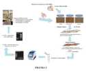
[0033] Figura 3. A Figura 3 é a representação esquemática do método de Sequenciamento de nova Geração (Fusion PCR Method – ION PGMTM) para identificação das sequências de aptâmeros obtidas por Cell-SELEX. A sequência adaptadora A em conjunto com a sequência chave (Key) e o Barcode são produzidas e fornecidas comercialmente e determinam a assinatura de cada conjunto de aptâmeros selecionados por Cell SELEX. A sequência adaptadora P1, também fornecida comercialmente, está presente em todas as sequências de aptâmeros. Primeiramente, a reação de ligação dos adaptadores aos aptâmeros é feita e em seguida um PCR em emulsão, com iniciadores complementares aos adaptadores A e P1, é realizado para amplificação exponencial destas sequências.[0033] Figure 3. Figure 3 is the schematic representation of the Next Generation Sequencing method (Fusion PCR Method – ION PGMTM) for identifying the aptamer sequences obtained by Cell-SELEX. The adapter sequence A together with the key sequence (Key) and the Barcode are commercially produced and supplied and determine the signature of each set of aptamers selected by Cell SELEX. The P1 adapter sequence, also commercially available, is present in all aptamer sequences. First, the binding reaction of the adapters to the aptamers is carried out and then an emulsion PCR, with primers complementary to the A and P1 adapters, is performed for exponential amplification of these sequences.
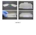
[0034] Figura 4. A Figura 4 mostra o N3DBioprinting®. (A) – Apresentação do produto; (B) – Base com os botões magnéticos; (C) – placa de cultivo dos esferoides 3D; e (D) – Formação dos esferoides (“printing”). Fonte: www.n3dbio.com[0034] Figure 4. Figure 4 shows the N3DBioprinting®. (A) – Presentation of the product; (B) – Base with magnetic buttons; (C) – 3D spheroid culture plate; and (D) – Formation of spheroids (“printing”). Source: www.n3dbio.com
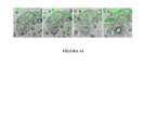
[0035] Figura 5. A Figura 5 mostra imagens de microscopia de células tumorais e controle não tumorais cultivadas em meio específico para cada tipo celular: A – LNCaP; B – RWPE-1; C – OVCAR-3; D – CHO-K1; E – MDA-MB-231; e F-MCF 10A. As culturas foram mantidas em estufa a 37ºC em atmosfera de 5% de CO2, utilizadas para os ensaios de Cell-SELEX. Características morfológicas das células em monocamada, visualizadas em microscópio de campo claro, aumento 10x.[0035] Figure 5. Figure 5 shows microscopy images of tumor cells and non-tumor control cultured in specific medium for each cell type: A – LNCaP; B – RWPE-1; C – OVCAR-3; D – CHO-K1; E – MDA-MB-231; and F-
[0036] Figura 6. A Figura 6 mostra gel de agarose 3% para verificação de amplificação de pool de aptâmeros no 12º round de CellSELEX após incubação com linhagem tumoral de próstata (LNCaP). A – PCR de verificação de melhor ciclagem com Cell-SELEX realizado a 37ºC – canais 1 a 5 – e Cell-SELEX realizado a 4ºC – canais 6 a 10. Os canais 1 e 6 correspondem a 4 ciclos de PCR; canais 2 e 7, 6 ciclos de PCR; canais 3 e 8, 8 ciclos de PCR; canal 4 e, 10 ciclos de PCR/ canais 5 e 10, 12 ciclos de PCR. B – PCR Preparativo (Primer Forward) realizado com o número de ciclos escolhido no PCR de verificação de melhor ciclo (10 ciclos) em Cell-SELEX realizado a 37ºC (canal 11) e a 4ºC (canal 12).[0036] Figure 6. Figure 6 shows 3% agarose gel for verification of aptamer pool amplification in the 12th round of CellSELEX after incubation with prostate tumor cell line (LNCaP). A – Better cycling verification PCR with Cell-SELEX performed at 37ºC –
[0037] Figura 7. A Figura 7 mostra a avaliação de ligação doseresposta com a linhagem tumoral de próstata, LNCaP, incubados com aptâmeros selecionados por Cell-SELEX após o 12º Round por 1h a 37ºC, nas concentrações de 5, 10 e 20µM de aptâmeros. A – Células incubadas com a biblioteca de aptâmeros; B – Células incubadas apenas com o primer marcado com FITC; C – Células incubadas com aptâmeros ssDNA 12R a 5µM; D – Células incubadas com aptâmeros ssDNA 12R a 10µM; E – Células incubadas com aptâmeros ssDNA 12R a 20 µM.[0037] Figure 7. Figure 7 shows the evaluation of dose-response binding with the prostate tumor cell line, LNCaP, incubated with aptamers selected by Cell-SELEX after the 12th Round for 1h at 37ºC, at concentrations of 5, 10 and 20µM of aptamers. A – Cells incubated with the aptamer library; B – Cells incubated only with the primer labeled with FITC; C – Cells incubated with
[0038] Figura 8. A Figura 8 mostra o ensaio de citometria para avaliação de enriquecimento do conjunto de aptâmeros ssDNA em células tumorais e controle não tumorais, LNCaP e RWPE-1 respectivamente, entre o 6º e 12º round de Cell-SELEX. (A) Histograma que representa a intensidade média de fluorescência de LNCaP tratadas com aptâmeros FITC do 6º ao 12º round; (B) Representação gráfica do percentual do número de células LNCaP fluorescentes (incubadas com aptâmeros FITC) dentro da população selecionada (R1); (C) Representação gráfica de intensidade média de fluorescência de células LNCaP dentro da população selecionada (R1); (D) Histograma que representa a intensidade média de fluorescência de LNCaP e RWPE-1 tratados com aptâmeros FITC no 12º round dentro da população selecionada (R1); (E) Representação gráfica do percentual do número de células LNCaP e RWPE-1 tratadas com aptâmeros FITC no 12º round dentro da população selecionada (R1).[0038] Figure 8. Figure 8 shows the cytometry assay for evaluating the enrichment of the ssDNA aptamer pool in tumor cells and non-tumor control, LNCaP and RWPE-1 respectively, between the 6th and 12th round of Cell-SELEX. (A) Histogram representing the mean fluorescence intensity of LNCaP treated with FITC aptamers from the 6th to the 12th round; (B) Graphic representation of the percentage of the number of fluorescent LNCaP cells (incubated with FITC aptamers) within the selected population (R1); (C) Graphic representation of mean fluorescence intensity of LNCaP cells within the selected population (R1); (D) Histogram representing the mean fluorescence intensity of LNCaP and RWPE-1 treated with FITC aptamers in the 12th round within the selected population (R1); (E) Graphic representation of the percentage of the number of LNCaP and RWPE-1 cells treated with FITC aptamers in the 12th round within the selected population (R1).
[0039] Figura 9. A Figura 9 mostra gel de agarose 3% para verificação de amplificação de pool de aptâmeros no 12º round de Cell-SELEX após incubação com linhagem tumoral de mama (MDA-MB-231). A – PCR de verificação de melhor ciclagem com Cell-SELEX realizado a 37ºC – canais 1 a 4 – O canal de número 1 corresponde a 6 ciclos de PCR; canal 2, 8 ciclos de PCR; canal 3, 10 ciclos de PCR; canal 4, 12 ciclos de PCR. B – PCR Preparativo (Primer Forward) realizado com o número ciclos escolhido no PCR de verificação de melhor ciclo (10 ciclos) em Cell-SELEX realizado a 37ºC (canal 5).[0039] Figure 9. Figure 9 shows 3% agarose gel for verification of aptamer pool amplification in the 12th round of Cell-SELEX after incubation with breast tumor cell line (MDA-MB-231). A – Better cycling verification PCR with Cell-SELEX performed at 37ºC –
[0040] Figura 10. A Figura 10 mostra o ensaio de citometria para avaliação de enriquecimento do conjunto de aptâmeros ssDNA em células tumorais e controle não tumorais, MDA-MB-231 e MCF-10A respectivamente, entre o 6º e 12º round de Cell-SELEX. (A) Histograma que representa a intensidade média de fluorescência de MDA-MB-231 tratadas com aptâmeros FITC do 6º ao 12º round; (B) Representação gráfica do percentual do número de células MDA-MB-231 fluorescentes (tratadas com aptâmeros FITC) dentro da população selecionada (R1); (C) Representação gráfica de intensidade média de fluorescência de células MDA-MB-231 dentro da população selecionada (R1); (D) Histograma que representa a intensidade média de fluorescência de MDA-MB-231 e MCF-10A tratados com aptâmeros FITC no 12º round dentro da população selecionada (R1); (E) Representação gráfica do percentual do número de células MDA-MB-231 e MCF-10A tratadas com aptâmeros FITC no 12º round dentro da população selecionada (R1).[0040] Figure 10. Figure 10 shows the cytometry assay for evaluating the enrichment of the ssDNA aptamer set in tumor cells and non-tumor control, MDA-MB-231 and MCF-10A respectively, between the 6th and 12th round of Cell -SELEX. (A) Histogram representing the mean fluorescence intensity of MDA-MB-231 treated with FITC aptamers from the 6th to the 12th round; (B) Graphic representation of the percentage number of fluorescent MDA-MB-231 cells (treated with FITC aptamers) within the selected population (R1); (C) Graphic representation of mean fluorescence intensity of MDA-MB-231 cells within the selected population (R1); (D) Histogram representing the mean fluorescence intensity of MDA-MB-231 and MCF-10A treated with FITC aptamers in the 12th round within the selected population (R1); (E) Graphic representation of the percentage number of MDA-MB-231 and MCF-10A cells treated with FITC aptamers in the 12th round within the selected population (R1).
[0041] Figura 11. A Figura 11 mostra gel de agarose 3% para verificação de amplificação de pool de aptâmeros no 16º round de CellSELEX após incubação com linhagem tumoral de ovário (OVCAR-3). A – PCR de verificação de melhor ciclagem com Cell-SELEX realizado a 37ºC – canais 1 a 4 – O canal de número 1 corresponde a 6 ciclos de PCR; canal 2, 8 ciclos de PCR; canal 3, 10 ciclos de PCR; canal 4, 12 ciclos de PCR. B – PCR Preparativo (Primer Forward) realizado com o número de ciclos escolhido no PCR de verificação de melhor ciclo (12 ciclos) em Cell-SELEX realizado a 37ºC (canal 5).[0041] Figure 11. Figure 11
[0042] Figura 12. A Figura 12 mostra o ensaio de citometria para avaliação de enriquecimento do conjunto de aptâmeros ssDNA em células tumorais e controle não tumorais, OVCAR-3 e CHO-K1 respectivamente, entre o 6º e 16º round de Cell-SELEX. (A) Histograma que representa a intensidade média de fluorescência de OVCAR-3 tratadas com aptâmeros FITC do 6º ao 16º round; (B) Representação gráfica do percentual do número de células OVCAR-3 fluorescentes (incubadas com aptâmeros FITC) dentro da população selecionada (R1); (C) Representação gráfica de intensidade média de fluorescência de células OVCAR-3 dentro da população selecionada (R1); (D) Histograma que representa a intensidade média de fluorescência de OVCAR-3 e CHO-K1 tratados com aptâmeros FITC no 16º round dentro da população selecionada (R1); (E) Representação gráfica do percentual do número de células OVCAR-3 e CHO-K1 tratadas com aptâmeros FITC no 16º round dentro da população selecionada (R1).[0042] Figure 12. Figure 12 shows the cytometry assay for evaluating the enrichment of the ssDNA aptamer set in tumor cells and non-tumor control, OVCAR-3 and CHO-K1 respectively, between the 6th and 16th round of Cell-SELEX . (A) Histogram representing the mean fluorescence intensity of OVCAR-3 treated with FITC aptamers from the 6th to the 16th round; (B) Graphic representation of the percentage number of fluorescent OVCAR-3 cells (incubated with FITC aptamers) within the selected population (R1); (C) Graphic representation of mean fluorescence intensity of OVCAR-3 cells within the selected population (R1); (D) Histogram representing the mean fluorescence intensity of OVCAR-3 and CHO-K1 treated with FITC aptamers in the 16th round within the selected population (R1); (E) Graphic representation of the percentage number of OVCAR-3 and CHO-K1 cells treated with FITC aptamers in the 16th round within the selected population (R1).
[0043] Figura 13. A Figura 13 mostra o ensaio de cinética de 30 a 240 minutos para avaliação de perfil de ligação de aptâmeros ssDNA 12R selecionados frente às células tumorais de próstata (LNCaP) em concentrações crescentes (1µM a 100µM), realizado no equipamento de análise de alto conteúdo (HCS) (aumento 10x).[0043] Figure 13. Figure 13 shows the kinetics assay from 30 to 240 minutes to evaluate the binding profile of selected
[0044] Figura 14. A Figura 14 mostra o perfil morfológico das células tumorais de próstata LNCaP frente à incubação de aptâmeros ssDNA 12R marcados com FITC a 1µM nos tempos de 30 a 240 minutos. Em destaque (setas) observa-se a alteração do perfil celular.[0044] Figure 14. Figure 14 shows the morphological profile of LNCaP prostate tumor cells against the incubation of
[0045] Figura 15. A Figura 15 mostra a avaliação dose-dependente de morfologia celular em microscópio ótico (20x) de células tumorais de próstata incubadas com aptâmeros-FITC nas concentrações de 0,25 a 50µM por 24 horas.[0045] Figure 15. Figure 15 shows the dose-dependent evaluation of cell morphology under light microscopy (20x) of prostate tumor cells incubated with aptamers-FITC at concentrations from 0.25 to 50µM for 24 hours.
[0046] Figura 16. A Figura 16 mostra a análise de viabilidade celular através do ensaio de Prestoblue® em células tumorais e controle de próstata incubadas com aptâmeros ssDNA 12R por 24 horas em concentrações crescentes (0,25 a 50 µM). A – Células tumorais (LNCaP); B – Células controle não tumorais (RWPE-1). O Triton X-100 foi utilizado como controle positivo para indução de perda de viabilidade celular. A biblioteca de aptâmeros (“biblio”, conjunto inicial total de aptâmeros utilizados no CellSELEX), foi utilizada como controle interno para verificação de viabilidade celular.[0046] Figure 16. Figure 16 shows the analysis of cell viability through the Prestoblue® assay in tumor cells and prostate control incubated with
[0047] Figura 17. A Figura 17 mostra a análise de proliferação celular através do ensaio de Cyquant® (Thermofisher®) em células tumorais e controle de próstata incubadas com aptâmeros ssDNA 12R por 24 horas em concentrações crescentes (0,25 a 100µM). A – Células tumorais (LNCaP); B – Células controle não tumorais (RWPE-1). O Triton X-100 foi utilizado como controle positivo para indução de perda de viabilidade celular e inibição da proliferação celular.[0047] Figure 17. Figure 17 shows the analysis of cell proliferation through the Cyquant® assay (Thermofisher®) in tumor cells and prostate control incubated with
[0048] Figura 18. A Figura 18 mostra a análise qualitativa de indução de morte celular por apoptose através da técnica de TUNEL® (Terminal deoxinucleotidil transferase Uracil Nick end Labeling) em células tumorais de próstata incubadas com aptâmeros 12R por 24 horas nas concentrações crescentes de 25µM e 50µM. O controle é a célula LNCaP sem o tratamento com aptâmeros. Em azul, núcleo celular marcado com DAPI (1:10.000) e em verde, marcação de DNA fragmentado com FTIC.[0048] Figure 18. Figure 18 shows the qualitative analysis of induction of cell death by apoptosis through the TUNEL® technique (Terminal deoxynucleotidyl transferase Uracil Nick end Labeling) in prostate tumor cells incubated with 12R aptamers for 24 hours at increasing concentrations of 25µM and 50µM. The control is the LNCaP cell without the aptamer treatment. In blue, cell nucleus stained with DAPI (1:10,000) and in green, fragmented DNA stained with FTIC.
[0049] Figura 19. A Figura 19 mostra o ensaio de Western Blot para investigar a ativação da via das caspases após incubação das células tumoral e controle não tumoral, LNCaP e RWPE-1 respectivamente, com aptâmeros ssDNA 25µM por 3 horas. Extratos proteicos totais de células LNCaP tratadas com aptâmeros ssDNA (+), células LNCaP não tratadas com aptâmeros ssDNA (-), células RWPE-1 tratadas com aptâmeros ssDNA (+) e, células RWPE-1 não tratadas com aptâmeros ssDNA (-). As análises de Western Blot e o histograma para verificação dos níveis expressão de Caspase 10 (A), Caspase 9 (B) e Bax (C) foram normalizados em relação aos níveis de GAPDH (controle endógeno). Padrão de massa molecular utilizado: PageRuler Plus Prestained Protein Ladder (Thermo Scientific®) (*P < 0.05).[0049] Figure 19. Figure 19 shows the Western Blot assay to investigate the activation of the caspase pathway after incubation of tumor and non-tumor control cells, LNCaP and RWPE-1 respectively, with 25µM ssDNA aptamers for 3 hours. Total protein extracts from LNCaP cells treated with ssDNA aptamers (+), LNCaP cells not treated with ssDNA aptamers (-), RWPE-1 cells treated with ssDNA aptamers (+) and, RWPE-1 cells not treated with ssDNA aptamers (-) . Western Blot analyzes and the histogram to verify Caspase 10 (A), Caspase 9 (B) and Bax (C) expression levels were normalized in relation to GAPDH levels (endogenous control). Molecular mass standard used: PageRuler Plus Prestained Protein Ladder (Thermo Scientific®) (*P < 0.05).
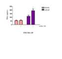
[0050] Figura 20. A Figura 20 mostra o ensaio de ELISA para avaliar a expressão de TRAIL em células LNCaP e RWPE-1 incubadas com aptâmeros ssDNA 12R por 3 horas.[0050] Figure 20. Figure 20 shows the ELISA assay to assess TRAIL expression in LNCaP and RWPE-1 cells incubated with
[0051] Figura 21. A Figura 21 mostra o conjunto de ssDNA aptâmeros 12R que induz ativação de caspase-3 em células tumorais de próstata, LNCaP. Detecção de atividade de caspase-3 através do ensaio em células vivas NucView® 488 and CF®594 Annexin V Dual Apoptosis Assay Kit. Marcação de caspase-3 ativada (verde) em células LNCaP incubadas com 25µM de aptâmeros ssDNA 12R nos tempos indicados (20-180 minutos com intervalo de 20 minutos). Marcação de anexina V (vermelho) para visualizar a exposição de fosfatidilserina; e calceína (azul) para observar células viáveis. Em J, K e L campo aumentado das imagens em A, B e C, respectivamente.[0051] Figure 21. Figure 21 shows the set of
[0052] Figura 22. A Figura 22 mostra a quantificação da detecção de atividade de caspase-3 através do ensaio em células vivas NucView® 488 and CF®594 Annexin V Dual Apoptosis Assay Kit. Marcação de caspase 3 ativada em células LNCaP (A) e RWPE-1 (B) incubadas com 25µM de aptâmeros ssDNA 12R nos tempos indicados (20-180 minutos com intervalo de 20 minutos), representados na linha de cor roxa; as células sem indução foram incubadas apenas com tampão de ligação, representadas pela cor azul; e as células tratadas com dexametasona a 1 µM estão representadas na cor laranja.[0052] Figure 22. Figure 22 shows quantification of caspase-3 activity detection using the NucView® 488 and CF®594 Annexin V Dual Apoptosis Assay Kit live cell assay.
[0053] Figura 23. A Figura 23 mostra o ensaio de avaliação de capacidade de formação de microesferas 3D em célula tumoral de ovário (OVCAR-3) e o controle não tumoral (CHO-K1) utilizando a tecnologia “n3D System technology”, desenvolvido pela Nano3D Biosciences, Inc®. Diferentes quantidades de OVCAR-3 e CHO-K1 (25x103 , 50x103 e 75x103 ) foram incubadas com as nanopartículas magnetizadas em uma placa magnética (Bioprinting) de 96 poços por duas horas. Após a formação dos esferoides, a base é retirada e a cultura é mantida no meio específico para cada tipo celular (aumento de 10x).[0053] Figure 23. Figure 23 shows the assay for evaluating the ability to form 3D microspheres in ovarian tumor cells (OVCAR-3) and non-tumor control (CHO-K1) using the “n3D System technology”, developed by Nano3D Biosciences, Inc®. Different amounts of OVCAR-3 and CHO-K1 (25x103 , 50x103 and 75x103 ) were incubated with the magnetized nanoparticles in a 96-well magnetic plate (Bioprinting) for two hours. After the formation of spheroids, the base is removed and the culture is maintained in the specific medium for each cell type (10x magnification).
[0054] Figura 24. A Figura 24 mostra o ensaio de avaliação de capacidade de formação de microesferas 3D em célula tumoral de próstata (LNCaP) e o controle não tumoral (RWPE-1) utilizando a tecnologia n3D System technology”, desenvolvido pela Nano3D Biosciences, Inc®. 50x103 células foram incubadas com as nanopartículas magnetizadas em uma placa magnética (Bioprinting) de 96 poços por duas horas. Após a formação dos esferoides, a base é retirada e a cultura é mantida no meio específico para cada tipo celular (aumento de 10x).[0054] Figure 24. Figure 24 shows the assay for evaluating the ability to form 3D microspheres in prostate tumor cells (LNCaP) and non-tumor control (RWPE-1) using n3D System technology”, developed by Nano3D Biosciences, Inc®. 50x103 cells were incubated with magnetized nanoparticles in a 96-well magnetic plate (Bioprinting) for two hours. After the formation of spheroids, the base is removed and the culture is maintained in the specific medium for each cell type (10x magnification).
[0055] Figura 25 A Figura 25 mostra a avaliação de cultura de microesferas de células tumorais e controle de ovário (OVCAR-3 e CHO-K1, respectivamente) incubadas com conjunto de aptâmeros ssDNA 16R por 24h. após 24 horas de cultivo dos esferóides (Figura 23), os aptâmeros ssDNA 16R 25 e 50 µM foram adicionados às culturas e mantidos por 24 horas. Após 24 horas de incubação com os aptâmeros ssDNA, os esferóides foram fixados com PFA 4% e marcado com DAPI para visualização do volume da microesfera (n=1).[0055] Figure 25 Figure 25 shows the evaluation of microsphere culture of tumor cells and ovarian control (OVCAR-3 and CHO-K1, respectively) incubated with
[0056] Figura 26. A Figura 26 mostra a avaliação de cultura de microesferas de células tumorais e controle de próstata (LNCaP e RWPE-1, respectivamente) incubadas com conjunto de aptâmeros ssDNA 12R por 24h. Após 24 horas de cultivo dos esferóides (Figura 24), os aptâmeros ssDNA 12R 25µM foram adicionados às culturas e mantidos por 24 horas. Após 24 horas de incubação com os aptâmeros ssDNA, os esferóides foram fixados com PFA 4% e marcado com DAPI para visualização do volume da microesfera (n=1).[0056] Figure 26. Figure 26 shows the evaluation of microsphere culture of tumor cells and prostate control (LNCaP and RWPE-1, respectively) incubated with
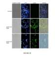
[0057] Figura 27. A Figura 27 mostra a avaliação do efeito do conjunto de aptâmeros ssDNA sob culturas 3D de células tumorais ovário e próstata (OVCAR-3 e LNCaP, respectivamente). Após 24 horas de incubação dos esferóides com os aptâmeros ssDNA 25µM, os esferoides das duas linhagens (OVCAR-3 e LNCaP) foram marcados com DAPI, LysoTrackerRed e Faloidina-FITC para visualização do núcleo, lisossomas e citoesqueleto, respectivamente.[0057] Figure 27. Figure 27 shows the evaluation of the effect of the ssDNA aptamer pool on 3D cultures of ovarian and prostate tumor cells (OVCAR-3 and LNCaP, respectively). After 24 hours of incubation of the spheroids with 25µM ssDNA aptamers, the spheroids of the two strains (OVCAR-3 and LNCaP) were labeled with DAPI, LysoTrackerRed and Phalloidin-FITC for visualization of the nucleus, lysosomes and cytoskeleton, respectively.
[0058] A não ser que sejam definidos de maneira diferente, todos os termos técnicos e científicos aqui utilizados têm o mesmo significado entendido por um técnico no assunto ao qual a invenção pertence. As técnicas convencionais de biologia molecular e imunologia são bem conhecidas de um técnico no assunto. O relatório descritivo também provê definições de termos para auxiliar na interpretação daquilo que é aqui descrito e das reivindicações. A não ser que seja indicado de forma diferente, todos os números expressando quantidades, porcentagens e proporções, e outros valores numéricos usados no relatório descritivo e nas reivindicações, devem ser entendidos como sendo modificados, em todos os casos, pelo termo “cerca de”. Assim, a não ser que seja indicado o contrário, os parâmetros numéricos mostrados no relatório descritivo e nas reivindicações são aproximações que podem variar, dependendo das propriedades a serem obtidas.[0058] Unless otherwise defined, all technical and scientific terms used herein have the same meaning as understood by one skilled in the art to which the invention pertains. Conventional molecular biology and immunology techniques are well known to a person skilled in the art. The specification also provides definitions of terms to aid in the interpretation of what is described herein and the claims. Unless otherwise indicated, all figures expressing amounts, percentages and proportions, and other numerical values used in the specification and claims, are to be understood to be modified, in all cases, by the term "about" . Thus, unless otherwise indicated, numerical parameters shown in the specification and claims are approximations that may vary depending on the properties to be obtained.
[0059] O termo “aptâmeros”, no contexto da presente invenção, refere-se a sequências de ácido nucleico que adotam uma estrutura 3D específica que permite que as mesmas se liguem a alvos moleculares com alta especificidade e afinidade.[0059] The term "aptamers", in the context of the present invention, refers to nucleic acid sequences that adopt a specific 3D structure that allows them to bind to molecular targets with high specificity and affinity.
[0060] O termo “ácido nucleico”, de acordo com a presente invenção, refere-se a qualquer tipo de ácido nucleico, tal como DNA e RNA, e a variantes dos mesmos, assim como combinações dos mesmos, modificações dos mesmos, incluindo nucleotídeos modificados etc. Os termos “ácido nucleico” e “oligonucleotídeo” e “polinucleotídeo” são usados de maneira intercambiável no contexto da presente invenção. Os ácidos nucleicos podem ser purificados de fontes naturais, produzidos com o uso de sistemas de expressão recombinante e, opcionalmente, purificados, quimicamente sintetizados, etc. Quando apropriado, por exemplo, no caso de moléculas quimicamente sintetizadas, os ácidos nucleicos podem compreender análogos de nucleosídeo tais como análogos que têm bases ou açúcares quimicamente modificados, modificações da cadeia principal, etc. Uma sequência de ácidos nucleicos é representada na direção 5’-3’ a não ser que indicado de outro modo. Como utilizado aqui, os símbolos para nucleotídeos e polinucleotídeos são os recomendados pela IUPAC-IUB Commission of Biochemical Nomenclature (Biochem. 9:4022, 1970).[0060] The term "nucleic acid", according to the present invention, refers to any type of nucleic acid, such as DNA and RNA, and variants thereof, as well as combinations thereof, modifications thereof, including modified nucleotides etc. The terms "nucleic acid" and "oligonucleotide" and "polynucleotide" are used interchangeably in the context of the present invention. Nucleic acids can be purified from natural sources, produced using recombinant expression systems, and optionally purified, chemically synthesized, etc. Where appropriate, for example in the case of chemically synthesized molecules, the nucleic acids may comprise nucleoside analogues such as analogues having chemically modified bases or sugars, backbone modifications, and the like. A nucleic acid sequence is represented in the 5'-3' direction unless otherwise indicated. As used herein, the symbols for nucleotides and polynucleotides are those recommended by the IUPAC-IUB Commission of Biochemical Nomenclature (Biochem. 9:4022, 1970).
[0061] Conforme usado na presente invenção, o emprego do termo “farmaceuticamente aceitável” significa, essencialmente, não ser tóxico ao indivíduo ao qual o material farmaceuticamente aceitável é administrado.[0061] As used in the present invention, use of the term "pharmaceutically acceptable" essentially means not being toxic to the individual to whom the pharmaceutically acceptable material is administered.
[0062] O termo “variante funcionalmente equivalente” refere-se a aptâmeros com sequências substancialmente similares às SEQ ID NOs: 1-10, 21-30 ou 41-50 mantendo a capacidade de se ligar e/ou inibir especificamente seu alvo. Uma variante funcionalmente equivalente do aptâmero da invenção pode ser uma sequência de ácidos nucleicos derivada das SEQ ID NOs: 1-10, 21-30 ou 31-40 que compreende a adição, substituição ou modificação de um ou mais nucleotídeos. A título de ilustração, as variantes funcionalmente equivalentes do aptâmero da invenção incluem sequências que compreendem a adição, remoção, substituição ou modificação de 1 a 3 nucleotídeos das sequências SEQ ID NOs: 1-10, 21-30 ou 41-50 e que mantêm uma capacidade de se ligar especificamente ao seu alvo e até inibi-lo.[0062] The term "functionally equivalent variant" refers to aptamers with sequences substantially similar to SEQ ID NOs: 1-10, 21-30 or 41-50 while retaining the ability to specifically bind and/or inhibit their target. A functionally equivalent variant of the aptamer of the invention can be a nucleic acid sequence derived from SEQ ID NOs: 1-10, 21-30 or 31-40 which comprises the addition, substitution or modification of one or more nucleotides. By way of illustration, functionally equivalent variants of the aptamer of the invention include sequences comprising the addition, deletion, substitution or modification of 1 to 3 nucleotides of sequences SEQ ID NOs: 1-10, 21-30 or 41-50 and which maintain an ability to specifically bind to its target and even inhibit it.
[0063] Mais especificamente, o termo “identidade” é definido como o grau de igualdade entre sequências de DNA ou RNA quando comparados nucleotídeo por nucleotídeo com uma sequência de referência.[0063] More specifically, the term “identity” is defined as the degree of similarity between DNA or RNA sequences when compared nucleotide by nucleotide with a reference sequence.
[0064] Na presente invenção, o termo “porcentagem de identidade de sequências” refere-se a comparações entre polinucleotídeos e é determinado por duas sequências idealmente alinhadas, sob determinados parâmetros de comparação. Este alinhamento pode compreender espaços (gaps), gerando intervalos quando comparadas à sequência de referência, que facilitam uma comparação adequada das mesmas. De maneira geral, o cálculo da porcentagem de identidade considera o número de posições onde o mesmo nucleotídeo ocorre nas sequências comparadas à sequência referência, sendo realizado através de diversos algoritmos de comparação de sequências e programas conhecidos no estado da arte. Tais algoritmos e programas incluem, mas não são limitados a BLAST e CLUSTAL, por exemplo.[0064] In the present invention, the term "percentage of sequence identity" refers to comparisons between polynucleotides and is determined by two ideally aligned sequences, under certain comparison parameters. This alignment may include spaces (gaps), generating intervals when compared to the reference sequence, which facilitate an adequate comparison of the same. In general, the calculation of the percentage of identity considers the number of positions where the same nucleotide occurs in the sequences compared to the reference sequence, being performed through several sequence comparison algorithms and programs known in the state of the art. Such algorithms and programs include, but are not limited to, BLAST and CLUSTAL, for example.
[0065] A presente invenção também inclui aptâmeros que compreendem sequências de nucleotídeos com uma identidade de sequência de pelo menos 70%, pelo menos 75%, pelo menos 80%, pelo menos 85%, pelo menos 90%, pelo menos 91%, pelo menos 92%, pelo menos 93%, pelo menos 94%, pelo menos 95%, pelo menos 96%, pelo menos 97%, pelo menos 98% ou pelo menos 99% com as sequências SEQ ID NOs: 1-10, 21-30 ou 41- 50 que, juntamente com as sequências das extremidades que se repetem, mantêm uma capacidade de se ligar especificamente ao seu alvo e inibi-lo.[0065] The present invention also includes aptamers comprising nucleotide sequences having a sequence identity of at least 70%, at least 75%, at least 80%, at least 85%, at least 90%, at least 91%, at least 92%, at least 93%, at least 94%, at least 95%, at least 96%, at least 97%, at least 98% or at least 99% with sequences SEQ ID NOs: 1-10, 21-30 or 41-50 which, together with the repeating end sequences, retain an ability to specifically bind to and inhibit its target.
[0066] Ao longo deste relatório descritivo, a menos que o contexto exija de outra forma, a palavra "compreendem", ou variações tais como "compreende" ou "compreendendo", será entendida como implicando na inclusão de uma etapa indicada ou elemento ou número inteiro ou grupo de elementos ou etapas ou números inteiros mas não a exclusão de qualquer outra etapa ou elemento ou número inteiro ou grupo de elementos ou inteiros.[0066] Throughout this specification, unless the context otherwise requires, the word "comprises", or variations such as "comprises" or "comprising", will be understood to imply the inclusion of an indicated step or element or whole number or group of elements or steps or integers but not the exclusion of any other step or element or whole number or group of elements or integers.
[0067] O termo "consiste em" ou "consistindo em" deve ser entendido no sentido de que um método, processo ou composição de matéria tem as etapas e/ou componentes citados e sem etapas ou componentes adicionais.[0067] The term "consisting of" or "consisting of" is to be understood in the sense that a method, process or composition of matter has the aforementioned steps and/or components and no additional steps or components.
[0068] No presente pedido, o reagente diagnóstico refere-se ao aptâmero utilizado para monitorar a presença e/ou o progressão da doença e a resposta à terapia.[0068] In the present application, the diagnostic reagent refers to the aptamer used to monitor the presence and/or progression of the disease and the response to therapy.
[0069] Os termos "tratar", "tratando" e "tratamento" referem-se a um método para aliviar ou anular uma doença e/ou os seus sintomas concomitantes.[0069] The terms "treat", "treating" and "treatment" refer to a method of alleviating or reversing a disease and/or its concomitant symptoms.
[0070] O termo “câncer” refere-se a uma proliferação anormal, autônoma e descontrolada de células de um determinado tecido do corpo. O termo “câncer” pode se referir, por exemplo, a linfomas, mielomas múltiplos, malignidades hematológicas, leucemias, neoplasmas e tumores sólidos e suas metástases. Em uma modalidade preferencial, os cânceres são de próstata, mama e ovário.[0070] The term “cancer” refers to an abnormal, autonomous and uncontrolled proliferation of cells of a particular tissue of the body. The term "cancer" may refer, for example, to lymphomas, multiple myelomas, hematologic malignancies, leukaemias, neoplasms, and solid tumors and their metastases. In a preferred embodiment, the cancers are prostate, breast, and ovarian.
[0071] De acordo com a presente invenção, “carreadores”, “excipientes” ou “solvente farmaceuticamente aceitável” busca incluir quaisquer e todos os solventes, meios de dispersão, revestimentos, agentes antibacterianos e antifúngicos, agentes isotônicos e de atraso de absorção e similares compatíveis com a administração farmacêutica. O uso de tais carreadores e veículos em substâncias farmaceuticamente ativas é bem conhecido na técnica. A não ser que qualquer carreador convencional seja incompatível com o composto ativo, o uso do mesmo nas composições da invenção é contemplado. Os veículos aceitáveis, excipientes ou estabilizadores aceitáveis não são tóxicos para o indivíduo nas doses e concentrações usadas e incluem tampões tais como fosfato, citrato e outros ácidos orgânicos; antioxidantes incluindo ácido ascórbico e metionina; conservantes (tais como cloreto de octadecildimetilbenzil amônio, cloreto de hexametônio, cloreto de benzalcônio, cloreto de benzetônio; álcool fenólico, butílico ou benzílico; alquil parabenos, tais como metil ou propil parabeno; catecol; resorcinol; ciclohexanol; 3-pentanol e m-cresol); polipeptídeos de peso molecular baixo (menos do que cerca de 10 aminoácidos); proteínas, tais como albumina sérica, gelatina ou imunoglobulinas; polímeros hidrofílicos tal como polivinilpirrolidona; aminoácidos tais como glicina, glutamina, asparagina, histidina, arginina ou lisina; monossacarídeos, dissacarídeos e outros carboidratos incluindo glicose ou dextrinas; agentes quelantes tal como EDTA; açúcares tais como sacarose, manitol, trealose ou sorbitol; contraíons formadores de sal tal como sódio; complexos de metal (por exemplo, complexos de Zn-proteína); e/ou tensoativos não iônicos tais como TWEEN™, PLURONICS™ ou polietileno glicol (PEG). Em uma concretização, o veículo preferido é polietilenoglicol (PEG).[0071] According to the present invention, "carriers", "excipients" or "pharmaceutically acceptable solvent" seeks to include any and all solvents, dispersion media, coatings, antibacterial and antifungal agents, isotonic and absorption delay agents and similar compatible with pharmaceutical administration. The use of such carriers and vehicles in pharmaceutically active substances is well known in the art. Unless any conventional carrier is incompatible with the active compound, use thereof in the compositions of the invention is contemplated. Acceptable carriers, excipients or stabilizers are not toxic to the individual at the doses and concentrations used and include buffers such as phosphate, citrate and other organic acids; antioxidants including ascorbic acid and methionine; preservatives (such as octadecyldimethylbenzyl ammonium chloride, hexamethonium chloride, benzalkonium chloride, benzethonium chloride; phenolic, butyl, or benzyl alcohol; alkyl parabens, such as methyl or propyl paraben; catechol; resorcinol; cyclohexanol; 3-pentanol and m- cresol); low molecular weight polypeptides (less than about 10 amino acids); proteins, such as serum albumin, gelatin or immunoglobulins; hydrophilic polymers such as polyvinylpyrrolidone; amino acids such as glycine, glutamine, asparagine, histidine, arginine or lysine; monosaccharides, disaccharides and other carbohydrates including glucose or dextrins; chelating agents such as EDTA; sugars such as sucrose, mannitol, trehalose or sorbitol; salt forming counterions such as sodium; metal complexes (eg Zn-protein complexes); and/or non-ionic surfactants such as TWEEN™, PLURONICS™ or polyethylene glycol (PEG). In one embodiment, the preferred carrier is polyethylene glycol (PEG).
[0072] De acordo com a presente invenção, os “ingredientes ativos” podem também ser incorporados na composição. Portanto, em uma concretização particular, a composição fornecida pela presente invenção pode também conter mais do que um ingrediente ativo conforme exigido para a indicação particular em questão, de preferência, aqueles com atividades complementares que não afetam adversamente um ao outro. Por exemplo, pode ser desejável fornecer, ademais, um agente quimioterapêutico, uma citocina, um agente analgésico, um agente anti-inflamatório ou um agente imunossupressor. A quantidade eficaz dos ditos outros ingredientes ativos depende, dentre outras coisas, da quantidade terapêutica dos aptâmeros que estão presentes na composição, da natureza e da gravidade da patologia a ser tratada, do indivíduo, etc.[0072] According to the present invention, the "active ingredients" can also be incorporated into the composition. Therefore, in a particular embodiment, the composition provided by the present invention may also contain more than one active ingredient as required for the particular indication in question, preferably those with complementary activities that do not adversely affect one another. For example, it may be desirable to additionally provide a chemotherapeutic agent, cytokine, analgesic agent, anti-inflammatory agent or immunosuppressive agent. The effective amount of said other active ingredients depends, among other things, on the therapeutic amount of the aptamers that are present in the composition, on the nature and severity of the pathology to be treated, on the individual, etc.
[0073] De acordo com a presente invenção, "fluroróforos" refere-se a um componente funcional de uma molécula que pode tornar uma outra molécula a ela conjugada fluorescente por meio de absorção de energia de um comprimento de onda específico, emitindo energia em diferentes comprimentos de onda. Dentre os possíveis fluoróforos utilizados na presente invenção podemos citar Isotiocianato de fluoresceína (FITC), Iodeto de Propídio, Rodamina, Tetrametilrodamina (TRITC) e outro derivados da Rodamina, Alexa Fluor 594, Texas Red, Alexa Fluor 647, Cy3, Cy5.[0073] According to the present invention, "fluorophores" refers to a functional component of a molecule that can make another molecule conjugated to it fluorescent by absorbing energy of a specific wavelength, emitting energy at different wavelengths. Among the possible fluorophores used in the present invention we can mention Fluorescein Isothiocyanate (FITC), Propidium Iodide, Rhodamine, Tetramethylrhodamine (TRITC) and other derivatives of Rhodamine, Alexa Fluor 594, Texas Red, Alexa Fluor 647, Cy3, Cy5.
[0074] De acordo com a presente invenção, "agentes de contrastes de imagem" são substâncias utilizadas para aumentar o contraste de estruturas ou fluidos em imagens. Dentre os possíveis agentes de contraste utilizados na presente invenção estão os contrastes iodados, os derivados de gadolínio, derivados de bário e os radioisótopos.[0074] According to the present invention, "image contrast agents" are substances used to enhance the contrast of structures or fluids in images. Among the possible contrast agents used in the present invention are iodinated contrasts, gadolinium derivatives, barium derivatives and radioisotopes.
[0075] Vale ressaltar que, com a presente invenção, qualquer molécula conjugada a ela, sendo essa molécula qualquer fluoróforo, qualquer radioisótopo ou qualquer agente de contraste de imagem são capazes de determinar células tumorais alvo, importantes no diagnóstico precoce de tumores.[0075] It is noteworthy that, with the present invention, any molecule conjugated to it, being that molecule any fluorophore, any radioisotope or any image contrast agent, are capable of determining target tumor cells, important in the early diagnosis of tumors.
[0076] Tal como empregado no presente documento, o termo "quantidade terapeuticamente eficaz" pode significar uma quantidade suficiente de aptâmero, agente anticancerígeno, um agente de distribuição ou composição de acordo com a presente invenção para inibir ou reduzir o número de células cancerígenas e/ou um ou mais sintomas de câncer. O técnico especialista no assunto estará ciente de que uma tal quantidade variará dependendo, por exemplo, do indivíduo em particular e/ou do tipo ou gravidade do nível da doença. O termo não deve ser interpretado no sentido de limitar a presente invenção a uma quantidade específica de aptâmero de DNA.[0076] As used herein, the term "therapeutically effective amount" can mean a sufficient amount of aptamer, anticancer agent, delivery agent or composition according to the present invention to inhibit or reduce the number of cancer cells and /or one or more symptoms of cancer. Those skilled in the art will be aware that such an amount will vary depending on, for example, the particular individual and/or the type or severity of the disease level. The term should not be construed to limit the present invention to a specific amount of DNA aptamer.
[0077] O termo "indivíduo" é aqui definido para incluir animais, tais como mamíferos. Em uma modalidade preferencial, o indivíduo é um ser humano.[0077] The term "individual" is defined herein to include animals, such as mammals. In a preferred embodiment, the subject is a human.
[0078] Embora a presente invenção possa ser suscetível a diferentes concretizações, é mostrada nos desenhos e na seguinte discussão detalhada, uma concretização preferida com o entendimento de que a presente descrição deve ser considerada uma exemplificação dos princípios da invenção e não pretende limitar a presente invenção ao que foi ilustrado e descrito aqui.[0078] Although the present invention may be susceptible to different embodiments, a preferred embodiment is shown in the drawings and the following detailed discussion, with the understanding that the present description is to be considered an exemplification of the principles of the invention and is not intended to limit the present invention to what has been illustrated and described here.
[0079] Em um primeiro aspecto, a invenção provê um aptâmero de ácido nucleico que compreende uma sequência de nucleotídeos com a fórmula geral (1), como segue, ou um sal farmaceuticamente aceitável do mesmo: TAGGGAAGAGAAGGACATATGAT-X1- TTGACTAGTACATGACCACTTGA (formula 1), em que X1 é a sequência de nucleotídeos como definida em qualquer uma das SEQ ID NO: 1-10, SEQ ID NO: 21-30 e SEQ ID NO: 41-50, ou uma sequência com pelo menos 90% de identidade às mesmas que apresente função equivalente às suas correspondentes. Em uma concretização, o ácido nucleico é DNA. Em outra concretização, o aptâmero é para uso como um medicamento ou um reagente diagnóstico. Em outra concretização, o aptâmero é para uso no diagnóstico e tratamento de câncer. Em outra concretização, o câncer é selecionado do grupo que consiste em câncer de próstata, ovário e mama. Em outra concretização, o câncer de mama é o câncer de mama triplo negativo.[0079] In a first aspect, the invention provides a nucleic acid aptamer comprising a nucleotide sequence with general formula (1), as follows, or a pharmaceutically acceptable salt thereof: TAGGGAAGAGAAGGACATATGAT-X1-TTGACTAGTACATGACCACTTGA (formula 1) , where X1 is the nucleotide sequence as defined in any one of SEQ ID NO: 1-10, SEQ ID NO: 21-30 and SEQ ID NO: 41-50, or a sequence with at least 90% identity to same ones that present function equivalent to their correspondents. In one embodiment, the nucleic acid is DNA. In another embodiment, the aptamer is for use as a medicament or a diagnostic reagent. In another embodiment, the aptamer is for use in diagnosing and treating cancer. In another embodiment, the cancer is selected from the group consisting of prostate, ovarian and breast cancer. In another embodiment, the breast cancer is triple negative breast cancer.
[0080] Em uma modalidade particular, o aptâmero aqui descrito consiste em entre 30 e 200 nucleotídeos, de preferência, entre 35 e 150 nucleotídeos, de maior preferência, entre 40 e 100 nucleotídeos, de ainda maior preferência, entre 45 e 80 nucleotídeos.[0080] In a particular embodiment, the aptamer described herein consists of between 30 and 200 nucleotides, preferably between 35 and 150 nucleotides, more preferably between 40 and 100 nucleotides, even more preferably between 45 and 80 nucleotides.
[0081] Os aptâmeros para uso no diagnóstico e tratamento de câncer de ovário podem ser como abaixo definidos: SEQ ID NO: 1: CATACACTCTTTATACTCTCATATTAACCA SEQ ID NO: 2: TCCCATTCTCTATAATAGTCCCCCACTATA SEQ ID NO: 3: ACGCTAATATCTGATTAATTACTTTTTTTTG SEQ ID NO: 4: GGTGTAGTTATTTGATATTCTTTATTGCTG SEQ ID NO: 5: TGGGATTAGTTTATGTTGTTTGTTGTTATT SEQ ID NO: 6: CACGGATATAATTATACCCTTTTAGTTTGA SEQ ID NO: 7: CAATACACTCTTTATAAACGTTTCCTTTCA SEQ ID NO: 8: GTATTGTTTTGTTGTTTCTTGTATAATTCG SEQ ID NO: 9: TGGTAGTATAGGTTGTTGTTGGTATTTATA SEQ ID NO: 10: TAGCGTTTTATAAATCTCTCCTTTATTGTG SEQ ID NO: 11: TAGGGAAGAGAAGGACATATGATCATACACTCTTTATACTCTCAT ATTAACCATTGACTAGTACATGACCACTTGA SEQ ID NO: 12: TAGGGAAGAGAAGGACATATGATTCCCATTCTCTATAATAGTCC CCCACTATATTGACTAGTACATGACCACTTGA SEQ ID NO: 13: TAGGGAAGAGAAGGACATATGATACGCTAATATCTGATTAATTA CTTTTTTTTGTTGACTAGTACATGACCACTTGA SEQ ID NO: 14: TAGGGAAGAGAAGGACATATGATGGTGTAGTTATTTGATATTCTT TATTGCTGTTGACTAGTACATGACCACTTGA SEQ ID NO: 15: TAGGGAAGAGAAGGACATATGATTGGGATTAGTTTATGTTGTTT GTTGTTATTTTGACTAGTACATGACCACTTGA SEQ ID NO: 16: TAGGGAAGAGAAGGACATATGATCACGGATATAATTATACCCTT TTAGTTTGATTGACTAGTACATGACCACTTGA SEQ ID NO: 17: TAGGGAAGAGAAGGACATATGATCAATACACTCTTTATAAACGT TTCCTTTCATTGACTAGTACATGACCACTTGA SEQ ID NO: 18: TAGGGAAGAGAAGGACATATGATGTATTGTTTTGTTGTTTCTTGT ATAATTCGTTGACTAGTACATGACCACTTGA SEQ ID NO: 19: TAGGGAAGAGAAGGACATATGATTGGTAGTATAGGTTGTTGTTG GTATTTATATTGACTAGTACATGACCACTTGA SEQ ID NO: 20: TAGGGAAGAGAAGGACATATGATTAGCGTTTTATAAATCTCTCCT TTATTGTGTTGACTAGTACATGACCACTTGA[0081] Aptamers for use in the diagnosis and treatment of ovarian cancer may be as defined below: SEQ ID NO: 1: CATACACTCTTTATACTTCCATATTAACCA SEQ ID NO: 2: TCCCATTCTCTATAATAGTCCCCCACTATA SEQ ID NO: 3: ACGCTAATATCTGATTAATTACTTTTTTTG SEQ ID NO: 4: GGTGTAGTTATTTGATATTCTTTATTGCTG SE Q ID NO: 5: TGGGATTAGTTTATGTTGTTTGTTGTTATT SEQ ID NO: 6: CACGGATATAATTATACCCTTTTAGTTTGA SEQ ID NO: 7: CAATACACTCTTTATAAACGTTTCCTTTCA SEQ ID NO: 8: GTATTGTTTTGTTGTTTCTTGTATAATTCG SEQ ID NO: 9: TGGTAGTATAGGTTGTTGTTGGTATTTATA SEQ ID NO: 10: TAGCGTTTTATAAATCTCTCCTTTATTGTG SEQ ID NO: 11: TAGGGAAGAGAAGGACATATGATCATACACTCTTTATACTCTCAT ATTAACCATTGACTAGTACATGACCACTTGA SEQ ID NO: 12: TAGGGAAGAGAAGGACATATGATTCCCATTCTCTATAATAGTCC CCCACTATATTGACTAGTACATGACCACTTGA SEQ ID NO: 13: TAGGGAAGAGAAGGACATATGATACGCTAATATCTGATTAATTA CTTTTTTTTGTTGACTAGTACATGACCACTTGA SEQ ID NO: 14: TAGGGAAGAGAAGGACATATGATGGTGTAGTTATTTGATATT CTT TATTGCTGTTGACTAGTACATGACCACTTGA SEQ ID NO: 15: TAGGGAAGAGAAGGACATATGATGGGATTAGTTTATGTTGTTT GTTGTTATTTTGACTAGTACATGACCACTTGA SEQ ID NO: 16: TAGGGAAGAGAAGGACATATGATCAGGATATAATTACCCTT TTAGTTTGATTGACTAGTACATGACCACTTGA SEQ ID NO: 17: TAGGGAAGAGAAGGACATATGATCAATACACTCTTTATAAACGT TTCCTTTCATTGACTAGTACATGACCACTTGA SEQ ID NO : 18: TAGGGAAGAGAAGGACATATGATGTATTGTTTTGTTGTTTCTTGT ATAATTCGTTGACTAGTACATGACCACTTGA SEQ ID NO: 19: TAGGGAAGAGAAGGACATATGATTGGTAGTATAGGTTGTTGTTG GTATTTATATTGACTAGTACATGACCACTTGA SEQ ID NO: 20: TAGGGAAGAGAAGGACATATGATTAGCGTTTTATAAA TCTCTCCT TTATTGTGTTGACTAGTACATGACCACTTGA
[0082] Importante salientar que as sequências em negrito são sequências que se repetem nas SEQ ID NO: 11 a 20. As SEQ ID NO: 1 a 10 são as regiões variáveis das SEQ ID NO: 11 a 20.[0082] It is important to note that the sequences in bold are sequences that are repeated in SEQ ID NO: 11 to 20. SEQ ID NO: 1 to 10 are the variable regions of SEQ ID NO: 11 to 20.
[0083] Os aptâmeros para uso no diagnóstico e tratamento de câncer de mama podem ser como abaixo definidos: SEQ ID NO: 21: ACGAGATCTCGAGGTAAGTCATACTTCGCA SEQ ID NO: 22: GTCACGAATATGATGAGGCTGACATGTCGG SEQ ID NO: 23: CGAATTGTCGAACGAGGCTCCTCCGAGGAT SEQ ID NO: 24: GCTACGAGTTTTGAGGTTTTGCATGGTTCG SEQ ID NO: 25: GCTACGAGTTTTTGAGGTTTTTGCATGGTTCG SEQ ID NO: 26 ACGAGTGTACCGAGGTTAAGTCACTTGGTC SEQ ID NO: 27: ACGCAGTGCGAATGAGGTTAATCATTTCTC SEQ ID NO: 28: GGAGTAAGAGCACCCCGGACCGCGCGACCG SEQ ID NO: 29: CGTAGAACCTGGGATGCATTTCAGCGAGTA SEQ ID NO: 30: GCAACCCGTCCCCCCACTAATTTACGCATCA SEQ ID NO: 31: TAGGGAAGAGAAGGACATATGATACGAGATCTCGAGGTAAGTCA TACTTCGCATTGACTAGTACATGACCACTTGA SEQ ID NO: 32: TAGGGAAGAGAAGGACATATGATGTCACGAATATGATGAGGCTG ACATGTCGGTTGACTAGTACATGACCACTTGA SEQ ID NO: 33: TAGGGAAGAGAAGGACATATGATCGAATTGTCGAACGAGGCTCC TCCGAGGATTTGACTAGTACATGACCACTTGA SEQ ID NO: 34: TAGGGAAGAGAAGGACATATGATGCTACGAGTTTTGAGGTTTTG CATGGTTCGTTGACTAGTACATGACCACTTGA SEQ ID NO: 35: TAGGGAAGAGAAGGACATATGATGCTACGAGTTTTTGAGGTTTT TGCATGGTTCGTTGACTAGTACATGACCACTTGA SEQ ID NO: 36: TAGGGAAGAGAAGGACATATGATACGAGTGTACCGAGGTTAAGT CACTTGGTCTTGACTAGTACATGACCACTTGA SEQ ID NO: 37: TAGGGAAGAGAAGGACATATGATACGCAGTGCGAATGAGGTTAATCATTTCTCTTGACTAGTACATGACCACTTGA SEQ ID NO: 38: TAGGGAAGAGAAGGACATATGATGGAGTAAGAGCACCCCGGAC CGCGCGACCGTTGACTAGTACATGACCACTTGA SEQ ID NO: 39: TAGGGAAGAGAAGGACATATGATCGTAGAACCTGGGATGCATTT CAGCGAGTATTGACTAGTACATGACCACTTGA SEQ ID NO: 40: TAGGGAAGAGAAGGACATATGATGCAACCCGTCCCCCCACTAAT TTACGCATCATTGACTAGTACATGACCACTTGA[0083] Aptamers for use in the diagnosis and treatment of breast cancer may be as defined below: SEQ ID NO: 21: ACGAGATCTCGAGGTAAGTCATACTTCGCA SEQ ID NO: 22: GTCACGAATATGATGAGGCTGACATGTCGG SEQ ID NO: 23: CGAATTGTCGAACGAGGCTCCTCCGAGGAT SEQ ID NO: 24: GCTACGAGTTTTGAGGTTTTGC ATGGTTCG SEQ ID NO: 25: GCTACGAGTTTTTTGAGGTTTTTGCATGGTTCG SEQ ID NO: 26 ACGAGTGTACCGAGGTTAAGTCACTTGGTC SEQ ID NO: 27: ACGCAGTGCGAATGAGGTTAATCATTTCTC SEQ ID NO: 28: GGAGTAAGAGCACCCCGGACCGCGCGACCG SEQ ID NO: 29: CGTAGAACCTGGGATGCATTTCAGCG AGTA SEQ ID NO: 30: GCAACCCGTCCCCCCACTAATTTACGCATCA SEQ ID NO: 31: TAGGGAAGAGAAGGACATATGATACGAGATCTCGAGGTAAGTCA TACTTCGCATTGACTAGTACATGACCACTTGA SEQ ID NO: 32: TAGGGAAGAGAAGGACATATGATGTCACGAATATGATGAGGCTG ACATGTCGGTTGACTAGTACATGACCACTTGA SEQ ID NO: 33: TAGGGAAGAGAAGGACATATGATGAATTGTCGAACGAGGCTCCTCCGAGGATTTGACTAGTACATGACCACTTGA SEQ ID NO: 34: TAGGGAAGAGAAGGACATATGATGCTACGAGTTTTGAGGTT TTG CATGGTTCGTTGACTAGTACATGACCACTTGA SEQ ID NO: 35: TAGGGAAGAGAAGGACATATGATGCTACGAGTTTTGAGGTTTT TGCATGGTTCGTTGACTAGTACATGACCACTTGA SEQ ID NO: 36: TAGGGAAGAGAAGGACATATGATACGAGTGTACCGAGGTTAAGT CACTTGGTCTTGACTAGTACATGACCACTTGA SEQ ID NO: 3 7: TAGGGAAGAGAAGGACATATGATACGCAGTGCGAATGAGGTTAATCATTTCTCTTGACTAGTACATGACACTTTGA SEQ ID NO: 38 : TAGGGAAGAGAAGGACATATGATGGAGTAAGAGCACCCCGGAC CGCGCGACCGTTGACTAGTACATGACCACTTGA SEQ ID NO: 39: TAGGGAAGAGAAGGACATATGATCGTAGAACCTGGGATGCATTT CAGCGAGTATTGACTAGTACATGACCACTTGA SEQ ID NO: 40: TAGGGAAGAGAAGGACATATGATGCAACCCGTCCCCCCACTAAT TTACG CATCATTGACTAGTACATGACACTTTGA
[0084] Importante salientar que as sequências em negrito são sequências que se repetem nas SEQ ID NO: 31 a 40. As SEQ ID NO: 21 a 30 são as regiões variáveis das SEQ ID NO: 31 a 40.[0084] It is important to note that the sequences in bold are sequences that are repeated in SEQ ID NO: 31 to 40. SEQ ID NO: 21 to 30 are the variable regions of SEQ ID NO: 31 to 40.
[0085] Os aptâmeros para uso no diagnóstico e tratamento de câncer de próstata podem ser como abaixo definidos: SEQ ID NO: 41: GCACGTCCCCCACTCCAACTAATCGCATCA SEQ ID NO: 42: AGACAACGCGTCCCCCACTAACCCCTGTCG SEQ ID NO: 43: CGGGTTGTAGTGGTGGCCCCATACCGTATT SEQ ID NO: 44 CCCCACCCCCCCCTCTTTGCAGTTGCTCTGG SEQ ID NO: 45: CACCGTCCCCCACTGCATCACGATCGTATG SEQ ID NO: 46: GGGCTAGGCAAACCCCGTCCCCCACTCTAT SEQ ID NO: 47: CGCGGCTCCGAAAGCTCGTTTACCCCCGAC SEQ ID NO: 48: CAGCCCCAGTCCCCCCACTCATTGTTGATCA SEQ ID NO: 49: CGGGTGTTTTATGCGGAGGGGTGTTACGGA SEQ ID NO: 50: GTCGCGGGAGAGTTTTTGCAATTGATGCTG SEQ ID NO: 51: TAGGGAAGAGAAGGACATATGATGCACGTCCCCCACTCCAACTA ATCGCATCATTGACTAGTACATGACCACTTGA SEQ ID NO: 52: TAGGGAAGAGAAGGACATATGATAGACAACGCGTCCCCCACTAA CCCCTGTCGTTGACTAGTACATGACCACTTGA SEQ ID NO: 53: TAGGGAAGAGAAGGACATATGATCGGGTTGTAGTGGTGGCCCCA TACCGTATTTTGACTAGTACATGACCACTTGA SEQ ID NO: 54: TAGGGAAGAGAAGGACATATGATCCCCACCCCCCCCTCTTTGCA GTTGCTCTGGTTGACTAGTACATGACCACTTGA SEQ ID NO: 55: TAGGGAAGAGAAGGACATATGATCACCGTCCCCCACTGCATCAC GATCGTATGTTGACTAGTACATGACCACTTGA SEQ ID NO: 56: TAGGGAAGAGAAGGACATATGATGGGCTAGGCAAACCCCGTCCC CCACTCTATTTGACTAGTACATGACCACTTGA SEQ ID NO: 57: TAGGGAAGAGAAGGACATATGATCGCGGCTCCGAAAGCTCGTTT ACCCCCGACTTGACTAGTACATGACCACTTGA SEQ ID NO: 58: TAGGGAAGAGAAGGACATATGATCAGCCCCAGTCCCCCCACTCATTGTTGATCATTGACTAGTACATGACCACTTGA SEQ ID NO: 59: TAGGGAAGAGAAGGACATATGATCGGGTGTTTTATGCGGAGGGG TGTTACGGATTGACTAGTACATGACCACTTGA SEQ ID NO: 60: TAGGGAAGAGAAGGACATATGATGTCGCGGGAGAGTTTTTGCAA TTGATGCTGTTGACTAGTACATGACCACTTGA[0085] Aptamers for use in the diagnosis and treatment of prostate cancer may be as defined below: SEQ ID NO: 41: GCACGTCCCCCACTCCAACTAATCGCATCA SEQ ID NO: 42: AGACAACGCGTCCCCCACTAACCCCTGTCG SEQ ID NO: 43: CGGGTTGTAGTGGTGGCCCCATACCGTATT SEQ ID NO: 44 CCCCACCCCCCCCTCTTTGCAG TTGCTCTGG SEQ ID SEQ ID NO: 45: CACCGTCCCCCACTGCATCACGATCGTATG SEQ ID NO: 46: GGGCTAGGCAAACCCCGTCCCCCACTCTAT SEQ ID NO: 47: CGCGGCTCCGAAAGCTCGTTTACCCCCGAC SEQ ID NO: 48: CAGCCCCAGTCCCCCCACTCATTGTTGATCA SEQ ID NO: 49: CGGGTGTTTTATGCGGAGGGGTGTTACGGA SEQ ID NO: 50: GTCGCGGGAGAGTTTTTGCAATTGATGCTG SEQ ID NO: 51: TAGGGAAGAGAAGGACATATGATGCACGTCCCCCACTCCAACTA ATCGCATCATTGACTAGTACATGACCACTTGA SEQ ID NO: 52: TAGGGAAGAGAAGGACATATGATAGACAACGCGTCCCCCACTAA CCCCTGTCGTTGACTAGTACATGACCACTTGA SEQ ID NO: 53: TAGGGAAGAGAAGGACATATGATCGGGTTGTAGTGGTGGCCCCA TACCGTATTTTGACTAGTACATGACCACTTGA SEQ ID NO: 54: TAGGGAAGAGAAGGACATATGATCCCACCCCCCCCTCTT TGCA GTTGCTCTGGTTGACTAGTACATGACCACTTGA SEQ ID NO: 55: TAGGGAAGAGAAGGACATATGATCATCACCGTCCCCCACTGCATCAC GATCGTATGTTGACTAGTACATGACCACTTGA SEQ ID NO: 56: TAGGGAAGAGAAGGACATATGATGGGCTAGGCAAACCCCGTCCC CCACTCTATTTGACTAGTACATGACCACTTGA SEQ ID NO: 57: TAGGGAAGAGAAGGACATATGATCGCGGCTCCGAAAGCTCGTTT ACCCCCGACTTGACTAGTACATGACCACTTGA SEQ ID NO: 58: TAGGGAAGAGAAGGACATATGATCAGCCCCAGTCCCCCCACTCATTGTTGATCATTGACTAGTACATGACCACTTGA SEQ ID NO: 59: TAGGGAAGAGAAGGACATATGATCGGGTGTTTTATGCGGAGGGG TGTTACGGATTGACTAGTACATGACCACTTGA SEQ ID NO: 60: TAGGGAAGAGAAGGACATATGATGTCGCGGGAGAGTTTTTGCA A TTGATGCTGTTGACTAGTACATGACACTTGA
[0086] Importante salientar que as sequências em negrito são sequências que se repetem nas SEQ ID NO: 51 a 60. As SEQ ID NO: 41 a 50 são as regiões variáveis das SEQ ID NO: 51 a 60.[0086] It is important to note that the sequences in bold are sequences that are repeated in SEQ ID NO: 51 to 60. SEQ ID NO: 41 to 50 are the variable regions of SEQ ID NO: 51 to 60.
[0087] A produção do aptâmero da invenção pode ser executada seguindo métodos convencionais na técnica. Os exemplos não limitantes de técnicas para a produção de aptâmeros incluem técnicas enzimáticas, tais como transcrição, sistemas de expressão recombinante e, principalmente, síntese química de fase sólida padrão (ou fase de solução), todos comercialmente disponíveis. Quando apropriado, por exemplo, no caso em que o aptâmero da invenção compreende variantes de ácido nucleico tais como aquelas descritas acima, análogos de nucleotídeo tais como análogos que têm açúcares ou bases quimicamente modificadas, modificações de cadeia principal, etc., o aptâmero da invenção será produzido por meio da síntese química. Os aptâmeros produzidos podem ser opcionalmente purificados por métodos que são bem conhecidos na técnica.[0087] The production of the aptamer of the invention can be carried out following methods conventional in the art. Non-limiting examples of techniques for producing aptamers include enzymatic techniques such as transcription, recombinant expression systems, and most notably standard solid phase (or solution phase) chemical synthesis, all of which are commercially available. Where appropriate, for example, in the case where the aptamer of the invention comprises nucleic acid variants such as those described above, nucleotide analogues such as analogues having chemically modified sugars or bases, backbone modifications, etc., the aptamer of the invention invention will be produced through chemical synthesis. The aptamers produced can optionally be purified by methods that are well known in the art.
[0088] Para a administração a um indivíduo que precisa de um aptâmero de ácido nucleico com a capacidade de reconhecer especificamente as linhagens tumorais LNCaP, MDA-MB-321 ou OVCAR-3 e que compreende uma sequência selecionada a partir do grupo que consiste de SEQ ID NO: 11 a SEQ ID NO: 20 ou uma variante funcionalmente equivalente das mesmas ou um complexo que compreende um aptâmero de ácido nucleico com a capacidade de reconhecer especificamente as linhagens tumorais LNCaP, MDA-MB-321 ou OVCAR-3 e que compreende uma sequência selecionada a partir do grupo que consiste de SEQ ID NO: 11 a SEQ ID NO: 20 ou uma variante funcionalmente equivalente das mesmas e um grupo funcional, os ditos aptâmeros e complexos podem ser formulados em composições adequadas.[0088] For administration to a subject in need of a nucleic acid aptamer having the ability to specifically recognize LNCaP, MDA-MB-321 or OVCAR-3 tumor lineages and comprising a sequence selected from the group consisting of SEQ ID NO: 11 to SEQ ID NO: 20 or a functionally equivalent variant thereof or a complex comprising a nucleic acid aptamer capable of specifically recognizing the LNCaP, MDA-MB-321 or OVCAR-3 tumor lineages and which comprises a sequence selected from the group consisting of SEQ ID NO: 11 to SEQ ID NO: 20 or a functionally equivalent variant thereof and a functional group, said aptamers and complexes can be formulated into suitable compositions.
[0089] Em outro aspecto, a presente invenção refere-se a uma composição que compreende pelo menos um aptâmero como definido acima. Em uma concretização, a composição compreende ainda um ou mais carreadores, excipientes ou solventes farmaceuticamente aceitáveis. Em outra concretização, a composição compreende adicionalmente um ou mais ingredientes ativos adicionais. Em outra concretização, a composição é para uso na detecção, diagnóstico ou tratamento de câncer. Em outra concretização, o câncer é selecionado do grupo que consiste em câncer de próstata, ovário e mama. Em uma concretização adicional, o câncer de mama é o câncer de mama triplo negativo.[0089] In another aspect, the present invention relates to a composition comprising at least one aptamer as defined above. In one embodiment, the composition further comprises one or more pharmaceutically acceptable carriers, excipients or solvents. In another embodiment, the composition additionally comprises one or more additional active ingredients. In another embodiment, the composition is for use in detecting, diagnosing or treating cancer. In another embodiment, the cancer is selected from the group consisting of prostate, ovarian and breast cancer. In a further embodiment, the breast cancer is triple negative breast cancer.
[0090] Em uma concretização, a composição é formulada com veículos que protegerão os ditos produtos da eliminação rápida do corpo, tal como uma formulação de liberação controlada, incluindo implantes e sistema de entrega microencapsulado. Polímeros biodegradáveis e biocompatíveis tais como acetato de etileno-vinila, polianidridos, ácido poliglicólico, colágeno, poliortoésteres e ácido poliláctico podem ser usados. Os métodos para a preparação de tais formulações serão evidentes para pessoas versadas na técnica.[0090] In one embodiment, the composition is formulated with carriers that will protect said products from rapid elimination from the body, such as a controlled release formulation, including implants and microencapsulated delivery system. Biodegradable and biocompatible polymers such as ethylene vinyl acetate, polyanhydrides, polyglycolic acid, collagen, polyorthoesters and polylactic acid can be used. Methods for preparing such formulations will be apparent to persons skilled in the art.
[0091] As composições fornecidas pela presente invenção podem ser administradas a um indivíduo por meio de qualquer via de administração adequada, tal como, por exemplo, por via parenteral.[0091] The compositions provided by the present invention can be administered to a subject via any suitable route of administration, such as, for example, parenterally.
[0092] O termo “parenteral”, no contexto da presente invenção, inclui a administração intravenosa, intraperitoneal, intramuscular ou subcutânea. A forma intravenosa da administração parenteral é geralmente preferencial.[0092] The term "parenteral", in the context of the present invention, includes intravenous, intraperitoneal, intramuscular or subcutaneous administration. The intravenous form of parenteral administration is generally preferred.
[0093] Ademais, as composições fornecidas pela presente invenção podem ser adequadamente administradas por infusão de pulso, por exemplo, com doses decrescentes do aptâmero da invenção. De preferência, a dosagem é fornecida por meio de injeções, de maior preferência, injeções intravenosas ou subcutâneas, dependendo em parte de se a administração é breve ou crônica.[0093] Furthermore, the compositions provided by the present invention may suitably be administered by pulse infusion, for example, with decreasing doses of the aptamer of the invention. Preferably, dosing is provided via injections, more preferably intravenous or subcutaneous injections, depending in part on whether the administration is brief or chronic.
[0094] A dosagem eficaz do agente anticancerígeno pode ser determinada de acordo com o peso, idade, sexo, estado de saúde, dieta, frequência de administração, o método de administração, excreção e gravidade de uma doença. A frequência de administração pode ser de uma a várias vezes ao dia.[0094] The effective dosage of the anticancer agent can be determined according to weight, age, sex, health status, diet, frequency of administration, method of administration, excretion and severity of a disease. The frequency of administration can be from one to several times a day.
[0095] Em outro aspecto, a presente invenção refere-se ao uso de um aptâmero como definido acima ou de uma composição como definida acima para manufatura de um medicamento para tratamento de câncer. Em uma concretização, o câncer é selecionado do grupo que consiste em câncer de próstata, ovário e mama. Em outra concretização, o câncer de mama é o triplo negativo.[0095] In another aspect, the present invention relates to the use of an aptamer as defined above or a composition as defined above for the manufacture of a medicament for treating cancer. In one embodiment, the cancer is selected from the group consisting of prostate, ovarian and breast cancer. In another embodiment, breast cancer is triple negative.
[0096] Em outro aspecto, a invenção refere-se a um kit diagnóstico que compreende os aptâmeros como acima definidos ou a composição como acima definida.[0096] In another aspect, the invention relates to a diagnostic kit comprising the aptamers as defined above or the composition as defined above.
[0097] Em outro aspecto, a invenção refere-se a método para detectar ou diagnosticar um tumor, que compreende contatar pelo menos um aptâmero como acima definido ou uma composição como acima definida com uma célula, tecido ou amostra de um indivíduo, e detectar a ligação do aptâmero à célula, tecido ou amostra. Em uma concretização, o aptâmero está associado a um fluoróforo, um agente de contraste ou um radioisótopo. Em outra concretização, a célula, tecido ou amostra é de câncer de próstata, ovário ou mama.[0097] In another aspect, the invention relates to a method for detecting or diagnosing a tumor, comprising contacting at least an aptamer as defined above or a composition as defined above with a cell, tissue or sample from an individual, and detecting the binding of the aptamer to the cell, tissue or sample. In one embodiment, the aptamer is associated with a fluorophore, a contrast agent or a radioisotope. In another embodiment, the cell, tissue or sample is from prostate, ovarian or breast cancer.
[0098] Em outro aspecto, a invenção refere-se a método para tratamento de câncer, que compreende a etapa de administrar a um indivíduo uma quantidade terapeuticamente eficaz de um aptâmero como acima definido ou de uma composição como acima definida.[0098] In another aspect, the invention relates to a method for treating cancer, comprising the step of administering to a subject a therapeutically effective amount of an aptamer as defined above or of a composition as defined above.
[0099] Como anteriormente mencionado, a presente invenção é descrita pelos exemplos não limitantes abaixo, que são meramente ilustrativas. Várias modificações e variações das concretizações são evidentes ao técnico no assunto, sem se afastar do espírito e do escopo da invenção.[0099] As previously mentioned, the present invention is described by the non-limiting examples below, which are merely illustrative. Various modifications and variations of embodiments are apparent to the person skilled in the art, without departing from the spirit and scope of the invention.
[00100] As linhagens tumoral e não tumoral de próstata, LNCaP e RWPE-1, respectivamente, foram cedidas gentilmente pela Dra. Etel Gimba, do INCA. As linhagens tumorais e não tumorais de mama, MDA-MB-231 e MCF-10A, respectivamente; e ovário, OVCAR-3 e CHO-K1, foram adquiridas comercialmente pelo Banco de Células do Rio de Janeiro-BCRJ. A descrição das linhagens e seus respectivos genótipos estão descritos na Tabela 1.[00100] The prostate tumor and non-tumor cell lines, LNCaP and RWPE-1, respectively, were kindly provided by Dr. Etel Gimba, from INCA. Breast tumor and non-tumor cell lines, MDA-MB-231 and MCF-10A, respectively; and ovary, OVCAR-3 and CHO-K1, were purchased commercially by Banco de Células do Rio de Janeiro-BCRJ. The description of the strains and their respective genotypes are described in Table 1.
[00101] Todas as linhagens foram autenticadas pelo Banco de Células do Rio de Janeiro - Associação Técnico Científica Paul Ehrlich APABCAM. Para facilitar o uso neste e em outros experimentos, a partir das linhagens recebidas do BCRJ e do INCA, as células foram cultivadas, conforme descrito a seguir, replicadas, congeladas e mantidas em nitrogênio líquido (N2).
[00102] As linhagens foram cultivadas de acordo com as orientações da ATCC (American Type Culture Collection). Para as culturas de LNCaP, MDA-MB-231, OVCAR-3 e CHO-K1, foi utilizado meio RPMI (Roswell Park Memorial Institute) 1640 (Sigma - 6504) suplementado com 2g/L de bicarbonato de sódio, 25mM de Hepes, 10% de soro fetal bovino e 1% de penicilina e estreptomicina (Sigma - P4333). Foram realizadas passagens semanais das células com plaqueamento de 0,4 x 10⁶ células em garrafas de cultivo de 25cm2 , mantidas a 37° C em atmosfera contendo 5% de CO2.[00102] The strains were cultivated according to the guidelines of the ATCC (American Type Culture Collection). For LNCaP, MDA-MB-231, OVCAR-3 and CHO-K1 cultures, RPMI medium (Roswell Park Memorial Institute) 1640 (Sigma - 6504) supplemented with 2g/L of sodium bicarbonate, 25mM of Hepes, 10% fetal bovine serum and 1% penicillin and streptomycin (Sigma - P4333). Weekly passages of cells were performed by plating 0.4 x 10⁶ cells in 25cm2 culture bottles, kept at 37° C in an atmosphere containing 5% CO2.
[00103] Para o cultivo de células RWPE-1, foi utilizado o meio Keratinocyte Serum Free Medium (Thermofisher -17005-042), suplementado com 50mg/L de extrato pituitário bovino (BPE), 5,0ug/L de fator de crescimento epidermal humano recombinante (rhEGF) e 1% de penicilina e estreptomicina (Sigma - P4333). Foram realizadas passagens semanais de células com plaqueamento de 0,4 x 10⁶ células em garrafas de cultivo de 25cm2 , mantidas a 37° C em atmosfera contendo 5% de CO2.[00103] For the cultivation of RWPE-1 cells, Keratinocyte Serum Free Medium (Thermofisher -17005-042) was used, supplemented with 50mg/L of bovine pituitary extract (BPE), 5.0ug/L of growth factor recombinant human epidermal (rhEGF) and 1% penicillin and streptomycin (Sigma - P4333). Weekly passages of cells were performed by plating 0.4 x 10⁶ cells in 25cm2 culture bottles, kept at 37° C in an atmosphere containing 5% CO2.
[00104] Para o cultivo de MCF10A, foi utilizado o meio Epithelial Cell Growth Medium (Lonza/Clonectics Corporation - CC-3150 – MEGM BulletKit), suplementado com BPE; rhEGF; hidrocortisona; insulina; penicilina/estreptomicina a 1% (Sigma - P4333) e toxina colérica a 10ng/mL (Sigma - C8052). As concentrações de BPE, rhEGF, hidrocortisona e insulina não foram fornecidas pelo fabricante. Foram realizadas passagens semanais de células com plaqueamento de 0,4 x 10⁶ células em garrafas de cultivo de 25cm2 , mantidas a 37° C em atmosfera contendo 5% de CO2.[00104] For the cultivation of MCF10A, Epithelial Cell Growth Medium (Lonza/Clonectics Corporation - CC-3150 – MEGM BulletKit) was used, supplemented with BPE; rhEGF; hydrocortisone; insulin; 1% penicillin/streptomycin (Sigma - P4333) and 10ng/mL cholera toxin (Sigma - C8052). BPE, rhEGF, hydrocortisone and insulin concentrations were not provided by the manufacturer. Weekly passages of cells were performed by plating 0.4 x 10⁶ cells in 25cm2 culture bottles, kept at 37° C in an atmosphere containing 5% CO2.
[00105] Em todas as linhagens do presente estudo foram realizados testes para verificação da presença de micoplasma nas culturas. O teste foi padronizado pelo nosso grupo através de PCR multiplex com os conjuntos de oligonucletídeos descrito por Timenetsky e colaboradores (2006) e Peres Cristo (2009). Como controle endógeno da reação de PCR, foi utilizado o conjunto de oligonucletídeos para o gene GAPDH .[00105] In all strains of the present study, tests were performed to verify the presence of mycoplasma in the cultures. The test was standardized by our group through multiplex PCR with sets of oligonucleotides described by Timenetsky et al. (2006) and Peres Cristo (2009). As an endogenous control of the PCR reaction, the set of oligonucleotides for the GAPDH gene was used.
[00106] Para a seleção do conjunto de aptâmeros específicos através do método de Cell-SELEX (CS) foi adquirida comercialmente uma biblioteca de aptâmeros construída pela empresa Trilink Biotechnologies® (EUA) contendo, aproximadamente, 1015 variantes randômicas de oligonucleotídeos sintéticos (ssDNA). Os aptâmeros são constituídos por duas sequencias flanqueadoras de 23 nucleotídeos (nt), e uma porção central, totalmente randômica, de 30 nt (Figura 1). A biblioteca teria sido normalizada para ter de fato todos os 4 nucleotídeos presentes em cerca de 25% de frequência em qualquer das posições randômicas.[00106] For the selection of the set of specific aptamers through the Cell-SELEX (CS) method, an aptamer library built by the company Trilink Biotechnologies® (USA) was commercially purchased, containing approximately 1015 random variants of synthetic oligonucleotides (ssDNA) . The aptamers consist of two flanking sequences of 23 nucleotides (nt), and a central portion, completely random, of 30 nt (Figure 1). The library would have been normalized to actually have all 4 nucleotides present at about 25% frequency at any of the random positions.
[00107] Todo o processo de padronização do CS foi realizado com base na metodologia descrita detalhadamente por Sefah e colaboradores (2010) (Figura 2).[00107] The entire CS standardization process was carried out based on the methodology described in detail by Sefah et al. (2010) (Figure 2).
[00108] As células tumorais e controle foram cultivadas a 5,0x 10⁶ células em placas de Petri de 60mmx15mm com volume final de 3mL de meio de cultura específico para cada tipo celular, conforme descrito na Figura 2, por 24 horas até 90% de confluência.[00108] Tumor and control cells were cultivated at 5.0x 10⁶ cells in Petri dishes of 60mmx15mm with a final volume of 3mL of specific culture medium for each cell type, as described in Figure 2, for 24 hours until 90% of confluence.
[00109] A biblioteca de aptâmeros de ssDNA (N30) foi eluída em 10μL de água milliQ para uma concentração final de 1mM. No preparo da biblioteca de aptâmeros para uso final, 10μL da biblioteca foram diluídos em 990μL de tampão de ligação (Tabela 2 - tampão de ligação = tampão de lavagem + 1mg/mL BSA), para uma concentração final de 10μM.


[00110] Primeiramente, após diluição, a biblioteca de aptâmeros foi aquecida a 95°C por 5 minutos para desfazer possíveis grumos de aptâmeros e colocada imediatamente no gelo para formação de estruturas 3D dos aptâmeros individuais. Em seguida, as culturas foram lavadas cuidadosamente, para evitar a perda de células aderidas, 2 vezes com 500μL de tampão de lavagem e então foram incubadas com 500μL da biblioteca de aptâmeros a 10 μM, diluída em 500μL da solução de ligação.[00110] First, after dilution, the aptamer library was heated at 95°C for 5 minutes to break up possible aptamer clumps and immediately placed on ice to form 3D structures of individual aptamers. Then, the cultures were carefully washed, to avoid loss of adhered cells, 2 times with 500μL of washing buffer and then they were incubated with 500μL of the 10μM aptamer library, diluted in 500μL of the ligation solution.
[00111] Para a linhagem tumoral de próstata, LNCaP, foram feitas três incubações distintas: uma a 37ºC (Placa 01- P1) com a biblioteca de aptâmeros, outra a 4 ºC (Placa 02- P2) com a biblioteca de aptâmeros, e a terceira placa de células foi incubada apenas com o tampão de ligação e sem aptâmeros a 37 ºC (Placa 03 -P3). Importante ressaltar que, para o processo de seleção de aptâmeros específicos para as linhagens MDA-MB-231 e OVCAR-3, foram realizadas apenas as incubações a 37ºC. A incubação das células com os aptâmeros foi realizada durante 1h em um agitador a 50 RPM. Após a incubação, o sobrenadante de cada placa, onde se encontram os aptâmeros não ligados às células, é recuperado e armazenado a -20 ºC para testes futuros, sendo considerado aptâmeros não ligados. Em seguida, as culturas foram lavadas 3 vezes com 500μl de tampão de lavagem, cuidadosamente, para evitar perda das células aderidas. Nesta etapa, adicionou-se 500μl de H2O milliQ para a lise celular e captura dos aptâmeros (ssDNAs) que se ligaram às células. O material foi raspado com um raspador de células, coletado e transferido para um microtubo de 1,5 ml, aquecido em um bloco de aquecimento a 95°C por 10min e centrifugado a 13000g por 5 minutos. O precipitado no fundo do tubo é descartado e o sobrenadante, contendo o conjunto de aptâmeros ssDNA resultantes da ligação às células, foram amplificados com diferentes números de ciclos de PCR.[00111] For the prostate tumor cell line, LNCaP, three distinct incubations were performed: one at 37°C (Plate 01-P1) with the aptamer library, another at 4°C (Plate 02-P2) with the aptamer library, and the third plate of cells was incubated with only the binding buffer and without aptamers at 37°C (Plate 03 -P3). It is important to point out that, for the selection process of specific aptamers for the MDA-MB-231 and OVCAR-3 strains, only incubations at 37ºC were performed. Cells were incubated with aptamers for 1 h in a shaker at 50 RPM. After incubation, the supernatant of each plate, where the aptamers not bound to the cells are found, is recovered and stored at -20ºC for future tests, being considered unbound aptamers. Then, the cultures were washed 3 times with 500μl of washing buffer, carefully, to avoid loss of adhered cells. In this step, 500μl of milliQ H2O was added for cell lysis and capture of aptamers (ssDNAs) that bound to cells. The material was scraped with a cell scraper, collected and transferred to a 1.5 ml microtube, heated in a heating block at 95°C for 10min and centrifuged at 13000g for 5 minutes. The precipitate at the bottom of the tube is discarded and the supernatant, containing the set of ssDNA aptamers resulting from cell binding, was amplified with different numbers of PCR cycles.
[00112] A amplificação da primeira rodada é feita nas seguintes condições (Tabela 3):
[00113] Um tubo com as soluções utilizadas na reação em cadeia de polimerase (PCR) é adicionado paralelamente, como controle negativo da reação (50μl). O volume total é dividido igualmente em 10 (dez) microtubos de 0,2mL, com 50μL para cada microtubo sob as seguintes condições de reação de PCR (Tabela 4).
[00114] Após o PCR para amplificação dos aptâmeros ssDNA selecionados na primeira rodada do Cell-SELEX (CS), um segundo PCR é realizado para a escolha do número de ciclos mais adequado para a amplificação desses aptâmeros. Os reagentes para o PCR e as condições de amplificação estão disponíveis nas Tabelas 5 e 6.
[00115] Um tubo com as soluções utilizadas na reação de PCR é adicionado paralelamente, como controle negativo da reação (50μL). O volume total é dividido igualmente em 5 (cinco) microtubos de 0,2mL, com 50uL em cada. Estes serão submetidos a diferentes números de ciclos (4, 6, 8, 10 ou 12 ciclos), nas seguintes condições de reação de PCR:
[00116] Os produtos amplificados foram avaliados em gel de agarose 3% através de eletroforese a 80V. Após escolher o ciclo com melhor qualidade de amplificação, um “PCR assimétrico” (ou melhor, reações de transcrição e acúmulo geométrico) é realizado, utilizando apenas o primer forward para multiplicação das sequencias de aptâmeros ssDNA selecionadas na primeira rodada do CS nas seguintes condições (Tabela 7):
[00117] O volume total é dividido igualmente em 10 microtubos de 0,2mL, com 50μL de reação em cada tubo. Estes serão submetidos ao número de ciclos selecionado na etapa anterior (10 ciclos), nas seguintes condições de reação de PCR (Tabela 8): Tabela 8: Condições de ciclagem para obtenção de sequências de aptâmeros ssDNA
[00118] Após a reação de multiplicação, todo o material obtido é reunido em um único microtubo e o resultado é verificado em gel de agarose 3% por mobilidade eletroforética a 80V. Em seguida, a nova biblioteca de aptâmeros ssDNA já parcialmente enriquecida contra os alvos é submetida a um processo de dessalinização para retirar sais, primers e demais reagentes em excesso utilizados nas reações de amplificação, feito de acordo com o protocolo do fabricante do produto, através de uma coluna de purificação de oligonucletídeos ou de pequenos fragmentos de DNA (illustraTM NAP-5 SephadexTM G-25 DNA Grade resin - GE Healthcare, UK) e eluído com o 500μL H2O ultrapura livre de DNAses e RNAses. Após o processo de purificação, os aptâmeros ssDNA foram concentrados em centrífuga a vácuo (Speedvac) e em seguida, estes foram quantificados pelo Qubit (ThermoFisher©) e armazenados a -20°C até a próxima rodada de CellSELEX.[00118] After the multiplication reaction, all the material obtained is gathered in a single microtube and the result is verified in a 3% agarose gel by electrophoretic mobility at 80V. Then, the new ssDNA aptamer library, already partially enriched against the targets, is subjected to a desalination process to remove salts, primers and other excess reagents used in the amplification reactions, done according to the product manufacturer's protocol, through of an oligonucleotide or small DNA fragment purification column (illustraTM NAP-5 SephadexTM G-25 DNA Grade resin - GE Healthcare, UK) and eluted with 500μL ultrapure H2O free of DNAses and RNases. After the purification process, the ssDNA aptamers were concentrated in a vacuum centrifuge (Speedvac) and then these were quantified by Qubit (ThermoFisher©) and stored at -20°C until the next CellSELEX run.
[00119] A partir da terceira rodada do CS, incluímos a etapa de seleção negativa, onde o conjunto de aptâmeros ssDNA selecionados com as células tumorais e eluídos, são incubadas imediatamente após com a linhagem não tumoral (RWPE-1, MCF-10A ou CHO-K1). Nessa etapa, os aptâmeros ssDNA selecionados a partir da raspagem das células tumorais (sobrenadante), são incubados no agitador com as células não tumorais por 1h a 37°C. Após a incubação, o sobrenadante é recolhido contendo, a princípio, aptâmeros que reconhecem células tumorais, mas onde foram subtraídos aqueles que reconhecem também células normais. Em seguida, dá-se início ao processo de amplificação por PCR para uma nova rodada de Cell-SELEX.[00119] From the third round of CS, we include the negative selection step, where the set of ssDNA aptamers selected with the tumor cells and eluted, are incubated immediately after with the non-tumor cell line (RWPE-1, MCF-10A or CHO-K1). In this step, the ssDNA aptamers selected from the scraping of tumor cells (supernatant) are incubated in the shaker with the non-tumor cells for 1 h at 37°C. After incubation, the supernatant is collected, containing, in principle, aptamers that recognize tumor cells, but from which those that also recognize normal cells were subtracted. Then, the PCR amplification process begins for a new round of Cell-SELEX.
[00120] A partir da quarta rodada do CS, na etapa de incubação dos aptâmeros com as células tumorais, é adicionado ao tampão de ligação 10% de soro fetal bovino (Gibco - ThermoFisher©) e ao longo das rodadas subsequentes, a concentração aumenta gradativamente até atingir 20% (10, 15 e 20%). Esta concentração de soro fetal bovino permanecerá no tampão de ligação até a última rodada do Cell-SELEX. Além disso, a partir da quarta rodada, o tempo de incubação com os aptâmeros frente às linhagens de células tumorais é reduzido de 1 hora para 45 minutos e segue para 30 minutos nas próximas rodadas. Após 12 rodadas de Cell-SELEX para as linhagens de próstata e mama e 16 rodadas de para a linhagem de ovário, a próxima etapa consiste na análise de enriquecimento de aptâmeros.[00120] From the fourth round of CS, in the stage of incubation of the aptamers with the tumor cells, 10% fetal bovine serum (Gibco - ThermoFisher©) is added to the binding buffer and throughout the subsequent rounds, the concentration increases gradually until reaching 20% (10, 15 and 20%). This concentration of fetal bovine serum will remain in the binding buffer until the last run of Cell-SELEX. In addition, from the fourth round, the incubation time with the aptamers against the tumor cell lines is reduced from 1 hour to 45 minutes and continues to 30 minutes in the next rounds. After 12 rounds of Cell-SELEX for the prostate and breast strains and 16 rounds of for the ovarian strain, the next step is the aptamer enrichment analysis.
[00121] Para o acompanhamento do processo de seleção e enriquecimento do conjunto de aptâmeros ssDNA específicos para as células tumorais, foram realizados ensaios de citometria de fluxo.[00121] To monitor the process of selection and enrichment of the set of ssDNA aptamers specific for tumor cells, flow cytometry assays were performed.
[00122] Num primeiro momento foi realizado o protocolo de PCR para amplificação dos aptâmeros específicos (forward e reverse), seguido de PCR assimétrico, com o primer forward marcado com Isotiocianato de fluoresceína (FITC) para a obtenção de sequências de aptâmeros ssDNA associadas ao marcador fluorescente. A obtenção de aptâmeros marcados foi realizado nas seguintes condições (Tabelas 9 e 10):
[00123] Foi realizado teste de ligação dose-resposta apenas com a linhagem tumoral de próstata, LNCaP. Os aptâmeros marcados com FITC eluídos em tampão de ligação e soro fetal bovino 20% foram incubados com 0,4 x 10⁶ células nas concentrações de 5, 10 e 20μM de aptâmeros por 1h a 37oC. Em seguida, as células foram centrifugadas por 5 minutos a 1200 rpm e o tampão de ligação foi descartado. As células foram lavadas 2 vezes com 500μL do tampão de lavagem e foi adicionado 500μL de tampão de lavagem às células, centrifugadas por 5 minutos a 1200 rpm, e as células foram ressuspensas em 400ul de tampão PBS 1x para, em seguida, serem analisados no citômetro de fluxo FACSCalibur (BD) da Plataforma de Citometria do IOC.[00123] A dose-response linkage test was performed only with the prostate tumor cell line, LNCaP. FITC-labeled aptamers eluted in binding buffer and 20% fetal bovine serum were incubated with 0.4 x 10⁶ cells at concentrations of 5, 10 and 20μM of aptamers for 1h at 37oC. Then the cells were centrifuged for 5 minutes at 1200 rpm and the binding buffer was discarded. The cells were washed 2 times with 500μL of washing buffer and 500μL of washing buffer was added to the cells, centrifuged for 5 minutes at 1200 rpm, and the cells were resuspended in 400ul of 1x PBS buffer to then be analyzed in the FACSCalibur (BD) flow cytometer from the IOC Cytometry Platform.
[00124] Após a determinação da concentração ideal de aptâmeros para avaliação do enriquecimento, foi avaliado o perfil de enriquecimento após a conclusão das 12 rodadas de Cell-SELEX para a linhagens de próstata (LNCaP e RWPE-1). As condições de preparo das células com os aptâmeros marcados com FITC são as mesmas descritas no parágrafo anterior. Em seguida, as células foram analisadas no citômetro de fluxo FACSCalibur (BD) da Plataforma de Citometria de Fluxo do Instituto Oswaldo Cruz (IOC).[00124] After determining the ideal concentration of aptamers for enrichment assessment, the enrichment profile was evaluated after the completion of the 12 rounds of Cell-SELEX for prostate cell lines (LNCaP and RWPE-1). The conditions for preparing cells with FITC-labeled aptamers are the same as those described in the previous paragraph. Then, the cells were analyzed in the FACSCalibur flow cytometer (BD) of the Flow Cytometry Platform of Instituto Oswaldo Cruz (IOC).
[00125] Para melhorar a eficiência no processo de identificação de aptâmeros selecionados a partir do Cell-SELEX, e atingir o objetivo de criar o banco de aptâmeros para as três linhagens tumorais do presente estudo, partiuse para o sequenciamento de nova geração.[00125] To improve efficiency in the process of identifying selected aptamers from Cell-SELEX, and achieve the goal of creating the aptamer bank for the three tumor cell lines of the present study, we started with next-generation sequencing.
[00126] Para a realização deste protocolo, foi utilizada a plataforma ION PGMTM System (ThermoFisher Scientific) para sequenciamento de produtos amplificados (Custom Fusion Primers – Ion AmpliSeqTM Technology). Foram sequenciadas as 5 últimas rodadas de cada seleção por Cell-SELEX (próstata, mama e ovário) e para isso, foi realizada a amplificação por PCR do conjunto de aptâmeros conforme descrito nas Tabelas 11, 12 e 13.

[00127] As amostras foram quantificadas (~13ng/μL) e encaminhadas para a plataforma de sequenciamento de Nova Geração da Fiocruz para o preparo e quantificação das bibliotecas geradas após a reação de fusão das sondas para o sequenciamento (Figura 3). As amostras foram analisadas no ION PGMTM System localizado no Laboratório de Alta Complexidade do Instituto Fernandes Figueira (IFF/FIOCRUZ) da Rede de Plataformas Tecnológicas da Fiocruz (VPPCB/FIOCRUZ).[00127] The samples were quantified (~13ng/μL) and sent to Fiocruz's New Generation sequencing platform for the preparation and quantification of the libraries generated after the fusion reaction of the probes for sequencing (Figure 3). The samples were analyzed in the ION PGMTM System located in the High Complexity Laboratory of the Fernandes Figueira Institute (IFF/FIOCRUZ) of the Fiocruz Technological Platforms Network (VPPCB/FIOCRUZ).
[00128] A análise das sequências e a montagem de scripts para análise de dados foi feita em colaboração com o Dr. Marcos Catanho da Plataforma de Bioinformática da Fiocruz-RJ.[00128] The analysis of the sequences and the assembly of scripts for data analysis was done in collaboration with Dr. Marcos Catanho from the Fiocruz-RJ Bioinformatics Platform.
[00129] Paralelamente aos ensaios de ligação dos aptâmeros às células tumorais por citometria de fluxo, foi realizada uma cinética de 30 a 360 minutos para acompanhar o perfil de ligação do pool de aptâmeros selecionados após 12 ciclos de enriquecimento, às células tumorais de próstata por microscopia High Content Screening (HCS Molecular Devices, LLC), localizado na Plataforma de Bioensaios do IOC. Primeiramente, 50 x 103 células LNCaP foram plaqueadas em placas de 96 canais de fundo chato. Após 24 horas em cultura, as células foram incubadas com o conjunto de ssDNA aptâmeros-FITC enriquecidos, na concentração de 20μM em solução de ligação. Em seguida, para o acompanhamento da cinética as células tumorais foram analisadas no HCS.[00129] In parallel with the binding assays of aptamers to tumor cells by flow cytometry, a kinetics of 30 to 360 minutes was performed to monitor the binding profile of the pool of aptamers selected after 12 cycles of enrichment, to prostate tumor cells by High Content Screening microscopy (HCS Molecular Devices, LLC), located at the IOC Bioassays Platform. First, 50 x 103 LNCaP cells were plated in flat-bottomed 96-channel plates. After 24 hours in culture, the cells were incubated with the enriched set of ssDNA aptamers-FITC, at a concentration of 20μM in ligation solution. Then, for monitoring the kinetics, the tumor cells were analyzed in the HCS.
[00130] Seguindo o racional do projeto, foi avaliada a viabilidade das células tumorais após incubação com o conjunto de aptâmeros ssDNA 12R por 24 horas pelo ensaio de PrestoblueTM (PrestoblueTM Cell Viability Reagent). O PrestoblueTM é uma solução baseada na resazurina que utiliza a capacidade redutora de células vivas para medir quantitativamente a proliferação celular. Quando as células estão vivas e saudáveis elas permanecem em um ambiente redutor no citosol. Nestas condições, o reagente é reduzido para resorufina de cor vermelha e altamente fluorescente. As mudanças podem ser detectadas através das medições de fluorescência e absorbância.[00130] Following the rationale of the project, the viability of tumor cells was evaluated after incubation with the set of
[00131] Primeiramente, 50 x103 células tumorais (LNCaP) e não tumorais (RWPE) foram plaqueadas em placas de 96 canalcanais e mantidas a 37° C em atmosfera contendo 5% de CO2 por 24 horas. Em seguida, as células foram incubadas com o conjunto de aptâmeros ssDNA 12R, em doses crescentes (0,25, 2,5, 12,5, 25, e 50μM) por 24 horas. Além dessas concentrações, foi utilizada a biblioteca original utilizada para o Cell-SELEX como controle negativo de atividade dos aptâmeros e, como controle positivo foi utilizado Triton 0,01% para induzir a morte celular. Após as 24 horas de incubação, os aptâmeros são retirados e em seguida é adicionado em cada canal 100μl de PrestoblueTM 1x concentrado diluído em meio de cultura e após 1 hora de incubação, a leitura das placas foi feita no FlexStation 3 (Molecular Devices®) na plataforma de Bioensaisos do IOC/FIOCRUZ.[00131] First, 50 x103 tumor cells (LNCaP) and non-tumor cells (RWPE) were plated in 96 channel plates and kept at 37° C in an atmosphere containing 5% CO2 for 24 hours. Then, the cells were incubated with the set of
[00132] Após observar a viabilidade celular, a próxima etapa foi observar o efeito dos aptâmeros ssDNA 12R sob a proliferação das células tumorais de próstata (LNCaP) e não tumorais (RWPE). Assim como no ensaio de viabilidade, 50 x 103 células foram pleaqueadas e mantidas em cultura por 24 horas a 37° C em atmosfera contendo 5% de CO2. As células tumorais e controle foram incubadas com os aptâmeros ssDNA 12R em doses crescentes (0,25, 2,5, 12,5, 25, 50 e 100μM) por 24 horas. Em seguida, as células foram incubadas com o reagente Cyquant® por 30 minutos e, posteriormente, a placa foi lida no FlexStation 3 (Molecular Devices®) lotado na plataforma de Bioensaisos do IOC/FIOCRUZ.[00132] After observing cell viability, the next step was to observe the effect of
[00133] Para determinação do possível mecanismo de morte celular, foi realizada a investigação de morte celular por apoptose através da técnica de TUNEL® (Terminal deoxinucleotidil transferase Uracil Nick End Labeling). As células apoptóticas podem ser microscopicamente reconhecidas através da aparente condensação da cromatina e fragmentação do DNA presente em estágios iniciais do processo de apoptose. A clivagem do DNA pode resultar em quebras na dupla fita do DNA e em fita simples (nicks). [001] Os procedimentos foram realizados de acordo com a descrição do kit comercial (In Situ Cell Death Detection Kit, Fluorescein Merk®). 50 x103 células tumorais foram plaqueadas em placas de 96 canais de fundo chato por 24 horas a 37° C em atmosfera contendo 5% de CO2. Em seguida, as células foram incubadas com o conjunto de aptâmeros ssDNA 12R por 24h nas concentrações de 25 e 50μM. Um canal com células tumorais foi mantida em meio de cultura como controle negativo.[00133] To determine the possible mechanism of cell death, the investigation of cell death by apoptosis was performed using the TUNEL® technique (Terminal deoxynucleotidyl transferase Uracil Nick End Labeling). Apoptotic cells can be microscopically recognized through the apparent chromatin condensation and DNA fragmentation present in the early stages of the apoptosis process. DNA cleavage can result in both double-stranded and single-stranded DNA breaks (nicks). [001] The procedures were performed according to the description of the commercial kit (In Situ Cell Death Detection Kit, Fluorescein Merk®). 50 x103 tumor cells were plated in flat-bottomed 96-channel plates for 24 hours at 37°C in an atmosphere containing 5% CO2. Then, the cells were incubated with the set of
[00134] Após a etapa de incubação com os aptâmeros ssDNA, as células foram lavadas 1 vez com PBS, e fixadas em paraformaldeído (PFA) 4% por 5 minutos a temperatura ambiente. Em seguida, as células foram novamente lavadas com PBS e permeabilizadas com Triton x100 0,1% em tampão citrato de sódio 0,1% por 2 minutos a 4oC (gelo). Em seguida, as células foram lavadas duas vezes com PBS e o reagente de TUNEL foi adicionado às células por 1 hora a 37oC. Após a incubação, as células foram lavadas 3 vezes com PBS, e então incubadas com a solução de DAPI (1:10.000) por 5 minutos a temperatura ambiente, lavadas 1 vez com PBS e finalmente, ressuspensas em 100μL de PBS para observação no microscópio de análise de alto conteúdo (HCS Molecular Devices) com excitação 450- 500nm e emissão a 515-565nm.[00134] After the incubation step with ssDNA aptamers, the cells were washed 1 time with PBS, and fixed in 4% paraformaldehyde (PFA) for 5 minutes at room temperature. Then, the cells were again washed with PBS and permeabilized with 0.1% Triton x100 in 0.1% sodium citrate buffer for 2 minutes at 4oC (ice). Then, the cells were washed twice with PBS and the TUNEL reagent was added to the cells for 1 hour at 37oC. After incubation, the cells were washed 3 times with PBS, and then incubated with the DAPI solution (1:10,000) for 5 minutes at room temperature, washed 1 time with PBS and finally, resuspended in 100μL of PBS for observation under the microscope. of high content analysis (HCS Molecular Devices) with excitation 450-500nm and emission at 515-565nm.
[00135] Para investigar os possíveis mecanismos de ativação de morte celular por apoptose foram avaliadas algumas proteínas da via de ativação extrínseca (Caspase 3, 8 e 10) ou intrínseca (Caspase 9, Bax e Bcl-2) de morte por apoptose. As células tumorais (LNCaP) e não tumorais (RWPE-1) foram tripsinizadas e plaqueadas em placas de Petri de 60mmx15mm com volume final de 3mL de meio de cultura. Após 24 horas, as células foram incubadas com o conjunto de aptâmeros ssDNA 12R, a 25μM por 3 horas a 37° C em atmosfera contendo 5% de CO2, de modo a se verificar o processo inicial de indução de morte. Após a incubação, o sobrenadante foi coletado e as células foram lavadas com tampão de lavagem. A extração de proteínas totais foi feita através da incubação do tampão RIPA de extração de proteínas por 30 minutos a 4oC. O material foi raspado, coletado e armazenado a -20oC. A quantificação de proteínas totais foi feita pelo Qubit 2.0® (Thermofisher Scientific).[00135] To investigate the possible activation mechanisms of cell death by apoptosis, some proteins of the extrinsic (
[00136] As proteínas totais (20µg) foram misturadas ao tampão de amostra, em volume final de 15μL, e aquecida a 100ºC por 5 minutos. As amostras foram submetidas a eletroforese em gel de poliacrilamida 12% (SDS-PAGE). As proteínas foram separadas eletroforeticamente em tampão de corrida Laemmli à 200V por 1 hora[00136] The total proteins (20μg) were mixed with the sample buffer, in a final volume of 15μL, and heated at 100ºC for 5 minutes. The samples were submitted to 12% polyacrylamide gel electrophoresis (SDS-PAGE). Proteins were electrophoretically separated in Laemmli running buffer at 200V for 1 hour
[00137] Após a eletroforese, as proteínas foram eletrotransferidas para uma membrana de nitrocelulose (Hybond C, GE®), em tampão de transferência à 100V por 1h à 4°C. Após a transferência, a membrana foi corada com kit de coloração reversível, MemCodeTM Reversible Protein Stain (Pierce), para a visualização da eficiência da transferência de proteínas. Posteriormente foi feita a saturação proteica da membrana utilizando-se uma solução de 5% leite desnatado em TBS-Tween 0,1% durante 2h em temperatura ambiente. Após o período de saturação a membrana foi lavada 2 vezes com TBS-Tween por 5 minutos cada. Em seguida, a membrana foi incubada com anticorpos primários (Tabela 14) diluídos em 5% leite desnatado em 0,1% TBS Tween, overnight à 4ºC. Após a incubação a membrana foi lavada duas vezes com 0,1% TBS-Tween e duas vezes com TBS 1x concentrado com duração de 5 minutos cada lavagem. Em seguida a membrana foi incubada com anticorpos secundários apropriados, conjugados anti-coelho ou anti-camundongo (animal em que foi preparado o anticorpo primário) (Tabela 13) conjugado a peroxidase, diluídos em 5% leite desnatado em 0,1% TBS-Tween, por 1h em temperatura ambiente. Após a incubação a membrana foi novamente lavada duas vezes com 0,1% TBS-Tween e duas vezes com TBS por 5 minutos.[00137] After electrophoresis, the proteins were electrotransferred to a nitrocellulose membrane (Hybond C, GE®), in transfer buffer at 100V for 1h at 4°C. After transfer, the membrane was stained with a reversible staining kit, MemCodeTM Reversible Protein Stain (Pierce), to visualize the efficiency of protein transfer. Subsequently, membrane protein saturation was performed using a solution of 5% skimmed milk in TBS-Tween 0.1% for 2 hours at room temperature. After the saturation period, the membrane was washed twice with TBS-Tween for 5 minutes each. Then, the membrane was incubated with primary antibodies (Table 14) diluted in 5% skimmed milk in 0.1% TBS Tween, overnight at 4°C. After incubation, the membrane was washed twice with 0.1% TBS-Tween and twice with 1x concentrated TBS, each wash lasting 5 minutes. Then the membrane was incubated with appropriate secondary antibodies, anti-rabbit or anti-mouse conjugates (animal in which the primary antibody was prepared) (Table 13) conjugated to peroxidase, diluted in 5% skimmed milk in 0.1% TBS- Tween, for 1 h at room temperature. After incubation, the membrane was again washed twice with 0.1% TBS-Tween and twice with TBS for 5 minutes.
[00138] A imunomarcação foi revelada por incubação de 5 minutos com solução de revelação SuperSignal® West Pico Chemiluminescent Substrate (Pierce) de acordo com as instruções do fabricante e em seguida exposta ao filme em diferentes tempos, revelada e fixada em soluções Kodak GBX. Os filmes foram escaneados no aparelho “GS-800® Calibrated Densitometer” (BioRad) e imagens com 600 dpi de resolução foram geradas. A análise densitométrica das bandas foi feita através do programa Quantity One® (versão 4.6.9) da BioRad.
[00139] A formulação das soluções utilizadas para o preparo das amostras e corrida dos géis, assim como as soluções utilizadas durante os ensaios de western blot estão descritas na Tabela 2.[00139] The formulation of the solutions used for preparing the samples and running the gels, as well as the solutions used during the western blot assays are described in Table 2.
[00140] Para avaliar a ativação da Caspase 3, protease efetora no processo de morte celular por apoptose, e que atua de forma irreversível na instabilidade e quebra do DNA, foi realizado um ensaio capaz de detectar a atividade de Caspase-3 em células vivas em tempo real. Foi utilizado o NucView® 488 and CF®594 Annexin V Dual Apoptosis Assay Kit for Live Cells que é um método eficiente para se observar a atividade de Caspase-3 utilizando-se o substrato fluorogênico da caspase permeável à membrana celular. A anexina V é um ligante natural da fosfatidilserina que, quando exposto na membrana externa, indica o início do processo de ativação de morte celular por apoptose.[00140] To evaluate the activation of
[00141] O kit comercial possui um ligante de caspase 3 ativa que emite fluorescência verde (488nm) mediante a ligação. No caso da anexina V, a mesma é marcada com radical fluorescente vermelho (594nm). No mesmo ensaio, para avaliarmos a viabilidade das células foi utilizada a calceína (435nm), que é capaz de medir tanto a atividade enzimática quanto a integridade da membrana celular.[00141] The commercial kit has an
[00142] Inicialmente, 50 x 103 células tumorais e não tumorais foram plaqueadas em placas de 96 poços de fundo por 24 horas. Em seguida, as células tumorais e controle foram separadas em três grupos: i) tratadas com o conjunto de aptâmeros ssDNA 12R 25μM; ii) tratadas com Dexametasona 1μM, controle positivo de indução de morte por apoptose; e iii) não tratadas (controle negativo), incubadas apenas com o tampão de ligação proveniente do próprio kit. Concomitantemente, foram adicionados na solução os marcadores de caspase 3 ativada a 2 μM, a anexina V (1:40) e a calceína a 20μM. Em seguida foi realizado o acompanhamento de cinética (20 a 180 minutos) no microscópio confocal Leica, modelo TCS SP8.[00142] Initially, 50 x 103 tumor and non-tumor cells were plated in 96 well bottom plates for 24 hours. Then, tumor and control cells were separated into three groups: i) treated with the set of
[00143] Para avaliar a atividade dos aptâmeros em uma estrutura celular mais complexa e próxima ao que ocorre in vivo, foi utilizada a tecnologia n3D System technology, desenvolvido pela Nano3D Biosciences, Inc®, para cultivarmos esferoides tridimensionais (3D) das células tumorais (LNCaP e OVCAR-3) e não tumorais (RWPE-1 e CHO-K1). Primeiramente, 1x 10⁶ células foram revestidas com as nanoesferas por 24 horas em cultura em placa de 6 poços. De acordo com o fabricante, a proporção ideal de nanoesferas é de 1 μL /10.000 células. Após esse período, as células foram tripsinizadas e cultivadas a 50 x103 células em placas de 96 poços definidas como “cell repelente” fornecidas pelo fabricante. A placa foi colocada sobre uma base especial que possui uma superfície magnética para cada poço até que se complete a formação dos esferóides (2 horas) (Figura 4). Após o “printing”, a base é retirada e os esferóides foram cultivados por 24 horas. Em seguida, os aptâmeros ssDNA 12R 25μM foram incubados com os esferóides 3D tumorais e controle por 24 horas.[00143] To evaluate the activity of aptamers in a more complex cellular structure and close to what occurs in vivo, we used the n3D System technology, developed by Nano3D Biosciences, Inc®, to cultivate three-dimensional (3D) spheroids from tumor cells ( LNCaP and OVCAR-3) and non-tumor (RWPE-1 and CHO-K1). First, 1x 10⁶ cells were coated with the nanospheres for 24 hours in culture in a 6-well plate. According to the manufacturer, the ideal proportion of nanospheres is 1 μL /10,000 cells. After this period, the cells were trypsinized and cultured at 50 x103 cells in 96-well plates defined as “cell repellent” provided by the manufacturer. The plate was placed on a special base that has a magnetic surface for each well until spheroid formation is completed (2 hours) (Figure 4). After “printing”, the base is removed and the spheroids were cultured for 24 hours. Then,
[00144] Para avaliar o efeito dos aptâmeros sob a arquitetura dos esferóides, foram utilizados marcadores de núcleo (DAPI- 1:10000 - Azul), de filamentos de actina (Faloidina- FITC 1:500) e lisossomos (LysoTracker Red a 5μM).[00144] To evaluate the effect of aptamers on the architecture of the spheroids, nuclear markers (DAPI- 1:10000 - Blue), actin filaments (Phalloidin- FITC 1:500) and lysosomes (LysoTracker Red at 5μM) were used. .
[00145] O objetivo é selecionar aptâmeros específicos, que reconhecem moléculas diferencialmente expressas na superfície das células tumorais, através da metodologia de Cell-SELEX, identificar aptâmeros tumor-específicos para linhagens tumorais de próstata (LNCaP), mama (MDA-MB-321) e ovário (OVCAR-3), e criar um banco de aptâmeros com potencial uso em diagnóstico e na terapia alvo-específica dos tumores acima descritos. Estes resultados têm a proposta de contribuir no diagnóstico e no desenvolvimento de novas estratégias terapêuticas com o mínimo de efeitos colaterais sobre as células saudáveis[00145] The objective is to select specific aptamers, which recognize molecules differentially expressed on the surface of tumor cells, through the methodology of Cell-SELEX, to identify tumor-specific aptamers for tumor lineages of prostate (LNCaP), breast (MDA-MB-321 ) and ovary (OVCAR-3), and create a bank of aptamers with potential use in diagnosis and target-specific therapy of the tumors described above. These results are intended to contribute to the diagnosis and development of new therapeutic strategies with minimal side effects on healthy cells.
[00146] As características morfológicas de cada tipo celular em cultura podem ser visualizadas na Figura 5. Células tumorais de próstata, LNCaP (Figura 5A), são células epiteliais pouco aderentes derivadas de metástases linfonodais, apresentam morfologia alongada, e perfil agregado em cultura (Horoszewicz et al., 1983). A RWPE-1 (Figura 5B) são células epiteliais de próstata não tumorais, derivada da zona periférica da próstata humana normal adulta, apresentam-se mais espraiadas na cultura, aderentes e não apresentam perfil de agregação em cultura (Bello et al., 1997). As células tumorais de ovário OVCAR-3 (Figura 5C), são células epiteliais aderentes, derivadas de ascite maligna de paciente com adenocarcinoma progressivo de ovário e formam colônias em culturas com baixa densidade de células (Hamilton et al., 1983). As células Cho-K1 (Figura 5D), foram utilizadas como as células controle não tumoral de ovário, são células epiteliais aderentes, de crescimento rápido em cultura e derivadas da biópsia de ovário de hamster (Puck et al., 1958). A linhagem tumoral de mama, MDA-MB-231 (Figura 5E), de origem de adenocarcinoma tem morfologia epitelial, derivada de um sítio metastático de pulmão, são células alongadas e aderentes em cultura (Brinkley et al., 1980). A linhagem não tumoral de mama, MCF10-A (Figura 5F), são células aderentes de crescimento lento, derivadas da glândula mamária (Soule and McGrath, 1991).[00146] The morphological characteristics of each cell type in culture can be seen in Figure 5. Prostate tumor cells, LNCaP (Figure 5A), are loosely adherent epithelial cells derived from lymph node metastases, show elongated morphology, and aggregated profile in culture ( Horoszewicz et al., 1983). RWPE-1 (Figure 5B) are non-tumor prostate epithelial cells, derived from the peripheral zone of the normal adult human prostate, they are more spread out in culture, adherent and do not present an aggregation profile in culture (Bello et al., 1997 ). OVCAR-3 ovarian tumor cells (Figure 5C) are adherent epithelial cells derived from malignant ascites of a patient with progressive ovarian adenocarcinoma and form colonies in cultures with low cell density (Hamilton et al., 1983). Cho-K1 cells (Figure 5D), used as non-tumor ovarian control cells, are adherent epithelial cells, rapidly growing in culture and derived from hamster ovary biopsy (Puck et al., 1958). The breast tumor cell line, MDA-MB-231 (Figure 5E), of adenocarcinoma origin, has epithelial morphology, derived from a lung metastatic site, are elongated and adherent cells in culture (Brinkley et al., 1980). The breast non-tumor cell line, MCF10-A (Figure 5F), are slow-growing adherent cells derived from the mammary gland (Soule and McGrath, 1991).
[00147] A metodologia do Cell-SELEX utilizada foi baseada nos métodos descritos por Sefah et al., 2010. O procedimento baseia-se em incubações sucessivas com as células de interesse, iniciando-se com a biblioteca de aptâmeros no primeiro round; e a partir do segundo round, utiliza-se o produto amplificado do round precedente eliminando-se as sequências reconhecidas pelas células controle não-tumorais de cada tipo tumoral, conforme descrito na metodologia.[00147] The Cell-SELEX methodology used was based on the methods described by Sefah et al., 2010. The procedure is based on successive incubations with the cells of interest, starting with the aptamer library in the first round; and from the second round, the amplified product of the previous round is used, eliminating the sequences recognized by non-tumor control cells of each tumor type, as described in the methodology.
[00148] A etapa de triagem e seleção de aptâmeros que reconhecem especificamente cada tipo tumoral foi realizada com as linhagens de próstata (LNCaP), mama (MDAMB-231) e ovário (OVCAR-3) por Cell-SELEX. O enriquecimento de aptâmeros específicos foi concluído após 12 rodadas para LNCaP e MDA-MB-231 e 16 rodadas para OVCAR-3.[00148] The screening and selection step of aptamers that specifically recognize each tumor type was performed with prostate (LNCaP), breast (MDAMB-231) and ovarian (OVCAR-3) cell lines by Cell-SELEX. Enrichment of specific aptamers was completed after 12 rounds for LNCaP and MDA-MB-231 and 16 rounds for OVCAR-3.
[00149] Foram realizadas 12 rodadas de Cell-Selex com as células LNCaP, com uma temperatura de incubação (37°C) (Figura 6).[00149] Twelve rounds of Cell-Selex were performed with LNCaP cells, with an incubation temperature (37°C) (Figure 6).
[00150] A seleção negativa com células controle não tumorais de próstata, RWPE-1, foi realizada a partir da 3ª rodada. O resultado que representa a amplificação para avalição de melhor quantidade de ciclos de PCR do conjunto de aptâmeros selecionados na 12ª rodada de Cell-Selex está demonstrado na Figura 6A e neste caso, foi determinado que 10 ciclos de PCR é a melhor condição de amplificação do conjunto de aptâmeros para LNCaP. A etapa seguinte consiste no ensaio de amplificação seletiva (ssDNA), caracterizado por ser um PCR assimétrico onde apenas uma das fitas é amplificada (Figura 6B).[00150] Negative selection with non-tumor prostate control cells, RWPE-1, was performed from the 3rd round. The result that represents the amplification for evaluating the best number of PCR cycles of the set of aptamers selected in the 12th round of Cell-Selex is shown in Figure 6A and in this case, it was determined that 10 cycles of PCR is the best amplification condition of the set of aptamers for LNCaP. The next step consists of the selective amplification assay (ssDNA), characterized by being an asymmetric PCR where only one of the strands is amplified (Figure 6B).
[00151] O acompanhamento da seleção e enriquecimento de aptâmeros específicos para as células tumorais foi realizado por ensaios de citometria de fluxo (Figura 7). Foi realizado teste de ligação dose-resposta (5, 10 e 20μM) apenas com a linhagem tumoral de próstata, LNCaP (Figura 7). Nestas condições, verificou-se que a melhor concentração para ligação de células tumorais reconhecidas pelos aptâmeros específicos foi a de 20μM, sendo então a concentração escolhida para os ensaios de enriquecimento posteriores para todos os tipos celulares.[00151] Monitoring the selection and enrichment of specific aptamers for tumor cells was performed by flow cytometry assays (Figure 7). A dose-response linkage test (5, 10 and 20μM) was performed only with the prostate tumor cell line, LNCaP (Figure 7). Under these conditions, it was found that the best concentration for binding tumor cells recognized by specific aptamers was 20μM, which was then the concentration chosen for subsequent enrichment assays for all cell types.
[00152] Para decidir a interrupção do procedimento de seleção dos aptâmeros, foi realizada incubação dos aptâmeros selecionados no 6º, 8º, 10º e 12º rounds de seleção (6R-12R) com as células tumorais e controle de próstata. Neste ensaio, foi possível observar o enriquecimento da ligação do pool de aptâmeros às células tumorais e diminuição da ligação às células nãotumorais nas três últimas rodadas de seleção (Figura 8). Foi observado que a ligação dos aptâmeros selecionados no 12R era específica às células tumorais com ausência de ligação às células não-tumorais, indicando que o enriquecimento e a seleção chegaram ao limite e dessa forma, o Cell-SELEX foi interrompido.[00152] To decide whether to interrupt the aptamer selection procedure, the selected aptamers were incubated in the 6th, 8th, 10th and 12th rounds of selection (6R-12R) with tumor cells and prostate control. In this assay, it was possible to observe the enrichment of binding of the pool of aptamers to tumor cells and decreased binding to non-tumor cells in the last three rounds of selection (Figure 8). It was observed that the binding of the selected aptamers in the 12R was specific to tumor cells with no binding to non-tumor cells, indicating that the enrichment and selection reached the limit and thus, the Cell-SELEX was stopped.
[00153] Após a conclusão das 12 rodadas de Cell-SELEX, utilizando aptâmeros marcados com FITC, foi avaliado o perfil de enriquecimento dos aptâmeros para as linhagens de próstata (LNCaP e RWPE-1). Nesse contexto, foi confirmado que as 12 rodadas de Cell-SELEX foram suficientes para o enriquecimento de aptâmeros (Figura 8).[00153] After completion of the 12 rounds of Cell-SELEX, using FITC-labeled aptamers, the enrichment profile of aptamers for prostate cell lines (LNCaP and RWPE-1) was evaluated. In this context, it was confirmed that the 12 rounds of Cell-SELEX were sufficient for the enrichment of aptamers (Figure 8).
[00154] Assim como descrito para tumor de próstata, foram realizadas 12 rodadas de incubação dos aptâmeros às células MDA-MB-231 a 37°C, com seleção negativa realizada a partir da 3ª rodada com células controle de mama não tumorais, MCF-10A. Os procedimentos de incubação da biblioteca de aptâmeros às células tumorais são semelhantes independentes do tipo tumoral.[00154] As described for prostate tumor, 12 rounds of incubation of aptamers to MDA-MB-231 cells at 37°C were performed, with negative selection performed from the 3rd round with non-tumor breast control cells, MCF- 10A. The procedures for incubating the aptamer library with tumor cells are similar regardless of tumor type.
[00155] No experimento de PCR para verificação de melhor ciclagem, 12 ciclos foi escolhido para o ensaio de PCR preparativo (amplificação seletiva) por apresentar produto amplificado e banda única (Figura 8). No ensaio de amplificação seletiva apenas utilizando o primer F, PCR preparativo, foram obtidas moléculas ssDNA específicas (Figura 8) que foram utilizadas no round subsequente.[00155] In the PCR experiment to verify better cycling, 12 cycles was chosen for the preparative PCR assay (selective amplification) because it presented amplified product and a single band (Figure 8). In the selective amplification assay using only the F primer, preparative PCR, specific ssDNA molecules were obtained (Figure 8) that were used in the subsequent round.
[00156] Da mesma forma que realizado para as células de próstata, para decidir a interrupção do procedimento de seleção dos aptâmeros, foi realizada a incubação dos aptâmeros selecionados no 6º, 8º, 10º e 12º rodada de seleção (6R-12R) com as células tumorais e controle de mama. Observamos enriquecimento da ligação do pool de aptâmeros às células tumorais e diminuição da ligação às células não-tumorais nos dois últimos rounds de seleção. Observamos a ligação dos aptâmeros do 12R especificamente às células tumorais com ausência de ligação às células não tumorais, indicando que o enriquecimento e a seleção chegaram ao limite e, dessa forma, o Cell-SELEX foi interrompido (Figura 10).[00156] In the same way as for prostate cells, in order to decide whether to interrupt the aptamer selection procedure, the selected aptamers were incubated in the 6th, 8th, 10th and 12th round of selection (6R-12R) with the tumor cells and breast control. We observed enrichment of binding of the aptamer pool to tumor cells and decreased binding to non-tumor cells in the last two rounds of selection. We observed binding of 12R aptamers specifically to tumor cells with no binding to non-tumor cells, indicating that enrichment and selection had reached a limit and thus Cell-SELEX was stopped (Figure 10).
[00157] Foram realizados 16 rounds de incubação dos aptâmeros às células OVCAR-3 a 37°C, com seleção negativa realizada a partir do 3º round com células controle de ovário não tumorais, CHO-K1. Os procedimentos de incubação da biblioteca de aptâmeros às células tumorais são semelhantes independentes do tipo tumoral. O resultado representativo das 16 rodadas está demonstrado na Figura 11. Pelo resultado do PCR de ciclos, foi escolhido 12 ciclos como o melhor para a realização do PCR preparativo (Figura 11) para obtenção de moléculas ssDNA, que foram então utilizadas na rodada subsequente.[00157] Sixteen rounds of incubation of aptamers to OVCAR-3 cells at 37°C were performed, with negative selection performed from the 3rd round with non-tumor ovarian control cells, CHO-K1. The procedures for incubating the aptamer library with tumor cells are similar regardless of tumor type. The representative result of the 16 rounds is shown in Figure 11. Based on the cycle PCR result, 12 cycles were chosen as the best for performing the preparative PCR (Figure 11) to obtain ssDNA molecules, which were then used in the subsequent round.
[00158] Da mesma forma que realizado para as células de próstata e mama, para decidir a interrupção do procedimento de seleção dos aptâmeros. Primeiramente, avaliamos o enriquecimento na 12ª rodada, assim como foi avaliado para as linhagens de próstata e mama. Nesse contexto, foi observado a ligação remanescente às células não-tumorais com os aptâmeros obtidos na 12ª rodada de seleção, o que nos levou a continuar as seleções até a 16ª rodada de Cell-SELEX. A avaliação de enriquecimento dos aptâmeros selecionados foi feita na 10ª, 12ª, 14ª e 16ª rodada de seleção (10R 16R) com as células tumorais e controle de ovário. Com aptâmeros obtidos na 16ª rodada observamos enriquecimento da ligação do pool de aptâmeros às células tumorais e diminuição da ligação às células não-tumorais nas duas últimas rodadas de seleção. Observamos a ligação dos aptâmeros do 16R especificamente às células tumorais com baixa ligação às células nãotumorais, indicando que o enriquecimento e a seleção chegaram ao limite e, dessa forma, o Cell-SELEX foi interrompido (Figura 12).[00158] In the same way as performed for prostate and breast cells, to decide whether to interrupt the aptamer selection procedure. First, we evaluated the enrichment in the 12th round, as it was evaluated for prostate and breast cell lines. In this context, the remaining binding to non-tumor cells with the aptamers obtained in the 12th round of selection was observed, which led us to continue the selections until the 16th round of Cell-SELEX. The enrichment assessment of the selected aptamers was performed in the 10th, 12th, 14th and 16th round of selection (
[00159] Optamos por utilizar o método de sequenciamento de nova geração Ion TorrentTM Personal Genome MachineTM (PGM) (ThermoFisher ScientificTM) como ferramenta para atingir o objetivo de criação do banco de aptâmeros específicos frente as linhagens de próstata, mama e ovário. LNCAP Para a identificação das sequências de aptâmeros selecionadas para as células LNCaP, foi realizado o sequenciamento das últimas cinco rodadas de seleção de aptâmeros a 37°C e 4°C através da tecnologia de sequenciamento de nova geração Ion Torrent Personal Genome Machine (PGM)(ThermoFisher Scientific ). A Tabela 15 descreve o resumo das análises das sequências obtidas nas últimas cinco rodadas (R8-R12) de seleção nas duas diferentes temperaturas. Para facilitar a compreensão, os aptâmeros selecionados a partir da incubação a 37°C, foram denominados de LNCaP1, enquanto os selecionados a partir da incubação a 4°C, LNCaP2. Com relação aos aptâmeros provenientes de LNCaP1, foi observado que o n° de sequências totais obtidas variou ao longo dos rounds, chegando a 7045 sequências na última rodada (Tabela 15). Da mesma forma, o número de sequências totais de aptâmeros provenientes de LNCaP2 também variou ao longo das rodadas, chegando a 10057 sequências na última rodada (Tabela 16). Observou-se ainda que o percentual de sequências obtidas que apresentavam os 2 adaptadores de reconhecimento dos primers e que estavam no tamanho esperado de 30 nucleotídeos também variou ao longo dos rounds (Tabelas 15 e 16): das 7045 sequências obtidas no último round de seleção de LNCaP1, 4658 apresentavam-se dentro do padrão esperado, com os dois adaptadores e tamanho da sequência randômica de 30 nucleotídeos, representando 66% do total de sequências obtidas. Das 10057 sequências obtidas no último round de seleção de LNCaP2, 7125 apresentavam-se dentro do padrão esperado, com os dois adaptadores e tamanho da sequência randômica de 30 nucleotídeos, representando 71% do total de sequências obtidas
[00160] Foram selecionadas as 10 sequências de aptâmeros com maior enriquecimento na última rodada de seleção a 37°C (Tabela 17) para a realização do perfil estrutural considerando a estrutura de menor energia livre, utilizando o programa Mfold.

[00161] A identificação das sequências de aptâmeros selecionadas nas últimas cinco rodadas de seleção realizada para as células MDA-MB-231 foi realizada através da técnica de sequenciamento de nova geração Ion Torrent. A Tabela 19 descreve o resumo da análise das sequências obtidas nas últimas cinco rodadas (R8-R12). Foi observado que o n° de sequências totais obtidas variou ao longo das rodadas, chegando a 10444 sequências na última rodada (Tabela 18). Ainda, observamos que o percentual de sequências obtidas que apresentavam os 2 adaptadores de reconhecimento dos primers e que estavam no tamanho esperado de cerca de 30 nucleotídeos também variou ao longo das rodadas: das 10444 sequências obtidas no último round de seleção, 7085 apresentavam-se dentro do padrão esperado, com os dois adaptadores e tamanho da sequência randômica de cerca de 30 nucleotídeos, representando 68% do total de sequências obtidas.
[00162] Foram selecionadas as 10 sequências de aptâmeros com maior enriquecimento no último round de seleção para a realização do perfil estrutural considerando a estrutura de menor energia livre, utilizando o programa Mfold (Tabela 19).

[00163] A identificação das sequências de aptâmeros selecionadas nas últimas cinco rodadas foi realizada para as células OVCAR-3 através da técnica de sequenciamento de nova geração Ion Torrent PGM. A Tabela 21 descreve o resumo da análise das sequências obtidas nos últimos cinco rounds (R12-R16). Foi observado que o n° de sequências totais obtidas variou ao longo dos rounds, chegando a 17110 sequências na última rodada (Tabela 20). Ainda, foi observado que o percentual de sequências obtidas que apresentavam os 2 adaptadores de reconhecimento dos primers e que estavam no tamanho esperado de cerca de 30 nucleotídeos também variou ao longo das rodadas das 17110 sequências obtidas no último round de seleção, 11067 apresentavam-se dentro do padrão esperado, com os dois adaptadores e tamanho da sequência randômica em torno de 30 nucleotídeos, representando 65% do total de sequências obtidas.
[00164] Foram selecionadas as 10 sequências de aptâmeros com maior enriquecimento na última rodada de seleção para a realização do perfil estrutural considerando a estrutura de menor energia livre, utilizando o programa Mfold (Tabela 21).

[00165] Primeiramente, é importante destacar que os resultados obtidos a partir desta etapa são oriundos dos aptâmeros selecionados por CellSELEX, com incubação a 37oC, frente à linhagem tumoral de próstata (LNCaP).[00165] First, it is important to highlight that the results obtained from this stage come from the aptamers selected by CellSELEX, with incubation at 37oC, against the prostate tumor lineage (LNCaP).
[00166] Em paralelo aos ensaios de ligação dos aptâmeros às células tumorais por citometria de fluxo, realizamos ainda uma cinética de 30 a 360 minutos para acompanhar o perfil de ligação do pool de aptâmeros às células tumorais de próstata por microscopia High Content Screening (HCS Molecular Devices, LLC), localizado na Plataforma de Bioensaios do IOC. Observamos células marcadas com os aptâmeros-FITC em concentrações crescentes de 1 a 100 M desde os primeiros 30 minutos até 240 minutos (4 horas) de incubação com os aptâmeros. Após 360 minutos não foi mais observada ligação dos aptâmeros às células (dados não mostrados). Uma observação interessante foi que as células positivamente marcadas com aptâmeros apresentavam-se arredondadas com perfil de perda de adesão, possivelmente indicando alteração na viabilidade das células (Figura 13).[00166] In parallel to the aptamer binding assays to tumor cells by flow cytometry, we also performed a kinetics of 30 to 360 minutes to monitor the binding profile of the aptamer pool to prostate tumor cells by High Content Screening (HCS) microscopy Molecular Devices, LLC), located on the IOC Bioassays Platform. We observed cells labeled with FITC-aptamers in increasing concentrations from 1 to 100 M from the first 30 minutes to 240 minutes (4 hours) of incubation with the aptamers. After 360 minutes, aptamer binding to cells was no longer observed (data not shown). An interesting observation was that cells positively stained with aptamers were rounded with a profile of loss of adhesion, possibly indicating a change in cell viability (Figure 13).
[00167] Numa análise mais detalhada da região destacada na Figura 14 (setas) observamos células ainda espraiadas marcadas com aptâmeros-FITC a 1μM nos tempos de 30 e 60 minutos, e após 120 e 240 minutos é possível notar a alteração do perfil celular para células arredondadas. Nas demais concentrações de aptâmeros as células marcadas apresentavam-se com perfil já arredondado desde os tempos iniciais de análise.[00167] In a more detailed analysis of the region highlighted in Figure 14 (arrows) we observed cells still spread out marked with aptamer-FITC at 1μM at times of 30 and 60 minutes, and after 120 and 240 minutes it is possible to notice the change in the cell profile to rounded cells. In the other concentrations of aptamers, the marked cells presented a profile already rounded since the initial analysis times.
[00168] Após a confirmação de que os aptâmeros selecionados ligavam-se às células tumorais de próstata (LNCaP), e que pareciam exercer algum efeito na viabilidade das células, foi realizada uma avaliação da morfologia celular após incubação do pool de aptâmeros 12R de forma dosedependente (0,25 a 50μM) por 24 horas. Observamos que após 24 horas de incubação com o pool de aptâmeros, as células apresentavam-se retraídas, com perfil de arredondamento a partir da concentração de 2,5μM, com efeito potencialmente letal na concentração de 50μM, no qual foram observadas poucas células aderidas e todas arredondadas (Figura 15).[00168] After confirming that the selected aptamers bound to prostate tumor cells (LNCaP), and that they seemed to exert some effect on cell viability, an evaluation of cell morphology was performed after incubation of the pool of 12R aptamers in a manner dose dependent (0.25 to 50μM) for 24 hours. We observed that after 24 hours of incubation with the pool of aptamers, the cells were retracted, with a rounding profile from the concentration of 2.5μM, with a potentially lethal effect at the concentration of 50μM, in which few adhered cells were observed and all rounded (Figure 15).
[00169] Para avaliarmos a viabilidade das células LNCaP após incubação com o conjunto de aptâmeros 12R, em doses crescentes (0,25 a 50μM) por 24 horas, foi realizado um ensaio com PrestoBlueTM (PrestoBlueTM Cell Viability Reagent). O PrestoBlueTM é uma solução baseada na resazurina que utiliza a capacidade redutora de células vivas para medir quantitativamente a viabilidade celular. Quando as células estão vivas e saudáveis elas permanecem em um ambiente redutor no citosol. Nestas condições, o reagente é reduzido para resorufina de cor vermelha e altamente fluorescente. Foi realizado um controle negativo da atividade do conjunto de aptâmeros, utilizando-se a biblioteca original dos aptâmeros prévia ao CellSELEX (biblio) e não foi observado nenhum efeito significativo (Figura 16A e 16B). A incubação das células tumorais de próstata com o conjunto de aptâmeros 12R por 24 horas levou a uma significativa perda de viabilidade celular nas concentrações de 12,5; 25 e 50 μM, chegando a mais de 50% de perda de viabilidade, com o valor de concentração inibitória de 42μM (IC50 = 42 μM) e sem efeito nas células controle não tumorais, RWPE-1 (Figura 16B).[00169] To assess the viability of LNCaP cells after incubation with the set of 12R aptamers, at increasing doses (0.25 to 50μM) for 24 hours, an assay was performed with PrestoBlueTM (PrestoBlueTM Cell Viability Reagent). PrestoBlueTM is a resazurin-based solution that uses the reducing capacity of live cells to quantitatively measure cell viability. When cells are alive and healthy they remain in a reducing environment in the cytosol. Under these conditions, the reagent is reduced to red and highly fluorescent resorufin. A negative control of the activity of the aptamer pool was performed using the original aptamer library prior to CellSELEX (biblio) and no significant effect was observed (Figure 16A and 16B). Incubation of prostate tumor cells with the 12R aptamer set for 24 hours led to a significant loss of cell viability at concentrations of 12.5; 25 and 50 μM, reaching more than 50% loss of viability, with the inhibitory concentration value of 42 μM (IC50 = 42 μM) and no effect on non-tumor control cells, RWPE-1 (Figure 16B).
[00170] O efeito de compostos com atividade anti-tumoral sob a proliferação celular é muito relevante e dessa forma decidimos testar se os aptâmeros selecionados no 12º round de seleção possui efeito na proliferação das células tumorais de próstata. As células tumorais e não tumorais de próstata foram incubadas com os aptâmeros ssDNA 12R em doses crescentes (0,25 a 100μM) por 24 horas. Após 24 horas em contato com o conjunto de aptâmeros, as células foram incubadas por 30 minutos com o reagente Cyquant® para determinação do perfil de proliferação celular. Foi observado que os aptâmeros a 25, 50 e 100μM inibiu a proliferação das células tumorais de próstata em cerca de 50%, sem efeito significativo nas células não tumorais (Figura 17)[00170] The effect of compounds with antitumor activity on cell proliferation is very relevant and thus we decided to test whether the aptamers selected in the 12th round of selection have an effect on the proliferation of prostate tumor cells. Prostate tumor and non-tumor cells were incubated with
[00171] A partir dos resultados observados de potencial efeito antiproliferativo e de inibição da viabilidade das células tumorais após incubação com o conjunto de aptâmeros ssDNA 12R, investigamos a indução de morte celular por apoptose através da técnica de TUNEL® (Terminal deoxinucleotidil transferase Uracil Nick End Labeling). Em análise qualitativa, observamos que as células tumorais incubadas com o conjunto de aptâmeros ssDNA 12R a 25 e 50μM por 24 horas apresentam-se em processo de morte por apoptose, como observado nas imagens da Figura 18, na qual são observados os núcleos das células positivamente corados em verde, indicando ativação do processo de apoptose.[00171] From the observed results of potential antiproliferative effect and inhibition of tumor cell viability after incubation with the set of
[00172] De forma a compreender as possíveis vias de indução de morte por apoptose, foram realizados ensaios de Western Blot para alguns dos efetores upstream para as vias de ativação extrínseca (Caspase 8 e 10) ou intrínseca (Caspase 9, Bax e Bcl-2) de morte por apoptose. Como o processo upstream de indução de morte deva ocorrer em tempo anterior ao efeito final da observação da morte das células tumorais, realizamos o ensaio em tempo anterior a 24 horas, testando o período de 3 horas para a ativação da via de sinalização indutora de morte já que havíamos observado a completa entrada dos aptâmeros pelas células tumorais nos ensaios de internalização. Assim, células tumorais e não tumorais de próstata foram então incubadas com o conjunto de ssDNA aptâmeros 12R por 3 horas de modo a se verificar o processo inicial de indução de morte.[00172] In order to understand the possible pathways of induction of death by apoptosis, Western Blot assays were performed for some of the upstream effectors for extrinsic (
[00173] Os resultados demonstram aumento da expressão de Caspase 10 nas células tumorais de próstata após incubação com o pool de aptâmeros ssDNA 12R (Figura 19A). Ainda, não foi possível observação de bandas nos ensaios realizados para detecção de Caspase 8 tanto nas células tumorais como nas não tumorais (dados não mostrados). Foi observada fraca detecção para Caspase 9 nas células tumorais de próstata, mas não houve qualquer alteração frente a incubação com o conjunto de ssDNA aptâmeros 12R (Figura 19B). Não foi detectada banda referente a Caspase 9 e 10 nas células RWPE-1 (Figura 19A e B), indicando ausência de indução de morte nas células não tumorais. Foi observada detecção de Bax nas células tumorais e não tumorais, sem qualquer efeito do conjunto de aptâmeros ssDNA 12R (Figura 19C). Também não foi possível observação de bandas nos ensaios realizados para detecção de Bcl-2 tanto nas células tumorais como nas não tumorais (dados não mostrados). Desta forma, conclui-se que a indução do processo de apoptose observado pelo ensaio de TUNEL após adição do pool de aptâmeros ssDNA 12R deve ser ocasionada pela via extrínseca de ativação, via Caspase 10.[00173] The results demonstrate increased
[00174] Para caracterizar o possível evento inicial ativador da via extrínseca, foi realizada a dosagem de TRAIL, através do ensaio de ELISA, secretada no sobrenadante das células tumorais e não tumorais com ou sem adição do conjunto de aptâmeros ssDNA 12R por 3 horas. Foi observado aumento da secreção de TRAIL pelas células tumorais de próstata em resposta à adição do pool de aptâmeros 12R (Figura 20) e ausência de efeito nas células não tumorais (RWPE-1).[00174] To characterize the possible initial activating event of the extrinsic pathway, TRAIL dosage was performed, through the ELISA assay, secreted in the supernatant of tumor and non-tumor cells with or without addition of the set of
[00175] Visto que o conjunto de aptâmeros ssDNA 12R induziram significativa perda de viabilidade celular, seguido de morte nas células tumorais por apoptose, realizamos um ensaio capaz de detectar a atividade de caspase 3 em células vivas em tempo real, o NucView® 488 and CF®594 Annexin V Dual Apoptosis Assay Kit for Live Cells é um método eficiente para observar a atividade de caspase 3 utilizando-se o substrato fluorogênico da caspase permeável à membrana celular. A Anexina V é um ligante natural da fosfatidilserina que, quando exposto na membrana, indica o início do processo de ativação de morte celular por apoptose. A ativação de Caspase-3 e a ligação da Anexina V foi acompanhada em cinética iniciando em 20 minutos até 180 minutos de incubação com o conjunto de ssDNA aptâmeros 12R nas células tumorais de próstata, LNCaP (Figura 21A-I). Para o ensaio, além de utilizar o conjunto de aptâmeros ssDNA 12R, utilizamos a dexametasona 1 M como controle positivo de indução de morte (Figura 22A e B). Nos primeiros 20 minutos foi possível observar um grande número de células tumorais viáveis que foram marcadas com azul de calceína, que é capaz de medir tanto a atividade enzimática quanto a integridade da membrana celular (Figura 21A).[00175] Since the set of
[00176] Além disso, observamos com 20 minutos de incubação do conjunto de aptâmeros que algumas células já apresentavam marcação por Anexina V, indicando a translocação de fosfatidilserina para o meio extracelular (Figura 21, em detalhe em J, seta branca), indicativo do início do processo de indução de morte celular. Após 40 minutos de incubação além da marcação de Anexina V na superfície das células, observamos a ativação da caspase 3 nas células (Figura 21, em detalhe em K, seta branca). As células LNCaP tem a ativação de caspase 3 de modo rápido apresentando aumento de 4x em relação ao tempo inicial com apenas 40 minutos de incubação com os aptâmeros ssDNA 12R a 25 M (Figura 22A). O pico de atividade do conjunto de aptâmeros ssDNA 12R foi em 80 minutos de incubação, alcançando a ativação 8 vezes maior que o tempo inicial de avaliação (Figura 22A). Por outro lado, o pico de atividade em células LNCaP induzida pela dexametasona a 1 M foi observado com 160 minutos alcançando 2 vezes mais atividade que o tempo inicial (Figura 22A). Curiosamente, as células RWPE-1 foram mais suscetíveis a atividade por dexametasona, com aumento da ativação de caspase 3 ao longo do período estudado, apresentando cerca de 8 vezes maior ativação aos 160 minutos do ensaio (Figura 22B).[00176] In addition, we observed 20 minutes of incubation of the aptamer set that some cells already showed marking by Annexin V, indicating the translocation of phosphatidylserine to the extracellular medium (Figure 21, in detail in J, white arrow), indicative of the initiation of the cell death induction process. After 40 minutes of incubation, in addition to the marking of Annexin V on the surface of the cells, we observed the activation of
[00177] Esses dados confirmam a indução de morte por apoptose das células tumorais de próstata, LNCaP, pela ação do conjunto de aptâmeros ssDNA 12R, com participação de caspase 3 ativada potencialmente pela via extrínseca de indução de morte, via caspase 10 e secreção de TRAIL.[00177] These data confirm the induction of death by apoptosis of prostate tumor cells, LNCaP, by the action of the set of
[00178] Para avaliar se os aptâmeros ssDNA manteriam sua atividade em uma estrutura celular mais complexa, fizemos esferoides 3D tanto nas linhagens de células tumorais quanto nas não tumorais de próstata e ovário. As microesferas foram cultivadas utilizando a tecnologia n3D System technology, desenvolvido pela Nano3D Biosciences, Inc.[00178] To assess whether ssDNA aptamers would maintain their activity in a more complex cell structure, we made 3D spheroids in both tumor and non-tumor cell lines of prostate and ovary. The microspheres were cultured using n3D System technology, developed by Nano3D Biosciences, Inc.
[00179] Num primeiro momento, foi testado o potencial das células incluídas no estudo, em formarem estruturas esferoides com a tecnologia Nano3D. Para isso, células de ovário tumorais e não tumorais em diferentes quantidades (25x103 , 50x103 e 75x103 ) incubadas com as nanopartículas magnetizadas foram submetidas a uma placa magnética (Bioprinting) por duas horas. Células de próstata tumorais e não tumorais foram induzidas a formação de esferas apenas com 50x103 células. Foi observada a formação de esferoides em todos os tipos de linhagens celulares aqui analisados (OVCAR3 e CHO-K1, Figura 23 e LNCaP e RWPE, Figura 24).[00179] At first, the potential of the cells included in the study to form spheroid structures with Nano3D technology was tested. For this, tumor and non-tumor ovarian cells in different amounts (25x103 , 50x103 and 75x103 ) incubated with magnetized nanoparticles were submitted to a magnetic plate (Bioprinting) for two hours. Tumor and non-tumor prostate cells were induced to form spheres with only 50x103 cells. Spheroid formation was observed in all types of cell lines analyzed here (OVCAR3 and CHO-K1, Figure 23 and LNCaP and RWPE, Figure 24).
[00180] Após 24 horas em cultura, os esferóides 3D de células tumorais de ovário e de próstata foram incubados por mais 24 horas com o conjunto de aptâmeros ssDNA selecionados especificamente para cada tipo tumoral. Nas análises comparativas dos esferoides de células OVCAR-3 e CHO-K1, foi observado que OVCAR-3 apresentou um tamanho 40% maior quando comparado aos esferoides de células controle não tumorais de ovário (CHO-K1). Com relação às análises de LNCaP e RWPE-1, foi observado que os esferoides de células LNCaP apresentaram tamanho 2,5 vezes maior quando comparados com os esferoides de células controle não tumorais de próstata (RWPE-1). Em ambos os casos, foram cultivados com o mesmo número de células inicial (Figura 25 e Figura 26). Ainda, foi observada uma sutil diminuição da microesfera de tumor de ovário após 24 horas de incubação com os aptâmeros ssDNA selecionados a 50 e 25μM (Figura 25), o que também pode ser observado com esferoides de tumor de próstata (Figura 26).[00180] After 24 hours in culture, the 3D spheroids of ovarian and prostate tumor cells were incubated for another 24 hours with the set of ssDNA aptamers selected specifically for each tumor type. In the comparative analyzes of spheroids from OVCAR-3 and CHO-K1 cells, it was observed that OVCAR-3 presented a 40% larger size when compared to spheroids from control non-tumor ovarian cells (CHO-K1). Regarding the LNCaP and RWPE-1 analyses, it was observed that the spheroids from LNCaP cells were 2.5 times larger when compared to the spheroids from non-tumor prostate control cells (RWPE-1). In both cases, they were cultured with the same initial number of cells (Figure 25 and Figure 26). Furthermore, a subtle decrease in the ovarian tumor microsphere was observed after 24 hours of incubation with the selected ssDNA aptamers at 50 and 25μM (Figure 25), which can also be observed with prostate tumor spheroids (Figure 26).
[00181] Além disso, para avaliar o efeito dos aptâmeros sob a arquitetura dos esferoides, foram utilizados marcadores de núcleo (DAPIazul), filamentos de actina (Faloidina-verde) e lisossomos (LysoTracker vermelho); este último para avaliarmos se a adição de aptâmeros aumentaria a atividade lisossomal, já que os aptâmeros podem ser internalizados via lisossomos. Nos esferoides tumorais de ovário foi observado aumento da marcação para filamentos de actina evidenciando alteração na estrutura do citoesqueleto e um aumento da atividade lisossomal observado pela maior intensidade de marcação por Lysotracker após a incubação com o conjunto de aptâmeros (Figura 27). Nos esferoides tumorais de próstata, o conjunto de aptâmeros também parece aumentar a marcação para filamentos de actina e ainda causa um aumento da atividade lisossomal observado pela marcação por Lysotracker. Além disso, foi observada a ruptura da microesfera, onde foi possível observar evidente extravasamento de células (Figura 27).[00181] In addition, to assess the effect of aptamers on the architecture of spheroids, nuclear markers (DAPIblue), actin filaments (Phalloidin-green) and lysosomes (LysoTracker red) were used; the latter to assess whether the addition of aptamers would increase lysosomal activity, since aptamers can be internalized via lysosomes. In ovarian tumor spheroids, an increase in actin filament staining was observed, showing alteration in the cytoskeletal structure and an increase in lysosomal activity observed by the greater intensity of staining by Lysotracker after incubation with the set of aptamers (Figure 27). In prostate tumor spheroids, the aptamer pool also seems to increase actin filament staining and even causes an increase in lysosomal activity observed by Lysotracker staining. In addition, the rupture of the microsphere was observed, where it was possible to observe evident cell extravasation (Figure 27).
[00182] Os documentos citados são incorporados aqui por referência em sua totalidade.[00182] The documents cited are incorporated herein by reference in their entirety.
[00183] Adams JM, Cory S. Bcl-2-regulated apoptosis: mechanism and therapeutic potential. Curr Opin Immunol. 2007 Oct;19(5):488-96. Epub 2007 Jul 12. Review. PubMed PMID: 17629468; PubMed Central PMCID: PMC2754308.[00183] Adams JM, Cory S. Bcl-2-regulated apoptosis: mechanism and therapeutic potential. Curr Opin Immunol. 2007 Oct;19(5):488-96. Epub 2007
[00184] Ahmadyousefi Y, Malih S, Mirzaee Y, Saidijam M. Nucleic acid aptamers in diagnosis of colorectal cancer. Biochimie. 2018 Sep 19;156:1-11. doi: 10.1016/j.biochi.2018.09.009. [Epub ahead of print] Review. PubMed PMID: 30244135.[00184] Ahmadyousefi Y, Malih S, Mirzaee Y, Saidijam M. Nucleic acid aptamers in diagnosis of colorectal cancer. Biochimie. 2018 Sep 19;156:1-11. doi: 10.1016/j.biochi.2018.09.009. [Epub ahead of print] Review. PubMed PMID: 30244135.
[00185] Alexandrov LB. Understanding the origins of human cancer. Science. 2015 Dec 4;350(6265):1175. doi: 10.1126/science.aad7363. PubMed PMID: 26785464.[00185] Alexandrov LB. Understanding the origins of human cancer. Science. 2015
[00186] Almeida M (2005). Fundamentos cito-fisiológicos da indeterminação do momento da morte. http://www.unifesp.br/dpato/medlegal/ fundamto.htm[00186] Almeida M (2005). Cytophysiological foundations of the indeterminacy of the time of death. http://www.unifesp.br/dpato/medlegal/ fundamto.htm
[00187] Amann A, Zwierzina M, Gamerith G, Bitsche M, Huber JM, Vogel GF, et al. Development of an innovative 3D cell culture system to study tumour stroma interactions in non-small cell lung cancer cells. PLoS One. 2014;9(3):e92511.[00187] Amann A, Zwierzina M, Gamerith G, Bitsche M, Huber JM, Vogel GF, et al. Development of an innovative 3D cell culture system to study tumor stroma interactions in non-small cell lung cancer cells. PLoS One. 2014;9(3):e92511.
[00188] Ameisen JC. On the origin, evolution, and nature of programmed cell death: a timeline of four billion years. Cell Death Differ. (2002) 9:367-93. American Joint Committee on Cancer. AJCC Cancer Staging Manual. 7th ed. New York, NY: Springer Science+Business Media; 2010[00188] Ameisen JC. On the origin, evolution, and nature of programmed cell death: a timeline of four billion years. Cell Death Differ. (2002) 9:367-93. American Joint Committee on Cancer. AJCC Cancer Staging Manual. 7th ed. New York, NY: Springer Science+Business Media; 2010
[00189] Anker CJ, Dechet C, Isaac JC, Akerley W, Shrieve DC. Smallcell carcinoma of the prostate. J Clin Oncol. 2008 Mar 1;26(7):1168-71. doi: 10.1200/JCO.2007.14.5409. PubMed PMID: 18309954.[00189] Anker CJ, Dechet C, Isaac JC, Akerley W, Shrieve DC. Smallcell carcinoma of the prostate. J Clin Oncol. 2008
[00190] Aravanis AM, Lee M, Klausner RD. Next-Generation Sequencing of Circulating Tumor DNA for Early Cancer Detection. Cell. 2017 Feb 9;168(4):571-574. doi: 10.1016/j.cell.2017.01.030. PubMed PMID: 28187279.[00190] Aravanis AM, Lee M, Klausner RD. Next-Generation Sequencing of Circulating Tumor DNA for Early Cancer Detection. Cell. 2017
[00191] Arisan ED, Obakan P, Coker-Gurkan A, Calcabrini A, Agostinelli E, Unsal NP. CDK inhibitors induce mitochondria-mediated apoptosis through the activation of polyamine catabolic pathway in LNCaP, DU145 and PC3 prostate cancer cells. Curr Pharm Des. 2014;20(2):180-8. PubMed PMID: 23701543.[00191] Arisan ED, Obakan P, Coker-Gurkan A, Calcabrini A, Agostinelli E, Unsal NP. CDK inhibitors induce mitochondria-mediated apoptosis through the activation of polyamine catabolic pathway in LNCaP, DU145 and PC3 prostate cancer cells. Curr Pharm Des. 2014;20(2):180-8. PubMed PMID: 23701543.
[00192] Avci-Adali M, Steinle H, Michel T, Schlensak C, Wendel HP. Potential capacity of aptamers to trigger immune activation in human blood. PLoS One. 2013;8(7):e68810. Published 2013 Jul 23. doi:10.1371/journal.pone.0068810.[00192] Avci-Adali M, Steinle H, Michel T, Schlensak C, Wendel HP. Potential capacity of aptamers to trigger immune activation in human blood. PLoS One. 2013;8(7):e68810. Published 2013 Jul 23. doi:10.1371/journal.pone.0068810.
[00193] Bahrami B, Hojjat-Farsangi M, Mohammadi H, Anvari E, Ghalamfarsa G, Yousefi M, Jadidi-Niaragh F. Nanoparticles and targeted drug delivery in cancer therapy. Immunol Lett. 2017 Oct; 190:64-83.doi: 10.1016/j.imlet.2017.07.015. Epub 2017 Jul 29. Review. PubMed PMID: 28760499.[00193] Bahrami B, Hojjat-Farsangi M, Mohammadi H, Anvari E, Ghalamfarsa G, Yousefi M, Jadidi-Niaragh F. Nanoparticles and targeted drug delivery in cancer therapy. Immunol Lett. 2017 Oct; 190:64-83.doi: 10.1016/j.imlet.2017.07.015. Epub 2017 Jul 29. Review. PubMed PMID: 28760499.
[00194] Baig FA, Hamid A, Mirza T, Syed S. Ductal and Acinar Adenocarcinoma of Prostate: Morphological and Immunohistochemical Characterization. Oman Med J. 2015 May;30(3):162-6. doi: 10.5001/omj.2015.36. PubMed PMID: 26171121; PubMed Central PMCID: PMC4459157.[00194] Baig FA, Hamid A, Mirza T, Syed S. Ductal and Acinar Adenocarcinoma of Prostate: Morphological and Immunohistochemical Characterization. Oman Med J. 2015 May;30(3):162-6. doi: 10.5001/omj.2015.36. PubMed PMID: 26171121; PubMed Central PMCID: PMC4459157.
[00195] Bates PJ, Choi EW, Nayak LV. G-rich oligonucleotides for cancer treatment. Methods Mol Biol. 2009;542:379-92. doi: 10.1007/978-1- 59745-561-9_21. PubMed PMID: 19565914.[00195] Bates PJ, Choi EW, Nayak LV. G-rich oligonucleotides for cancer treatment. Methods Mol Biol. 2009;542:379-92. doi: 10.1007/978-1-59745-561-9_21. PubMed PMID: 19565914.
[00196] Bates PJ, Laber DA, Miller DM, Thomas SD, Trent JO. Discovery and development of the G-rich oligonucleotide AS1411 as a novel treatment for cancer. Exp Mol Pathol. 2009 Jun;86(3):151-64. doi: 10.1016/j.yexmp.2009.01.004. Epub 2009 Jan 20. Review. PubMed PMID: 19454272; PubMed Central PMCID: PMC2716701.[00196] Bates PJ, Laber DA, Miller DM, Thomas SD, Trent JO. Discovery and development of the G-rich oligonucleotide AS1411 as a novel treatment for cancer. Exp Mol Pathol. 2009 Jun;86(3):151-64. doi: 10.1016/j.yexmp.2009.01.004. Epub 2009
[00197] Bates PJ, Reyes-Reyes EM, Malik MT, Murphy EM, O'Toole MG, Trent JO. Gquadruplex oligonucleotide AS1411 as a cancer-targeting agent: Uses and mechanisms. Biochim Biophys Acta Gen Subj. 2017 May;1861(5 Pt B):1414 1428. doi: 10.1016/j.bbagen.2016.12.015. Epub 2016 Dec 20. Review. PubMed PMID: 28007579.[00197] Bates PJ, Reyes-Reyes EM, Malik MT, Murphy EM, O'Toole MG, Trent JO. Gquadruplex oligonucleotide AS1411 as a cancer-targeting agent: Uses and mechanisms. Biochim Biophys Acta Gen Subj. 2017 May;1861(5 Pt B):1414 1428. doi: 10.1016/j.bbagen.2016.12.015. Epub 2016
[00198] Beier R, Boschke E, Labudde D. New strategies for evaluation and analysis of SELEX experiments. Biomed Res Int. 2014;2014:849743. doi: 10.1155/2014/849743. Epub 2014 Mar 19. PubMed PMID: 24779017; PubMed Central PMCID: PMC3977542.[00198] Beier R, Boschke E, Labudde D. New strategies for evaluation and analysis of SELEX experiments. Biomed Res Int. 2014;2014:849743. doi: 10.1155/2014/849743. Epub 2014 Mar 19. PubMed PMID: 24779017; PubMed Central PMCID: PMC3977542.
[00199] Bello D, Webber MM, Kleinman HK, Wartinger DD, Rhim JS. Androgen responsive adult human prostatic epithelial cell lines immortalized by human papillomavirus 18. Carcinogenesis. 1997 Jun;18(6):1215-23. PubMed PMID: 9214605.[00199] Bello D, Webber MM, Kleinman HK, Wartinger DD, Rhim JS. Androgen responsive adult human prostatic epithelial cell lines immortalized by human papillomavirus 18. Carcinogenesis. 1997 Jun;18(6):1215-23. PubMed PMID: 9214605.
[00200] Berezovski MV, Lechmann M, Musheev MU, Mak TW, Krylov SN (2008). Aptamerfacilitated biomarker discovery (AptaBiD). J Am Chem Soc. 2008 Jul 16;130(28):9137-43. doi:10.1021/ja801951p.[00200] Berezovski MV, Lechmann M, Musheev MU, Mak TW, Krylov SN (2008). Aptamerfacilitated biomarker discovery (AptaBiD). J Am Chem Soc. 2008
[00201] Bergsbaken T, Fink SL, Cookson BT. Pyroptosis: host cell death and inflammation. Nat Rev Microbiol. 2009 Feb;7(2):99-109. doi: 10.1038/nrmicro2070. Review. PubMed PMID: 19148178; PubMed Central PMCID: PMC2910423.[00201] Bergsbaken T, Fink SL, Cookson BT. Pyroptosis: host cell death and inflammation. Nat Rev Microbiol. 2009 Feb;7(2):99-109. doi: 10.1038/nrmicro2070. Review. PubMed PMID: 19148178; PubMed Central PMCID: PMC2910423.
[00202] Bhattacharya R, Banerjee K, Mukherjee N, Sen M, Mukhopadhyay A. From molecular insight to therapeutic strategy: The holistic approach for treating triple negative breast cancer. Pathol Res Pract. 2017 Mar;213(3):177-182. doi: 10.1016/j.prp.2017.01.001. Epub 2017 Jan 19. Review. PubMed PMID: 28215644.[00202] Bhattacharya R, Banerjee K, Mukherjee N, Sen M, Mukhopadhyay A. From molecular insight to therapeutic strategy: The holistic approach for treating triple negative breast cancer. Pathol Res Pract. 2017 Mar;213(3):177-182. doi: 10.1016/j.prp.2017.01.001. Epub 2017 Jan 19. Review. PubMed PMID: 28215644.
[00203] Birch JR, Racher AJ. Antibody production. Adv. Drug Deliv. Rev. 2006, 58, 671-685.[00203] Birch JR, Racher AJ. Antibody production. Adv. Drug Deli. Rev. 2006, 58, 671-685.
[00204] Bivalkar-Mehla SVJ, Mehla R, Abreha M, Kanwar JR, Tikoo A, Chauhan A. Viral Rna silencing suppressors (Rss): Novel strategy of viruses to ablate the host Rna interference (Rnai) defense system. (2010) Virus Res 155:1 9[00204] Bivalkar-Mehla SVJ, Mehla R, Abreha M, Kanwar JR, Tikoo A, Chauhan A. Viral Rna silencing suppressors (Rss): Novel strategy of viruses to ablate the host Rna interference (Rnai) defense system. (2010) Virus Res 155:1 9
[00205] Blind M, Blank M. Aptamer Selection Technology and Recent Advances. Mol Ther Nucleic Acids. 2015;4:e223. doi: 10.1038/mtna.2014.74. Epub 2016 Dec 6. Review. PubMed PMID: 28110747; PubMed Central PMCID: PMC4345306[00205] Blind M, Blank M. Aptamer Selection Technology and Recent Advances. Mol Ther Nucleic Acids. 2015;4:e223. doi: 10.1038/mtna.2014.74. Epub 2016
[00206] Bock LC, Griffin LC, Latham JA, Vermaas EH, Toole JJ. Selection of singlestranded DNA molecules that bind and inhibit human thrombin. Nature. 1992 Feb 6;355(6360):564-6. PubMed PMID: 1741036[00206] Bock LC, Griffin LC, Latham JA, Vermaas EH, Toole JJ. Selection of singlestranded DNA molecules that bind and inhibit human thrombin. Nature. 1992
[00207] Bourzac K. Nanotechnology: Carrying drugs. Nature. 2012 Nov 22;491(7425):S58-60. PubMed PMID: 23320289.[00207] Bourzac K. Nanotechnology: Carrying drugs. Nature. 2012 Nov 22;491(7425):S58-60. PubMed PMID: 23320289.
[00208] Bras M, Queenan B, Susin SA. Programmed cell death via mitochondria: different modes of dying. Biochemistry (Mosc). 2005 Feb;70(2):231-9. Review. PubMed PMID: 15807663.[00208] Bras M, Queenan B, Susin SA. Programmed cell death via mitochondria: different modes of dying. Biochemistry (Mosc). 2005 Feb;70(2):231-9. Review. PubMed PMID: 15807663.
[00209] Bray F, Ferlay J, Soerjomataram I, Siegel RL, Torre LA, Jemal A. Global cancer statistics 2018: GLOBOCAN estimates of incidence and mortality worldwide for 36 cancers in 185 countries. CA Cancer J Clin. 2018 Sep 12. doi: 10.3322/caac.21492. [Epub ahead of print] PubMed PMID: 30207593.[00209] Bray F, Ferlay J, Soerjomataram I, Siegel RL, Torre LA, Jemal A. Global cancer statistics 2018: GLOBOCAN estimates of incidence and mortality worldwide for 36 cancers in 185 countries. CA Cancer J Clin. 2018
[00210] Brinkley BR, Beall PT, Wible LJ, Mace ML, Turner DS, Cailleau RM. Variations in cell form and cytoskeleton in human breast carcinoma cells in vitro. Cancer Res. 1980 Sep;40(9):3118-29. PubMed PMID: 7000337.[00210] Brinkley BR, Beall PT, Wible LJ, Mace ML, Turner DS, Cailleau RM. Variations in cell form and cytoskeleton in human breast carcinoma cells in vitro. Cancer Res. 1980 Sep;40(9):3118-29. PubMed PMID: 7000337.
[00211] Bruno JG, Carrillo MP, Richarte AM, Phillips T, Andrews C, Lee JS. Development, screening, and analysis of DNA aptamer libraries potentially useful for diagnosis and passive immunity of arboviruses. BMC Res Notes. 2012 Nov 13;5:633. doi: 10.1186/1756-0500-5-633. PubMed PMID: 23148669; PubMed Central PMCID: PMC3517355.[00211] Bruno JG, Carrillo MP, Richarte AM, Phillips T, Andrews C, Lee JS. Development, screening, and analysis of DNA aptamer libraries potentially useful for diagnosis and passive immunity of arboviruses. BMC Res Notes. 2012 Nov 13;5:633. doi: 10.1186/1756-0500-5-633. PubMed PMID: 23148669; PubMed Central PMCID: PMC3517355.
[00212] Bruno JG. Aptamer biotin streptavidin C1q complexes can trigger the classical complement pathway to kill cancer cells. In Vitro Cell Dev Biol Anim. 2010 Feb;46(2):107-13. doi: 10.1007/s11626-009-9257-7. PubMed PMID: 19915929.[00212] Bruno JG. Aptamer biotin streptavidin C1q complexes can trigger the classical complement pathway to kill cancer cells. In Vitro Cell Dev Biol Anim. 2010 Feb;46(2):107-13. doi: 10.1007/s11626-009-9257-7. PubMed PMID: 19915929.
[00213] Bruno JG. Potential Inherent Stimulation of the Innate Immune System by Nucleic Acid Aptamers and Possible Corrective Approaches. Pharmaceuticals (Basel). 2018 Jun 23;11(3). pii: E62. doi: 10.3390/ph11030062. Review. PubMed PMID: 29937498; PubMed Central PMCID: PMC6161019.[00213] Bruno JG. Potential Inherent Stimulation of the Innate Immune System by Nucleic Acid Aptamers and Possible Corrective Approaches. Pharmaceuticals (Basel). 2018 Jun 23;11(3). pii: E62. doi: 10.3390/ph11030062. Review. PubMed PMID: 29937498; PubMed Central PMCID: PMC6161019.
[00214] Burlacu A, Jinga V, Gafencu AV, Simionescu M. Severity of oxidative stress generates different mechanisms of endothelial cell death. Cell Tissue Res. 2001 Dec;306(3):409-16. Epub 2001 Oct 23. PubMed PMID: 11735041.[00214] Burlacu A, Jinga V, Gafencu AV, Simionescu M. Severity of oxidative stress generates different mechanisms of endothelial cell death. Cell Tissue Res. 2001 Dec;306(3):409-16. Epub 2001 Oct 23. PubMed PMID: 11735041.
[00215] Burlacu A, Jinga V, Gafencu AV, Simionescu M. Severity of oxidative stress generates different mechanisms of endothelial cell death. Cell Tissue Res. 2001 Dec;306(3):409-16. Epub 2001 Oct 23. PubMed PMID: 11735041.[00215] Burlacu A, Jinga V, Gafencu AV, Simionescu M. Severity of oxidative stress generates different mechanisms of endothelial cell death. Cell Tissue Res. 2001 Dec;306(3):409-16. Epub 2001 Oct 23. PubMed PMID: 11735041.
[00216] Cai Z, Liu ZG. Execution of RIPK3-regulated necrosis. Mol Cell Oncol. 2014 Oct 29;1(2): e960759. doi: 10.4161/23723548.2014.960759. eCollection 2014 Apr-Jun. Review. PubMed PMID: 27308332; PubMed Central PMCID: PMC4905176.[00216] Cai Z, Liu ZG. Execution of RIPK3-regulated necrosis. Mol Cell Oncol. 2014 Oct 29;1(2): e960759. doi: 10.4161/23723548.2014.960759. eCollection 2014 Apr-Jun. Review. PubMed PMID: 27308332; PubMed Central PMCID: PMC4905176.
[00217] Calhoun BC, Booth CN. Atypical apocrine adenosis diagnosed on breast core biopsy: implications for management. Hum Pathol. 2014 Oct;45(10):2130- 5. doi: 10.1016/j.humpath.2014.06.010. Epub 2014 Jun 27. PubMed PMID: 25106711.[00217] Calhoun BC, Booth CN. Atypical apocrine adenosis diagnosed on breast core biopsy: implications for management. Hum Pathol. 2014 Oct;45(10):2130- 5. doi: 10.1016/j.humpath.2014.06.010. Epub 2014 Jun 27. PubMed PMID: 25106711.
[00218] Caroli J, Taccioli C, De La Fuente A, Serafini P, Bicciato S. APTANI: computational tool to select aptamers through sequence-structure motif analysis of HT-SELEX data. Bioinformatics. 2016 Jan 15;32(2):161-4. doi: 10.1093/bioinformatics/btv545. Epub 2015 Sep 22. PubMed PMID: 26395772.[00218] Caroli J, Taccioli C, De La Fuente A, Serafini P, Bicciato S. APTANI: computational tool to select aptamers through sequence-structure motif analysis of HT-SELEX data. Bioinformatics. 2016 Jan 15;32(2):161-4. doi: 10.1093/bioinformatics/btv545. Epub 2015 Sep 22. PubMed PMID: 26395772.
[00219] Catuogno S, Esposito CL, de Franciscis V. Developing Aptamers by Cell Based SELEX. Methods Mol Biol. 2016;1380:33-46. doi: 10.1007/978-1 4939-3197-2_3. PubMed PMID: 26552814.[00219] Catuogno S, Esposito CL, de Franciscis V. Developing Aptamers by Cell Based SELEX. Methods Mol Biol. 2016;1380:33-46. doi: 10.1007/978-1 4939-3197-2_3. PubMed PMID: 26552814.
[00220] Cerchia L, Esposito CL, Camorani S, Rienzo A, Stasio L, Insabato L, Affuso A, de Franciscis V. Targeting Axl with an high-affinity inhibitory aptamer. Mol Ther. 2012 Dec;20(12):2291-303. doi: 10.1038/mt.2012.163. Epub 2012 Aug 21. PubMed PMID: 22910292; PubMed Central PMCID: PMC3519989.[00220] Cerchia L, Esposito CL, Camorani S, Rienzo A, Stasio L, Insabato L, Affuso A, de Franciscis V. Targeting Axl with a high-affinity inhibitory aptamer. Mol Ther. 2012 Dec;20(12):2291-303. doi: 10.1038/mt.2012.163. Epub 2012 Aug 21. PubMed PMID: 22910292; PubMed Central PMCID: PMC3519989.
[00221] Cerqueira OL, Truesdell P, Baldassarre T, Vilella-Arias SA, Watt K, Meens J, Chander H, Osório CA, Soares FA, Reis EM, Craig AW. CIP4 promotes metastasis in triple-negative breast cancer and is associated with poor patient prognosis. Oncotarget. 2015 Apr 20;6(11):9397-408. PubMed PMID: 25823823; PubMed Central PMCID: PMC4496225.[00221] Cerqueira OL, Truesdell P, Baldassarre T, Vilella-Arias SA, Watt K, Meens J, Chander H, Osório CA, Soares FA, Reis EM, Craig AW. CIP4 promotes metastasis in triple-negative breast cancer and is associated with poor patient prognosis. Oncotarget. 2015
[00222] Cheang MC, Chia SK, Voduc D, Gao D, Leung S, Snider J, et al. KI67 index, HER2 status, and prognosis of patients with luminal B breast cancer. J Natl Cancer Inst. 2009;101(10):736-50.[00222] Cheang MC, Chia SK, Voduc D, Gao D, Leung S, Snider J, et al. KI67 index, HER2 status, and prognosis of patients with luminal B breast cancer. J Natl Cancer Inst. 2009;101(10):736-50.
[00223] Chen C, Zhou S, Cai Y, Tang F. Nucleic acid aptamer application in diagnosis and therapy of colorectal cancer based on cellSELEX technology. NPJ Precis Oncol. 2017 Nov 14;1(1):37. doi: 10.1038/s41698-017-0041-y. eCollection 2017. Review. PubMed PMID: 29872716; PubMed Central PMCID: PMC5871892.[00223] Chen C, Zhou S, Cai Y, Tang F. Nucleic acid aptamer application in diagnosis and therapy of colorectal cancer based on cellSELEX technology. NPJ Precis Oncol. 2017
[00224] Chen CH, Chernis GA, Hoang VQ, Landgraf R. Inhibition of heregulin signaling by an aptamer that preferentially binds to the oligomeric form of human epidermal growth factor receptor-3. Proc Natl Acad Sci U S A. 2003 Aug 5;100(16):9226-31. Epub 2003 Jul 21. PubMed PMID: 12874383; PubMed Central PMCID: PMC170900.[00224] Chen CH, Chernis GA, Hoang VQ, Landgraf R. Inhibition of heregulin signaling by an aptamer that preferentially binds to the oligomeric form of human epidermal growth factor receptor-3. Proc Natl Acad Sci U S A. 2003
[00225] Chen Y, Lin JS. The application of aptamer in apoptosis. Biochimie. 2017;132:1-8.[00225] Chen Y, Lin JS. The application of aptamer in apoptosis. Biochimie. 2017;132:1-8.
[00226] Cho H, Mariotto AB, Schwartz LM, Luo J, Woloshin S. When do changes in cancer survival mean progress? The insight from population incidence and mortality. J Natl Cancer Inst Monogr. 2014 Nov;2014(49):187- 97. doi: 10.1093/jncimonographs/lgu014. PubMed PMID: 25417232; PubMed Central PMCID: PMC4841163.[00226] Cho H, Mariotto AB, Schwartz LM, Luo J, Woloshin S. When do changes in cancer survival mean progress? The insight from population incidence and mortality. J Natl Cancer Inst Monogr. 2014 Nov;2014(49):187- 97. doi: 10.1093/jncimonographs/lgu014. PubMed PMID: 25417232; PubMed Central PMCID: PMC4841163.
[00227] Cho M, Xiao Y, Nie J, Stewart R, Csordas AT, Oh SS, Thomson JA, Soh HT. Quantitative selection of DNA aptamers through microfluidic selection and high-throughput sequencing. Proc Natl Acad Sci U S A. 2010 Aug 31;107(35):15373-8. doi: 10.1073/pnas.1009331107. Epub 2010 Aug 12.[00227] Cho M, Xiao Y, Nie J, Stewart R, Csordas AT, Oh SS, Thomson JA, Soh HT. Quantitative selection of DNA aptamers through microfluidic selection and high-throughput sequencing. Proc Natl Acad Sci U S A. 2010 Aug 31;107(35):15373-8. doi: 10.1073/pnas.1009331107. Epub 2010
[00228] Erratum in: Proc Natl Acad Sci U S A. 2011 Mar 29;108(13):5472. PubMed PMID: 20705898; PubMed Central PMCID: PMC2932614.[00228] Erratum in: Proc Natl Acad Sci U S A. 2011 Mar 29;108(13):5472. PubMed PMID: 20705898; PubMed Central PMCID: PMC2932614.
[00229] Chughtai B, Forde JC, Thomas DD, Laor L, Hossack T, Woo HH, Te AE, Kaplan SA. Benign prostatic hyperplasia. Nat Rev Dis Primers. 2016 May 5;2:16031. doi:10.1038/nrdp.2016.31. Review. PubMed PMID: 27147135. Cirqueira MB, Moreira MA, Soares LR, Cysneiros MA, Vilela MH, Freitas Junior R. Effect of Ki-67 on Immunohistochemical Classification of Luminal A to Luminal B Subtypes of Breast Carcinoma. Breast J. 2015 Sep- Oct;21(5):465-72. doi: 10.1111/tbj.12441. Epub 2015 Jul 3. PubMed PMID: 26138552.[00229] Chughtai B, Forde JC, Thomas DD, Laor L, Hossack T, Woo HH, Te AE, Kaplan SA. Benign prostatic hyperplasia. Nat Rev Dis Primers. 2016 May 5;2:16031. doi:10.1038/nrdp.2016.31. Review. PubMed PMID: 27147135. Cirqueira MB, Moreira MA, Soares LR, Cysneiros MA, Vilela MH, Freitas Junior R. Effect of Ki-67 on Immunohistochemical Classification of Luminal A to Luminal B Subtypes of Breast Carcinoma. Breast J. 2015 Sep- Oct;21(5):465-72. doi: 10.1111/tbj.12441. Epub 2015
[00230] Cirqueira MB, Moreira MAR, Soares LR, Freitas-Junior R. Molecular subtypes of breast câncer. 2011. FEMINA. Out; 39 (10):499-503.[00230] Cirqueira MB, Moreira MAR, Soares LR, Freitas-Junior R. Molecular subtypes of breast cancer. 2011. FEMALE. Out; 39(10):499-503.
[00231] Civit L, Taghdisi SM, Jonczyk A, Haßel SK, Gröber C, Blank M, Stunden HJ, Beyer M, Schultze J, Latz E, Mayer G. Systematic evaluation of cell-SELEX enriched aptamers binding to breast cancer cells. Biochimie. 2018 Feb;145:53-62. doi: 10.1016/j.biochi.2017.10.007. Epub 2017 Oct 18. PubMed PMID: 29054799.[00231] Civit L, Taghdisi SM, Jonczyk A, Haßel SK, Gröber C, Blank M, Stunden HJ, Beyer M, Schultze J, Latz E, Mayer G. Systematic evaluation of cell-SELEX enriched aptamers binding to breast cancer cells. Biochimie. 2018 Feb;145:53-62. doi: 10.1016/j.biochi.2017.10.007. Epub 2017 Oct 18. PubMed PMID: 29054799.
[00232] Cohen GM. Caspases: the executioners of apoptosis. (1997) Biochem J. 326:16[00232] Cohen GM. Caspases: the executioners of apoptosis. (1997) Biochem J. 326:16
[00233] Coulter LR, Landree MA, Cooper TA. Identification of a new class of exonic splicing enhancers by in vivo selection. Mol Cell Biol. 1997 Apr;17(4):2143- 50. Erratum in: Mol Cell Biol 1997 Jun;17(6):3468. PubMed PMID: 9121463; PubMed Central PMCID: PMC232062.[00233] Coulter LR, Landree MA, Cooper TA. Identification of a new class of exonic splicing enhancers by in vivo selection. Mol Cell Biol. 1997 Apr;17(4):2143-50. Erratum in: Mol Cell Biol 1997 Jun;17(6):3468. PubMed PMID: 9121463; PubMed Central PMCID: PMC232062.
[00234] Cruz-Toledo J, McKeague M, Zhang X, Giamberardino A, McConnell E, Francis T, DeRosa MC, Dumontier M. Aptamer Base: a collaborative knowledge base to describe aptamers and SELEX experiments. Database (Oxford). 2012 Mar 20;2012:bas006. doi: 10.1093/database/bas006. Print 2012. PubMed PMID: 22434840; PubMed Central PMCID: PMC3308162.[00234] Cruz-Toledo J, McKeague M, Zhang X, Giamberardino A, McConnell E, Francis T, DeRosa MC, Dumontier M. Aptamer Base: a collaborative knowledge base to describe aptamers and SELEX experiments. Database (Oxford). 2012
[00235] Dai CL, Tiwari AK, Wu CP, Su XD, Wang SR, Liu DG, Ashby CR Jr, Huang Y, Robey RW, Liang YJ, Chen LM, Shi CJ, Ambudkar SV, Chen ZS, Fu LW., Lapatinib (Tykerb, GW572016) reverses multidrug resistance in cancer cells by inhibiting the activity of ATP-binding cassette subfamily B member 1 and G member 2. Cancer Res. 2008 Oct 1;68(19):7905-14. doi: 10.1158/0008-5472.CAN-08-0499. Erratum in: Cancer Res. 2008 Dec 15;68(24):10387. PubMed PMID: 18829547; PubMed Central PMCID: PMC2652245.[00235] Dai CL, Tiwari AK, Wu CP, Su XD, Wang SR, Liu DG, Ashby CR Jr, Huang Y, Robey RW, Liang YJ, Chen LM, Shi CJ, Ambudkar SV, Chen ZS, Fu LW., Lapatinib (Tykerb, GW572016) reverses multidrug resistance in cancer cells by inhibiting the activity of ATP-binding cassette
[00236] Dai X, Li T, Bai Z, Yang Y, Liu X, Zhan J, Shi B. Breast cancer intrinsic subtype classification, clinical use and future trends. Am J Cancer Res. 2015 Sep 15;5(10):2929-43. eCollection 2015. Review. PubMed PMID: 26693050; PubMed Central PMCID: PMC4656721.[00236] Dai X, Li T, Bai Z, Yang Y, Liu X, Zhan J, Shi B. Breast cancer intrinsic subtype classification, clinical use and future trends. Am J Cancer Res. 2015 Sep 15;5(10):2929-43. eCollection 2015. Review. PubMed PMID: 26693050; PubMed Central PMCID: PMC4656721.
[00237] Daniels DA, Chen H, Hicke BJ, Swiderek KM, Gold L. A tenascin-c aptamer identified by tumor Cell Selex: Systematic evolution of ligands by exponential enrichment. Proceedings of the National Academy of Sciences of the United States of America. 2003; 100: 15416 21. https://doi. org/10.1073/pnas.2136683100.[00237] Daniels DA, Chen H, Hicke BJ, Swiderek KM, Gold L. A tenascin-c aptamer identified by tumor Cell Selex: Systematic evolution of ligands by exponential enrichment. Proceedings of the National Academy of Sciences of the United States of America. 2003; 100: 15416 21. https://doi. org/10.1073/pnas.2136683100.
[00238] Daniels DA, Chen H, Hicke BJ, Swiderek KM, Gold L. A tenascin-c aptamer identified by tumor Cell Selex: Systematic evolution of ligands by exponential enrichment. Proceedings of the National Academy of Sciences of the United States of America. 2003; 100: 15416 21. https://doi.org/10.1073/pnas.2136683100.[00238] Daniels DA, Chen H, Hicke BJ, Swiderek KM, Gold L. A tenascin-c aptamer identified by tumor Cell Selex: Systematic evolution of ligands by exponential enrichment. Proceedings of the National Academy of Sciences of the United States of America. 2003; 100: 15416 21. https://doi.org/10.1073/pnas.2136683100.
[00239] Dausse E, Taouji S, Evadé L, Di Primo C, Chevet E, Toulmé JJ. HAPIscreen, a method for high-throughput aptamer identification. J Nanobiotechnology. 2011 Jun 3;9:25. doi: 10.1186/1477-3155-9-25. PubMed PMID: 21639912; PubMed Central PMCID: PMC3127992.[00239] Dausse E, Taouji S, Evadé L, Di Primo C, Chevet E, Toulmé JJ. HAPIscreen, a method for high-throughput aptamer identification. J Nanobiotechnology. 2011
[00240] Dausse E, Taouji S, Evadé L, Di Primo C, Chevet E, Toulmé JJ. HAPIscreen, a method for high-throughput aptamer identification. J Nanobiotechnology. 2011 Jun 3;9:25. doi: 10.1186/1477-3155-9-25. PubMed PMID: 21639912; PubMed Central PMCID: PMC3127992.[00240] Dausse E, Taouji S, Evadé L, Di Primo C, Chevet E, Toulmé JJ. HAPIscreen, a method for high-throughput aptamer identification. J Nanobiotechnology. 2011
[00241] De Abreu FB, Schwartz GN, Wells WA, Tsongalis GJ. Personalized therapy for breast cancer. Clin Genet. 2014 Jul;86(1):62-7. doi: 10.1111/cge.12381. Epub 2014 Apr 9. Review. PubMed PMID: 24635704.[00241] De Abreu FB, Schwartz GN, Wells WA, Tsongalis GJ. Personalized therapy for breast cancer. Clin Genet. 2014 Jul;86(1):62-7. doi: 10.1111/cge.12381. Epub 2014
[00242] de Almagro MC, Vucic D. Necroptosis: Pathway diversity and characteristics. Semin Cell Dev Biol. 2015 Mar; 39:56-62. doi: 10.1016/j.semcdb.2015.02.002. Epub 2015 Feb 13. Review. PubMed PMID: 25683283.[00242] de Almagro MC, Vucic D. Necroptosis: Pathway diversity and characteristics. Semin Cell Dev Biol. 2015 Mar; 39:56-62. doi: 10.1016/j.semcdb.2015.02.002. Epub 2015 Feb 13. Review. PubMed PMID: 25683283.
[00243] de Barros AP, Takiya CM, Garzoni LR, Leal-Ferreira ML, Dutra HS, Chiarini LB, et al. Osteoblasts and bone marrow mesenchymal stromal cells control hematopoietic stem cell migration and proliferation in 3D in vitro model. PLoS One. 2010;5(2):e9093.[00243] de Barros AP, Takiya CM, Garzoni LR, Leal-Ferreira ML, Dutra HS, Chiarini LB, et al. Osteoblasts and bone marrow mesenchymal stromal cells control hematopoietic stem cell migration and proliferation in 3D in vitro model. PLoS One. 2010;5(2):e9093.
[00244] De Marzo AM, Platz EA, Sutcliffe S, et al: Inflammation in prostate carcinogenesis. Nat Rev Cancer 7: 256-269, 2007[00244] De Marzo AM, Platz EA, Sutcliffe S, et al: Inflammation in prostate carcinogenesis. Nat Rev Cancer 7: 256-269, 2007
[00245] Dent R, Trudeau M, Pritchard KI, Hanna WM, Kahn HK, Sawka CA, Lickley LA, Rawlinson E, Sun P, Narod SA. Triple-negative breast cancer: clinical features and patterns of recurrence. Clin Cancer Res. 2007 Aug 1;13(15 Pt1):4429-34. PubMed PMID: 17671126.[00245] Dent R, Trudeau M, Pritchard KI, Hanna WM, Kahn HK, Sawka CA, Lickley LA, Rawlinson E, Sun P, Narod SA. Triple-negative breast cancer: clinical features and patterns of recurrence. Clin Cancer Res. 2007
[00246] Di Filippo M, Bernardi G (2009) The Early Apoptotic DNA Fragmentation Targets a Small Number of Specific Open Chromatin Regions. PLOS ONE 4(4): e5010. https://doi.org/10.1371/journal.pone.0005010[00246] Di Filippo M, Bernardi G (2009) The Early Apoptotic DNA Fragmentation Targets a Small Number of Specific Open Chromatin Regions. PLOS ONE 4(4): e5010. https://doi.org/10.1371/journal.pone.0005010
[00247] Du C, Fang M, Li Y, Li L, Wang X. Smac, a mitochondrial protein that promotes cytochrome c-dependent caspase activation by eliminating IAP inhibition. (2000) Cell 102:33 42.[00247] Du C, Fang M, Li Y, Li L, Wang X. Smac, a mitochondrial protein that promotes cytochrome c-dependent caspase activation by eliminating IAP inhibition. (2000) Cell 102:33 42.
[00248] Dupont DM, Larsen N, Jensen JK, Andreasen PA, Kjems J. Characterisation of aptamer-target interactions by branched selection and high-throughput sequencing of SELEX pools. Nucleic Acids Res. 2015 Dec 2;43(21):e139. doi: 10.1093/nar/gkv700. Epub 2015 Jul 10. PubMed PMID: 26163061; PubMed Central PMCID: PMC4666376.[00248] Dupont DM, Larsen N, Jensen JK, Andreasen PA, Kjems J. Characterization of aptamer-target interactions by branched selection and high-throughput sequencing of SELEX pools. Nucleic Acids Res. 2015
[00249] Duque-Parra JE. Note on the origin and history of the term "apoptosis". (2005) Anat Rec B New Anat. 283:2-4.[00249] Duke-Parra JE. Note on the origin and history of the term "apoptosis". (2005) Anat Rec B New Anat. 283:2-4.
[00250] Eaton RM, Shallcross JA, Mael LE, Mears KS, Minkoff L, Scoville DJ, Whelan RJ. Selection of DNA aptamers for ovarian cancer biomarker HE4 using CESELEX and high-throughput sequencing. Anal Bioanal Chem. 2015 Sep;407(23):6965-73. doi: 10.1007/s00216-015-8665-7. Epub 2015 Apr 12. PubMed PMID: 25863801; PubMed Central PMCID: PMC4551533.[00250] Eaton RM, Shallcross JA, Mael LE, Mears KS, Minkoff L, Scoville DJ, Whelan RJ. Selection of DNA aptamers for ovarian cancer biomarker HE4 using CESELEX and high-throughput sequencing. Anal Bioanal Chem. 2015 Sep;407(23):6965-73. doi: 10.1007/s00216-015-8665-7. Epub 2015
[00251] Eckhart L, Ballaun C, Hermann M, VandeBerg JL, Sipos W, Uthman A et al. Identification of novel mammalian caspases reveals an important role of gene loss in shaping the human caspase repertoire. Mol Biol Evol 2008; 25: 831 841.[00251] Eckhart L, Ballaun C, Hermann M, VandeBerg JL, Sipos W, Uthman A et al. Identification of novel mammalian caspases reveals an important role of gene loss in shaping the human caspase repertoire. Mol Biol Evol 2008; 25: 831 841.
[00252] Ellington AD, Szostak JW. In vitro selection of rna molecules that bind specific ligands. Nature. 1990; 346: 818 - 22[00252] Ellington AD, Szostak JW. In vitro selection of rna molecules that bind specific ligands. Nature. nineteen ninety; 346: 818 - 22
[00253] Ellington AD, Szostak JW. In vitro selection of RNA molecules that bind specific ligands. Nature. 1990 Aug 30;346(6287):818-22. PubMed PMID: 1697402. Ellington AD, Szostak JW. Selection in vitro of single-stranded DNA molecules that fold into specific ligand-binding structures. Nature. 1992 Feb[00253] Ellington AD, Szostak JW. In vitro selection of RNA molecules that bind specific ligands. Nature. 1990
[00254] 27;355(6363):850-2. PubMed PMID: 1538766.[00254] 27;355(6363):850-2. PubMed PMID: 1538766.
[00255] Elmore S. Apoptosis: a review of programmed cell death. Toxicol Pathol. 2007 Jun;35(4):495-516. Review. PubMed PMID: 17562483; PubMed Central PMCID: PMC2117903.[00255] Elmore S. Apoptosis: a review of programmed cell death. Toxicol Pathol. 2007 Jun;35(4):495-516. Review. PubMed PMID: 17562483; PubMed Central PMCID: PMC2117903.
[00256] Enari M, Sakahira H, Yokoyama H, Okawa K, Iwamatsu A, Nagata S. A caspaseactivated DNase that degrades DNA during apoptosis, and its inhibitor ICAD. (1998) Nature 391:43-50.[00256] Enari M, Sakahira H, Yokoyama H, Okawa K, Iwamatsu A, Nagata S. A caspaseactivated DNase that degrades DNA during apoptosis, and its inhibitor ICAD. (1998) Nature 391:43-50.
[00257] Esposito CL, Passaro D, Longobardo I, Condorelli G, Marotta P, Affuso A, de Franciscis V, Cerchia L. A neutralizing RNA aptamer against EGFR causes selective apoptotic cell death. PLoS One. 2011;6(9):e24071. doi: 10.1371/journal.pone.0024071. Epub 2011 Sep 6. PubMed PMID: 21915281; PubMed Central PMCID: PMC3167817.[00257] Esposito CL, Passaro D, Longobardo I, Condorelli G, Marotta P, Affuso A, de Franciscis V, Cerchia L. A neutralizing RNA aptamer against EGFR causes selective apoptotic cell death. PLoS One. 2011;6(9):e24071. doi: 10.1371/journal.pone.0024071. Epub 2011
[00258] Faria M, Ulrich H (2002). The use of synthetic oligonucleotides as protein inhibitors and anticode drugs in cancer therapy: accomplishments and limitations. Curr Cancer Drug Targets. Dec;2(4):355- 68.[00258] Faria M, Ulrich H (2002). The use of synthetic oligonucleotides as protein inhibitors and anticode drugs in cancer therapy: accomplishments and limitations. Curr Cancer Drug Targets. Dec;2(4):355-68.
[00259] Ferlay, J., Steliarova-Foucher, E., Lortet-Tieulent, J., Rosso, S., Coebergh, J. W., Comber, H., Bray, F. (2013). Cancer incidence and mortality patterns in Europe: Estimates for 40 countries in 2012. European Journal of Cancer, 49(6), 1374-1403. Acesso em 20 de 9 de 2018, disponível em https://sciencedirect.com/science/article/pii/s0959804913000075[00259] Ferlay, J., Steliarova-Foucher, E., Lortet-Tieulent, J., Rosso, S., Coebergh, J. W., Comber, H., Bray, F. (2013). Cancer incidence and mortality patterns in Europe: Estimates for 40 countries in 2012. European Journal of Cancer, 49(6), 1374-1403. Accessed 9/20/2018, available at https://sciencedirect.com/science/article/pii/s0959804913000075
[00260] Ferlay, J., Steliarova-Foucher, E., Lortet-Tieulent, J., Rosso, S., Coebergh, J. W., Comber, H., Bray, F. (2013). Cancer incidence and mortality patterns in Europe: Estimates for 40 countries in 2012. European Journal of Cancer, 49(6), 1374-1403.[00260] Ferlay, J., Steliarova-Foucher, E., Lortet-Tieulent, J., Rosso, S., Coebergh, J. W., Comber, H., Bray, F. (2013). Cancer incidence and mortality patterns in Europe: Estimates for 40 countries in 2012. European Journal of Cancer, 49(6), 1374-1403.
[00261] Ferreira CS, Matthews CS, Missailidis S. DNA aptamers that bind to MUC1 tumour marker: design and characterization of MUC1-binding single-stranded DNA aptamers. Tumour Biol. 2006;27(6):289-301. Epub 2006 Oct 9. PubMed PMID: 17033199.[00261] Ferreira CS, Matthews CS, Missailidis S. DNA aptamers that bind to MUC1 tumor marker: design and characterization of MUC1-binding single-stranded DNA aptamers. Tumor Biol. 2006;27(6):289-301. Epub 2006
[00262] Fink SL, Bergsbaken T, Cookson BT. Anthrax lethal toxin and Salmonella elicit the common cell death pathway of caspase-1-dependent pyroptosis via distinct mechanisms. Proc Natl Acad Sci USA 2008; 105: 4312 4317.[00262] Fink SL, Bergsbaken T, Cookson BT. Anthrax lethal toxin and Salmonella elicit the common cell death pathway of caspase-1-dependent pyroptosis via distinct mechanisms. Proc Natl Acad Sci USA 2008; 105: 4312 4317.
[00263] Fink SL, Cookson BT. Apoptosis, pyroptosis, and necrosis: mechanistic description of dead and dying eukaryotic cells. Infect Immun. 2005 Apr;73(4):1907-16. Review. PubMed PMID: 15784530; PubMed Central PMCID: PMC1087413.[00263] Fink SL, Cookson BT. Apoptosis, pyroptosis, and necrosis: mechanistic description of dead and dying eukaryotic cells. Infect Immun. 2005 Apr;73(4):1907-16. Review. PubMed PMID: 15784530; PubMed Central PMCID: PMC1087413.
[00264] Galluzzi L, Vitale I, Aaronson SA, Abrams JM, Adam D, Agostinis P, Alnemri ES, Altucci L, Amelio I, Andrews DW, Annicchiarico Petruzzelli M, Antonov AV, Arama E, Baehrecke EH, Barlev NA, Bazan NG, Bernassola F, Bertrand MJM, Bianchi K, Blagosklonny MV, Blomgren K, Borner C, Boya P, Brenner C, Campanella M, Candi E, Carmona-Gutierrez D, Cecconi F, Chan FK, Chandel NS, Cheng EH, Chipuk JE, Cidlowski JA, Ciechanover A, Cohen GM, Conrad M, Cubillos-Ruiz JR, Czabotar PE, D'Angiolella V, Dawson TM, Dawson VL, De Laurenzi V, De Maria R, Debatin KM, DeBerardinis RJ, Deshmukh M, Di Daniele N, Di Virgilio F, Dixit VM, Dixon SJ, Duckett CS, Dynlacht BD, El-Deiry WS, Elrod JW, Fimia GM, Fulda S, García-Sáez AJ, Garg AD, Garrido C, Gavathiotis E, Golstein P, Gottlieb E, Green DR, Greene LA, Gronemeyer H, Gross A, Hajnoczky G, Hardwick JM, Harris IS, Hengartner MO, Hetz C, Ichijo H, Jäättelä M, Joseph B, Jost PJ, Juin PP, Kaiser WJ, Karin M, Kaufmann T, Kepp O, Kimchi A, Kitsis RN, Klionsky DJ, Knight RA, Kumar S, Lee SW, Lemasters JJ, Levine B, Linkermann A, Lipton SA, Lockshin RA, López-Otín C, Lowe SW, Luedde T, Lugli E, MacFarlane M, Madeo F, Malewicz M, Malorni W, Manic G, Marine JC, Martin SJ, Martinou JC, Medema JP, Mehlen P, Meier P, Melino S, Miao EA, Molkentin JD, Moll UM, MuñozPinedo C, Nagata S, Nuñez G, Oberst A, Oren M, Overholtzer M, Pagano M, Panaretakis T, Pasparakis M, Penninger JM, Pereira DM, Pervaiz S, Peter ME, Piacentini M, Pinton P, Prehn JHM, Puthalakath H, Rabinovich GA, Rehm M, Rizzuto R, Rodrigues CMP, Rubinsztein DC, Rudel T, Ryan KM, Sayan E, Scorrano L, Shao F, Shi Y, Silke J, Simon HU, Sistigu A, Stockwell BR, Strasser A, Szabadkai G, Tait SWG, Tang D, Tavernarakis N, Thorburn A, Tsujimoto Y, Turk B, Vanden Berghe T, Vandenabeele P, Vander Heiden MG, Villunger A, Virgin HW, Vousden KH, Vucic D, Wagner EF, Walczak H, Wallach D, Wang Y, Wells JA, Wood W, Yuan J, Zakeri Z, Zhivotovsky B, Zitvogel L, Melino G, Kroemer G. Molecular mechanisms of cell death: recommendations of the Nomenclature Committee on Cell Death 2018. Cell Death Differ. 2018 Mar;25(3):486-541. doi: 10.1038/s41418-017-0012-4. Epub 2018 Jan 23. Review. PubMed PMID: 29362479; PubMed Central PMCID: PMC5864239[00264] Galluzzi L, Vitale I, Aaronson SA, Abrams JM, Adam D, Agostinis P, Alnemri ES, Altucci L, Amelio I, Andrews DW, Annicchiarico Petruzzelli M, Antonov AV, Arama E, Baehrecke EH, Barlev NA, Bazan NG, Bernassola F, Bertrand MJM, Bianchi K, Blagosklonny MV, Blomgren K, Borner C, Boya P, Brenner C, Campanella M, Candi E, Carmona-Gutierrez D, Cecconi F, Chan FK, Chandel NS, Cheng EH, Chipuk JE, Cidlowski JA, Ciechanover A, Cohen GM, Conrad M, Cubillos-Ruiz JR, Czabotar PE, D'Angiolella V, Dawson TM, Dawson VL, De Laurenzi V, De Maria R, Debatin KM, DeBerardinis RJ, Deshmukh M, Di Daniele N, Di Virgilio F, Dixit VM, Dixon SJ, Duckett CS, Dynlacht BD, El-Deiry WS, Elrod JW, Fimia GM, Fulda S, García-Sáez AJ, Garg AD, Garrido C, Gavathiotis E, Golstein P , Gottlieb E, Green DR, Greene LA, Gronemeyer H, Gross A, Hajnoczky G, Hardwick JM, Harris IS, Hengartner MO, Hetz C, Ichijo H, Jäättelä M, Joseph B, Jost PJ, Juin PP, Kaiser WJ, Karin M, Kaufmann T, Kepp O, Kimchi A, Kitsis RN, Klionsky DJ, Knight RA, Kumar S, Lee SW, Lemasters JJ, Levine B, Linkermann A, Lipton SA, Lockshin RA, López-Otín C, Lowe SW, Luedde T, Lugli E, MacFarlane M, Madeo F, Malewicz M, Malorni W, Manic G, Marine JC, Martin SJ, Martinou JC, Medema JP, Mehlen P, Meier P, Melino S, Miao EA, Molkentin JD, Moll UM, MuñozPinedo C, Nagata S, Nuñez G, Oberst A, Oren M, Overholtzer M, Pagano M, Panaretakis T, Pasparakis M, Penninger JM, Pereira DM, Pervaiz S, Peter ME, Piacentini M, Pinton P, Prehn JHM, Puthalakath H , Rabinovich GA, Rehm M, Rizzuto R, Rodrigues CMP, Rubinsztein DC, Rudel T, Ryan KM, Sayan E, Scorrano L, Shao F, Shi Y, Silke J, Simon HU, Sistigu A, Stockwell BR, Strasser A, Szabadkai G, Tait SWG, Tang D, Tavernarakis N, Thorburn A, Tsujimoto Y, Turk B, Vanden Berghe T, Vandenabeele P, Vander Heiden MG, Villunger A, Virgin HW, Vousden KH, Vucic D, Wagner EF, Walczak H, Wallach D, Wang Y, Wells JA, Wood W, Yuan J, Zakeri Z, Zhivotovsky B, Zitvogel L, Melino G, Kroemer G. Molecular mechanisms of cell death: recommendations of the Nomenclature Committee on Cell Death 2018. Cell Death Differ. 2018 Mar;25(3):486-541. doi: 10.1038/s41418-017-0012-4. Epub 2018 Jan 23. Review. PubMed PMID: 29362479; PubMed Central PMCID: PMC5864239
[00265] Galluzzi L, Vitale I, Abrams JM, Alnemri ES, Baehrecke EH, Blagosklonny MV, Dawson TM, Dawson VL, El-Deiry WS, Fulda S, Gottlieb E, Green DR, Hengartner MO, Kepp O, Knight RA, Kumar S, Lipton SA, Lu X, Madeo F, Malorni W, Mehlen P, Nuñez G, Peter ME, Piacentini M, Rubinsztein DC, Shi Y, Simon HU, Vandenabeele P, White E, Yuan J, Zhivotovsky B, Melino G, Kroemer G. Molecular definitions of cell death subroutines: recommendations of the Nomenclature Committee on Cell Death 2012. Cell Death Differ. 2012 Jan;19(1):107-20. doi: 10.1038/cdd.2011.96. Epub 2011 Jul 15. Review. PubMed PMID: 21760595; PubMed Central PMCID: PMC3252826.[00265] Galluzzi L, Vitale I, Abrams JM, Alnemri ES, Baehrecke EH, Blagosklonny MV, Dawson TM, Dawson VL, El-Deiry WS, Fulda S, Gottlieb E, Green DR, Hengartner MO, Kepp O, Knight RA, Kumar S, Lipton SA, Lu X, Madeo F, Malorni W, Mehlen P, Nuñez G, Peter ME, Piacentini M, Rubinsztein DC, Shi Y, Simon HU, Vandenabeele P, White E, Yuan J, Zhivotovsky B, Melino G , Kroemer G. Molecular definitions of cell death subroutines: recommendations of the Nomenclature Committee on Cell Death 2012. Cell Death Differ. 2012 Jan;19(1):107-20. doi: 10.1038/cdd.2011.96. Epub 2011 Jul 15. Review. PubMed PMID: 21760595; PubMed Central PMCID: PMC3252826.
[00266] Germer C., & Mann B. (2013). [Robotics: innovation in minimally invasive surgery] . Chirurg, 84(8), 633-634. doi:10.1007/s00104- 012-2439-y[00266] Germer C., & Mann B. (2013). [Robotics: innovation in minimally invasive surgery] . Chirurg, 84(8), 633-634. doi:10.1007/s00104-012-2439-y
[00267] Girvan AC, Teng Y, Casson LK, Thomas SD, Jüliger S, Ball MW, Klein JB, Pierce WM Jr, Barve SS, Bates PJ. AGRO100 inhibits activation of nuclear factorkappaB (NF-kappaB) by forming a complex with NF-kappaB essential modulator (NEMO) and nucleolin. Mol Cancer Ther. 2006 Jul;5(7):1790-9. PubMed PMID: 16891465.[00267] Girvan AC, Teng Y, Casson LK, Thomas SD, Jüliger S, Ball MW, Klein JB, Pierce WM Jr, Barve SS, Bates PJ. AGRO100 inhibits activation of nuclear factorkappaB (NF-kappaB) by forming a complex with NF-kappaB essential modulator (NEMO) and nucleolin. Mol Cancer Ther. 2006 Jul;5(7):1790-9. PubMed PMID: 16891465.
[00268] Gissberg O, Zaghloul EM, Lundin KE, Nguyen CH, LandrasGuetta C, Wengel J, Zain R, Smith CI. Delivery, Effect on Cell Viability, and Plasticity of Modified Aptamer Constructs. Nucleic Acid Ther. 2016 Jun;26(3):183-9. doi: 10.1089/nat.2015.0592. Epub 2016 Feb 9. PubMed PMID: 26859550.[00268] Gissberg O, Zaghloul EM, Lundin KE, Nguyen CH, LandrasGuetta C, Wengel J, Zain R, Smith CI. Delivery, Effect on Cell Viability, and Plasticity of Modified Aptamer Constructs. Nucleic Acid Ther. 2016 Jun;26(3):183-9. doi: 10.1089/nat.2015.0592. Epub 2016
[00269] Gold L, Ayers D, Bertino J, Bock C, Bock A, Brody EN, Carter J, Dalby AB, Eaton BE, Fitzwater T, Flather D, Forbes A, Foreman T, Fowler C, Gawande B, Goss M, Gunn M, Gupta S, Halladay D, Heil J, Heilig J, Hicke B, Husar G, Janjic N, Jarvis T, Jennings S, Katilius E, Keeney TR, Kim N, Koch TH, Kraemer S, Kroiss L, Le N, Levine D, Lindsey W, Lollo B, Mayfield W, Mehan M, Mehler R, Nelson SK, Nelson M, Nieuwlandt D, Nikrad M, Ochsner U, Ostroff RM, Otis M, Parker T, Pietrasiewicz S, Resnicow DI, Rohloff J, Sanders G, Sattin S, Schneider D, Singer B, Stanton M, Sterkel A, Stewart A, Stratford S, Vaught JD, Vrkljan M, Walker JJ, Watrobka M, Waugh S, Weiss A, Wilcox SK, Wolfson A, Wolk SK, Zhang C, Zichi D. Aptamer-based multiplexed proteomic technology for biomarker discovery. PLoSOne. 2010 Dec 7;5(12):e15004. doi: 10.1371/journal.pone.0015004. PubMed PMID: 21165148; PubMed Central PMCID: PMC3000457.[00269] Gold L, Ayers D, Bertino J, Bock C, Bock A, Brody EN, Carter J, Dalby AB, Eaton BE, Fitzwater T, Flather D, Forbes A, Foreman T, Fowler C, Gawande B, Goss M , Gunn M, Gupta S, Halladay D, Heil J, Heilig J, Hicke B, Husar G, Janjic N, Jarvis T, Jennings S, Katilius E, Keeney TR, Kim N, Koch TH, Kraemer S, Kroiss L, Le N, Levine D, Lindsey W, Lollo B, Mayfield W, Mehan M, Mehler R, Nelson SK, Nelson M, Nieuwlandt D, Nikrad M, Ochsner U, Ostroff RM, Otis M, Parker T, Pietrasiewicz S, Resnicow DI, Rohloff J, Sanders G, Sattin S, Schneider D, Singer B, Stanton M, Sterkel A, Stewart A, Stratford S, Vaught JD, Vrkljan M, Walker JJ, Watrobka M, Waugh S, Weiss A, Wilcox SK, Wolfson A , Wolk SK, Zhang C, Zichi D. Aptamer-based multiplexed proteomic technology for biomarker discovery. PLoSOne. 2010
[00270] Goldie SJ, Daniels N. Model-based analyses to compare health and economic outcomes of cancer control: inclusion of disparities. J Natl Cancer Inst. 2011 Sep 21;103(18):1373-86. doi: 10.1093/jnci/djr303. Epub 2011 Sep 6. PubMed PMID: 21900120; PubMed Central PMCID: PMC3176779.[00270] Goldie SJ, Daniels N. Model-based analyzes to compare health and economic outcomes of cancer control: inclusion of disparities. J Natl Cancer Inst. 2011 Sep 21;103(18):1373-86. doi: 10.1093/jnci/djr303. Epub 2011
[00271] Gragoudas E.S., Adamis A.P., Cunningham E.T., Jr., Feinsod M., Guyer D.R., VEGF Inhibition Study in Ocular Neovascularization Clinical Trial Group Pegaptanib for neovascular age-related macular degeneration. N. Engl. J. Med. 2004;351:2805-2816.[00271] Gragoudas E.S., Adamis A.P., Cunningham E.T., Jr., Feinsod M., Guyer D.R., VEGF Inhibition Study in Ocular Neovascularization Clinical Trial Group Pegaptanib for neovascular age-related macular degeneration. N. Engl. J. Med. 2004;351:2805-2816.
[00272] Grandér D, Kharaziha P, Laane E, Pokrovskaja K, Panaretakis T. Autophagy as the main means of cytotoxicity by glucocorticoids in hematological malignancies. Autophagy. 2009 Nov;5(8):1198-200. Epub 2009 Nov 18. PubMed PMID: 19855186.[00272] Grandér D, Kharaziha P, Laane E, Pokrovskaja K, Panaretakis T. Autophagy as the main means of cytotoxicity by glucocorticoids in hematological malignancies. Autophagy. 2009 Nov;5(8):1198-200. Epub 2009 Nov 18. PubMed PMID: 19855186.
[00273] Grandér D, Kharaziha P, Laane E, Pokrovskaja K, Panaretakis T. Autophagy as the main means of cytotoxicity by glucocorticoids in hematological malignancies. Autophagy. 2009 Nov;5(8):1198-200. Epub 2009 Nov 18. PubMed PMID: 19855186.[00273] Grandér D, Kharaziha P, Laane E, Pokrovskaja K, Panaretakis T. Autophagy as the main means of cytotoxicity by glucocorticoids in hematological malignancies. Autophagy. 2009 Nov;5(8):1198-200. Epub 2009 Nov 18. PubMed PMID: 19855186.
[00274] Grivicich I, Regner A, Zanoni C, Correa LP, Jotz GP, Henriques JA, Schwartsmann G, da Rocha AB. Hsp70 response to 5-fluorouracil treatment in human colon cancer cell lines. Int J Colorectal Dis. 2007 Oct;22(10):1201-8. Epub 2007 Mar 28. PubMed PMID: 17390142.[00274] Grivicich I, Regner A, Zanoni C, Correa LP, Jotz GP, Henriques JA, Schwartsmann G, da Rocha AB. Hsp70 response to 5-fluorouracil treatment in human colon cancer cell lines. Int J Colorectal Dis. 2007 Oct;22(10):1201-8. Epub 2007 Mar 28. PubMed PMID: 17390142.
[00275] Grunewald T, Ledermann JA. Targeted Therapies for Ovarian Cancer. Best Pract Res Clin Obstet Gynaecol. 2017 May;41:139-152. doi:10.1016/j.bpobgyn.2016.12.001. Epub 2016 Dec 8. Review. PubMed PMID: 28111228.[00275] Grunewald T, Ledermann JA. Targeted Therapies for Ovarian Cancer. Best Pract Res Clin Obstet Gynaecol. 2017 May;41:139-152. doi:10.1016/j.bpobgyn.2016.12.001. Epub 2016
[00276] Guisier F, Bohn P, Patout M, Piton N, Farah I, Vera P, et al. (2017) In- and ex-vivo molecular imaging of apoptosis to assess sensitivity of non-small cell lung cancer to EGFR inhibitors using probe-based confocal laser endomicroscopy. PLoS ONE 12(7): e0180576. https://doi.org/10.1371/ journal.pone.0180576[00276] Guisier F, Bohn P, Patout M, Piton N, Farah I, Vera P, et al. (2017) In- and ex-vivo molecular imaging of apoptosis to assess sensitivity of non-small cell lung cancer to EGFR inhibitors using probe-based confocal laser endomicroscopy. PLoS ONE 12(7): e0180576. https://doi.org/10.1371/journal.pone.0180576
[00277] Haghighi M, Khanahmad H, Palizban A. Selection and Characterization of Single-Stranded DNA Aptamers Binding Human B-Cell Surface Protein CD20 by Cell-SELEX. Molecules. 2018 Mar 21;23(4). pii: E715. doi: 10.3390/molecules23040715. PubMed PMID: 29561802; PubMed Central PMCID: PMC6017093.[00277] Haghighi M, Khanahmad H, Palizban A. Selection and Characterization of Single-Stranded DNA Aptamers Binding Human B-Cell Surface Protein CD20 by Cell-SELEX. molecules. 2018 Mar 21;23(4). pii: E715. doi: 10.3390/molecules23040715. PubMed PMID: 29561802; PubMed Central PMCID: PMC6017093.
[00278] Hamilton TC, Young RC, McKoy WM, Grotzinger KR, Green JA, Chu EW, Whang-Peng J, Rogan AM, Green WR, Ozols RF. Characterization of a human ovarian carcinoma cell line (NIH:OVCAR-3) with androgen and estrogen receptors. Cancer Res. 1983 Nov;43(11):5379-89. PubMed PMID: 6604576.[00278] Hamilton TC, Young RC, McKoy WM, Grotzinger KR, Green JA, Chu EW, Whang-Peng J, Rogan AM, Green WR, Ozols RF. Characterization of a human ovarian carcinoma cell line (NIH:OVCAR-3) with androgen and estrogen receptors. Cancer Res. 1983 Nov;43(11):5379-89. PubMed PMID: 6604576.
[00279] Han K, Liang Z, Zhou N. Design strategies for aptamer-based biosensors. Sensors 2010, 10, 4541 4557.[00279] Han K, Liang Z, Zhou N. Design strategies for aptamer-based biosensors.
[00280] Hanahan D, Weinberg RA. Hallmarks of cancer: the next generation. Cell. 2011 Mar 4;144(5):646-74. doi: 10.1016/j.cell.2011.02.013. Review. PubMed PMID: 21376230.[00280] Hanahan D, Weinberg RA. Hallmarks of cancer: the next generation. Cell. 2011
[00281] Hanahan D, Weinberg RA. The hallmarks of cancer. Cell. 2000 Jan 7;100(1):57-70. Review. PubMed PMID: 10647931.[00281] Hanahan D, Weinberg RA. The hallmarks of cancer. Cell. 2000
[00282] Hanahan D. Rethinking the war on cancer. Lancet. 2014 Feb 8;383(9916):558-63. doi: 10.1016/S0140-6736(13)62226-6. Epub 2013 Dec 16. Review. PubMed PMID: 24351321.[00282] Hanahan D. Rethinking the war on cancer. Lancet. 2014
[00283] Hau, H., Khanal, D., Rogers, L., Suchowerska, N., Kumar, R., Sridhar, S., McKenzie, D., & Chrzanowski, W. Dose enhancement and cytotoxicity of gold nanoparticles in colon cancer cells when irradiated with kilo- and mega-voltage radiation. Bioeng. Transl. Med. In press (2016).[00283] Hau, H., Khanal, D., Rogers, L., Suchowerska, N., Kumar, R., Sridhar, S., McKenzie, D., & Chrzanowski, W. Dose enhancement and cytotoxicity of gold nanoparticles in colon cancer cells when irradiated with kilo- and mega-voltage radiation. Bioeng. Transl. Med. Inpress (2016).
[00284] Hengartner MO. The biochemistry of apoptosis. Nature. 2000; 407:770-76.[00284] Hengartner MO. The biochemistry of apoptosis. Nature. 2000; 407:770-76.
[00285] Henry NL, Hayes DF. Cancer biomarkers. Molecular Oncology. 2012; 6: 140 46. https://doi.org/10.1016/j. molonc.2012.01.010.[00285] Henry NL, Hayes DF. Cancer biomarkers. Molecular Oncology. 2012; 6: 140 46. https://doi.org/10.1016/j. molonc.2012.01.010.
[00286] Hill MM, Adrain C, Martin SJ: Portrait of a killer: The mitochondrial apoptosome emerges from the shadows. (2003) Mol Interv 3:19-26.[00286] Hill MM, Adrain C, Martin SJ: Portrait of a killer: The mitochondrial apoptosome emerges from the shadows. (2003) Mol Interv 3:19-26.
[00287] Hirata H, Takahashi A, Kobayashi S, Yonehara S, SawaiH, Okazaki T, Yamamoto K, Sasada, M. Caspases are activated in a branched protease cascade and control distinct downstream processes in Fas-induced apoptosis. (1998) J Exp Med. 187:587 600.[00287] Hirata H, Takahashi A, Kobayashi S, Yonehara S, SawaiH, Okazaki T, Yamamoto K, Sasada, M. Caspases are activated in a branched protease cascade and control distinct downstream processes in Fas-induced apoptosis. (1998) J Exp Med. 187:587600.
[00288] Hofacker, I.L. RNA consensus structure prediction with RNAalifold. Methods Mol. Biol. 2007, 395, 527 544.[00288] Hofacker, I.L. RNA consensus structure prediction with RNAalifold. Methods Mol. Biol. 2007, 395, 527 544.
[00289] Holler N, Zaru R, Micheau O, Thome M, Attinger A, Valitutti S, Bodmer JL, Schneider P, Seed B, Tschopp J. Fas triggers an alternative, caspase-8-independent cell death pathway using the kinase RIP as effector molecule. Nat Immunol 2000; 1:489-95; PMID:11101870; http://dx.doi.org/10.1038/82732.[00289] Holler N, Zaru R, Micheau O, Thome M, Attinger A, Valitutti S, Bodmer JL, Schneider P, Seed B, Tschopp J. Fas triggers an alternative, caspase-8-independent death cell pathway using the kinase RIP the effector molecule.
[00290] Horoszewicz JS, Leong SS, Kawinski E, Karr JP, Rosenthal H, Chu TM, Mirand EA, Murphy GP. LNCaP model of human prostatic carcinoma. Cancer Res. 1983 Apr;43(4):1809-18. PubMed PMID: 6831420. Howell SJ, Johnston SR, Howell A. The use of selective estrogen receptor modulators and selective estrogen receptor down-regulators in breast cancer. Best Pract Res Clin Endocrinol Metab. 2004 Mar;18(1):47-66. Review. PubMed PMID: 14687597. https://www.cancer.org[00290] Horoszewicz JS, Leong SS, Kawinski E, Karr JP, Rosenthal H, Chu TM, Mirand EA, Murphy GP. LNCaP model of human prostatic carcinoma. Cancer Res. 1983 Apr;43(4):1809-18. PubMed PMID: 6831420. Howell SJ, Johnston SR, Howell A. The use of selective estrogen receptor modulators and selective estrogen receptor down-regulators in breast cancer. Best Pract Res Clin Endocrinol Metab. 2004 Mar;18(1):47-66. Review. PubMed PMID: 14687597. https://www.cancer.org
[00291] Hu Y, Duan J, Zhan Q, Wang F, Lu X, Yang XD. Novel MUC1 aptamer selectively delivers cytotoxic agent to cancer cells in vitro. PLoS One. 2012;7(2):e31970.[00291] Hu Y, Duan J, Zhan Q, Wang F, Lu X, Yang XD. Novel MUC1 aptamer selectively delivers cytotoxic agent to cancer cells in vitro. PLoS One. 2012;7(2):e31970.
[00292] Huang PW, Lee C-H, Lin P-L. Classifying pathological prostate images by fractal analysis. In: Chatterjee A, Siarry P, eds. Computational Intelligence in Image Processing. Heidelberg, Germany: Springer-Verlag Berlin Heidelberg; 2013:253-263.[00292] Huang PW, Lee C-H, Lin P-L. Classifying pathological prostate images by fractal analysis. In: Chatterjee A, Siarry P, eds. Computational Intelligence in Image Processing. Heidelberg, Germany: Springer-Verlag Berlin Heidelberg; 2013:253-263.
[00293] Huang YF, Shangguan D, Liu H, Phillips JA, Zhang X, Chen Y, Tan W. Molecular assembly of an aptamer-drug conjugate for targeted drug delivery to tumor cells. Chembiochem. 2009 Mar 23;10(5):862-8. doi: 10.1002/cbic.200800805.[00293] Huang YF, Shangguan D, Liu H, Phillips JA, Zhang X, Chen Y, Tan W. Molecular assembly of an aptamer-drug conjugate for targeted drug delivery to tumor cells. Chembiochem. 2009 Mar 23;10(5):862-8. doi: 10.1002/cbic.200800805.
[00294] Hwang MS, Baek WK. Glucosamine induces autophagic cell death through the stimulation of ER stress in human glioma cancer cells. Biochem Biophys Res Commun.2010 Aug 13;399(1):111-6. doi: 10.1016/j.bbrc.2010.07.050. Epub 2010 Jul 17. PubMed PMID: 20643103.[00294] Hwang MS, Baek WK. Glucosamine induces autophagic cell death through the stimulation of ER stress in human glioma cancer cells. Biochem Biophys Res Commun.2010 Aug 13;399(1):111-6. doi: 10.1016/j.bbrc.2010.07.050. Epub 2010 Jul 17. PubMed PMID: 20643103.
[00295] Iaboni M, Russo V, Fontanella R, Roscigno G, Fiore D, Donnarumma E, Esposito CL, Quintavalle C, Giangrande PH, de Franciscis V, Condorelli G. AptamermiRNA-212 Conjugate Sensitizes NSCLC Cells to TRAIL. Mol Ther Nucleic Acids. 2016 Mar 8;5:e289. doi: 10.1038/mtna.2016.5. PubMed PMID: 27111415; PubMed Central PMCID: PMC5014461.[00295] Iaboni M, Russo V, Fontanella R, Roscigno G, Fiore D, Donnarumma E, Esposito CL, Quintavalle C, Giangrande PH, de Franciscis V, Condorelli G. AptamermiRNA-212 Conjugate Sensitizes NSCLC Cells to TRAIL. Mol Ther Nucleic Acids. 2016
[00296] Ingargiola M, Runge R, Heldt JM, Freudenberg R, Steinbach J, Cordes N, et al. Potential of a Cetuximab-based radioimmunotherapy combined with external irradiation manifests in a 3-D cell assay. International journal of cancer Journal international du cancer. 2014;135(4):968-80.[00296] Ingargiola M, Runge R, Heldt JM, Freudenberg R, Steinbach J, Cordes N, et al. Potential of a Cetuximab-based radioimmunotherapy combined with external irradiation manifests in a 3-D cell assay. International journal of cancer Journal international du cancer. 2014;135(4):968-80.
[00297] Ireson CR, Kelland LR. Discovery and development of anticancer aptamers. Mol. Cancer Ther. 2006, 5, 2957 2962.[00297] Ireson CR, Kelland LR. Discovery and development of anticancer aptamers. mol. Cancer Ther. 2006, 5, 2957 2962.
[00298] Iwai Y, Hamanishi J, Chamoto K, Honjo T. Cancer immunotherapies targeting the PD-1 signaling pathway. J Biomed Sci. 2017 Apr 4;24(1):26. doi: 10.1186/s12929-017-0329-9. Review. PubMed PMID: 28376884; PubMed Central PMCID: PMC5381059.[00298] Iwai Y, Hamanishi J, Chamoto K, Honjo T. Cancer immunotherapies targeting the PD-1 signaling pathway. J Biomed Sci. 2017
[00299] James ER, Green DR. Manipulation of apoptosis in the hostparasite interaction. Trends Parasitol. 2004 Jun;20(6):280-7. Review. PubMed PMID: 15147679.[00299] James ER, Green DR. Manipulation of apoptosis in the hostparasite interaction. Trends Parasitol. 2004 Jun;20(6):280-7. Review. PubMed PMID: 15147679.
[00300] Jayasena SD. Aptamers: An emerging class of molecules that rival antibodies in diagnostics. Clin. Chem. 1999, 45, 1628 1650.[00300] Jayasena SD. Aptamers: An emerging class of molecules that rival antibodies in diagnostics. clinic chem. 1999, 45, 1628 1650.
[00301] Jemaa BA, Bouraoui Y, Oueslati R. Insight into the heterogeneity of prostate cancer through PSA-PSMA prostate clones: mechanisms and consequences. Histol Histopathol. 2014 Oct;29(10):1263-80. doi: 10.14670/HH-29.1263. Epub 2014 Apr 30. Review. PubMed PMID: 24788382.[00301] Jemaa BA, Bouraoui Y, Oueslati R. Insight into the heterogeneity of prostate cancer through PSA-PSMA prostate clones: mechanisms and consequences. Histol Histopathol. 2014 Oct;29(10):1263-80. doi: 10.14670/HH-29.1263. Epub 2014
[00302] Jun Luo, David J. Duggan, Yidong Chen, Jurga Sauvageot, Charles M. Ewing, Michael L. Bittner, Jeffrey M. Trent and William B. Isaacs .Human Prostate Cancer and Benign Prostatic Hyperplasia. Cancer Res June 15 2001 (61) (12) 4683-4688.[00302] Jun Luo, David J. Duggan, Yidong Chen, Jurga Sauvageot, Charles M. Ewing, Michael L. Bittner, Jeffrey M. Trent and William B. Isaacs .Human Prostate Cancer and Benign Prostatic Hyperplasia. Cancer Res June 15 2001 (61) (12) 4683-4688.
[00303] Kampen KR. (2011) The discovery and early understanding of leukemia. Leuk Res. Jan;36(1):6-13. doi: 10.1016/j.leukres.2011.09.028[00303] Kampen KR. (2011) The discovery and early understanding of leukemia. Leuk Res. Jan;36(1):6-13. doi: 10.1016/j.leukres.2011.09.028
[00304] Kanwar JR, Mohan RR, Kanwar RK, Roy K, Bawa R (2010). Applications of aptamers in nanodelivery systems in cancer, eye and inflammatory diseases. Nanomedicine (Lond). Nov;5(9):1435-45. doi: 10.2217/nnm.10.115.[00304] Kanwar JR, Mohan RR, Kanwar RK, Roy K, Bawa R (2010). Applications of aptamers in nanodelivery systems in cancer, eye and inflammatory diseases. Nanomedicine (Lond). Nov;5(9):1435-45. doi: 10.2217/nnm.10.115.
[00305] Kanwar JR, Roy K, Kanwar RK. Chimeric aptamers in cancer cell-targeted drug delivery. (2011) Crit Rev Biochem Mol Biol. Dec;46(6):459-77.[00305] Kanwar JR, Roy K, Kanwar RK. Chimeric aptamers in cancer cell-targeted drug delivery. (2011) Crit Rev Biochem Mol Biol. Dec;46(6):459-77.
[00306] Kanwar JR, Roy K, Kanwar RK. Chimeric aptamers in cancer cell-targeted drug delivery. Crit Rev Biochem Mol Biol. 2011 Dec;46(6):459-77. doi: 10.3109/10409238.2011.614592. Epub 2011 Sep 28. Review. PubMed PMID: 21955150; PubMed Central PMCID: PMC3233271.[00306] Kanwar JR, Roy K, Kanwar RK. Chimeric aptamers in cancer cell-targeted drug delivery. Crit Rev Biochem Mol Biol. 2011 Dec;46(6):459-77. doi: 10.3109/10409238.2011.614592. Epub 2011 Sep 28. Review. PubMed PMID: 21955150; PubMed Central PMCID: PMC3233271.
[00307] Kayagaki N, Warming S, Lamkanfi M, Vande Walle L, Louie S, Dong J, Newton K, Qu Y, Liu J, Heldens S, Zhang J, Lee WP, RooseGirma M, Dixit VM. Noncanonical inflammasome activation targets caspase11. Nature. 2011 Oct 16;479(7371):117-21. doi: 10.1038/nature10558. PubMed PMID: 22002608.[00307] Kayagaki N, Warming S, Lamkanfi M, Vande Walle L, Louie S, Dong J, Newton K, Qu Y, Liu J, Heldens S, Zhang J, Lee WP, RooseGirma M, Dixit VM. Noncanonical inflammasome activation targets caspase11. Nature. 2011
[00308] Kayagaki, N., Warming, S., Lamkanfi, M., Vande Walle, L., Louie, S., Dong, J., Newton, K., Qu, Y., Liu, J., Heldens, S. et al. (2011). Non-canonical inflammasome activation targets caspase-11. Nature 479, 117- 121.[00308] Kayagaki, N., Warming, S., Lamkanfi, M., Vande Walle, L., Louie, S., Dong, J., Newton, K., Qu, Y., Liu, J., Heldens , S. et al. (2011). Non-canonical inflammasome activation targets caspase-11. Nature 479, 117-121.
[00309] Kelm JM, Timmins NE, Brown CJ, Fussenegger M, Nielsen LK. Method for generation of homogeneous multicellular tumor spheroids applicable to a wide variety of cell types. Biotechnology and bioengineering. 2003;83(2):173-80.[00309] Kelm JM, Timmins NE, Brown CJ, Fussenegger M, Nielsen LK. Method for generation of homogeneous multicellular tumor spheroids applicable to a wide variety of cell types. Biotechnology and bioengineering. 2003;83(2):173-80.
[00310] Kennecke H, Yerushalmi R, Woods R, Cheang MCU, Voduc D, Speers CH, et al. Metastatic Behavior of Breast Cancer Subtypes. J Clin Oncol. 2010;28(20):3271-7[00310] Kennecke H, Yerushalmi R, Woods R, Cheang MCU, Voduc D, Speers CH, et al. Metastatic Behavior of Breast Cancer Subtypes. J Clin Oncol. 2010;28(20):3271-7
[00311] Kepp O, Galluzzi L, Zitvogel L, Kroemer G. Pyroptosis - a cell death modality of its kind? Eur J Immunol. 2010 Mar;40(3):627-30. doi: 10.1002/eji.200940160. Review. PubMed PMID: 20201017.[00311] Kepp O, Galluzzi L, Zitvogel L, Kroemer G. Pyroptosis - a cell death modality of its kind? Eur J Immunol. 2010 Mar;40(3):627-30. doi: 10.1002/eji.200940160. Review. PubMed PMID: 20201017.
[00312] Kim JY, Cho CH, Song HS. Targeted therapy of ovarian cancer including immune check point inhibitor. Korean J Intern Med. 2017 Sep;32(5):798-804. doi: 10.3904/kjim.2017.008. Epub 2017 Aug 22. Review. PubMed PMID: 28823141; PubMed Central PMCID: PMC5583460.[00312] Kim JY, Cho CH, Song HS. Targeted therapy of ovarian cancer including immune check point inhibitor. Korean J Intern Med. 2017 Sep;32(5):798-804. doi: 10.3904/kjim.2017.008. Epub 2017 Aug 22. Review. PubMed PMID: 28823141; PubMed Central PMCID: PMC5583460.
[00313] Kopp R.P., Freedland S.J. and Parsons J.K. (2011). Associations of benign prostatic hyperplasia with prostate cancer: the debate continues. Eur. Urol. 60, 699-700.[00313] Kopp R.P., Freedland S.J. and Parsons J.K. (2011). Associations of benign prostatic hyperplasia with prostate cancer: the debate continues. eur. Urol. 60, 699-700.
[00314] Korff T, Augustin HG. Integration of endothelial cells in multicellular spheroids prevents apoptosis and induces differentiation. The Journal of cell biology.1998;143(5):1341-52.[00314] Korff T, Augustin HG. Integration of endothelial cells in multicellular spheroids prevents apoptosis and induces differentiation. The Journal of cell biology.1998;143(5):1341-52.
[00315] Kristeleit RS, Miller RE, Kohn EC. Gynecologic Cancers: Emerging Novel Strategies for Targeting DNA Repair Deficiency. Am Soc Clin Oncol Educ Book. 2016;35:e259-68. doi: 10.14694/EDBK_159086. Review. PubMed PMID: 27249731.[00315] Kristeleit RS, Miller RE, Kohn EC. Gynecologic Cancers: Emerging Novel Strategies for Targeting DNA Repair Deficiency. Am Soc Clin Oncol Educ Book. 2016;35:e259-68. doi: 10.14694/EDBK_159086. Review. PubMed PMID: 27249731.
[00316] Laane E, Tamm KP, Buentke E, Ito K, Kharaziha P, Oscarsson J, Corcoran M, Björklund AC, Hultenby K, Lundin J, Heyman M, Söderhäll S, Mazur J, Porwit A, Pandolfi PP, Zhivotovsky B, Panaretakis T, Grandér D. Cell death induced by dexamethasone in lymphoid leukemia is mediated through initiation of autophagy. Cell Death Differ. 2009 Jul;16(7):1018-29. doi: 10.1038/cdd.2009.46. Epub 2009 Apr 24. Erratum in: Cell Death Differ. 2009 Jul;16(7):1071. PubMed PMID: 19390558.[00316] Laane E, Tamm KP, Buentke E, Ito K, Kharaziha P, Oscarsson J, Corcoran M, Björklund AC, Hultenby K, Lundin J, Heyman M, Söderhäll S, Mazur J, Porwit A, Pandolfi PP, Zhivotovsky B , Panaretakis T, Grandér D. Cell death induced by dexamethasone in lymphoid leukemia is mediated through initiation of autophagy. Cell Death Differ. 2009 Jul;16(7):1018-29. doi: 10.1038/cdd.2009.46. Epub 2009 Apr 24. Erratum in: Cell Death Differ. 2009 Jul;16(7):1071. PubMed PMID: 19390558.
[00317] Lai YT, DeStefano JJ. DNA aptamers to human immunodeficiency virus reverse transcriptase selected by a primer-free SELEX method: characterization and comparison with other aptamers. Nucleic Acid Ther. 2012 Jun;22(3):162-76. doi: 10.1089/nat.2011.0327. Epub 2012 May 3. PubMed PMID: 22554064; PubMed Central PMCID: PMC3423876.[00317] Lai YT, DeStefano JJ. DNA aptamers to human immunodeficiency virus reverse transcriptase selected by a primer-free SELEX method: characterization and comparison with other aptamers. Nucleic Acid Ther. 2012 Jun;22(3):162-76. doi: 10.1089/nat.2011.0327. Epub 2012 May 3. PubMed PMID: 22554064; PubMed Central PMCID: PMC3423876.
[00318] Lao YH, Phua KK, Leong KW. Aptamer nanomedicine for cancer therapeutics: barriers and potential for translation. ACS Nano. 2015 Mar 24;9(3):2235-54. doi: 10.1021/nn507494p. Epub 2015 Mar 9. Review. PubMed PMID: 25731717.[00318] Lao YH, Phua KK, Leong KW. Aptamer nanomedicine for cancer therapeutics: barriers and potential for translation. ACS Nano. 2015 Mar 24;9(3):2235-54. doi:10.1021/nn507494p. Epub 2015
[00319] Lassalle HP, Marchal S, Guillemin F, Reinhard A, Bezdetnaya L. Aptamers as remarkable diagnostic and therapeutic agents in cancer treatment. Curr Drug Metab. 2012 Oct;13(8):1130-44. Review. PubMed PMID: 22380008.[00319] Lassalle HP, Marchal S, Guillemin F, Reinhard A, Bezdetnaya L. Aptamers as remarkable diagnostic and therapeutic agents in cancer treatment. Curr Drug Metab. 2012 Oct;13(8):1130-44. Review. PubMed PMID: 22380008.
[00320] Lauridsen LH, Doessing HB, Long KS, Nielsen AT. A Capture-SELEX Strategy for Multiplexed Selection of RNA Aptamers Against Small Molecules. Methods Mol Biol. 2018;1671:291-306. doi: 10.1007/978-1-4939-7295-1_18. PubMed PMID: 29170966.[00320] Lauridsen LH, Doessing HB, Long KS, Nielsen AT. A Capture-SELEX Strategy for Multiplexed Selection of RNA Aptamers Against Small Molecules. Methods Mol Biol. 2018;1671:291-306. doi: 10.1007/978-1-4939-7295-1_18. PubMed PMID: 29170966.
[00321] Leach JC, Wang A, Ye K, Jin S. A RNA-DNA hybrid aptamer for nanoparticlebased prostate tumor targeted drug delivery. International Journal of Molecular Sciences. 2016; 17: 380. https://doi.org/10.3390/ ijms17030380[00321] Leach JC, Wang A, Ye K, Jin S. An RNA-DNA hybrid aptamer for nanoparticlebased prostate tumor targeted drug delivery. International Journal of Molecular Sciences. 2016; 17: 380. https://doi.org/10.3390/ijms17030380
[00322] Lee SH, Shen MM. Cell types of origin for prostate cancer. Curr Opin Cell Biol. 2015 Dec;37:35-41. doi: 10.1016/j.ceb.2015.10.002. Epub 2015 Nov 11. Review. PubMed PMID: 26506127.[00322] Lee SH, Shen MM. Cell types of origin for prostate cancer. Curr Opin Cell Biol. 2015 Dec;37:35-41. doi: 10.1016/j.ceb.2015.10.002. Epub 2015
[00323] Lehmann BD, Bauer JA, Chen X, Sanders ME, Chakravarthy AB, Shyr Y, Pietenpol JA. Identification of human triple-negative breast cancer subtypes and preclinical models for selection of targeted therapies. J Clin Invest. 2011 Jul;121(7):2750-67. doi: 10.1172/JCI45014. PubMed PMID: 21633166; PubMed Central PMCID: PMC3127435.[00323] Lehmann BD, Bauer JA, Chen X, Sanders ME, Chakravarthy AB, Shyr Y, Pietenpol JA. Identification of human triple-negative breast cancer subtypes and preclinical models for selection of targeted therapies. J Clin Invest. 2011 Jul;121(7):2750-67. doi: 10.1172/JCI45014. PubMed PMID: 21633166; PubMed Central PMCID: PMC3127435.
[00324] Leonard F, Godin B. 3D In Vitro Model for Breast Cancer Research Using Magnetic Levitation and Bioprinting Method. Methods Mol Biol. In press (2016)[00324] Leonard F, Godin B. 3D In Vitro Model for Breast Cancer Research Using Magnetic Levitation and Bioprinting Method. Methods Mol Biol. In Press (2016)
[00325] Li N, Nguyen HH, Byrom M, Ellington AD. Inhibition of cell proliferation by an anti-EGFR aptamer. PLoS One. 2011;6(6):e20299. doi: 10.1371/journal.pone.0020299. Epub 2011 Jun 8. PubMed PMID: 21687663; PubMed Central PMCID: PMC3110755.[00325] Li N, Nguyen HH, Byrom M, Ellington AD. Inhibition of cell proliferation by an anti-EGFR aptamer. PLoS One. 2011;6(6):e20299. doi: 10.1371/journal.pone.0020299. Epub 2011
[00326] Li X, Yu Y, Ji Q, Qiu L. Targeted delivery of anticancer drugs by aptamer AS1411 mediated Pluronic F127/cyclodextrin-linked polymer composite micelles. Nanomedicine. 2015 Jan;11(1):175-84. doi: 10.1016/j.nano.2014.08.013. Epub 2014 Sep 16. PubMed PMID: 25218928.[00326] Li X, Yu Y, Ji Q, Qiu L. Targeted delivery of anticancer drugs by aptamer AS1411 mediated Pluronic F127/cyclodextrin-linked polymer composite micelles. Nanomedicine. 2015 Jan;11(1):175-84. doi: 10.1016/j.nano.2014.08.013. Epub 2014
[00327] Lin Y, Choksi S, Shen HM, Yang QF, Hur GM, Kim YS, Tran JH, Nedospasov SA, Liu ZG. Tumor necrosis factor-induced nonapoptotic cell death requires receptor-interacting protein-mediated cellular reactive oxygen species accumulation. J Biol Chem 2004; 279:10822-8; PMID:14701813; http://dx.doi.org/10.1074/jbc.M313141200[00327] Lin Y, Choksi S, Shen HM, Yang QF, Hur GM, Kim YS, Tran JH, Nedospasov SA, Liu ZG. Tumor necrosis factor-induced nonapoptotic cell death requires receptor-interacting protein-mediated cellular reactive oxygen species accumulation. J Biol Chem 2004; 279:10822-8; PMID:14701813; http://dx.doi.org/10.1074/jbc.M313141200
[00328] Liu FW, Tewari KS. New Targeted Agents in Gynecologic Cancers: Synthetic Lethality, Homologous Recombination Deficiency, and PARP Inhibitors. Curr Treat Options Oncol. 2016 Mar;17(3):12. doi: 10.1007/s11864-015-0378-9. Review. PubMed PMID: 26931795; PubMed Central PMCID: PMC5927601.[00328] Liu FW, Tewari KS. New Targeted Agents in Gynecologic Cancers: Synthetic Lethality, Homologous Recombination Deficiency, and PARP Inhibitors. Curr Treat Options Oncol. 2016 Mar;17(3):12. doi: 10.1007/s11864-015-0378-9. Review. PubMed PMID: 26931795; PubMed Central PMCID: PMC5927601.
[00329] Liu J, Wei T, Zhao J, Huang Y, Deng H, Kumar A, Wang C, Liang Z, Ma X, Liang XJ. Multifunctional aptamer-based nanoparticles for targeted drug delivery to circumvent cancer resistance. Biomaterials. 2016 Jun;91:44-56. doi: 10.1016/j.biomaterials.2016.03.013. Epub 2016 Mar 10. PubMed PMID: 26994877.[00329] Liu J, Wei T, Zhao J, Huang Y, Deng H, Kumar A, Wang C, Liang Z, Ma X, Liang XJ. Multifunctional aptamer-based nanoparticles for targeted drug delivery to circumvent cancer resistance. Biomaterials. 2016 Jun;91:44-56. doi: 10.1016/j.biomaterials.2016.03.013. Epub 2016
[00330] Liu M, Yu X, Chen Z, Yang T, Yang D, Liu Q, Du K, Li B, Wang Z, Li S, Deng Y, He N. Aptamer selection and applications for breast cancer diagnostics and therapy. J Nanobiotechnology. 2017 Nov 13;15(1):81. doi: 10.1186/s12951-017-0311-4. Review. PubMed PMID: 29132385; PubMed Central PMCID: PMC5683342.[00330] Liu M, Yu X, Chen Z, Yang T, Yang D, Liu Q, Du K, Li B, Wang Z, Li S, Deng Y, He N. Aptamer selection and applications for breast cancer diagnostics and therapy. J Nanobiotechnology. 2017 Nov 13;15(1):81. doi: 10.1186/s12951-017-0311-4. Review. PubMed PMID: 29132385; PubMed Central PMCID: PMC5683342.
[00331] Liu M, Yu X, Chen Z, Yang T, Yang D, Liu Q, Du K, Li B, Wang Z, Li S, Deng Y, He N. Aptamer selection and applications for breast cancer diagnostics and therapy. J Nanobiotechnology. 2017 Nov 13;15(1):81. doi: 10.1186/s12951-017-0311-4. Review. PubMed PMID: 29132385; PubMed Central PMCID: PMC5683342.[00331] Liu M, Yu X, Chen Z, Yang T, Yang D, Liu Q, Du K, Li B, Wang Z, Li S, Deng Y, He N. Aptamer selection and applications for breast cancer diagnostics and therapy. J Nanobiotechnology. 2017 Nov 13;15(1):81. doi: 10.1186/s12951-017-0311-4. Review. PubMed PMID: 29132385; PubMed Central PMCID: PMC5683342.
[00332] Liu X, Wang F, Aizen R, Yehezkeli O, Willner I. Graphene oxide/nucleic-acidstabilized silver nanoclusters: Functional hybrid materials for optical aptamer sensing and multiplexed analysis of pathogenic dnas. J Am Chem Soc. 2013; 135: 11832-9.[00332] Liu X, Wang F, Aizen R, Yehezkeli O, Willner I. Graphene oxide/nucleic-acidstabilized silver nanoclusters: Functional hybrid materials for optical aptamer sensing and multiplexed analysis of pathogenic dnas. J Am Chem Soc. 2013; 135: 11832-9.
[00333] Locksley R. M., Killeen N., and Lenardo M. J. The TNF and TNF receptor superfamilies: integrating mammalian biology. (2001) Cell 104, 487 501.[00333] Locksley R. M., Killeen N., and Lenardo M. J. The TNF and TNF receptor superfamilies: integrating mammalian biology. (2001)
[00334] Locksley RM, Killeen N, Lenardo MJ. The TNF and TNF receptor superfamilies: integrating mammalian biology. Cell. 2001 Feb 23;104(4):487-501. Review. PubMed PMID: 11239407.[00334] Locksley RM, Killeen N, Lenardo MJ. The TNF and TNF receptor superfamilies: integrating mammalian biology. Cell. 2001 Feb 23;104(4):487-501. Review. PubMed PMID: 11239407.
[00335] M Braden A, V Stankowski R, M Engel J, A Onitilo A. Breast cancer biomarkers: risk assessment, diagnosis, prognosis, prediction of treatment efficacy and toxicity, and recurrence. Curr Pharm Des. 2014;20(30):4879-98. Review. PubMed PMID: 24283956.[00335] M Braden A, V Stankowski R, M Engel J, A Onitilo A. Breast cancer biomarkers: risk assessment, diagnosis, prognosis, prediction of treatment efficacy and toxicity, and recurrence. Curr Pharm Des. 2014;20(30):4879-98. Review. PubMed PMID: 24283956.
[00336] Ma J, Wang MG, Mao AH, Zeng JY, Liu YQ, Wang XQ, Ma J, Tian YJ, Ma N, Yang N, Wang L, Liao SQ (2013). Target replacement strategy for selection of DNA aptamers against the Fc region of mouse IgG. Genet Mol Res. Apr 25;12(2):1399-410. doi: 10.4238/2013.April.25.11.[00336] Ma J, Wang MG, Mao AH, Zeng JY, Liu YQ, Wang XQ, Ma J, Tian YJ, Ma N, Yang N, Wang L, Liao SQ (2013). Target replacement strategy for selection of DNA aptamers against the Fc region of mouse IgG. Genet Mol Res. Apr 25;12(2):1399-410. doi: 10.4238/2013.April.25.11.
[00337] Ma J, Wang MG, Mao AH, Zeng JY, Liu YQ, Wang XQ, Ma J, Tian YJ, Ma N, Yang N, Wang L, Liao SQ (2013). Target replacement strategy for selection of DNA aptamers against the Fc region of mouse IgG. Genet Mol Res. Apr 25;12(2):1399-410. doi: 10.4238/2013.April.25.11.[00337] Ma J, Wang MG, Mao AH, Zeng JY, Liu YQ, Wang XQ, Ma J, Tian YJ, Ma N, Yang N, Wang L, Liao SQ (2013). Target replacement strategy for selection of DNA aptamers against the Fc region of mouse IgG. Genet Mol Res. Apr 25;12(2):1399-410. doi: 10.4238/2013.April.25.11.
[00338] Magalhães, JP. How ageing processes influence cancer. (2013) Nature Reviews Cancer, 13(5), 357 65.[00338] Magalhães, JP. How aging processes influence cancer. (2013) Nature Reviews Cancer, 13(5), 357 65.
[00339] Magalhaes. (2013). How ageing processes influence cancer. Nature Reviews Cancer, 13(5), 357 65.[00339] Magalhaes. (2013). How aging processes influence cancer. Nature Reviews Cancer, 13(5), 357 65.
[00340] Mairal T, Ozalp VC, Lozano Sánchez P, Mir M, Katakis I, O'Sullivan CK (2008). Aptamers: molecular tools for analytical applications. Anal Bioanal Chem. Feb;390(4):989-1007. Epub 2007 Jun 21. Review. PubMed PMID: 17581746.[00340] Mairal T, Ozalp VC, Lozano Sánchez P, Mir M, Katakis I, O'Sullivan CK (2008). Aptamers: molecular tools for analytical applications. Anal Bioanal Chem. Feb;390(4):989-1007. Epub 2007 Jun 21. Review. PubMed PMID: 17581746.
[00341] Majumder P, Gomes KN, Ulrich H. Aptamers: from bench side research towards patented molecules with therapeutic applications. Expert Opin Ther Pat. 2009 Nov;19(11):1603-13. doi: 10.1517/13543770903313746. Review. PubMed PMID: 19852719.[00341] Majumder P, Gomes KN, Ulrich H. Aptamers: from bench side research towards patented molecules with therapeutic applications. Expert Opinion Ther Pat. 2009 Nov;19(11):1603-13. doi: 10.1517/13543770903313746. Review. PubMed PMID: 19852719.
[00342] Malik SS, Batool R, Masood N, Yasmin A. Risk factors for prostate cancer: A multifactorial case-control study. Curr Probl Cancer. 2018 May - Jun;42(3):337-343. doi: 10.1016/j.currproblcancer.2018.01.014. Epub 2018 Feb 9. PubMed PMID: 29433825.[00342] Malik SS, Batool R, Masood N, Yasmin A. Risk factors for prostate cancer: A multifactorial case-control study. Curr Probl Cancer. 2018 May - Jun;42(3):337-343. doi: 10.1016/j.currproblcancer.2018.01.014. Epub 2018
[00343] Mantovani A, Allavena P, Sica A and Balkwill F: Cancerrelated inflammation. Nature 454: 436-444, 2008.[00343] Mantovani A, Allavena P, Sica A and Balkwill F: Cancerrelated inflammation. Nature 454: 436-444, 2008.
[00344] Marusyk A, Polyak K. Tumor heterogeneity: causes and consequences. Biochim Biophys Acta 2010; 1805:105[00344] Marusyk A, Polyak K. Tumor heterogeneity: causes and consequences. Biochim Biophys Acta 2010; 1805:105
[00345] Marzo I, Susin SA, Petit PX, Ravagnan L, Brenner C, Larochette N, Zamzami N, Kroemer G Caspases disrupt mitochondrial membrane barrier function. (1998) FEBS Lett. 427:198-202.[00345] Marzo I, Susin SA, Petit PX, Ravagnan L, Brenner C, Larochette N, Zamzami N, Kroemer G Caspases disrupt mitochondrial membrane barrier function. (1998) FEBS Lett. 427:198-202.
[00346] Mascini M. Aptamers and their applications. Anal. Bioanal. Chem. 2008, 390, 987-988.[00346] Mascini M. Aptamers and their applications. Anal. Bioanal. chem. 2008, 390, 987-988.
[00347] Masoudi-Nejad A, Asgari Y. Metabolic cancer biology: structural-based analysis of cancer as a metabolic disease, new sights and opportunities for disease treatment. Semin Cancer Biol. 2015 Feb;30:21-9. doi: 10.1016/j.semcancer.2014.01.007. Epub 2014 Feb 2. Review. PubMed PMID: 24495661.[00347] Masoudi-Nejad A, Asgari Y. Metabolic cancer biology: structural-based analysis of cancer as a metabolic disease, new sights and opportunities for disease treatment. Semin Cancer Biol. 2015 Feb;30:21-9. doi: 10.1016/j.semcancer.2014.01.007. Epub 2014
[00348] McDonald ES, Clark AS, Tchou J, Zhang P, Freedman GM. Clinical Diagnosis and Management of Breast Cancer. J Nucl Med. 2016 Feb;57 Suppl 1:9S-16S. doi: 10.2967/jnumed.115.157834. Review. PubMed PMID: 26834110.[00348] McDonald ES, Clark AS, Tchou J, Zhang P, Freedman GM. Clinical Diagnosis and Management of Breast Cancer. J Nucl Med. 2016 Feb;57 Suppl 1:9S-16S. doi:10.2967/jnumed.115.157834. Review. PubMed PMID: 26834110.
[00349] McDonnell TJ, Chari NS, Cho-Vega JH, Troncoso P, Wang X, Bueso-Ramos CE, Coombes K, Brisbay S, Lopez R, Prendergast G, Logothetis C, Do KA. Biomarker expression patterns that correlate with high grade features in treatment naive, organ-confined prostate cancer. BMC Med Genomics. 2008 Jan 31;1:1. doi: 10.1186/1755-8794-1-1. PubMed PMID: 18237448; PubMed Central PMCID: PMC2227949.[00349] McDonnell TJ, Chari NS, Cho-Vega JH, Troncoso P, Wang X, Bueso-Ramos CE, Coombes K, Brisbay S, Lopez R, Prendergast G, Logothetis C, Do KA. Biomarker expression patterns that correlate with high grade features in treatment naive, organ-confined prostate cancer. BMC Med Genomics. 2008 Jan 31;1:1. doi: 10.1186/1755-8794-1-1. PubMed PMID: 18237448; PubMed Central PMCID: PMC2227949.
[00350] Medley CD, Bamrungsap S, Tan W, Smith JE. Aptamerconjugated nanoparticles for cancer cell detection. Anal Chem. 2011 Feb 1;83(3):727-34. doi: 10.1021/ac102263v. Epub 2011 Jan 10. PubMed PMID: 21218774; PubMed Central PMCID: PMC3059367.[00350] Medley CD, Bamrungsap S, Tan W, Smith JE. Aptamerconjugated nanoparticles for cancer cell detection. Anal Chem. 2011
[00351] Meier P, Finch A, Evan G. Apoptosis in development. Nature. 2000 Oct 12;407(6805):796-801. Review. PubMed PMID: 11048731[00351] Meier P, Finch A, Evan G. Apoptosis in development. Nature. 2000
[00352] Meier P, Finch A, Evan G. Apoptosis in development. Nature. 2000 Oct 12;407(6805):796-801. Review. PubMed PMID: 11048731.[00352] Meier P, Finch A, Evan G. Apoptosis in development. Nature. 2000
[00353] Merrimen JL, Evans AJ, Srigley JR. Preneoplasia in the prostate gland with emphasis on high grade prostatic intraepithelial neoplasia. Pathology. 2013 Apr;45(3):251-63. doi: 10.1097/PAT.0b013e32835f6134. Review. PubMed PMID: 23478231.[00353] Merrimen JL, Evans AJ, Srigley JR. Preneoplasia in the prostate gland with emphasis on high grade prostatic intraepithelial neoplasia. Pathology. 2013 Apr;45(3):251-63. doi: 10.1097/PAT.0b013e32835f6134. Review. PubMed PMID: 23478231.
[00354] Mi J, Zhang XW, Rabbani ZN, Liu YM, Reddy SK, Su Z, Salahuddin FK, Viles K, Giangrande PH, Dewhirst MW, Sullenger BA, Kontos CD, Clary BM. RNA aptamer-targeted inhibition of NF-kappa B suppresses non-small cell lung cancer resistance to doxorubicin. Mol. Ther., 2008, 16, 66-73.[00354] Mi J, Zhang XW, Rabbani ZN, Liu YM, Reddy SK, Su Z, Salahuddin FK, Viles K, Giangrande PH, Dewhirst MW, Sullenger BA, Kontos CD, Clary BM. RNA aptamer-targeted inhibition of NF-kappa B suppresses non-small cell lung cancer resistance to doxorubicin. mol. Ther., 2008, 16, 66-73.
[00355] Mi Z, Guo H, Russell MB, Liu Y, Sullenger BA, Kuo PC. RNA aptamer blockade of osteopontin inhibits growth and metastasis of MDA-MB231 breast cancer cells. Mol Ther. 2009 Jan;17(1):153-61. doi: 10.1038/mt.2008.235. Epub 2008 Nov 4. PubMed PMID: 18985031; PubMed Central PMCID: PMC2834992.[00355] Mi Z, Guo H, Russell MB, Liu Y, Sullenger BA, Kuo PC. RNA aptamer blockade of osteopontin inhibits growth and metastasis of MDA-MB231 breast cancer cells. Mol Ther. 2009 Jan;17(1):153-61. doi: 10.1038/mt.2008.235. Epub 2008
[00356] Morita Y, Leslie M, Kameyama H, Volk DE, Tanaka T. Aptamer Therapeutics in Cancer: Current and Future. Cancers (Basel). 2018 Mar 19;10(3). pii: E80. doi: 10.3390/cancers10030080. Review. PubMed PMID: 29562664; PubMed Central PMCID: PMC5876655.[00356] Morita Y, Leslie M, Kameyama H, Volk DE, Tanaka T. Aptamer Therapeutics in Cancer: Current and Future. Cancers (Basel). 2018 Mar 19;10(3). pii: E80. doi: 10.3390/cancers10030080. Review. PubMed PMID: 29562664; PubMed Central PMCID: PMC5876655.
[00357] Morrison J, Thoma C, Goodall RJ, Lyons TJ, Gaitskell K, Wiggans AJ, Bryant A. Epidermal growth factor receptor blockers for the treatment of ovarian cancer. Cochrane Database Syst Rev. 2018 Oct 16;10:CD007927. doi: 10.1002/14651858.CD007927.pub4. [Epub ahead of print] Review. PubMed PMID: 30321910.[00357] Morrison J, Thoma C, Goodall RJ, Lyons TJ, Gaitskell K, Wiggans AJ, Bryant A. Epidermal growth factor receptor blockers for the treatment of ovarian cancer. Cochrane Database Syst Rev. 2018
[00358] Mottet N, Bastian PJ, Bellmunt J, Van Den Bergh RCN, Bolla M, Van Casteren NJ, et al. Guidelines on Prostate Cancer [Internet] . 2014 [cited 2018 Oct 10] . Available from: https://uroweb.org/wpcontent/uploads/1607-Prostate-Cancer_LRV3.pdf[00358] Mottet N, Bastian PJ, Bellmunt J, Van Den Bergh RCN, Bolla M, Van Casteren NJ, et al. Guidelines on Prostate Cancer [Internet] . 2014 [cited 2018 Oct 10] . Available from: https://uroweb.org/wpcontent/uploads/1607-Prostate-Cancer_LRV3.pdf
[00359] Muzio M, Chinnaiyan AM, Kischkel FC, O’Rourke K, Schevchenko A, Ni J et al. FLICE, a novel FADD-homologous ICE/CED-3- like protease, is recruited to the CD95 (Fas/APO-1) death inducing signaling complex. Cell 1996; 85: 817 827[00359] Muzio M, Chinnaiyan AM, Kischkel FC, O’Rourke K, Schevchenko A, Ni J et al. FLICE, a novel FADD-homologous ICE/CED-3-like protease, is recruited to the CD95 (Fas/APO-1) death inducing signaling complex. Cell 1996; 85: 817 827
[00360] Nabavinia MS, Gholoobi A, Charbgoo F, Nabavinia M, Ramezani M, Abnous K. Anti-MUC1 aptamer: A potential opportunity for cancer treatment. Med Res Rev. 2017 Nov;37(6):1518-1539. doi: 10.1002/med.21462. Epub 2017 Jul 31. Review. PubMed PMID: 28759115.[00360] Nabavinia MS, Gholoobi A, Charbgoo F, Nabavinia M, Ramezani M, Abnous K. Anti-MUC1 aptamer: A potential opportunity for cancer treatment. Med Res Rev. 2017 Nov;37(6):1518-1539. doi: 10.1002/med.21462. Epub 2017 Jul 31. Review. PubMed PMID: 28759115.
[00361] Ng E.W., Shima D.T., Calias P., Cunningham E.T., Jr., Guyer D.R., Adamis A.P. Pegaptanib, a targeted anti-VEGF aptamer for ocular vascular disease. Nat. Rev. Drug Discov. 2006;5:123-132.[00361] Ng E.W., Shima D.T., Calias P., Cunningham ET., Jr., Guyer D.R., Adamis A.P. Pegaptanib, a targeted anti-VEGF aptamer for ocular vascular disease. Nat. Rev. Drug Discov. 2006;5:123-132.
[00362] Nowell PC. The clonal evolution of tumor cell populations. Science. 1976;194:23-8.[00362] Nowell PC. The clonal evolution of tumor cell populations. Science. 1976;194:23-8.
[00363] Nowell PC. The clonal evolution of tumor cell populations. Science. 1976 Oct 1;194(4260):23-8. PubMed PMID: 959840.[00363] Nowell PC. The clonal evolution of tumor cell populations. Science. 1976
[00364] Nowsheen S, Aziz K, Panayiotidis MI, Georgakilas AG. Molecular markers for cancer prognosis and treatment: have we struck gold? Cancer Lett. 2012 Dec 31;327(1-2):142-52. doi: 10.1016/j.canlet.2011.11.022. Epub 2011 Nov 25. Review. PubMed PMID: 22120674.[00364] Nowsheen S, Aziz K, Panayiotidis MI, Georgakilas AG. Molecular markers for cancer prognosis and treatment: have we struck gold? Cancer Lett. 2012 Dec 31;327(1-2):142-52. doi: 10.1016/j.canlet.2011.11.022. Epub 2011 Nov 25. Review. PubMed PMID: 22120674.
[00365] Ohuchi S. Cell-SELEX Technology. Biores Open Access. 2012 Dec;1(6):265-72. doi: 10.1089/biores.2012.0253. PubMed PMID: 23515081; PubMed Central PMCID: PMC3559206.[00365] Ohuchi S. Cell-SELEX Technology. Biores Open Access. 2012 Dec;1(6):265-72. doi: 10.1089/biores.2012.0253. PubMed PMID: 23515081; PubMed Central PMCID: PMC3559206.
[00366] O'Malley RP, Mariano TM, Siekierka J, Mathews MB. A mechanism for the control of protein synthesis by adenovirus VA RNAI. Cell. 1986 Feb 14;44(3):391-400. PubMed PMID: 3943131.[00366] O'Malley RP, Mariano TM, Siekierka J, Mathews MB. A mechanism for the control of protein synthesis by adenovirus VA RNAI. Cell. 1986
[00367] Orrenius S. Mitochondrial regulation of apoptotic cell death. Toxicol Lett. 2004 Apr 1;149(1-3):19-23. Review. PubMed PMID: 15093244.[00367] Orrenius S. Mitochondrial regulation of apoptotic cell death. Toxicol Lett. 2004
[00368] O'Shaughnessy J. Extending survival with chemotherapy in metastatic breast cancer. Oncologist. 2005;10 Suppl 3:20-9. Review. PubMed PMID: 16368868.[00368] O'Shaughnessy J. Extending survival with chemotherapy in metastatic breast cancer. Oncologist. 2005;10 Suppl 3:20-9. Review. PubMed PMID: 16368868.
[00369] O'Shaughnessy JA. Pegylated liposomal doxorubicin in the treatment of breast cancer. Clin Breast Cancer. 2003 Dec;4(5):318-28. Review. PubMed PMID: 14715106.[00369] O'Shaughnessy JA. Pegylated liposomal doxorubicin in the treatment of breast cancer. Clin Breast Cancer. 2003 Dec;4(5):318-28. Review. PubMed PMID: 14715106.
[00370] Parker C, Gillessen S, Heidenreich A, Horwich A; ESMO Guidelines Committee. Cancer of the prostate: ESMO Clinical Practice Guidelines for diagnosis, treatment and follow-up. Ann Oncol. 2015 Sep;26 Suppl 5:v69-77. doi: 10.1093/annonc/mdv222. Epub 2015 Jul 22. Erratum in: Ann Oncol. 2016 Sep;27(Suppl 5):v148. PubMed PMID: 26205393.[00370] Parker C, Gillessen S, Heidenreich A, Horwich A; ESMO Guidelines Committee. Cancer of the prostate: ESMO Clinical Practice Guidelines for diagnosis, treatment and follow-up. Ann Oncol. 2015 Sep;26 Suppl 5:v69-77. doi: 10.1093/annonc/mdv222. Epub 2015 Jul 22. Erratum in: Ann Oncol. 2016 Sep;27(Suppl 5):v148. PubMed PMID: 26205393.
[00371] Pereira RL, Nascimento IC, Santos AP, Ogusuku IEY, Lameu C, Mayer G, Ulrich H. Aptamers: novelty tools for cancer biology. Oncotarget. 2018 Jun 1;9(42):26934-26953. doi: 10.18632/oncotarget.25260. eCollection 2018 Jun 1. Review. PubMed PMID: 29928493; PubMed Central PMCID: PMC6003562.[00371] Pereira RL, Nascimento IC, Santos AP, Ogusuku IEY, Lameu C, Mayer G, Ulrich H. Aptamers: novelty tools for cancer biology. Oncotarget. 2018
[00372] Peres Cristo, GS, Lopes dos Santos, S. Utilização da PCR na detecção de Mycoplasma sp. em culturas de células contínuas e de Trypanosoma cruzi. Revista de Biologia e Ciências da Terra [Internet] . 2009;9(1):72-76. Recuperado de: http://www.redalyc.org/ articulo.oa?id=50016921007[00372] Peres Cristo, GS, Lopes dos Santos, S. Use of PCR in the detection of Mycoplasma sp. in continuous cell cultures and Trypanosoma cruzi. Journal of Biology and Earth Sciences [Internet] . 2009;9(1):72-76. Retrieved from: http://www.redalyc.org/articulao.oa?id=50016921007
[00373] Phillips JA, Lopez-Colon D, Zhu Z, Xu Y, Tan W. Applications of aptamers in cancer cell biology. (2008) Anal Chim Acta. Jul 28;621(2):101-8.[00373] Phillips JA, Lopez-Colon D, Zhu Z, Xu Y, Tan W. Applications of aptamers in cancer cell biology. (2008) Anal Chim Acta. Jul 28;621(2):101-8.
[00374] Physical Sciences - Oncology Centers Network, Agus DB, Alexander JF, Arap W, Ashili S, Aslan JE, Austin RH, Backman V, Bethel KJ, Bonneau R, Chen WC, Chen-Tanyolac C, Choi NC, Curley SA, Dallas M, Damania D, Davies PC, Decuzzi P, Dickinson L, Estevez-Salmeron L, Estrella V, Ferrari M, Fischbach C, Foo J, Fraley SI, Frantz C, Fuhrmann A, Gascard P, Gatenby RA, Geng Y, Gerecht S, Gillies RJ, Godin B, Grady WM, Greenfield A, Hemphill C, Hempstead BL, Hielscher A, Hillis WD, Holland EC, Ibrahim- Hashim A, Jacks T, Johnson RH, Joo A, Katz JE, Kelbauskas L, Kesselman C, King MR, Konstantopoulos K, Kraning-Rush CM, Kuhn P, Kung K, Kwee B, Lakins JN, Lambert G, Liao D, Licht JD, Liphardt JT, Liu L, Lloyd MC, Lyubimova A, Mallick P, Marko J, McCarty OJ, Meldrum DR, Michor F, Mumenthaler SM, Nandakumar V, O'Halloran TV, Oh S, Pasqualini R, Paszek MJ, Philips KG, Poultney CS, Rana K, Reinhart-King CA, Ros R, Semenza GL, Senechal P, Shuler ML, Srinivasan S, Staunton JR, Stypula Y, Subramanian H, Tlsty TD, Tormoen GW, Tseng Y, van Oudenaarden A, Verbridge SS, Wan JC, Weaver VM, Widom J, Will C, Wirtz D, Wojtkowiak J, Wu PH. A physical sciences network characterization of non-tumorigenic and metastatic cells. Sci Rep. 2013;3:1449. doi: 10.1038/srep01449. PubMed PMID: 23618955; PubMed Central PMCID: PMC3636513.[00374] Physical Sciences - Oncology Centers Network, Agus DB, Alexander JF, Arap W, Ashili S, Aslan JE, Austin RH, Backman V, Bethel KJ, Bonneau R, Chen WC, Chen-Tanyolac C, Choi NC, Curley SA , Dallas M, Damania D, Davies PC, Decuzzi P, Dickinson L, Estevez-Salmeron L, Estrella V, Ferrari M, Fischbach C, Foo J, Fraley SI, Frantz C, Fuhrmann A, Gascard P, Gatenby RA, Geng Y , Gerecht S, Gillies RJ, Godin B, Grady WM, Greenfield A, Hemphill C, Hempstead BL, Hielscher A, Hillis WD, Holland EC, Ibrahim-Hashim A, Jacks T, Johnson RH, Joo A, Katz JE, Kelbauskas L , Kesselman C, King MR, Konstantopoulos K, Kraning-Rush CM, Kuhn P, Kung K, Kwee B, Lakins JN, Lambert G, Liao D, Licht JD, Liphardt JT, Liu L, Lloyd MC, Lyubimova A, Mallick P , Marko J, McCarty OJ, Meldrum DR, Michor F, Mumenthaler SM, Nandakumar V, O'Halloran TV, Oh S, Pasqualini R, Paszek MJ, Philips KG, Poultney CS, Rana K, Reinhart-King CA, Ros R, Semenza GL, Senechal P, Shuler ML, Srinivasan S, Staunton JR, Stypula Y, Subramanian H, Tlsty TD, Tormoen GW, Tseng Y, van Oudenaarden A, Verbridge SS, Wan JC, Weaver VM, Widom J, Will C, Wirtz D, Wojtkowiak J, Wu PH. A physical sciences network characterization of non-tumorigenic and metastatic cells. Sci Rep. 2013;3:1449. doi:10.1038/srep01449. PubMed PMID: 23618955; PubMed Central PMCID: PMC3636513.
[00375] Portugal J, Mansilla S, Bataller M. Mechanisms of druginduced mitotic catastrophe in cancer cells. Curr Pharm Des. 2010 Jan;16(1):69-78. Review. PubMed[00375] Portugal J, Mansilla S, Bataller M. Mechanisms of drug-induced mitotic catastrophe in cancer cells. Curr Pharm Des. 2010 Jan;16(1):69-78. Review. PubMed
[00376] PUCK TT, CIECIURA SJ, ROBINSON A. Genetics of somatic mammalian cells. III. Long-term cultivation of euploid cells from human and animal subjects. J Exp Med. 1958 Dec 1;108(6):945-56. PubMed PMID: 13598821; PubMed Central PMCID: PMC2136918.[00376] PUCK TT, CIECIURA SJ, ROBINSON A. Genetics of somatic mammalian cells. III. Long-term cultivation of euploid cells from human and animal subjects. J Exp Med. 1958
[00377] Quang NN, Perret G, Ducongé F. Applications of HighThroughput Sequencing for In Vitro Selection and Characterization of Aptamers. Pharmaceuticals (Basel). 2016 Dec 10;9(4). pii: E76. Review. PubMed PMID: 27973417; PubMed Central PMCID: PMC5198051.[00377] Quang NN, Perret G, Ducongé F. Applications of HighThroughput Sequencing for In Vitro Selection and Characterization of Aptamers. Pharmaceuticals (Basel). 2016
[00378] Que-Gewirth NS, Sullenger BA. Gene therapy progress and prospects: RNA aptamers. Gene Ther. 2007 Feb;14(4):283-91. Review. PubMed PMID: 17279100.[00378] Que-Gewirth NS, Sullenger BA. Gene therapy progress and prospects: RNA aptamers. Gene Ther. 2007 Feb;14(4):283-91. Review. PubMed PMID: 17279100.
[00379] Ranganath RM, Nagashree NR. Role of programmed cell death in development. Int Rev Cytol. 2001; 202:159-242. Rashid S. Hallmarks of Cancer Cell. (2017) doi:10.1007/978-981-10-2579-2_2[00379] Ranganath RM, Nagashree NR. Role of programmed cell death in development. Int Rev Cytol. 2001; 202:159-242. Rashid S. Hallmarks of Cancer Cell. (2017) doi:10.1007/978-981-10-2579-2_2
[00380] Ray P, Viles KD, Soule EE, Woodruff RS. Application of aptamers for targeted therapeutics. Arch Immunol Ther Exp (Warsz). 2013 Aug;61(4):255-71. doi: 10.1007/s00005-013-0227-0. Epub 2013 Apr 7. Review. PubMed PMID: 23563807.[00380] Ray P, Viles KD, Soule EE, Woodruff RS. Application of aptamers for targeted therapeutics. Arch Immunol Ther Exp (Warsz). 2013 Aug;61(4):255-71. doi: 10.1007/s00005-013-0227-0. Epub 2013
[00381] Ray P, White RR. Aptamers for Targeted Drug Delivery. Pharmaceuticals (Basel). 2010 May 27;3(6):1761-1778. Review. PubMed PMID: 27713328; PubMed Central PMCID: PMC4033951.[00381] Ray P, White RR. Aptamers for Targeted Drug Delivery. Pharmaceuticals (Basel). 2010 May 27;3(6):1761-1778. Review. PubMed PMID: 27713328; PubMed Central PMCID: PMC4033951.
[00382] Reid BM, Permuth JB, Sellers TA. Epidemiology of ovarian cancer: a review. Cancer Biol Med. 2017 Feb;14(1):9-32. doi: 10.20892/j.issn.2095-3941.2016.0084. PubMed PMID: 28443200; PubMed Central PMCID: PMC5365187.[00382] Reid BM, Permuth JB, Sellers TA. Epidemiology of ovarian cancer: a review. Cancer Biol Med. 2017 Feb;14(1):9-32. doi: 10.20892/j.issn.2095-3941.2016.0084. PubMed PMID: 28443200; PubMed Central PMCID: PMC5365187.
[00383] Rérole AL, Gobbo J, De Thonel A, Schmitt E, Pais de Barros JP, Hamm nn A, Lanneau D, Fourmaux E, Demidov ON, Micheau O, Lagrost L, Colas P, Kroemer G, Garrido C. Peptides and aptamers targeting HSP70: a novel approach for anticancer chemotherapy. Cancer Res. 2011 Jan 15;71(2):484-95. doi: 10.1158/0008-5472.CAN-10-1443. Epub 2011 Jan 11. Erratum in: Cancer Res. 2015 Mar 1;75(5):902. Deminov, Oleg [corrected to Demidov, Oleg N] . PubMed PMID: 21224349.[00383] Rérole AL, Gobbo J, De Thonel A, Schmitt E, Pais de Barros JP, Hamm nn A, Lanneau D, Fourmaux E, Demidov ON, Micheau O, Lagrost L, Colas P, Kroemer G, Garrido C. Peptides and aptamers targeting HSP70: a novel approach for anticancer chemotherapy. Cancer Res. 2011 Jan 15;71(2):484-95. doi: 10.1158/0008-5472.CAN-10-1443. Epub 2011
[00384] Rosenberg SA, Dudley ME. Adoptive cell therapy for the treatment of patients with metastatic melanoma. Curr Opin Immunol. 2009 Apr;21(2):233-40. doi: 10.1016/j.coi.2009.03.002. Epub 2009 Mar 21. Review. PubMed PMID: 19304471; PubMed Central PMCID: PMC3459355.[00384] Rosenberg SA, Dudley ME. Adoptive cell therapy for the treatment of patients with metastatic melanoma. Curr Opin Immunol. 2009 Apr;21(2):233-40. doi: 10.1016/j.coi.2009.03.002. Epub 2009 Mar 21. Review. PubMed PMID: 19304471; PubMed Central PMCID: PMC3459355.
[00385] Rowinsky EK. Targeted induction of apoptosis in cancer management: the emerging role of tumor necrosis factor-related apoptosisinducing ligand receptor activating agents. J Clin Oncol. 2005 Dec 20;23(36):9394-407. Review. PubMed PMID: 16361639.[00385] Rowinsky EK. Targeted induction of apoptosis in cancer management: the emerging role of tumor necrosis factor-related apoptosis inducing ligand receptor activating agents. J Clin Oncol. 2005
[00386] Sakamoto T, Ennifar E, Nakamura Y. Thermodynamic study of aptamers binding to their target proteins. Biochimie. 2018 Feb;145:91-97. doi: 10.1016/j.biochi.2017.10.010. Epub 2017 Oct 18. Review. PubMed PMID: 29054802.[00386] Sakamoto T, Ennifar E, Nakamura Y. Thermodynamic study of aptamers binding to their target proteins. Biochimie. 2018 Feb;145:91-97. doi: 10.1016/j.biochi.2017.10.010. Epub 2017 Oct 18. Review. PubMed PMID: 29054802.
[00387] Sanger F, Nicklen S, Coulson AR. DNA sequencing with chain-terminating inhibitors. Proc Natl Acad Sci U S A. 1977 Dec;74(12):5463-7. PubMed PMID: 271968; PubMed Central PMCID: PMC431765.[00387] Sanger F, Nicklen S, Coulson AR. DNA sequencing with chain-terminating inhibitors. Proc Natl Acad Sci U S A. 1977 Dec;74(12):5463-7. PubMed PMID: 271968; PubMed Central PMCID: PMC431765.
[00388] Saraste A, Pulkki K. Morphologic and biochemical hallmarks of apoptosis. Cardiovasc Res. 2000 Feb;45(3):528-37. Review. PubMed PMID: 10728374.[00388] Saraste A, Pulkki K. Morphologic and biochemical hallmarks of apoptosis. Cardiovasc Res. 2000 Feb;45(3):528-37. Review. PubMed PMID: 10728374.
[00389] Sayers TJ. Targeting the extrinsic apoptosis signaling pathway for cancer therapy. Cancer Immunol Immunother. 2011 Aug;60(8):1173-80. doi: 10.1007/s00262-011-1008-4. Epub 2011 Apr 6. Review. PubMed PMID: 21626033.[00389] Sayers TJ. Targeting the extrinsic apoptosis signaling pathway for cancer therapy. Cancer Immunol Immunother. 2011 Aug;60(8):1173-80. doi: 10.1007/s00262-011-1008-4. Epub 2011
[00390] Schmadeka R, Harmon BE, Singh M. Triple-negative breast carcinoma: current and emerging concepts. Am J Clin Pathol. 2014 Apr;141(4):462-77. doi: 10.1309/AJCPQN8GZ8SILKGN. Review. PubMed PMID: 24619745.[00390] Schmadeka R, Harmon BE, Singh M. Triple-negative breast carcinoma: current and emerging concepts. Am J Clin Pathol. 2014 Apr;141(4):462-77. doi: 10.1309/AJCPQN8GZ8SILKGN. Review. PubMed PMID: 24619745.
[00391] Schmadeka R, Harmon BE, Singh M. Triple-negative breast carcinoma: current and emerging concepts. Am J Clin Pathol. 2014 Apr;141(4):462-77. doi: 10.1309/AJCPQN8GZ8SILKGN. Review. PubMed PMID: 24619745.[00391] Schmadeka R, Harmon BE, Singh M. Triple-negative breast carcinoma: current and emerging concepts. Am J Clin Pathol. 2014 Apr;141(4):462-77. doi: 10.1309/AJCPQN8GZ8SILKGN. Review. PubMed PMID: 24619745.
[00392] Sciarra A, Di Silverio F, Salciccia S, Autran Gomez AM, Gentilucci A, Gentile V. Inflammation and chronic prostatic diseases: evidence for a link? Eur Urol. 2007 Oct;52(4):964-72. Epub 2007 Jul 2. Review. PubMed PMID: 17618043.[00392] Sciarra A, Di Silverio F, Salciccia S, Autran Gomez AM, Gentilucci A, Gentile V. Inflammation and chronic prostatic diseases: evidence for a link? Eur Urol. 2007 Oct;52(4):964-72. Epub 2007
[00393] Sefah K, Shangguan D, Xiong X, O'Donoghue MB, Tan W. Development of DNA aptamers using Cell-SELEX (2010). Nat Protoc. Jun;5(6):1169-85. doi:10.1038/nprot.2010.66.[00393] Sefah K, Shangguan D, Xiong X, O'Donoghue MB, Tan W. Development of DNA aptamers using Cell-SELEX (2010). Nat Protoc. Jun;5(6):1169-85. doi:10.1038/nprot.2010.66.
[00394] Sefah K, Shangguan D, Xiong X, O'Donoghue MB, Tan W. Development of DNA aptamers using Cell-SELEX. Nat Protoc. 2010 Jun;5(6):1169-85. doi: 10.1038/nprot.2010.66. Epub 2010 Jun 3. PubMed PMID: 20539292.[00394] Sefah K, Shangguan D, Xiong X, O'Donoghue MB, Tan W. Development of DNA aptamers using Cell-SELEX. Nat Protoc. 2010 Jun;5(6):1169-85. doi: 10.1038/nprot.2010.66. Epub 2010
[00395] Senkus E, Kyriakides S, Ohno S, Penault-Llorca F, Poortmans P, Rutgers E, Zackrisson S, Cardoso F; ESMO Guidelines Committee. Primary breast cancer: ESMO Clinical Practice Guidelines for diagnosis, treatment and follow-up. Ann Oncol. 2015 Sep;26 Suppl 5:v8-30. doi: 10.1093/annonc/mdv298. PubMed PMID: 26314782.[00395] Senkus E, Kyriakides S, Ohno S, Penault-Llorca F, Poortmans P, Rutgers E, Zackrisson S, Cardoso F; ESMO Guidelines Committee. Primary breast cancer: ESMO Clinical Practice Guidelines for diagnosis, treatment and follow-up. Ann Oncol. 2015 Sep;26 Suppl 5:v8-30. doi: 10.1093/annonc/mdv298. PubMed PMID: 26314782.
[00396] Shalini S, Dorstyn L, Dawar S, Kumar S. Old, new and emerging functions of caspases. Cell Death Differ. 2015 Apr;22(4):526-39. doi: 10.1038/cdd.2014.216. Epub 2014 Dec 19. Review. PubMed PMID: 25526085; PubMed Central PMCID: PMC4356345.[00396] Shalini S, Dorstyn L, Dawar S, Kumar S. Old, new and emerging functions of caspases. Cell Death Differ. 2015 Apr;22(4):526-39. doi: 10.1038/cdd.2014.216. Epub 2014 Dec 19. Review. PubMed PMID: 25526085; PubMed Central PMCID: PMC4356345.
[00397] Shen Y, Zhang J, Hao W, Wang T, Liu J, Xie Y, Xu S, Liu H. Copolymer micelles function as pH-responsive nanocarriers to enhance the cytotoxicity of a HER2 aptamer in HER2-positive breast cancer cells. Int J Nanomedicine. 2018 Jan 25;13:537-553[00397] Shen Y, Zhang J, Hao W, Wang T, Liu J, Xie Y, Xu S, Liu H. Copolymer micelles function as pH-responsive nanocarriers to enhance the cytotoxicity of a HER2 aptamer in HER2-positive breast cancer cells . Int J Nanomedicine. 2018 Jan 25;13:537-553
[00398] Shi J, Kantoff PW, Wooster R, Farokhzad OC (2017). Cancer nanomedicine: progress,challenges and opportunities. Nat Rev Cancer. Jan;17(1):20-37. doi:10.1038/nrc.2016.108.[00398] Shi J, Kantoff PW, Wooster R, Farokhzad OC (2017). Cancer nanomedicine: progress, challenges and opportunities. Nat Rev Cancer. Jan;17(1):20-37. doi:10.1038/nrc.2016.108.
[00399] Shi L, Chevolot Y, Souteyrand E, Laurenceau E. Autoantibodies against heat shock proteins as biomarkers for the diagnosis and prognosis of cancer. Cancer Biomark. 2017;18(2):105-116. doi: 10.3233/CBM-160117. Review. PubMed PMID: 27983529.[00399] Shi L, Chevolot Y, Souteyrand E, Laurenceau E. Autoantibodies against heat shock proteins as biomarkers for the diagnosis and prognosis of cancer. Cancer Biomark. 2017;18(2):105-116. doi: 10.3233/CBM-160117. Review. PubMed PMID: 27983529.
[00400] Sloviter RS. Apoptosis: a guide for the perplexed. Trends Pharmacol Sci. 2002 Jan;23(1):19-24. PubMed PMID: 11804647.[00400] Slovite RS. Apoptosis: a guide for the perplexed. Trends Pharmacol Sci. 2002 Jan;23(1):19-24. PubMed PMID: 11804647.
[00401] Song KM, Lee S, Ban C. Aptamers and their biological applications. Sensors (Basel). 2012;12(1):612-31. doi: 10.3390/s120100612. Epub 2012 Jan 9. Review. PubMed PMID: 22368488; PubMed Central PMCID: PMC3279232.[00401] Song KM, Lee S, Ban C. Aptamers and their biological applications. Sensors (Basel). 2012;12(1):612-31. doi: 10.3390/s120100612. Epub 2012
[00402] Sotiriou C, Neo SY, McShane LM, Korn EL, Long PM, Jazaeri A, et al. Breast cancer classification and prognosis based on gene expression profiles from a population-based study. Proc Natl Acad Sci U S A. 2003;100(18):10393-8.[00402] Sotiriou C, Neo SY, McShane LM, Korn EL, Long PM, Jazaeri A, et al. Breast cancer classification and prognosis based on gene expression profiles from a population-based study. Proc Natl Acad Sci U S A. 2003;100(18):10393-8.
[00403] Sotiriou C, Pusztai L. Gene-expression signatures in breast cancer. N Engl J Med. 2009 Feb 19;360(8):790-800. doi: 10.1056/NEJMra0801289. Review. PubMed PMID: 19228622.[00403] Sotiriou C, Pusztai L. Gene-expression signatures in breast cancer. N Engl J Med. 2009 Feb 19;360(8):790-800. doi: 10.1056/NEJMra0801289. Review. PubMed PMID: 19228622.
[00404] Soule H, McGrath CM. Immortal human mammary epithelial cell lines. US Patent 5,026,637 dated Jun 25 1991[00404] Soule H, McGrath CM. Immortal human mammary epithelial cell lines. US Patent 5,026,637 dated Jun 25 1991
[00405] Souza AG, Marangoni K, Fujimura PT, Alves PT, Silva MJ, Bastos VA, Goulart LR, Goulart VA. 3D Cell-SELEX: Development of RNA aptamers as molecular probes for PC-3 tumor cell line. Exp Cell Res. 2016 Feb 15;341(2):147-56. doi: 10.1016/j.yexcr.2016.01.015. Epub 2016 Jan 25. PubMed PMID: 26821206.[00405] Souza AG, Marangoni K, Fujimura PT, Alves PT, Silva MJ, Bastos VA, Goulart LR, Goulart VA. 3D Cell-SELEX: Development of RNA aptamers as molecular probes for PC-3 tumor cell line. Exp Cell Res. 2016 Feb 15;341(2):147-56. doi: 10.1016/j.yexcr.2016.01.015. Epub 2016 Jan 25. PubMed PMID: 26821206.
[00406] Souza AG, Marangoni K, Fujimura PT, Alves PT, Silva MJ, Bastos VA, Goulart LR, Goulart VA. 3D Cell-SELEX: Development of RNA aptamers as molecular probes for PC-3 tumor cell line. Exp Cell Res. 2016 Feb 15;341(2):147-56. doi: 10.1016/j.yexcr.2016.01.015. Epub 2016 Jan 25. PubMed PMID: 26821206.[00406] Souza AG, Marangoni K, Fujimura PT, Alves PT, Silva MJ, Bastos VA, Goulart LR, Goulart VA. 3D Cell-SELEX: Development of RNA aptamers as molecular probes for PC-3 tumor cell line. Exp Cell Res. 2016 Feb 15;341(2):147-56. doi: 10.1016/j.yexcr.2016.01.015. Epub 2016 Jan 25. PubMed PMID: 26821206.
[00407] Souza AG, Silva IBB, Campos-Fernandez E, Barcelos LS, Souza JB, Marangoni K, Goulart LR, Alonso-Goulart V. Comparative Assay of 2D and 3D Cell Culture Models: Proliferation, Gene Expression and Anticancer Drug Response. Curr Pharm Des. 2018;24(15):1689-1694. doi: 10.2174/1381612824666180404152304. PubMed PMID: 29623827.[00407] Souza AG, Silva IBB, Campos-Fernandez E, Barcelos LS, Souza JB, Marangoni K, Goulart LR, Alonso-Goulart V. Comparative Assay of 2D and 3D Cell Culture Models: Proliferation, Gene Expression and Anticancer Drug Response. Curr Pharm Des. 2018;24(15):1689-1694. doi: 10.2174/1381612824666180404152304. PubMed PMID: 29623827.
[00408] Stein CA, Castanotto D. FDA-Approved Oligonucleotide Therapies in 2017. Mol Ther. 2017 May 3;25(5):1069-1075. doi: 10.1016/j.ymthe.2017.03.023. Epub 2017 Mar 31. Review. PubMed PMID: 28366767; PubMed Central PMCID: PMC5417833.[00408] Stein CA, Castanotto D. FDA-Approved Oligonucleotide Therapies in 2017. Mol Ther. 2017 May 3;25(5):1069-1075. doi: 10.1016/j.ymthe.2017.03.023. Epub 2017 Mar 31. Review. PubMed PMID: 28366767; PubMed Central PMCID: PMC5417833.
[00409] Sun H, Zhu X, Lu PY, Rosato RR, Tan W, Zu Y. Oligonucleotide aptamer: new tools for targeted cancer therapy. Mol Ther Nucleic Acids. 2014 Aug 5;3:e182. doi: 10.1038/mtna.2014.32. PubMed PMID: 25093706; PubMed Central PMCID: PMC4221593.[00409] Sun H, Zhu X, Lu PY, Rosato RR, Tan W, Zu Y. Oligonucleotide aptamer: new tools for targeted cancer therapy. Mol Ther Nucleic Acids. 2014
[00410] Sung KE, Su X, Berthier E, Pehlke C, Friedl A, Beebe DJ. Understanding the impact of 2D and 3D fibroblast cultures on in vitro breast cancer models. PLoS One. 2013;8(10):e76373.[00410] Sung KE, Su X, Berthier E, Pehlke C, Friedl A, Beebe DJ. Understanding the impact of 2D and 3D fibroblast cultures on in vitro breast cancer models. PLoS One. 2013;8(10):e76373.
[00411] Tait SW, Ichim G, Green DR. Die another way--non-apoptotic mechanisms of cell death. J Cell Sci. 2014 May 15;127(Pt 10):2135-44. doi: 10.1242/jcs.093575. Review. PubMed PMID: 24833670; PubMed Central PMCID: PMC4021468.[00411] Tait SW, Ichim G, Green DR. Die another way--non-apoptotic mechanisms of cell death. J Cell Sci. 2014 May 15;127(Pt 10):2135-44. doi: 10.1242/jcs.093575. Review. PubMed PMID: 24833670; PubMed Central PMCID: PMC4021468.
[00412] Takahashi A: Caspase: Executioner and undertaker of apoptosis. (1999) Int J Hematol 70:226- 232[00412] Takahashi A: Caspase: Executioner and undertaker of apoptosis. (1999) Int J Hematol 70:226-232
[00413] Tang Y, Wang Y, Kiani MF, Wang B. Classification, Treatment Strategy, and Associated Drug Resistance in Breast Cancer. Clin Breast Cancer. 2016 Oct;16(5):335-343. doi: 10.1016/j.clbc.2016.05.012. Epub 2016 May 13. Review. PubMed PMID: 27268750.[00413] Tang Y, Wang Y, Kiani MF, Wang B. Classification, Treatment Strategy, and Associated Drug Resistance in Breast Cancer. Clin Breast Cancer. 2016 Oct;16(5):335-343. doi: 10.1016/j.clbc.2016.05.012. Epub 2016 May 13. Review. PubMed PMID: 27268750.
[00414] Tao Z, Shi A, Lu C, Song T, Zhang Z, Zhao J. Breast Cancer: Epidemiology and Etiology. Cell Biochem Biophys. 2015 Jun;72(2):333-8. doi: 10.1007/s12013-014-0459-6. Review. PubMed PMID: 25543329.[00414] Tao Z, Shi A, Lu C, Song T, Zhang Z, Zhao J. Breast Cancer: Epidemiology and Etiology. Cell Biochem Biophys. 2015 Jun;72(2):333-8. doi: 10.1007/s12013-014-0459-6. Review. PubMed PMID: 25543329.
[00415] Timenetsky J, Santos LM, Buzinhani M, Mettifogo E. Detection of multiple mycoplasma infection in cell cultures by PCR. Braz J Med Biol Res. 2006 Jul;39(7):907-14. PubMed PMID: 16862282.[00415] Timenetsky J, Santos LM, Buzinhani M, Mettifogo E. Detection of multiple mycoplasma infection in cell cultures by PCR. Braz J Med Biol Res. 2006 Jul;39(7):907-14. PubMed PMID: 16862282.
[00416] Torre LA, Bray F, Siegel RL, Ferlay J, Lortet-Tieulent J, Jemal A. Global cancer statistics, 2012. CA Cancer J Clin. 2015 Mar;65(2):87-108. doi:10.3322/caac.21262. Epub 2015 Feb 4. PubMed PMID: 25651787.[00416] Torre LA, Bray F, Siegel RL, Ferlay J, Lortet-Tieulent J, Jemal A. Global cancer statistics, 2012. CA Cancer J Clin. 2015 Mar;65(2):87-108. doi:10.3322/caac.21262. Epub 2015
[00417] Torre LA, Trabert B, DeSantis CE, Miller KD, Samimi G, Runowicz CD, Gaudet MM, Jemal A, Siegel RL. Ovarian cancer statistics, 2018. CA Cancer J Clin. 2018 Jul;68(4):284-296. doi: 10.3322/caac.21456. Epub 2018 May 29. PubMed PMID: 29809280.[00417] Torre LA, Trabert B, DeSantis CE, Miller KD, Samimi G, Runowicz CD, Gaudet MM, Jemal A, Siegel RL. Ovarian cancer statistics, 2018. CA Cancer J Clin. 2018 Jul;68(4):284-296. doi: 10.3322/caac.21456. Epub 2018 May 29. PubMed PMID: 29809280.
[00418] Toulmé JJ, Azéma L, Darfeuille F, Dausse E, Durand G, Paurelle O. Aptamers in Bordeaux 2017: An exceptional "millésime". Biochimie. 2018 Feb;145:2-7. doi: 10.1016/j.biochi.2017.11.015. Epub 2017 Nov 24. PubMed PMID: 29180020.[00418] Toulmé JJ, Azéma L, Darfeuille F, Dausse E, Durand G, Paurelle O. Aptamers in Bordeaux 2017: An exceptional "millésime". Biochimie. 2018 Feb;145:2-7. doi: 10.1016/j.biochi.2017.11.015. Epub 2017 Nov 24. PubMed PMID: 29180020.
[00419] Toulmé JJ, Giangrande PH, Mayer G, Suess B, Ducongé F, Sullenger B, de Franciscis V, Darfeuille F, Peyrin E. Aptamers in Bordeaux, 24-25 June 2016. Pharmaceuticals (Basel). 2017 Jan 20;10(1). pii: E14. doi: 10.3390/ph10010014. PubMed PMID: 28117671; PubMed Central PMCID: PMC5374418.[00419] Toulmé JJ, Giangrande PH, Mayer G, Suess B, Ducongé F, Sullenger B, de Franciscis V, Darfeuille F, Peyrin E. Aptamers in Bordeaux, 24-25 June 2016. Pharmaceuticals (Basel). 2017
[00420] Tuerk C, Gold L. Systematic evolution of ligands by exponential enrichment: Rna ligands to bacteriophage t4 DNA polymerase. Science. 1990; 249: 505-10.[00420] Tuerk C, Gold L. Systematic evolution of ligands by exponential enrichment: Rna ligands to bacteriophage t4 DNA polymerase. Science. nineteen ninety; 249: 505-10.
[00421] Vakifahmetoglu H, Olsson M, Zhivotovsky B. Death through a tragedy: mitotic catastrophe. Cell Death Differ. 2008 Jul;15(7):1153-62. doi: 10.1038/cdd.2008.47.Epub 2008 Apr 11. Review. PubMed PMID: 18404154.[00421] Vakifahmetoglu H, Olsson M, Zhivotovsky B. Death through a tragedy: mitotic catastrophe. Cell Death Differ. 2008 Jul;15(7):1153-62. doi: 10.1038/cdd.2008.47.Epub 2008
[00422] Van Bokhoven, A., Varella Garcia, M., Korch, C., Johannes, W. U., Smith, E. E., Miller, H. L., Nordeen, S. K., Miller, G. J. & Lucia, M. S. (2003) Molecular characterization of human prostate carcinoma cell lines. Prostate 57, 205-225[00422] Van Bokhoven, A., Varella Garcia, M., Korch, C., Johannes, W. U., Smith, E. E., Miller, H. L., Nordeen, S. K., Miller, G. J. & Lucia, M. S. (2003) Molecular characterization of human prostate carcinoma cell lines. Prostate 57, 205-225
[00423] Vandenabeele P, Galluzzi L, Vanden Berghe T, Kroemer G. Molecular mechanisms of necroptosis: an ordered cellular explosion. Nat Rev Mol Cell Biol. 2010 Oct;11(10):700-14. doi: 10.1038/nrm2970. Epub 2010 Sep 8. Review. PubMed PMID: 20823910.[00423] Vandenabeele P, Galluzzi L, Vanden Berghe T, Kroemer G. Molecular mechanisms of necroptosis: an ordered cellular explosion. Nat Rev Mol Cell Biol. 2010 Oct;11(10):700-14. doi: 10.1038/nrm2970. Epub 2010
[00424] Vandenabeele P, Galluzzi L, Vanden Berghe T, Kroemer G. Molecular mechanisms of necroptosis: an ordered cellular explosion. Nat Rev Mol Cell Biol 2010; 11: 700 714[00424] Vandenabeele P, Galluzzi L, Vanden Berghe T, Kroemer G. Molecular mechanisms of necroptosis: an ordered cellular explosion. Nat Rev Mol Cell Biol 2010; 11: 700 714
[00425] Vandghanooni S, Eskandani M, Barar J, Omidi Y. Bispecific therapeutic aptamers for targeted therapy of cancer: a review on cellular perspective. J Mol Med (Berl). 2018 Sep;96(9):885-902. doi: 10.1007/s00109-018-1669-y. Epub 2018 Jul 28. Review. PubMed PMID: 30056527.[00425] Vandghanooni S, Eskandani M, Barar J, Omidi Y. Bispecific therapeutic aptamers for targeted therapy of cancer: a review on cellular perspective. J Mol Med (Berl). 2018 Sep;96(9):885-902. doi: 10.1007/s00109-018-1669-y. Epub 2018 Jul 28. Review. PubMed PMID: 30056527.
[00426] Vater A, Klussmann S. Toward third-generation aptamers: Spiegelmers and their therapeutic prospects. Curr Opin Drug Discov Devel. 2003 Mar;6(2):253-61. Review. PubMed PMID: 12669461.[00426] Vater A, Klussmann S. Toward third-generation aptamers: Spiegelmers and their therapeutic prospects. Curr Opin Drug Discov Devel. 2003 Mar;6(2):253-61. Review. PubMed PMID: 12669461.
[00427] Vaught JD, Bock C, Carter J, Fitzwater T, Otis M, Schneider D, Rolando J, Waugh S, Wilcox SK, Eaton BE. Expanding the chemistry of DNA for in vitro selection. J Am Chem Soc. 2010 Mar 31;132(12):4141-51. doi: 10.1021/ja908035g.PubMed PMID: 20201573.[00427] Vaught JD, Bock C, Carter J, Fitzwater T, Otis M, Schneider D, Rolando J, Waugh S, Wilcox SK, Eaton BE. Expanding the chemistry of DNA for in vitro selection. J Am Chem Soc. 2010 Mar 31;132(12):4141-51. doi: 10.1021/ja908035g.PubMed PMID: 20201573.
[00428] Vecchi L, Alves Pereira Zóia M, Goss Santos T, de Oliveira Beserra A, Colaço Ramos CM, França Matias Colombo B, Paiva Maia YC, Piana de Andrade V, Teixeira Soares Mota S, Gonçalves de Araújo T, Van Petten de Vasconcelos Azevedo F, Soares FA, Oliani SM, Goulart LR. Inhibition of the AnxA1/FPR1 autocrine axis reduces MDA-MB-231 breast cancer cell growth and aggressiveness in vitro and in vivo. Biochim Biophys Acta Mol Cell Res. 2018 Sep; 1865 (9):1368-1382. doi: 10.1016/j.bbamcr.2018.06.010. Epub 2018 Jun 20. PubMed PMID: 29932988.[00428] Vecchi L, Alves Pereira Zóia M, Goss Santos T, de Oliveira Beserra A, Colaço Ramos CM, França Matias Colombo B, Paiva Maia YC, Piana de Andrade V, Teixeira Soares Mota S, Gonçalves de Araújo T, Van Petten de Vasconcelos Azevedo F, Soares FA, Oliani SM, Goulart LR. Inhibition of the AnxA1/FPR1 autocrine axis reduces MDA-MB-231 breast cancer cell growth and aggressiveness in vitro and in vivo. Biochim Biophys Acta Mol Cell Res. 2018 Sep; 1865(9):1368-1382. doi: 10.1016/j.bbamcr.2018.06.010. Epub 2018
[00429] Verhagen AM, Ekert PG, Pakusch M, Silk J, Connolly LM, Reid GE, Moritz RL, Simpson RJ, Vaux DL. Identification of DIABLO, a mammalian protein that promotes apoptosis by binding to and antagonizing IAP proteins. (2000) Cell 102:43 53[00429] Verhagen AM, Ekert PG, Pakusch M, Silk J, Connolly LM, Reid GE, Moritz RL, Simpson RJ, Vaux DL. Identification of DIABLO, a mammalian protein that promotes apoptosis by binding to and antagonizing IAP proteins. (2000) Cell 102:4353
[00430] Vidi PA, Chandramouly G, Gray M, Wang L, Liu E, Kim JJ, et al. Interconnected contribution of tissue morphogenesis and the nuclear protein NuMA to the DNA damage response. Journal of cell science. 2012;125(Pt 2):350-61.[00430] Vidi PA, Chandramouly G, Gray M, Wang L, Liu E, Kim JJ, et al. Interconnected contribution of tissue morphogenesis and the nuclear protein NuMA to the DNA damage response. Journal of cell science. 2012;125(Pt 2):350-61.
[00431] Vidic M, Smuc T, Janez N, Blank M, Accetto T, Mavri J, Nascimento IC, Nery AA, Ulrich H, Lah TT. In Silico Selection Approach to Develop DNA Aptamers for a Stem-like Cell Subpopulation of Non-small Lung Cancer Adenocarcinoma Cell Line A549. Radiol Oncol. 2018 Mar 25;52(2):152-159. doi: 10.2478/raon-2018-0014. eCollection 2018 Jun. PubMed PMID: 30018518; PubMed Central PMCID: PMC6043879.[00431] Vidic M, Smuc T, Janez N, Blank M, Accetto T, Mavri J, Nascimento IC, Nery AA, Ulrich H, Lah TT. In Silico Selection Approach to Develop DNA Aptamers for a Stem-like Cell Subpopulation of Non-small Lung Cancer Adenocarcinoma Cell Line A549. Radiol Oncol. 2018 Mar 25;52(2):152-159. doi: 10.2478/raon-2018-0014. eCollection 2018 Jun. PubMed PMID: 30018518; PubMed Central PMCID: PMC6043879.
[00432] Vozenin-Brotons MC, Sivan V, Gault N, Renard C, Geffrotin C, Delanian S, et al. Antifibrotic action of Cu/Zn SOD is mediated by TGFbeta1 repression and phenotypic reversion of myofibroblasts. Free radical biology & medicine. 2001;30(1):30-42.[00432] Vozenin-Brotons MC, Sivan V, Gault N, Renard C, Geffrotin C, Delanian S, et al. Antifibrotic action of Cu/Zn SOD is mediated by TGFbeta1 repression and phenotypic reversion of myofibroblasts. Free radical biology & medicine. 2001;30(1):30-42.
[00433] Vuong D, Simpson PT, Green B, Cummings MC, Lakhani SR. Molecular classification of breast cancer. Virchows Arch. 2014 Jul;465(1):1- 14. doi: 10.1007/s00428-014-1593-7. Epub 2014 May 31. PubMed PMID: 24878755.[00433] Vuong D, Simpson PT, Green B, Cummings MC, Lakhani SR. Molecular classification of breast cancer. Virchows Arch. 2014 Jul;465(1):1- 14. doi: 10.1007/s00428-014-1593-7. Epub 2014 May 31. PubMed PMID: 24878755.
[00434] Walczak H, Miller RE, Ariail K, et al. Tumoricidal activity of tumor necrosis factorrelated apoptosis-inducing ligand in vivo. Nat Med 1999;5:157 63.[00434] Walczak H, Miller RE, Ariail K, et al. Tumoricidal activity of tumor necrosis factorrelated apoptosis-inducing ligand in vivo. Nat Med 1999;5:157 63.
[00435] Wang X. The expanding role of mitochondria in apoptosis. Genes Dev. 2001 Nov 15;15(22):2922-33. Review. PubMed PMID: 11711427[00435] Wang X. The expanding role of mitochondria in apoptosis. Genes Dev. 2001 Nov 15;15(22):2922-33. Review. PubMed PMID: 11711427
[00436] Wang ZW, Wu HB, Mao ZF, Hu XP, Zhang H, Hu ZP, Ren ZL. In vitro selection and identification of ssDNA aptamers recognizing the Ras protein. Mol Med Rep. 2014 Sep;10(3):1481-8. doi: 10.3892/mmr.2014.2337. Epub 2014 Jun 16. PubMed PMID: 24938205.[00436] Wang ZW, Wu HB, Mao ZF, Hu XP, Zhang H, Hu ZP, Ren ZL. In vitro selection and identification of ssDNA aptamers recognizing the Ras protein. Mol Med Rep. 2014 Sep;10(3):1481-8. doi: 10.3892/mmr.2014.2337. Epub 2014
[00437] Webb PM, Jordan SJ. Epidemiology of epithelial ovarian cancer. Best Pract Res Clin Obstet Gynaecol. 2017 May;41:3-14. doi: 10.1016/j.bpobgyn.2016.08.006. Epub 2016 Oct 3. Review. PubMed PMID: 27743768.[00437] Webb PM, Jordan SJ. Epidemiology of epithelial ovarian cancer. Best Pract Res Clin Obstet Gynaecol. 2017 May;41:3-14. doi: 10.1016/j.bpobgyn.2016.08.006. Epub 2016
[00438] Weigelt B, Baehner FL and Reis-Filho JS. The contribution of gene expression profiling to breast cancer classification, prognostication and prediction: a retrospective of the last decade. J Pathol 2010; 220: 263-280.[00438] Weigelt B, Baehner FL and Reis-Filho JS. The contribution of gene expression profiling to breast cancer classification, prognostication and prediction: a retrospective of the last decade. J Pathol 2010; 220: 263-280.
[00439] Wiesener S, Falkenberg T, Hegyi G, Hök J, Roberti di Sarsina P, Fønnebø V (2012). Legal status and regulation of complementary and alternative medicine in Europe. Forsch Komplementmed. 2012;19 Suppl 2:29-36. doi: 10.1159/000343125 (EUR Lex, 2012).[00439] Wiesener S, Falkenberg T, Hegyi G, Hök J, Roberti di Sarsina P, Fønnebø V (2012). Legal status and regulation of complementary and alternative medicine in Europe. Forsch Komplementmed. 2012;19 Suppl 2:29-36. doi: 10.1159/000343125 (EUR Lex, 2012).
[00440] Wiley SR, Schooley K, Smolak PJ, et al. Identification and characterization of a new member of the TNF family that induces apoptosis. 1995 Imm unity 1995;3:673 82.[00440] Wiley SR, Schooley K, Smolak PJ, et al. Identification and characterization of a new member of the TNF family that induces apoptosis. 1995 Imm unity 1995;3:673 82.
[00441] Wong RS. Apoptosis in cancer: from pathogenesis to treatment. J Exp Clin Cancer Res. 2011 Sep 26;30:87. doi: 10.1186/1756- 9966-30-87. Review. PubMed PMID: 21943236; PubMed Central PMCID: PMC3197541.[00441] Wong RS. Apoptosis in cancer: from pathogenesis to treatment. J Exp Clin Cancer Res. 2011 Sep 26;30:87. doi: 10.1186/1756-9966-30-87. Review. PubMed PMID: 21943236; PubMed Central PMCID: PMC3197541.
[00442] Wu X, Chen J, Wu M, Zhao JX. Aptamers: active targeting ligands for cancer diagnosis and therapy. Theranostics. 2015 Jan 20;5(4):322-44. doi: 10.7150/thno.10257. eCollection 2015. Review. PubMed PMID: 25699094; PubMed Central PMCID: PMC4329498. www.cancerresearchuk.org. www.inca.gov.br[00442] Wu X, Chen J, Wu M, Zhao JX. Aptamers: active targeting ligands for cancer diagnosis and therapy. Theranostics. 2015
[00443] Xiao Z, Shangguan D, Cao Z, Fang X, Tan W. Cells pecific internalization study of an aptamer from whole cell selection. Chemistry. 2008; 14: 1769 75. https://doi. org/10.1002/chem.200701330.[00443] Xiao Z, Shangguan D, Cao Z, Fang X, Tan W. Cells specific internalization study of an aptamer from whole cell selection. Chemistry. 2008; 14: 1769 75. https://doi. org/10.1002/chem.200701330.
[00444] Xu R, Boudreau A, Bissell MJ. Tissue architecture and function: dynamic reciprocity via extra- and intra-cellular matrices. Cancer metastasis reviews. 2009;28(1-2):167-76.[00444] Xu R, Boudreau A, Bissell MJ. Tissue architecture and function: dynamic reciprocity via extra- and intra-cellular matrices. Cancer metastasis reviews. 2009;28(1-2):167-76.
[00445] Yang G, Nowsheen S, Aziz K, Georgakilas AG. Toxicity and adverse effects of Tamoxifen and other anti-estrogen drugs. Pharmacol Ther. 2013 Sep;139(3):392-404. doi: 10.1016/j.pharmthera.2013.05.005. Epub 2013 May 24. Review. PubMed PMID: 23711794.[00445] Yang G, Nowsheen S, Aziz K, Georgakilas AG. Toxicity and adverse effects of Tamoxifen and other anti-estrogen drugs. Pharmacol Ther. 2013 Sep;139(3):392-404. doi: 10.1016/j.pharmthera.2013.05.005. Epub 2013 May 24. Review. PubMed PMID: 23711794.
[00446] Yao H, He G, Yan S, Chen C, Song L, Rosol TJ, Deng X. Triple-negative breast cancer: is there a treatment on the horizon? Oncotarget. 2017 Jan 3;8(1):1913-1924. doi: 10.18632/oncotarget.12284. Review. PubMed PMID: 27765921; PubMed Central PMCID: PMC5352107.[00446] Yao H, He G, Yan S, Chen C, Song L, Rosol TJ, Deng X. Triple-negative breast cancer: is there a treatment on the horizon? Oncotarget. 2017
[00447] Yardley DA, Brufsky A, Coleman RE, Conte PF, Cortes J, Glück S, Nabholtz JM, O'Shaughnessy J, Beck RM, Ko A, Renschler MF, Barton D, Harbeck N. Phase II/III weekly nab-paclitaxel plus gemcitabine or carboplatin versus gemcitabine/carboplatin as first-line treatment of patients with metastatic triple-negative breast cancer (the tnAcity study): study protocol for a randomized controlled trial. Trials. 2015 Dec 16;16:575. doi: 10.1186/s13063-015-1101-7. Erratum in: Trials. 2016;17:63. PubMed PMID: 26673577; PubMed Central PMCID: PMC4682258.[00447] Yardley DA, Brufsky A, Coleman RE, Conte PF, Cortes J, Glück S, Nabholtz JM, O'Shaughnessy J, Beck RM, Ko A, Renschler MF, Barton D, Harbeck N. Phase II/III weekly nab -paclitaxel plus gemcitabine or carboplatin versus gemcitabine/carboplatin as first-line treatment of patients with metastatic triple-negative breast cancer (the tnAcity study): study protocol for a randomized controlled trial. Trials. 2015
[00448] Yersal O, Barutca S. Biological subtypes of breast cancer: Prognostic and therapeutic implications. World J Clin Oncol. 2014 Aug 10; 5(3): 412 424. Published online 2014 Aug 10. doi: 10.5306/wjco.v5.i3.412 PMCID: PMC4127612[00448] Yersal O, Barutca S. Biological subtypes of breast cancer: Prognostic and therapeutic implications. World J Clin Oncol. 2014
[00449] Zhang L, Huang Y, Zhuo W, Zhu Y, Zhu B, Chen Z. Identification and characterization of biomarkers and their functions for Lapatinib-resistant breast cancer. Med Oncol. 2017 May;34(5):89. doi: 10.1007/s12032-017-0953-y. Epub 2017 Apr 9. PubMed PMID: 28393315.[00449] Zhang L, Huang Y, Zhuo W, Zhu Y, Zhu B, Chen Z. Identification and characterization of biomarkers and their functions for Lapatinib-resistant breast cancer. Med Oncol. 2017 May;34(5):89. doi: 10.1007/s12032-017-0953-y. Epub 2017
[00450] Zhang L, Wan S, Jiang Y, Wang Y, Fu T, Liu Q, Cao Z, Qiu L, Tan W. Molecular Elucidation of Disease Biomarkers at the Interface of Chemistry and Biology. J Am Chem Soc. 2017 Feb 22;139(7):2532-2540. doi: 10.1021/jacs.6b10646. Epub 2017 Feb 7. PubMed PMID: 28121431; PubMed Central PMCID: PMC5519284.[00450] Zhang L, Wan S, Jiang Y, Wang Y, Fu T, Liu Q, Cao Z, Qiu L, Tan W. Molecular Elucidation of Disease Biomarkers at the Interface of Chemistry and Biology. J Am Chem Soc. 2017 Feb 22;139(7):2532-2540. doi: 10.1021/jacs.6b10646. Epub 2017
[00451] Ziegler YS, Moresco JJ, Tu PG, Yates JR 3rd, Nardulli AM (2014). Plasma membrane proteomics of human breast cancer cell lines identifies potential targets for breast cancer diagnosis and treatment. PLoS One. Jul 16;9(7):e102341. doi:10.1371/journal.pone. 0102341.[00451] Ziegler YS, Moresco JJ, Tu PG, Yates JR 3rd, Nardulli AM (2014). Plasma membrane proteomics of human breast cancer cell lines identify potential targets for breast cancer diagnosis and treatment. PLoS One.
[00452] Zuker, M. Mfold web server for nucleic acid folding and hybridization prediction. Nucleic Acids Res. 2003,31, 3406 3415.[00452] Zuker, M. Mfold web server for nucleic acid folding and hybridization prediction. Nucleic Acids Res. 2003,31, 3406 3415.
Claims (20)
Translated fromPortuguesePriority Applications (5)
| Application Number | Priority Date | Filing Date | Title |
|---|---|---|---|
| BR102021018527-9ABR102021018527A2 (en) | 2021-09-17 | 2021-09-17 | NUCLEIC ACID APTAMER, COMPOSITION, USE OF AN APTAMER, DIAGNOSTIC KIT, METHOD FOR DETECTING OR DIAGNOSING A TUMOR, AND, METHOD FOR TREATMENT OF CANCER |
| EP22868485.8AEP4407031A2 (en) | 2021-09-17 | 2022-09-06 | Nucleic acid aptamer, composition, use of aptamer, diagnostic kit, method for detecting or diagnosing tumours and cancer treatment method |
| CN202280063089.9ACN118215736A (en) | 2021-09-17 | 2022-09-06 | Nucleic acid aptamer, composition, use of aptamer, diagnostic kit, method for detecting or diagnosing tumor, and cancer treatment method |
| PCT/BR2022/050356WO2023039654A2 (en) | 2021-09-17 | 2022-09-06 | Nucleic acid aptamer, composition, use of aptamer, diagnostic kit, method for detecting or diagnosing tumours and cancer treatment method |
| US18/691,706US20250263729A1 (en) | 2021-09-17 | 2022-09-06 | Nucleic acid aptamer, composition, use of aptamer, diagnostic kit, method for detecting or diagnosing tumours and cancer treatment method |
Applications Claiming Priority (1)
| Application Number | Priority Date | Filing Date | Title |
|---|---|---|---|
| BR102021018527-9ABR102021018527A2 (en) | 2021-09-17 | 2021-09-17 | NUCLEIC ACID APTAMER, COMPOSITION, USE OF AN APTAMER, DIAGNOSTIC KIT, METHOD FOR DETECTING OR DIAGNOSING A TUMOR, AND, METHOD FOR TREATMENT OF CANCER |
Publications (1)
| Publication Number | Publication Date |
|---|---|
| BR102021018527A2true BR102021018527A2 (en) | 2023-03-28 |
Family
ID=85602067
Family Applications (1)
| Application Number | Title | Priority Date | Filing Date |
|---|---|---|---|
| BR102021018527-9ABR102021018527A2 (en) | 2021-09-17 | 2021-09-17 | NUCLEIC ACID APTAMER, COMPOSITION, USE OF AN APTAMER, DIAGNOSTIC KIT, METHOD FOR DETECTING OR DIAGNOSING A TUMOR, AND, METHOD FOR TREATMENT OF CANCER |
Country Status (5)
| Country | Link |
|---|---|
| US (1) | US20250263729A1 (en) |
| EP (1) | EP4407031A2 (en) |
| CN (1) | CN118215736A (en) |
| BR (1) | BR102021018527A2 (en) |
| WO (1) | WO2023039654A2 (en) |
Family Cites Families (25)
| Publication number | Priority date | Publication date | Assignee | Title |
|---|---|---|---|---|
| US5026637A (en) | 1989-02-28 | 1991-06-25 | Herbert Soule | Immortal human mammary epithelial cell lines |
| WO2001070979A2 (en)* | 2000-03-21 | 2001-09-27 | Millennium Pharmaceuticals, Inc. | Genes, compositions, kits, and method for identification, assessment, prevention and therapy of ovarian cancer |
| WO2002058534A2 (en)* | 2000-11-20 | 2002-08-01 | Corixa Corporation | Compositions and methods for the therapy and diagnosis of colon cancer |
| US20090005268A1 (en)* | 2005-07-18 | 2009-01-01 | Epigenomics Ag | Compositions and Methods for Cancer Diagnostics Comprising Pan-Cancer Markers |
| WO2009089598A2 (en)* | 2008-01-18 | 2009-07-23 | Katholieke Universiteit Leuven | Msmb-gene methylation based diagnosis, staging and prognosis of prostate cancer |
| ES2647612T3 (en)* | 2010-06-04 | 2017-12-22 | Chronix Biomedical | Biomarkers of circulating nucleic acids associated with prostate cancer |
| HK1206636A1 (en)* | 2012-04-02 | 2016-01-15 | Modernatx, Inc. | Modified polynucleotides for the production of oncology-related proteins and peptides |
| US10942184B2 (en) | 2012-10-23 | 2021-03-09 | Caris Science, Inc. | Aptamers and uses thereof |
| WO2014068408A2 (en)* | 2012-10-23 | 2014-05-08 | Caris Life Sciences Switzerland Holdings, S.A.R.L. | Aptamers and uses thereof |
| EP2808392A1 (en) | 2013-05-28 | 2014-12-03 | Rheinische Friedrich-Wilhelms-Universität Bonn | Aptamers and use of the aptamers in the diagnosis and treatment of cancer |
| CN103343127B (en)* | 2013-07-24 | 2015-06-03 | 厦门大学 | Aptamer LXL-2 for breast cancer cell MDA-MB-231 and applications of aptamer |
| CN103333896B (en)* | 2013-07-24 | 2015-05-13 | 厦门大学 | Nucleic acid aptamer LXL-1 of breast cancer cell MDA-MB-231 and application thereof |
| CN103343128B (en)* | 2013-07-24 | 2015-04-22 | 厦门大学 | Aptamer LXL-3 for breast cancer cell MDA-MB-231 and applications of aptamer |
| TWI623617B (en)* | 2014-08-29 | 2018-05-11 | 國立清華大學 | A suitable body for ovarian cancer and its application |
| US11008575B2 (en)* | 2015-07-28 | 2021-05-18 | The University Of North Carolina At Charlotte | DNA aptamers against cancer and uses thereof in delivery of therapy and diagnosis of cancer |
| CA3038502A1 (en)* | 2016-09-27 | 2018-04-05 | Caris Science, Inc. | Oligonucleotide probes and uses thereof |
| TWI642778B (en)* | 2016-11-17 | 2018-12-01 | 國立清華大學 | Aptamer specific to ovarian cancer and detection method for ovarian cancer |
| BR102017001563A2 (en)* | 2017-01-24 | 2020-02-18 | Universidade Federal de Uberlândia | RNA APTAMERS AND THEIR DIAGNOSTIC AND THERAPEUTIC APPLICATIONS IN PROSTATE CANCER |
| KR20180094764A (en) | 2017-02-16 | 2018-08-24 | 한양대학교 산학협력단 | Cancer antigen 125 specific aptamer |
| EP3663404A1 (en)* | 2018-12-03 | 2020-06-10 | Fundación para la Investigación Biomédica del Hospital Universitario Ramón y Cajal | Aptamers and the use thereof in the treatment of cancer |
| EP3683310A1 (en)* | 2019-01-21 | 2020-07-22 | Rheinische Friedrich-Wilhelms-Universität Bonn | Aptamer and use of the aptamer in the diagnosis and treatment of cancer |
| GB201908591D0 (en)* | 2019-06-14 | 2019-07-31 | Ucl Business Plc | Methods for cancer diagnosis |
| US20210199660A1 (en)* | 2019-11-22 | 2021-07-01 | 10X Genomics, Inc. | Biomarkers of breast cancer |
| GB202009220D0 (en)* | 2020-06-17 | 2020-07-29 | Ucl Business Ltd | Methods for detecting and predicting cancer |
| GB202009222D0 (en)* | 2020-06-17 | 2020-07-29 | Ucl Business Plc | Methods for detecting and predicting cancer |
- 2021
- 2021-09-17BRBR102021018527-9Apatent/BR102021018527A2/enunknown
- 2022
- 2022-09-06EPEP22868485.8Apatent/EP4407031A2/enactivePending
- 2022-09-06USUS18/691,706patent/US20250263729A1/enactivePending
- 2022-09-06WOPCT/BR2022/050356patent/WO2023039654A2/ennot_activeCeased
- 2022-09-06CNCN202280063089.9Apatent/CN118215736A/enactivePending
Also Published As
| Publication number | Publication date |
|---|---|
| WO2023039654A2 (en) | 2023-03-23 |
| EP4407031A2 (en) | 2024-07-31 |
| WO2023039654A3 (en) | 2023-05-04 |
| US20250263729A1 (en) | 2025-08-21 |
| CN118215736A (en) | 2024-06-18 |
Similar Documents
| Publication | Publication Date | Title |
|---|---|---|
| Zhang et al. | Extracellular vesicle-mediated delivery of miR-101 inhibits lung metastasis in osteosarcoma | |
| Ghaemmaghami et al. | Role of exosomes in malignant glioma: microRNAs and proteins in pathogenesis and diagnosis | |
| Cheng et al. | Circulating myocardial microRNAs from infarcted hearts are carried in exosomes and mobilise bone marrow progenitor cells | |
| Zhu et al. | The novel GINS4 axis promotes gastric cancer growth and progression by activating Rac1 and CDC42 | |
| Yu et al. | N4-acetylcytidine modification of lncRNA CTC-490G23. 2 promotes cancer metastasis through interacting with PTBP1 to increase CD44 alternative splicing | |
| Zhan et al. | miR-145 sensitizes gallbladder cancer to cisplatin by regulating multidrug resistance associated protein 1 | |
| Sun et al. | Exosomal circRNA as a novel potential therapeutic target for multiple myeloma-related myocardial damage | |
| Glémain et al. | Neutrophil-derived extracellular vesicles induce endothelial inflammation and damage through the transfer of miRNAs | |
| Moody et al. | MicroRNA-31 regulates chemosensitivity in malignant pleural mesothelioma | |
| Martinelli et al. | Activation of the SDF1/CXCR4 pathway retards muscle atrophy during cancer cachexia | |
| Zhang et al. | Angiogenesis is inhibited by arsenic trioxide through downregulation of the CircHIPK3/miR-149-5p/FOXO1/VEGF functional module in rheumatoid arthritis | |
| Wang et al. | Acquisition of radioresistance in docetaxel-resistant human lung adenocarcinoma cells is linked with dysregulation of miR-451/c-Myc-survivin/rad-51 signaling | |
| Li et al. | Mesenchymal stem cell-derived extracellular vesicles transfer miR-598 to inhibit the growth and metastasis of non-small-cell lung cancer by targeting THBS2 | |
| Tsolou et al. | ‘Stemness’ and ‘senescence’related escape pathways are dose dependent in lung cancer cells surviving post irradiation | |
| Xie et al. | microRNA-889 is downregulated by histone deacetylase inhibitors and confers resistance to natural killer cytotoxicity in hepatocellular carcinoma cells | |
| Ru et al. | IRGM is a novel regulator of PD-L1 via promoting S6K1-mediated phosphorylation of YBX1 in hepatocellular carcinoma | |
| Ni et al. | MiR-221-3p-mediated downregulation of MDM2 reverses the paclitaxel resistance of non-small cell lung cancer in vitro and in vivo | |
| Wei et al. | RETRACTED ARTICLE: MicroRNA-215 enhances invasion and migration by targeting retinoblastoma tumor suppressor gene 1 in high-grade glioma | |
| Mi et al. | Coatomer subunit beta 2 (COPB2), identified by label-free quantitative proteomics, regulates cell proliferation and apoptosis in human prostate carcinoma cells | |
| Ao et al. | Effects of PKM2 gene silencing on the proliferation and apoptosis of colorectal cancer LS-147T and SW620 cells | |
| Meng et al. | Ultrasound-microbubbles-mediated microRNA-449a inhibits lung cancer cell growth via the regulation of Notch1 | |
| CN105555956A (en) | Aptamers against glioma cells | |
| He et al. | Down-regulation of GP130 signaling sensitizes bladder cancer to cisplatin by impairing Ku70 DNA repair signaling and promoting apoptosis | |
| Ma et al. | CircEGFR reduces the sensitivity of pirarubicin and regulates the malignant progression of triple-negative breast cancer via the miR-1299/EGFR axis | |
| Kuang et al. | A G-quadruplex-binding platinum complex induces cancer mitochondrial dysfunction through dual-targeting mitochondrial and nuclear G4 enriched genome |
Legal Events
| Date | Code | Title | Description |
|---|---|---|---|
| B03A | Publication of a patent application or of a certificate of addition of invention [chapter 3.1 patent gazette] |